Article

Neurogenesis during Brittle Star Arm Regeneration Is Characterised by a Conserved Set of Key Developmental Genes

1
Department of Genetics, Evolution and Environment, University College London, Darwin Building, Gower Street, London WC1E 6BT, UK
2
Center for Regenerative Therapies Dresden (CRTD), Technische Universität (TU) Dresden, 01307 Dresden, Germany
3
Center for Life’s Origins and Evolution, University College London, London WC1E 6BT, UK
*
Authors to whom correspondence should be addressed.
Biology 2022, 11(9), 1360; https://doi.org/10.3390/biology11091360
Submission received: 13 August 2022 / Revised: 6 September 2022 / Accepted: 8 September 2022 / Published: 16 September 2022
(This article belongs to the Special Issue The Evolution of Regeneration)

Abstract

:

Simple Summary

Injuries to the central nervous system most often lead to irreversible damage in humans. Brittle stars are marine animals related to sea stars and sea urchins, and are one of our closest evolutionary relatives among invertebrates. Extraordinarily, they can perfectly regenerate their nerves even after completely severing the nerve cord after arm amputation. Understanding what genes and cellular mechanisms are used for this natural repair process in the brittle star might lead to new insights to guide strategies for therapeutics to improve outcomes for central nervous system injuries in humans.

Abstract

Neural regeneration is very limited in humans but extremely efficient in echinoderms. The brittle star Amphiura filiformis can regenerate both components of its central nervous system as well as the peripheral system, and understanding the molecular mechanisms underlying this ability is key for evolutionary comparisons not only within the echinoderm group, but also wider within deuterostomes. Here we characterise the neural regeneration of this brittle star using a combination of immunohistochemistry, in situ hybridization and Nanostring nCounter to determine the spatial and temporal expression of evolutionary conserved neural genes. We find that key genes crucial for the embryonic development of the nervous system in sea urchins and other animals are also expressed in the regenerating nervous system of the adult brittle star in a hierarchic and spatio-temporally restricted manner.

1. Introduction

Regeneration of the nervous system is a question that fascinated scientists for decades, especially considering that axons of the central nervous system (CNS) notoriously fail to regenerate in humans compared to peripheral nerves. This is due to a variety of limiting factors such as the lack of attractive or trophic factors in the adult brain and spinal cord, or in fact the presence of inhibitory factors of neurite growth in the CNS [1]. In contrast, various other invertebrate and vertebrate species show high regenerative capacity from whole brain regeneration in planarian worms after amputating the animals in half [2], to regeneration of the severed spinal cord [3] or parts of the brain in zebrafish and salamanders [4,5,6]. However, many of the instances of adult neurogenesis observed in these various animal taxa are seen to use an array of different regenerative mechanisms and cellular processes, including stem and progenitor cell proliferation, dedifferentiation of existing cells and even transdifferentiation of cells to replace lost neuronal subtypes [7,8,9,10]. The evolutionary origin of adult neurogenesis is thus not clear and open questions remain concerning the conservation of the regenerative mechanisms displayed by these different animal models and the ability to generate new neurons in homeostatic conditions in mammals. Therefore, understanding the different mechanisms of regeneration and neurogenesis in a variety of animals would improve the potential to find molecular or cellular targets for the development of regenerative therapies for repair of injuries to the CNS.
Brittle stars are members of the echinoderm phylum of marine invertebrates characterised by their pentaradial body plan organisation. Echinoderms belong to the ambulacrarians, which together form a phylogenetic sister group with chordates, and remarkably can regenerate most adult structures including their central nervous system. Both the generation of new neurons under normal physiological conditions as well as neurogenesis during post-traumatic regeneration has been extensively studied in related echinoderms—sea cucumbers [11,12,13,14,15], sea urchins [16,17] and sea stars [18,19,20,21], but to a lesser degree in brittle stars. The brittle star has however been shown to be an excellent model to study the cellular and molecular mechanisms of arm regeneration and embryonic development allowing, therefore a direct comparison of the two processes in the same species. The brittle star body is formed by a central disc that contains the visceral mass and five segmented arms. Each arm segment is a complex structure composed of different organs and tissue types including the radial nerve cord, intervertebral muscles, podia, spines, skeletal shields and vertebrae, ligaments and a radial water canal, all of which can regenerate perfectly after autotomy or amputation to generate again a completely functional arm. Several studies focused on the regeneration of the skeletal elements in the brittle star Amphiura filiformis, which showed that the regeneration of the adult skeleton re-uses parts of the established embryonic skeletogenesis gene regulatory network [22,23,24]. Proliferative cells, likely from the coelomic epithelium, differentiate first into migratory skeletal precursor cells and then mature cells capable of depositing the biomineralised skeleton [25,26,27]. Brittle star embryonic neural development has been described in the species Amphipholis kochii [28] and Amphiura filiformis [29] revealing the extent of the larval and juvenile nervous system. Additionally, recent work has described in detail the anatomy and expression of cell-type specific markers in the nervous system of the adult arm in two brittle star species—Amphipholis kochii and Ophioderma brevispinum [30,31]. Remarkably, these studies revealed a high degree of conservation of the neuroanatomical architecture of the peripheral nerves and the central radial nerve cord, as well as the heterogeneity of ophiuroid radial glial cells between two species which diversified over 250 million years ago [32,33].
Here, we elucidate the potential molecular drivers of neuronal growth and repatterning during arm regeneration in the brittle star by looking at the expression of key transcription factors regulating neurogenesis across the animal kingdom. Group B SRY-related HMG box genes (soxB genes) have been shown to first regulate the formation of the embryonic neurectoderm and also to maintain neurectodermal cells in a progenitor state in a variety of animals from Nematostella, through sea urchins to vertebrates [16,34,35,36]. Following the formation of neuronal progenitors, members of the superfamily of basic helix-loop-helix (bHLH) transciption factors such as atonal and neuroD are involved in the specification of these cells and differentiation into subsets of neurons [37,38,39]. This has also as recently been neatly demonstrated in the developing octopus brain and although the sequence of expression of bHLH genes, as well as the extent of differentiation of the progenitors in which they are expressed varies slightly across species, they all direct neural progenitor commitment [40]. After the stage of neural fate acquisition, regulatory genes such as musashi, and embryonic lethal abnormal vision (elav) are involved in the final differentiation stages of neurogenesis [16,17,41]. Elav protein expression has already been shown to be expressed in virtually all neurons of the radial nerve cord in the intact brittle star arm [30]. In sea urchins specifically, six3 has been shown to be required for the development of all neurons in the embryo [16,42]. Finally, pax6 expression has not been extensively studied in the sea urchin embryo but it is expressed in adult sea urchin tube feet where it is presumed to have a role in photoreception [43], and thus is also an indicator of the final differentiation and maturation of the sensory nervous system. We thus intend to expand our understanding of neural regeneration in brittle stars by studying the expression of genes required for driving the specification and differentiation of neuronal subtypes as well as the patterning of the intricate nervous system at different regeneration stages of the arms of the brittle star Amphiura filiformis.

2. Methods

2.1. Animal Maintenance and Collection

Amphiura filiformis species were obtained in the fjord close to the Kristineberg Center for Marine Research and Innovation, Sweden, at depths of 20–60 m using a Petersen mud grab. Before any experiments, animals were always left to acclimatise for a few days in flow-through tanks (14 °C) in London in filtered artificial seawater (Instant Ocean, Acquarium Systems; 30‰ salinity). During all manipulations live Amphiura were anaesthetised in a solution of 3.5% MgCl2·6H2O in a 50:50 solution of filtered sea water (FSW) to milliQ H2O. For 50% differentiation index (DI) and 95% DI regenerated stages arms were amputated at the intersegmental boundary at a fixed distance from the disc dependent on its size in accordance with standardised tables composed previously [44] or at 1 cm from the disc for stages 1–5 [26]. A maximum of 2 arms out of 5 were cut per brittle star to minimize stress placed on the animal. The animals were then returned to sea water and left to regenerate for a given amount of time to reach the desired differentiation index (blastema, 50%, 95%) [44] or early regeneration stage [26]. Following this step, the arms were collected either for whole mount in situ hybridisation experiments or for RNA extraction. For the former, the regenerates were cut along with a few pre- regenerative segments retained as a control. For RNA extraction, 30 regenerates per stage were used and the amputation was limited to the tissue of interest only (1 mature segment for non-regenerating and stage 1 samples, regenerated tissue only for stages 2–5, several segments of proximal or distal tissue excluding distal cap structure) as described. Tissue for WMISH were then fixed in 4% PFA overnight at 4 °C and washed with 1X PBST. For long term storage the regenerates were put in 100% methanol at −20 °C. Samples for RNA were placed in RLT (Qiagen) and stored at −80 °C until extraction.

2.2. Immunohistochemistry

Regenerating arm samples that were fixed for WMISH as above in 4% PFA were used for the majority of antibody experiments with the exception of the samples for anti-synaptotagmin B staining, which were fixed in PFA for 15 min at room temperature and postfixed in 100% methanol for 1 min. Arms were first rehydrated using a descending Ethanol: H2O gradient (70%, 50%, 30%) at room temperature (RT). Following 3 PBST (phosphate buffered saline +0.1% Tween-20) washes (RT), a permeabilization step involving PBSTX (PBS with a 1:100 Triton-X100) was used for 1hr at RT before washing with a blocking buffer (PBST + 4% Goat serum) for 30 mins at RT. The primary antibodies used in this work are listed in Table S1. The samples were incubated in primary antibody solution overnight at 4 °C, washed in PBST and incubated in secondary antibody solution (1:1000) for 2 h at room temperature (Table S1). DAPI was used to label nuclei. All arms were oriented and imaged from the oral side, where the regenerating RNC is best visible, unless otherwise specified. Imaging was carried out using a Zeiss AxioImager A1 microscope, Zeiss light-sheet Z1 (Carl Zeiss, Jena, Germany) or Leica TCS SP2 confocal microscope (Leica, Wetzlar, Germany).

2.3. Whole-Mount In Situ Hybridisation

WMISH was carried out according to the protocol outlined previously [24], with no modifications. All arms were oriented and imaged from the oral side, where the regenerating RNC is best visible, unless otherwise specified. Imaging of WMISH samples was carried out using a Zeiss AxioImager A1 microscope. A list of probes used in this work is provided in Supplementary Materials (Table S2)

2.4. NanoString—nCounter Analysis

The NanoString nCounter dataset used in this study was created, validated and described in detail in a previous publication [24]. Briefly, total RNA was extracted, and cDNA was synthesised from a pool of regenerating arms as described before [24,25]. The samples were processed in the Nanostring facility at UCL using 100 ng of total RNA/sample and with the nCounter (NanoString Technologies, Seattle, WA, USA) according to manufacturer’s instructions. The results were analysed using the nSolver software 4.0 (NanoString) and quantified as described previously [24].

3. Results

The anatomy and composition of the brittle star arm has been described in detail in the species Amphipholis kochii and Ophioderma brevispinum, suggesting that the architecture of the brittle star arm central and peripheral nerves is conserved within ophiuroids [30]. Together with this study, previous work describing the arm anatomy specifically in Amphiura filiformis [26,27,45] confirm that the overall organisation of the arm nervous system in A. filiformis is similar to that in other brittle stars. The central nervous system composed mainly of the radial nerve cord is positioned in the oral part of the segments and extends throughout the arm (Figure 1A). Interestingly, after amputation there is a high degree of proliferation observed specifically at the oral position within the segments corresponding to the position of the radial nerve cord [26]. To characterise the organisation of the nervous system in regenerating arms of A. filiformis following amputation we stained for antibodies marking different neuronal subpopulations in the different regions of a 50% stage regenerating arm, which showcases all the levels of segment maturation. Anti-synaptotagmin B (SynB) staining highlights not only the radial nerve cord running through the middle oral side of the proximal differentiating segments of the regenerating arm (Figure 1B), but also the peripheral nerves strongly expressing this protein in rings at the base of podia (Figure 1D), which then extend projections along the podium into the tip (Figure 1D’), similar to what has been observed in the mature segment of O. brevispinum [30]. Interestingly, there is no SynB signal observed in the newly-formed distal undifferentiated segments (Figure 1C), except for the distal tip, which contains some mature synaptotagmin-expressing cells (Figure 1C’). This observation is in line with the proposed regeneration mechanism suggested for the brittle star, whereby a differentiated terminal structure is first formed and then intercalation of newly emerging segments between the stump and the distal tip takes place to rebuild the lost arm [26]. Acetylated-tubulin immunoreactivity is primarily observed in putative ciliated cells in the outermost structures of the arm including podia, spines and the growing arm segments (Figure S1), but strong labelling can also be seen in proximal muscle nerves (Figure S1, arrows) similarly to the case in O. brevispinum. Gamma-tubulin is detectable in neural projections extending within both podia and spines (Figure S1). Serotonergic neurons can be found in localised tracts along the aboral side of the arm segments and additionally a few single serotonin-positive cells are localised to the tips of the spines. Large clusters of serotonin-positive cells are also found at the tips of the podia (Figure S1).
We then proceeded to determine the expression patterns of key regulatory genes underlying neurogenesis during early and late stages of brittle star arm regeneration. During early stages of regeneration (stage 2–3) of A. filiformis, Afi-elav, Afi-soxC, Afi-soxb1, Afi-six3, Afi-pax6 (Figure 2; early stages panels) and Afi-neuroD (Figure S2; early stages panels) have a strong expression in the distal region of the regenerate. Afi-elav, Afi-soxB1 and Afi-neuroD then become localised to only a few cells at the tip (stage 4–5), while Afi-pax6 and Afi-soxC are strongly expressed in the developing distal structure. Interestingly, their expression precedes the morphological differentiation of the distal structures including the terminal podium surrounded by the terminal ossicle [26]. Afi-atonal and Afi-nkx2.1 have no detectable expression at this stage (Figure S2). In addition to strong distal expression, Afi-six3 is also expressed in a line of cells corresponding to the regenerating radial nerve cord and Afi-pax6 is also expressed in podia primordia. Afi-soxB2 is only localised to the epidermis at first, where sensory neurons will appear, then once the arms reach stage 4 it begins to be expressed in a highly specific repetitive pattern in a more lateral position relative to the expression of Afi-six3 in the regenerating radial nerve cord. It appears that this pattern precedes the morphological differentiation of the newly forming metameric units (Figure 2; early stages panel). Afi-musashi, a gene encoding for a RNA-binding protein highly expressed in CNS neural progenitor cells [46], is expressed in the epidermis and distal tip of stage 2–3 arms, and then has strong expression in the distal end of stage 5 arms throughout the radial nerve cord as well as patterned expression in podia primordia in the proximal end of the stage 5 arm (Figure S2). At late stages of regeneration Afi-elav Afi-nkx2.1, Afi-pax6 and Afi-musashi are all expressed in the distalmost structure, although Afi-pax6 and Afi-musashi show expression only in the terminal podium (Figure 2 and Figure S2; 50% distal panel). The remaining genes are not expressed in this structure but rather mark distinct territories in the newly forming and differentiating neuronal domains. Afi-elav, Afi-soxB1, Afi-soxC, Afi-six3, Afi-atonal, Afi-neuroD, Afi-musashi and Afi-nkx2.1 share a highly localised segmental pattern of expression in the oral side of the regenerating arm corresponding to the cell bodies of the radial nerve cord, which are localised in the centre of individual segments and do not span inter-segment regions compared to their axons (Figure 2; 50% proximal panel; [30]). Afi-soxB2 is expressed in the epidermis of the distal region and then together with Afi-pax6 and Afi-musashi, is expressed in the regenerating podia although in very different patterns—Afi-musashi is localised to the tip of the podium, Afi-soxB2 is expressed in scattered single cells in the podia and Afi-pax6 is expressed all along the length of the podia, corresponding to the nerve plexus. Afi-elav, Afi-soxB1, Afi-soxB2, Afi-soxC and Afi-pax6 are also expressed in the podia of adult non-regenerating brittle star arms though in distinct patterns (Figure 2; podium panel). Afi-elav, Afi-soxC and Afi-pax6 are expressed in a ring around the base of the tip of the podium, while Afi-soxB1 is expressed at the very distal tip of the podium. The expression patterns of these neuronal genes reveal the positions of different elements of the nervous system such as the epidermis, podia and the terminal podium, which likely give rise to different subtypes of peripheral sensory neurons, and the cells in the radial nerve cord.
Next, we wanted to study in depth the quantitative changes in expression of the various neuronal genes throughout regeneration using a high-throughput gene expression analysis system—the nanostring nCounter. We used probes from a previously published codeset [24] and looked at the emerging patterns of gene expression at early and late stages of arm regeneration (stage 1–24 h post amputation, 48 hpa, and 72 hpa; blastema stages 3, 4 and 5 and late regeneration stages from 50% differentiated arms’ proximal and distal segments [26]). Sample collection was carried out as outlined in Figure 3A; note that for the distal segments of the 50% regenerated arms we excluded the distal cap structure, which shows signs of differentiation [26].
Many of the neuronal genes are strongly downregulated 24 h post amputation relative to the non-injured, non-regenerating arm, namely Afi-elav, Afi-soxB2, Afi-soxB1, Afi-musashi, Afi-neuroD, and Afi-six3 (Figure 3B). Additionally, we observed many Delta/Notch signaling pathway components, such as Afi-delta, Afi-notch, and Afi-serrate as well as the FGF signaling gene previously shown to be expressed in the regenerating brittle star radial nerve cord Afi-fgfr1 [24], being downregulated at this timepoint as well (Figure 3B). After 48 hpa a few genes are beginning to become upregulated, such as the FGF pathway ligand Afi-fgf9/16/20 and neuronal genes Afi-soxB2, Afi-soxB1 and Afi-soxC (Figure 3B). At the blastemal stages additional upregulation of Afi-musashi, Afi-brn1/2/3 and Afi-six3 can also be detected together with Afi-fgfr1 and Afi-serrate (Figure 3B). Consistently, Afi-nkx2.1, which could not be detected in the early stages using in situ hybridisation (Figure S2) also shows no expression using the nanostring until late stages of regeneration (Figure 3B). In the 50% proximal and distal segments, most of the neuronal genes show the highest levels of expression. Afi-elav and Afi-pax6 are only strongly expressed at this stage compared to earlier stages (Figure 3B). It is worth noting that two well-known developmental genes important for the specification of an embryonic neural territory called the apical organ, namely Afi-foxJ1 and Afi-foxQ2 are not expressed in adult brittle star arms. Interestingly, when we looked at the expression of proliferation-associated and stem-cell associated genes such as cyclins (Afi-cycA, Afi-cycB and Afi-cycE), the transcription factors Afi-pcna, Afi-mycb and Afi-piwi, they are also strongly downregulated right after amputation and begin to be turned on between 48 and 72 hpa, and are very strongly upregulated in blastemal stages and late regeneration stages (Figure 3C). This is consistent with EdU-labeling, which shows a shutdown of proliferation at 24 hpa and a progressively higher number of cells in S-phase starting at 72 hpa and throughout the arm regeneration in Amphiura filiformis [26]. We thus observe two main patterns of the dynamics of gene expression during regeneration as shown in Figure 3D. Proliferation and stem-cell associated genes are generally not highly expressed in non-regenerating arms, then start to be switched on around 48 hpa, and are highly expressed in blastema stages 3–4 as well as in undifferentiated distal segments of late stage regenerating arms. There is a lower level of expression of these genes in the proximal segments of 50% regenerated arms, which are undergoing differentiation (Figure 3D). On the other hand, we see genes with the opposite expression pattern, which generally are expressed at lower levels throughout regeneration compared to non-injured arms with the exception of the proximal segments of the 50% stage regenerates (Figure 3D). These opposing patterns of expression are suggestive of the potential role of these genes in the onset of regeneration, such as proliferation or cell fate specification (early stage patterns in red) or alternatively in the differentiation, patterning or maintenance of cell types at late stages and in fully differentiated non-regenerating arms (late stage patterns in blue). Altogether, these data reveal the strength of looking at the gene expression patterns at strict developmental stages in trying to identify potential functions that the genes may have during the process of arm regeneration in the brittle star.

4. Discussion

The ability to regenerate a complete central nervous system is extraordinary among deuterostomes, and so understanding how it occurs in brittle stars may yield important information to focus efforts for regenerative medicine. We show here that the brittle star Amphiura filiformis can completely restore not only the complete central radial nerve cord and its projections but also peripheral nerves which provide a sensory function for the podia. This regeneration is characterised by a timely expression of known developmental genes, which have been shown to be important for neuronal ectoderm specification, differentiation and maturation of the array of mature neuronal subtypes during development in various species. Although unlike in the context of development, the timing of expression of these genes during regeneration does not follow the same straightforward order. Supporting previous hypotheses concerning the distalisation-intercalation mode of the brittle star arm regeneration [26], neurogenic gene expression patterns seem to confirm the formation of a distal structure very early on during regeneration (stage 2/3), which although not yet prominent histologically, is already defined by a co-expression of not only the early genes which specify the neuroectodermal territory such as Afi-six3 and Afi-soxB1, but also genes known to be important for the final differentiation stage of neurons such as Afi-musashi and Afi-elav (summarised in Figure 4). At stage 4/5 this terminal differentiated structure is even more obvious with the co-expression in this specific territory of the majority of genes studied including Afi-pax6, which most likely correlated to the formation of the terminal podium. Nevertheless, the region just below this terminal structure both at the early stages and late stages of regeneration (50% distal) is free from neurogenic gene expression until clear segments and/or podia appear. It seems then that there is a growth zone just proximal to the terminal tip that adds new metameric units in which the new radial nerve cord and its axonal extensions begin to differentiate and mature, reminiscent of the recently proposed mechanism of indeterminate homeostatic growth for adult brittle star arms [31]. This appears to be supported by the opposing temporal gene expression of putative stem cell and proliferation-related genes such as Afi-mycb, Afi-piwi and Afi-vasa compared to the neuronal genes Afi-elav, Afi-neuroD and Afi-pax6 (Figure 3). Together these data suggest that although a clear terminal structure is formed at the distal-most end, the region just underneath it is likely to be the growth zone where a high degree of cell proliferation [26] is responsible for the regeneration of the radial nerve cord. It is however not clear if it arises from a pool of progenitor cells or whether the extending nerve cord from the non-amputated part of the arm (seen histologically and with the expression of Afi-six3) can dedifferentiate to give rise to new neurons. It would also be very interesting to understand whether the early regeneration of the nerve cord drives the formation of the whole arm, such as the nerve dependent regeneration of many animals including salamander limbs [47,48] and even starfish [49]. Afi-soxB2 expression suggests its role in the laying out of the future metameric units as well as the formation of the peripheral neuroectodermal territories such as the ciliated epidermis and podia. This is in line with the role of soxB2 in posterior growth in annelids [50] and ciliogenesis and patterning of the sea urchin embryo [51]. Perhaps the nerve is a driving force for patterning the new metameric units and could even be a source for signalling through pathways known to be present in brittle star regenerating arms such as FGF [24] and Delta/Notch [52].

5. Conclusions

We provided a first glimpse into the anatomy and regulatory genes involved in brittle star neuronal regeneration, which will provide a platform for understanding adult neurogenesis in deuterostomes using an experimental model which so readily regrows and repatterns components of both the central and peripheral nervous system.

Supplementary Materials

The supporting information can be downloaded at: https://www.mdpi.com/article/10.3390/biology11091360/s1. Figure S1: Nervous system antibody staining in regenerating arms of A. filiformis, Figure S2: Whole mount in situ hybridisation of Afi-atonal, Afi-musashi, Afi-neuroD and Afi-nkx2.1 in A. filiformis. Table S1: Primary and secondary antibodies used in this study and relative sources, Table S2: List of probes used for WMISH. References [53,54] are cited in Supplementary Materials.

Author Contributions

Conceptualization, A.C. and P.O.; Formal analysis, A.C.; Investigation, A.C., J.T. and P.O.; Methodology, A.C. and P.O.; Project administration, P.O.; Supervision, P.O.; Writing—original draft, A.C., Writing—review and editing, P.O. and A.C. All authors have read and agreed to the published version of the manuscript.

Funding

This work was partly funded by the KVA fund SL2015-0048 from the Royal Swedish Academy of Sciences and the EU FP7 Research Infrastructures Initiative ASSEMBLE (227799). AC was funded by a Wellcome Trust PhD fellowship grant (099745/Z/12/Z).

Institutional Review Board Statement

Not applicable.

Informed Consent Statement

Not applicable.

Data Availability Statement

The datasets of the current study are available from the corresponding author on reasonable request.

Acknowledgments

We thank the staff at the Kristineberg Center for Marine Research and Innovation, especially Sam Dupont, Olga Ortega-Martinez and Ursula Schwarz, for helpful discussions and assistance during animal and sample collection. We thank Dominic Lynch and Katarina Zimmer for help with cloning the experimental probes. We thank Wendy Hart for help with the Nanostring nCounter experiments at UCL. Finally, we thank Johannes Girstmair for helpful comments and help with figure preparation.

Conflicts of Interest

The authors declare no conflict of interest.

References

  1. Silver, J.; Schwab, M.E.; Popovich, P.G. Central Nervous System Regenerative Failure: Role of Oligodendrocytes, Astrocytes, and Microglia. Cold Spring Harb. Perspect Biol. 2015, 7, a020602. [Google Scholar] [CrossRef] [PubMed]
  2. Agata, K.; Umesono, Y. Brain regeneration from pluripotent stem cells in planarian. Phil. Trans. R. Soc. B 2008, 363, 2071–2078. [Google Scholar] [CrossRef] [PubMed]
  3. Diaz Quiroz, J.F.; Echeverri, K. Spinal cord regeneration: Where fish, frogs and salamanders lead the way, can we follow? Biochem. J. 2013, 451, 353–364. [Google Scholar] [CrossRef] [PubMed]
  4. Zambusi, A.; Ninkovic, J. Regeneration of the central nervous system-principles from brain regeneration in adult zebrafish. World J. Stem. Cells 2020, 12, 8–24. [Google Scholar] [CrossRef] [PubMed]
  5. Lange, C.; Brand, M. Vertebrate brain regeneration—A community effort of fate-restricted precursor cell types. Curr. Opin. Genet. Dev. 2020, 64, 101–108. [Google Scholar] [CrossRef]
  6. Lust, K.; Tanaka, E.M. A Comparative Perspective on Brain Regeneration in Amphibians and Teleost Fish. Dev. Neurobiol. 2019, 79, 424–436. [Google Scholar] [CrossRef]
  7. Wagner, D.E.; Wang, I.E.; Reddien, P.W. Clonogenic neoblasts are pluripotent adult stem cells that underlie planarian regeneration. Science 2011, 332, 811–816. [Google Scholar] [CrossRef]
  8. Shin, J.; Berg, D.A.; Zhu, Y.; Shin, J.Y.; Song, J.; Bonaguidi, M.A.; Enikolopov, G.; Nauen, D.W.; Christian, K.M.; Ming, G.L.; et al. Single-Cell RNA-Seq with Waterfall Reveals Molecular Cascades underlying Adult Neurogenesis. Cell Stem Cell 2015, 17, 360–372. [Google Scholar] [CrossRef]
  9. Molinaro, A.M.; Pearson, B.J. In silico lineage tracing through single cell transcriptomics identifies a neural stem cell population in planarians. Genome Biol. 2016, 17, 87. [Google Scholar] [CrossRef]
  10. Mollinari, C.; Zhao, J.; Lupacchini, L.; Garaci, E.; Merlo, D.; Pei, G. Transdifferentiation: A new promise for neurodegenerative diseases. Cell Death Dis. 2018, 9, 830. [Google Scholar] [CrossRef] [Green Version]
  11. Mashanov, V.S.; Zueva, O.R.; García-Arrarás, J.E. Inhibition of cell proliferation does not slow down echinoderm neural regeneration. Front. Zool. 2017, 14, 12. [Google Scholar] [CrossRef] [PubMed]
  12. Mashanov, V.S.; Zueva, O.R.; Heinzeller, T. Regeneration of the radial nerve cord in a holothurian: A promising new model system for studying post-traumatic recovery in the adult nervous system. Tissue Cell 2008, 40, 351–372. [Google Scholar] [CrossRef] [PubMed]
  13. Mashanov, V.S.; Zueva, O.R.; García-Arrarás, J.E. Transcriptomic changes during regeneration of the central nervous system in an echinoderm. BMC Genom. 2014, 15, 357. [Google Scholar] [CrossRef] [PubMed]
  14. Tossas, K.; Qi-Huang, S.; Cuyar, E.; García-Arrarás, J.E. Temporal and spatial analysis of enteric nervous system regeneration in the sea cucumber Holothuria glaberrima. Regeneration 2014, 1, 10–26. [Google Scholar] [CrossRef]
  15. San Miguel-Ruiz, J.E.; Maldonado-Soto, A.R.; García-Arrarás, J.E. Regeneration of the radial nerve cord in the sea cucumber Holothuria glaberrima. BMC Dev. Biol. 2009, 9, 3. [Google Scholar] [CrossRef]
  16. Burke, R.; Angerer, L.; Elphick, M.; Humphrey, G.; Yaguchi, S.; Kiyama, T.; Liang, S.; Mu, X.; Agca, C.; Klein, W.; et al. A genomic view of the sea urchin nervous system. Dev. Biol. 2006, 300, 434–460. [Google Scholar] [CrossRef]
  17. Garner, S.; Zysk, I.; Byrne, G.; Kramer, M.; Moller, D.; Taylor, V.; Burke, R.D. Neurogenesis in sea urchin embryos and the diversity of deuterostome neurogenic mechanisms. Development 2015, 143, 286–297. [Google Scholar] [CrossRef]
  18. Yankura, K.A.; Koechlein, C.S.; Cryan, A.F.; Cheatle, A.; Hinman, V.F. Gene regulatory network for neurogenesis in a sea star embryo connects broad neural specification and localized patterning. Proc. Natl. Acad. Sci. USA 2013, 110, 8591–8596. [Google Scholar] [CrossRef]
  19. Jarvela, A.M.C.; Yankura, K.A.; Hinman, V.F. A gene regulatory network for apical organ neurogenesis and its spatial control in sea star embryos. Development 2016, 143, 4214–4223. [Google Scholar] [CrossRef]
  20. Byrne, M.; Mazzone, F.; Elphick, M.R.; Thorndyke, M.C.; Cisternas, P. Expression of the neuropeptide SALMFamide-1 during regeneration of the seastar radial nerve cord following arm autotomy. Proc. R. Soc. B Biol. Sci. 2019, 286, 20182701. [Google Scholar] [CrossRef] [Green Version]
  21. Zheng, M.; Zueva, O.; Hinman, V.F. Regeneration of the larval sea star nervous system by wounding induced respecification to the sox2 lineage. eLife 2022, 11, e72983. [Google Scholar] [CrossRef] [PubMed]
  22. Dylus, D.V.; Czarkwiani, A.; Stångberg, J.; Ortega-Martinez, O.; Dupont, S.; Oliveri, P. Large-scale gene expression study in the ophiuroid Amphiura filiformis provides insights into evolution of gene regulatory networks. Evodevo 2016, 7, 2. [Google Scholar] [CrossRef] [PubMed]
  23. Dylus, D.V.; Czarkwiani, A.; Blowes, L.M.; Elphick, M.R.; Oliveri, P. Developmental transcriptomics of the brittle star Amphiura filiformis reveals gene regulatory network rewiring in echinoderm larval skeleton evolution. Genome Biol. 2018, 19, 26. [Google Scholar] [CrossRef] [PubMed]
  24. Czarkwiani, A.; Dylus, D.V.; Carballo, L.; Oliveri, P. FGF signalling plays similar roles in development and regeneration of the skeleton in the brittle star Amphiura filiformis. Development 2021, 148, dev180760. [Google Scholar] [CrossRef] [PubMed]
  25. Czarkwiani, A.; Dylus, D.V.; Oliveri, P. Expression of skeletogenic genes during arm regeneration in the brittle star Amphiura filiformis. Gene Expr. Patterns 2013, 13, 464–472. [Google Scholar] [CrossRef]
  26. Czarkwiani, A.; Ferrario, C.; Dylus, D.V.; Sugni, M.; Oliveri, P. Skeletal regeneration in the brittle star Amphiura filiformis. Front. Zool. 2016, 13, 18. [Google Scholar] [CrossRef]
  27. Piovani, L.; Czarkwiani, A.; Ferrario, C.; Sugni, M.; Oliveri, P. Ultrastructural and molecular analysis of the origin and differentiation of cells mediating brittle star skeletal regeneration. BMC Biol. 2021, 19, 9. [Google Scholar] [CrossRef]
  28. Hirokawa, T.; Komatsu, M.; Nakajima, Y. Development of the nervous system in the brittle star Amphipholis kochii. Dev. Genes Evol. 2008, 218, 15–21. [Google Scholar] [CrossRef]
  29. Dupont, S.; Thorndyke, W.; Thorndyke, M.C.; Burke, R.D. Neural development of the brittlestar Amphiura filiformis. Dev. Genes Evol. 2009, 219, 159–166. [Google Scholar] [CrossRef]
  30. Zueva, O.; Khoury, M.; Heinzeller, T.; Mashanova, D.; Mashanov, V. The complex simplicity of the brittle star nervous system. Front. Zool. 2018, 15, 1. [Google Scholar] [CrossRef] [Green Version]
  31. Mashanov, V.; Whaley, L.; Davis, K.; Heinzeller, T.; Machado, D.J.; Reid, R.W.; Kofsky, J.; Janies, D. A subterminal growth zone at arm tip likely underlies life-long indeterminate growth in brittle stars. Front. Zool. 2022, 19, 15. [Google Scholar] [CrossRef] [PubMed]
  32. O’Hara, T.; Hugall, A.; Thuy, B.; Moussalli, A. Phylogenomic Resolution of the Class Ophiuroidea Unlocks a Global Microfossil Record. Curr. Biol. 2014, 24, 1874–1879. [Google Scholar] [CrossRef] [PubMed]
  33. Thuy, B.; Stöhr, S. A new morphological phylogeny of the Ophiuroidea (Echinodermata) accords with molecular evidence and renders microfossils accessible for cladistics. PLoS ONE 2016, 11, e0156140. [Google Scholar] [CrossRef] [PubMed]
  34. Rentzsch, F.; Layden, M.; Manuel, M. The cellular and molecular basis of cnidarian neurogenesis. WIREs Dev. Biol. 2016, 6, e257. [Google Scholar] [CrossRef] [PubMed]
  35. Guth, S.I.E.; Wegner, M. Having it both ways: Sox protein function between conservation and innovation. Cell. Mol. Life Sci. 2008, 65, 3000–3018. [Google Scholar] [CrossRef] [PubMed]
  36. Sarkar, A.; Hochedlinger, K. The Sox family of transcription factors: Versatile regulators of stem and progenitor cell fate. Cell Stem Cell 2013, 12, 15–30. [Google Scholar] [CrossRef]
  37. Powell, L.M.; Jarman, A.P. Context dependence of proneural bHLH proteins. Curr. Opin. Genet. Dev. 2008, 18, 411–417. [Google Scholar] [CrossRef]
  38. Simionato, E.; Ledent, V.; Richards, G.; Thomas-Chollier, M.; Kerner, P.; Coornaert, D.; Degnan, B.M.; Vervoort, M. Origin and diversification of the basic helix-loop-helix gene family in metazoans: Insights from comparative genomics. BMC Evol. Biol. 2007, 7, 33. [Google Scholar] [CrossRef]
  39. Ledent, V.; Vervoort, M. The basic helix-loop-helix protein family: Comparative genomics and phylogenetic analysis. Genome Res. 2001, 11, 754–770. [Google Scholar] [CrossRef]
  40. Deryckere, A.; Styfhals, R.; Elagoz, A.M.; Maes, G.E.; Seuntjens, E. Identification of neural progenitor cells and their progeny reveals long distance migration in the developing octopus brain. eLife 2021, 10, e69161. [Google Scholar] [CrossRef]
  41. Pascale, A.; Amadio, M.; Quattrone, A. Defining a neuron: Neuronal ELAV proteins. Cell. Mol. Life Sci. 2008, 65, 128–140. [Google Scholar] [CrossRef] [PubMed]
  42. Wei, Z.; Yaguchi, J.; Yaguchi, S.; Angerer, R.C.; Angerer, L.M. The sea urchin animal pole domain is a Six3-dependent neurogenic patterning center. Development 2009, 136, 1179–1189. [Google Scholar] [CrossRef] [PubMed]
  43. Lesser, M.P.; Carleton, K.L.; Bottger, S.A.; Barry, T.M.; Walker, C.W. Sea urchin tube feet are photosensory organs that express a rhabdomeric-like opsin and Pax6. Proc. R. Soc. B Biol. Sci. 2011, 278, 3371–3379. [Google Scholar] [CrossRef] [PubMed]
  44. Dupont, S.; Thorndyke, M.C. Growth or differentiation? Adaptive regeneration in the brittlestar Amphiura filiformis. J. Exp. Biol. 2006, 209, 3873–3881. [Google Scholar] [CrossRef]
  45. Biressi, A.C.M.; Zou, T.; Dupont, S.; Dahlberg, C.; Di Benedetto, C.; Bonasoro, F.; Thorndyke, M.; Carnevali, M.C. Wound healing and arm regeneration in Ophioderma longicaudum and Amphiura filiformis (Ophiuroidea, Echinodermata): Comparative morphogenesis and histogenesis. Zoomorphology 2010, 129, 1–19. [Google Scholar] [CrossRef]
  46. Kaneko, Y.; Sakakibara, S.-I.; Imai, T.; Suzuki, A.; Nakamura, Y.; Sawamoto, K.; Ogawa, Y.; Toyama, Y.; Miyata, T.; Okano, H. Musashi1: An evolutionally conserved marker for CNS progenitor cells including neural stem cells. Dev. Neurosci. 2000, 22, 139–153. [Google Scholar] [CrossRef]
  47. Farkas, J.E.; Freitas, P.D.; Bryant, D.M.; Whited, J.L.; Monaghan, J.R. Neuregulin-1 signaling is essential for nerve-dependent axolotl limb regeneration. Development 2016, 143, 2724–2731. [Google Scholar] [CrossRef]
  48. Kumar, A.; Brockes, J.P. Nerve dependence in tissue, organ, and appendage regeneration. Trends Neurosci. 2012, 35, 691–699. [Google Scholar] [CrossRef]
  49. Huet, P.M. Le rôle du système nerveux an cours de la régénération du bras chez une Etoile de mer: Asterina gibbosa Penn.(Echinoderme, Astéride). J. Embryol. Exp. Morphol. 1975, 33, 535–552. [Google Scholar]
  50. Sur, A.; Magie, C.R.; Seaver, E.C.; Meyer, N.P. Spatiotemporal regulation of nervous system development in the annelid Capitella teleta. Evodevo 2017, 8, 13. [Google Scholar] [CrossRef]
  51. Anishchenko, E.; Arnone, M.I.; D’Aniello, S. SoxB2 in sea urchin development: Implications in neurogenesis, ciliogenesis and skeletal patterning. Evodevo 2018, 9, 5. [Google Scholar] [CrossRef] [PubMed] [Green Version]
  52. Mashanov, V.; Akiona, J.; Khoury, M.; Ferrier, J.; Reid, R.; Machado, D.J.; Zueva, O.; Janies, D. Active Notch signaling is required for arm regeneration in a brittle star. PLoS ONE 2020, 15, e0232981. [Google Scholar] [CrossRef] [PubMed]
  53. Annunziata, R.; Arnone, M.I. A dynamic regulatory network explains ParaHox gene control of gut patterning in the sea urchin. Development 2014, 141, 2462–2472. [Google Scholar] [CrossRef] [PubMed]
  54. Nakajima, Y.; Kaneko, H.; Murray, G.; Burke, R.D. Divergent patterns of neural development in larval echinoids and asteroids. Evol. Dev. 2004, 6, 95–104. [Google Scholar] [CrossRef]
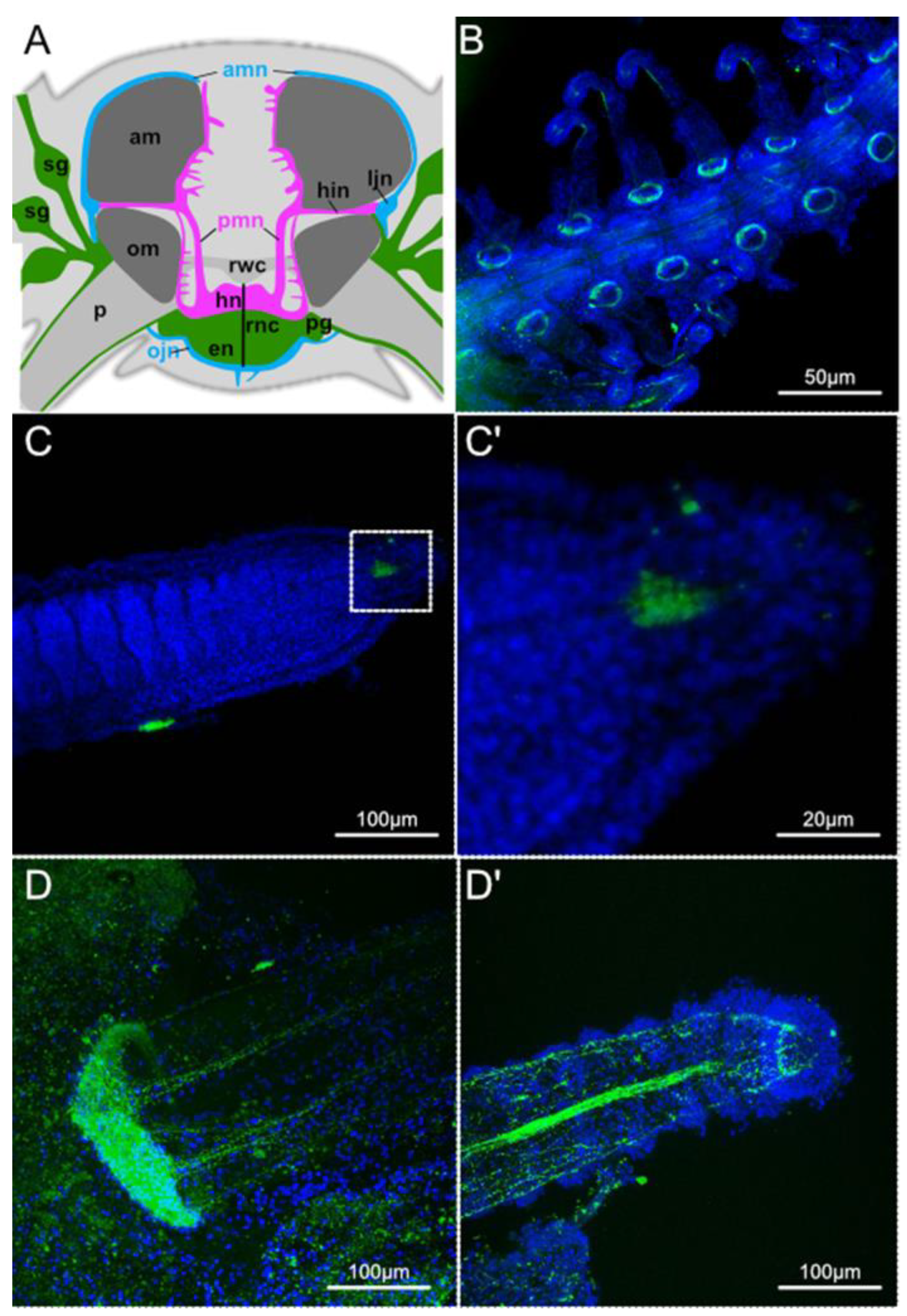
Figure 1. Nervous system in the brittle star arm. (A) Schematic diagram of the distribution of the nervous system in a mature arm segment of a brittle star, adapted from [30] under the terms of Creative Commons Attribution 4.0 International Licence. (B) SynB staining in regen-erating 50% proximal segments of A. filiformis. (C) SynB staining in regenerating 50% distal seg-ments of A. filiformis. (C’) Magnification of (C). (D) SynB staining in mature podium base of A. fili-formis. (D’) Tip of the same podium from (D). am—aboral intervertebral muscle; amn—aboral mixed nerve; en—epineural epithelium, hn—hyponeural epithelium, hin—horizontal intermuscular hyponeural nerve; ljn—lateral juxtaligametal node; ojn—oral juxtaligamental node; om—oral in-tervertebral muscle; p—podium; pg—podial ganglion; pmn—proximal muscle nerve; rnc—radial nerve cord; rwc—radial water-vascular canal; sg—spine ganglion.
Biology 11 01360 g001
Figure 2. Whole mount in situ hybridisation of Afi-elav, Afi-soxB1, Afi-soxB2, Afi-soxC, Afi-pax6, and Afi-six3 in A. filiformis at early (stage 2/3 and stage 4/5) and late (50% proximal and distal segments) stages of arm regeneration and podia showing a variety of patterns within the different regions of the regenerating nervous system. OV—oral view, OLV—oral-lateral view, LV—lateral view. Scale bar 100 um.
Biology 11 01360 g002
Figure 3. Dynamics of gene expression during arm regeneration of the brittle star Amphiura filiformis. (A) Schematic diagram representing the tissue collection procedure for generating quantitative expression data using the nanoString nCounter for different stages of arm regeneration. Red dashed lines indicate the tissue being collected at each stage. (B) Heatmap and clustering analysis of quantitative data for nervous system gene expression at different stages of regeneration relative to the non-regenerating arm segments obtained using the nSolver package. (C) Heatmap of nanoString data for proliferation and stem cell gene expression at different stages of regeneration relative to the non-regenerating arm segments. (D) Comparison of the two main observed patterns of expression changes during regeneration using stem cell/proliferation genes Afi-mycb, Afi-piwi and Afi-vasa and nervous system differentiation genes Afi-elav, Afi-neuroD and Afi-pax6. Note the opposite trend in expression suggestive of the different functionality of the genes during regeneration. Expressed as abundance relative to maximum expression (%). NR—non-regenerating, hpa—hours post amputation, St—stage, prox—proximal, dist—distal.
Biology 11 01360 g003
Figure 4. Summary of neurogenic gene expression patterns during early and late regenerative stages of the brittle star A. filiformis.
Biology 11 01360 g004
Publisher’s Note: MDPI stays neutral with regard to jurisdictional claims in published maps and institutional affiliations.