Article

Concurrent Mutations in SF3B1 and PHF6 in Myeloid Neoplasms

1
Department of Hematopathology, The University of Texas MD Anderson Cancer Center, Houston, TX 77030, USA
2
Department of Leukemia, The University of Texas MD Anderson Cancer Center, Houston, TX 77030, USA
*
Authors to whom correspondence should be addressed.
Biology 2023, 12(1), 13; https://doi.org/10.3390/biology12010013
Submission received: 17 November 2022 / Revised: 16 December 2022 / Accepted: 17 December 2022 / Published: 21 December 2022
(This article belongs to the Section Cancer Biology)

Abstract

:

Simple Summary

The advent of next-generation sequencing has elucidated the understanding of the genetic landscape of myeloid neoplasms. Most myeloid neoplasms carry more than one gene mutation at their initial presentation or develop them during the disease course. The patterns of and interactions between these mutated genes are of great research interest and may improve our ability to diagnose and prognosticate patients. It is known that certain gene mutations have a cooperative effect in the pathogenesis of myeloid neoplasms, whereas other gene mutations are mutually exclusive. A commonly cited example of mutually exclusive mutations is SF3B1 and PHF6. This observation, however, has never been rigorously assessed. Since SF3B1 and PHF6 mutations can both serve as drivers of mutations and play key roles in the development of myeloid neoplasms, it is of clinical importance to clarify this issue. We report the clinicopathologic and molecular features of 21 myeloid neoplasms with double SF3B1 and PHF6 mutations. We summarize that concurrent mutations in SF3B1 and PHF6 are rare, but they do exist in a variety of myeloid neoplasms. SF3B1 mutations usually occur as early initiating events whereas PHF6 mutations occur late, with a role in disease progression. The presence of a larger number of other co-mutated genes suggests that the neoplastic cells have gone through active clonal evolution.

Abstract

It has been reported that gene mutations in SF3B1 and PHF6 are mutually exclusive. However, this observation has never been rigorously assessed. We report the clinicopathologic and molecular genetic features of 21 cases of myeloid neoplasms with double mutations in SF3B1 and PHF6, including 9 (43%) with myelodysplastic syndrome, 5 (24%) with acute myeloid leukemia, 4 (19%) with myeloproliferative neoplasms, and 3 (14%) with myelodysplastic/myeloproliferative neoplasms. Multilineage dysplasia with ring sideroblasts, increased blasts, and myelofibrosis are common morphologic findings. All cases but one had diploid or non-complex karyotypes. SF3B1 mutations were detected in the first analysis of all the patients. PHF6 mutations occurred either concurrently with SF3B1 mutations or in subsequent follow-up samples and are associated with disease progression and impending death in most cases. Most cases had co-mutations, the most common being ASXL1, RUNX1, TET2, and NRAS. With a median follow-up of 39 months (range, 3-155), 17 (81%) patients died, 3 were in complete remission, and 1 had persistent myelodysplastic syndrome. The median overall survival was 51 months. In summary, concurrent mutations in SF3B1 and PHF6 are rare, but they do exist in a variety of myeloid neoplasms, with roles as early initiating events and in disease progression, respectively.

1. Introduction

With the advent of next-generation sequencing (NGS) technologies, the genetic landscape of myeloid neoplasms has become increasingly elucidated. Gene mutations in myeloid neoplasms affect a number of cellular pathways, including epigenetic modification (ASXL1, DNMT3A, EZH2, IDH1, IDH2, TET2), RNA splicing (SF3B1, SRSF2, U2AF1, ZRSR2), transcriptional regulation (CEBPA, ETV6, PHF6, RUNX1, TP53, WT1), and proliferative signaling pathways (FLT3, JAK2, KRAS, NRAS). It is also known that certain combinations of gene mutations have a cooperative effect in the pathogenesis of myeloid neoplasms, whereas other gene mutations are thought to be mutually exclusive [1,2,3,4].
Splicing factor 3b subunit 1 (SF3B1) is one of several genes involved in RNA splicing that is mutated in a variety of myeloid neoplasms. SF3B1 mutations occur most commonly in myelodysplastic syndromes (MDS) and are best known for their association with ring sideroblasts (RS), which are signature phenotypes [5]. PHD finger protein 6 (PHF6) is involved in transcriptional regulation and the wild-type protein localizes to the nucleolus, associates with ribosomal RNA (rRNA) promoters, and suppresses rRNA transcription. Mutations of PHF6 have been described in congenital syndromes and hematopoietic neoplasms [6,7].
A commonly cited example in the literature of mutually exclusive gene mutations is SF3B1 and PHF6 [6]. This observation, however, has never been rigorously assessed, and data from a recent study showed both SF3B1 and PHF6 mutations in 2 of 75 (2.7%) cases of acute myeloid leukemia (AML) [7]. From this study, it is unclear whether these mutations were detected simultaneously in the same sample or sequentially in different samples from the same patient. Since SF3B1 and PHF6 mutations can both serve as driver mutations and play key roles in the development of a variety of myeloid neoplasms, it is of clinical importance to clarify the issue of their potential mutual exclusivity. The aim of this study is to report a series of myeloid neoplasms in which concurrent SF3B1 and PHF6 mutations were detected, along with their clinicopathologic and molecular features.

2. Materials and Methods

2.1. Study Group

Following institutional review board approval, we retrospectively screened myeloid neoplasms that were assessed using a targeted 81-gene NGS panel test [8] performed as a part of the routine workup at our institution from January 1, 2017 through September 30, 2021. Clinical and laboratory data were obtained by review of the medical records. The study was conducted in accord with the Declaration of Helsinki.

2.2. Morphologic Evaluation

We reviewed hematoxylin-eosin stained core biopsy and clot specimens as well as Wright-Giemsa stained peripheral blood (PB) smears and bone marrow (BM) aspirate smears/touch imprints. Differential counts of 200- and 500-cells were performed manually on PB and BM smears, respectively. Cytochemical stains for iron and histochemical stains for reticulin and collagen were performed using standard methods.

2.3. Cytogenetic Analysis

Cytogenetic analysis was performed on metaphase cells prepared from BM aspirate specimens cultured for 24–48 h without mitogens, using standard techniques. Giemsa-banded metaphases were analyzed, and the results were reported using the International System for Human Cytogenetic Nomenclature, 2020.

2.4. Next-generation Sequencing

Genomic DNA was PCR-amplified and subjected to mutation analysis using a panel of 81 genes that commonly mutated in hematopoietic neoplasms on an Illumina MiSeq NGS platform (Illumina Inc., San Diego, CA, USA), as described previously [8]. The panel of 81 genes included ANKRD26, ASXL1, ASXL2, BCOR, BCORL1, BRAF, BRINP3, CALR, CBL, CBLB, CBLC, CEBPA, CREBBP, CRLF2, CSF3R, CUX1, DDX41, DNMT3A, EED, ELANE, ETNK1, ETV6, EZH2, FBXW7, FLT3, GATA1, GATA2, GFI1, GNAS, HNRNPK, HRAS, IDH1, IDH2, IKZF1, IL2RG, IL7R, JAK1, JAK2, JAK3, KDM6A, KIT, KMT2A, KRAS, MAP2K1, MPL, NF1, NOTCH1, NPM1, NRAS, PAX5, PHF6, PIGA, PML, PRPF40B, PTEN, PTPN11, RAD21, RARA, RUNX1, SETBP1, SF1, SF3A1, SF3B1, SH2B3, SMC1A, SMC3, SRSF2, STAG1, STAG2, STAT3, STAT5A, STAT5B, SUZ12, TERC, TERT, TET2, TP53, U2AF1, U2AF2, WT1, and ZRSR2.

3. Results

3.1. Concurrent SF3B1 and PHF6 Mutations Occur at Low Frequency in Myeloid Neoplasms

Among a total of 6478 cases identified within the study period, SF3B1 mutations were detected in 562 patients and PHF6 mutations were detected in 245 patients, accounting for 8.7% and 3.8% of all patients, respectively. Most of these patients had been tested multiple times during diagnosis and/or follow-up visits (median = 4; range 1–11). All test results were retrieved for analysis.
Sixty-seven distinct SF3B1 mutations were detected, most of which were located in the H5 and H6 of the so-called HEAT (Huntingtin, elongation factor 3, protein phosphatase 2A, and the yeast PI3-kinase TOR1) motifs [9]. Over 98% of these mutations were missense mutations, and nearly half (49%) were K700E mutation. Other common mutations occurred at codon K666 (22%; 65% of which were K666N), codon R625 (7%), and codon H662 (5%).
A total of 198 distinct mutations in PHF6 were detected. These mutations were widely spanned across the whole coding region but were relatively more concentrated in the two PHD-like zinc finger domains [6]. The most frequent mutations included R274 (R274Q and R274*) in 8%, I314 in 6%, R225* in 4%, R116* in 4%, C297 in 4%, and R342* in 3% of myeloid neoplasms. About 28% of these mutations were nonsense mutations, likely resulting in the inactivation of this tumor suppressor gene.
We identified 23 neoplasms that had mutations in both SF3B1 and PHF6, representing 0.4% of all neoplasms assessed. Among all the cases with SF3B1 mutation in this study, these 23 cases accounted for 4%, compared with other frequently mutated genes in the SF3B1-mutated group, such as TET2 (18%), DNMT3A (17%), JAK2 (16%), ASXL1 (13%), and TP53 (13%). Among all the cases with PHF6 mutations, these 23 patients accounted for 9%, compared with other mutated genes in the PHF6-mutated group, such as ASXL1 (40%), TET2 (34%), RUNX1 (25%), DNMT3A (19%), NRAS (17%), TP53 (16%), and JAK2 (14%). Further analysis revealed that, in two patients the SF3B1 and PHF6 mutations were never detected simultaneously in the same sample, suggesting that mutations of these two genes likely did not arise from the same clone. For that reason, these two cases were excluded, leaving a study cohort of 21 cases that account for 0.3% of all myeloid neoplasms assessed during the study period.

3.2. Concurrent SF3B1 and PHF6 Mutations Occur in a Variety of Myeloid Neoplasms

The study group included 15 men and 6 women with a median age of 69 years (range, 51–84). The pathologic diagnoses encompassed the full spectrum of myeloid neoplasms and included 9 (43%) cases with MDS, 5 (24%) with AML, 4 (19%) with myeloproliferative neoplasms (MPN), and 3 (14%) with MDS/MPN. All (100%) of the MDS cases had increased RS and 4 (44%) had excess blasts (≥5%, MDS-EB). Most AML patients (3/5, 60%) had AML with myelodysplasia-related changes (AML-MRC), one had AML with maturation, and one had AML with minimal differentiation. All three (100%) patients with MPN had primary myelofibrosis (PMF); one patient did not have a BM biopsy done at our hospital. One patient with MDS/MPN presented in the accelerated phase, and the other two came to us after chemotherapy. The diagnosis and clinicopathologic features are summarized in Table 1.

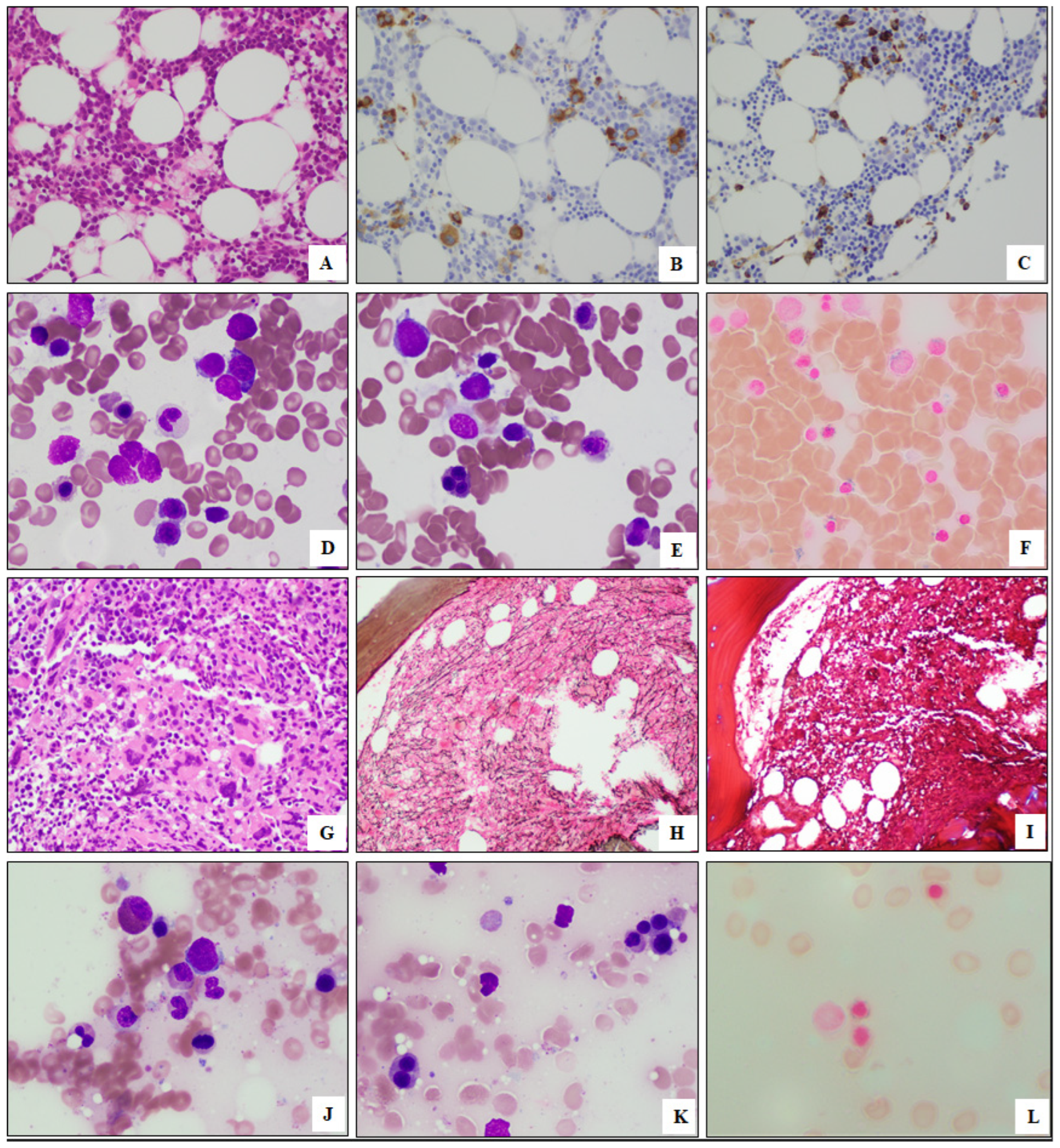
3.3. Morphologic Findings of Myeloid Neoplasms with SF3B1 and PHF6 Mutations

The BM biopsies were hypercellular (median cellularity, 80%). Dysplasia was common in all disease entities, with all MDS/MPN (3/3, 100%) and most MDS (8/9, 89%), AML (3/4, 75%), and MPN (2/3, 67%) having dysplasia involving ≥2 lineages. Increased RS (≥5%) were noted in all cases assessed except one PMF case. Apart from AML, increased blasts (≥5%) were detected in 2/3 (67%) of MDS/MPN, 4/9 (44%) of MDS, and 1/3 (33%) of MPN cases. Leukemic transformation occurred in 6/9 (67%) of MDS and 2/3 (67%) of MDS/MPN cases, with a median interval of 37 months from the initial diagnosis to the development of leukemia (range, 2-141). Myelofibrosis was also a common finding, seen in 3/3 (100%) of MPN, 3/3 (100%) of MDS/MPN, 5/6 (83%) of MDS, and 3/4 (75%) of AML cases. The morphologic findings from representative cases are illustrated in Figure 1.

3.4. Cytogenetic Findings of Myeloid Neoplasms with SF3B1 and PHF6 Mutations

Among 20 patients with cytogenetic data available, 12 (60%) had a normal karyotype, 7 (40%) had simple abnormal karyotypes with 1–2 chromosomal aberration(s), and 1 (10%, AML) had a complex karyotype with 3 chromosomal aberrations. No specific recurrent karyotypic abnormalities were identified, except for del20q, which was detected in three (15%) cases (2 PMF and 1 MDS/MPN) (Table 1).

3.5. Molecular Findings of Myeloid Neoplasms with SF3B1 and PHF6 Mutations

In nine patients, NGS was performed at the time of the initial diagnosis or within 3 months of the diagnosis, and SF3B1 mutations were present in all of these nine (100%) patients. In the remaining 12 patients, NGS was performed 11–126 months (median, 12) after the initial diagnosis and SF3B1 mutations were detected at first test in all 12 (100%) patients. Overall, SF3B1 mutations were detected in the first analysis of all 21 (100%) patients. PHF6 mutations occurred either concurrently with SF3B1 mutations (n = 14, 67%) or in subsequent follow-up samples (n = 7, 33%, median interval between the two mutations, 10 months; range, 5–21 months). In the latter scenario, the emergence of PHF6 mutations was accompanied by an increased blast count in two MDS/MPN cases and one MDS case, leukemia transformation in one MDS case, and clonal cytogenetic evolution with +8 in one MPN case. Moreover, the appearance of PHF6 mutations was associated with impending death in several cases (two MDS/MPN, one MDS, and one MPN), with these patients succumbing to the disease shortly after the emergence of PHF6 mutations.
All SF3B1 mutations were missense mutations, with the hotspots at codon K700 (n = 8) and K666 (n = 7). One MDS case had two SF3B1 mutations. PHF6 mutations consisted of 16 missense mutations, 7 nonsense mutations, 7 frameshifts, and 1 splice mutation, without hotspots. Four cases had more than one PHF6 mutations. The distribution of SF3B1 and PHF6 mutations is illustrated in Figure 2A.
The VAF of SF3B1 mutations ranged from 2–45% (median, 32%) with 18 (86%) cases having a VAF of ≥5%. The VAF of PHF6 mutations ranged from 1–85% (median, 13%), with 14 (67%) cases having a VAF of ≥5%. In 12 cases, the VAF levels of both mutations were ≥5%, and both mutations were maintained at substantial levels in these cases until the last follow-up.
We also evaluated the frequencies of other coexisting mutations in these cases (Figure 2B). In addition to SF3B1 and PHF6, all but one PMF case had at least one other gene mutation. The median number of mutated genes in this cohort was 7, higher than that of the SF3B1-mutated group (median = 3.5) or the PHF6-mutated group (median = 5). The most common coexisting mutations affected TET2 (n = 8), ASXL1 (n = 7), DNMT3A (n = 7), and RUNX1 (n = 7). However, in three cases (one MDS, one AML, and one PMF), mutations of PHF6 and SF3B1 had nearly equal VAFs and hence were likely in the same dominant clone; there were also no or fewer (≤2) other genes involved, with zero in PMF cases, one in an MDS case, and two in AML cases (Figure 2B). TP53 mutations were detected in three (14%) cases, and the mutations were either with low VAFs (<2%) and transient (in an MDS case and an MDS/MPN case) or persistent at low levels (VAF<3%, in an MDS case), suggesting that TP53 may not be a key player in these cases. The VAFs of all mutations from each of these cases are provided in Supplementary Table S1. Mutation clone sizes in a representative test of each of the 12 myeloid neoplasms cases with a significant level of concurrent SF3B1 and PHF6 mutations are plotted in Figure 2C.

3.6. Clinical Findings of Myeloidoutcomes of Myeloid Neoplasms with SF3B1 and PHF6 Mutations

All patients received chemotherapy, and four MDS patients additionally received allogeneic stem cell transplantation (ASCT). With a median follow-up of 39 months (range, 3–155), the clinical outcomes of these patients were poor. Seventeen (81%) patients died by the end of the study interval, including two of the four MDS patients who underwent transplantation. Three were in complete remission, including two MDS patients after transplantation and one AML patient after chemotherapy. One patient had persistent MDS (Table 1). Most patients, except for four MDS (after transplantation) and four AML patients, never achieved complete remission. Two MDS patients and one AML patient shortly relapsed. The median overall survival (OS) of these patients was 51 months.

4. Discussion

In recent years, large-scale analysis using NGS has become feasible in clinical practice and a large amount of data on gene mutation patterns in myeloid neoplasms has accumulated. These data have shown that most myeloid neoplasms have more than one gene mutation [1,2,3,10,11]. The patterns of and interactions between these mutated genes is of great research interest and may improve our ability to diagnose and prognosticate patients with myeloid neoplasms [4,12].
In this study, we analyzed the NGS panel test results of 6478 patients to address the question of whether SF3B1 and PHF6 mutations are mutually exclusive in myeloid neoplasms. Our findings suggest that these double-mutated cases are uncommon (<1%), but far from mutually exclusive, in contrast with the literature [6]. In this study, these cases account for roughly 4% of all cases with SF3B1 mutations or 9% of all cases with PHF6 mutations. These neoplasms also usually harbor a larger number of other co-mutated genes, suggesting that the neoplastic cells have gone through active clonal evolution. The disease distribution of these cases was generally consistent with the literature’s reported frequencies in cases with SF3B1 [5] or PHF6 [6,7] mutations separately, except that MPN cases seemed slightly more frequent. One possible explanation for myeloid neoplasms with both SF3B1 and PHF6 mutations not having been reported in the literature may be the lack of large-scale studies of myeloid neoplasms that have been assessed for PHF6 mutations.
SF3B1 is one of the most commonly mutated genes in MDS. SF3B1 mutations are particularly common (>80%) in cases of MDS with RS and confer a favorable prognosis [13]. SF3B1 mutations have also been reported in approximately 20% of patients with MDS with isolated del(5q), without any significant impact on their overall survival [14]. In AML with RS, on the other hand, SF3B1 mutations have been associated with an inferior clinical outcome [5]. SF3B1 mutations have also been reported in about 10% of BCR-ABL1–negative MPNs [13], but its prognostic significance is still unclear, with contradictory reports. Some researchers reported a role of SF3B1 mutations in myelofibrotic progression in patients with polycythemia vera and essential thrombocythemia [15], whereas others concluded that SF3B1 mutations did not correlate with fibrotic evolution [16]. SF3B1 mutations are disease-defining in MDS/MPN with RS and thrombocytosis (MDS/MPN-RS-T) and are seen in about 5% of chronic myelomonocytic leukemia (CMML), without a well-established association with leukemic transformation and overall survival [17]. Moreover, SF3B1 mutations also occur frequently in non-myeloid neoplasms such as chronic lymphocytic leukemia/small lymphocytic lymphoma, and have been associated with resistance to fludarabine therapy and poor clinical outcomes [18]. It has been reported that an SF3B1 mutation is an early event in myeloid neoplasms, and in MDS/MPN, SF3B1 mutations seem to represent founder mutations followed by secondary hits in genes involved in other signaling pathways, such as JAK2 [17]. In keeping with this, SF3B1 mutations were detected in the first tests in all cases in this study. A recent study suggested that not all SF3B1 mutations are equal. The most common SF3B1 K700E mutation confers a favorable prognosis in MDS cases and is associated with a lower risk of progression to AML compared with patients with wild-type SF3B1 [2,14], whereas a K666N mutation has been found to be associated with a poorer prognosis [19]. Only eight (38%) of the SF3B1 mutations in this SF3B1 and PHF6 double-mutated cohort were K700E, and K666 mutation occurred in a similar percentage of patients (n = 7, 33%). All patients with SF3B1 mutations at codon K666 died at the end of the follow-up period, though their OS varied largely. Moreover, we noticed that in cases with PHF6 mutations at a substantial level and in dominant clones, the co-mutated SF3B1 was more likely to be of the K666 type. This may lead to speculation that SF3B1 K666 mutations could provide a preferred microenvironment for PHF6 mutated clones to gain a competition edge in during clonal evolution.
Somatic PHF6 mutations occur more frequently in T-lymphoblastic leukemia/lymphoma and have only been reported in approximately 3% of myeloid neoplasms [6,20], which is consistent with our findings in this study. The potential role of PHF6 mutations in the pathogenesis of myeloid neoplasms is poorly understood, but PHF6 appears to act as a tumor suppressor gene, regulating the transcription of signaling genes and the DNA damage response [7]. As myeloid neoplasms with PHF6 mutations are rare, the clinical characteristics of patients with these myeloid neoplasms are much less established compared to those of patients with myeloid neoplasms with SF3B1 mutations. The findings in the current study contribute more data to this entity. Myeloid neoplasms with SF3B1 and PHF6 double mutations tend to show morphologic evidence of myelodysplasia, increased reticulin fibrosis, an increased blast count, and the risk of leukemia transformation. The prognostic significance of PHF6 mutations remains unclear. Some studies showed that PHF6 mutations predicted a shorter overall survival in patients with intermediate-risk AML [20], whereas others showed that PHF6 mutations had no prognostic impact when compared with patients with wild-type PHF6 [5]. Noticeably, the clinical outcome of these double-mutated cases in this study was very poor, with a median survival more in keeping with that of patients with PHF6-mutated neoplasms than that of SF3B1-mutated neoplasms.
Another reported feature of PHF6 mutations is that these mutations are associated with an increased number of other mutated genes (>3 besides PHF6), including ASXL1, EZH2, RUNX1, TET2, U2AF1, and ZRSR2 [6], but are mutually exclusive with SF3B1 [5,21]. The findings in this study are consistent with earlier reports and confirm that PHF6 mutations are significantly associated with ASXL1 and RUNX1 mutations. However, in the double mutation cohort, we noticed that once both mutations reached dominant clone levels, there tended to be fewer other genes involved, suggesting that the combination of SF3B1 and PHF6 mutations may be sufficiently powerful to drive the disease’s progression. In addition, low frequencies and very low levels of TP53 mutations were detected in these double-mutated cases. This finding could imply that mutations in PHF6, also acting as a tumor suppressor gene, may have been more important in the pathogenesis than TP53 during clonal evolution. Moreover, PHF6 mutations tend to occur late in the course of myeloid neoplasms, either concurrently with or subsequently to SF3B1 mutations in all cases, and are associated with disease progression in a subset of the cases. All of these data suggest that PHF6 mutations in myeloid neoplasms often arise late as a result of clonal evolution and that increasing levels of PHF6 mutations are associated with poor clinical outcomes. It seems reasonable to suggest that the coexistence of these two mutations contributes to poor clinical outcomes.
A limitation of this study is that we did not perform single-cell sequencing and therefore we cannot prove with certainty that the SF3B1 and PHF6 mutations resided in the same clone. We are also aware of the fact that most of the cases in this study had other mutated genes, which could be confounding factors for the assessment of the clinical findings of this cohort. Nevertheless, the findings in this study show that myeloid neoplasms with SF3B1 and PHF6 mutations do occur uncommonly, and seem to be associated with some distinctive features.

5. Conclusions

In summary, concurrent mutations in the SF3B1 and PHF6 genes are rare in myeloid neoplasms, but they do exist. The simultaneous occurrence of both mutations most likely is the result of the active clonal evolution of the neoplastic cells with no specific associated disease type preference. It is possible that myeloid neoplasms with SF3B1 and PHF6 double mutations may represent a unique entity among myeloid neoplasms.

Supplementary Materials

The following supporting information can be downloaded at: https://www.mdpi.com/article/10.3390/biology12010013/s1, Table S1: Variant allele frequency of mutations detected in 21 patients with myeloid neoplasms with SF3B1 and PHF6 mutations during study period.

Author Contributions

Conceptualization, Z.Z. and C.C.Y.; methodology, Z.Z., M.J.R., K.P.P., R.L. and C.C.Y.; investigation, Z.Z. and C.C.Y.; resources, Z.Z., L.J.M., S.G., M.J.R., C.Y.O., S.L., R.K.-S., F.Z.J., G.G.-M., K.S.C., K.P.P. and C.C.Y.; data curation, Z.Z. and C.C.Y.; writing—original draft preparation, Z.Z. and C.C.Y.; writing—review and editing, Z.Z., L.J.M. and C.C.Y. All authors have read and agreed to the published version of the manuscript.

Funding

This research received no external funding.

Institutional Review Board Statement

The study was conducted in accordance with the Declaration of Helsinki, and approved by the Institutional Review Board of The University of Texas MD Anderson Cancer Center (DR10-0299).

Informed Consent Statement

The Institutional Review Board has waived the requirement for informed consent for this retrospective study.

Data Availability Statement

The data presented in this study are available on request from the corresponding authors. The data are not publicly available due to privacy.

Acknowledgments

The authors would like to thank all the reviewers and editors who participated in the review.

Conflicts of Interest

The authors declare no conflict of interest.

References

  1. McClure, R.F.; Ewalt, M.D.; Crow, J.; Temple-Smolkin, R.L.; Pullambhatla, M.; Sargent, R.; Kim, A.S. Clinical Significance of DNA Variants in Chronic Myeloid Neoplasms: A Report of the Association for Molecular Pathology. J. Mol. Diagn. 2018, 20, 717–737. [Google Scholar] [CrossRef] [PubMed] [Green Version]
  2. Schwind, S.; Jentzsch, M.; Kubasch, A.S.; Metzeler, K.H.; Platzbecker, U. Myelodysplastic syndromes: Biological and therapeutic consequences of the evolving molecular aberrations landscape. Neoplasia 2021, 23, 1101–1109. [Google Scholar] [CrossRef] [PubMed]
  3. Wang, M.; Yang, C.; Zhang, L.; Schaar, D.G. Molecular Mutations and Their Cooccurrences in Cytogenetically Normal Acute Myeloid Leukemia. Stem Cells Int. 2017, 2017, 6962379. [Google Scholar] [CrossRef] [PubMed] [Green Version]
  4. Papaemmanuil, E.; Gerstung, M.; Bullinger, L.; Gaidzik, V.I.; Paschka, P.; Roberts, N.D.; Potter, N.E.; Heuser, M.; Thol, F.; Bolli, N.; et al. Genomic Classification and Prognosis in Acute Myeloid Leukemia. N. Engl. J. Med. 2016, 374, 2209–2221. [Google Scholar] [CrossRef] [PubMed]
  5. Venable, E.R.; Chen, D.; Chen, C.P.; Bessonen, K.R.; Nguyen, P.L.; Oliveira, J.L.; Reichard, K.K.; Hoyer, J.D.; Althoff, S.D.; Roh, D.J.; et al. Pathologic Spectrum and Molecular Landscape of Myeloid Disorders Harboring SF3B1 Mutations. Am. J. Clin. Pathol. 2021, 156, 679–690. [Google Scholar] [CrossRef] [PubMed]
  6. Mori, T.; Nagata, Y.; Makishima, H.; Sanada, M.; Shiozawa, Y.; Kon, A.; Yoshizato, T.; Sato-Otsubo, A.; Kataoka, K.; Shiraishi, Y.; et al. Somatic PHF6 mutations in 1760 cases with various myeloid neoplasms. Leukemia 2016, 30, 2270–2273. [Google Scholar] [CrossRef]
  7. Kurzer, J.H.; Weinberg, O.K. PHF6 Mutations in Hematologic Malignancies. Front. Oncol. 2021, 11, 704471. [Google Scholar] [CrossRef]
  8. Patel, K.P.; Ruiz-Cordero, R.; Chen, W.; Routbort, M.J.; Floyd, K.; Rodriguez, S.; Galbincea, J.; Barkoh, B.A.; Hatfield, D.; Khogeer, H.; et al. Ultra-Rapid Reporting of GENomic Targets (URGENTseq): Clinical Next-Generation Sequencing Results within 48 hours of Sample Collection. J. Mol. Diagn. 2019, 21, 89–98. [Google Scholar] [CrossRef]
  9. Papaemmanuil, E.; Cazzola, M.; Boultwood, J.; Malcovati, L.; Vyas, P.; Bowen, D.; Pellagatti, A.; Wainscoat, J.S.; Hellstrom-Lindberg, E.; Gambacorti-Passerini, C.; et al. Somatic SF3B1 mutation in myelodysplasia with ring sideroblasts. N. Engl. J. Med. 2011, 365, 1384–1395. [Google Scholar] [CrossRef] [Green Version]
  10. Haferlach, T.; Nagata, Y.; Grossmann, V.; Okuno, Y.; Bacher, U.; Nagae, G.; Schnittger, S.; Sanada, M.; Kon, A.; Alpermann, T.; et al. Landscape of genetic lesions in 944 patients with myelodysplastic syndromes. Leukemia 2014, 28, 241–247. [Google Scholar] [CrossRef]
  11. Papaemmanuil, E.; Gerstung, M.; Malcovati, L.; Tauro, S.; Gundem, G.; Van Loo, P.; Yoon, C.J.; Ellis, P.; Wedge, D.C.; Pellagatti, A.; et al. Clinical and biological implications of driver mutations in myelodysplastic syndromes. Blood 2013, 122, 3616–3627, quiz 3699. [Google Scholar] [CrossRef] [PubMed] [Green Version]
  12. Loghavi, S.; Zuo, Z.; Ravandi, F.; Kantarjian, H.M.; Bueso-Ramos, C.; Zhang, L.; Singh, R.R.; Patel, K.P.; Medeiros, L.J.; Stingo, F.; et al. Clinical features of de novo acute myeloid leukemia with concurrent DNMT3A, FLT3 and NPM1 mutations. J. Hematol. Oncol. 2014, 7, 74. [Google Scholar] [CrossRef] [PubMed] [Green Version]
  13. Lasho, T.L.; Finke, C.M.; Hanson, C.A.; Jimma, T.; Knudson, R.A.; Ketterling, R.P.; Pardanani, A.; Tefferi, A. SF3B1 mutations in primary myelofibrosis: Clinical, histopathology and genetic correlates among 155 patients. Leukemia 2012, 26, 1135–1137. [Google Scholar] [CrossRef] [Green Version]
  14. Malcovati, L.; Stevenson, K.; Papaemmanuil, E.; Neuberg, D.; Bejar, R.; Boultwood, J.; Bowen, D.T.; Campbell, P.J.; Ebert, B.L.; Fenaux, P.; et al. SF3B1-mutant MDS as a distinct disease subtype: A proposal from the International Working Group for the Prognosis of MDS. Blood 2020, 136, 157–170. [Google Scholar] [CrossRef]
  15. Zhao, L.P.; Daltro de Oliveira, R.; Marcault, C.; Soret, J.; Gauthier, N.; Verger, E.; Cassinat, B.; Kiladjian, J.; Benajiba, L. SF3B1 mutations in the driver clone increase the risk of evolution to myelofibrosis in patients with myeloproliferative neopalsms (MPN). Blood 2020, 136, 1. [Google Scholar]
  16. Boiocchi, L.; Hasserjian, R.P.; Pozdnyakova, O.; Wong, W.J.; Lennerz, J.K.; Le, L.P.; Dias-Santagata, D.; Iafrate, A.J.; Hobbs, G.S.; Nardi, V. Clinicopathological and molecular features of SF3B1-mutated myeloproliferative neoplasms. Hum. Pathol. 2019, 86, 1–11. [Google Scholar] [CrossRef]
  17. Palomo, L.; Meggendorfer, M.; Hutter, S.; Twardziok, S.; Adema, V.; Fuhrmann, I.; Fuster-Tormo, F.; Xicoy, B.; Zamora, L.; Acha, P.; et al. Molecular landscape and clonal architecture of adult myelodysplastic/myeloproliferative neoplasms. Blood 2020, 136, 1851–1862. [Google Scholar] [CrossRef]
  18. Rossi, D.; Bruscaggin, A.; Spina, V.; Rasi, S.; Khiabanian, H.; Messina, M.; Fangazio, M.; Vaisitti, T.; Monti, S.; Chiaretti, S.; et al. Mutations of the SF3B1 splicing factor in chronic lymphocytic leukemia: Association with progression and fludarabine-refractoriness. Blood 2011, 118, 6904–6908. [Google Scholar] [CrossRef] [Green Version]
  19. Dalton, W.B.; Helmenstine, E.; Pieterse, L.; Li, B.; Gocke, C.D.; Donaldson, J.; Xiao, Z.; Gondek, L.P.; Ghiaur, G.; Gojo, I.; et al. The K666N mutation in SF3B1 is associated with increased progression of MDS and distinct RNA splicing. Blood Adv. 2020, 4, 1192–1196. [Google Scholar] [CrossRef] [PubMed]
  20. Patel, J.P.; Gonen, M.; Figueroa, M.E.; Fernandez, H.; Sun, Z.; Racevskis, J.; Van Vlierberghe, P.; Dolgalev, I.; Thomas, S.; Aminova, O.; et al. Prognostic relevance of integrated genetic profiling in acute myeloid leukemia. N. Engl. J. Med. 2012, 366, 1079–1089. [Google Scholar] [CrossRef] [Green Version]
  21. Chien, K.S.; Kanagal-Shamanna, R.; Naqvi, K.; Sasaki, K.; Alvarado, Y.; Takahashi, K.; Borthakur, G.M.; Jabbour, E.; Routbor, M.; Pierce, S.A.; et al. The impact of PHF6 mutations in myelodysplastic syndromes, chronic myelomonocytic leukemia, and acute myeloid leukemia. Blood 2019, 134, 1436–1439. [Google Scholar] [CrossRef]
Figure 1. Morphologic features of representative cases of myeloid neoplasms with SF3B1 and PHF6 mutations. (AF) A case of myelodysplastic syndrome with multilineage dysplasia, increased blasts, and fibrosis. The bone marrow is hypercellular for age (A,H&E) with dysmegakaryopoiesis (B, CD61) and increased blasts (C, CD34). The aspirate smear shows dysgranuopoiesis (D, Wright-Giemsa), dyserythropoiesis (E, Wright-Giemsa), and increased ring sideroblasts (F, Prussian Blue) (AC, 200×; DF, 500×). (GL) A case of primary myelofibrosis. The bone marrow is hypercellular (G,H&E), with reticulin fibrosis (H), without collagen fibrosis (I). The aspirate smear shows dysgranulopoiesis (J, Wright-Giemsa), dyserythropoiesis (K, Wright-Giemsa), and increased ring sideroblasts (L, Prussian Blue) (GI, 200×; JL, 500×).
Biology 12 00013 g001
Figure 2. Molecular features of cases of myeloid neoplasms with SF3B1 and PHF6 mutations. (A) The location and number of SF3B1 and PHF6 mutations. (B) The mutation profile heatmap. The cases were divided into four categories: myelodysplastic syndrome (MDS), acute myeloid leukemia (AML), myeloproliferative neoplasms (MPN), and myelodysplastic/myeloproliferative neoplasms (MDS/MPN). Each column represents a single case. Overall, the most frequent concurrent mutations were TET2, ASXL1, DNMT3A, and RUNX1. (C) Mutation clone size of each case. Normalized variant allele frequencies (VAFs) in a representative test of each of the 12 myeloid neoplasms cases with a significant level of concurrent SF3B1 and PHF6 mutations are shown.
Biology 12 00013 g002aBiology 12 00013 g002b
Table 1. Clinicopathologic and molecular genetic features of 21 patients with myeloid neoplasms with SF3B1 and PHF6 mutations.
CaseAgeSexWBCHbPlateletLDHKaryotypeSF3B1PHF6SCTFU (m)Outcome
MDS
169M8.210.2234NAinv(17)(p13q23)
[20/20]
K666MK21fs, R116*, F198fs, C242Y, G291E, C297R, C297YNo39Died
251M8.912.1116204del(11q)[9/20]R625C, D781GC305S, L324fsYes14CR
356M7.09.851020746,XY[20]E783KC212GYes155Died
477M2.08.624413046,XY[20]S779PC280fsNo38MDS
577M6.310.2153124del(13)(q12q14)
[3/19]
K666TQ7*No65Died
684M11.17.48856846,XY[20]K666NR319*No3Died
774M1.79.841159add(1)(p36.1)
[2/20]
R625CH229fs, C283*Yes39CR
862M7.29.933644146,XY[20]K700ER225*Yes71Died
968M3.17.66317746,XY[20]K700EI290TNo28Died
AML
1070F6.96.9513546,XX[20]K700Ec.585+1G>A p.?No11Died
1182M2.59.15313046,XY[20]I704NH302RNo90Died
1282M1.211.3111232–Y,+1, der(1;13)(q10;q10)
[4/11]
K666NR274QNo51Died
1375M1.08.05218946,XY[20]K700EK130fsNo13Died
1458F5.88.919912646,XX[20]K700EH239RNo9CR
MPN
1567F2.98.639247del(20)(q11.2q13.3)
[20]
K666NH239YNo57Died
1659M7.19.056375del(20)(q11.2q13.3)
[20]
K666TI314TNo37Died
1776M3.69.319626646,XY[20]K666QR274QNo41Died
1858MNANANANANAK700EE27fs, R225*, Y240CNo72Died
MDS/MPN
1973F11.57.22598746,XX[20]G740EC215SNo16Died
2069F17.25.259566646,XX[20]K700EF263*No87Died
2166F60.49.126570del(20)(q11.2q13.1)
[6/20]
K700EI314TNo20Died
AML—acute myeloid leukemia; CR —complete remission; F—female; FU (m)—follow-up (months); Hb—hemoglobin; LDH—lactate dehydrogenase; M—male; MDS—myelodysplastic syndrome; MDS/MPN—myelodysplastic/myeloproliferative neoplasms; MPN—myeloproliferative neoplasms; NA—not available; SCT—stem cell transplantation; WBC—white blood cells. Reference ranges: WBC, 4.0–11.0 K/uL; Hb, 14.0–18.0 g/dL for men and 12.0–16.0 g/dL for women; platelet, 140–440 K/uL; LDH, 135–225 IU/L.
Disclaimer/Publisher’s Note: The statements, opinions and data contained in all publications are solely those of the individual author(s) and contributor(s) and not of MDPI and/or the editor(s). MDPI and/or the editor(s) disclaim responsibility for any injury to people or property resulting from any ideas, methods, instructions or products referred to in the content.

