Article

M.tb-Rv2462c of Mycobacterium tuberculosis Shows Chaperone-like Activity and Plays a Role in Stress Adaptation and Immunomodulation

1
Microbial Pathogenesis and Microbiome Lab, Department of Microbiology, Central University of Rajasthan, Ajmer 305817, India
2
Department of Biochemistry, School of Chemical and Life Sciences, Jamia Hamdard, New Delhi 110062, India
3
Department of Immunology and Microbiology, University of Colorado School of Medicine, Anschutz Medical Campus, Aurora, CO 80045, USA
*
Authors to whom correspondence should be addressed.
Biology 2023, 12(1), 69; https://doi.org/10.3390/biology12010069
Submission received: 25 November 2022 / Revised: 28 December 2022 / Accepted: 28 December 2022 / Published: 30 December 2022
(This article belongs to the Section Microbiology)

Abstract

:

Simple Summary

Mycobacterium tuberculosis protein tig (Rv2462c) is present in membrane fraction along with the cytosol. It has been previously reported to assist in the folding of nascent polypeptides. We have investigated the role of Mycobacterium tuberculosis tig in stress adaptation; for example, it exhibits roles in thermal and oxidative shock, survival within macrophages, biofilm formation and immunological responses. Our findings establish the contribution of tig to tolerance and adaptation to host-generated stresses, implying its involvement in the infection cycle of Mycobacterium tuberculosis. These attributes of Tig highlights its significance in Mycobacterium tuberculosis virulence and make it an important target for novel therapeutic interventions against tuberculosis.

Abstract

Mycobacterium tuberculosis (M.tb)-encoded factors protect it against host-generated stresses and support its survival in the hostile host environment. M.tb possesses two peptidyl-prolyl cis-trans isomerases and a probable trigger factor encoded by Rv2462c which has an FKBP-like PPIase domain. PPIases are known to assist the folding of peptidyl-prolyl bonds and are involved in various cellular processes important for bacterial survival in host-generated stresses. In this study, we aim to functionally characterize Rv2462c of M.tb. Our data suggest that the trigger factor of M.tb exhibits chaperone activity both in vitro and in vivo. Heterologous expression of M.tb-Rv2462c locus into Mycobacterium smegmatis enhanced its survival within macrophages, adaptation to oxidative stress and biofilm formation. M.tb-trigger factor has strong immunomodulatory potential and modifies the cytokine profile of the host towards the proinflammatory axis.

1. Introduction

Despite our best efforts, Mycobacterium tuberculosis (M.tb) is a human peril causing millions of deaths every year due to tuberculosis (TB). Evasion of host defences helps it to survive in hostile macrophages [1]. The World Health Organization (WHO) has estimated that about 25% of the world’s population is affected by latent TB. Though mostly asymptomatic, 5–10% progress into active infections [2]. Efforts to discover new antibiotics against multidrug-resistant M.tb has been marked as a global priority by WHO [3]. Various stress responding factors including heat shock proteins, foldases and chaperones contribute to virulence thereby significantly aided in its pathogenicity by immune evasion and colonization of host macrophages [4]. However, the mechanisms employed by the pathogen to survive the stresses of the macrophage microenvironment have not been well elucidated yet. Its better understanding may help in the containment of M.tb infection and may suggest novel therapeutic interventions.
Trigger factors across diverse bacteria are essential for the co-translational folding of many nascent polypeptides [5]. The bacterial trigger factor chaperones bind to the 50S ribosomes near the peptide exit channel, enabling its interaction with nascent polypeptides, and facilitating their folding [6,7]. In vitro, the trigger factor refolds denatured RNase T1 and Glyceraldehyde-3-phosphate dehydrogenase (GAPDH) as a prolyl isomerase [8,9].
In an attempt to identify M.tb encoded protein responsible for interactions between host and pathogen, stress adaptation, and immunomodulation, we identified and characterized Rv2462c locus of M.tb. It has an FKBP-like or PPIase domain which may confer chaperone-like activity and is postulated to be involved in cellular processes [10]. This protein has been previously reported to be a probable trigger factor protein of M.tb [11]. The percentage amino acid coverage of tig with has 7.1% amino acid sequence identity with PPIase A and 3.8% identity with PPIase B within M.tb genome. Prior studies have revealed that M.tb-Rv2462c is involved in the maintenance of open conformations of newly synthesized proteins, which indicates its chaperonic nature [12]. It has also been observed that nutrient deprivation enhances the expression level of this protein [13]. Inorganic phosphate limitation, especially, is believed to regulate the concentration of M.tb-Rv2462c. It has been further reported that M.tb-Rv2462c may contribute significantly to the pathogenicity of M.tb, making it significant for further studies [12,14].
In this study, we have characterized the M.tb-Tig (Rv2462c) and explored its immunomodulatory potential and role in promoting M.tb survival under intracellular stresses by triggering the host immune system and modifying the cytokine secretion profile of the host, highlighting the significance of the protein in M.tb pathogenesis. Here, we have demonstrated that the recombinant Tig (Rv2462c), when expressed in E. coli, displays chaperone-like activity and significantly improves survival chances under high-temperature conditions. When expressed in M. smegmatis, it results in enhanced biofilm formation and increased survivability under oxidative stress. Our observations point towards the involvement of M.tb-Tig in stress adaptation, which forms an important component of the pathogenicity of M.tb.

2. Materials and Methods

2.1. Bacterial Strains and Cell Lines

E. coli BL-21 strain from Novagen was used for protein expression. M.tb-H37Rv genomic DNA was a gift from Prof. Rakesh Bhatnagar’s laboratory in JNU, New Delhi, India. THP-1 cell was obtained from National Centre for Cell Sciences, Pune, India.
M. smegmatis mc2 155 was initially procured from ATCC (Cat#700084). 7H9 broth with 10% OADC and 0.05% tween-80 was used to grow mycobacterium strains.

2.2. Cloning and Purification of M.tb-Rv2462c by Ni-NTA Column

M.tb-Rv2462c gene locus was amplified by using forward (CTAGCTAGCGTGAAGAGCACCGTCGAGCAGTT) and reverse primers (CCGCTCGAGTCGAGTCACGTTGTCGCTTCGTC) and cloned in pET28a vector [15]. From tags of both terminals, only N-terminal poly-his tag was utilized while C-terminal tag was suppressed during expression by stop codon of the cloned gene. The His-tagged M.tb-Rv2462c gene was expressed in BL21 cells. Recombinant protein was purified using the Ni-NTA column as described earlier [16]. Then, the eluted fraction was dialyzed to exchange buffer at 4 °C as described earlier [16]. The his-tag has small size and was not cleaved from the rTig after purification.

2.3. Enzyme Activity

Enzyme activity of recombinant trigger factor (rTig) was measured using a spectrophotometric assay used previously [15]. In summary, rTig catalyses cis-trans isomerization of Ala-Pro bond. The trans form is taken as substrate by coupled enzyme α-chymotrypsin to produce chromogenic product. Thus, isomerization of Ala-Pro bond was seen as an enhancement in the absorbance at 390 nm at 15 °C. Absorbance was documented every 1.5 s for 100 s and plotted graphically against time. Control reaction was used in absence of rTig [15].

2.4. Chaperone-like Activity of M.tb Rv2462c

Restoration of enzyme activity of heat-denatured proteins is considered a key function of chaperones. To determine if enzyme activity of thermally aggregated NdeI can be recovered by the M.tb-Rv2462c protein, 200 ng of the pET22b vector was digested by NdeI with and without thermally aggregated NdeI at 37 °C for 2 h. Heat-denaturation of NdeI was achieved by submerging 10 U of the enzyme at 60 °C for 10 min. The digested plasmid was then electrophoresed in agarose gel containing ethidium bromide stain and visualized by the Gel Doc system (Bio-Rad, Kidlington, UK) [15,17].
To explore the hydrophobicity surface of M.tb-rTig, ANS probe was used and monitored by fluorescence spectroscopy [15,18]. Briefly, in presence of rTig, ANS fluorescence was evaluated by excitation at 390 nm and subsequent emission measurements ranging from 450 to 550 nm [17]. The fluorescence of protein-bound ANS dye was measured after mixing 0.5 mg/mL of the recombinant tig with 50 μM ANS and incubated at RT for 30 min. Emission spectrum of the ANS alone was used as control.
Heat-shock of E. coli was given as described earlier [19]. Fold survival in the presence and absence of heat shock was calculated, with the vector control (pET22b) being treated as one fold in the normalisation process [17].

2.5. Expression of M.tb-Rv2462c in Mycobacterium Smegmatis

To ascertain the impact of Rv2462c on the survivability of mycobacterium inside the host, we assayed recombinant M. smegmatis strains, expressing M.tb-Rv2462c, for growth within THP-1 monocyte cells, in vitro. The E. coli-mycobacterium shuttle vector pSTKT having UV15 promoter with tetO sequence (inducible with anhydrotetracycline) was used to construct pST-Rv2462c. pST-Rv2462c was electroporated into Mycobacterium smegmatis mc2 155 as described earlier [20].

2.6. Examining In Vitro Growth and Effect of H2O2

Mycobacterium smegmatis strains (Ms_VC, Ms_WT and Ms_tig) were grown in 7H9 broth containing 10% OADC to an OD600 of 0.8–1.0. The grown cultures were diluted to a ratio of hundred 1:100. Then, the cultures were grown to an OD600 of 0.4 and treated with 7 mM H2O2 for 3 h. The cultures were diluted 1:100 and plated on 7H10 agar plates.

2.7. Intracellular Replication of M. Smegmatis Strains in THP-1 Cells

Human THP-1 monocyte cells cultured in RPMI 1640 medium were differentiated into macrophages by phorbol 12-myristate 13-acetate (PMA) [21] and grown by incubating at 37 °C and 5% CO2. M. smegmatis cultures (Ms_WT, Ms_tig and Ms_VC) were then used to infect the monolayers of THP-1 for 4 h at a 50 MoI. Then, the infected THP-1 cells were treated with Gentamicin (20 μg/mL) for 30 min. Further, the THP-1 cells were washed twice with RPMI medium and fresh medium was added and incubated for 72 h in CO2 incubator. The cells were then taken out carefully at different time points and centrifuged for 3 min at 1000 rpm. The cells were washed two times in RPMI medium and lysed in sterile water. The lysates were plated on 7H10 agar plates and kept at 37 °C for 4 days.

2.8. Biofilm Estimation

M. smegmatis strains (Ms_tig, Ms_VC and Ms_WT) were grown in 7H9 media supplemented with 0.05% Tween 80, 10% OADC and 0.001% glycerol. The cultures were diluted to an OD600 of 0.08 and incubated at 37 °C in a test tube. Further, 20 ng/mL of anhydrotetracycline was added to all of the M. smegmatis strains. To allow pellicle development at the liquid-air interface, M. smegmatis strains were subsequently incubated as static culture for 7 days in growth medium without Tween 80.
Biofilm formation was quantified using the crystal violet assay [22]. Anhydrotetracycline-induced Ms_tig, Ms_VC and Ms_WT were cultured as static phase in a sterile, flat-bottomed 96-well microtiter plate. After 7 days, the media below the pellicle was aspirated out. Further, 125 μL of 0.1% crystal violet dye was used to stain the pellicle. The stained pellicle was then washed with water three times before adding 30% acetic acid. The plates were incubated for 15 min at room temperature, before recording the absorbance spectrophotometrically at 550 nm.

2.9. Prediction of Antigenicity

In silico analysis of the antigenicity of M.tb-tig was performed with a tool developed by the Immunomedicine group of UCM, Spain. This tool utilizes the method described to determine the antigenic peptides [23]. The occurrence of amino acid residues in previously established segmental epitopes has been tabulated and forms the basis of the prediction. A minimum of eight residues is essential for a segment to be considered significant.

2.10. Immune Assay

The presence of immunogenic proteins induces the secretion of cytokines from macrophages. Cytokine secretion levels were assayed by Enzyme-Linked Immunosorbent Assay (ELISA) method as previously reported [21,24]. THP-1 cells were differentiated into macrophages by adding PMA (20 ng/mL) for 24 h in 5% CO2 at 37 °C [20,25,26]. Then, differentiated THP-1 cells (2 × 10⁴ cells/cm2) were treated with M.tb-tig protein (2.5–20 µg) for 48 h. LPS was used as positive control while heat-denatured M.tb-tig was used as negative control. The culture supernatants were used to quantify the levels of cytokines using ELISA as described earlier [17].

3. Results

3.1. rTig of M.tb Is Enzymatically Active

The molecular mass of purified recombinant M.tb rTig was found to be 50 kDa on 10% SDS-PAGE (Figure 1A). α chymotrypsin and a chromogenic peptide were used in a coupled assay to assess the enzyme activity of rTig. The rise in the isomerization activity rate before reaching saturation shows rTig is active (Figure 1A). Alignment of amino acid sequence of trigger factors of E. coli K-12, M. smegmatis and M.tb H37Rv shows the similarities among the three. As usual, M. smegmatis trigger factor has higher match with M.tb than E. coli K-12 (Figure 1B).

3.2. rTig Displays Properties of a Chaperone Protein

Previous studies have established the importance of surface hydrophobicity for chaperone activity [17]. 1-anilinonaphthalene-8-sulfonate (ANS) is a hydrophobic selective dye bond with cationic groups of polyamino acids and proteins by the formation of ion pairs. It binds to the hydrophobic sites buried in a protein surface and has long been used for characterizing protein-binding sites. The sulfonate group of ANS interacts with cationic groups of lysine and arginine, causing a hypsochromic shift, thereby, enhancing fluorescence intensity and blue shift of fluorescence emission [27]. In our study, a shift in the emission wavelength, associated with enhanced fluorescence intensity was observed in case of ANS bound to rTig (Figure 2A). This observation establishes the presence of surface hydrophobicity in rTig, which is an attribute of chaperone-like proteins.
Protecting proteins from thermal aggregation and preserving their functional activity is characteristic to most chaperones [17]. The chaperone function of rTig was investigated by examining its ability to restore the enzymatic activity of the heat denatured NdeI. After heat denaturation, NdeI failed to digest pET22b plasmid (Figure 2, lane 3), but retained the activity after addition of rTig (lanes 4). BSA, used as negative control, failed to display a similar restorative effect on heat-denatured NdeI (Figure 2B; lane 5). Restoration of enzymatic activity of heat-denatured NdeI by rTig suggests its role in protein refolding.
To understand the chaperone-like role of M.tb-rTig, in vitro, we examined if the M.tb-rTig has the ability to rescue E. coli BL21 (DE3) strain after exposure to heat stress. There was no significant difference in the CFU count between uninduced or vector control strains (Figure 2C,D). We observed a significant increase in the CFU count of the induced culture as compared to the uninduced and vector control after an hour of heat shock (Figure 2D). The number of CFU decreased when incubated for two hours, but was still significantly higher than controls. These data suggest that rTig can protect the bacterium from thermal stress and might function as chaperone.

3.3. M.tb Tig Helps Mycobacterial Survival within THP-1 Macrophages

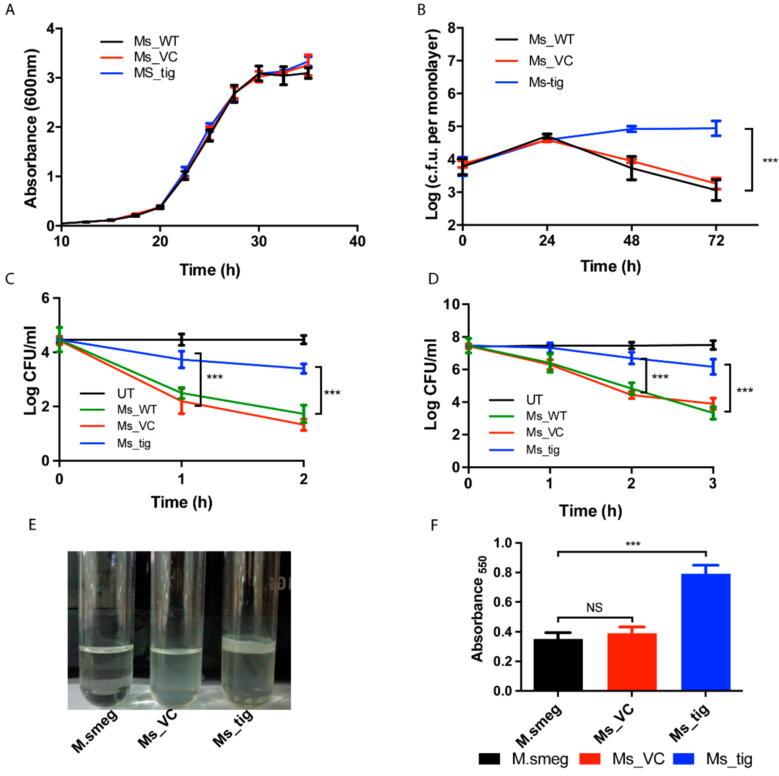
We aim to determine the influence of M.tb-Rv2462c on the ability of mycobacterium to adapt and survive in host cells. Under normal conditions, the overexpression of M.tb-Tig (Ms_Tig) has no impact on the growth pattern of M. smegmatis compared to controls (Ms_VC, Ms_WT) (Figure 3A).
To assess the role of M.tb-Tig on the survival of the pathogen inside host cells, we compared CFU of M. smegmatis strains (Ms_Tig, Ms_VC and Ms_WT) after infecting THP-1 cells. A steady rise in the CFU count of Ms-Tig was observed up to 72 h (p < 0.001) while a decline in the CFU was observed for Ms_WT and Ms_VC after 24 h (Figure 3B). Our observations suggest that rTig contributes to intracellular adaptation and growth of mycobacterium in human macrophages.
Intracellular mycobacteria usually experience oxidative stress. For examining the possible impact of M.tb-Tig on the survival of M. smegmatis under hydrogen peroxide stress, we treated the M. smegmatis cultures with 7 mM of H2O2 for 3 h. The higher CFU count in Ms_Tig compared to Ms_VC and Ms_WT strains suggest that M.tb-Tig offer protection against oxidative stress. (Figure 3D) and might play a role in stress adaptation of the mycobacterium. For heat stress experiment, different M. smegmatis strains grown till 0.6 OD600 with and without anhydrotetracycline were exposed to 50 °C temperature for 2 h. Further, survival under temperature stress is a significant attribute of chaperone proteins. M. smegmatis expressing M.tb trigger factor show improved survival under a temperature stress of 50 °C compared to controls (Figure 3C).
To further consolidate the contribution of M.tb tig in virulence and stress adaptation of the mycobacteria, we studied the possible role of the protein in modulating biofilm formation. As described previously, M. smegmatis expressing rTig, wild-type M. smegmatis and Ms_VC, used as control, were induced when the cells were grown both with and without anhydrotetracycline. The results demonstrated a noteworthy contrast in biofilm production by the three strains. Enhanced biofilm formation of nearly 2-fold (p < 0.005) was exhibited by Ms_Tig (Figure 3E,F). This suggests that M.tb. rTig is involved in the development of biofilms.

3.4. Exposure of THP-1 Cell with M.tb Tig Elicited Pro-Inflammatory Responses

Antigenicity profiling using the antigenicity prediction tool revealed the amino acid sequence of M.tb Tig exhibits high antigenicity and multiple antigenic stretches (Figure 4A,B). Furthermore, we examined the role of M.tb-rTig in the regulation of different cytokine secretions. LPS, the endotoxin contaminant of the recombinant protein rTig was removed by polymyxin B agarose beads. The differentiated THP-1 cells were treated with different dosage (2.5 to 20 µg/mL) of M.tb-Tig. The dose-dependent increased levels of TNF-α and IL-6 were observed by the treatment of M.tb-Tig. These results revealed that M.tb-Rv2462c elicited an increased secretion of proinflammatory cytokine by macrophages (Figure 4C,D). However, no significant changes were observed in the levels of IL-10 (Figure 4E). These results are comparable to the immune-modulatory function of other mycobacterial PPIases [20].

4. Discussion

The current study was aimed at functionally characterizing the M.tb trigger factor rTig and exploring its role in M.tb pathogenesis by studying its contribution to immunomodulation and stress adaptation of the pathogen. The disease progression and outcome of tuberculosis are influenced by a complicated interplay between the virulence factors of the pathogen and immune response of the host. Alongside their role in protein folding and secretion, cell signalling and apoptosis, bacterial chaperones are involved in virulence and stress adaptation. Additionally, in this study, we have tried to focus precisely on these moonlighting activities of the trigger factor protein of M.tb. Bacterial pathogens such as M.tb, H. pylori and Clostridium difficile, use chaperones such as Hsp60 and Hsp70 as adhesins, to attach to the host tissue. Regulation of host cell activity by cell signalling has been exhibited by molecular chaperones such as Cpn60 and Hsp70. Cpn60, Hsp70, Hsp90 and Cpn10 display extracellular signalling in pathogens such as M.tb, M. paratuberculosis, E. coli, Chlamydia trachomatis and H. pylori. It has been demonstrated earlier that M.tb secretes Cpn10 when inside the phagolysosome of the macrophage. The overexpression of Hsps in a mutant of the intracellular pathogen, S. typhimurium has previously illustrated the importance of chaperones in stress adaptation [28,29,30,31].
The trigger factor, encoded by the tig gene, is highly conserved across diverse bacterial species. This ribosome-associated molecular chaperone was initially discovered in vitro, in Gram-negative bacteria, folding the precursor of the outer membrane protein A (pro-OmpA) into a conformation suitable for translocation [19,32]. It has now been linked to the virulence of many pathogens. In this study, we investigated the chaperone-like activity and immunological role of the trigger factor protein of M.tb. The binding of molecular chaperones to misfolded proteins is guided by surface hydrophobicity. The distinctive blue shift in the emission maxima, and enhanced absorbance by ANS-binding to rTig suggests the presence of surface hydrophobicity similar to chaperones. Earlier studies have reported that the trigger factor protects against thermal denaturation in E. coli [33]. Our current study demonstrates that M.tb tig protects the cells against high-temperatures and aids in the restoration of enzyme activity of thermally denatured proteins. Incubation with rTig restores enzyme activity of NdeI, pointing towards its expected role in protein refolding. After getting satisfactory evidence of rTig’s chaperone-like nature, in vitro, we proceeded to investigate the activity of the protein, in vivo. Additionally, indeed, E. coli transformed with rTig displays a significantly increased survival after heat shock as compared to the controls.
To assess the role in stress adaptation and to demonstrate chaperone-like activity, the heterologous expression of M.tb trigger factor in E. coli and M. smegmatis expression systems was carried out. Using the vector alone and uninduced controls, as well as the fact that they do have sequence and therefore structural variations, the influence of the trigger factors of the expression systems was accounted for.
M.tb has the ability to resist stress and survive inside the host. We explored the contribution of rTig in protecting M. smegmatis cells under conditions of oxidative stress. In comparison to M. smegmatis controls, we observed a significant increase in the survival of M. smegmatis strain expressing M.tb tig. Our observations lie in line with in vitro results, indicative of rTig acting as a chaperone-like protein which also contributes to stress adaptation.
The overproduction of pro-inflammatory cytokines by human macrophages is considered to be a hallmark of tuberculosis. Antimicrobial activity against the pathogen is affected primarily by granulomas, whose formation and maintenance are believed to be modulated by TNF-α. Previous studies have also linked TNF-α production with intracellular viability and virulence [34]. IL-6 is involved in inflammation and pathology and is thus believed to play a role in disease progression in tuberculosis [35,36]. THP-1 cells were treated with increased doses of rTig, which amplified the secretion of pro-inflammatory cytokines such as TNF-α and IL-6. However, no significant changes were observed in the secretion of anti-inflammatory cytokine, IL-10. M. smegmatis cells transformed with rTig displayed enhanced survival in THP-1 cells. This points towards a crucial role of rTig in the pathobiology of tuberculosis. There are seemingly paradoxical role of pro-inflammatory cytokines vis a vis host benefits/survival. Pro-inflammatory cytokines could be pro-host as having anti-bacterial effects and anti-host as being toxic to host. Additionally, in case of tuberculosis, pro-inflammatory cytokines released earlier in the infection result in accumulation of immune cells around the granuloma resulting in spread of the pathogen.
IL-6 is not a pure pro-inflammatory, but rather has an anti-inflammatory tinge as it inhibits endotoxin-induced TNF-α and IL-1 production in mononuclear cells and induce the soluble receptors for TNF-α which is its natural inhibitors [37]. Furthermore, the macrophage polarization is no longer considered to be existing in the dichotomy of pro and anti-inflammation. They exist in the more flexible and dynamic in response to the macroenvironment and may exist in the intermediate polarization state rather than canonical binaries of pro- and anti-states [38].
The E. coli trigger factor also enhances the binding efficiency of the chaperone GroEL [19,39,40]. In Gram-positive bacteria Streptococcus pyogenes, the homologue of the trigger factor, RopA, is critical for the secretion and maturation of SpeB [41,42]. Deficiency of LuxS, a modulator of biofilm formation and acid and oxidative stress tolerance in S. mutans, was found to induce the upregulation of its trigger factor, strongly suggesting its involvement in the virulence of the pathogen [19,43]. In the swine pathogen Streptococcus suis serotype 2, tig encodes a virulence regulator, associated with the expression of the pathogen’s virulence genes. The tig mutants of S. suis serotype 2 influence its adaptive capacity against environmental stresses [19].
Previous research demonstrated the role of Mycobacterium abscessus and P. aeruginosa in the production of biofilms in cystic fibrosis as well as a virulence factor in UPEC isolates. The likelihood of biofilm development within the host tissues is suggested by the occurrence of extracellular M.tb in biofilm-such as structures inside the lung lesions of guinea pigs infected with M.tb [44]. In the current study, M. smegmatis expressing rTig significantly upregulates biofilm production, in comparison to the strains lacking rTig. Our findings establish the involvement of rTig in biofilm production.
Cell surface proteins play crucial role in the regulation of biofilm formation. Tig (Rv2462c) has been reported in the cell wall fraction along with cytosolic fraction in different proteomic profiling [45,46,47]. The Tig overexpression in M. smegmatis, then, might have altered its physiochemical properties or working as a surface-associated adhesin-like molecule, promoting biofilm formation. For example, localization of Lap A protein to cell surface enhances the biofilm formation Pseudomonas fluorescens [48]. Similarly, BpfA–BpfG–BpfD proteins governs biofilm formation in Shewanella oneidensis [49]. Some previous studies in Staphylococcus aureus have shown role of Tig biofilm formation. Tig acts as a chaperone protein and help in folding of proteins essential in biofilm formation [50].
The results of this study on M.tb tig is in accordance with the findings of previous studies. Surface hydrophobicity and the ability to restore enzyme activity of heat-denatured NdeI establishes the expected chaperonic nature of rTig. The rTig protein considerably increases survivability under high-temperature conditions, which was suggestive of its involvement in the virulence of the pathogen. This was further established by the observations that the presence of the protein distinctly increased tolerance to oxidative stress (H2O2).

5. Conclusions

Our study demonstrates the chaperonic nature and immunomodulatory potential of the M.tb trigger factor protein. Our findings also establish its contribution to tolerance and adaptation to host-generated stresses, implying its involvement in the infection cycle of M.tb. Revelations of these attributes of M.tb-Tig highlights its significance in M.tb virulence and make it an important target for novel therapeutic interventions against tuberculosis, thus fulfilling the primary aim of this study. Further studies would provide a better comprehension of the mechanisms employed by the protein to enable the intracellular lifestyle and enhanced pathogenicity of M.tb.

Author Contributions

This study was designed by D.T. and S.P.; D.T., S.K. and S.P. have conceptualized the work. M.K., R.R., A.B. and P.J. have carried out the experiments and analysed the data. R.R., S.P. and D.T. wrote the manuscript. All authors have read and agreed to the published version of the manuscript.

Funding

This research was funded by DBT Biocare grant of DBT, GoI, grant number BT/PR30553/BIC/101/1123/2018.

Institutional Review Board Statement

Not applicable.

Informed Consent Statement

Not applicable.

Data Availability Statement

Not applicable.

Acknowledgments

M.K. would like to thank ICCR for providing fellowship. We thank Sayeed ur Rehman, Jamia Hamdard for helping in biophysical work and data interpretation.

Conflicts of Interest

The authors declare no competing interests.

References

  1. Ehrt, S.; Schnappinger, D. Mycobacterial survival strategies in the phagosome: Defence against host stresses. Cell. Microbiol. 2009, 11, 1170–1178. [Google Scholar] [CrossRef] [Green Version]
  2. Glaziou, P.; Floyd, K.; Raviglione, M.C. Global Epidemiology of Tuberculosis. Semin. Respir. Crit. Care Med. 2018, 39, 271–285. [Google Scholar] [CrossRef] [PubMed] [Green Version]
  3. Tacconelli, E.; Carrara, E.; Savoldi, A.; Harbarth, S.; Mendelson, M.; Monnet, D.L.; Pulcini, C.; Kahlmeter, G.; Kluytmans, J.; Carmeli, Y.; et al. Discovery, research, and development of new antibiotics: The WHO priority list of antibiotic-resistant bacteria and tuberculosis. Lancet. Infect. Dis. 2018, 18, 318–327. [Google Scholar] [CrossRef] [PubMed]
  4. Santoro, M.G.; Amici, C.; Rossi, A. Role of Heat Shock Proteins in Viral Infection. In Prokaryotic and Eukaryotic Heat Shock Proteins in Infectious Disease; Springer: Berlin/Heidelberg, Germany, 2009; Volume 4, pp. 51–84. [Google Scholar] [CrossRef]
  5. Kramer, G.; Boehringer, D.; Ban, N.; Bukau, B. The ribosome as a platform for co-translational processing, folding and targeting of newly synthesized proteins. Nat. Struct. Mol. Biol. 2009, 16, 589–597. [Google Scholar] [CrossRef] [PubMed]
  6. Stoller, G.; Rücknagel, K.P.; Nierhaus, K.H.; Schmid, F.X.; Fischer, G.; Rahfeld, J.U. A ribosome-associated peptidyl-prolyl cis/trans isomerase identified as the trigger factor. EMBO J. 1995, 14, 4939–4948. [Google Scholar] [CrossRef]
  7. Ferbitz, L.; Maier, T.; Patzelt, H.; Bukau, B.; Deuerling, E.; Ban, N. Trigger factor in complex with the ribosome forms a molecular cradle for nascent proteins. Nature 2004, 431, 590–596. [Google Scholar] [CrossRef] [Green Version]
  8. Merz, F.; Hoffmann, A.; Rutkowska, A.; Zachmann-Brand, B.; Bukau, B.; Deuerling, E. The C-terminal domain of Escherichia coli trigger factor represents the central module of its chaperone activity. J. Biol. Chem. 2006, 281, 31963–31971. [Google Scholar] [CrossRef]
  9. Huang, G.C.; Li, Z.Y.; Zhou, J.M.; Fischer, G. Assisted folding of D-glyceraldehyde-3-phosphate dehydrogenase by trigger factor. Protein Sci. A Publ. Protein Soc. 2000, 9, 1254–1261. [Google Scholar] [CrossRef] [Green Version]
  10. Li, Z.; Wu, D.; Zhan, B.; Hu, X.; Gan, J.; Ji, C.; Li, J. Structural insights into the complex of trigger factor chaperone and ribosomal protein S7 from Mycobacterium tuberculosis. Biochem. Biophys. Res. Commun. 2019, 512, 838–844. [Google Scholar] [CrossRef]
  11. Cole, S.T.; Brosch, R.; Parkhill, J.; Garnier, T.; Churcher, C.; Harris, D.; Gordon, S.V.; Eiglmeier, K.; Gas, S.; Barry, C.E., 3rd; et al. Deciphering the biology of Mycobacterium tuberculosis from the complete genome sequence. Nature 1998, 393, 537–544. [Google Scholar] [CrossRef] [Green Version]
  12. Sharma, D.; Kumar, B.; Lata, M.; Joshi, B.; Venkatesan, K.; Shukla, S.; Bisht, D. Comparative Proteomic Analysis of Aminoglycosides Resistant and Susceptible Mycobacterium tuberculosis Clinical Isolates for Exploring Potential Drug Targets. PLoS ONE 2015, 10, e0139414. [Google Scholar] [CrossRef]
  13. Hampshire, T.; Soneji, S.; Bacon, J.; James, B.W.; Hinds, J.; Laing, K.; Stabler, R.A.; Marsh, P.D.; Butcher, P.D. Stationary phase gene expression of Mycobacterium tuberculosis following a progressive nutrient depletion: A model for persistent organisms? Tuberculosis 2004, 84, 228–238. [Google Scholar] [CrossRef] [Green Version]
  14. Rifat, D.; Bishai, W.R.; Karakousis, P.C. Phosphate depletion: A novel trigger for Mycobacterium tuberculosis persistence. J. Infect. Dis. 2009, 200, 1126–1135. [Google Scholar] [CrossRef] [Green Version]
  15. Pandey, S.; Sharma, A.; Tripathi, D.; Kumar, A.; Khubaib, M.; Bhuwan, M.; Chaudhuri, T.K.; Hasnain, S.E.; Ehtesham, N.Z. Mycobacterium tuberculosis peptidyl-prolyl isomerases also exhibit chaperone like activity in-vitro and in-vivo. PLoS ONE 2016, 11, e0150288. [Google Scholar] [CrossRef]
  16. Perez, C.; Maier, T. Expression, Purification, and Structural Biology of Membrane Proteins; Springer: Berlin/Heidelberg, Germany, 2020. [Google Scholar]
  17. Suragani, M.; Aadinarayana, V.D.; Pinjari, A.B.; Tanneeru, K.; Guruprasad, L.; Banerjee, S.; Pandey, S.; Chaudhuri, T.K.; Ehtesham, N.Z. Human resistin, a proinflammatory cytokine, shows chaperone-like activity. Proc. Natl. Acad. Sci. USA 2013, 110, 20467–20472. [Google Scholar] [CrossRef] [Green Version]
  18. Fischer, G.; Bang, H.; Mech, C. Determination of enzymatic catalysis for the cis-trans-isomerization of peptide binding in proline-containing peptides. Biomed. Biochim. Acta 1984, 43, 1101–1111. [Google Scholar]
  19. Wu, T.; Zhao, Z.; Zhang, L.; Ma, H.; Lu, K.; Ren, W.; Liu, Z.; Chang, H.; Bei, W.; Qiu, Y.; et al. Trigger Factor of Streptococcus Suis Is Involved in Stress Tolerance and Virulence. Microb. Pathog. 2011, 51, 69–76. [Google Scholar] [CrossRef]
  20. Pandey, S.; Tripathi, D.; Khubaib, M.; Kumar, A.; Sheikh, J.A.; Sumanlatha, G.; Ehtesham, N.Z.; Hasnain, S.E. Mycobacterium tuberculosis Peptidyl-Prolyl Isomerases Are Immunogenic, Alter Cytokine Profile and Aid in Intracellular Survival. Front. Cell. Infect. Microbiol. 2017, 7, 38. [Google Scholar] [CrossRef] [Green Version]
  21. Nair, S.; Ramaswamy, P.A.; Ghosh, S.; Joshi, D.C.; Pathak, N.; Siddiqui, I.; Sharma, P.; Hasnain, S.E.; Mande, S.C.; Mukhopadhyay, S. The PPE18 of Mycobacterium tuberculosis interacts with TLR2 and activates IL-10 induction in macrophage. J. Immunol. 2009, 183, 6269–6281. [Google Scholar] [CrossRef] [Green Version]
  22. O’Toole, G.A. Microtiter dish biofilm formation assay. J. Vis. Exp. 2011, 47, e2437. [Google Scholar] [CrossRef]
  23. Kolaskar, A.S.; Tongaonkar, P.C. A semi-empirical method for prediction of antigenic determinants on protein antigens. FEBS Lett. 1990, 276, 172–174. [Google Scholar] [CrossRef] [PubMed] [Green Version]
  24. Banerjee, S.; Nandyala, A.; Podili, R.; Katoch, V.; Murthy, K.; Hasnain, S.E. Mycobacterium tuberculosis (Mtb) isocitrate dehydrogenases show strong B cell response and distinguish vaccinated controls from TB patients. Proc. Natl. Acad. Sci. USA 2004, 101, 12652–12657. [Google Scholar] [CrossRef] [PubMed] [Green Version]
  25. Garg, R.; Tripathi, D.; Kant, S.; Chandra, H.; Bhatnagar, R.; Banerjee, N. The conserved hypothetical protein Rv0574c is required for cell wall integrity, stress tolerance, and virulence of Mycobacterium tuberculosis. Infect. Immun. 2015, 83, 120–129. [Google Scholar] [CrossRef] [Green Version]
  26. Schulz, C.; Lai, X.; Bertrams, W.; Jung, A.L.; Sittka-Stark, A.; Herkt, C.E.; Janga, H.; Zscheppang, K.; Stielow, C.; Schulte, L.; et al. Thp-1-Derived Macrophages Render Lung Epithelial Cells Hypo-Responsive to Legionella Pneumophila—A Systems Biology Study. Sci. Rep. 2017, 7, 1–15. [Google Scholar] [CrossRef] [PubMed]
  27. Gasymov, O.K.; Glasgow, B.J. Ans Fluorescence: Potential to Augment the Identification of the External Binding Sites of Proteins. Biochim. Biophys. Acta 2007, 1774, 403–411. [Google Scholar] [CrossRef] [Green Version]
  28. Vanghele, M.; Ganea, E. The role of bacterial molecular chaperones in pathogen survival within the host. Rom. J. Biochem. 2010, 47, 100. [Google Scholar]
  29. Buchmeier, N.A.; Heffron, F. Induction of Salmonella stress proteins upon infection of macrophages. Science 1990, 248, 730–732. [Google Scholar] [CrossRef]
  30. Henderson, B.; Allan, E.; Coates, A.R. Stress wars: The direct role of host and bacterial molecular chaperones in bacterial infection. Infect. Immun. 2006, 74, 3693–3706. [Google Scholar] [CrossRef] [Green Version]
  31. Fossati, G.; Izzo, G.; Rizzi, E.; Gancia, E.; Modena, D.; Moras, M.L.; Niccolai, N.; Giannozzi, E.; Spiga, O.; Bono, L.; et al. Mycobacterium tuberculosis chaperonin 10 is secreted in the macrophage phagosome: Is secretion due to dissociation and adoption of a partially helical structure at the membrane? J. Bacteriol. 2003, 185, 4256–4267. [Google Scholar] [CrossRef] [Green Version]
  32. Crooke, E.; Wickner, W. Trigger factor: A soluble protein that folds pro-OmpA into a membrane-assembly-competent form. Proc. Natl. Acad. Sci. USA 1987, 84, 5216–5220. [Google Scholar] [CrossRef] [Green Version]
  33. Kandror, O.; Goldberg, A.L. Trigger factor is induced upon cold shock and enhances viability of Escherichia coli at low temperatures. Proc. Natl. Acad. Sci. USA 1997, 94, 4978–4981. [Google Scholar] [CrossRef]
  34. Newman, G.W.; Gan, H.X.; McCarthy, P.L., Jr.; Remold, H.G. Survival of human macrophages infected with Mycobacterium avium intracellulare correlates with increased production of tumor necrosis factor-alpha and IL-6. J. Immunol. 1991, 147, 3942–3948. [Google Scholar]
  35. Tsao, T.C.; Hong, J.; Huang, C.; Yang, P.; Liao, S.K.; Chang, K.S. Increased TNF-alpha, IL-1 beta and IL-6 levels in the bronchoalveolar lavage fluid with the upregulation of their mRNA in macrophages lavaged from patients with active pulmonary tuberculosis. Tuber. Lung Dis. 1999, 79, 279–285. [Google Scholar] [CrossRef]
  36. Ilonidis, G.; Parapanisiou, E.; Anogeianaki, A.; Giavazis, I.; Theofilogiannakos, E.K.; Tsekoura, P.; Kidonopoulou, K.; Trakatelli, C.; Polimenidis, Z.; Conti, P.; et al. Interleukin-1beta (IL-1 beta), interleukin 6 (IL-6) and tumor necrosis factor (TNF) in plasma and pleural fluid of pneumonia, lung cancer and tuberculous pleuritis. J. Biol. Regul. Homeost. Agents 2006, 20, 41–46. [Google Scholar]
  37. van der Poll, T. Pro-Inflammatory Cytokines: Double-Edged Swords in the Pathogenesis of Bacterial Infection. In Mechanisms of Organ Dysfunction in Critical Illness; Evans, T.W., Fink, M.P., Eds.; Springer: Berlin/Heidelberg, Germany, 2002; pp. 146–158. [Google Scholar] [CrossRef]
  38. Mosser, D.M.; Edwards, J.P. Exploring the full spectrum of macrophage activation. Nat. Rev. Immunol. 2008, 8, 958–969. [Google Scholar] [CrossRef]
  39. Kandror, O.; Sherman, M.; Goldberg, A. Rapid degradation of an abnormal protein in Escherichia coli proceeds through repeated cycles of association with GroEL. J. Biol. Chem. 1999, 274, 37743–37749. [Google Scholar] [CrossRef] [Green Version]
  40. Kandror, O.; Sherman, M.; Rhode, M.; Goldberg, A.L. Trigger factor is involved in GroEL-dependent protein degradation in Escherichia coli and promotes binding of GroEL to unfolded proteins. EMBO J. 1995, 14, 6021–6027. [Google Scholar] [CrossRef]
  41. Lyon, W.R.; Caparon, M.G. Trigger factor-mediated prolyl isomerization influences maturation of the Streptococcus pyogenes cysteine protease. J. Bacteriol. 2003, 185, 3661–3667. [Google Scholar] [CrossRef] [Green Version]
  42. Lyon, W.R.; Gibson, C.M.; Caparon, M.G. A role for trigger factor and an rgg-like regulator in the transcription, secretion and processing of the cysteine proteinase of Streptococcus pyogenes. EMBO J. 1998, 17, 6263–6275. [Google Scholar] [CrossRef] [Green Version]
  43. Wen, Z.T.; Burne, R.A. LuxS-mediated signaling in Streptococcus mutans is involved in regulation of acid and oxidative stress tolerance and biofilm formation. J. Bacteriol. 2004, 186, 2682–2691. [Google Scholar] [CrossRef] [Green Version]
  44. Lenaerts, A.J.; Hoff, D.; Aly, S.; Ehlers, S.; Andries, K.; Cantarero, L.; Orme, I.M.; Basaraba, R.J. Location of Persisting Mycobacteria in a Guinea Pig Model of Tuberculosis Revealed by R207910. Antimicrob. Agents Chemother. 2007, 51, 3338–3345. [Google Scholar] [CrossRef] [PubMed] [Green Version]
  45. Rosenkrands, I.; Weldingh, K.; Jacobsen, S.; Hansen, C.V.; Florio, W.; Gianetri, I.; Andersen, P. Mapping and Identification of Mycobacterium Tuberculosis Proteins by Two-Dimensional Gel Electrophoresis, Microsequencing and Immunodetection. Electrophoresis 2000, 21, 935–948. [Google Scholar] [CrossRef]
  46. Rosenkrands, I.; King, A.; Weldingh, K.; Moniatte, M.; Moertz, E.; Andersen, P. Towards the Proteome of Mycobacterium Tuberculosis. Electrophoresis 2000, 21, 3740–3756. [Google Scholar] [CrossRef] [PubMed]
  47. Jungblut, P.R.; Schaible, U.E.; Mollenkopf, H.J.; Zimny-Arndt, U.; Raupach, B.; Mattow, J.; Halada, P.; Lamer, S.; Hagens, K.; Kaufmann, S.H. Comparative proteome analysis of Mycobacterium tuberculosis and Mycobacterium bovis BCG strains: Towards functional genomics of microbial pathogens. Mol. Microbiol. 1999, 33, 1103–1117. [Google Scholar] [CrossRef]
  48. Boyd, C.D.; Smith, T.J.; El-Kirat-Chatel, S.; Newell, P.D.; Dufrene, Y.F.; O’Toole, G.A. Structural features of the Pseudomonas fluorescens biofilm adhesin LapA required for LapG-dependent cleavage, biofilm formation, and cell surface localization. J. Bacteriol. 2014, 196, 2775–2788. [Google Scholar] [CrossRef] [Green Version]
  49. Zhou, G.; Yuan, J.; Gao, H. Regulation of biofilm formation by BpfA, BpfD, and BpfG in Shewanella oneidensis. Front. Microbiol. 2015, 6, 790. [Google Scholar] [CrossRef]
  50. Keogh, R.A.; Zapf, R.L.; Frey, A.; Marino, E.C.; Null, G.G.; Wiemels, R.E.; Holzschu, D.L.; Shaw, L.N.; Carroll, R.K. Staphylococcus aureus Trigger Factor Is Involved in Biofilm Formation and Cooperates with the Chaperone PpiB. J. Bacteriol. 2021, 203, e00681-20. [Google Scholar] [CrossRef]
Figure 1. Purified rTig is enzymatically active. (A): Isomerization activity of rTig was measured using a small chromogenic peptide and α chymotrypsin as coupled enzyme and juxtaposed with the rate of spontaneous cis-trans isomerization in the absence of the rTig. Purified rTig was found to weigh around 50 kDa on 10% SDS PAGE. (B): Alignment of trigger factors of Escherichia coli K-12, M. smegmatis and M. smegmatis H37Rv using T-Coffee protein alignment server of EMBL-EBI. The matching residues are highlighted.
Biology 12 00069 g001
Figure 2. M.tb Rv2462c (rTig) exhibits in vitro and in vivo chaperone-like activity. (A): ANS-bound to M.tb Rv2462c (rTig) confirmed surface hydrophobicity. Fluorescence excitation was monitored in the range of 400–650 nm. Fluorescence spectra of control (ANS buffer) and ANS bound to rTig were recorded in red and blue colour, respectively. (B): rTig protected heat-denatured NdeI protein M, GeneRuler 1KB marker. Lane 1, multiple bands of undigested pET22b (5.3 kb), Lane 2, digested pET22b by NdeI, Lane 3, digested pET22b by heat denatured NdeI. Lane 4, digested by thermally denatured NdeI in the presence of Rv2462c protein. Lane 5, digested pET22b by heat-denatured NdeI in the presence of BSA protein. (C,D): rTig rescues E. coli cells from heat shock: Transformed E. coli cells (with pET22b alone and pET22b-Rv2462c) underwent heat treatment (50 °C). At one and two hours, bacterial growth was determined by the CFU count. E. coli expressing Rv2462c displayed a greater number of CFU compared to vector control.
Biology 12 00069 g002
Figure 3. Role of rTig in stress adaptation and biofilm production. (A): M. smegmatis carrying rTig shows no change in growth characteristics, in vitro. Bacterial growth (OD600) of Ms_WT, Ms_VC and Ms_tig was plotted. Data are the mean ± standard deviation of data derived from three distinct experiments. (B): M. smegmatis expressing M.tb Rv2462c show enhanced survival within THP-1 cells. Human THP-1 cells were infected with M. smegmatis mc2 155 (Ms_WT), M. smegmatis vector control (Ms_VC) and those transformed with M.tb Rv2462c (Ms_tig) and plated at different time intervals. The log of c.f.u per monolayer of THP-1 cells was computed at designated time points. The results represent three individual experiments (*** p < 0.001). They show the mean ± SEM of three triplicate wells. (C,D): Growth characteristics of M. smegmatis under temperature and oxidative stress. Ms_WT, Ms_VC and Ms_tig cultures were grown up to an O.D. of 0.4. They were then subjected to different temperature (50 °C or 37 °C) (panel-C) H2O2 treatment (panel-D) and log c.f.u. was computed after pre-determined time intervals. The results shown represent three individual experiments (*** p < 0.001) and are the mean ± SEM of three triplicate wells. (E,F): rTig encoded by M.tb Rv2462c promotes the development of pellicle and production of biofilm in M. smegmatis. Upon induction with anhydrotetracycline to express rTig of M.tb, pellicle formation at the liquid air junction was observed and quantified in M.smeg, Ms_VC and Ms_tig.
Biology 12 00069 g003
Figure 4. (A): Analysis of rTig using antigenicity prediction tool. (B): Major antigenic stretches. (C), (D,E): M.tb Rv2462c enhances the production of pro-inflammatory cytokines by human monocytes. Concentration-dependent rise in the secretion of pro-inflammatory cytokines ((C): TNF-α; (D): IL-6) as a function of treatment of THP-1 cells with rTig. No significant changes were observed in IL-10 secretion. The mean ± SD of three replicates has been represented here. LPS was taken as the positive control. (*** p < 0.001).
Biology 12 00069 g004
Disclaimer/Publisher’s Note: The statements, opinions and data contained in all publications are solely those of the individual author(s) and contributor(s) and not of MDPI and/or the editor(s). MDPI and/or the editor(s) disclaim responsibility for any injury to people or property resulting from any ideas, methods, instructions or products referred to in the content.