Article

The First Survival Score for Patients Treated with Whole-Brain Radiotherapy Plus Simultaneous Integrated Boost for Brain Metastases

1
Department of Radiation Oncology, University of Lubeck, 23562 Lubeck, Germany
2
Department of Radiation Oncology, Mayo Clinic, Phoenix, AZ 85054, USA
*
Author to whom correspondence should be addressed.
Biology 2023, 12(4), 585; https://doi.org/10.3390/biology12040585
Submission received: 16 March 2023 / Revised: 9 April 2023 / Accepted: 11 April 2023 / Published: 12 April 2023
(This article belongs to the Section Cancer Biology)

Abstract

:

Simple Summary

A model was generated to judge survival after whole-brain radiotherapy plus simultaneous boost (WBRT+SIB) for cerebral metastases. Initially, three models, each including three prognostic groups, were created and compared for accuracy in prognosticating death ≤ 6 and survival ≥ 6 months using the corresponding positive predictive values (PPVs). PPVs to predict death ≤ 6 and survival ≥ 6 months were 85% and 57% for Model 1 (considering performance status = KPS and the number of lesions), 83% and 75% for Model 2 (KPS, lesions, age), and 86% and 78% for Model 3 (KPS, lesions, age, extra-cerebral metastases), respectively. The three models were accurate in predicting death ≤ 6 months and Models 2 and 3 were more precise than Model 1 in predicting survival ≥6 months. Model 2 appears preferable for many patients. Patients with poor survival prognoses may not benefit from adding SIB.

Abstract

A modern approach for brain metastases includes whole-brain radiotherapy plus simultaneous boost (WBRT+SIB). We developed a survival score in 128 patients treated with WBRT+SIB. Three models, each including three prognostic groups, were created. Positive predictive values (PPVs) for death ≤6 and survival ≥6 months were calculated. On multivariate analyses, performance score (KPS) and the number of brain metastases were significantly associated with survival. On univariate analyses, age showed a strong trend, and extra-cerebral cranial metastases a trend. In Model 1 (KPS, number of lesions), compared groups had 6-month survival rates of 15%, 38% and 57%. In Model 2 (KPS, lesions, age), rates were 17%, 33% and 75%, and in Model 3 (KPS, lesions, age, extra-cerebral metastases), 14%, 34% and 78%. PPVs for death ≤6 and survival ≥6 months were 85% and 57% (Model 1), 83% and 75% (Model 2), and 86% and 78% (Model 3). Thus, all models were accurate in predicting death ≤ 6 months; poor-prognosis patients may not benefit from SIB. Models 2 and 3 were superior in predicting survival ≥ 6 months. Given that Model 3 requires more data (including extensive staging), Model 2 is considered favorable for many patients. If extra-cerebral metastases are already known or extensive staging has been performed, Model 3 can also be used.

1. Introduction

Cerebral metastases affect a significant number of patients following their diagnosis of cancer [1,2]. The majority of these patients are treated with radiation therapy, either alone or in combination with other treatment modalities. Radiotherapy techniques for brain metastases include whole-brain radiotherapy (WBRT) and local radiation approaches, namely stereotactic radiosurgery (one fraction with a high dose of 15–24 Gy) and fractionated stereotactic radiotherapy (generally 27–30 Gy given in 3 to 5 fractions) [1,2]. In previous studies, these techniques were combined as a sequential approach (WBRT plus a stereotactic boost) for very few cerebral metastases [3,4]. In those studies, the addition of a stereotactic boost to WBRT improved intra-cerebral control. Another approach combines WBRT with an integrated boost (SIB). Modern radiation therapy techniques, such as volumetric modulated arc therapy (VMAT), can generate steep dose gradients between the outer margin of the metastases and the surrounding normal tissues [5]. Thus, VMAT allows the delivery of lower doses to the whole brain and increased doses to the metastatic lesions in one treatment plan during the same radiation session [5]. This approach generally reduces treatment time and dose to normal organs [5,6,7,8,9]. Earlier feasibility studies using helical tomotherapy produced promising results [10,11] that were confirmed in more recent studies using intensity-modulated radiation therapy (IMRT) [12,13,14]. In a study from 2022, even the combination of WBRT+SIB and the tyrosine kinase inhibitor apatinib appeared safe [15]. Moreover, in several studies, WBRT+SIB was similarly effective as stereotactic radiotherapy, WBRT, or a combination of both with respect to local control or intra-cerebral control [6,16,17,18,19]. Some studies even suggested that WBRT+SIB resulted in improved outcomes compared to WBRT with or without stereotactic radiosurgery [7,20,21,22]. Based on these results, interest has increased in using WBRT+SIB, including the combination with hippocampus-sparing techniques to reduce the risk of neurocognitive decline [18,19,20,21,22,23,24,25]. However, not all patients considered candidates for WBRT may benefit from the addition of a SIB, especially patients with poor prognoses. To identify patients who may not benefit from an additional SIB, we developed a survival score specifically for patients who are considered for WBRT+SIB.

2. Patients and Methods
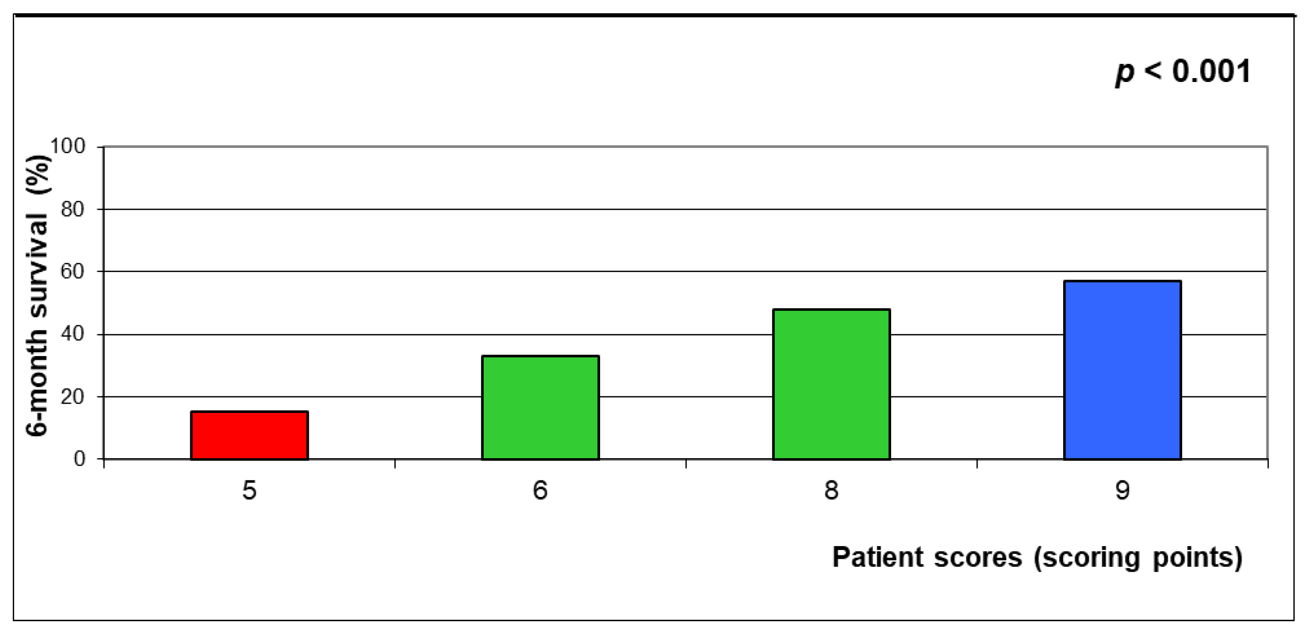
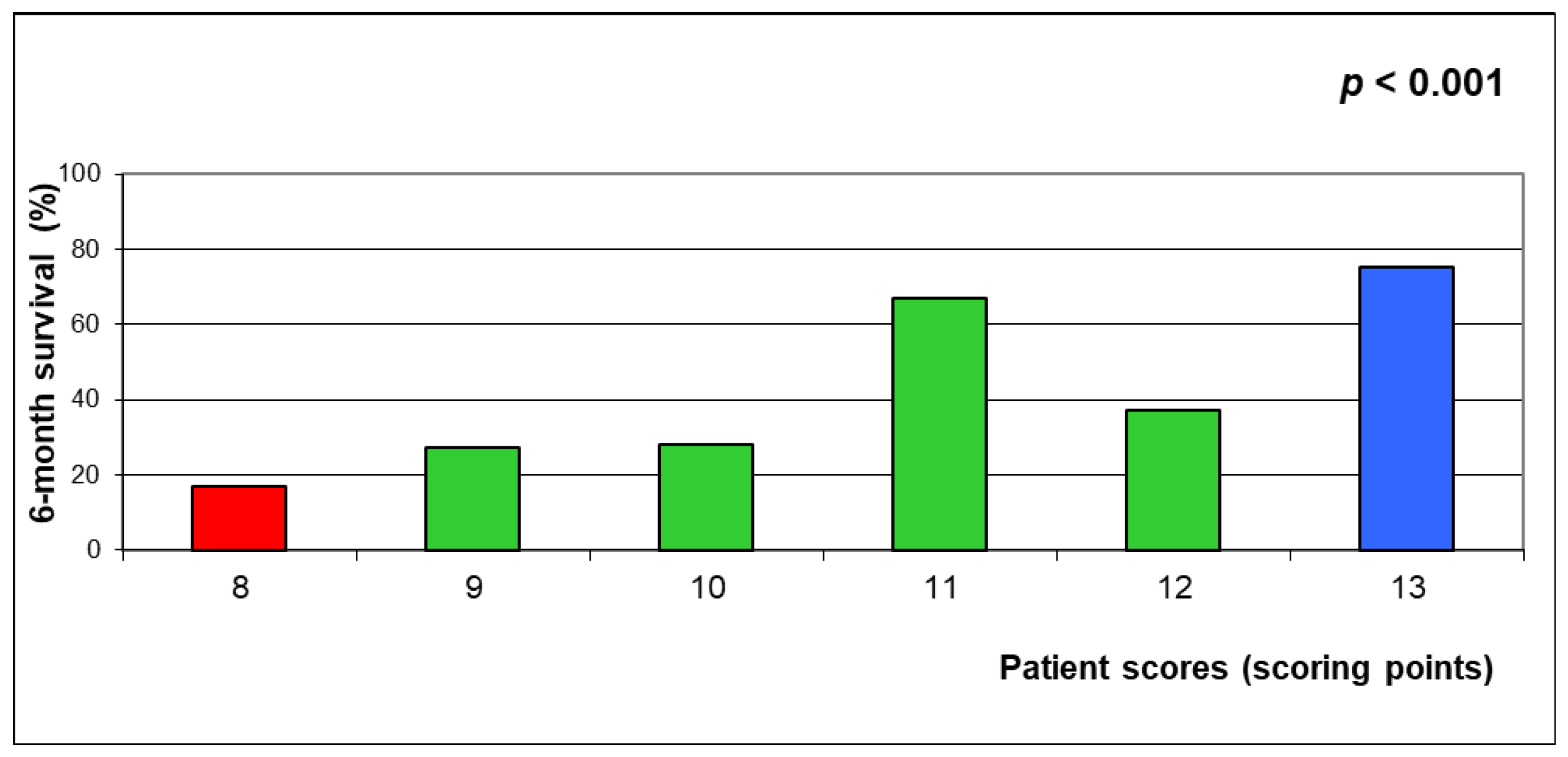
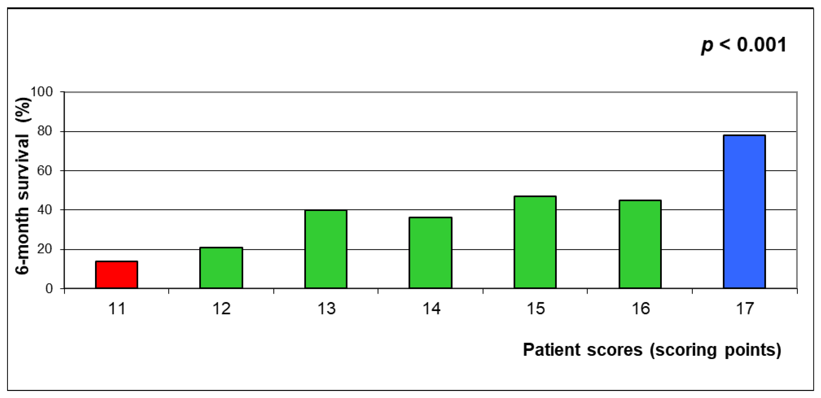
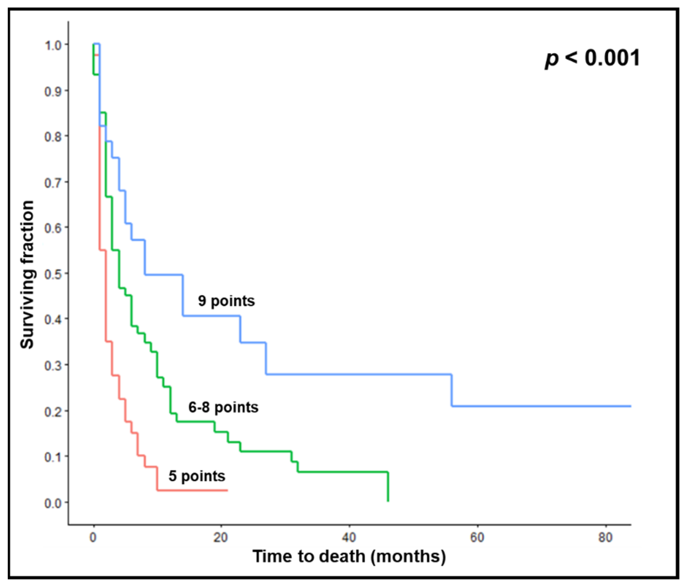
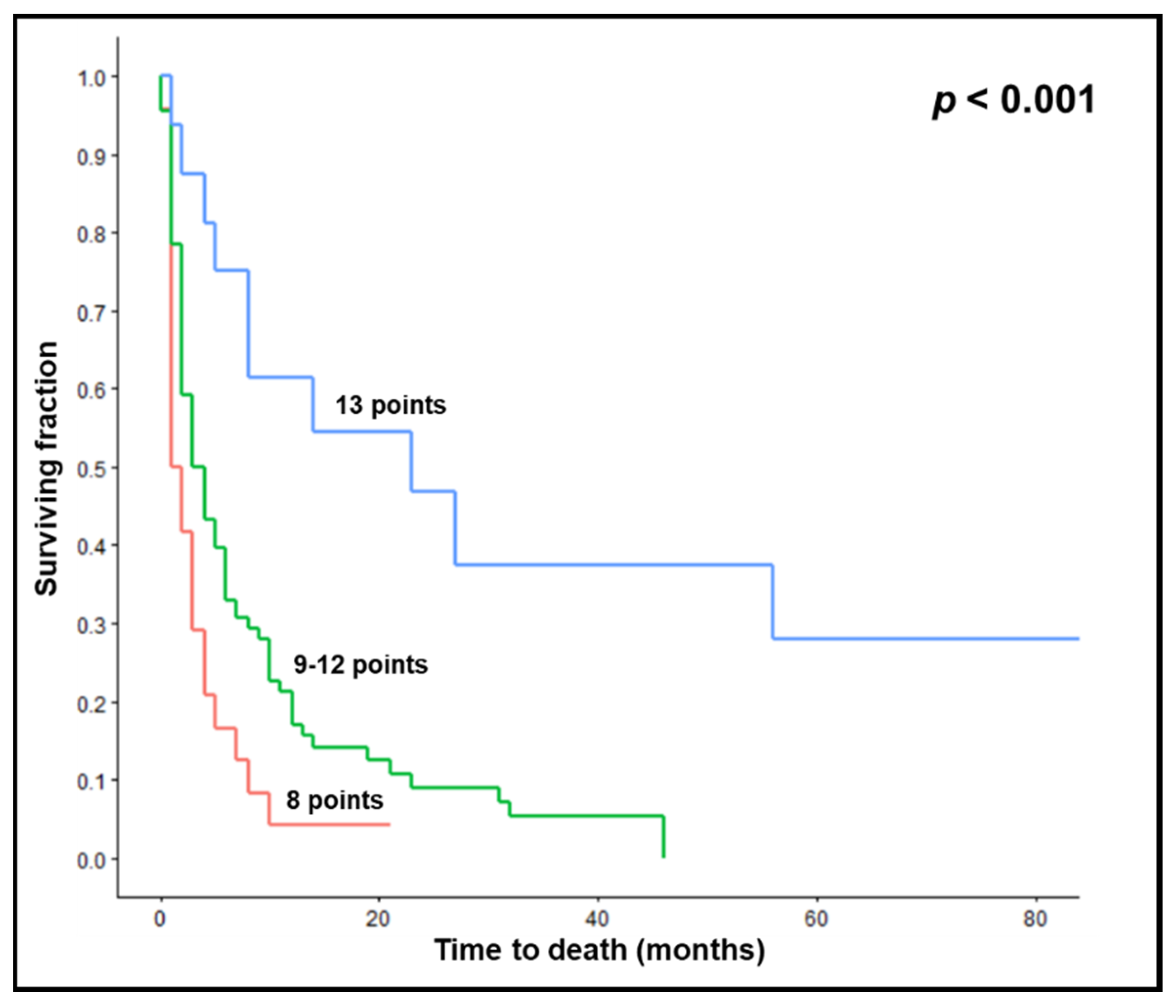
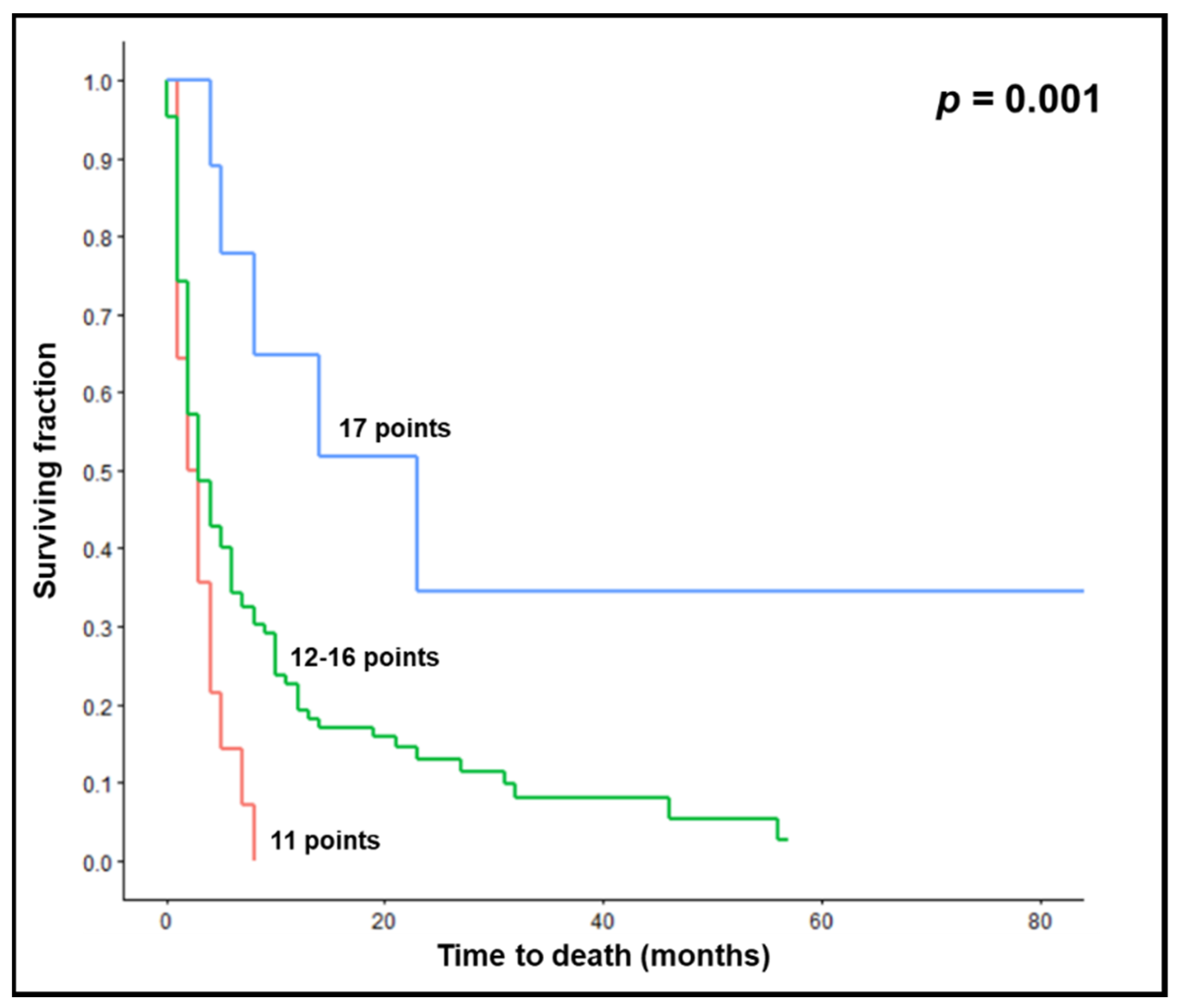
One-hundred-and-twenty-eight patients with newly diagnosed metastases to the brain, who were not considered candidates for neurosurgical resection or stereotactic radiotherapy by a multidisciplinary tumor board, were analyzed. This retrospective study received approval from the Ethics Committee of the University of Lübeck, Lübeck, Germany (22-059). All 128 patients were treated with WBRT plus a SIB to metastatic sites between 2014 and 2021 using VMAT (an example of a treatment plan given in Figure 1). Eighty-seven patients had 1–5 brain metastases, 25 patients had 6–10 lesions, and 16 patients had >10 lesions, respectively. The radiation regimens were either 14 × 2.5 Gy of WBRT plus a SIB of 0.5 Gy per fraction, resulting in a total dose of 42 Gy to the metastatic sites (49 patients), or 18 × 2.0 Gy of WBRT plus a SIB of 0.5 Gy per fraction resulting in a total dose of 45 Gy to the brain lesions (79 patients). The corresponding biologically effective doses using an alpha/beta ratio of 12 Gy for control of brain metastases were 42.3 Gy12 to the entire brain and 52.5 Gy12 to the metastases, respectively, for 14 × 2.5 Gy of WBRT plus a SIB of 0.5 Gy per fraction, and 42.0 Gy12 and 54.4 Gy12, respectively, for 18 × 2.0 Gy of WBRT plus a SIB of 0.5 Gy per fraction.
Ten factors (Table 1) were investigated for associations with survival, including the year of treatment (2014–2018 vs. 2019–2021), time between diagnosis of malignancy and the first fraction of radiation therapy (0–1 vs. ≥2 months), radiotherapy regimen (14 × 2.5 Gy of WBRT plus SIB vs. 18 × 2.0 Gy of WBRT plus SIB), systemic treatment with 6 months before radiotherapy (no vs. yes), age at radiotherapy (≤64 vs. ≥65 years, median = 64 years), gender (female vs. male), Karnofsky performance score (KPS ≤80 vs. 90–100), primary tumor entity (breast cancer vs. non-small cell lung cancer vs. small-cell lung cancer vs. less radiosensitive tumors including kidney cancer and melanoma vs. other types), number of brain lesions (1–3 vs. ≥4, median = 3), and the existence of extra-cerebral metastases (no vs. yes). The year of treatment was included as a parameter since the use of targeted therapies has increased in recent years, particularly for patients with non-small cell lung cancer [15]. Often, these targeted therapies lead to improved survival.
Patients were followed until they died or for at least 6 months after the end of their radiotherapy course. For univariate analyses, we used the Kaplan-Meier method supplemented by the log-rank test (BlueSky Statistics 10 GA, Chicago, IL, USA). Characteristics found significant in univariate analyses (p < 0.05) or indicating a strong trend (p < 0.07) or a trend (p < 0.14) were included in multivariate analyses (Cox proportional hazards model). Those factors that achieved significance in the Cox proportional hazards model were considered independent predictors of survival. Therefore, we felt that it was appropriate to perform both univariate and multivariate analyses. To provide the most precise survival score, three different models were calculated. Model 1 included only factors being significant on both univariate and multivariate analyses (=independent factors), Model 2 included factors that indicated at least a strong trend in univariate analyses, and Model 3 included factors indicating a trend. For each factor included in one of the three models, the 6-month survival rates (in %) were divided by 10. Scoring points obtained from this procedure (factor scores) were added individually for each patient (patient scores). Higher patient scores represented better 6-month survival. Thus, the three models are not mathematical models but were created from real patient data, namely from the patients’ 6-month survival rates. Therefore, we can estimate the probability of dying within 6 months following WBRT+SIB and living 6 or more months following WBRT+SIB.
Based on the corresponding 6-month survival rates, three groups (poor, intermediate, and favorable prognosis) were created. The models were compared for diagnostic accuracy by calculating positive predictive values (PPVs) to correctly prognosticate death during the 6 months following radiotherapy (least favorable prognostic groups) and survival for 6 or more months (most favorable prognostic groups).
PPVs to correctly predict death within 6 months were calculated as follows:
PPV = [patients dying/(patients dying + patients not dying)] × 100
PPVs to correctly predict survival for at least 6 months were calculated as follows:
PPV = [patients surviving/(patients surviving + patients not surviving)] × 100

3. Results

On univariate analyses, KPS of 90–100 (p < 0.001) and only 1–3 brain lesions (p = 0.006) were significantly associated with better survival. Age ≤ 64 years (p = 0.061) showed a strong trend, and no metastases outside the brain (p = 0.128) showed a trend. The results of the complete univariate analyses, including the survival rates at 3, 6, and 12 months are summarized in Table 2. On multivariate analyses, KPS [hazard ratio (HR) 0.40, 95% confidence interval (CI) 0.27–0.61, p < 0.001) and the number of lesions (HR 1.81, 95% CI 1.23–2.65, p = 0.002) remained significant, whereas age (HR 1.12, 95% CI 0.76–1.66, p = 0.57) and extra-cerebral metastases (HR: 1.19, 95% CI: 0.79–1.78, p = 0.40) did not reach significance. The scoring points for each factor are shown in Table 3. These factor scores were valid for each model. Since the models included different numbers of factors, patient scores (obtained after the addition of the factor scores) were also different. Patient scores ranged between 5 and 9 points in Model 1, between 8 and 13 points in Model 2, and between 11 and 17 points in Model 3.
The 6-months survival rates of the patient scores of the three models are shown in Figure 2, Figure 3 and Figure 4.
Based on the 6-month survival rates of the patient scores, three groups with different survival outcomes were created for each model. In Model 1 (including KPS and the number of lesions), groups were 5 points, 6–8 points, and 9 points with 6-month survival rates of 15%, 38%, and 57%, respectively (p < 0.001, Figure 5). In Model 2 (including KPS, number of lesions, and age), groups were 8 points, 9–12 points, and 13 points with 6-month survival rates of 17%, 33%, and 75%, respectively (p < 0.001, Figure 6). In Model 3 (including KPS, number of lesions, age, and extra-cerebral metastases), groups were 11 points, 12–16 points, and 17 points, with 6-month survival rates of 14%, 34%, and 78%, respectively (p = 0.001, Figure 7). The PPVs for accurate prediction of death during 6 months and survival for 6 or more months were 85% and 57% for Model 1, 83% and 75% for Model 2, and 86% and 78% for Model 3, respectively.

4. Discussion

Different techniques and regimens are available for radiotherapy of brain metastases. A regimen that is increasingly used includes WBRT plus a SIB. Compared to WBRT plus a sequential boost, WBRT+SIB has two advantages, i.e., a reduced treatment time and dose to normal tissues [5,6,7,8,9]. Several studies have shown that WBRT+SIB is feasible. Fifteen years ago, Tomita et al. observed no acute toxicity or late complications (median follow-up = 10 months) in seven patients with 1–4 brain metastases treated with 10 × 3 Gy of WBRT and a SIB of 2.0 Gy per fraction to metastatic locations using helical tomotherapy [10]. In 2011, the results of a phase I trial were reported [11]. Patients (n = 48) had 1–3 brain metastases and received 10 × 3 Gy of WBRT plus a SIB with increasing doses ranging between 0.5 and 3.0 Gy per fraction (3 + 3 design). No patient, including those receiving 60 Gy in 10 fractions to the metastatic lesions, experienced dose-limiting neurological toxicity [10]. A similar approach was chosen by Ferro et al. in 2017 [12]. A total of 30 patients were treated with 10 × 3 Gy of WBRT plus a SIB for 1–4 brain metastases using IMRT. SIB doses ranged between 0.5 and 2.0 Gy per fraction. Three patients experienced dose-limiting toxicity, including one grade 3 dermatitis at dose level 2, one grade neurological toxicity at dose level 4, and one intracerebral hemorrhage at dose level 4, respectively [12]. In the retrospective study of Dong et al., 15 × 2.5 Gy of WBRT plus a SIB of 1.0 Gy per fraction was found feasible and safe in 46 patients with 1–7 brain metastases from non-small cell lung cancer [13]. Moreover, in a more recent phase II trial of 50 patients with 1–8 brain metastases receiving 10 × 2.0 Gy of hippocampus-sparing-WBRT plus a SIB of 4.0 Gy per fraction using IMRT or VMAT, only three patients experienced grade 3 toxicities, including nausea, emesis, and necrosis or headache [14]. Moreover, in a recent retrospective multi-center study, the combination of WBRT+SIB and the tyrosine kinase inhibitor apatinib was both feasible and effective [15]. In that study, oral apatinib was started one week before radiation therapy and administered during and for one week following this treatment. Two of 16 patients (12.5%) developed a grade 3 toxicity, namely oral mucositis and hypertension. At three months following treatment, intra-cerebral control was 100%. Median survival and intra-cerebral progression-free survival times were 26 months and 16.5 months, respectively [15].
In addition to demonstrating the feasibility of WBRT+SIB, several studies have shown that this approach leads to local and intra-cerebral control rates at least comparable to other radiation approaches, namely WBRT alone, stereotactic radiotherapy alone, or WBRT plus stereotactic radiotherapy [6,16,17,18,19]. Some data suggests that WBRT+SIB may be superior to WBRT with or without stereotactic radiosurgery [7,20,21,22]. For example, in the most recent study, WBRT+SIB resulted in longer median intra-cerebral progression-free survival than WBRT, followed by stereotactic radiosurgery (91 vs. 5.0 months, p = 0.001) [22]. However, despite increasing interest in using WBRT+SIB, particularly with hippocampus-sparing techniques, not all patients may benefit from this approach.
Survival scores can facilitate identifying patients who may or may not benefit from a WBRT+SIB. Several scores were already developed for patients with brain metastases, including tools for specific techniques and approaches [26,27,28,29,30,31,32]. However, no such tool exists for patients receiving WBRT+SIB. We developed a survival score in a cohort of 128 patients treated with WBRT+SIB and compared three models, including different prognostic factors. Model 1 included two factors, namely KPS and the number of lesions. Model 2 was supplemented by age, and Model 3 was supplemented by age and extra-cerebral metastases. These prognostic factors were previously used in different constellations for survival scores that were designed for patients irradiated for brain metastases but not specifically for those patients receiving WBRT+SIB [26,27,28,29,30,31,32]. These findings demonstrate the consistency of the results in our present study. However, our results should be interpreted with caution, given the retrospective nature of this study.
The main purpose of the three models investigated in this study was the prediction of death within 6 months following WBRT+SIB. The second goal was the prediction of survival for at least 6 months. All three models provided high accuracy regarding the prediction of death within 6 months, with PPVs of 85%, 83%, and 86%, respectively. Regarding the second goal, Model 2 and Model 3 were superior to Model 1 (PPVs of 75% and 78%, respectively, compared to 57%). When comparing Models 2 and 3, the differences regarding the accuracy to predict death ≤6 and survival ≥6 months appear marginal. Given that Model 3 requires more data (including extensive staging to properly judge the presence of extra-cerebral metastases) and is only marginally more accurate than Model 2, Model 2 appears favorable for a considerable number of patients. If extra-cerebral metastases have already been diagnosed or extensive staging is already available without a diagnosis of metastases outside the brain, Model 3 can also be used. When using model 2, those patients with 8 points likely did not benefit from the addition of a SIB since the 6-month survival rate was poor (17%), and the median survival time was only 1.5 months. This likely holds true also for many patients with 9–12 points (6-month survival rate of 33% and median survival time of 3.5 months, respectively). Thus, only patients with 13 points appear as good candidates for adding a SIB. When using Model 3, patients with 17 points appear suitable for the addition of a SIB since the 6-month survival rates of the other two groups (11 points and 12–16 points) were only 14% and 34%, respectively.
However, as stated above, if one considers following the treatment suggestions based on this study’s findings, its limitations must be kept in mind, most of all the retrospective nature of the data used to generate the predictive models. Moreover, many patients with up to 3 or 4 cerebral lesions are candidates for stereotactic radiotherapy with stereotactic radiosurgery in one fraction or multi-fraction stereotactic radiotherapy alone rather than for WBRT+SIB [1,2]. In a study from 2007 including 186 patients with 1–3 brain metastases, 18–25 Gy of stereotactic radiosurgery alone resulted in similar survival and better local control (risk ratio = 1.63, p < 0.01) than 30–40 Gy (2–3 Gy per fraction) of WBRT alone [33]. To further improve radiation therapy outcomes for a limited number of cerebral metastases, several studies investigated the combination of stereotactic radiosurgery and WBRT and compared this approach to stereotactic radiosurgery alone [34,35,36,37,38,39]. In these studies, the addition of WBRT resulted in better intra-cerebral control but not in better survival. Moreover, in two randomized trials, the addition of WBRT resulted in significantly more neuro-cognitive deficits [38,39]. Therefore, many radiation oncologists prefer stereotactic radiosurgery alone rather than stereotactic radiosurgery in combination with WBRT for 1–3 cerebral metastases. This holds particularly true for patients with a low or intermediate risk of developing new brain metastases outside the irradiated cerebral areas within a short period of time. In 2014, a predictive model including three groups was presented that estimated the risk of new brain metastases within 6 months after stereotactic radiotherapy alone [40]. The risk of new cerebral lesions during 6 months post radiation therapy was 36%, 55%, and 80%, respectively, for patients with 16–17, 18–20 and 21–22 points). If stereotactic radiotherapy is used for very few cerebral metastases, multi-fraction stereotactic radiotherapy, for example, with 3 × 9 Gy or 5 × 6 Gy, is preferred over single-fraction treatment for lesions >2 cm in largest diameter since multi-fraction stereotactic irradiation results in less radiation necrosis than single-fraction radiosurgery [1,41]. In a study with 289 consecutive patients with lesions >2 cm from Italy, the cumulative radiation necrosis rates were 18% after single-fraction radiosurgery and 9% after multi-fraction treatment, respectively [41].

5. Conclusions

A new survival score was developed for patients with brain metastases assigned to WBRT+SIB. Three models were created and compared for accuracy. All models accurately predicted death ≤ 6 months with PPVs of 85%, 83%, and 86%, respectively. This can help identify patients who may not benefit from WBRT+SIB. Regarding the prediction of survival for at least 6 months, Model 2 and Model 3 were superior to Model 1, with PPVs of 75% and 78% compared to 57%. Given that Model 3 requires extensive staging to properly judge the existence of metastases outside the brain and is only marginally more accurate than Model 2, Model 2 appears favorable for many patients. If metastases outside the brain are already known and/or extensive staging has already been performed, Model 3 can also be used. Validation of these Models in a prospective cohort of patients is warranted.

Author Contributions

Conceptualization, D.R., L.J., E.M.W., F.C. and N.Y.Y.; methodology, D.R., L.J., E.M.W., F.C. and N.Y.Y.; validation, D.R., L.J. and E.M.W.; formal analysis, D.R. and N.Y.Y.; investigation, D.R. and L.J.; resources, D.R.; writing—original draft preparation, D.R. and N.Y.Y.; writing—review and editing, D.R., L.J., E.M.W., F.C. and N.Y.Y.; visualization, D.R., L.J., E.M.W., F.C. and N.Y.Y. All authors have read and agreed to the published version of the manuscript.

Funding

This research received no external funding.

Institutional Review Board Statement

The study was approved by the Ethics Committee of the University of Lübeck, Germany (22-059).

Informed Consent Statement

Since the study is retrospective in nature, written informed consent was not required from the patients alive according to the responsible ethics committee.

Data Availability Statement

The data analyzed for this paper cannot be shared due to data protection regulations. According to the ethics committee, only evaluation of anonymized data is allowed for this study.

Conflicts of Interest

The authors declare no conflict of interest related to this study.

References

  1. Gondi, V.; Bauman, G.; Bradfield, L.; Burri, S.H.; Cabrera, A.R.; Cunningham, D.A.; Eaton, B.R.; Hattangadi-Gluth, J.A.; Kim, M.M.; Kotecha, R.; et al. Radiation therapy for brain metastases: An ASTRO clinical practice guideline. Pract. Radiat. Oncol. 2022, 12, 265–282. [Google Scholar] [CrossRef] [PubMed]
  2. Tsao, M.N.; Rades, D.; Wirth, A.; Lo, S.S.; Danielson, B.L.; Gaspar, L.E.; Sperduto, P.W.; Vogelbaum, M.A.; Radawski, J.D.; Wang, J.Z.; et al. Radiotherapeutic and surgical management for newly diagnosed brain metastasis(es): An American Society for Radiation Oncology evidence-based guideline. Pract. Radiat. Oncol. 2012, 2, 210–225. [Google Scholar] [CrossRef] [PubMed] [Green Version]
  3. Andrews, D.W.; Scott, C.B.; Sperduto, P.W.; Flanders, A.E.; Gaspar, L.E.; Schell, M.C.; Werner-Wasik, M.; Demas, W.; Ryu, J.; Bahary, J.P.; et al. Whole brain radiation therapy with or without stereotactic radiosurgery boost for patients with one to three brain metastases: Phase III results of the RTOG 9508 randomised trial. Lancet 2004, 363, 1665–1672. [Google Scholar] [CrossRef]
  4. Rades, D.; Janssen, S.; Dziggel, L.; Blanck, O.; Bajrovic, A.; Veninga, T.; Schild, S.E. A matched-pair study comparing whole-brain irradiation alone to radiosurgery or fractionated stereotactic radiotherapy alone in patients irradiated for up to three brain metastases. BMC Cancer 2017, 17, 30. [Google Scholar] [CrossRef] [Green Version]
  5. Mantovani, C.; Gastino, A.; Cerrato, M.; Badellino, S.; Ricardi, U.; Levis, M. Modern radiation therapy for the management of brain metastases from non-small cell lung cancer: Current approaches and future directions. Front. Oncol. 2021, 11, 772789. [Google Scholar] [CrossRef]
  6. Awad, R.; Fogarty, G.; Hong, A.; Kelly, P.; Ng, D.; Santos, D.; Haydu, L. Hippocampal avoidance with volumetric modulated arc therapy in melanoma brain metastases—The first Australian experience. Radiat. Oncol. 2013, 8, 62. [Google Scholar] [CrossRef] [PubMed] [Green Version]
  7. Dobi, Á.; Fodor, E.; Maráz, A.; Együd, Z.; Cserháti, A.; Tiszlavicz, L.; Reisz, Z.; Barzó, P.; Varga, Z.; Hideghéty, K. Boost irradiation integrated to whole brain radiotherapy in the management of brain metastases. Pathol. Oncol. Res. 2020, 26, 149–157. [Google Scholar] [CrossRef] [Green Version]
  8. Lagerwaard, F.J.; van der Hoorn, E.A.; Verbakel, W.F.; Haasbeek, C.J.; Slotman, B.J.; Senan, S. Whole-brain radiotherapy with simultaneous integrated boost to multiple brain metastases using volumetric modulated arc therapy. Int. J. Radiat. Oncol. Biol. Phys. 2009, 75, 253–259. [Google Scholar] [CrossRef]
  9. Hsu, F.; Carolan, H.; Nichol, A.; Cao, F.; Nuraney, N.; Lee, R.; Gete, E.; Wong, F.; Schmuland, M.; Heran, M.; et al. Whole brain radiotherapy with hippocampal avoidance and simultaneous integrated boost for 1-3 brain metastases: A feasibility study using volumetric modulated arc therapy. Int. J. Radiat. Oncol. Biol. Phys. 2010, 76, 1480–1485. [Google Scholar] [CrossRef]
  10. Tomita, N.; Kodaira, T.; Tachibana, H.; Nakamura, T.; Nakahara, R.; Inokuchi, H.; Shibamoto, Y. Helical tomotherapy for brain metastases: Dosimetric evaluation of treatment plans and early clinical results. Technol. Cancer Res. Treat. 2008, 7, 417–424. [Google Scholar] [CrossRef]
  11. Rodrigues, G.; Yartsev, S.; Yaremko, B.; Perera, F.; Dar, A.R.; Hammond, A.; Lock, M.; Yu, E.; Ash, R.; Caudrelier, J.M.; et al. Phase I trial of simultaneous in-field boost with helical tomotherapy for patients with one to three brain metastases. Int. J. Radiat. Oncol. Biol. Phys. 2011, 80, 1128–1133. [Google Scholar] [CrossRef] [PubMed]
  12. Ferro, M.; Chiesa, S.; Macchia, G.; Cilla, S.; Bertini, F.; Frezza, G.; Farioli, A.; Cammelli, S.; Balducci, M.; Ianiro, A.; et al. Intensity modulated radiation therapy with simultaneous integrated boost in patients with brain oligometastases: A phase 1 study (ISIDE-BM-1). Int. J Radiat. Oncol. Biol. Phys. 2017, 97, 82–90. [Google Scholar] [CrossRef] [PubMed]
  13. Dong, Y.; Zhang, Y.; Zhang, T.; Fan, M.; Zhu, J.; Li, B.; Huang, W. Feasibility and efficacy of simultaneous integrated boost intensity-modulated radiation therapy based on MRI-CT fusion in patients with brain metastases of non-small cell lung cancer. J. Cancer 2018, 9, 4477–4483. [Google Scholar] [CrossRef] [PubMed]
  14. Westover, K.D.; Mendel, J.T.; Dan, T.; Kumar, K.; Gao, A.; Pulipparacharuv, S.; Iyengar, P.; Nedzi, L.; Hannan, R.; Anderson, J.; et al. Phase II trial of hippocampal-sparing whole brain irradiation with simultaneous integrated boost for metastatic cancer. Neuro Oncol. 2020, 22, 1831–1839. [Google Scholar] [CrossRef] [PubMed]
  15. Ma, J.; Bi, J.; Tuo, X.; Pi, G.; Li, Y.; Li, Y.; Zeng, F.; Gong, H.; Hu, D.; Han, G. Efficacy and safety of apatinib combined with whole-brain radiation therapy with a simultaneous integrated boost for brain metastases from non-small cell lung cancer: A multicenter retrospective study. J. Thorac. Dis. 2022, 14, 455–463. [Google Scholar] [CrossRef]
  16. Vargo, J.A.; Plants, B.A.; Mihailidis, D.N.; Mallah, J.; Plants, M.; Welch, C.A.; Clark, G.M.; Farinash, L.J.; Raja, P.; Harmon, M.B.; et al. Early clinical outcomes for 3 radiation techniques for brain metastases: Focal versus whole-brain. Pract. Radiat. Oncol. 2011, 1, 261–270. [Google Scholar] [CrossRef]
  17. Oehlke, O.; Wucherpfennig, D.; Fels, F.; Frings, L.; Egger, K.; Weyerbrock, A.; Proki, V.; Nieder, C.; Grosu, A.L. Whole brain irradiation with hippocampal sparing and dose escalation on multiple brain metastases: Local tumour control and survival. Strahlenther. Onkol. 2015, 191, 461–469. [Google Scholar] [CrossRef]
  18. Lebow, E.S.; Hwang, W.L.; Zieminski, S.; Wang, Y.; Niemierko, A.; Mehan, W.A., Jr.; Oh, K.S.; Khandekar, M.; Willers, H.; Shih, H.A. Early experience with hippocampal avoidance whole brain radiation therapy and simultaneous integrated boost for brain metastases. J. Neurooncol. 2020, 148, 81–88. [Google Scholar] [CrossRef]
  19. Zhong, J.; Waldman, A.D.; Kandula, S.; Eaton, B.R.; Prabhu, R.S.; Huff, S.B.; Shu, H.G. Outcomes of whole-brain radiation with simultaneous in-field boost (SIB) for the treatment of brain metastases. J. Neurooncol. 2020, 147, 117–123. [Google Scholar] [CrossRef]
  20. Popp, I.; Rau, S.; Hintz, M.; Schneider, J.; Bilger, A.; Fennell, J.T.; Heiland, D.H.; Rothe, T.; Egger, K.; Nieder, C.; et al. Hippocampus-avoidance whole-brain radiation therapy with a simultaneous integrated boost for multiple brain metastases. Cancer 2020, 126, 2694–2703. [Google Scholar] [CrossRef] [Green Version]
  21. Du, T.Q.; Li, X.; Zhong, W.S.; Tian, J.D.; Zhao, Y.X.; Liu, D. Brain metastases of lung cancer: Comparison of survival outcomes among whole brain radiotherapy, whole brain radiotherapy with consecutive boost, and simultaneous integrated boost. J. Cancer Res. Clin. Oncol. 2021, 147, 569–577. [Google Scholar] [CrossRef] [PubMed]
  22. Lin, B.; Huang, D.; Du, H.; Fan, J.; Zhang, Y.; Feng, G.; Gao, F.; Du, X.B. Whole-brain radiation therapy with simultaneous integrated boost versus whole-brain radiation therapy plus stereotactic radiosurgery for the treatment of brain metastasis from lung cancer. Front. Oncol. 2021, 11, 631422. [Google Scholar] [CrossRef] [PubMed]
  23. Grosu, A.L.; Frings, L.; Bentsalo, I.; Oehlke, O.; Brenner, F.; Bilger, A.; Fennell, J.T.; Rothe, T.; Schneider-Fuchs, S.; Graf, E.; et al. Whole-brain irradiation with hippocampal sparing and dose escalation on metastases: Neurocognitive testing and biological imaging (HIPPORAD)—A phase II prospective randomized multicenter trial (NOA-14, ARO 2015-3, DKTK-ROG). BMC Cancer 2020, 20, 532. [Google Scholar] [CrossRef]
  24. Chia, B.S.H.; Leong, J.Y.; Ong, A.L.K.; Lim, C.; Poon, S.H.; Chua, M.L.K.; Chua, K.L.M.; Kusumawidjaja, G.; Chua, E.T.; Wong, F.Y.; et al. Randomised prospective phase II trial in multiple brain metastases comparing outcomes between hippocampal avoidance whole brain radiotherapy with or without simultaneous integrated boost: HA-SIB-WBRT study protocol. BMC Cancer 2020, 20, 1045. [Google Scholar] [CrossRef]
  25. Popp, I.; Grosu, A.L.; Fennell, J.T.; Fischer, M.; Baltas, D.; Wiehle, R. Optimization of hippocampus sparing during whole brain radiation therapy with simultaneous integrated boost-tutorial and efficacy of complete directional hippocampal blocking. Strahlenther. Onkol. 2022, 198, 537–546. [Google Scholar] [CrossRef] [PubMed]
  26. Gaspar, L.; Scott, C.; Rotman, M.; Asbell, S.; Phillips, T.; Wasserman, T.; McKenna, W.G.; Byhardt, R. Recursive partitioning analysis (RPA) of prognostic factors in three Radiation Therapy Oncology Group (RTOG) brain metastases trials. Int. J. Radiat. Oncol. Biol. Phys. 1997, 37, 745–751. [Google Scholar] [CrossRef]
  27. Weltman, E.; Salvajoli, J.V.; Brandt, R.A.; de Morais Hanriot, R.; Prisco, F.E.; Cruz, J.C.; de Oliveira Borges, S.R.; Wajsbrot, D.B. Radiosurgery for brain metastases: A score index for predicting prognosis. Int. J. Radiat. Oncol. Biol. Phys. 2000, 46, 1155–1161. [Google Scholar] [CrossRef]
  28. Rades, D.; Dunst, J.; Schild, S.E. A new scoring system to predicting the survival of patients treated with whole-brain radiotherapy for brain metastases. Strahlenther. Onkol. 2008, 184, 251–255. [Google Scholar] [CrossRef]
  29. Sperduto, P.W.; Berkey, B.; Gaspar, L.E.; Mehta, M.; Curran, W. A new prognostic index and comparison to three other indices for patients with brain metastases: An analysis of 1,960 patients in the RTOG database. Int. J. Radiat. Oncol. Biol. Phys. 2008, 70, 510–514. [Google Scholar] [CrossRef]
  30. Rades, D.; Dziggel, L.; Nagy, V.; Segedin, B.; Lohynska, R.; Veninga, T.; Khoa, M.T.; Trang, N.T.; Schild, S.E. A new survival score for patients with brain metastases who received whole-brain radiotherapy (WBRT) alone. Radiother. Oncol. 2013, 108, 123–127. [Google Scholar] [CrossRef]
  31. Lorenzoni, J.; Devriendt, D.; Massager, N.; David, P.; Ruíz, S.; Vanderlinden, B.; Van Houtte, P.; Brotchi, J.; Levivier, M. Radiosurgery for treatment of brain metastases: Estimation of patient eligibility using three stratification systems. Int. J. Radiat. Oncol. Biol. Phys. 2004, 60, 218–224. [Google Scholar] [CrossRef] [PubMed]
  32. Serizawa, T.; Higuchi, Y.; Nagano, O.; Matsuda, S.; Ono, J.; Saeki, N.; Hirai, T.; Miyakawa, A.; Shibamoto, Y. A new grading system focusing on neurological outcomes for brain metastases treated with stereotactic radiosurgery: The modified Basic Score for Brain Metastases. J. Neurosurg. 2014, 121 (Suppl. S2), 35–43. [Google Scholar] [CrossRef] [PubMed] [Green Version]
  33. Rades, D.; Pluemer, A.; Veninga, T.; Hanssens, P.; Dunst, J.; Schild, S.E. Whole-brain radiotherapy versus stereotactic radiosurgery for patients in recursive partitioning analysis classes 1 and 2 with 1 to 3 brain metastases. Cancer 2007, 110, 2285–2292. [Google Scholar] [CrossRef] [PubMed]
  34. Aoyama, H.; Shirato, H.; Tago, M.; Nakagawa, K.; Toyoda, T.; Hatano, K.; Kenjyo, M.; Oya, N.; Hirota, S.; Shioura, H.; et al. Stereotactic radiosurgery plus whole-brain radiation therapy vs stereotactic radiosurgery alone for treatment of brain metastases: A randomized controlled trial. JAMA 2006, 295, 2483–2491. [Google Scholar] [CrossRef] [PubMed]
  35. Rades, D.; Kueter, J.D.; Hornung, D.; Veninga, T.; Hanssens, P.; Schild, S.E.; Dunst, J. Comparison of stereotactic radiosurgery (SRS) alone and whole brain radiotherapy (WBRT) plus a stereotactic boost (WBRT+SRS) for one to three brain metastases. Strahlenther. Onkol. 2008, 184, 655–662. [Google Scholar] [CrossRef] [PubMed]
  36. Kocher, M.; Soffietti, R.; Abacioglu, U.; Villà, S.; Fauchon, F.; Baumert, B.G.; Fariselli, L.; Tzuk-Shina, T.; Kortmann, R.D.; Carrie, C.; et al. Adjuvant whole-brain radiotherapy versus observation after radiosurgery or surgical resection of one to three cerebral metastases: Results of the EORTC 22952-26001 study. J. Clin. Oncol. 2011, 29, 134–141. [Google Scholar] [CrossRef] [Green Version]
  37. Rades, D.; Hornung, D.; Veninga, T.; Schild, S.; Gliemroth, J. Single brain metastasis: Radiosurgery alone compared with radiosurgery plus up-front whole-brain radiotherapy. Cancer 2012, 118, 2980–2985. [Google Scholar] [CrossRef] [Green Version]
  38. Chang, E.L.; Wefel, J.S.; Hess, K.R.; Allen, P.K.; Lang, F.F.; Kornguth, D.G.; Arbuckle, R.B.; Swint, J.M.; Shiu, A.S.; Maor, M.H.; et al. Neurocognition in patients with brain metastases treated with radiosurgery or radiosurgery plus whole-brain irradiation: A randomised controlled trial. Lancet Oncol. 2009, 10, 1037–1044. [Google Scholar] [CrossRef]
  39. Brown, P.D.; Jaeckle, K.; Ballman, K.V.; Farace, E.; Cerhan, J.H.; Anderson, S.K.; Carrero, X.W.; Barker, F.G., 2nd; Deming, R.; Burri, S.H.; et al. Effect of Radiosurgery alone vs radiosurgery with whole brain radiation therapy on cognitive function in patients with 1 to 3 brain metastases: A randomized clinical trial. JAMA 2016, 316, 401–409. [Google Scholar] [CrossRef]
  40. Huttenlocher, S.; Dziggel, L.; Horning, D.; Blanck, O.; Schild, S.E.; Rades, D. A new prognostic instrument to predict the probability of developing new cerebral metastases after radiosurgery alone. Radiat. Oncol. 2014, 9, 267. [Google Scholar] [CrossRef] [Green Version]
  41. Minniti, G.; Scaringi, C.; Paolini, S.; Lanzetta, G.; Romano, A.; Cicone, F.; Osti, M.; Enrici, R.M.; Esposito, V. Single-fraction versus multifraction (3 × 9 Gy) stereotactic radiosurgery for large (>2 cm) brain metastases: A comparative analysis of local control and risk of radiation-induced brain necrosis. Int. J. Radiat. Oncol. Biol. Phys. 2016, 95, 1142–1148. [Google Scholar] [CrossRef] [PubMed]
Figure 1. Example of a treatment plan of whole-brain radiotherapy plus a simultaneous integrated boost to the metastatic lesions using a hippocampus-sparing technique (Department of Radiation Oncology, University of Lübeck, Germany).
Biology 12 00585 g001
Figure 2. Model 1 (including KPS and number of lesions): Six-month survival rates of patient scores ranging between 5 and 9 points.
Biology 12 00585 g002
Figure 3. Model 2 (including KPS, number of lesions, and age): Six-month survival rates of patient scores ranging between 8 and 13 points.
Biology 12 00585 g003
Figure 4. Model 3 (including KPS, number of lesions, age, and extra-cerebral metastases): Six-month survival rates of patient scores ranging between 11 and 17 points.
Biology 12 00585 g004
Figure 5. Model 1: Kaplan-Meier curves for the survival of the three groups (5, 6–8 and 9 points).
Biology 12 00585 g005
Figure 6. Model 2: Kaplan-Meier curves for survival of the three groups (8, 9–12, and 13 points).
Biology 12 00585 g006
Figure 7. Model 3: Kaplan-Meier curves for survival of the three groups (11, 12–16 and 17 points).
Biology 12 00585 g007
Table 1. Distribution of the factors analyzed for survival.
FactorNumber of PatientsProportion (%)
Year of treatment
2014–20187256
2019–20215644
Time between tumor diagnosis and RT
0–1 months7055
≥2 months5845
RT regimen
14 × 2.5 Gy of WBRT + SIB4938
18 × 2.0 Gy of WBRT + SIB7962
Pre-RT systemic treatment
No7962
Yes4938
Age at RT
≤64 years6551
≥65 years6349
Gender
Female4938
Male7962
Karnofsky performance score
≤807962
90–1004938
Type of primary tumor
Breast cancer86
Non-small cell lung cancer7760
Small-cell lung cancer1814
Less radiosensitive tumors1310
Other types129
Number of brain lesions
1–36752
≥46148
Extra-cerebral metastases
No4737
Yes8163
RT: Radiotherapy; WBRT: Whole-Brain Radiotherapy; SIB: Simultaneous Integrated Boost.
Table 2. Survival rates of the investigated factors at 3, 6, and 12 months following radiotherapy.
FactorSurvival Rate (%)p-Value
at 3
Months
at 6
Months
at 12
Months
Year of treatment 0.34
2014–2018513623
2019–2021503417
Time between tumor diagnosis and RT 0.56
0–1 months564023
≥2 months452918
RT regimen 0.55
14 x 2.5 Gy of WBRT + SIB553722
18 x 2.0 Gy of WBRT + SIB483419
Pre-RT systemic treatment 0.84
No523523
Yes493515
Age at RT 0.061
≤64 years553825
≥65 years463216
Gender 0.30
Female433330
Male563715
Karnofsky performance score <0.001
≤80372410
90–100735337
Type of primary tumor 0.26
Breast cancer503825
Non-small cell lung cancer533824
Small-cell lung cancer50280
Less radiosensitive tumors624623
Other types251717
Number of brain lesions 0.006
1–3584331
≥443269
Extra-cerebral metastases 0.128
No554031
Yes483214
RT: Radiotherapy; WBRT: Whole-Brain Radiotherapy; SIB: Simultaneous Integrated Boost. Bold p-values are significant.
Table 3. Six-month survival rates and related scoring points.
FactorSurvival Rate
at 6 Months (%)
Scoring
Points
Karnofsky performance score
90–100535
≤80242
Number of brain lesions
1–3434
≥4263
Age at radiotherapy
≤64 years384
≥65 years323
Extra-cerebral metastases
No404
Yes323
Disclaimer/Publisher’s Note: The statements, opinions and data contained in all publications are solely those of the individual author(s) and contributor(s) and not of MDPI and/or the editor(s). MDPI and/or the editor(s) disclaim responsibility for any injury to people or property resulting from any ideas, methods, instructions or products referred to in the content.

