Communication

Whole Genome Analysis and Assessment of the Metabolic Potential of Gordonia rubripertincta Strain 112, a Degrader of Aromatic and Aliphatic Compounds

by
Ekaterina Frantsuzova
1,
Alexander Bogun
1,2,
Viktor Solomentsev
1,2,
Anna Vetrova
1,
Rostislav Streletskii
3,
Inna Solyanikova
1,4 and
Yanina Delegan
1,*
1
Institute of Biochemistry and Physiology of Microorganisms, Federal Research Center “Pushchino Scientific Center for Biological Research of Russian Academy of Sciences” (FRC PSCBR RAS), 142290 Pushchino, Moscow Region, Russia
2
State Research Center for Applied Microbiology and Biotechnology, 142279 Obolensk, Moscow Region, Russia
3
Laboratory of Ecological Soil Science, Faculty of Soil Science, Lomonosov Moscow State University, 119991 Moscow, Russia
4
Regional Microbiological Center, Belgorod State University, 308015 Belgorod, Russia
*
Author to whom correspondence should be addressed.
Biology 2023, 12(5), 721; https://doi.org/10.3390/biology12050721
Submission received: 18 April 2023 / Revised: 11 May 2023 / Accepted: 13 May 2023 / Published: 15 May 2023
(This article belongs to the Section Microbiology)

Abstract

:

Simple Summary

In the modern world, natural ecosystems are subjected to significant anthropogenic and technogenic stress. As a result of industrial processes, pollutants of various chemical structures are discharged into water and soil ecosystems. Many bacteria are able to utilize pollutants, thus contributing to the remediation of polluted ecosystems. The Gordonia rubripertincta strain 112 is interesting as a destroyer of aromatic and aliphatic compounds. The study of the molecular mechanisms of its functioning will allow us to use it effectively in green technologies in the future. We have shown that the strain is able to quickly cope with toxic pollutants without stress and growth inhibition.

Abstract

The application of Gordonia strains in biotechnologies of environmental purification as degraders of pollutants of different chemical structures is an interesting research topic. The strain Gordonia rubripertincta 112 (IEGM112) is capable of utilizing diesel fuel, alkanes, and aromatic compounds. The aim of this work was to study the potential of G. rubripertincta 112 as a degrader of aromatic and aliphatic compounds and analyze its complete genome in comparison with other known G. rubripertincta strains. The genome had a total length of 5.28 Mb and contained 4861 genes in total, of which 4799 were coding sequences (CDS). The genome contained 62 RNA genes in total, of which 50 were tRNAs, three were ncRNAs, and nine were rRNAs. The strain bears plasmid elements with a total length of 189,570 nucleotides (plasmid p1517). The strain can utilize 10.79 ± 1.17% of hexadecane and 16.14 ± 0.16% of decane over 3 days of cultivation. In the genome of the strain, we have found metabolic pathways of alkane (cytochrome P450 hydroxylases) and catechol (ortho- and meta-pathways) degradation. These results will help us to further approach the fundamental study of the processes occurring in the strain cells and to enrich our knowledge of the catabolic capabilities of G. rubripertincta.

1. Introduction

Actinobacteria of the genus Gordonia are a significant part of microbial communities formed in various ecosystems, such as soils, waters, and sediments [1,2,3,4,5]. It can be said that due to their amazing metabolic flexibility, Gordonia strains are distributed everywhere. Representatives of the genus Gordonia have been isolated from the soil of the cold desert in India (G. terrae, EU333873.1), the sand of Namibia (G. namibiensis, [6]), and even from the intestinal tract of Periplaneta americana (G. terrae, [7]). Species of Gordonia are resistant to desiccation [8] and the metal content in the habitat [9], and they are capable of growing in saline media [10].
Gordonia strains are interesting for application in biotechnologies of environmental purification as degraders of pollutants of different chemical structures, such as alkanes [11,12,13,14], aromatic hydrocarbons [15,16] and their derivatives [17,18], thiophenes [19,20,21], and phthalates [22,23,24,25,26]. The type strains of several Gordonia species were isolated from soils contaminated with thiophenes (G. amicalis [27]), tar (G. alkanivorans [28]), from automobile tires (G. polyisoprenivorans [29], G. westfalica [30]), oil-producing wells (G. paraffinivorans [31]), and activated sludge [23].
Little is known about the metabolic capabilities of Gordonia rubripertincta representatives. The G. rubripertincta strain CWB2 is a promising producer of siderophores [32,33,34,35,36] and is capable of utilizing styrene [37,38,39]. A strain of the G. rubripertincta SD5 strain has the ability to utilize di-(2-ethylhexyl) phthalate [40]. The ability of G. rubripertincta to utilize alkanes has been shown only by Trögl et al. [41] who used the CWB2 strain as an example. The authors studied the microbial degradation of a C10-C40 mixture of alkanes in a model soil experiment. Thus, presently, the potential of G. rubripertincta strains as degraders of various pollutants has been very poorly studied.
The strain Gordonia rubripertincta 112 (IEGM112) was isolated in 1980 from crude oil-contaminated soil in the territory of Ukraine. The aim of this work is to study the potential of the G. rubripertincta strain 112 as a degrader of aromatic and aliphatic compounds and analyze its genome in comparison with other known G. rubripertincta strains.
The catabolism of alkanes in the representatives of the genus Gordonia can be proceeded by both terminal and subterminal oxidation. During terminal oxidation, alcohol, aldehyde, and acid are sequentially formed from alkanes. Thus, Liu et al. [12] observed the formation of hexadecanol and hexadecanoic acid in the cells of the Gordonia sihwaniensis strain utilizing hexadecane. However, the authors also recorded the presence of hexadecene in the cells, which suggested the possibility of an alternative pathway of alkane catabolism.
The subterminal oxidation of alkanes was observed by Kotani et al. [42] in Gordonia sp. TY-5. The strain oxidized propane to 2-propanol. Then, 2-propanol was converted to acetone, and then under the control of Baeyer–Villiger monooxygenase, it was transformed into an ester [43]. Examples of degradation of longer alkanes by Gordonia strains through subterminal oxidation are not currently available in the literature.
It has been reported in the literature that Gordonia strains use two genetic systems for terminal alkane oxidation: alkB for long and cytochrome P450 (CYP153) for short ones [12,13,44]. The alkB genetic system is usually represented by a single copy, while CYP153 can have several copies. For example, we previously showed that the Gordonia amicalis 1D strain contains one copy of alkB and two copies of CYP153, which allows it to utilize alkanes with a length range from C10 to C36 [45].
Among Gordonia, it is also known that there may be no alkB genes at all. The entire process of alkane catabolism in such strains is controlled by the alkane monooxygenases of the CYP153 family. For example, this is a characteristic of G. alkanivorans strains [20]. It is now known that both alkB and CYP153 genes are involved in alkane catabolism with a medium chain length. In actinobacteria, the alkB system controls the oxidation of C16–C40 alkanes, while CYP153 oxidizes alkanes with a shorter chain (shorter than C14) [46,47].
The ability to utilize aromatic compounds (naphthalene and its derivatives), unlike alkanes, is infrequent in Gordonia. Lin et al. [16] described the organization of an operon involved in naphthalene catabolism. One of the strains in our laboratory collection, G. polyisoprenivorans 135 [15], is also capable of utilizing naphthalene and its derivatives. The organization of the naphthalene operon in strain 135 is different from the one of Gordonia sp. strain CC-NAPH129-6 in the work of Lin et al. Catechol degradation among Gordonia was previously observed only in G. polyisoprenivorans [18].
In this work, we plan to study the organization of the genetic systems of alkane and catechol catabolism in the G. rubripertincta strain 112 and compare them with the organization of similar systems in the genomes of other Gordonia strains. These results will help us to further approach the fundamental study of the processes occurring in the strain cells and to enrich our knowledge of the catabolic capabilities of G. rubripertincta.

2. Materials and Methods

2.1. Bacterial Strain

The strain Gordonia rubripertincta 112 (IEGM112) can be found in the IEGM Regional Specialized Collection of Alkanotrophic Microorganisms (Perm, Russia) and in the collection of the laboratory of the physiology of microorganisms IBPM RAS (Pushchino, Moscow Region, Russia).

2.2. Chemicals

High-purity-grade (>98%) catechol, dichloromethane, decane, and hexadecane were obtained from Sigma-Aldrich (Burlington, MA, USA). All the other reagents were of analytical grade.

2.3. Growth Media and Conditions

The strain G. rubripertincta 112 was grown at 27 °C in a liquid mineral medium with decane (7.5 mL/L), hexadecane (7.5 mL/L), catechol (0.1 g/L), benzoate (1 g/L), or potassium acetate (10% w/w) as the sole carbon and energy source on an orbital shaker at 180 rpm.
To obtain the inoculum, the strain was grown in a mineral medium with acetate for 20 h. The biomass was precipitated and washed with sterile distilled water before inoculating it into experimental flasks. The resulting precipitate was resuspended in a mineral medium until a concentration of 108 CFU/mL was reached using a turbidity standard. The inoculum was added to the experimental flasks to the final concentration of 106 CFU/mL.
The following mineral medium was used in this study: K2HPO4—8.71 g/L, 5 M NH4Cl solution—1 mL/L, 0.1 M Na2SO4 solution—1 mL/L, 62 mM MgCl2 solution—1 mL/L, 1 mM CaCl2 solution—1 mL/L, 0.005 mM (NH4)6Mo7O24 × 4H2O solution, micronutrients—1 mL (micronutrient composition in g/L: ZnO—0.41 g, FeCl3 × 6H2O—5.4 g, MnCl2 × 4H2O—2 g, CuCl2 × 2H2O—0.17 g, CoCl2 × 6H2O—0.48 g, H3BO3—0.06 g), pH 7.0.
Lysogeny broth (LB) medium [48], consisting of (per liter of distilled water) 10 g of tryptone, 5 g of yeast extract, 5 g of NaCl, and 15 g of agar (PanReac, Barcelona, Spain), was used to obtain individual colonies and assess bacterial abundance.
To evaluate the efficiency of aliphatic hydrocarbon degradation by G. rubripertincta 112, the bacterium was cultured at 27 °C in a liquid mineral medium with decane or hexadecane for 3 days. All the experiments were performed in three independent biological replicates.

2.4. Determination of Hydrocarbon Concentration

The samples were analyzed using the equipment of the Collective Use Center, Soil Science Faculty, and Lomonosov Moscow State University. Decane or hexadecane was extracted from the culture medium with dichloromethane (1:2, v/v). To stop all biological processes, pre-acidification of the culture medium with sulfuric acid to pH 2 was performed. Decane and hexadecane were measured using a gas chromatography system (Agilent 6890, Agilent Technologies, Santa Clara, CA, USA) equipped with a flame ionization detector. The chromatographic column was DB-1 (30 m × 0.25 mm id, 0.25 µm). The oven temperature program was from 40 °C with an increase of 15 °C/min and 11.7 °C/min for decane and hexadecane, respectively.
Absolute calibration with analytical standards was used for quantitation. The correlation coefficient was 0.98. The validity of the results was confirmed using one-factor analysis of variance (ANOVA), which was p = 0.05. The samples were diluted 100-fold before the assay was utilized. All of the results were derived from five independent replicates.
The degree of decane/hexadecane biodegradation (D) was calculated using the following formula:
D = (C0 − Ci)/C0 × 100 [%],
where C0 is the concentration of hydrocarbon in the experiment without microorganisms (abiotic control), and Ci is the concentration of hydrocarbon in the experiment with microorganisms after 72 h of growth.

2.5. Genome Sequencing and Analysis

Genomic DNA was isolated from a fresh culture biomass (a colony) of Gordonia rubripertincta 112 grown on LB agar using a DNeasy Blood & Tissue Kit (QIAGEN, 69506). Sequencing was performed on an MGI platform (DNBSEQ-G400) using the DNBSEQ-G400RS High-throughput Sequencing Set (FCL PE150) (2 × 150 bp). A paired-end library was prepared with the MGIEasy Universal DNA Library Prep Set. We obtained 11,794,002 paired-end reads.
The raw reads were filtered using Trimmomatic v. 0.39 [49] and assembled using SPAdes v. 3.15.4 [50]. Contigs shorter than 500 bp were removed. We have obtained 87 contigs (Table 1).
The ANI value was calculated using the EzBioCloud ANI Calculator [51]. DNA-DNA hybridization (DDH) was calculated using the Genome-to-Genome Distance Calculator (GGDC) [52]. The genome was annotated with the NCBI Prokaryotic Genome Annotation Pipeline (PGAP) version 4.6 [53], Prokka [54], and RAST [55]. The genome contained 4861 genes in total, of which 4799 were coding sequences (CDS). Out of a total of 4799 CDS, 4715 were CDS with protein and 84 were without protein (pseudogenes). The genome contained 62 RNA genes in total, of which 50 were tRNAs, 3 ncRNAs, and 9 rRNAs. The genome data can be accessed in the GenBank database under the accession number JARUXG000000000 (BioProject PRJNA953757, BioSample SAMN34123077).
The whole-genome tree was built using the TYGS web service [https://tygs.dsmz.de/ (accessed on 1 May 2023)]. TYGS uses FastME 2.1.4 [56] to build the trees from Genome BLAST Distance Phylogeny (GBDP) distances calculated from genome sequences [57] and the “greedy-with-trimming” algorithm [58]. Alignment maps were built using Mauve ver. 2.4.0, (21 December 2014) [59]. For pangenome analysis and unique genes search, we used OrthoVenn [60]. The functional annotation of the genome was carried out using KEGG [61]. The search for clusters of secondary metabolite production was carried out using Antismash [62]. CheckM v. 1.2.2 [63] was used for assessing the quality of genomes. Metabolic pathways were drawn using ChemDraw Ultra ver. 12.0.2.1076.

3. Results and Discussion

3.1. Identification of Strain 112

On agarized media, strain 112 forms small round colonies of a pink–orange color (Figure S1). The strain can grow within a temperature range from 10 to 40 °C (optimum 28–30 °C) and within a pH range from 6.3 to 8.4 (optimum 7.3). Strain 112 was originally assigned to the genus Rhodococcus [64], but it was later reidentified as presumably belonging to the species Gordonia rubripertincta. For a more reliable identification, we calculated the ANI and DDH parameters between the genomes of strain 112 and the G. rubripertincta strain ATCC14352. In addition, we compared the genome of strain 112 with other G. rubripertincta strains from the Genbank database.
As of now (April 2023), there are eight G. rubripertincta genomes in the Genbank database, including strain 112 (https://www.ncbi.nlm.nih.gov/genome/browse#!/prokaryotes/12310/ (accessed on 30 April 2023)) (Table 2).
The sequences which were designated to be ATCC14352 (JAAXPB000000000.1) and NBRC101908 (BAHB00000000.1) belong to the same strain, the type strain of G. rubripertincta. For a further work, we took the ATCC14352 (JAAXPB000000000.1) genome (Table 3). The genome of strain IEGM1388 (JAPWIE000000000.1) has a very low DDH value with the genome of the G. rubripertincta strain ATCC14352 (14.60%), which led us to assume that strain IEGM1388 does not belong to G. rubripertincta at all. A BLAST search for 16S rRNA and gyrB gene sequences showed that strain IEGM1388 is a member of the genus Williamsia, its closest relative being Williamsia sp. NRRL B-15444R (JN201861.1). We do not consider strain IEGM1388 to be G. rubripertincta hereafter.
On the phylogenetic tree, strain 112 is also clustered with G. rubripertincta (Figure 1).
Thus, strain 112 reliably belongs to G. rubripertincta. Its closest relative is the Gordonia rubripertincta type strain ATCC14352.

3.2. The Plasmid of the Gordonia rubripertincta Strain 112

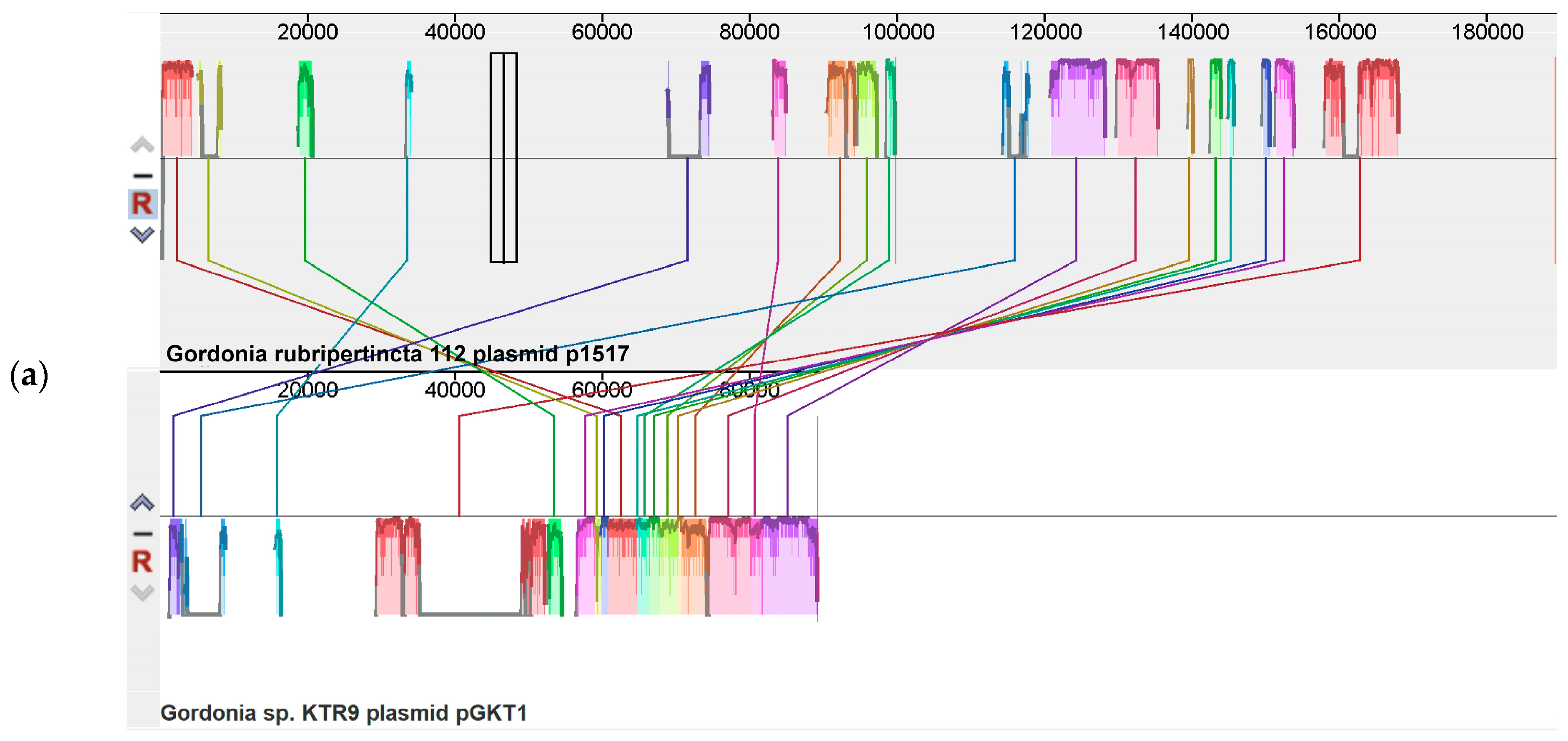
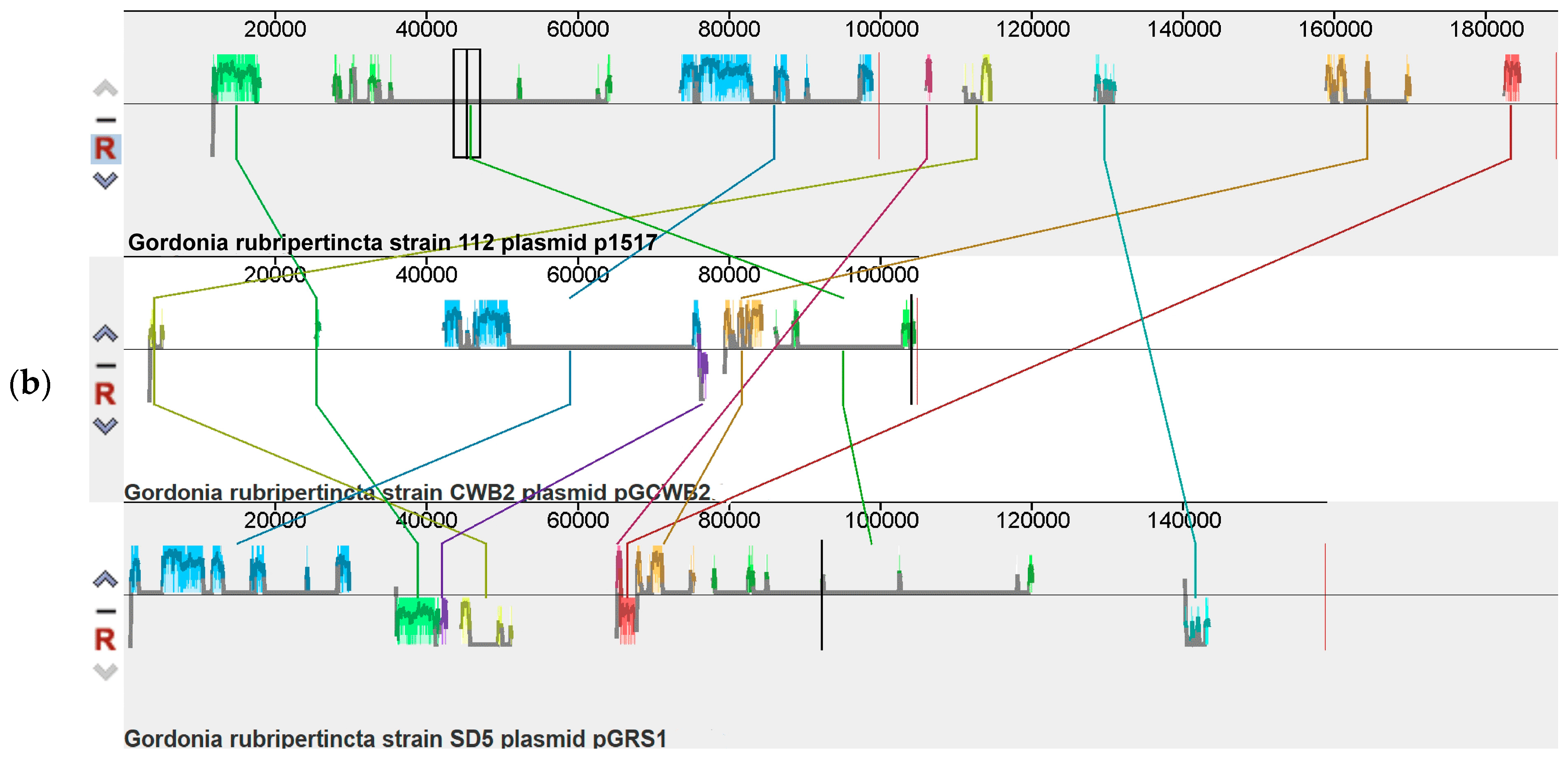
Contigs 15 and 17 of strain 112 were identified as plasmid elements with a total length of 189,570 nucleotides (plasmid p1517). Plasmid p1517 is most related to plasmid pGKT1 of Gordonia sp. KTR9 (NC_018582.1) (Figure 2a). No significant kinship was observed with the plasmids of G. rubripertincta SD5 and CWB2 strains (Figure 2b).
Plasmid p1517 of the G. rubripertincta strain 112 is predominantly composed of genes encoding hypothetical proteins. It also contains genes encoding mobile genome elements, plasmid maintenance, and separation genes. The genes responsible for metal transport (lead, cadmium, zinc, mercury, manganese) and metal resistance (cadmium, cobalt, zinc) are localized on plasmid p1517.
Plasmids pGCWB2 of G. rubripertincta CWB2 and pGRS1 of G. rubripertincta SD5, unlike plasmid p1517 of strain G. rubripertincta 112, contain catabolic genes. Plasmid pGCWB2 (CP022581.1) contains:
  • Phenylacetate catabolism gene cluster;
  • Styrene monooxygenase StyA, 3 beta-hydroxysteroid dehydrogenase/Delta 5-->4-isomerase;
  • Genes involved in type IV secretory system Conjugative DNA transfer.
Plasmid pGRS1 of G. rubripertincta SD5 contains genes of the aromatic compound catabolism pathway, namely aromatic ring-hydroxylating dioxygenase (alpha and beta subunits), alpha/beta fold hydrolase, extradiol dioxygenase, and ferredoxin.

3.3. Pangenome Analysis of Gordonia rubripertincta Strains

The pangenome of strain 112 and its closest relatives (DDH > 80%) G. rubripertincta ATCC14352, CWB2, and W3S5 was analyzed. All genomes used for pangenome analysis had completeness above 99% and contamination levels of 0.03–0.54%.
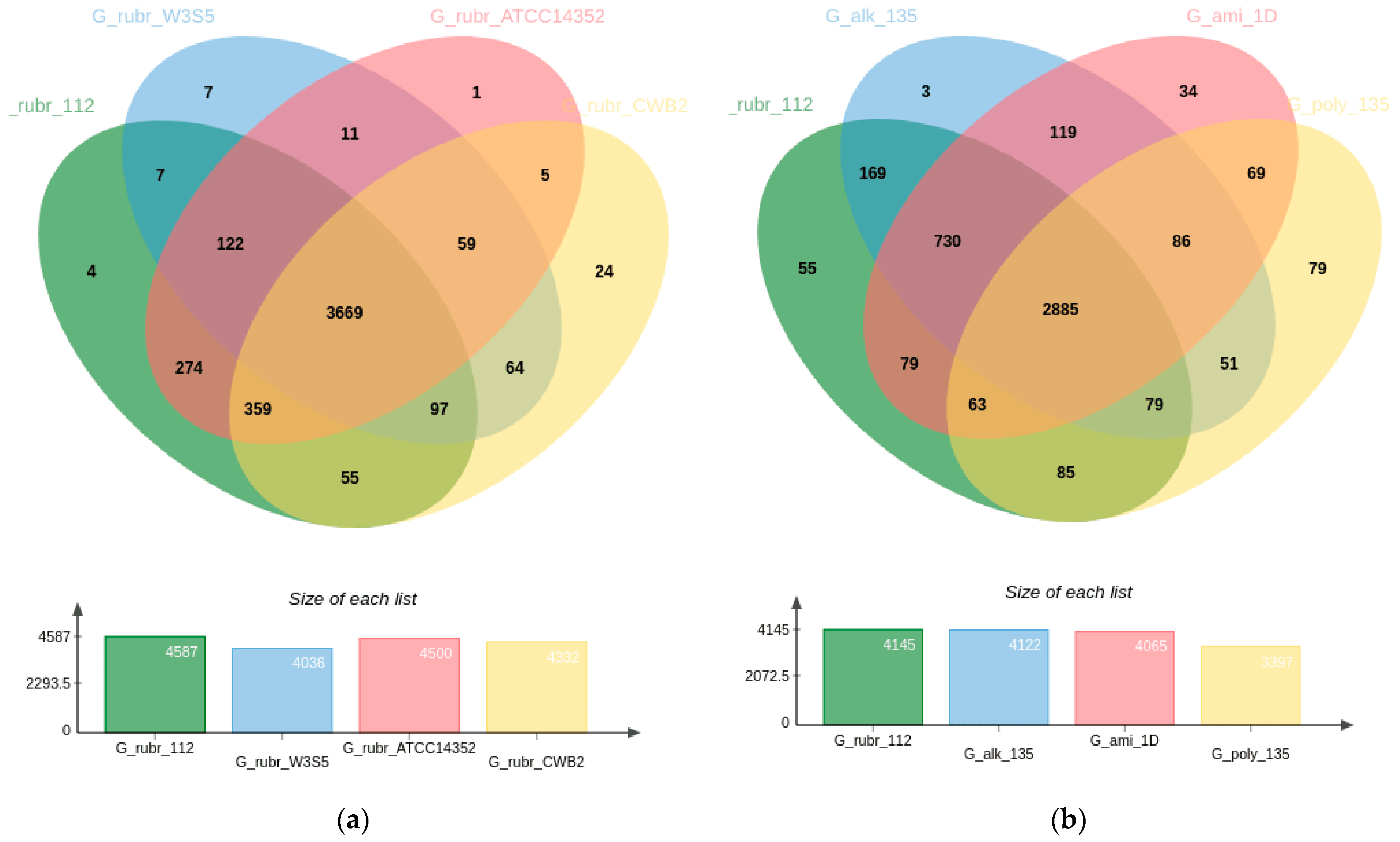
The pangenome of the strains is represented by 4758 genes, of which 3669 of the genes (77.11%) are core (genes that all our strains have). The result obtained may indicate the heterogeneity of G. rubripertincta species (Figure 3). For comparison, in a previous study [65], we showed that R. qingshengii accounts for 86% of the pangenome.
The CWB2 strain has the greatest number of unique coding sequences among the four G. rubripertincta strains analyzed. Among the 24 amino acid sequences unique to it and not found in three other strains (Figure 3a), there are products with nitrilotriacetate monooxygenase activity (four amino acid sequences), oxidoreductase activity (five aa sequences), and aflatoxin biosynthetic process (two aa sequences). Strain 112 has four unique amino acid sequences, but their function could not be determined. Strain 112 and ATCC14352 are the closest relatives of all G. rubripertincta studied, so we expected to see the greatest number of amino acid sequences unique to the pair (274 sequences, Table S1A).
In addition, we studied the pangenome of Gordonia strains from other species in our laboratory collection. All the strains taken for analysis had one thing in common: the ability to degrade alkanes. Strain G. polyisoprenivorans 135 utilized chloroaromatic compounds [18], strain G. alkanivorans 135 utilized thiophenes as the sole source of sulfur [20], and strain G. amicalis 1D utilized alkanes up to C36 [45]. Strain G. rubripertincta 112 was the only one of all the listed strains that utilized short alkanes (C8-C12) more actively than alkanes with a C16+ chain length.
The pangenome of the strains of four different Gordonia species is 4586 genes, of which 2885 (62.90%) derive from the core (Figure 3b). Among the different Gordonia species analyzed, strain G. rubripertincta 112 is the most related to strain G. alkanivorans 135. The G_rubr_112/G_alk_135 pair has 169 genes unique to the pair which were not found in the other strains analyzed (Table S1B). Among the genes unique to the G_rubr_112/G_alk_135 pair, it is interesting to note the genes belonging to the following categories (Table 4).
A BLAST search of the nucleotide (and amino acid, with similar results) sequence of the 01466 gene of strain G. rubripertincta 112 showed that this sequence with the parameters query cover > 90% and per. ident > 90% is characteristic only of some (not all) of the strains of G. rubripertincta and G. alkanivorans (Table 5).
Changing the search mode from highly similar sequences (megablast) to somewhat similar sequences (blastn) reveals organisms with sequences remotely related to the gene, but their query coverage and percent identity are too low (<70%) to speak of unconditional relatedness. The product of chorismatase catalyzes the hydrolysis of isochorismate into 2,3-dihydro-2,3-dihydroxybenzoate and pyruvate [66].
The steroid catabolism genes (GO:0006694, GO:0008202, GO:0006707) in both strains are located one after another and presumably represent an operon of the following structure: flavin-dependent monooxygenase, reductase subunit HsaB → 4,5:9,10-diseco-3-hydroxy-5,9,17-trioxoandrosta-1(10),2-diene-4-oate hydrolase → hypothetical protein → 3-ketosteroid-9-alpha-monooxygenase, ferredoxin reductase component → 3-oxosteroid 1-dehydrogenase. A BLAST search for the gene sequences of this operon showed that they, similarly to isochorismatase, occur in individual representatives of G. rubripertincta and G. alkanivorans.
There is no information in the literature on the phenotypic traits or the ability of G. alkanivorans to utilize steroids. However, the genes responsible for this ability are found in them. Besides G. alkanivorans, the gene cluster kst, responsible for the catabolism of steroid compounds, is found in the following representatives of the genus Gordonia: G. rubripertincta, G. bronchialis, G. insulae, G. mangrovi, G. araii, G. namibiensis, G. sputi, G. crocea, and others. An experimental confirmation of this ability in Gordonia was performed for G. cholesterolivorans [67] and Gordonia neofelifaecis [68,69]. The process of the catabolism of steroid compounds has been studied in the most detail in the strain G. rubripertincta CWB2 strain [38]. Our strain G. rubripertincta 112, similar to other representatives of this species, possesses a set of genes for steroid catabolism; however, the study of the phenotypic manifestations of this process in strain 112 was not included in the task of this work.

3.4. Functional Annotation of the Genome of the Strain 112

Out of 4787 genes, 2212 (46.2%) were functionally annotated (Figure 4).
The result of the functional annotation shows that strain 112 possesses the genes of all metabolic pathways required for autonomous culture existence. We also performed functional annotation of the genomes of other G. rubripertincta from the Genbank database (Table 6) to compare the representation of the metabolic pathways of interest in the genomes of the strains of this species.
The «Xenobiotics degradation and metabolism» category in strain 112 contains 77 genes. The most frequently occurring genes (>10 genes) in the category are the following functional clusters:
Benzoate degradation—23 CDS.
Xylene degradation—10 CDS.
Steroid degradation—11 CDS.

3.4.1. Diversity of Aromatic Compound Catabolism Genes in the Genome of Gordonia rubripertincta 112

We observed in strain 112 the ability to grow on benzoate and catechol as the sole source of carbon and energy. Catechol degradation by the strain can occur via both the ortho- and meta-pathways; the corresponding genes are found in the strain genome (Figure S2).
The xylene metabolism category contains no genes encoding the enzymes of the first reaction of this process. In addition, we did not find the strain’s ability to grow on xylenes. The metabolic pathway for xylene degradation in strain 112 begins from the middle, with the methylbenzoate conversion reaction catalyzed by benzoate/toluate 1,2-dioxygenase [EC:1.14.12.10 1.14.12.-]. One of the main metabolites of this process is methylcatechol (three- or four-substituted), and the further pathway is similar to catechol catabolism.
The genomes of all G. rubripertincta strains contain two copies of the catechol 1,2-dioxygenase (C1,2DO) genes (Table 7). The copies have 86% query coverage and 58.33% identity between them.
Genes of the catechol catabolism meta-pathway (in particular, catechol 2,3-dioxygenase (C2,3DO)) are present in three of the six G. rubripertincta strains. The amino acid sequence of the C2,3DO gene in strain W3S5 differs from that in strains ATCC14352 and 112. At 99% coverage, the percent identity between them is 83.02%.
There are currently 55 known species of the genus Gordonia (https://lpsn.dsmz.de/ (accessed on 30 March 2023)). Genes of only the ortho-pathway of catechol catabolism (in particular, C1,2DO) are present in the genomes of the strains of 22 species (Table S2). Genes of both ortho (C1,2DO) and meta (C2,3DO) pathways were found in the genomes of the representatives of 10 species. The prevalence of C1,2DO in Gordonia genomes allows this gene to be used as a phylogenetic marker for the identification of strains of this genus. Shen et al. [70] suggested that the catA gene encoding C1,2DO evolves faster than the rrn operon or gyrB gene, so catA is a more sensitive marker for species identification. Gordonia malaquae is the only species in the genus Gordonia whose strains have only C2,3DO genes but no C1,2DO genes.
The enzymatic activity of C2,3DO in the work of Silva et al. [17] in the G. polyisoprenivorans strain was higher than that of C1,2DO under most of the conditions tested (pH, temperature, time course, ion effect). However, it is worth noting that the authors used complex LB medium with anthracene supplementation to cultivate the strain. Considering that LB medium components are more available, and a preferable substrate compared to anthracene, it is impossible to draw unequivocal conclusions about the process of aromatic compound utilization and enzyme activity under such conditions. Solyanikova et al. [71] also showed that when the cells of the G. polyisoprenivorans strain grew on a medium with benzoate, the activity of catechol 1,2-dioxygenase was 0.850 U/(mg of protein), and the activity of catechol 2,3-dioxygenase was absent. Thus, the simultaneous maintenance of the two metabolic pathways of catechol catabolism in Gordonia genomes does not mean that both pathways will be involved. Moreover, C2,3DO may be a redundant metabolic pathway in Gordonia strains.
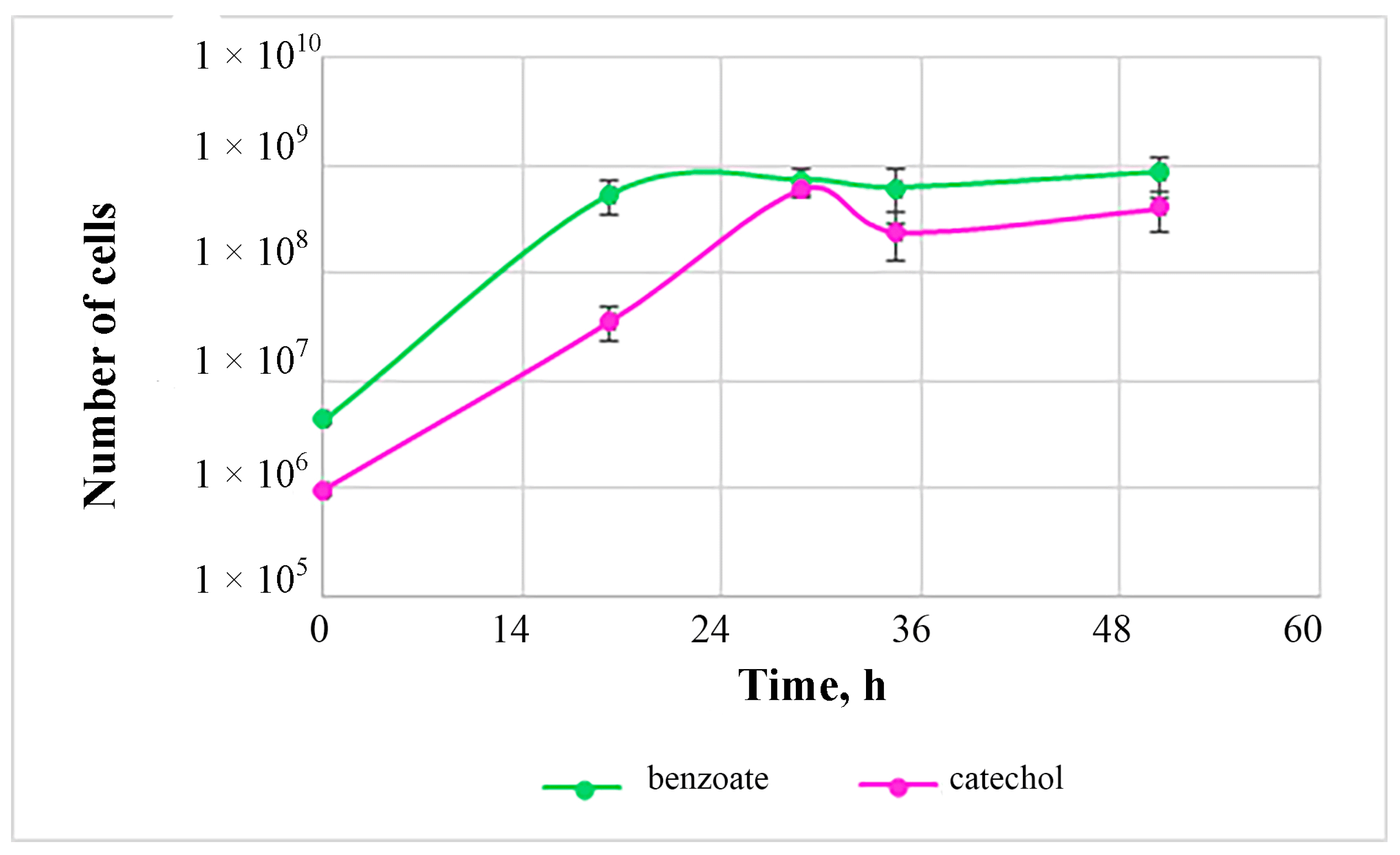
The cell number during growth on catechol reaches a maximum after 24 h of growth, after which the culture enters the stationary phase (Figure 5).
When growing on benzoate, we observed the maximum number of cells after 30 h of growth. Thus, when utilizing aromatic compounds, the development of the periodic culture of strain 112 proceeds rather quickly, which allows us to consider the biotechnological promise of this strain for the utilization of these compounds.

3.4.2. Diversity of Alkane Catabolism Genes in Strain Gordonia rubripertincta 112

Strain 112 is capable of utilizing alkanes with a C10-C16 chain length, and the growth on short alkanes (decane (C10) as an example) is observed not only in vapors, but also in direct contact of the microorganism with the substrate. Growth on alkanes longer than C16 was not observed in the strain; therefore, we assumed that it, similarly to G. alkanivorans strains, lacked alkB genes, and its ability to degrade alkanes was controlled by CYP153 genes. An analysis of the annotated genome confirmed our assumptions: strain 112 indeed lacks alkB genes.
We analyzed the genomes of the other G. rubripertincta strains from the Genbank database to understand whether the absence of alkB genes is a strain feature or a species-specific pattern. All G. rubripertincta strains whose genomes were sequenced lack the alkB operon.
The genome of strain 112 revealed four operons with a structure typical of the alkane hydroxylating cluster of actinobacteria: cytochrome P450 hydroxylase, ferredoxin, and ferredoxin reductase. An analysis of the prevalence of hydroxylase genes from these operons among Gordonia showed that one of them (PRJNA953757:QBL07_23005) is not found in other Gordonia. The presence of this gene is characteristic of some Rhodococcus (Table 8), so we can assume that this operon CYP153 was obtained by strain 112 as a result of a horizontal transfer from Rhodococcus strains.
The GC content of the region where the described operon is located (NODE_27_length_46626) is 65.7%, which is 2% lower than the GC content of the contigs that have the highest affinity to the genome regions of the typical strain G. rubripertincta. This observation suggests that some elements of contig 27 were acquired during horizontal transfer.

3.5. Peculiarities of Alkane Catabolism by Strain G. rubripertincta 112

It is known that the toxicity of the aliphatic hydrocarbons decreases with an increasing number of carbon atoms due to a decrease in their volatility [72]. Over 3 days, the abiotic loss of decane and hexadecane in the system without microorganisms was 62.87% and 1.21%, respectively (Table 9).
The degree of degradation of decane and hexadecane relative to the control system without microorganisms was 10.79 and 16.14%, respectively. It can be noted that, despite the high abiotic loss of decane, the degree of its degradation was higher compared to the degree of hexadecane degradation.
Considering that the genome of strain 112 lacks alkB genes, and only cytochrome P450 hydroxylases are used for alkane degradation, it was not surprising that the strain is unable to oxidize alkanes longer than C16. Its abilities as an alkane degrader could be exploited in soils contaminated with light fractions of oil and petroleum products. The alkane catabolism genes in strain 112 are located on the chromosome, which suggests their stable maintenance during both non-selective cultivation and remediation processes in soil. In the genomes of other known G. rubripertincta strains, alkane destruction genes are also localized on chromosomes; however, there are no data on the ability of these strains to utilize alkanes. Thus, at this time, only G. rubripertincta 112 and CWB2 have experimentally confirmed the ability to utilize alkanes. As for catechol degradation, strain 112 is currently the only representative of G. rubripertincta in which this ability has been experimentally confirmed.

4. Conclusions

This study provides an improved understanding of the genomic organization of the Gordonia rubripertincta strain and its metabolic capabilities. The ability of the strain of this genus to utilize aromatic compounds was confirmed experimentally for the first time, and the genetic pathways involved in this process were described. The ability to utilize alkanes of different chain lengths, including short ones (decane), was described. The results provide a powerful basis for further transcriptome experiments and the study of the regulation of the catabolism of various pollutants by G. rubripertincta 112.

Supplementary Materials

The following supporting information can be downloaded at: https://www.mdpi.com/article/10.3390/biology12050721/s1, Figure S1: Growth of G. rubripertincta strain 112 (a) on agarized LB medium, (b) in liquid mineral medium with decane (left flask) and hexadecane (right flask); Table S1A: Biological process summary of genes unique for pair G_rubr_112/G_rubr_ATCC14352, Table S1B: Biological process summary of genes unique for pair G_rubr_112/G_alk_135; Figure S2: Schemes of (a) ortho- and (b) meta-pathways of catechol degradation; Table S2: Distribution of catechol dioxygenase genes in the genomes of representatives of different Gordonia species.

Author Contributions

Conceptualization, Y.D. and A.V.; methodology, E.F., A.B. and R.S.; software, Y.D.; validation, A.B., Y.D. and I.S.; formal analysis, A.V.; investigation, E.F., V.S. and R.S.; data curation, Y.D.; writing—original draft preparation, E.F. and Y.D.; writing—review and editing, A.B. and A.V.; visualization, E.F.; supervision, Y.D.; project administration, Y.D.; funding acquisition, Y.D. All authors have read and agreed to the published version of the manuscript.

Funding

This work was financially supported by the Russian Science Foundation, grant number 22-74-10082.

Institutional Review Board Statement

Not applicable.

Informed Consent Statement

Not applicable.

Data Availability Statement

The data on genome sequence of the strain Gordonia rubripertincta 112 can be found in the Genbank database under the accession number JARUXG000000000 (BioProject PRJNA953757, BioSample SAMN34123077).

Conflicts of Interest

The authors declare no conflict of interest.

References

  1. Cha, J.-H.; Cha, C.-J. Gordonia alkaliphila sp. nov., an actinomycete isolated from tidal flat sediment. Int. J. Syst. Evol. Microbiol. 2013, 63 Pt 1, 327–331. [Google Scholar] [CrossRef]
  2. He, Y.; Lyu, L.; Hu, Z.; Yu, Z.; Shao, Z. Gordonia tangerina sp. nov., isolated from seawater. Int. J. Syst. Evol. Microbiol. 2022, 72, 005632. [Google Scholar] [CrossRef] [PubMed]
  3. Kim, H.; Dong, K.; Kim, J.; Lee, S. Characteristics of crude oil-degrading bacteria Gordonia iterans isolated from marine coastal in Taean sediment. Microbiologyopen 2019, 8, e00754. [Google Scholar] [CrossRef]
  4. Sangkanu, S.; Suriyachadkun, C.; Phongpaichit, S. Gordonia sediminis sp. nov., an actinomycete isolated from mangrove sediment. Int. J. Syst. Evol. Microbiol. 2019, 69, 1814–1820. [Google Scholar] [CrossRef] [PubMed]
  5. Shen, F.-T.; Goodfellow, M.; Jones, A.L.; Chen, Y.-P.; Arun, A.B.; Lai, W.-A.; Rekha, P.D.; Young, C.-C. Gordonia soli sp. nov., a novel actinomycete isolated from soil. Int. J. Syst. Evol. Microbiol. 2006, 56, 2597–2601. [Google Scholar] [CrossRef] [PubMed]
  6. Brandão, P.F.B.; Maldonado, L.A.; Ward, A.C.; Bull, A.T.; Goodfellow, M. Gordonia namibiensis sp. nov., a Novel Nitrile MetabolisingActinomycete Recovered from an African Sand. Syst. Appl. Microbiol. 2001, 24, 510–515. [Google Scholar] [CrossRef] [PubMed]
  7. Ma, Y.; Xu, M.; Liu, H.; Yu, T.; Guo, P.; Liu, W.; Jin, X. Antimicrobial compounds were isolated from the secondary metabolites of Gordonia, a resident of intestinal tract of Periplaneta americana. AMB Express 2021, 11, 111. [Google Scholar] [CrossRef]
  8. Mohammadipanah, F.; Wink, J. Actinobacteria from Arid and Desert Habitats: Diversity and Biological Activity. Front. Microbiol. 2016, 6, 1541. [Google Scholar] [CrossRef]
  9. Kepenek, E.S.; Severcan, M.; Gozen, A.G.; Severcan, F. Discrimination of heavy metal acclimated environmental strains by chemometric analysis of FTIR spectra. Ecotoxicol. Environ. Saf. 2020, 202, 110953. [Google Scholar] [CrossRef]
  10. Kayasth, M.; Kumar, V.; Gera, R. Gordonia sp.: A salt tolerant bacterial inoculant for growth promotion of pearl millet under saline soil conditions. 3 Biotech 2014, 4, 553–557. [Google Scholar] [CrossRef]
  11. Ehiosun, K.I.; Godin, S.; Urios, L.; Lobinski, R.; Grimaud, R. Degradation of long-chain alkanes through biofilm formation by bacteria isolated from oil-polluted soil. Int. Biodeterior. Biodegradation 2022, 175, 105508. [Google Scholar] [CrossRef]
  12. Liu, Y.; Wu, J.; Liu, Y.; Wu, X. Biological Process of Alkane Degradation by Gordonia sihwaniensis. ACS Omega 2022, 7, 55–63. [Google Scholar] [CrossRef] [PubMed]
  13. Lo Piccolo, L.; De Pasquale, C.; Fodale, R.; Puglia, A.M.; Quatrini, P. Involvement of an Alkane Hydroxylase System of Gordonia sp. Strain SoCg in Degradation of Solid n-Alkanes. Appl. Environ. Microbiol. 2011, 77, 1204–1213. [Google Scholar] [CrossRef] [PubMed]
  14. Silva, N.M.; de Oliveira, A.M.S.A.; Pegorin, S.; Giusti, C.E.; Ferrari, V.B.; Barbosa, D.; Martins, L.F.; Morais, C.; Setubal, J.C.; Vasconcellos, S.P.; et al. Characterization of novel hydrocarbon-degrading Gordonia paraffinivorans and Gordonia sihwensis strains isolated from composting. PLoS ONE 2019, 14, e0215396. [Google Scholar] [CrossRef] [PubMed]
  15. Frantsuzova, E.; Solomentsev, V.; Vetrova, A.; Travkin, V.; Solyanikova, I.; Delegan, Y. Complete Genome Sequence of Gordonia polyisoprenivorans 135, a Promising Degrader of Aromatic Compounds. Microbiol. Resour. Announc. 2023, 12, e0005823. [Google Scholar] [CrossRef]
  16. Lin, C.-L.; Shen, F.-T.; Tan, C.-C.; Huang, C.-C.; Chen, B.-Y.; Arun, A.; Young, C.-C. Characterization of Gordonia sp. strain CC-NAPH129-6 capable of naphthalene degradation. Microbiol. Res. 2012, 167, 395–404. [Google Scholar] [CrossRef]
  17. Silva, A.S.; de Oliveira Camargo, F.A.; Andreazza, R.; Jacques, R.J.S.; Baldoni, D.B.; Bento, F.M. Enzymatic activity of catechol 1,2-dioxygenase and catechol 2,3-dioxygenase produced by Gordonia polyisoprenivorans. Química Nova 2012, 35, 1587–1592. [Google Scholar] [CrossRef]
  18. Suzina, N.E.; Sorokin, V.V.; Polivtseva, V.N.; Klyueva, V.V.; Emelyanova, E.V.; Solyanikova, I.P. From Rest to Growth: Life Collisions of Gordonia polyisoprenivorans 135. Microorganisms 2022, 10, 465. [Google Scholar] [CrossRef]
  19. Akhtar, N.; Akhtar, K.; Ghauri, M.A. Biodesulfurization of Thiophenic Compounds by a 2-Hydroxybiphenyl-Resistant Gordonia sp. HS126-4N Carrying dszABC Genes. Curr. Microbiol. 2018, 75, 597–603. [Google Scholar] [CrossRef]
  20. Delegan, Y.; Kocharovskaya, Y.; Frantsuzova, E.; Streletskii, R.; Vetrova, A. Characterization and genomic analysis of Gordonia alkanivorans 135, a promising dibenzothiophene-degrading strain. Biotechnol. Rep. 2021, 29, e00591. [Google Scholar] [CrossRef]
  21. Wang, W.; Ma, T.; Lian, K.; Zhang, Y.; Tian, H.; Ji, K.; Li, G. Genetic Analysis of Benzothiophene Biodesulfurization Pathway of Gordonia terrae Strain C-6. PLoS ONE 2013, 8, e84386. [Google Scholar] [CrossRef]
  22. Hu, T.; Yang, C.; Hou, Z.; Liu, T.; Mei, X.; Zheng, L.; Zhong, W. Phthalate Esters Metabolic Strain Gordonia sp. GZ-YC7, a Potential Soil Degrader for High Concentration Di-(2-ethylhexyl) Phthalate. Microorganisms 2022, 10, 641. [Google Scholar] [CrossRef]
  23. Jin, D.; Kong, X.; Jia, M.; Yu, X.; Wang, X.; Zhuang, X.; Deng, Y.; Bai, Z. Gordonia phthalatica sp. nov., a di-n-butyl phthalate-degrading bacterium isolated from activated sludge. Int. J. Syst. Evol. Microbiol. 2017, 67, 5128–5133. [Google Scholar] [CrossRef]
  24. Kanaujiya, D.K.; Sivashanmugam, S.; Pakshirajan, K. Biodegradation and toxicity removal of phthalate mixture by Gordonia sp. in a continuous stirred tank bioreactor system. Environ. Technol. Innov. 2022, 26, 102324. [Google Scholar] [CrossRef]
  25. Nahurira, R.; Ren, L.; Song, J.; Jia, Y.; Wang, J.; Fan, S.; Wang, H.; Yan, Y. Degradation of Di(2-Ethylhexyl) Phthalate by a Novel Gordonia alkanivorans Strain YC-RL2. Curr. Microbiol. 2017, 74, 309–319. [Google Scholar] [CrossRef] [PubMed]
  26. Sarkar, J.; Chowdhury, P.P.; Dutta, T.K. Complete degradation of di-n-octyl phthalate by Gordonia sp. strain Dop5. Chemosphere 2013, 90, 2571–2577. [Google Scholar] [CrossRef] [PubMed]
  27. Kim, S.B.; Brown, R.; Oldfield, C.; Gilbert, S.C.; Iliarionov, S.; Goodfellow, M. Gordonia amicalis sp. nov., a novel dibenzothiophene-desulphurizing actinomycete. Int. J. Syst. Evol. Microbiol. 2000, 50, 2031–2036. [Google Scholar] [CrossRef]
  28. Kummer, C.; Schumann, P.; Stackebrandt, E. Gordonia alkanivorans sp. nov., isolated from tar-contaminated soil. Int. J. Syst. Evol. Microbiol. 1999, 49, 1513–1522. [Google Scholar] [CrossRef] [PubMed]
  29. Linos, A.; Steinbüchel, A.; Spröer, C.; Kroppenstedt, R.M. Gordonia polyisoprenivorans sp. nov., a rubber-degrading actinomycete isolated from an automobile tyre. Int. J. Syst. Evol. Microbiol. 1999, 49, 1785–1791. [Google Scholar] [CrossRef]
  30. Linos, A.; Berekaa, M.M.; Steinbüchel, A.; Kim, K.K.; Sproer, C.; Kroppenstedt, R.M. Gordonia westfalica sp. nov., a novel rubber-degrading actinomycete. Int. J. Syst. Evol. Microbiol. 2002, 52, 1133–1139. [Google Scholar] [CrossRef]
  31. Xue, Y.; Sun, X.; Zhou, P.; Liu, R.; Liang, F.; Ma, Y. Gordonia paraffinivorans sp. nov., a hydrocarbon-degrading actinomycete isolated from an oil-producing well. Int. J. Syst. Evol. Microbiol. 2003, 53, 1643–1646. [Google Scholar] [CrossRef] [PubMed]
  32. Esuola, C.O.; Babalola, O.O.; Heine, T.; Schwabe, R.; Schlömann, M.; Tischler, D. Identification and characterization of a FAD-dependent putrescine N-hydroxylase (GorA) from Gordonia rubripertincta CWB2. J. Mol. Catal. B Enzym. 2016, 134, 378–389. [Google Scholar] [CrossRef]
  33. Hofmann, M.; Martin del Campo, J.S.; Sobrado, P.; Tischler, D. Biosynthesis of desferrioxamine siderophores initiated by decarboxylases: A functional investigation of two lysine/ornithine-decarboxylases from Gordonia rubripertincta CWB2 and Pimelobacter simplex 3E. Arch. Biochem. Biophys. 2020, 689, 108429. [Google Scholar] [CrossRef] [PubMed]
  34. Schwabe, R.; Obst, B.; Mehnert, M.; Tischler, D.; Wiche, O. Gallium Mobilization in Soil by Bacterial Metallophores. Solid State Phenom. 2017, 262, 513–516. [Google Scholar] [CrossRef]
  35. Schwabe, R.; Senges, C.H.R.; Bandow, J.E.; Heine, T.; Lehmann, H.; Wiche, O.; Schlömann, M.; Levicán, G.; Tischler, D. Data on metal-chelating, -immobilisation and biosorption properties by Gordonia rubripertincta CWB2 in dependency on rare earth adaptation. Data Brief 2020, 31, 105739. [Google Scholar] [CrossRef] [PubMed]
  36. Schwabe, R.; Senges, C.H.R.; Bandow, J.E.; Heine, T.; Lehmann, H.; Wiche, O.; Schlömann, M.; Levicán, G.; Tischler, D. Cultivation dependent formation of siderophores by Gordonia rubripertincta CWB2. Microbiol. Res. 2020, 238, 126481. [Google Scholar] [CrossRef] [PubMed]
  37. Eggerichs, D.; Mügge, C.; Mayweg, J.; Apfel, U.-P.; Tischler, D. Enantioselective Epoxidation by Flavoprotein Monooxygenases Supported by Organic Solvents. Catalysts 2020, 10, 568. [Google Scholar] [CrossRef]
  38. Heine, T.; Zimmerling, J.; Ballmann, A.; Kleeberg, S.B.; Rückert, C.; Busche, T.; Winkler, A.; Kalinowski, J.; Poetsch, A.; Scholtissek, A.; et al. On the Enigma of Glutathione-Dependent Styrene Degradation in Gordonia rubripertincta CWB2. Appl. Environ. Microbiol. 2018, 84, e00154-18. [Google Scholar] [CrossRef] [PubMed]
  39. Zimmerling, J.; Oelschlägel, M.; Großmann, C.; Voitel, M.; Schlömann, M.; Tischler, D. Biochemical Characterization of Phenylacetaldehyde Dehydrogenases from Styrene-degrading Soil Bacteria. Appl. Biochem. Biotechnol. 2020, 193, 650–667. [Google Scholar] [CrossRef] [PubMed]
  40. Han, S.-S.; Kang, H.K.; Jo, B.Y.; Ryu, B.-G.; Jin, H.M.; Chung, E.J.; Jung, J.Y. Complete Genome Sequence of Gordonia rubripertincta SD5, a Soil Bacterium Isolated from a Di-(2-Ethylhexyl) Phthalate-Degrading Enrichment Culture. Genome Announc. 2020, 9, e01087-20. [Google Scholar] [CrossRef]
  41. Trögl, J.; Esuola, C.O.; Kříženecká, S.; Kuráň, P.; Seidlová, L.; Veronesi-Dáňová, P.; Popelka, J.; Babalola, O.O.; Hrabák, P.; Czinnerová, M.; et al. Biodegradation of High Concentrations of Aliphatic Hydrocarbons in Soil from a Petroleum Refinery: Implications for Applicability of New Actinobacterial Strains. Appl. Sci. 2018, 8, 1855. [Google Scholar] [CrossRef]
  42. Kotani, T.; Yamamoto, T.; Yurimoto, H.; Sakai, Y.; Kato, N. PropaneMonooxygenase and NAD + -Dependent Secondary AlcoholDehydrogenase in Propane Metabolism by Gordonia sp. Strain TY-5. J. Bacteriol. 2003, 185, 7120–7128. [Google Scholar] [CrossRef] [PubMed]
  43. Kotani, T.; Yurimoto, H.; Kato, N.; Sakai, Y. Novel Acetone Metabolism in a Propane-Utilizing Bacterium, Gordonia sp. Strain TY-5. J. Bacteriol. 2007, 189, 886–893. [Google Scholar] [CrossRef]
  44. van Beilen, J.B.; Funhoff, E.G.; van Loon, A.; Just, A.; Kaysser, L.; Bouza, M.; Holtackers, R.; Röthlisberger, M.; Li, Z.; Witholt, B. Cytochrome P450 Alkane Hydroxylases of the CYP153 Family Are Common in Alkane-Degrading Eubacteria Lacking Integral Membrane Alkane Hydroxylases. Appl. Environ. Microbiol. 2006, 72, 59–65. [Google Scholar] [CrossRef]
  45. Delegan, Y.A.; Valentovich, L.N.; Shafieva, S.M.; Ganbarov, K.G.; Filonov, A.E.; Vainstein, M.B. Characterization and genomic analysis of highly efficient thermotolerant oil-degrading bacterium Gordonia sp. 1D. Folia Microbiol. 2018, 64, 41–48. [Google Scholar] [CrossRef] [PubMed]
  46. Nie, Y.; Liang, J.; Fang, H.; Tang, Y.-Q.; Wu, X.-L. Two Novel Alkane Hydroxylase-Rubredoxin Fusion Genes Isolated from a Dietzia Bacterium and the Functions of Fused Rubredoxin Domains in Long-Chain n-Alkane Degradation. Appl. Environ. Microbiol. 2011, 77, 7279–7288. [Google Scholar] [CrossRef]
  47. Nie, Y.; Liang, J.-L.; Fang, H.; Tang, Y.-Q.; Wu, X.-L. Characterization of a CYP153 alkane hydroxylase gene in a Gram-positive Dietzia sp. DQ12-45-1b and its “team role” with alkW1 in alkane degradation. Appl. Microbiol. Biotechnol. 2014, 98, 163–173. [Google Scholar] [CrossRef] [PubMed]
  48. Bertani, G. STUDIES ON LYSOGENESIS I. J. Bacteriol. 1951, 62, 293–300. [Google Scholar] [CrossRef]
  49. Bolger, A.M.; Lohse, M.; Usadel, B. Trimmomatic: A flexible trimmer for Illumina sequence data. Bioinformatics 2014, 30, 2114–2120. [Google Scholar] [CrossRef]
  50. Bankevich, A.; Nurk, S.; Antipov, D.; Gurevich, A.A.; Dvorkin, M.; Kulikov, A.S.; Lesin, V.M.; Nikolenko, S.I.; Pham, S.; Prjibelski, A.D.; et al. SPAdes: A New Genome Assembly Algorithm and Its Applications to Single-Cell Sequencing. J. Comput. Biol. 2012, 19, 455–477. [Google Scholar] [CrossRef]
  51. Yoon, S.-H.; Ha, S.-M.; Kwon, S.; Lim, J.; Kim, Y.; Seo, H.; Chun, J. Introducing EzBioCloud: A taxonomically united database of 16S rRNA gene sequences and whole-genome assemblies. Int. J. Syst. Evol. Microbiol. 2017, 67, 1613–1617. [Google Scholar] [CrossRef] [PubMed]
  52. Meier-Kolthoff, J.P.; Auch, A.F.; Klenk, H.-P.; Göker, M. Genome sequence-based species delimitation with confidence intervals and improved distance functions. BMC Bioinform. 2013, 14, 60. [Google Scholar] [CrossRef] [PubMed]
  53. Tatusova, T.; DiCuccio, M.; Badretdin, A.; Chetvernin, V.; Nawrocki, E.P.; Zaslavsky, L.; Lomsadze, A.; Pruitt, K.D.; Borodovsky, M.; Ostell, J. NCBI prokaryotic genome annotation pipeline. Nucleic Acids Res. 2016, 44, 6614–6624. [Google Scholar] [CrossRef] [PubMed]
  54. Seemann, T. Prokka: Rapid prokaryotic genome annotation. Bioinformatics 2014, 30, 2068–2069. [Google Scholar] [CrossRef] [PubMed]
  55. Aziz, R.K.; Bartels, D.; Best, A.A.; DeJongh, M.; Disz, T.; Edwards, R.A.; Formsma, K.; Gerdes, S.; Glass, E.M.; Kubal, M.; et al. The RAST Server: Rapid Annotations using Subsystems Technology. BMC Genom. 2008, 9, 75. [Google Scholar] [CrossRef] [PubMed]
  56. Lefort, V.; Desper, R.; Gascuel, O. FastME 2.0: A Comprehensive, Accurate, and Fast Distance-Based Phylogeny Inference Program: Table 1. Mol. Biol. Evol. 2015, 32, 2798–2800. [Google Scholar] [CrossRef]
  57. Meier-Kolthoff, J.P.; Göker, M. TYGS is an automated high-throughput platform for state-of-the-art genome-based taxonomy. Nat. Commun. 2019, 10, 2182. [Google Scholar] [CrossRef]
  58. Reeve, W.; O’hara, G.; Chain, P.; Ardley, J.; Brau, L.; Nandesena, K.; Tiwari, R.; Malfatti, S.; Kiss, H.; Lapidus, A.; et al. Complete genome sequence of Rhizobium leguminosarum bv trifolii strain WSM2304, an effective microsymbiont of the South American clover Trifolium polymorphum. Stand. Genom. Sci. 2010, 2, 66–76. [Google Scholar] [CrossRef]
  59. Darling, A.C.E.; Mau, B.; Blattner, F.R.; Perna, N.T. Mauve: Multiple Alignment of Conserved Genomic Sequence with Rearrangements. Genome Res. 2004, 14, 1394–1403. [Google Scholar] [CrossRef]
  60. Xu, L.; Dong, Z.; Fang, L.; Luo, Y.; Wei, Z.; Guo, H.; Zhang, G.; Gu, Y.Q.; Coleman-Derr, D.; Xia, Q.; et al. OrthoVenn2: A web server for whole-genome comparison and annotation of orthologous clusters across multiple species. Nucleic Acids Res. 2019, 47, W52–W58. [Google Scholar] [CrossRef]
  61. Kanehisa, M. KEGG: Kyoto Encyclopedia of Genes and Genomes. Nucleic Acids Res. 2000, 28, 27–30. [Google Scholar] [CrossRef]
  62. Blin, K.; Shaw, S.; Kloosterman, A.M.; Charlop-Powers, Z.; van Wezel, G.P.; Medema, M.H.; Weber, T. antiSMASH 6.0: Improving cluster detection and comparison capabilities. Nucleic Acids Res. 2021, 49, W29–W35. [Google Scholar] [CrossRef] [PubMed]
  63. Parks, D.H.; Imelfort, M.; Skennerton, C.T.; Hugenholtz, P.; Tyson, G.W. CheckM: Assessing the quality of microbial genomes recovered from isolates, single cells, and metagenomes. Genome Res. 2015, 25, 1043–1055. [Google Scholar] [CrossRef] [PubMed]
  64. Ivshina, I.B.; Kevorkov, N.N.; Koblova, I.V.; Nesterenko, O.A.; Kvasnikov, E.I.; Kasumova, S.A. Identification of bacteria belonging to the genus Rhodococcus using the technique of immunodiffusion. Mikrobiologiya 1980, 51, 636–641. [Google Scholar]
  65. Iminova, L.; Delegan, Y.; Frantsuzova, E.; Bogun, A.; Zvonarev, A.; Suzina, N.; Anbumani, S.; Solyanikova, I. Physiological and biochemical characterization and genome analysis of Rhodococcus qingshengii strain 7B capable of crude oil degradation and plant stimulation. Biotechnol. Rep. 2022, 35, e00741. [Google Scholar] [CrossRef]
  66. Goral, A.M.; Tkaczuk, K.L.; Chruszcz, M.; Kagan, O.; Savchenko, A.; Minor, W. Crystal structure of a putative isochorismatase hydrolase from Oleispira antarctica. J. Struct. Funct. Genom. 2012, 13, 27–36. [Google Scholar] [CrossRef] [PubMed]
  67. Drzyzga, O.; Fernández de las Heras, L.; Morales, V.; Navarro Llorens, J.M.; Perera, J. Cholesterol Degradation by Gordonia cholesterolivorans. Appl. Environ. Microbiol. 2011, 77, 4802–4810. [Google Scholar] [CrossRef]
  68. Ge, F.; Li, W.; Chen, G.; Liu, Y.; Zhang, G.; Yong, B.; Wang, Q.; Wang, N.; Huang, Z.; Li, W.; et al. Draft Genome Sequence of Gordonia neofelifaecis NRRL B-59395, a Cholesterol-Degrading Actinomycete. J. Bacteriol. 2011, 193, 5045–5046. [Google Scholar] [CrossRef]
  69. Zhang, Q.; Ren, Y.; He, J.; Cheng, S.; Yuan, J.; Ge, F.; Li, W.; Zhang, Y.; Xie, G. Multiplicity of 3-ketosteroid Δ1-dehydrogenase enzymes in Gordonia neofelifaecis NRRL B-59395 with preferences for different steroids. Ann. Microbiol. 2015, 65, 1961–1971. [Google Scholar] [CrossRef]
  70. Shen, F.-T.; Lin, J.-L.; Huang, C.-C.; Ho, Y.-N.; Arun, A.B.; Young, L.-S.; Young, C.-C. Molecular detection and phylogenetic analysis of the catechol 1,2-dioxygenase gene from Gordonia spp. Syst. Appl. Microbiol. 2009, 32, 291–300. [Google Scholar] [CrossRef]
  71. Solyanikova, I.P.; Emelyanova, E.V.; Shumkova, E.S.; Egorova, D.O.; Korsakova, E.S.; Plotnikova, E.G.; Golovleva, L.A. Peculiarities of the degradation of benzoate and its chloro- and hydroxy-substituted analogs by actinobacteria. Int. Biodeterior. Biodegradation 2015, 100, 155–164. [Google Scholar] [CrossRef]
  72. Macaya, C.C.; Durán, R.E.; Hernández, L.; Rodríguez-Castro, L.; Barra-Sanhueza, B.; Dorochesi, F.; Seeger, M. Bioremediation of Petroleum. In Reference Module in Life Sciences; Elsevier: Amsterdam, The Netherlands, 2019. [Google Scholar] [CrossRef]
Figure 1. Whole-genome tree demonstrating the position of strain 112 within the genus G. rubripertincta and within the genus Gordonia in general.
Biology 12 00721 g001
Figure 2. Mauve visualization of locally collinear blocks identified between p1517 of G. rubripertincta 112 and relative plasmids: (a) pGKT1 of Gordonia sp. KTR9, (b) pGCWB2 of G. rubripertincta CWB2 and pGRS1 of G. rubripertincta SD5. Vertical bars mark boundaries between elements.
Biology 12 00721 g002aBiology 12 00721 g002b
Figure 3. Venn diagram depicting pangenome of (a) Gordonia rubripertincta strains 112, ATCC14352, CWB2 and W3S5, (b) G. rubripertincta 112, G. amicalis 1D, G. alkanivorans 135, and G. polyisoprenivorans 135.
Biology 12 00721 g003
Figure 4. Number of genes associated with general functional categories in the genome of strain 112 based on KEGG classification.
Biology 12 00721 g004
Figure 5. Growth curve of strain 112 on mineral medium with benzoate (green) and catechol (purple).
Biology 12 00721 g005
Table 1. Metrics of the genome assembly of the strain G. rubripertincta 112.
ParameterLength
Genome size, bp5,281,129
The longest contig, bp845,133
The shortest contig, bp502
N50, bp233,583
N75, bp130,856
N90, bp59,209
Table 2. Comparison of G. rubripertincta genomes deposited in the Genbank database.
GenBank Accession
Number
Strain NameChromosome Size, MbPlasmid Size, kb
JARUXG000000000Gordonia rubripertincta 1125.09189
JAAXPB000000000.1Gordonia rubripertincta ATCC14352T5.70-
JAFFGU000000000.1Gordonia rubripertincta BP-2955.15-
CP022580.1Gordonia rubripertincta CWB25.23105
Table 3. ANI and DDH values between Gordonia rubripertincta 112 and relative strains.
Strain NameANI Value, %DDH Value, %
Gordonia rubripertincta ATCC14352T99.9898.90
Gordonia rubripertincta BP-29598.1878.40
Gordonia rubripertincta CWB298.0886.40
Gordonia rubripertincta SD598.4377.70
Gordonia rubripertincta W3S598.0681.80
Table 4. Genes unique for the pair of strains G_rubr_112/G_alk_135 and their functions.
Gene Onthology (GO) Category IdentifierFunctionGene Identifier According to the
GenBank Annotation
112135
0019439aromatic compound catabolic process0146600188
0006707cholesterol catabolic process0012603312
0008202steroid metabolic process0012703311
0006694steroid biosynthetic process00124
00123
03314
03315
0009712catechol-containing compound metabolic process0116301469
Table 5. Frequency of 01466 isochorismatase gene in Gordonia. The sequence from strain 112 was taken as a reference.
StrainSequence NameQuery Cover, %Per. Ident, %
G. alkanivorans 135isochorismatase family protein100100
G. rubripertincta SD5isochorismatase family protein10097.29
G. alkanivorans YC-RL2isochorismatase10096.68
G. alkanivorans GH-1isochorismatase10096.68
G. rubripertincta CWB2maleamate amidohydrolase6996.98
Table 6. Number of functionally annotated genes in genomes of known G. rubripertincta strains. In cases where the strain genome is assembled to the complete level and has plasmids, the data are presented in the sum for the chromosome and plasmid.
GenBank Accession NumberStrain NameTotal Number of GenesFunctionally Annotated GenesFunctionally Annotated Genes, %
JARUXG000000000Gordonia rubripertincta 1124787221246.2
JAAXPB000000000.1Gordonia rubripertincta ATCC14352T5023235246.8
JAFFGU000000000.1Gordonia rubripertincta BP-2954650208644.9
CP022580.1Gordonia rubripertincta CWB24707209244.4
CP059694.1Gordonia rubripertincta SD54670205844.1
VLNS00000000.1Gordonia rubripertincta W3S54252199346.9
Table 7. Representation of catechol dioxygenase genes in genomes of G. rubripertincta strains.
StrainC1,2DOC2,3DO
ATCC1435221
W3S521
BP-2952-
SD52-
CWB22-
11221
Table 8. Strains in whose genomes the genes most related to the CYP153 hydroxylase gene PRJNA953757:QBL07_23005 were found.
StrainGenbank Acc NumberQuery Cover, %Percent Identity, %
Rhodococcus pseudokoreensis R79CP0706199374.96
Rhodococcus sp. USK10CP0760489474.80
Rhodococcus opacus B4AP0111159474.19
Table 9. Abiotic loss and degree of degradation of aliphatic hydrocarbons by G. rubripertincta strain 112.
Growth SubstrateAbiotic Loss, %Degradation Degree, %
hexadecane1.21 ± 0.1310.79 ± 1.17
decane62.87 ± 0.41 16.14 ± 0.16
Disclaimer/Publisher’s Note: The statements, opinions and data contained in all publications are solely those of the individual author(s) and contributor(s) and not of MDPI and/or the editor(s). MDPI and/or the editor(s) disclaim responsibility for any injury to people or property resulting from any ideas, methods, instructions or products referred to in the content.