Article

Hydrogen Sulfide Prevents LPS-Induced Depression-like Behavior through the Suppression of NLRP3 Inflammasome and Pyroptosis and the Improvement of Mitochondrial Function in the Hippocampus of Mice

1
School of Kinesiology, Shanghai University of Sport, Shanghai 200438, China
2
National Clinical Research Center for Geriatric Disorders, Central South University Xiangya Hospital, Changsha 410008, China
3
International Collaborative Research Center for Medical Metabolomics, Central South University Xiangya Hospital, Changsha 410008, China
*
Authors to whom correspondence should be addressed.
Biology 2023, 12(8), 1092; https://doi.org/10.3390/biology12081092
Submission received: 22 June 2023 / Revised: 26 July 2023 / Accepted: 2 August 2023 / Published: 5 August 2023

Abstract

:

Simple Summary

Depression is a significant public health problem, and its pathogenesis is associated with inflammation in the central nervous system. In this study, we investigated the effects of H2S donor NaHS treatment on depression-like behavior caused by lipopolysaccharide (LPS) and its potential mechanisms. It was found that H2S treatment prevented LPS-induced depression-like behavior. LPS resulted in NF-κB and NLRP3 inflammasome activation and pyroptosis in the hippocampus and led to hippocampal mitochondrial dysfunction, which could be reversed with H2S treatment. Our data indicate that H2S prevents LPS-induced depression-like behaviors via the inhibition of neuroinflammation and pyroptosis and improving mitochondrial function. H2S could be a promising therapeutic reagent for depression.

Abstract

Hydrogen sulfide (H2S) has been implicated to have antidepressive effects. We sought to investigate the prevention effects of H2S donor NaHS on depression-like behavior induced by lipopolysaccharide (LPS) in mice and its potential mechanisms. Sucrose preference, force swimming, open field, and elevate zero maze were used to evaluate depression-like behavior. NF-κB and NLRP3 inflammasome activation and mitochondrial function in the hippocampus were determined. It was found that depression-like behavior induced by LPS was prevented by NaHS pretreatment. LPS caused NF-κB and NLRP3 inflammasome activation in the hippocampus as evidenced by increased phosphorylated-p65 levels and increased NLRP3, ASC, caspase-1, and mature IL-1β levels in the hippocampus, which were also blocked by NaHS. LPS increased GSDMD-N levels and TUNEL-positive cells in the hippocampus, which was prevented by NaHS. Abnormal mitochondrial morphology in the hippocampus was found in LPS-treated mice. Mitochondrial membrane potential and ATP production were reduced, and ROS production was increased in the hippocampus of LPS-treated mice. NaHS pretreatment improved impaired mitochondrial morphology and increased membrane potential and ATP production and reduced ROS production in the hippocampus of LPS-treated mice. Our data indicate that H2S prevents LPS-induced depression-like behaviors by inhibiting NLRP3 inflammasome activation and pyroptosis and improving mitochondrial function in the hippocampus.

Graphical Abstract

1. Introduction

Major depressive disorder (MDD) is a kind of widespread psychological disorder characterized by a high incidence, high recurrence rate, low awareness rate, and high suicide mortality rate [1]. So far, the pathogenesis of depression is still unclear. However, many theories have been developed, such as imbalances of monoaminergic neurotransmitters, impaired hippocampal neurogenesis, mitochondrial dysfunction, and neuroinflammation, etc. [2,3].
Of note, it has been implicated that neuroinflammation would play a crucial role in the onset of depressive symptoms in certain instances [4]. Elevated levels of proinflammatory cytokines, including interleukin (IL)-1β, IL-6, and interferon-γ (IFN-γ), were found in cerebrospinal fluid and peripheral circulation in certain patients with MDD. Numerous studies reveal that inflammation, especially in chronic unexpected mild stress (CUMS)-induced [5,6] and LPS-induced models [7,8,9], aggravates the development of depressive-like behavior in certain brain regions. In the ovariectomized (OVX) model, elevated concentrations of the proinflammatory cytokines IL-6 and IFN-γ, as well as activated NF-κB, are linked to depressive-like behavior in the hippocampus [10]. Thus, the inhibition of neuroinflammation would be a promising therapeutic strategy for MDD.
Nucleotide-binding oligomerization domain-like receptor protein 3 (NLRP3) is a cytoplasmic pattern-recognition receptor (PRR) that undergoes self-oligomerization. It recruits apoptosis-associated speck-like proteins containing a caspase recruitment domain (ASC) and then combines with pro-caspase-1 to form the NLRP3/ASC/pro-caspase-1 protein complex, thereby promoting IL-1β and IL-18 maturation and gasdermin D (GSDMD)-dependent pyroptosis [11]. In patients with MDD, NLRP3 inflammasome is increased in mononuclear cells in peripheral circulation [12]. Our previous study found that NLRP3 inflammasome activation causes depression-like behaviors in the hippocampus of OVX mice [10]. Some studies have shown that NLRP3 inflammasome is activated in some other brain regions in LPS- and CUMS-induced depression models [13,14].
An increasing body of evidence shows that mitochondrial dysfunction is a significantly critical step in the onset and progression of depression [15]. Changes in mitochondrial function and morphology are revealed in the brains of patients with MDD [16]. Various studies have indicated that mitochondrial dysfunction may lead to depression-like behavior induced by LPS, chronic stress, and prenatal stress in rodent models [15,17,18,19,20,21]. In fact, abnormal mitochondrial function can lead to inflammation, and vice versa. The interplay between mitochondrial dysfunction and inflammation has been implicated in MDD development [22,23].
Hydrogen sulfide (H2S) is a variety of newfound gas-signaling molecule and has many biological functions including vasodilation, angiogenesis, cytoprotection, and anti-inflammation [24]. Some studies have shown that H2S can ameliorate LPS-induced and CUMS-induced depression-like behavior by suppressing microglia activation and endoplasmic reticulum stress, respectively [25,26,27,28,29]. However, whether H2S alleviation of depression-like behavior is associated with restraining the interplay between mitochondria dysfunction and inflammation remains to be elucidated.
The purpose of the present study was to investigate mechanisms underlying H2S prevention of depression-like behavior and to examine whether NLRP3 inflammasome and mitochondrial function contribute to it. To achieve this, we firstly observed the prevention effects of H2S donor NaHS on LPS-induced depression-like behavior and inflammation in the hippocampus of mice, and then examined mitochondrial function and NLRP3 inflammasome activation in the hippocampus in response to LPS and NaHS pretreatment.

2. Materials and Methods

2.1. Animals and Treatment

ICR mice weighing about 40 g (male, 8 weeks of age) were obtained from Gem Pharmatech (Nanjing, China). Animals were maintained in an SPF animal room at a temperature of 22 ± 2 °C with a 12 h light–dark cycle. Animals were provided with a standard pellet diet and water ab libitum. All animal experiments were approved by the Ethical Committee of Experimental Animals of the Shanghai University of Sport.
The mice were randomly divided into the following three groups, control group, LPS group, and LPS + NaHS group (n = 10 in each group). All animals were allowed to habituate for seven days before the following experiment. The mice of the LPS + NaHS group received an intraperitoneal (i.p.) injection of NaHS (5.6 mg/kg) for seven days. On the last day, the mice were also treated with LPS (0.83 mg/kg, ip). The mice of the LPS group were administrated with saline (i.p) for 6 days, and on the seventh day, the animals were injected with LPS (0.83 mg/kg, ip). Control mice received saline treatment for seven days. LPS (Escherichia coli 0111: B4) and NaHS were purchased from Sigma-Aldrich (Livonia, MI, USA) and dissolved in saline. The dosage of LPS and NaHS were chosen according to the literature [8,18,25,30]. Twenty-four hours after the last injection, the animals underwent behavior tests. After the behavior tests, the mice were firstly anesthetized with isoflurane and then cervical dislocation was quickly performed. The bilateral hippocampal tissues were quickly dissected from the brains. For Western blotting analysis, all samples were stored at −80 °C until analysis. For mitochondrial function, experiments were performed as soon as possible to keep the samples fresh. For TUNEL and immunofluorescence (IF) analysis, the mice were perfused with 4% paraformaldehyde (PFA) at the apex of the heart. After a series of procedures, the extracted brains were embedded with OCT compound. Then, serial slices of the hippocampus were cryosectioned (30 μm per slice) along the sagittal axis across the hippocampus using the freezing microtome (Leica, Wetzlar, Germany). All sections were frozen at −20 °C until analysis.

2.2. Behavior Analysis

All behavioral tests were conducted during 9:00–13:00 for mice. Before measurements, the mice were acclimated in the behavioral laboratory for at least two hours. In each test, animal behavior was recorded and analyzed using ANY-maze™ tracking software (Stoelting Co., Wooddale, IL, USA).

2.2.1. Sucrose Preference Test (SPT)

Mice were single-housed and habituated with 1% sucrose solution for 3 days before the test [30]. Briefly, 72 h–48 h before the test, two bottles (50 mL tubes with stoppers) of 1% sucrose solution were placed in every cage for 24 h. At 48 h–24 h before the test, one bottle of 1% sucrose solution and one bottle of water were placed in every cage. Then, the mice were deprived of water and food for 24 h, followed by the test. The experimental animals had free access to two bottles containing 1% sucrose solution and water, respectively. The position of the bottles was interchanged every 12 h. After 24 h, the results were calculated with the following formula: percentage of sucrose preference = the weight of sucrose water consumption/the weight of total liquid intake.

2.2.2. Forced Swimming Test (FST)

As previously mentioned, this test was conducted [31]. Briefly, each mouse was put into a transparent cylinder filled with 24 °C water. The test lasted 6 min and was divided into a pretest (the first 2 min) and a test (the last 4 min). Finally, the immobile time was recorded.

2.2.3. Open Field Test (OFT)

The test was performed as described previously [32]. Briefly, each mouse was placed into the center of mental box (50 cm × 50 cm × 50 cm). The area ratio of the central zone to the peripheral zone was 9:25. Every session lasted for 6 min divided into pretest (the first 1 min) and test (the last 5 min); 75% alcohol was sprayed uniformly after each test. Finally, the shuttle distance and the number of entrances to the central zone were recorded.

2.2.4. Elevate Zero Maze Test (EZM)

The elevated zero maze consisted of two open arms and two closed arms, as described previously [33]. The maze was placed 50 cm above the floor. The camera was kept above the maze. Each mouse was placed in the same position and allowed to explore the maze for 6 min. Each session was divided into a pretest (the first 1 min) and a test (the last 5 min); 75% alcohol was sprayed uniformly when the test ended. Finally, the results were calculated with the formula: percentage of entering open arms = the number of entering open arms/(the number of entering open arms + the number of entering closed arms).

2.2.5. Integrated Behavioral Z-Normalization

The z-score was calculated for each mouse by computing how many standard deviations σ a given observation X (percentage of sucrose preference for SPT, immobile time for FST, shuttle distance and the number of times entering the central zone for OFT, and percentage of times entering the open arms for EZM) was from the group mean (μ). Finally, the z-scores for depression were averaged within each test such that each test had the same weight [17,34].

2.3. Mitochondria Isolation and Measurement of Mitochondrial Function

Hippocampal mitochondria were isolated using Tissue Mitochondria Isolation Kit (C3606, Beyotime, Shanghai, China) as described previously [30]. Briefly, the fresh hippocampal samples were rapidly placed into the PBS solution and washed twice. The samples were then homogenated with reagent A. After centrifugation of 1000× g for 5 min at 4 °C, the supernatant was subjected to centrifugation at 11,000× g for 10 min at 4 °C. The supernatants were discarded, and mitochondrial pellets were collected.
The mitochondrial membrane potential (MMP) was evaluated using the enhanced MMP assay kit with JC-1 (C2003S, Beyotime, Shanghai, China). The ATP concentration was assessed using an enhanced ATP Assay Kit (S0027, Beyotime, Shanghai, China). The ROS level was measured by the MitoSOX™ Red mitochondrial superoxide indicator (M36008, Invitrogen, Cheshire, UK).

2.4. Mitochondrial Respirometry

The fresh hippocampus was gently homogenized in an ice-cold MiR05 respiration medium with a PBI shredder. Next, the mixed solution was added into chambers for high-resolution respirometry (Oroboros O2K, Oroboros Instruments, Innsbruck, Austria) [17,35]. The substrate-uncoupler-inhibitor-titration (SUIT-) protocol was performed at 37 °C and 360–380 μM oxygen concentration.
Basal respiration (no additives, Routine) was measured when respiration was stabilized. Next, pyruvate (2000 mM), malate (400 mM), glutamate (2000 mM), and ADP (500 mM) were added to determine the oxidative phosphorylation capacity of complex I (CI OXPHOS). Succinate (1000 mM), as the substrate of complex II, was then added to measure the OXPHOS of complex I and complex II (CI&CII OXPHOS). Subsequently, protonophore carbonyl cyanide-m-chlorophenylhydrazone (CCCP, 1.0 mM) was titrated to assess the maximal uncoupled respiratory capacity of the electron transfer system (ETS). After that, the CII-supported uncoupled respiratory function (CII ETS) was evaluated after adding rotenone (1 mM). Residual oxygen consumption (ROX) was detected after the titration of antimycin A (5 mM) and malonic acid (2000 mM).

2.5. Electron Microscopy

Fresh hippocampal tissues were fixed lasting 24 h with the 2.5% glutaraldehyde solution. Following dehydration, the tissue was post-fixed using osmium tetroxide, and embedded in epoxy resin. The samples were cut into 50 nm slices, re-stained with lead citrate and uranyl acetate, and examined using a Hitachi H-7700 TEM (Hitachi, Tokyo, Japan).

2.6. Western Blotting Analysis

RIPA buffer containing phosphatase and protease inhibitors were used to lyse hippocampal tissues. After ultrasonic grinding and centrifugation, the supernatant was used for protein quantification with the BCA protein assay kit. A 30 μg protein of each sample was loaded and separated by 10% or 12.5% SDS-PAGE and then transferred to polyvinylidene difluoride (PVDF) membranes (Millipore, Bedford, MA, USA). Subsequently, the PVDF membranes were blocked with 5% BSA solution, followed by incubation with various primary antibodies (Supplementary Table S1) at 4 °C overnight. Following the washing procedure, the membranes were incubated with secondary antibodies lasting 1 h at room temperature. The enhanced chemiluminescence (ECL) and an image analyzer (Tanon, Shanghai, China) were used to detect these blots. Finally, Image J software 1.53 (NIH, Bethesda, MD, USA) was utilized to quantify them.

2.7. TdT-Mediated dUTP Nick-End Labeling (TUNEL) Assay

Programmed death cells contain DNA fragments, so they were detected using TUNEL assay. Frozen sections of hippocampal tissue were perfused with 4% paraformaldehyde for fixation for 10 min. Next, freshly prepared membrane permeabilization solution and balancing buffer were transferred to the sections. Then, sections were stained with the TUNEL reaction mixture (G1502, Service, Wuhan, China) and incubated for 2 h in the dark at 37 °C. The fluorescence was quantified using fluorescence microscopy (Olympus, Tokyo, Japan).

2.8. Statistical Analysis

All the data were presented as the mean ± SD. The normal distribution was evaluated using a Shapiro–Wilk test. One-way ANOVA followed by a post hoc test were used to evaluate the statistical differences between groups using SPSS 25.0 (SPSS, Chicago, IL, USA). The statistical significance threshold was set as p < 0.05.

3. Results

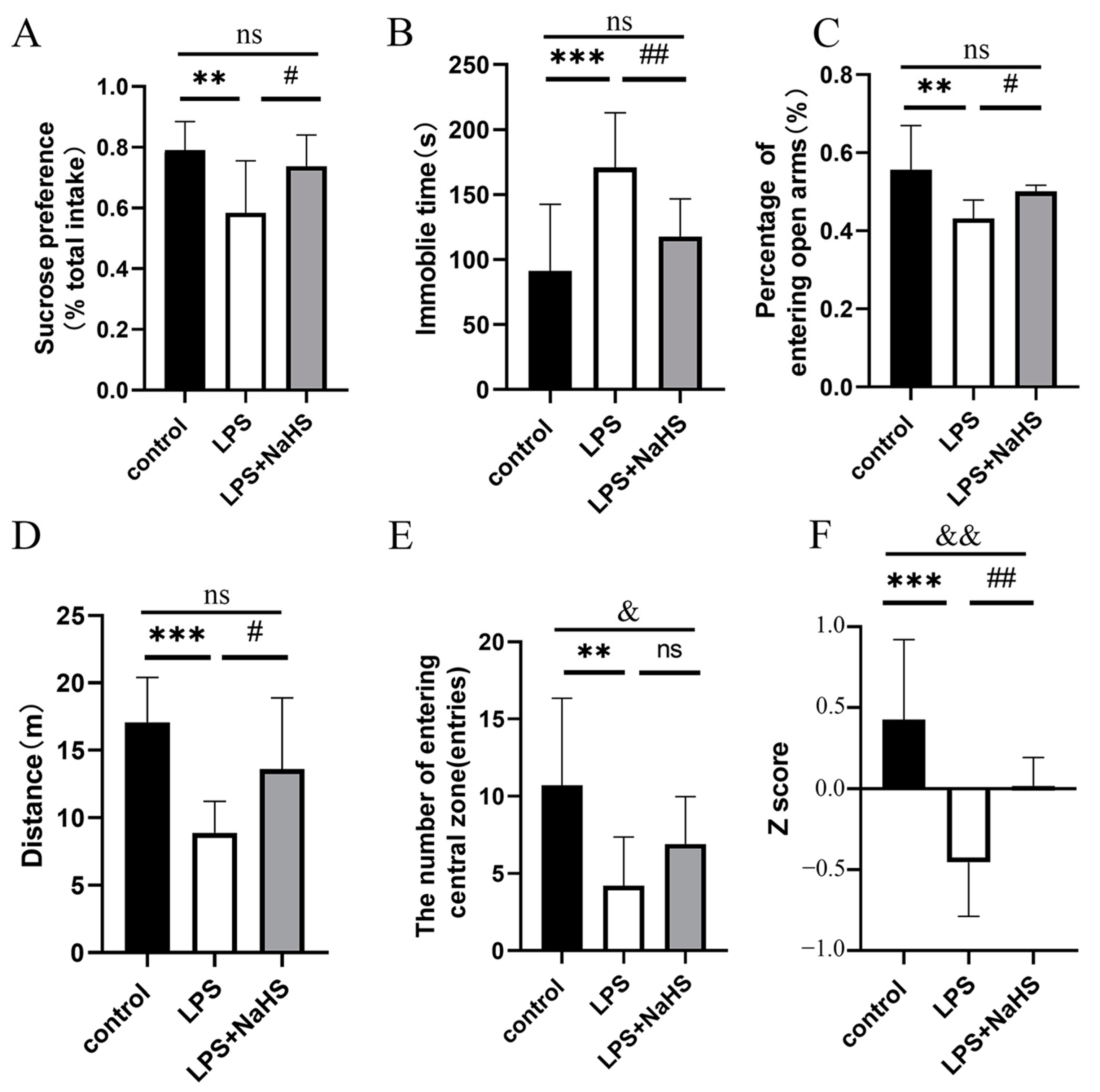
3.1. NaHS Pretreatment Prevents LPS-Induced Depression- and Anxiety-like Behavior

As shown in Figure 1, NaHS pretreatment efficiently ameliorated the effects of LPS on anhedonia and despair, which was evaluated using SPT and FST (Figure 1A,B). As for anxiety behavior, LPS treatment resulted in a decreased percentage of times entering the open arms in the EZM, reduced shuttle distance, and lowered time in the center zone in the OFT. NaHS significantly increased shuttle distance and did not affect the percentage of times entering the open arms in the EZM and time in the center zone in the OFT in LPS-treated mice (Figure 1C–E). Z-normalization analysis showed that the individual z-scores of mice pretreated with NaHS were significantly lower than those of mice treated without NaHS (Figure 1F), indicating an antidepressant effect of NaHS pretreatment.

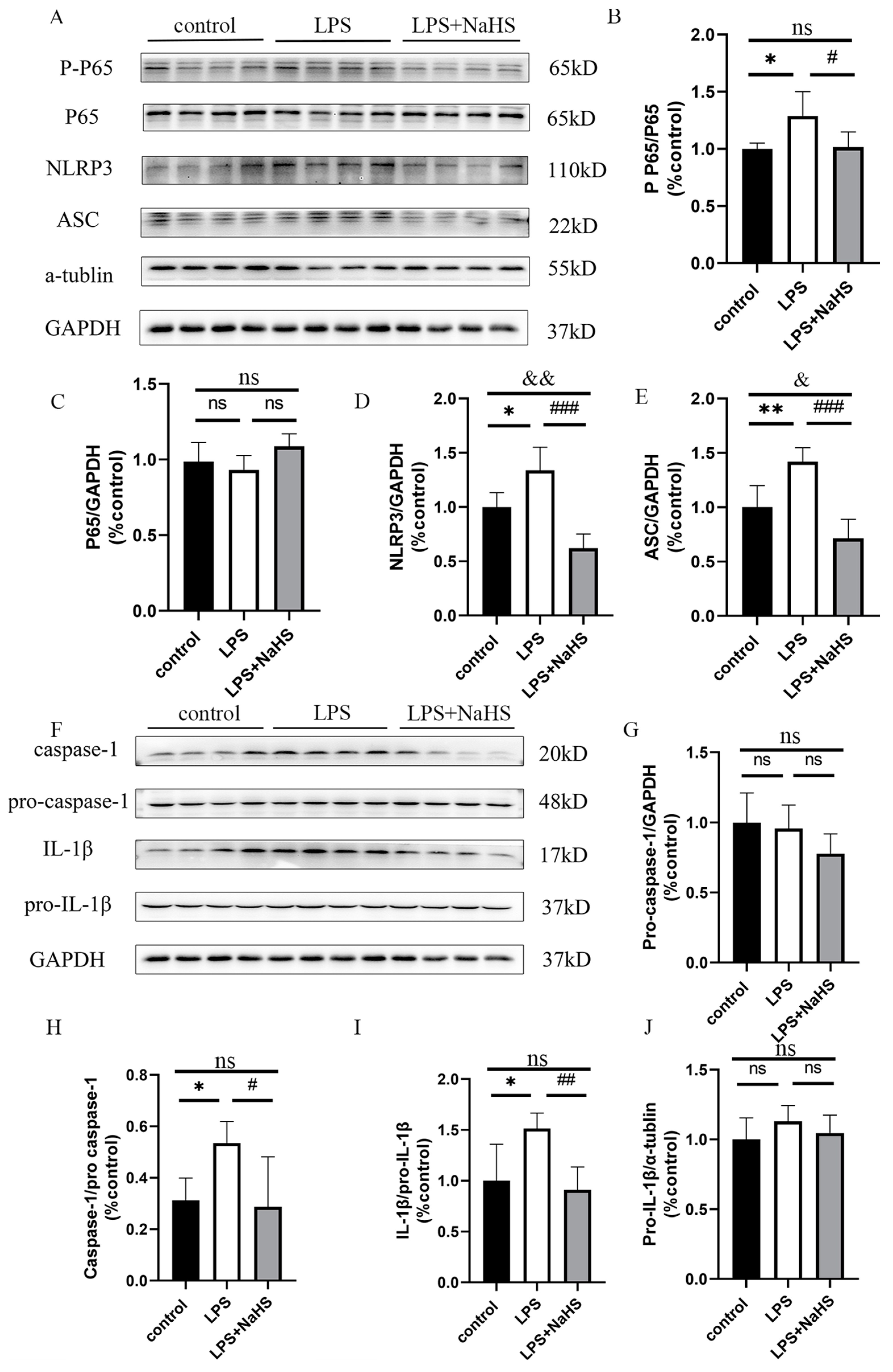
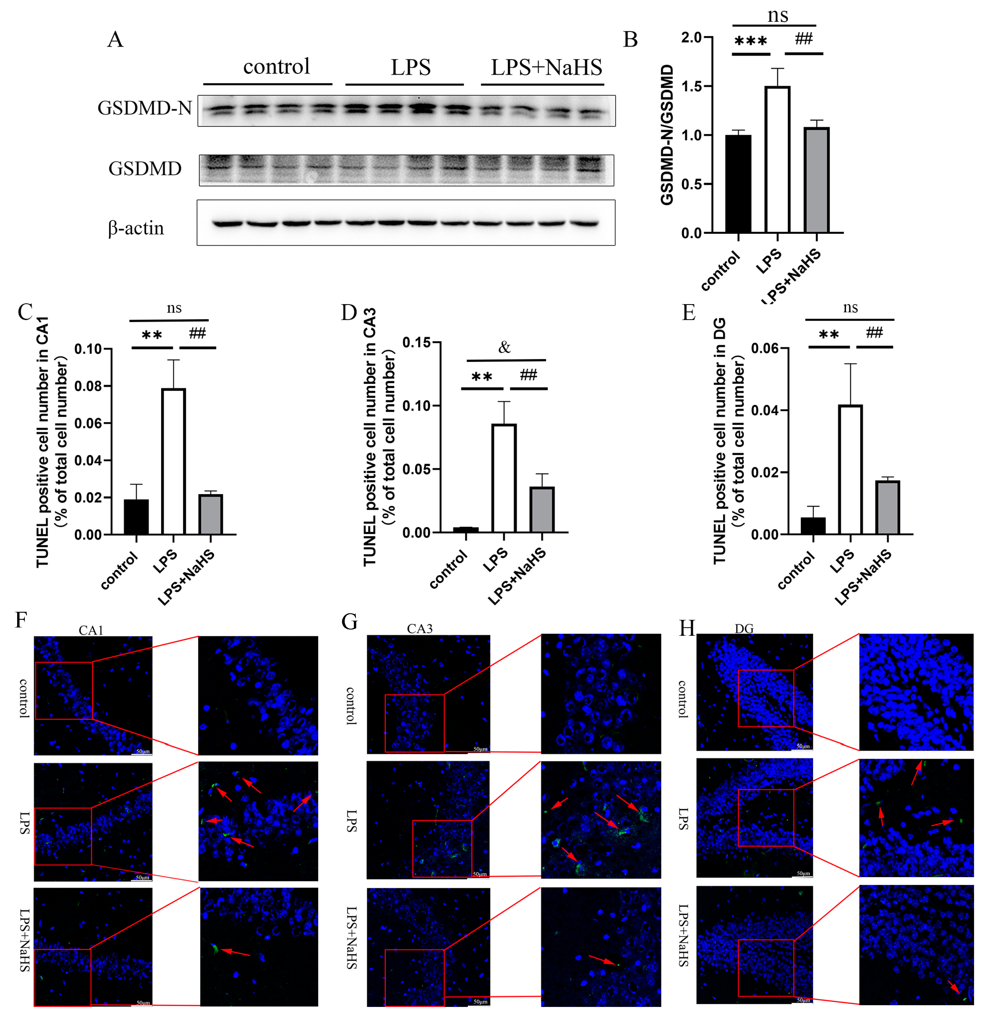
3.2. H2S Pretreatment Alleviates NF-κB and NLRP3 Inflammasome Activation and Pyroptosis in the Hippocampus of LPS-Treated Mice

As shown in Figure 2A–C, LPS-induced NF-κB activation in the hippocampus as evidenced by increased p-p65 levels, which could be suppressed by NaHS pretreatment, suggesting that H2S prevented neuroinflammation in the hippocampus in LPS-treated mice. LPS treatment also induced NLRP3 inflammasome activation as evidenced by increased NLRP3, ASC, and caspase-1 levels, and IL-1β maturation, which is also prevented by NaHS pretreatment (Figure 2 and Supplemental Figures S1–S13). In addition, the levels of NLRP3 and ASC were lower in the NaHS + LPS group than those in the control group.
NLRP3 inflammasome activation also leads to the cleavage of full-length GSDMD to generate the GSDMD N-terminal, thereby leading to pyroptosis. An increase in the level of the GSDMD N-terminus was found in LPS-treated mice, which was significantly inhibited by NaHS pretreatment (Figure 3A,B and Supplemental Figures S14–S16). TUNEL staining showed that LPS increased the percentage of positive staining cells in the region of CA1, CA3, and DG, which also decreased in the NaHS pretreatment group, suggesting that NaHS prevents NLRP3 inflammasome activation and pyroptosis caused by LPS (Figure 3C–H).

3.3. H2S Prevents Mitochondrial Damage in the Hippocampus of LPS-Treated Mice

As shown in Figure 4A, LPS caused the mitochondrial swelling and blurring of ridges in hippocampal neurons, which could be improved by NaHS pretreatment. Consistently, LPS resulted in mitochondrial membrane potential, and ATP production was reduced, while ROS production was increased in the hippocampus models, which was significantly improved by NaHS pretreatment (Figure 4B–D).
Mitochondrial respirometry showed that CI leak, CI OXPHOS, CI&II OXPHOS, CI&II ETS, and CII ETS were significantly lower in the LPS treatment group than those of the control group. With the treatment of NaHS, the oxygen consumption rates for CI OXPHOS, CI&II OXPHOS, and CI&II ETS were obviously increased, but CII ETS was not significantly affected (Figure 5).

4. Discussion

The present study has shown that LPS could lead to depression-like behavior, which could be reversed by H2S treatment. Moreover, we have also demonstrated that NF-κB and NLRP3 inflammasome activation and pyroptosis as well as mitochondrial dysfunction in the hippocampus in mice, could be prevented by H2S treatment.
Many studies have proposed that sterile inflammation has a crucial role in the pathogenesis of depression [36,37,38]. Peripheral LPS treatment causes a systemic immune response, which eventually induces behavior deficits after 24 h [8]. As mentioned, the inflammatory responses occur in the central nervous system (CNS) in patients with MDD [39,40] and rodent models of depression, including the LPS-induced depression-like mouse model [37,41,42]. We also found that inflammation occurred consistently in the hippocampus of LPS-treated mice, and it was suppressed when the depression-like behavior was improved.
NLRP3 inflammasome was very crucial in the onset of various inflammatory responses [23]. As mentioned, NLRP3 inflammasome activation occurs in the CNS in the LPS-induced depression-like mouse model. The present study also revealed NLRP3 inflammasome activation in the hippocampus upon LPS treatment. It has also been shown that the blockage of NLRP3 inflammasome activation by specific NLRP3 inflammasome inhibitors could alleviate depression-like behavior in mice [7,10,41,43]. We consistently found that hippocampal NLRP3 inflammasome activation was prevented in LPS-treated mice with H2S pretreatment. It therefore indicates that the suppression of hippocampal NLRP3 inflammasome activation contributes to the H2S prevention of LPS-induced depression-like behavior.
Pyroptosis, a form of programmed necrosis, is linked to a general innate immune effector mechanism in psychological disorders [44,45]. It is known that NLRP3 inflammasome induces pyroptosis in various tissues [46]. The full-length GSDMD protein is cleaved to produce the GSDMD N-terminus as a result of NLRP3 inflammasome activation, which causes cell necrosis [37,44,47]. In this study, we found that pyroptosis occurred in the hippocampus of the LPS-induced model. Moreover, pyroptosis in the hippocampus was suppressed by H2S pretreatment. Interestingly, another study has also shown that H2S suppresses hypoxia-induced pyroptosis in the adrenal glands of mice [48]. Thus, our data indicate that hippocampal pyroptosis contributes to LPS-induced depression-like behavior and the suppression of pyroptosis is associated with the H2S prevention of depression-like behavior.
As mentioned, mitochondrial dysfunction plays a critical role in neuropsychiatric disorders including MDD [49]. LPS can induce mitochondrial abnormalities in the CNS of the LPS-induced depression-like mouse model. We consistently found that abnormal mitochondria morphology and function in the hippocampus of LPS-treated mice. Of note, there were a few studies on the effects of H2S on mitochondrial function in the CNS in depression-like models, although some studies have demonstrated that H2S can alleviate mitochondrial dysfunction in various tissues [50,51,52]. In the present study, we showed that H2S treatment reversed LPS-induced abnormal mitochondrial morphology and function in the hippocampus. Mitochondria can activate the NLRP3 inflammasome in two ways: in an ROS-dependent and ROS-independent manner. ROS can lead to producing oxidized mtDNA; the latter activates NLRP3 inflammasome. Three mitochondrial proteins, cardiolipin, mitochondrial antiviral signaling protein (MAVS), and mitofusin 2 (MFN 2), have been implicated in the activation of NLRP3 inflammasome in an ROS-independent manner [23]. It is well known that inflammation can cause mitochondrial dysfunction in various tissues [53,54]. Thus, our above findings that H2S pretreatment improves mitochondrial function and the suppression of NLRP3 inflammasome activation indicate that H2S arrests the interplay of mitochondrial dysfunction in the hippocampus of the LPS-induced depression-like model.
Many studies have demonstrated that H2S has therapeutic effects on depression-like behavior in various animal models of depression [25,26,27,28]. Of note, some studies also show that the endogenous H2S level in the brain is reduced in various rodent models of depression [26,28,29]. Kumar et al. [26] reported that the H2S level in the hippocampus is reduced upon LPS treatment, which contributes to depression-like behavior in mice. Thus, NaHS pretreatment could prevent a reduced H2S level in the hippocampus.
There are several limitations in the present study. In addition to the hippocampus, other brain regions such as the prefrontal cortex and amygdala also play an important role in depression pathogenesis [55,56,57]. It would be of interest to investigate the effects of H2S on these brain regions in depression-like models. Furthermore, the cell types responsible for NLRP3 inflammasome activation and mitochondrial dysfunction caused by LPS have not been clarified, since both microglia and neurons have been shown to play roles in LPS-induced depression-like behavior. Mitochondria are highly dynamic organelles, and their normal function depends on the regulation of the mitochondrial quality control system in depression [42]. This should also be investigated in future studies.

5. Conclusions

In conclusion, our study reveals that NLRP3 inflammasome activation and pyroptosis as well as abnormal mitochondrial morphology and function occur in the hippocampus of the LPS-induced depression-like mouse model. H2S pretreatment prevents LPS-induced depression-like behavior through the inhibition of neuroinflammation and pyroptosis and improving mitochondrial function in the hippocampus. Our study immediately highlights that H2S can be a promising prevention reagent for depression.

Supplementary Materials

The following supporting information can be downloaded at: https://www.mdpi.com/article/10.3390/biology12081092/s1, Table S1: The antibodies employed in this study; Figure S1: Western blot membrane of P65 (~65 kDa) protein detected with anti-NF-κB (C22B4; 1:1000; CST, USA) antibody; Figure S2: Western blot membrane of P-P65 (~65 kDa) protein detected with anti-P-NF-κB (S536; 1:500; CST, USA) antibody; Figure S3: Western blot membrane of NLRP3 (~110 kDa) protein detected with anti-NLRP3 (ab214185; 1:1000; Abcam, UK) antibody; Figure S4: Western blot membrane of NLRP3 (~110 kDa) protein detected with anti-NLRP3 (ab214185; 1:1000; Abcam, UK) antibody; Figure S5: Western blot membrane of GAPDH (~37 kDa) protein detected with anti-GAPDH (MB001; 1:10000; Bioworld, USA) antibody; Figure S6: Western blot membrane of ASC (~22 kDa) protein detected with anti-ASC (A11433; 1:1000; ABclonal, China) antibody; Figure S7: Western blot membrane of GAPDH (~37 kDa) protein detected with anti-GAPDH (MB001; 1:10000; Bioworld, USA) antibody; Figure S8: Western blot membrane of pro-caspase-1 (~48 kDa) protein detected with anti-caspase-1 (A0964; 1:1000; ABclonal, China) antibody; Figure S9: Western blot membrane of caspase-1 (~20 kDa) protein detected with anti-caspase-1 (A0964; 1:1000; ABclonal, China) antibody; Figure S10: Western blot membrane of GAPDH (~37 kDa) protein detected with anti-GAPDH (MB001; 1:10000; Bioworld, USA) antibody; Figure S11: Western blot membrane of pro-IL-1β (~37 kDa) protein detected with anti-IL-1β (A11369; 1:1000; ABclonal, China) antibody; Figure S12: Western blot membrane of IL-1β (~17 kDa) protein detected with anti-IL-1β (A11369; 1:1000; ABclonal, China) antibody; Figure S13: Western blot membrane of α-tublin (~55 kDa) protein detected with anti-α-tublin (BS1699; 1:10000; Bioworld, USA) antibody; Figure S14: Western blot membrane of GSDMD (~53 kDa) protein detected with anti-GSDMD (A20197; 1:1000; ABclonal, China) antibody; Figure S15: Western blot membrane of GSDMD-N (~37 kDa) protein detected with anti-GSDMD-N (A20197; 1:1000; ABclonal, China) antibody; Figure S16: Western blot membrane of β-actin (~43 kDa) protein detected with anti-β-actin (BS6007M; 1:10000; Bioworld, USA) antibody.

Author Contributions

X.N. and J.L. designed the study and wrote the manuscript. P.B. performed the experiments, analyzed the data, and wrote a draft. Y.G. assisted in the animal experiments. Y.W. and M.X. assisted in the sampling. Z.Q. assisted in data analysis. All authors have read and agreed to the published version of the manuscript.

Funding

This research was funded by the National Natural Science Foundation of China (No. 31971102) to Jianqiang Lu, and the National Natural Science Foundation of China (No.32170864) to Xin Ni.

Institutional Review Board Statement

Animal experimental manipulations were approved by the Animal Ethics Committee of Shanghai University of Sport. The ethics committee approval number is 102772022DW006.

Informed Consent Statement

Not applicable.

Data Availability Statement

Not applicable.

Conflicts of Interest

The authors declare no conflict of interest.

References

  1. Malhi, G.S.; Mann, J.J. Depression. Lancet 2018, 392, 2299–2312. [Google Scholar] [PubMed]
  2. Gardner, A.; Boles, R.G. Beyond the serotonin hypothesis: Mitochondria, inflammation and neurodegeneration in major depression and affective spectrum disorders. Prog. Neuropsychopharmacol. Biol. Psychiatry 2011, 35, 730–743. [Google Scholar] [CrossRef]
  3. Peng, G.J.; Tian, J.S.; Gao, X.X.; Zhou, Y.Z.; Qin, X.M. Research on the Pathological Mechanism and Drug Treatment Mechanism of Depression. Curr. Neuropharmacol. 2015, 13, 514–523. [Google Scholar] [CrossRef] [PubMed] [Green Version]
  4. Eyre, H.; Baune, B.T. Neuroplastic changes in depression: A role for the immune system. Psychoneuroendocrinology 2012, 37, 1397–1416. [Google Scholar] [CrossRef] [PubMed]
  5. Lu, Y.; Xu, X.; Jiang, T.; Jin, L.; Zhao, X.D.; Cheng, J.H.; Jin, X.J.; Ma, J.; Piao, H.N.; Piao, L.X. Sertraline ameliorates inflammation in CUMS mice and inhibits TNF-α-induced inflammation in microglia cells. Int. Immunopharmacol. 2019, 67, 119–128. [Google Scholar] [CrossRef] [PubMed]
  6. Leng, L.; Zhuang, K.; Liu, Z.; Huang, C.; Gao, Y.; Chen, G.; Lin, H.; Hu, Y.; Wu, D.; Shi, M.; et al. Menin Deficiency Leads to Depressive-like Behaviors in Mice by Modulating Astrocyte-Mediated Neuroinflammation. Neuron 2018, 100, 551–563.e557. [Google Scholar] [CrossRef] [Green Version]
  7. Ali, T.; Rahman, S.U.; Hao, Q.; Li, W.; Liu, Z.; Ali Shah, F.; Murtaza, I.; Zhang, Z.; Yang, X.; Liu, G.; et al. Melatonin prevents neuroinflammation and relieves depression by attenuating autophagy impairment through FOXO3a regulation. J. Pineal Res. 2020, 69, e12667. [Google Scholar] [CrossRef]
  8. Frenois, F.; Moreau, M.; O’Connor, J.; Lawson, M.; Micon, C.; Lestage, J.; Kelley, K.W.; Dantzer, R.; Castanon, N. Lipopolysaccharide induces delayed FosB/DeltaFosB immunostaining within the mouse extended amygdala, hippocampus and hypothalamus, that parallel the expression of depressive-like behavior. Psychoneuroendocrinology 2007, 32, 516–531. [Google Scholar] [CrossRef] [Green Version]
  9. Li, W.; Ali, T.; He, K.; Liu, Z.; Shah, F.A.; Ren, Q.; Liu, Y.; Jiang, A.; Li, S. Ibrutinib alleviates LPS-induced neuroinflammation and synaptic defects in a mouse model of depression. Brain Behav. Immun. 2021, 92, 10–24. [Google Scholar] [CrossRef] [PubMed]
  10. Xu, Y.; Sheng, H.; Bao, Q.; Wang, Y.; Lu, J.; Ni, X. NLRP3 inflammasome activation mediates estrogen deficiency-induced depression- and anxiety-like behavior and hippocampal inflammation in mice. Brain Behav. Immun. 2016, 56, 175–186. [Google Scholar] [CrossRef]
  11. Paik, S.; Kim, J.K.; Silwal, P.; Sasakawa, C.; Jo, E.K. An update on the regulatory mechanisms of NLRP3 inflammasome activation. Cell Mol. Immunol. 2021, 18, 1141–1160. [Google Scholar] [CrossRef] [PubMed]
  12. Alcocer-Gómez, E.; de Miguel, M.; Casas-Barquero, N.; Núñez-Vasco, J.; Sánchez-Alcazar, J.A.; Fernández-Rodríguez, A.; Cordero, M.D. NLRP3 inflammasome is activated in mononuclear blood cells from patients with major depressive disorder. Brain Behav. Immun. 2014, 36, 111–117. [Google Scholar] [CrossRef] [PubMed]
  13. Brkic, Z.; Milosavljevic, M.; Glavonic, E.; Adzic, M. Mitochondrial signaling in inflammation-induced depressive behavior in female and male rats: The role of glucocorticoid receptor. Brain Res. Bull. 2019, 150, 317–327. [Google Scholar] [CrossRef]
  14. Xia, C.Y.; Guo, Y.X.; Lian, W.W.; Yan, Y.; Ma, B.Z.; Cheng, Y.C.; Xu, J.K.; He, J.; Zhang, W.K. The NLRP3 inflammasome in depression: Potential mechanisms and therapies. Pharmacol. Res. 2023, 187, 106625. [Google Scholar] [CrossRef] [PubMed]
  15. Wu, T.; Huang, Y.; Gong, Y.; Xu, Y.; Lu, J.; Sheng, H.; Ni, X. Treadmill Exercise Ameliorates Depression-Like Behavior in the Rats With Prenatal Dexamethasone Exposure: The Role of Hippocampal Mitochondria. Front. Neurosci. 2019, 13, 264. [Google Scholar] [CrossRef] [PubMed] [Green Version]
  16. Scaini, G.; Mason, B.L.; Diaz, A.P.; Jha, M.K.; Soares, J.C.; Trivedi, M.H.; Quevedo, J. Dysregulation of mitochondrial dynamics, mitophagy and apoptosis in major depressive disorder: Does inflammation play a role? Mol. Psychiatry 2021, 27, 1095–1102. [Google Scholar] [CrossRef] [PubMed]
  17. Gebara, E.; Zanoletti, O.; Ghosal, S.; Grosse, J.; Schneider, B.L.; Knott, G.; Astori, S.; Sandi, C. Mitofusin-2 in the Nucleus Accumbens Regulates Anxiety and Depression-like Behaviors Through Mitochondrial and Neuronal Actions. Biol. Psychiatry 2021, 89, 1033–1044. [Google Scholar] [CrossRef]
  18. Wang, Y.; Ni, J.; Gao, C.; Xie, L.; Zhai, L.; Cui, G.; Yin, X. Mitochondrial transplantation attenuates lipopolysaccharide- induced depression-like behaviors. Prog. Neuropsychopharmacol. Biol. Psychiatry 2019, 93, 240–249. [Google Scholar] [CrossRef]
  19. Jin, X.; Zhu, L.; Lu, S.; Li, C.; Bai, M.; Xu, E.; Shen, J.; Li, Y. Baicalin ameliorates CUMS-induced depression-like behaviors through activating AMPK/PGC-1α pathway and enhancing NIX-mediated mitophagy in mice. Eur. J. Pharmacol. 2023, 938, 175435. [Google Scholar] [CrossRef]
  20. Głombik, K.; Stachowicz, A.; Olszanecki, R.; Ślusarczyk, J.; Trojan, E.; Lasoń, W.; Kubera, M.; Budziszewska, B.; Spedding, M.; Basta-Kaim, A. The effect of chronic tianeptine administration on the brain mitochondria: Direct links with an animal model of depression. Mol. Neurobiol. 2016, 53, 7351–7362. [Google Scholar] [CrossRef] [Green Version]
  21. Liu, W.; Wang, H.; Xue, X.; Xia, J.; Liu, J.; Qi, Z.; Ji, L. OGT-related mitochondrial motility is associated with sex differences and exercise effects in depression induced by prenatal exposure to glucocorticoids. J. Affect. Disord. 2018, 226, 203–215. [Google Scholar] [CrossRef]
  22. Visentin, A.P.V.; Colombo, R.; Scotton, E.; Fracasso, D.S.; da Rosa, A.R.; Branco, C.S.; Salvador, M. Targeting Inflammatory-Mitochondrial Response in Major Depression: Current Evidence and Further Challenges. Oxid. Med. Cell Longev. 2020, 2020, 2972968. [Google Scholar] [CrossRef] [PubMed] [Green Version]
  23. Swanson, K.V.; Deng, M.; Ting, J.P. The NLRP3 inflammasome: Molecular activation and regulation to therapeutics. Nat. Rev. Immunol. 2019, 19, 477–489. [Google Scholar] [CrossRef]
  24. Murphy, B.; Bhattacharya, R.; Mukherjee, P. Hydrogen sulfide signaling in mitochondria and disease. FASEB J. 2019, 33, 13098–13125. [Google Scholar] [CrossRef] [PubMed] [Green Version]
  25. Liu, S.Y.; Li, D.; Zeng, H.Y.; Kan, L.Y.; Zou, W.; Zhang, P.; Gu, H.F.; Tang, X.Q. Hydrogen Sulfide Inhibits Chronic Unpredictable Mild Stress-Induced Depressive-Like Behavior by Upregulation of Sirt-1: Involvement in Suppression of Hippocampal Endoplasmic Reticulum Stress. Int. J. Neuropsychopharmacol. 2017, 20, 867–876. [Google Scholar] [CrossRef] [Green Version]
  26. Kumar, M.; Arora, P.; Sandhir, R. Hydrogen Sulfide Reverses LPS-Induced Behavioral Deficits by Suppressing Microglial Activation and Promoting M2 Polarization. J. Neuroimmune Pharmacol. 2021, 16, 483–499. [Google Scholar] [CrossRef]
  27. Ruilian, L.; Honglin, Q.; Jun, X.; Jianxin, L.; Qingyun, B.; Yilin, C.; Haifeng, M. H(2)S-mediated aerobic exercise antagonizes the hippocampal inflammatory response in CUMS-depressed mice. J. Affect. Disord. 2021, 283, 410–419. [Google Scholar] [CrossRef] [PubMed]
  28. Wei, L.; Kan, L.Y.; Zeng, H.Y.; Tang, Y.Y.; Huang, H.L.; Xie, M.; Zou, W.; Wang, C.Y.; Zhang, P.; Tang, X.Q. BDNF/TrkB Pathway Mediates the Antidepressant-Like Role of H(2)S in CUMS-Exposed Rats by Inhibition of Hippocampal ER Stress. Neuromolecular Med. 2018, 20, 252–261. [Google Scholar] [CrossRef]
  29. Tan, H.; Zou, W.; Jiang, J.; Tian, Y.; Xiao, Z.; Bi, L.; Zeng, H.; Tang, X. Disturbance of hippocampal H2S generation contributes to CUMS-induced depression-like behavior: Involvement in endoplasmic reticulum stress of hippocampus. Acta Biochim. Biophys. Sin. 2015, 47, 285–291. [Google Scholar] [CrossRef] [Green Version]
  30. Chen, W.J.; Du, J.K.; Hu, X.; Yu, Q.; Li, D.X.; Wang, C.N.; Zhu, X.Y.; Liu, Y.J. Protective effects of resveratrol on mitochondrial function in the hippocampus improves inflammation-induced depressive-like behavior. Physiol. Behav. 2017, 182, 54–61. [Google Scholar] [CrossRef]
  31. Yankelevitch-Yahav, R.; Franko, M.; Huly, A.; Doron, R. The forced swim test as a model of depressive-like behavior. J. Vis. Exp. 2015, 97, e52587. [Google Scholar]
  32. Seibenhener, M.L.; Wooten, M.C. Use of the Open Field Maze to Measure Locomotor and Anxiety-like Behavior in Mice. J. Vis. Exp. 2015, 96, e52434. [Google Scholar]
  33. Tucker, L.B.; McCabe, J.T. Behavior of Male and Female C57BL/6J Mice Is More Consistent with Repeated Trials in the Elevated Zero Maze than in the Elevated Plus Maze. Front. Behav. Neurosci. 2017, 11, 13. [Google Scholar] [CrossRef] [Green Version]
  34. Guilloux, J.P.; Seney, M.; Edgar, N.; Sibille, E. Integrated behavioral z-scoring increases the sensitivity and reliability of behavioral phenotyping in mice: Relevance to emotionality and sex. J. Neurosci. Methods 2011, 197, 21–31. [Google Scholar] [CrossRef] [Green Version]
  35. Burtscher, J.; Zangrandi, L.; Schwarzer, C.; Gnaiger, E. Differences in mitochondrial function in homogenated samples from healthy and epileptic specific brain tissues revealed by high-resolution respirometry. Mitochondrion 2015, 25, 104–112. [Google Scholar] [CrossRef]
  36. Miller, A.H.; Maletic, V.; Raison, C.L. Inflammation and its discontents: The role of cytokines in the pathophysiology of major depression. Biol. Psychiatry 2009, 65, 732–741. [Google Scholar] [CrossRef] [PubMed] [Green Version]
  37. Li, S.; Sun, Y.; Song, M.; Song, Y.; Fang, Y.; Zhang, Q.; Li, X.; Song, N.; Ding, J.; Lu, M.; et al. NLRP3/caspase-1/GSDMD-mediated pyroptosis exerts a crucial role in astrocyte pathological injury in mouse model of depression. JCI Insight 2021, 6, e146852. [Google Scholar] [CrossRef]
  38. Zhao, S.; Li, X.; Wang, J.; Wang, H. The Role of the Effects of Autophagy on NLRP3 Inflammasome in Inflammatory Nervous System Diseases. Front. Cell Dev. Biol. 2021, 9, 657478. [Google Scholar] [CrossRef]
  39. Borsini, A.; Nicolaou, A.; Camacho-Muñoz, D.; Kendall, A.C.; Di Benedetto, M.G.; Giacobbe, J.; Su, K.P.; Pariante, C.M. Omega-3 polyunsaturated fatty acids protect against inflammation through production of LOX and CYP450 lipid mediators: Relevance for major depression and for human hippocampal neurogenesis. Mol. Psychiatry 2021, 26, 6773–6788. [Google Scholar] [CrossRef]
  40. Pitharouli, M.C.; Hagenaars, S.P.; Glanville, K.P.; Coleman, J.R.I.; Hotopf, M.; Lewis, C.M.; Pariante, C.M. Elevated C-Reactive Protein in Patients With Depression, Independent of Genetic, Health, and Psychosocial Factors: Results From the UK Biobank. Am. J. Psychiatry 2021, 178, 522–529. [Google Scholar] [CrossRef]
  41. Han, X.; Xu, T.; Fang, Q.; Zhang, H.; Yue, L.; Hu, G.; Sun, L. Quercetin hinders microglial activation to alleviate neurotoxicity via the interplay between NLRP3 inflammasome and mitophagy. Redox Biol. 2021, 44, 102010. [Google Scholar] [CrossRef] [PubMed]
  42. Yue, N.; Huang, H.; Zhu, X.; Han, Q.; Wang, Y.; Li, B.; Liu, Q.; Wu, G.; Zhang, Y.; Yu, J. Activation of P2X7 receptor and NLRP3 inflammasome assembly in hippocampal glial cells mediates chronic stress-induced depressive-like behaviors. J. Neuroinflammation 2017, 14, 102. [Google Scholar] [CrossRef] [PubMed] [Green Version]
  43. Arioz, B.I.; Tastan, B.; Tarakcioglu, E.; Tufekci, K.U.; Olcum, M.; Ersoy, N.; Bagriyanik, A.; Genc, K.; Genc, S. Melatonin Attenuates LPS-Induced Acute Depressive-Like Behaviors and Microglial NLRP3 Inflammasome Activation Through the SIRT1/Nrf2 Pathway. Front. Immunol. 2019, 10, 1511. [Google Scholar] [CrossRef]
  44. Shi, J.; Gao, W.; Shao, F. Pyroptosis: Gasdermin-Mediated Programmed Necrotic Cell Death. Trends Biochem. Sci. 2017, 42, 245–254. [Google Scholar] [CrossRef] [PubMed]
  45. Jin, X.; Jin, H.; Shi, Y.; Guo, Y.; Zhang, H. Pyroptosis, a novel mechanism implicated in cataracts. Mol. Med. Rep. 2018, 18, 2277–2285. [Google Scholar] [CrossRef] [Green Version]
  46. Yu, P.; Zhang, X.; Liu, N.; Tang, L.; Peng, C.; Chen, X. Pyroptosis: Mechanisms and diseases. Signal Transduct. Target. Ther. 2021, 6, 128. [Google Scholar] [CrossRef] [PubMed]
  47. Li, Y.; Song, W.; Tong, Y.; Zhang, X.; Zhao, J.; Gao, X.; Yong, J.; Wang, H. Isoliquiritin ameliorates depression by suppressing NLRP3-mediated pyroptosis via miRNA-27a/SYK/NF-κB axis. J. Neuroinflammation 2021, 18, 1. [Google Scholar] [CrossRef] [PubMed]
  48. Zhang, N.; Zhou, Z.; Huang, Y.; Wang, G.; Tang, Z.; Lu, J.; Wang, C.; Ni, X. Reduced hydrogen sulfide production contributes to adrenal insufficiency induced by hypoxia via modulation of NLRP3 inflammasome activation. Redox Rep. 2023, 28, 2163354. [Google Scholar] [CrossRef]
  49. Pei, L.; Wallace, D.C. Mitochondrial Etiology of Neuropsychiatric Disorders. Biol. Psychiatry 2018, 83, 722–730. [Google Scholar] [CrossRef]
  50. Zhao, F.L.; Fang, F.; Qiao, P.F.; Yan, N.; Gao, D.; Yan, Y. AP39, a Mitochondria-Targeted Hydrogen Sulfide Donor, Supports Cellular Bioenergetics and Protects against Alzheimer’s Disease by Preserving Mitochondrial Function in APP/PS1 Mice and Neurons. Oxid. Med. Cell Longev. 2016, 2016, 8360738. [Google Scholar] [CrossRef] [Green Version]
  51. Lohakul, J.; Jeayeng, S.; Chaiprasongsuk, A.; Torregrossa, R.; Wood, M.E.; Saelim, M.; Thangboonjit, W.; Whiteman, M.; Panich, U. Mitochondria-Targeted Hydrogen Sulfide Delivery Molecules Protect Against UVA-Induced Photoaging in Human Dermal Fibroblasts, and in Mouse Skin In Vivo. Antioxid. Redox Signal 2021, 36, 1268–1288. [Google Scholar] [CrossRef] [PubMed]
  52. Latorre, E.; Torregrossa, R.; Wood, M.E.; Whiteman, M.; Harries, L.W. Mitochondria-targeted hydrogen sulfide attenuates endothelial senescence by selective induction of splicing factors HNRNPD and SRSF2. Aging (Albany N. Y.) 2018, 10, 1666–1681. [Google Scholar] [CrossRef] [PubMed]
  53. Missiroli, S.; Genovese, I.; Perrone, M.; Vezzani, B.; Vitto, V.A.M.; Giorgi, C. The Role of Mitochondria in Inflammation: From Cancer to Neurodegenerative Disorders. J. Clin. Med. 2020, 9, 740. [Google Scholar] [CrossRef] [Green Version]
  54. Yue, L.; Yao, H. Mitochondrial dysfunction in inflammatory responses and cellular senescence: Pathogenesis and pharmacological targets for chronic lung diseases. Br. J. Pharmacol. 2016, 173, 2305–2318. [Google Scholar] [CrossRef] [PubMed] [Green Version]
  55. Pizzagalli, D.A.; Roberts, A.C. Prefrontal cortex and depression. Neuropsychopharmacology 2022, 47, 225–246. [Google Scholar] [CrossRef] [PubMed]
  56. Grogans, S.E.; Fox, A.S.; Shackman, A.J. The Amygdala and Depression: A Sober Reconsideration. Am. J. Psychiatry 2022, 179, 454–457. [Google Scholar] [CrossRef] [PubMed]
  57. Liu, W.; Ge, T.; Leng, Y.; Pan, Z.; Fan, J.; Yang, W.; Cui, R. The Role of Neural Plasticity in Depression: From Hippocampus to Prefrontal Cortex. Neural Plast. 2017, 2017, 6871089. [Google Scholar] [CrossRef] [Green Version]
Figure 1. H2S attenuates depression-like and anxiety-like behavior caused by LPS in the mouse model. (A) Sucrose preference in SPT. (B) Immobile time in FST. (C) Percentage of number of entries in open arm in EZM. (D) Total distance traveled in OFT. (E) The number of times entering the central zone. (F) Integrated z-scores of depression across depression-related variables. All data are expressed as mean ± SD. ** p < 0.01, *** p < 0.001; # p < 0.05, ## p < 0.01; & p < 0.05, && p < 0.01; ns: no significance.
Biology 12 01092 g001
Figure 2. H2S prevents NF-κB and NLRP3 inflammasome activation in the hippocampus of LPS-treated mice. (A,F) Western blotting images of various proteins. (BE,GJ) Quantitative diagrams of Western blot analysis. All data are expressed as mean ± SD. * p < 0.05, ** p < 0.01; # p < 0.05, ## p < 0.01, ### p < 0.001; & p < 0.05, && p < 0.01; ns: no significance.
Biology 12 01092 g002
Figure 3. H2S suppresses pyroptosis in the hippocampus of LPS-treated mice. (A) Representative images of GSDMD-N and GSDMD blotting. (B) Quantitative diagram of Western blot analysis. (CE) Percentage of TUNEL positive cells in CA1, CA3, and DG regions. (FH) Representative images of TUNEL positive cells in CA1, CA3, and DG regions (×200). Red arrows indicate the TUNEL-positive cells. All data are expressed as mean ± SD. ** p < 0.01, *** p < 0.001; ## p < 0.01; & p < 0.05, ns: no significance.
Biology 12 01092 g003
Figure 4. H2S improves mitochondrial morphology and function in the hippocampus of LPS-treated mice. (A) Representative TEM images of mitochondria in the hippocampus (×20 k). Red arrows indicate the local magnification of the mitochondria. (BD) Mitochondrial function: membrane potential (B), ATP production (C), and ROS production (D). All data are expressed as mean ± SD. * p < 0.05, ** p < 0.01, *** p < 0.001; & p < 0.05, ns: no significance.
Biology 12 01092 g004
Figure 5. H2S improves the respiratory rate of mitochondrial complexes of the hippocampus in the LPS-induced depression model. (A) Representative tracing of the determination of OXPHOS complex I and II. Blue line: real-time oxygen consumption. Red line: real-time respiratory rate. (B) Statistical data of complex I and II. All data are expressed as mean ± SD. * p < 0.05, ** p < 0.01; # p < 0.05, ns: no significance.
Biology 12 01092 g005
Disclaimer/Publisher’s Note: The statements, opinions and data contained in all publications are solely those of the individual author(s) and contributor(s) and not of MDPI and/or the editor(s). MDPI and/or the editor(s) disclaim responsibility for any injury to people or property resulting from any ideas, methods, instructions or products referred to in the content.

