Article

Three Copies of zbed1 Specific in Chromosome W Are Essential for Female-Biased Sexual Size Dimorphism in Cynoglossus semilaevis

1
Jiangsu Key Laboratory of Marine Bioresources and Environment, Jiangsu Ocean University, Lianyungang 222000, China
2
National Key Laboratory of Mariculture Biobreeding and Sustainable Goods, Yellow Sea Fisheries Research Institute, Chinese Academy of Fishery Sciences, Qingdao 266071, China
3
College of Fisheries and Life Science, Dalian Ocean University, Dalian 116023, China
4
College of Fisheries and Life Science, Shanghai Ocean University, Shanghai 201306, China
5
Fisheries College, Zhejiang Ocean University, Zhoushan 316022, China
*
Author to whom correspondence should be addressed.
Biology 2024, 13(3), 141; https://doi.org/10.3390/biology13030141
Submission received: 29 January 2024 / Revised: 17 February 2024 / Accepted: 19 February 2024 / Published: 23 February 2024
(This article belongs to the Section Marine Biology)

Abstract

:

Simple Summary

The sex chromosome, especially specific in one sex, is considered to determine the sexual size dimorphism (SSD), a dimorphic sexual difference in the body size. For Cynoglossus semilaevis, a flatfish unique in China, the important role of female-specific chromosome W in its female-biased SSD was previously implied. Furthermore, a W chromosome-specific gene zbed1 was identified to potentially regulate female-biased SSD in C. semilaevis. However, the chromosome’s location, family members, and detailed network information are still unknown. At present, the genome-wide identification of ZBED family members and dPCR experiment both confirm that three copies of the zbed1 gene are located in chromosome W, with no zbed1 gene in chromosome Z. Phylogenetic analysis for ZBED family revealed an existence of ZBED9 in the fish. Nine members were uncovered from C. semilaevis, clustering into three kinds, ZBED1, ZBED4 and ZBEDX, which is less than the eleven kinds of ZBED members in mammals. For the chromosome-W-specific zbed1, qPCR verified its predominant expression in the female brain and pituitary tissues. The dual luciferase activity test showed that transcription factor c/ebpα could significantly enhance the transcriptional activity of zbed1 promoter, which is opposite to its effect on the male determinant factor dmrt1. In addition, after zbed1 interfered in the brain cells, piwil1, esr2 and wnt7b were up-regulated, while cell-cycle-related genes (tbp, cdk2, cdk4, cdk6, ccng1 and ccndx) were down-regulated. It is suggested that the cell proliferation function of zbed1 may be realized by regulating esr2, piwil1, cell cycle and the Wnt pathway.

Abstract

The sex chromosome, especially specific in one sex, generally determines sexual size dimorphism (SSD), a phenomenon with dimorphic sexual difference in the body size. For Cynoglossus semilaevis, a flatfish in China, although the importance of chromosome W and its specific gene zbed1 in female-biased SSD have been suggested, its family members and regulation information are still unknown. At present, three zbed1 copies gene were identified on chromosome W, with no gametologs. Phylogenetic analysis for the ZBED family revealed an existence of ZBED9 in the fish. Nine members were uncovered from C. semilaevis, clustering into three kinds, ZBED1, ZBED4 and ZBEDX, which is less than the eleven kinds of ZBED members in mammals. The predominant expression of zbed1 in the female brain and pituitary tissues was further verified by qPCR. Transcription factor c/ebpα could significantly enhance the transcriptional activity of zbed1 promoter, which is opposite to its effect on the male determinant factor-dmrt1. When zbed1 was interfered with, piwil1, esr2 and wnt7b were up-regulated, while cell-cycle-related genes, including cdk4 and ccng1, were down-regulated. Thus, zbed1 is involved in cell proliferation by regulating esr2, piwil1, cell cycle and the Wnt pathway. Further research on their interactions would be helpful to understand fish SSD.

1. Introduction

The phenomenon of body size varying greatly between females and males is known as sexual size dimorphism (SSD), which exists widely across multiple species, including arthropods, fish, reptiles, birds, and mammals [1,2,3]. The formation of SSD is usually related to the allometric growth and natural selection in different sexes [4]. Sex steroids and the somatotrophic axis (mainly GH-IGF1 system) have been reported to influence the sexual allometric growth [5,6,7].
Since 1984, Rice and others have put forward the theory of “sex chromosome dominating gender abnormality” [8]. Increasing evidence has indicated that the sex chromosome may be a dominant factor for SSD. For instance, mice with two X chromosomes have characteristics such as heavier body weight than mice with only one X chromosome [9]. In Portuguese Water Dogs, the interaction of CHM marker on the chromosome X and igf1 gene on the chromosome 15 could result in male-biased SSD [10]. Studies have also found that the singing control area of the male brain of zebra finch is usually 5–6 times larger than that of the female [11,12], which is mainly controlled by sex chromosomes [13]. The sexual dimorphic singing behavior of another bird, the white-waisted wingbird, was regulated by the Z-chromosome-linked gene erbin through its influence on brain cell proliferation and neuronal differentiation [14]. In drosphila, except for the classical insulin/IGF pathway (IIS) [7,15], the sex-determining gene sxl in chromosome X has also been proven to regulate female-biased SSD by cell-autonomy and non-cell-autonomy mechanisms [16,17]. Yet, in the seed beetle, a Y-linked TOR provides a male-specific opportunity to affect male body size and thereby sexual size dimorphism (SSD) [18].On the other hand, there is no significant correlation between sex chromosome types of radial fin fish and fin decoration characteristics of male fish [19,20]. In view of this, it is worth investigating whether a sex chromosome plays a role in the female-biased SSD of the Chinese female heterogamete flatfish, Cynoglossus semilaevis (ZW♀/ZZ♂) [21,22].
Our previous transcriptomic analysis of female and male C. semilaevis has screened 204 genes on chromosome W [23], implying the important role of chromosome W in its female-biased SSD. Further GWAS has identified the chromosome-W-linked transcription factor zinc finger BED domain-containing protein 1 (zbed1) from C. semilaevis [24]. The Zbed1 gene, a homologue of DREF in Drosophila, was firstly identified by Ohshima in 2003 as a key transcription factor for cell proliferation and can participate in DNA replication and cell proliferation by regulating histone H1 ribosomal protein RP [25,26].
In mammals, eleven ZBED family members have been identified and derived from “molecularly domesticated” hAT DNA transposons, which are divided into two sub-families (ZBED5 and ZBED7–9 belong to the Buster sub-family, while the others were assigned to Ac sub-family) [27]. Besides zbed1, other members, including zbed2, zbed3, zbed4, and zbed6, are also involved in regulating brain development, growth, insulin metabolism, interferon response pathway, and so on [28,29,30,31,32]. For example, the zbed3 protein assumes a pivotal function in the modulation of the Wnt/β-catenin pathway in mammalian organisms [31], zbed4 is closely related to retinal morphogenesis and isopathways [29], and zbed6 can regulate cell proliferation, differentiation and growth by inhibiting igf2 and other genes [30].
However, only three members of the ZBED family, ZBED1, ZBED4 and ZBEDX, have been identified in most fish [33], which seems to be contradictory to fish-specific genome duplication [34]. Until now, only two members of the ZBED family, ZBED1 and ZBED4, have been found in C. semilaevis. Whether there are other members is not clear.
Therefore, the present study aims to firstly identify C. semilaevis ZBED family members from a genome-wide perspective, which is also helpful to confirm whether zbed1 has its homologue gene in chromosome Z and whether any other members exist in this fish. The spatiotemporal expression pattern and transcriptional regulation of zbed1 are subsequently examined. Finally, the knockdown effect of zbed1 is studied to reveal its molecular pathways involved in sexual size dimorphism.

2. Materials and Methods

2.1. Ethics Statement

MS-222 was used to anesthetize fish individuals prior to the experiments in the present study. The Animal Care and Use Committee of the Yellow Sea Fisheries Research Institute, Chinese Academy of Fishery Sciences approved the sampling and treatment of C. semilaevis in the present study.

2.2. Cell Culture and Transfection

HEK 293T cells derived from human embryonic kidneys and C. semilaevis brain cells obtained from female individuals were utilized in this study. HEK 293T cells were cultured in DMEM medium supplemented with 10% fetal bovine serum (FBS) (Bovogen, Melbourne, Australia), and maintained at 37 °C with 5% CO2. Additionally, 1% antibiotics were added to the culture. C. semilaevis cells were cultured in L-15 medium supplemented with 10% FBS, 5 ng/mL bFGF (Beyotime, Shanghai, China), 5 ng/mL LIF (Beyotime, Shanghai, China), and 1% antibiotics at 24 °C [35]. The day before transfection, the cells were incubated in 24-well or 12-well plates with a density ranging from 60% to 80%. Lipo8000TM Transfection Reagent (Beyotime, Shanghai, China) and riboFECTTM CP Transfection Kit (Ribobio, Guangzhou, China) were used for the transfection of plasmids and small interfering RNAs (siRNAs), respectively [36].

2.3. Experimental Samples

The C. semilaevis samples were obtained from Haiyang Yellow Sea Fisheries Limited Company in Shandong, and the genetic sex was determined combining visual observation and a molecular identification method with the previously reported primers Cs-SEX-F and Cs-SEX-R (Table 1) [37]. After dissection, the gonad, kidney, intestine, brain, pituitary, gill, spleen, muscle, skin, and liver tissues were separately collected from 4 female and 4 male one-year-old (1 Y) individuals. The brain tissue of fish at different stages, including three-month-old (3 M), five-month-old (5 M), eight-month-old (8 M), one-year-old (1 Y), 1.5-year-old (1.5 Y), and two-year-old fish (2 Y), were also picked. Furthermore, the embryos at various classic developmental periods such as cleavage period, blastocyst period, gastrula period, segmentation period, pharyngula period and early larval were also harvested. They were stored at −20 °C in RNA preservation solution (TaKaRa, Tokyo, Japan) or 100% ethanol for DNA/RNA extraction, respectively.

2.4. DNA Extraction, RNA Extraction, and cDNA Synthesis

The genomic DNA was extracted using TIANamp Marine Animal DNA Kit (TIANGEN, Beijing, China). Total RNA was extracted by TRIzol (Invitrogen, Waltham, MA, USA) according to standard protocol. The quality, concentration, and integrity of DNA/RNA were determined by agarose gel electrophoresis and NanoVuePlus (GE Healthcare, Little Chalfont, Buckinghamshire, UK). The PrimeScriptTM RT reagent kit with gDNA Eraser (Perfect Real Time) (TaKaRa, Tokyo, Japan) was used to make cDNA. CDS of zbed1 was cloned using primers (zbed1-cds-F and zbed1-cds-R) (Table 1). The PCR amplification was carried out for 10 s at 98 °C, followed by 40 cycles at 68 °C for 30 s, and 7 s at 72 °C. The PCR product was subcloned to pEASY-T1 vector and sequenced by Qingdao Ruibo Company (Ruibo, Beijing, China).

2.5. Identification of Three Multi-Copy Genes of Cs-zbed1 Gene and dPCR

The design of taqman probes and primers targeting the genome sequences of zbed1 and myosin heavy chain 6 (myh6), a single-copy internal reference gene in teleost [38], was conducted by Sangon Biotech Company (Sangon, Shanghai, China). We validated the amplification efficiency of Taqman probes and primers by qPCR. The taqman–PCR reaction system contained 1 μL of DNA, 2.5 μL of each primer, 0.2 μL of taqman probe, 10 μL of KOD mix, and 3.4 μL of ddH2O. Next, PCR was performed for 10 s at 98 °C, followed by 40 cycles of 5 s at 58 °C and 30 s at 68 °C.
Furthermore, the digital PCR (dPCR) experiment was performed using the Naica Lite Automatic Microchip Digital PCR System (Stilla Technologies, Paris, France). The amplification system in sapphire chips comprised 25 μL of perfeCTa Multiplex qPCR Toughmix, 2.5 μL of taqman primer, 2.5 μL of Fluorescein, 0.625 μL of taqman probe, 5 μL of DNA, and 11.875 μL of ddH2O. After incubating at 95 °C for 3 min, the samples were denatured for 10 s at 95 °C for 40 cycles, and then annealed at 58 °C for 1 min. The number of droplets in sapphire chips were imaged by the Naica Prism2 reader and the fluorescence data were measured by Crystal Miner_v3.1.6.3 software to determine the positivity or negativity. Subsequently, the fluorescence intensity distribution of the droplet and the copy number of each sample were further analyzed by Crystal Reader_Prism3_v3.1.6.3.SP1 software. Finally, the copy number of the target gene was determined by comparing it to the copy number of the internal reference gene.

2.6. Phylogenetic Analysis of ZBED Family

To conduct a phylogenetic analysis of the ZBED family, the ZF_BED (PF02892) and Dimer_Tnp_hAT (PF05699) domains of PFAM database were firstly used to search in C. semilaevis genome. Combining BLAST and HomoloGene search in the NCBI database, ZBED family members of C. semilaevis, and other teleosts and mammals were obtained and submitted to MEGA 7.0 software for phylogenetic tree construction with the neighbor joining method. Furthermore, the tree was modified and visualized by EvolView Old versions Online website (https://www.evolgenius.info/evolview/#/treeview, accessed on 6 December 2023). The protein–protein interaction (PPI) network for ZBED family members based on mammals and fish was built according to the STRING database (https://string-db.org/cgi, accessed on 10 April 2023), with a confidence level of 0.25.

2.7. Expression Patterns of zbed1

We designed qPCR primers (zbed1-qPCR-F/R) (Table 1) for detecting the expression patterns of C. semilaevis zbed1. β-actin was utilized as an internal reference gene. The reaction was conducted using the Ex-Taq with SYBR® Green Realtime PCR Master Mix (TOYOBO, Osaka, Japan) on a 7500 fast real-time PCR system (ABI, Los Angeles, CA, USA). The 2−ΔΔCt method was applied to determine the relative mRNA expression level. Four different female individuals were used as biological repeats and the data were analyzed with SPSS 25.0 (IBM Corp, Armonk, NY, USA) using one-way ANOVA and multiple comparison by the Wohler and Duncan methods, and p-value < 0.05 was considered the threshold for statistical significance.

2.8. Promoter Activity Analysis of zbed1

The Zbed1 promoter region was amplified using primers (zbed1-pro-F, zbed1-pro-R) and cloned into HindIII-digested pGL3-Basic with the TOROIVD® One Step Fusion Cloning MIX SeamLess cloning kit (TOROIVD, Shanghai, China) to generate the recombinant plasmid pGL3-zbed1-pro.
HEK 293T cells were separately transfected with the plasmids pGL3-zbed1-pro, pGL3-control, and pGL3-basic using Lipo8000TM Transfection Reagent at 800 ng per well in 24-well plates. We used a 40 ng/well concentration of the pRL-TK plasmid as an internal reference. The firefly and Renilla luciferase activities in these cells were measured by using the Dual-Luciferase Reporter Gene Assay Kit (Beyotime, Shanghai, China) and a Varioskan Flash spectral scanning multimode reader (Thermo, Vantaa, Finland) after 48 h. Triplicates of each experiment were performed. The data were analyzed with SPSS 25.0 (IBM Corp, Armonk, NY, USA) using t-test. The data of each co-transfection were compared with the original promoter and p-value < 0.05 was considered the threshold for statistical significance.
Furthermore, the possible transcription factors binding to zbed1 promoter were predicted using online tools PROMO (http://alggen.lsi.upc.es/cgi-bin/promo_v3/promo/promoinit.cgi?dirDB=TF_8.3, accessed on 6 October 2022) and JASPAR (http://jaspar.genereg.net/, accessed on 6 October 2022). The CDS of candidate transcription factors were cloned and ligated to pcDNA3.1. The binding site mutations were performed using a rapid site-directed mutagenesis kit (TIANGEN, Beijing, China). The co-transfection of pGL3-zbed1-pro and transcription factor plasmids were carried out and the luciferase reporter assay was processed as described above.

2.9. Design and Transfection of RNAi in Female C. semilaevis Brain Cell Lines

Based on the zbed1 mRNA sequences, one siRNA (Table 1) was designed and ordered from Guangzhou RiboBio Co., Ltd. (Ribobio, Guangzhou, China). By using the riboFECTTM CP Transfection Kit (Ribobio, Guangzhou, China), the negative control (RNAi-NC), positive control (RNAi-cy3), and one siRNA for zbed1 were transfected into female brain cells. We diluted 3 μL of siRNA (20 μM) with 60 μL of CP buffer and 5 μL of CP reagent before adding it to each well of a 12-well plate. A total of 48 h after transfection, total RNA extraction, cDNA synthesis, and qPCR experiment were carried out with the above-described methods. The data were analyzed with SPSS 25.0 (IBM Corp, Armonk, NY, USA) using t-test. The data of each downstream gene were compared with NC and p-value < 0.05 was considered the threshold for statistical significance.

3. Results

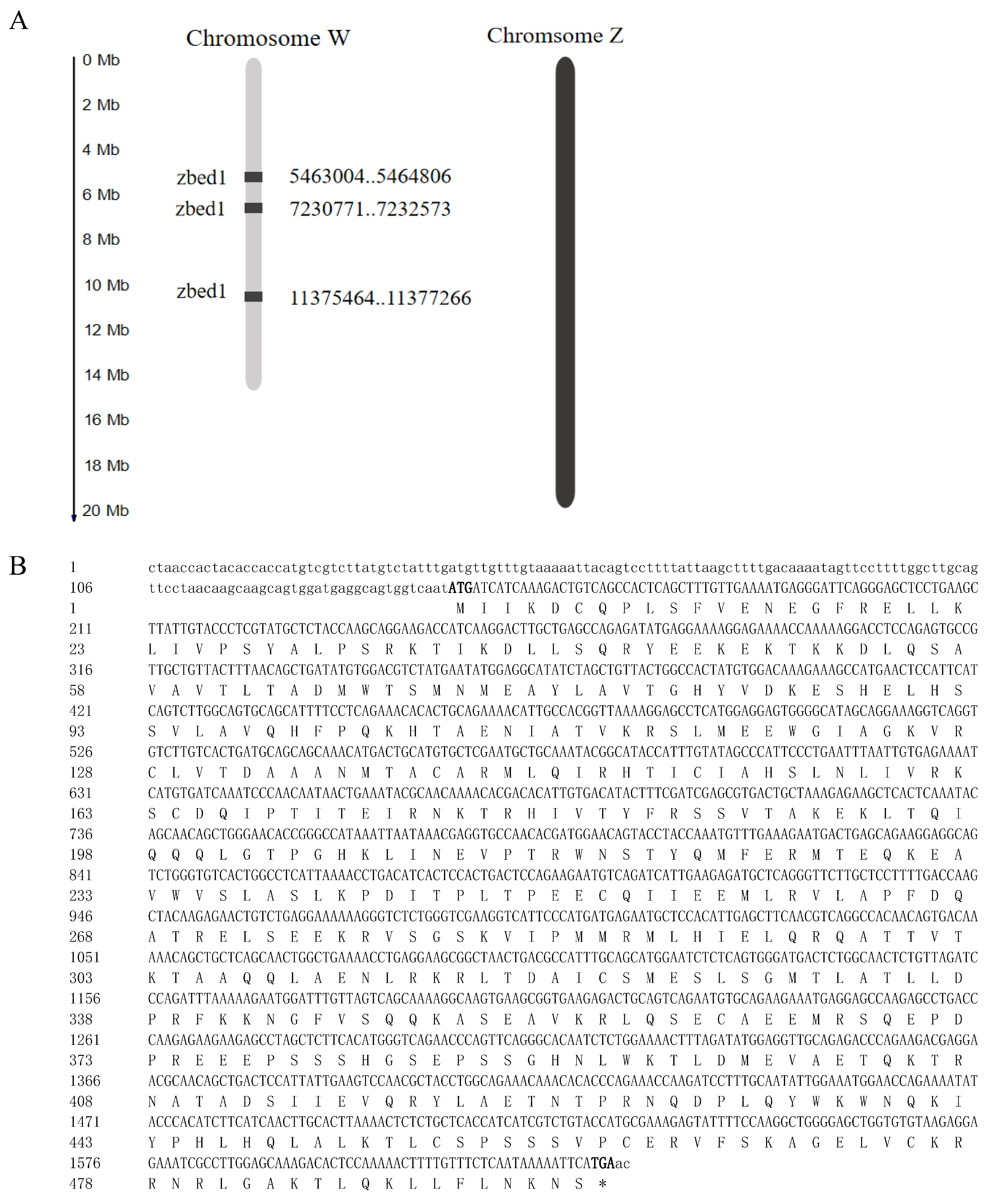
3.1. Multiple Copies of zbed1 on W Chromosome and Phylogenetic Tree of ZBED Family

By screening domains ZF_BED and Dimer_Tnp_hAT among the C. semilaevis genome, three copies of the zbed1 gene were identified on chromosome W (Figure 1). Briefly, three copies of the zbed1 gene were located on the W chromosome of C. semilaevis: Gene ID 103397026 at W_5463004-5464806; Gene ID 107990198 at W_7230771-7232573; and Gene ID 103397195 at W_11375464-11377266. PCR cloning and sequencing confirmed that the length for each zbed1 gene was 1802 bp, with three exons and two introns. In addition, the cDNA for each zbed1 gene was 1491 bp, encoding 496 amino acids.
To verify whether they were three copies of zbed1 in the C. semilaevis and whether there were gametologs on the chromosome Z, taqman primers and probes were designed based on zbed1 and myh6, a single-copy gene located on the chromosome 7 of C. semilaevis. Firstly, PCR experiment by taqman primers revealed that only female-specific target bands were amplified for zbed1, while for the autosome gene myh6, female and male target bands were both observed from the genomic DNA template (Figure 2A). Furthermore, digital PCR was employed for the confirmation of the gene copy number of zbed1 in the female individuals. The obvious distinction was detected between the number of positive droplets and negative droplets (no template control, NTC) generated by the digital PCR system in the same detection channel, demonstrating that the system exhibited consistent droplet generation and reliable repeatability (Figure 2B). The results showed that the copy number of the zbed1 gene was three times or more that of myh6 in three female individuals (Figure 2C).
As expected, the phylogenetic tree of ZBED homologues from different species displayed eleven sub-branches (Figure 3A). Then, the sub-branches of Ac (ZBED1, ZBED4, ZBED6cl, ZBED6, ZBEDX, ZBED2, and ZBED3) and Buster (ZBED7, ZBED8, ZBED5, and ZBED9) transposons were clustered into two big branches. In addition to ZBED1, ZBED4 and ZBEDX, which were widely found in fish, we also retrieved several fish ZBED5-like sequences from the NCBI database, and the phylogenetic results revealed that these sequences gathered with mammalian ZBED9 proteins, which suggested that they might be fish ZBED9. Similarly, three ZBED4-like of C. semilaevis were grouped into the ZBEDX sub-branch, indicating that they may be correctly called ZBEDX. Thus, only four ZBED1, one ZBED4, and four ZBEDX were identified in C. semilaevis, of which the three copies of ZBED1 were located on chromosome W and ZBED1-like (ZBED1l) was on chromosome 10 (Figure 3A, Table 2). In each sub-branch of ZBED1, ZBED4, and ZBEDX, the sequences of C. semilaevis and other fishes clustered together, followed by clustering with mammalian proteins.
The expression heatmap (Figure 3B) based on our previous two-year-old transcriptomic data [23] revealed that zbed1 was predominantly expressed in the female brain and pituitary. Zbed1l was highly expressed in the pituitary gland and male gonad. The expression of zbed4 mainly focused on the male pituitary gland, brain, and gonad. As for zbedx, a high expression level was detected in the gonad and liver.

3.2. Interaction Network Analysis of ZBEDs

For a better understanding of ZBED’s biological activity and complex regulatory network, a protein–protein interaction (PPI) network was constructed (Figure 4). This network predicted the interactions among 11 members of the ZBED family (ZBED1-9, ZBEDX, and ZBED6CL) and 25 other proteins, including IGF2 and its associated members (IGF1R, IGF2R, IGFBP1-3, IGFBP5-6), several zinc finger proteins including zinc finger protein 396 (ZNF396) and ZNF444, dehydrogenase/reductase X-linked (DHRSX), Karyopherin subunit alpha 1 (KPNA1), Small ubiquitin-related modifier 3-like (SUMO3A), and Acetylserotonin O-methyltransferase-like (ASMTL). Importantly, ZBED1 exhibited a complex interaction with other ZBED members. However, ZBEDX did not interact with other family members.

3.3. The Expression of zbed1 in Different Tissues and at Different Developmental Stages

The qPCR results indicated that zbed1 was highly expressed at the blastocyst and gastrula periods of embryonic development (Figure 5A). Furthermore, the zbed1 gene exhibited widespread expression in different tissues of female fish, with particularly high levels in the pituitary and brain (Figure 5B). The expression pattern of zbed1 in the female brain at different developmental stages showed that the highest level was detected at 2 Y (Figure 5C).

3.4. Knock-Down Effects on zbed1 and Other Related Genes by RNAi Transfection in Brain Cells

Female C. semilaevis brain cells were used for RNAi experiments to investigate the potential knock-down impact of zbed1. Within the exon region, one siRNA was designed. A high transfection efficiency (>90%) of RNAi-cy3 was observed (Figure 6A). qPCR analysis was performed to evaluate the expression levels of zbed1 and its related genes, such as piwi-like RNA-mediated gene silencing 1 (piwil1), estrogen receptor 2 (esr2), wingless-type MMTV integration site family, member 7B (wnt7b), TATA box binding protein (tbp), cyclin-dependent kinase 2 (cdk2), cyclin-dependent kinase 4 (cdk4), cyclin-dependent kinase 6 (cdk6), cyclin G1 (ccng1), and cyclin Dx (ccndx). In female brain cells, siRNA1 exhibited a significant knock-down effect (Figure 6B). Following this, piwil1, esr2 and wnt7b demonstrated up-regulation, while tbp, cdk2, cdk4, cdk6, ccng1 and ccndx displayed down-regulation (Figure 6C).

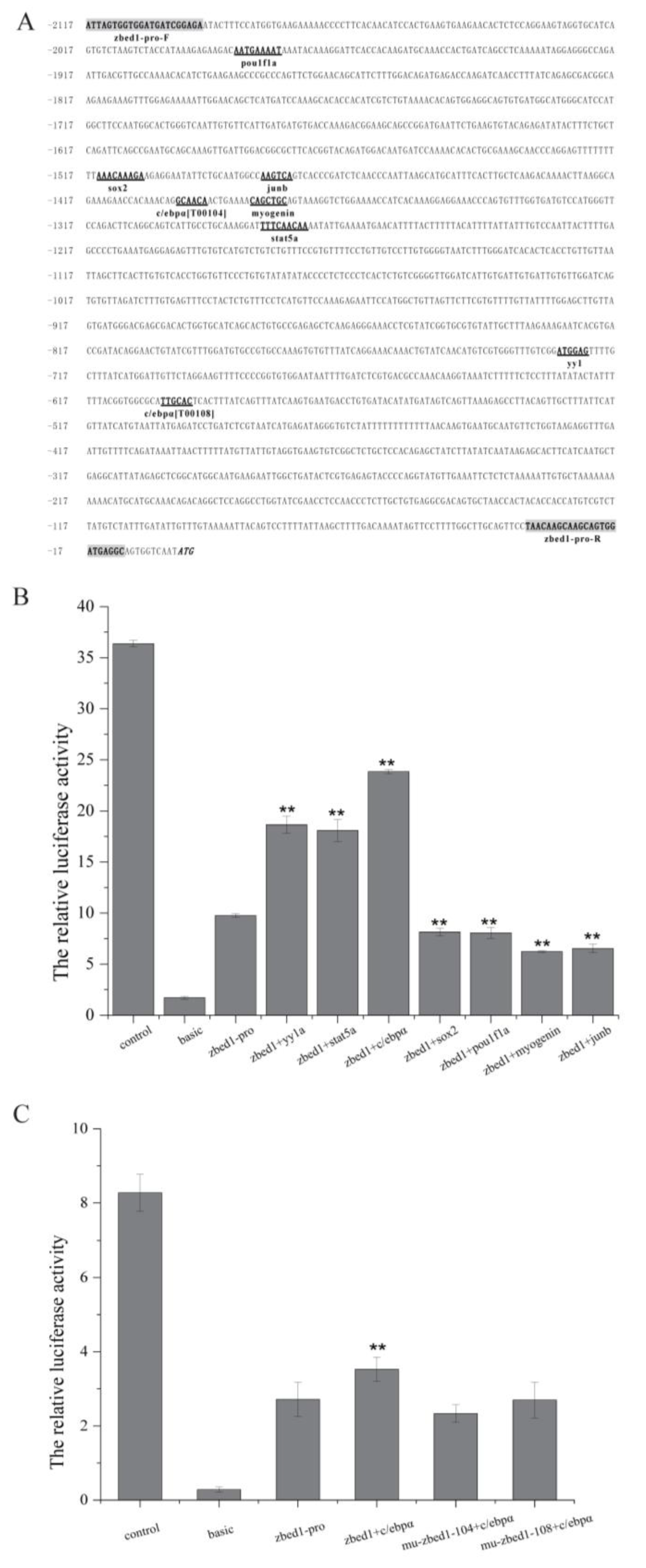
3.5. Site-Directed Mutagenesis of zbed1 Promoter Activity and Transcription Factor Sites

A promoter region of zbed1 was cloned at the upstream 11–2117 bp of the start codon and named zbed1-pro. In PROMO and JASPAR analysis, several transcription-factor-binding sites were predicted, including Yin Yang 1A (yy1a), signal transducer and activator of transcription 5A (stat5a), CCAAT/enhancer-binding protein Alpha (c/ebpα), SRY-related high-mobility-group box protein 2 (sox2), and POU domain, class 1, transcription factor 1 (pou1f1), activator protein 1 (AP-1) family members, include two major types of protein transcription factors (junb and fos), as well as myogenin in the MyoD family (Figure 7A).
The dual-luciferase assay demonstrated that the zbed1 promoter was highly active in promoting firefly luciferase transcription (Figure 7B). The subsequent co-transfection assay revealed that the zbed1 promoter activity exhibited significant up-regulation upon binding with the transcription factors yy1a and c/ebpα, whereas a significant decrease in promoter activity was observed upon binding with sox2, pou1f1a, myogenin, and junb.
Given the crucial involvement of c/ebpα in growth and development, an additional investigation was conducted to ascertain the influence of c/ebpα transcription factor on the zbed1 promoter. It involved the introduction of mutation for the two c/ebpα sites on the promoter, followed by co-transfection with c/ebpα plasmid. The results revealed that no up-regulation effect was observed once the two binding sites were mutated (Figure 7C).

4. Discussion

More than 600 fish species exhibit sexual size dimorphism (SSD) [22]. This phenomenon often leads to serious male or female growth disadvantages, resulting in high breeding costs and limiting the feasibility of the fish-breeding industry. To investigate the related mechanism mediating SSD will be meaningful for the molecular breeding of fish with high-quality growth traits.
The identification of zbed1 is particularly interesting because it is a homologue in Drosophila (DREF) and human (hDREF/zbed1) [39]. Through our research, we found that zbed1 had three identical copies on the W chromosome of C. semilaevis, and no homologous gene was found on the Z chromosome. There are two important domains in zbed1, namely the amino terminal BED zinc finger domain and the carboxyl terminal hATC domain, which provides important structural support for the zbed1 molecule to distribute in the nucleus with a granular structure [40].
Unlike the eleven ZBED members identified in the mammals, only three members (ZBED1, ZBED4 and ZBEDX) have been identified in the vast majority of fish. The present study revealed the existence a new ZBED member, ZBED9, in this fish species, which broadened the current knowledge regarding the fish ZBED family [33]. More numbers or more functions for duplicate ZBED members would be found with increasing research on fish genomics.
Consistent with our previous transcriptomic data [24], the qPCR results revealed that zbed1 was significantly expressed in the female brain and pituitary. Differently, human zbed1 was widely expressed in all tissues from three primary germ layers, with a high expression in the digestive and reproductive systems [41]. Therefore, we speculated that the regulation of the W chromosome gene zbed1 on female-biased SSD in C. semilaevis may be realized by its high specific expression in female brain neurons and pituitary hormone cells, similar to the sexual dimorphic singing behavior of birds [14] and female-biased SSD of drosphila [16].
It was also found that C. semilaevis zbed1 was significantly highly expressed in the blastocyst period and gastrula period and reached its peak in the gastrula period. In zebrafish, the early development of the neural tube begins from the blastocyst period [42,43], and the formation of neuroectoderm and neural plate in the gastrula period lays the foundation for the neural tube [44,45]. Thus, it was suggested that zbed1 was involved in the early development of the nervous system of C. semilaevis.
The subsequent analysis of transcriptional regulation suggested that c/ebpα may serve as a crucial transcription factor in activating the transcriptional activity of zbed1. As a conserved transcription factor involved in cellular growth and differentiation [46], c/ebpα plays a role in the early differentiation of gonads and acts as a suppressor of the male determinant gene dmrt1 in C. semilaevis [47]. This implies that c/ebpα has an opposing effect on dmrt1 and zbed1, leading us to propose that zbed1 may be a significant gene associated with the regulation of female behavior.
As for the knockdown effect of zbed1 on cell proliferation and growth performance, important genes derived from the cell cycle, Wnt pathway and other pathways according to our previous DAP-seq analysis [24] were determined to be regulated by zbed1 RNAi in the brain cell line. Wnt7b, one important factor of the Wnt pathway, was upregulated after the knockdown of zbed1. A Wnt transduction abnormality will cause nervous system developmental defects and lead to many neurological diseases [48,49,50,51]. It is suggested that zbed1 may regulate the development of the nervous system of C. semilaevis by regulating the expression of wnt7b. In addition, as the main receptor for estrogen, the expression of esr2 was also increased after the knockdown of zbed1 in the brain cells. In mammals, esr2 may inhibit granulosa cell proliferation by mediating the effect on cyclin-dependent kinase inhibitor 1a (cnkn1a) [52]. It was hypothesized that zbed1 may exert control over cell proliferation by reducing the expression of esr2.
Notably, the knockdown of zbed1 resulted in the decrease in several genes involved in the cell cycle, including cdk2, cdk4, cdk6, ccng1, and ccndx. The activation of the cell cycle pathway has been proposed as a significant factor contributing to the SSD in C. semilaevis [53]. Our findings suggest that zbed1, an upstream transcription factor located on the W chromosome, may regulate the activation of the cell cycle.

5. Conclusions

In this study, we identified three copies of zbed1 on the W chromosome of C. semilaevis, which displayed a female-biased expression pattern in the brain and pituitary. The experiments demonstrated that the transcription factor c/ebpα can activate the transcriptional activity of the zbed1 promoter. It is also suggested that zbed1 may play a vital function in cell proliferation by regulating esr2, piwil1, the cell cycle, and the Wnt pathway. Further investigations into the interaction among esr2, the Wnt pathway, and zbed1 will contribute to a better understanding of the mechanism underlying sexual size dimorphism in C. semilaevis.

Author Contributions

The experiments were conceived and designed by N.W. Fish samples were collected by Y.S. and J.W. qPCR analysis was performed by Y.S. and Q.Z. Phylogenetic tree construction was carried out by X.L. and Y.S. The dPCR analysis to validate the presence of multi-copy genes was conducted by N.W. and Y.S. PPI network analysis was conducted by N.W. and Y.S. Promoter activity analysis and site-directed mutagenesis of transcription factors were performed by Y.S. and J.M. The RNAi knock-down experiment was carried out by Y.S. and J.M. This paper was written, revised and discussed by Y.S., X.L., W.X. and N.W. All authors have read and agreed to the published version of the manuscript.

Funding

This work was supported by the National Key Research and Development Program of China (2022YFF1000303), Key Research and Development Project of Shandong Province (2021LZGC028, 2023ZLYS02), Taishan Young Scholar Project of Shandong Province (tsqn202211266), and the Central Public-Interest Scientific Institution Basal Research Fund, CAFS (2023TD20).

Institutional Review Board Statement

We followed the guidelines for the Care and Use of Laboratory Animals at the Yellow Sea Fisheries Research Institute, Chinese Academy of Fishery Sciences, during all experimental procedures.

Informed Consent Statement

Not applicable.

Data Availability Statement

Data are contained within the article.

Conflicts of Interest

The authors declare no conflicts of interest.

References

  1. Cox, R.M.; Butler, M.A.; John-Alder, H.B. The evolution of sexual size dimorphism in reptiles. In Sex, Size and Gender Roles: Evolutionary Studies of Sexual Size Dimorphism; Fairbairn, D.J., Blanckenhorn, W.U., Székely, T., Eds.; Oxford University Press: Oxford, UK, 2007; pp. 38–49. [Google Scholar]
  2. Lindenfors, P.; Gittleman, J.L.; Jones, K.E. Sexual size dimorphism in mammals. In Sex, Size and Gender Roles: Evolutionary Studies of Sexual Size Dimorphism; Fairbairn, D.J., Blanckenhorn, W.U., Székely, T., Eds.; Oxford University Press: Oxford, UK, 2007; pp. 16–26. [Google Scholar]
  3. Székely, T.; Lislevand, T.; Figuerola, J. Sexual size dimorphism in birds. In Sex, Size and Gender Roles: Evolutionary Studies of Sexual Size Dimorphism; Fairbairn, D.J., Blanckenhorn, W.U., Székely, T., Eds.; Oxford University Press: Oxford, UK, 2007; pp. 27–37. [Google Scholar]
  4. Badyaev, A.V. Growing apart: An ontogenetic perspective on the evolution of sexual size dimorphism. Trends Ecol. Evol. 2002, 17, 369–378. [Google Scholar] [CrossRef]
  5. Zhang, M.; Yang, Q.; Shi, R.; Wang, J.; Zhang, Z.; Yang, Y.; Li, W.; Chen, S.; Wang, N. Effects of long-term sex steroid hormones (estradiol and testosterone)-supplemented feeds on the growth performance of Chinese tongue sole (Cynoglossus semilaevis). Fish Physiol. Biochem. 2022, 48, 1365–1375. [Google Scholar] [CrossRef] [PubMed]
  6. Cox, R.M.; Stenquist, D.S.; Calsbeek, R. Testosterone, growth and the evolution of sexual size dimorphism. J. Evol. Biol. 2009, 22, 1586–1598. [Google Scholar] [CrossRef] [PubMed]
  7. Millington, J.W.; Brownrigg, G.P.; Chao, C.; Sun, Z.; Basner-Collins, P.J.; Wat, L.W.; Hudry, B.; Miguel-Aliaga, I.; Rideout, E.J. Female-biased upregulation of insulin pathway activity mediates the sex difference in Drosophila body size plasticity. eLife 2021, 10, e58341. [Google Scholar] [CrossRef] [PubMed]
  8. Rice, W.R. Sex chromosomes and the evolution of sexual dimorphism. Evolution 1984, 38, 735–742. [Google Scholar] [CrossRef]
  9. Chen, X.; McClusky, R.; Chen, J.; Beaven, S.W.; Tontonoz, P.; Arnold, A.P.; Reue, K. The number of x chromosomes causes sex differences in adiposity in mice. PLoS Genet. 2012, 8, e1002709. [Google Scholar] [CrossRef] [PubMed]
  10. Chase, K.; Carrier, D.R.; Adler, F.R.; Ostrander, E.A.; Lark, K.G. Interaction between the X chromosome and an autosome regulates size sexual dimorphism in Portuguese Water Dogs. Genome Res. 2005, 15, 1820–1824. [Google Scholar] [CrossRef]
  11. Singh Alvarado, J.; Goffinet, J.; Michael, V.; Liberti, W., 3rd; Hatfield, J.; Gardner, T.; Pearson, J.; Mooney, R. Neural dynamics underlying birdsong practice and performance. Nature 2021, 599, 635–639. [Google Scholar] [CrossRef]
  12. Nottebohm, F.; Arnold, A.P. Sexual dimorphism in vocal control areas of the songbird brain. Science 1976, 194, 211–213. [Google Scholar] [CrossRef]
  13. Agate, R.J.; Grisham, W.; Wade, J.; Mann, S.; Wingfield, J.; Schanen, C.; Palotie, A.; Arnold, A.P. Neural, not gonadal, origin of brain sex differences in a gynandromorphic finch. Proc. Natl. Acad. Sci. USA 2003, 100, 4873–4878. [Google Scholar] [CrossRef]
  14. Zhao, Y.; Zhang, X.; Wang, R.; Bing, J.; Wu, F.; Zhang, Y.; Xu, J.; Han, Z.; Zhang, X.; Zeng, S. Erbin and ErbB2 play roles in the sexual differentiation of the song system nucleus HVC in bengalese finches (Lonchura Striata var. domestica). Dev. Neurobiol. 2018, 78, 15–38. [Google Scholar] [CrossRef]
  15. Testa, N.D.; Ghosh, S.M.; Shingleton, A.W. Sex-specific weight loss mediates sexual size dimorphism in Drosophila melanogaster. PLoS ONE 2013, 8, e58936. [Google Scholar] [CrossRef]
  16. Sawala, A.; Gould, A.P. The sex of specific neurons controls female body growth in Drosophila. PLoS Biol. 2017, 15, e2002252. [Google Scholar] [CrossRef]
  17. Sawala, A.; Gould, A.P. Sex-lethal in neurons controls female body growth in Drosophila. Fly 2018, 12, 133–141. [Google Scholar] [CrossRef]
  18. Kaufmann, P.; Wiberg, R.A.W.; Papachristos, K.; Scofield, D.G.; Tellgren-Roth, C.; Immonen, E. Y-Linked Copy Number Polymorphism of Target of Rapamycin Is Associated with Sexual Size Dimorphism in Seed Beetles. Mol. Biol. Evol. 2023, 40, msad167. [Google Scholar] [CrossRef]
  19. Mank, J.E.; Hall, D.W.; Kirkpatrick, M.; Avise, J.C. Sex chromosomes and male ornaments: A comparative evaluation in ray-finned fishes. Proc. Biol. Sci. 2006, 273, 233–236. [Google Scholar] [CrossRef] [PubMed]
  20. Poissant, J.; Davis, C.S.; Malenfant, R.M.; Hogg, J.T.; Coltman, D.W. QTL mapping for sexually dimorphic fitness-related traits in wild bighorn sheep. Heredity 2012, 108, 256–263. [Google Scholar] [CrossRef] [PubMed]
  21. Shao, C.; Li, Q.; Chen, S.; Zhang, P.; Lian, J.; Hu, Q.; Sun, B.; Jin, L.; Liu, S.; Wang, Z.; et al. Epigenetic modification and inheritance in sexual reversal of fish. Genome Res. 2014, 24, 604–615. [Google Scholar] [CrossRef] [PubMed]
  22. Horne, C.R.; Hirst, A.G.; Atkinson, D. Selection for increased male size predicts variation in sexual size dimorphism among fish species. Proc. Biol. Sci. 2020, 287, 20192640. [Google Scholar] [CrossRef]
  23. Wang, N.; Wang, R.; Wang, R.; Chen, S. Transcriptomics analysis revealing candidate networks and genes for the body size sexual dimorphism of Chinese tongue sole (Cynoglossus semilaevis). Funct. Integr. Genom. 2018, 18, 327–339. [Google Scholar] [CrossRef]
  24. Wang, N.; Gao, J.; Liu, Y.; Shi, R.; Chen, S. Identification of crucial factors involved in Cynoglossus semilaevis sexual size dimorphism by GWAS and demonstration of zbed1 regulatory network by DAP-seq. Genomics 2022, 114, 110376. [Google Scholar] [CrossRef]
  25. Ohshima, N.; Takahashi, M.; Hirose, F. Identification of a human homologue of the DREF transcription factor with a potential role in regulation of the histone H1 gene. J. Biol. Chem. 2003, 278, 22928–22938. [Google Scholar] [CrossRef]
  26. Yamashita, D.; Sano, Y.; Adachi, Y.; Okamoto, Y.; Osada, H.; Takahashi, T.; Yamaguchi, T.; Osumi, T.; Hirose, F. hDREF regulates cell proliferation and expression of ribosomal protein genes. Mol. Cell. Biol. 2007, 27, 2003–2013. [Google Scholar] [CrossRef]
  27. Jin, Y.; Li, R.; Zhang, Z.; Ren, J.; Song, X.; Zhang, G. ZBED1/DREF: A transcription factor that regulates cell proliferation. Oncol. Lett. 2020, 20, 137. [Google Scholar] [CrossRef]
  28. Somerville, T.D.D.; Xu, Y.; Wu, X.S.; Maia-Silva, D.; Hur, S.K.; de Almeida, L.M.N.; Preall, J.B.; Koo, P.K.; Vakoc, C.R. ZBED2 is an antagonist of interferon regulatory factor 1 and modifies cell identity in pancreatic cancer. Proc. Natl. Acad. Sci. USA 2020, 117, 11471–11482. [Google Scholar] [CrossRef]
  29. Saghizadeh, M.; Akhmedov, N.B.; Yamashita, C.K.; Gribanova, Y.; Theendakara, V.; Mendoza, E.; Nelson, S.F.; Ljubimov, A.V.; Farber, D.B. ZBED4, a BED-type zinc-finger protein in the cones of the human retina. Investig. Ophthalmol. Vis. Sci. 2009, 50, 3580–3588. [Google Scholar] [CrossRef] [PubMed]
  30. Markljung, E.; Jiang, L.; Jaffe, J.D.; Mikkelsen, T.S.; Wallerman, O.; Larhammar, M.; Zhang, X.; Wang, L.; Saenz-Vash, V.; Gnirke, A.; et al. ZBED6, a novel transcription factor derived from a domesticated DNA transposon regulates IGF2 expression and muscle growth. PLoS Biol. 2009, 7, e1000256. [Google Scholar] [CrossRef] [PubMed]
  31. Ruan, X.; Liu, G.; Zhou, J.; Chen, P.; Sun, C.; Liu, W.; Wu, C.; Hou, L.; Yin, B.; Qiang, B.; et al. Zbed3 Is Indispensable for Wnt Signaling Regulation of Cortical Layers Formation in Developing Brain. Cereb. Cortex 2021, 31, 4078–4091. [Google Scholar] [CrossRef] [PubMed]
  32. Jia, Y.; Yuan, L.; Hu, W.; Luo, Y.; Suo, L.; Yang, M.; Chen, S.; Wang, Y.; Liu, H.; Yang, G.; et al. Zinc-finger BED domain-containing 3 (Zbed3) is a novel secreted protein associated with insulin resistance in humans. J. Intern. Med. 2014, 275, 522–533. [Google Scholar] [CrossRef] [PubMed]
  33. Zeng, C.W.; Sheu, J.C.; Tsai, H.J. Genomic Structure, Protein Character, Phylogenic Implication, and Embryonic Expression Pattern of a Zebrafish New Member of Zinc Finger BED-Type Gene Family. Genes 2023, 14, 179. [Google Scholar] [CrossRef] [PubMed]
  34. Jaillon, O.; Aury, J.M.; Brunet, F.; Petit, J.L.; Stange-Thomann, N.; Mauceli, E.; Bouneau, L.; Fischer, C.; Ozouf-Costaz, C.; Bernot, A.; et al. Genome duplication in the teleost fish Tetraodon nigroviridis reveals the early vertebrate proto-karyotype. Nature 2004, 431, 946–957. [Google Scholar] [CrossRef]
  35. Sun, Y.; Zhang, M.; Cheng, P.; Gong, Z.; Li, X.; Wang, N.; Wei, M.; Xu, X.; Xu, W. pitpβ_w Encoding Phosphatidylinositol Transfer Protein Is Involved in Female Differentiation of Chinese Tongue Sole, Cynoglossus semilaevis. Front. Genet. 2022, 13, 861763. [Google Scholar] [CrossRef]
  36. Cheng, P.; Chen, Z.; Xu, W.; Wang, N.; Yang, Q.; Shi, R.; Li, X.; Cui, Z.; Cheng, J.; Chen, S. Potential Involvement of ewsr1-w Gene in Ovarian Development of Chinese Tongue Sole, Cynoglossus semilaevis. Animals 2022, 12, 2503. [Google Scholar] [CrossRef] [PubMed]
  37. Yang, L.; Songlin, C.; Fengtao, G.; Liang, M.M.; Qiaomu, H.; Wentao, S.; Changwei, S.; WeiQun, L. SCAR-transformation of sex-specific SSR marker and its application in half-smooth tongue sole (Cynoglossus semiliaevis). J. Agric. Biotechnol. 2014, 22, 787–792. [Google Scholar]
  38. Li, C.; Ortí, G.; Zhang, G.; Lu, G. A practical approach to phylogenomics: The phylogeny of ray-finned fish (Actinopterygii) as a case study. BMC Evol. Biol. 2007, 7, 44. [Google Scholar] [CrossRef] [PubMed]
  39. Yamashita, D.; Moriuchi, T.; Osumi, T.; Hirose, F. Transcription Factor hDREF Is a Novel SUMO E3 Ligase of Mi2α. J. Biol. Chem. 2016, 291, 11619–11634. [Google Scholar] [CrossRef]
  40. Yamashita, D.; Komori, H.; Higuchi, Y.; Yamaguchi, T.; Osumi, T.; Hirose, F. Human DNA replication-related element binding factor (hDREF) self-association via hATC domain is necessary for its nuclear accumulation and DNA binding. J. Biol. Chem. 2007, 282, 7563–7575. [Google Scholar] [CrossRef]
  41. Hansen, S.V.; Traynor, S.; Ditzel, H.J.; Gjerstorff, M.F. Human DREF/ZBED1 is a nuclear protein widely expressed in multiple cell types derived from all three primary germ layers. PLoS ONE 2018, 13, e0205461. [Google Scholar] [CrossRef] [PubMed]
  42. Kimmel, C.B.; Law, R.D. Cell lineage of zebrafish blastomeres: III. Clonal analyses of the blastula and gastrula stages. Dev. Biol. 1985, 108, 94–101. [Google Scholar] [CrossRef]
  43. Kimmel, C.B.; Law, R.D. Cell lineage of zebrafish blastomeres: II. Formation of the yolk syncytial layer. Dev. Biol. 1985, 108, 86–93. [Google Scholar] [CrossRef]
  44. Woo, K.; Shih, J.; Fraser, S.E. Fate maps of the zebrafish embryo. Curr. Opin. Genet. Dev. 1995, 5, 439–443. [Google Scholar] [CrossRef]
  45. Strähle, U.; Blader, P. Early neurogenesis in the zebrafish embryo. FASEB J. 1994, 8, 692–698. [Google Scholar] [CrossRef]
  46. Ren, W.; Guo, J.; Jiang, F.; Lu, J.; Ding, Y.; Li, A.; Liang, X.; Jia, W. CCAAT/enhancer-binding protein α is a crucial regulator of human fat mass and obesity associated gene transcription and expression. Biomed Res. Int. 2014, 2014, 406909. [Google Scholar] [CrossRef]
  47. Wang, Q.; Wang, R.; Feng, B.; Li, S.; Mahboob, S.; Shao, C. Cloning and functional analysis of c/ebpα as negative regulator of dmrt1 in Chinese tongue sole (Cynoglossus semilaevis). Gene 2021, 768, 145321. [Google Scholar] [CrossRef]
  48. Lambert, C.; Cisternas, P.; Inestrosa, N.C. Role of Wnt Signaling in Central Nervous System Injury. Mol. Neurobiol. 2016, 53, 2297–2311. [Google Scholar] [CrossRef]
  49. Huang, C.; Fu, X.H.; Zhou, D.; Li, J.M. The Role of Wnt/β-Catenin Signaling Pathway in Disrupted Hippocampal Neurogenesis of Temporal Lobe Epilepsy: A Potential Therapeutic Target? Neurochem. Res. 2015, 40, 1319–1332. [Google Scholar] [CrossRef]
  50. Arredondo, S.B.; Valenzuela-Bezanilla, D.; Mardones, M.D.; Varela-Nallar, L. Role of Wnt Signaling in Adult Hippocampal Neurogenesis in Health and Disease. Front. Cell Dev. Biol. 2020, 8, 860. [Google Scholar] [CrossRef] [PubMed]
  51. Sun, X.; Peng, X.; Cao, Y.; Zhou, Y.; Sun, Y. ADNP promotes neural differentiation by modulating Wnt/β-catenin signaling. Nat. Commun. 2020, 11, 2984. [Google Scholar] [CrossRef] [PubMed]
  52. Wang, Y.; Liu, W.; Du, J.; Yu, Y.; Liang, N.; Liang, M.; Yao, G.; Cui, S.; Huang, H.; Sun, F. NGF promotes mouse granulosa cell proliferation by inhibiting ESR2 mediated down-regulation of CDKN1A. Mol. Cell. Endocrinol. 2015, 406, 68–77. [Google Scholar] [CrossRef] [PubMed]
  53. Wang, N.; Liu, Y.; Wang, R.; Chen, S. The linkage of cell cycle and DNA replication with growth difference in female Chinese tongue sole (Cynoglossus semilaevis): Analysis from transcriptomic study and WGCNA. Comp. Biochem. Physiol. Part D Genom. Proteom. 2021, 39, 100833. [Google Scholar] [CrossRef]
Figure 1. The genomic location and sequences of Cynoglossus semilaevis zbed1 gene. (A) Location of zbed1 gene on the sex chromosome of C. semilaevis. There were three copies of zbed1 at different locations on the W chromosome and none on the Z chromosome. (B) Sequence information for C. semilaevis zbed1 cDNA and protein. The start and stop codons are shown in bold.
Biology 13 00141 g001
Figure 2. The specificity of zbed1 in the females and the confirmation of its copy number. (A) PCR experiment result for the detection of zbed1 and myh6 from genomic DNA. F1-3 and M1-3 indicated three female and male individuals, respectively. M was the maker. (B) Droplet distribution map. Zbed1, myh6 and NTC droplets were clearly distinguished under the detection channel. The blue line showed the fluorescence threshold. (C) The copy numbers of zbed1 and the single-copy gene myh6 in three female individuals.
Biology 13 00141 g002
Figure 3. Phylogenetic tree and expression heatmap of ZBED family genes. (A) Phylogenetic tree of ZBEDs from Cynoglossus semilaevis (Cs), Homo sapiens (Hs), Mus musculus (Mm), Danio rerio (Dr), Oryzias latipes (Ol), Oreochromis niloticus (On), Epinephelus lanceolatus (El), Scophthalmus maximus (Sm), Xenopus tropicalis (Xt), Bos taurus (Bt), and Pan troglodytes (Pt). The percentage of replicate trees in which associated taxa clustered together in the bootstrap test (1000 replicates) is shown next to the branches. Eleven sub-clusters were indicated in different colors. Then, the sub-branches of Ac (ZBED1, ZBED4, ZBED6cl, ZBED6, ZBEDX, ZBED2, and ZBED3) and Buster (ZBED7, ZBED8, ZBED5, and ZBED9) transposons were clustered into two big branches. In each sub-branch of ZBED1, ZBED4, and ZBEDX, the sequences of C. semilaevis and other fishes clustered together, followed by clustering with mammalian proteins. CsZBEDs were marked in red stars. (B) Heatmap of zbed mRNA abundances in different tissues of C. semilaevis female and male individuals. FB: female brain, MB: male brain, FP: female pituitary, MP: male pituitary, FG: female gonad, MG: male gonad, FL: female liver, ML: male liver.
Biology 13 00141 g003
Figure 4. Protein interaction network among ZBED’s family members. Nodes indicate the interactive proteins; edges indicate both functional and physical protein associations; and different colors indicate the various types of interaction evidence.
Biology 13 00141 g004
Figure 5. Spatiotemporal expression patterns of zbed1 gene in C. semilaevis. The letters a–h represent significance. There are significant differences between columns with different letters in each picture. (A) Relative mRNA expression patterns of zbed1 in C. semilaevis embryos at five periods (cleavage, blastocyst, gastrula, segmentation, pharyngula and early larval). (B) Relative mRNA expression patterns of zbed1 in 11 tissues (brain, pituitary, gill, gonad, heart, intestine, kidney, liver, muscle, skin, and spleen) of female C. semilaevis. (C) Relative mRNA expression patterns of zbed1 in the brain of female C. semilaevis at six developmental stages including three-month-old (3 M), five-month-old (5 M), eight-month-old (8 M), one-year-old (1 Y), 1.5-year-old (1.5 Y), and two-year-old fish (2 Y). The data were analyzed with SPSS 25.0 (IBM Corp, Armonk, NY, USA) using one-way ANOVA and multiple comparison by Wohler and Duncan methods, and p-value < 0.05 was considered the threshold for statistical significance.
Biology 13 00141 g005
Figure 6. The knockdown effect of zbed1 on the female C. semilaevis brain cells. (A) RNAi transfection efficiency in brain cells. (B) Interference efficiency of zbed1 siRNA. (C) The expression patterns of genes in female brain cells after transfection with zbed1 siRNA. The data in (B,C) were analyzed with SPSS 25.0 (IBM Corp, Armonk, NY, USA) using t-test. The data of each downstream gene were compared with NC and p-value < 0.05 was considered the threshold for statistical significance (double asterisk, p < 0.01).
Biology 13 00141 g006
Figure 7. Structure, activity, and transcription factor analysis of C. semilaevis zbed1 promoter. (A) Sequence structure and putative transcription-factor-binding sites (i.e., pou1f1a, sox2, junB, c/ebpα, myogenin, yy1, and stat5a) on zbed1 promoter. Two primers of the promoter are shown in shadow. (B) Transcription activity of zbed1 promoter and co-transfection with transcription factors in HEK 293T cells. (C) The luciferase activity after co-transfection with the transcription factor c/ebpα and mutated zbed1 promoter. The significance is indicated by asterisks. The data in (B,C) were analyzed with SPSS 25.0 (IBM Corp, Armonk, NY, USA) using t-test. The data of each co-transfection were compared with the original promoter and p-value < 0.05 was considered the threshold for statistical significance (double asterisk, p < 0.01).
Biology 13 00141 g007
Table 1. The primers used in present study.
PrimersInformationSequences (5′–3′)
Cs-SEX-Fsex detectionCCTAAATGATGGATGTAGATTCTGTC
Cs-SEX-Rsex detectionGATCCAGAGAAAATAAACCCAGG
zbed1-cds-FCDS cloningATGATCATCAAAGACTGTCAGCC
zbed1-cds-RCDS cloningTCATGAATTTTTATTGAGAAACA
zbed1-pro-Fpromoter cloningAGATCTGCGATCTAAGTAAGCTATTAGTGGTGGATGATCGGAGA
zbed1-pro-Rpromoter cloningCAACAGTACCGGAATGCCAAGCTGCCTCATCCACTGCTTGCTTGTTA
zbed1-mu-c/ebpα104-Fpromoter mutationAAAGAACCACAAACAGCGATCAACT
zbed1-mu-c/ebpα104-Rpromoter mutationAGTTGATCGCTGTTTGTGGTTCTTT
zbed1-mu-c/ebpα108-Fpromoter mutationTGGCGCATACCAGTCACTTTATCAG
zbed1-mu-c/ebpα108-Rpromoter mutationCTGATAAAGTGACTGGTATGCGCCA
zbed1- site1RNAi site1GCAACAGCTGACTCCATTA
RNAi-NCnegative control (nc)CTGAAGATCCGGCTCATCA
zbed1-qPCR-FqPCRCTCCAGAGTGCCGTTGC
zbed1-qPCR-RqPCRGTTCATGGCTTTCTTTGTCC
actin-FqPCRTTCCAGCCTTCCTTCCTT
actin-RqPCRTACCTCCAGACAGCACAG
esr2-qPCR-FqPCRGATTAGGAGAAGGTGGAGAAGG
esr2-qPCR-RqPCRGGTAACCAGAGGCATAGTCGTG
ccng1-qPCR-FqPCRAGTGACTACGCCAACACCAAAT
ccng1-qPCR-RqPCRGATGGTAGGCAGATGAGCGATT
ccndx-qPCR-FqPCRCCTTGTCCTTGCCTATCTC
ccndx-qPCR-RqPCRGACGCCTCAAAGTTGTTCT
piwill-qPCR-FqPCRCATCCAACTGTCGGCCAACTAT
piwill-qPCR-RqPCRTCGGCAATCTATTAGGCAGGAA
cdk4-qPCR-FqPCRCGCCAGTATGCAGTATGA
cdk4-qPCR-RqPCRTCTTGAGCAGAGCCACCT
cdk2-qPCR-FqPCRCACTGGTATCCCTCTGCC
cdk2-qPCR-RqPCRGAAGTCGGCGAGTTTGAT
cdk6-qPCR-FqPCRTACCACCCGAGACCATTA
cdk6-qPCR-RqPCRTAGATTCGAGCCAGACCA
tbp-qPCR-FqPCRAAACAGTAACAGGCTCCAC
tbp-qPCR-RqPCRTCCAGTTTACAGCCAAGAT
wnt7b-qPCR-FqPCRAGCAGCATTCACCTACGC
wnt7b-qPCR-RqPCRCTTCCAGCCTTCCTCTTG
zbed1-taqman-FTaqman primerCTCTGGCAACTCTGTTAGATCC
zbed1-taqman-RTaqman primerGCTCTTGGCTCCTCATTTCT
zbed1-taqman-probeTaqman probeAAAGGCAAGTGAAGCGGTGAAGAGAC 5′6-FAM, 3′BHQ1
myh6-taqman-FTaqman primerACAAGTGGCTTCCTGTCTATG
myh6-taqman-RTaqman primerGCGTTATCGGAGATGGAGAAA
myh6-taqman-probeTaqman probeTAAGAAGAGAAGCGAGGCTCCACCTC 5′6-FAM, 3′BHQ1
Table 2. The ZBED family members identified from C. semilaevis genome.
NameGene IDProtein IDGene Length
(bp)
ORF Length (bp) Amino Acid
Length (aa)
ChrLocationNo. of
Exons
No. of Introns
zbed1103397026XP_016898185.1 18021491496W5,463,004–5,464,80632
zbed1103397195XP_016898198.118021491496W11,375,464–11,377,26632
zbed1107990198XP_016898129.118021491496W7,230,771–7,232,57332
zbed1l103384517XP_016891160.219901537511102,017,183–2,0191,7221
zbed4103382917XP_024912923.1744237501249826,160,975–26,168,41632
zbedx103384547XP_024914808.171672163720102,351,855–2,359,02165
zbedx103397244XP_008333685.2524481161386W13,905,364–13,957,81121
zbedx112486373XP_024908564.113181089362W10,396,182–10,397,49921
zbedx103392902XP_024920030.1692141550499176,577,362–6,646,57576
Disclaimer/Publisher’s Note: The statements, opinions and data contained in all publications are solely those of the individual author(s) and contributor(s) and not of MDPI and/or the editor(s). MDPI and/or the editor(s) disclaim responsibility for any injury to people or property resulting from any ideas, methods, instructions or products referred to in the content.

