Commentary

Quantification of Thermal Acclimation in Immune Functions in Ectothermic Animals

by
Franziska C. Sandmeier
Biology Department, Colorado State University-Pueblo, Pueblo, CO 81001, USA
Biology 2024, 13(3), 179; https://doi.org/10.3390/biology13030179
Submission received: 18 January 2024 / Revised: 7 March 2024 / Accepted: 7 March 2024 / Published: 9 March 2024

Abstract

:

Simple Summary

This commentary focuses on experimental techniques used by researchers to understand how the immune system of an animal can acclimate, or change, to function across different body temperatures. I categorized studies as using three different, broad techniques: (1) allowing animal body temperatures to change naturally; (2) manipulating body temperatures of live animals in a controlled environment; and (3) manipulating temperatures of immune components (plasma, cells, etc.) in laboratory assays. I reviewed how the current literature used these techniques, with the conclusion that combinations of the three different techniques increased a mechanistic understanding of acclimation. In particular, cell-based techniques seem under-utilized and can lead to a greater understanding of how an animal may be changing the numbers of cells in their bodies or changing the cells themselves to function across different temperatures.

Abstract

This short review focuses on current experimental designs to quantify immune acclimation in animals. Especially in the face of rapidly changing thermal regimes, thermal acclimation of immune function has the potential to impact host–pathogen relationships and the fitness of hosts. While much of the field of ecoimmunology has focused on vertebrates and insects, broad interest in how animals can acclimate to temperatures spans taxa. The literature shows a recent increase in thermal acclimation studies in the past six years. I categorized studies as focusing on (1) natural thermal variation in the environment (e.g., seasonal), (2) in vivo manipulation of animals in captive conditions, and (3) in vitro assays using biological samples taken from wild or captive animals. I detail the strengths and weaknesses of these approaches, with an emphasis on mechanisms of acclimation at different levels of organization (organismal and cellular). These two mechanisms are not mutually exclusive, and a greater combination of the three techniques listed above will increase our knowledge of the diversity of mechanisms used by animals to acclimate to changing thermal regimes. Finally, I suggest that functional assays of immune system cells (such as quantification of phagocytosis) are an accessible and non-taxa-specific way to tease apart the effects of animals upregulating quantities of immune effectors (cells) and changes in the function of immune effectors (cellular performance) due to structural changes in cells such as those of membranes and enzymes.

1. Introduction

Thermal changes in the environment are broadly important to the physiology and ecology of animal taxa, but particularly to ectotherms [1,2]. In recent years, ecoimmunologists have emphasized the importance of understanding thermal effects on the immune system, the microbiome, and their interaction [2,3,4]. However, clear patterns across taxa have not emerged [2,5]. Many species will experience changing thermal regimes and environmental conditions in the face of global climate change [6,7]. Ecothermic vertebrates such as amphibians and reptiles, as well as invertebrate taxa such as corals, have experienced some of the largest species declines at the intersection of climate change and disease [1,6]. Across taxa, conservation biologists have placed an emphasis on understanding how temperature, including predictable and unpredictable changes, impacts the ecoimmunology and disease ecology of host–pathogen systems [8,9,10].
In this commentary and review, I suggest that thermal studies on immune function may lack a clearly stated experimental framework and tools to understand underlying mechanisms. I hope to address three main points in methods of quantifying the thermal acclimation of immune functions, specifically in ectothermic animals. (1) I propose a framework of general mechanisms of thermal acclimation in animals and how these mechanisms can lead to a myriad of changes in immune mechanisms and the associated microbiomes. (2) I suggest experimental designs that can explicitly address these different mechanisms at a greater level of depth. Through a small review of this literature, I emphasize both strengths and possible holes in our current understanding of the thermal acclimation of immune function in ectotherms. (3) Finally, I suggest that assays based on quantifying cell-based functions, such as phagocytosis, of immune cells allow researchers to understand thermal acclimation in terms of the up- and down-regulation of cells on the level of the whole organism as well as changes in cell function.
Physiological ecologists have studied thermal acclimation in organisms for decades, including the creation of thermal performance curves for functions such as movement and metabolic rates (Figure 1a) [11]. These curves are especially useful for quantifying these functions at peak performance, the temperature at peak performance, and the thermal breadth of the function, often measured at 2/3 or 3/4 of performance (Figure 1a) [11,12,13]. Instead of using this type of function from classical physiological ecology, immune functions could include phagocytosis by immune cells, killing of bacteria in a challenge experiment, melanization and encapsulation in insects, etc. [5,14,15]. Thermal acclimation, in general, is mediated by the nervous and endocrine systems of animals on the level of the whole organism and at the cellular level [11,16,17]. At the level of the whole organism, changes in nervous or hormonal signals can effect large changes such as changes in respiration, metabolic rate, size of organs and tissues (spleen, fat bodies), and organ-specific production of cells and molecules [11,18,19]. At the cellular level, due to either organismal signals or direct reactions to changing temperature, acclimation can include changes to the cell membrane, the enzymatic structure of the cell, and changes to organelles such as mitochondria [11,19,20]. These two levels of acclimation may occur at the same time, but this also allows for differential cell-based acclimation, especially for systems as complex as the immune system [5,17,21]. For this reason, different immune mechanisms may respond with vastly different thermal performance curves and even different rates of thermal acclimation [2,5,17].
Therefore, we can visualize two general strategies that ectothermic animals can use to acclimate the function of the immune system to either increasing or decreasing temperatures (Figure 1b). They can increase or decrease immune cells or proteins, or they can change the thermal optima or thermal breadth of these cells or proteins via structural changes (Figure 1b). In reality, animals likely use a combination of these simplified strategies. Animals that have adapted to living in environments with large temperature fluctuations may also have pre-formed or constitutive components of the immune system that have a wide thermal breadth of performance [14,15,22]. In ecoimmunology, biases toward “hotter is better” were embedded into some early theories, such as the lag hypothesis, which predicts that animals need to up-regulate cells to maintain immunity at cooler temperatures [23]. However, evidence suggests that some specific immune cells do not show this pattern and have low thermal optima [14,24,25]. Overall, many species—especially animals that have evolved in temperate regions with fluctuating temperatures and varying levels of behavioral thermoregulation—may be a mosaic or composition of immune cells that range in thermal optima and breadth. Given the complexity of the immune system, including webs of interaction among cells and redundancy in important functions of immune defense [21,26], animals may have a combination of immune functions that work differentially well across their breadth of body temperatures.
The microbiome of an animal may also respond to temperature fluctuations in a similar way [27,28,29]. The turnover of species due to a change in temperature would be similar to up- or downregulating certain host cells. Similar to host cells, certain bacteria may be able to change surface and enzymatic components to survive and function in a different thermal regime [11,19]. Given the growing recognition of the importance of the microbiome in maintaining animal health, the overall thermal acclimation of the microbiome is an important consideration in understanding the acclimation of immunity [4,29,30]. The interaction of immune cells and the commensal microbial community may show further complexities as each acclimates and changes to the thermal regime of the animal [31]. Commensal microbes may also interact with pathogens in a thermally dependent manner [27]. This type of study seems relatively new, and the focus has primarily been on microbiome composition in corals and sponges [e.g., [32,33]], or in the gastrointestinal microbiome composition of vertebrates [e.g., [3,34]].
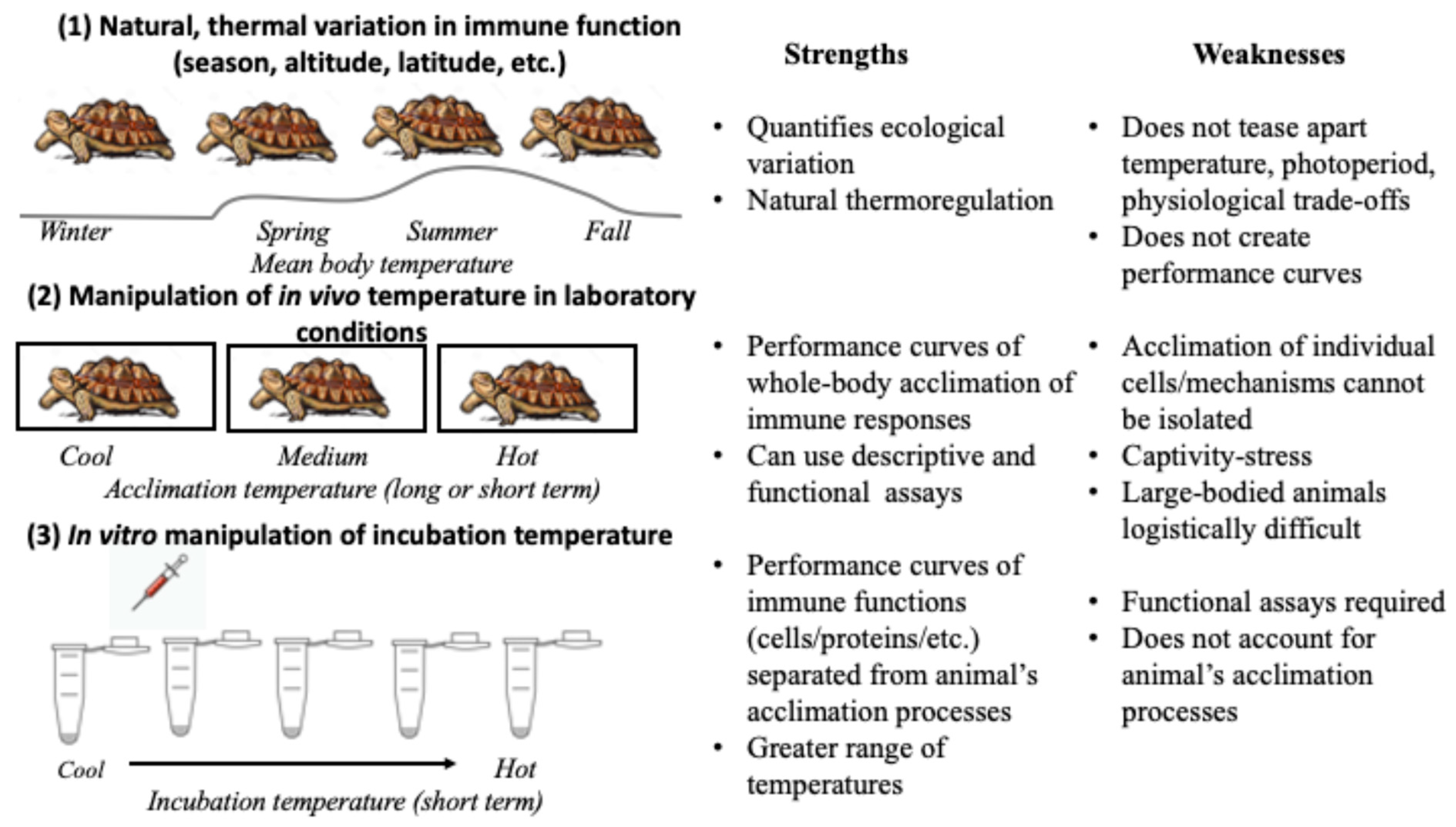
This idea that animals may function as a mosaic of cells with different thermal performances—in both host cells and their commensal microbial community—suggests that study designs also need to take this complexity into consideration. Three types of study design have commonly been used to quantify the thermal acclimation of immune responses in ectothermic animals (Figure 2). The first approach is to study the natural changes in body temperatures of animals, due to factors such as season, altitude, or latitude [24,35,36]. It is the most ecologically relevant approach but the least reductionistic and often does not provide mechanistic explanations [8]. The second approach includes bringing animals into a controlled setting to change their thermal environment [5,10,24]. Both long- and short-term acclimation can be quantified in this way, which also allows the animal to coordinate changes via the nervous and endocrine systems to what might be unpredictable changes in the environmental temperature [5]. Finally, in vitro assays of blood/interstitial fluid or isolated cells/proteins can be used in functional immune assays across an array of incubation temperatures, which can also include a step to allow for acclimation to different thermal regimes. This is the most reductionist approach, but it allows researchers to isolate the effects of temperature on a molecular or cellular level [5,35].

2. Literature Review

To understand the use of these approaches in the literature, I conducted a literature review using three sets of keywords in Web of Science. The purpose of this review was not to exhaustively review all the literature—some taxa-specific literature was likely missed—but to understand general patterns in the literature that emphasized contributions to the general theory of acclimation of immune responses. The search terms “immun* acclimat* temperature* ectotherm*” generated 62 hits, “Immun* acclimat* thermal* ectotherm*” generated 50, and “Immun* thermal* performance* ectotherm*” generated 44 hits, many of which were overlapping hits. Asterisks allow the search engine to find all words that include the root-term. I then sorted all references to only include papers focused on immunology in response to true acclimation to temperatures. Thus, research that quantified the temperature preference of animals after an experimental manipulation was excluded. I also excluded growing, valuable literature that only included thermal performance curves, without addressing changes to performance curves due to acclimation [e.g., [37,38]]. If papers included thermal performance curves, with acclimation at the level of the whole animal, I did include these under in vitro assays combined with the other experimental techniques. I included papers that broadly quantified changes in gene expression and protein synthesis, if these studies included specific genes known to be important to immune function. There was a clear inflection point in the number of papers published annually in and after 2017, with a consistent, increased publication rate in the most recent six years. In all, four publications were reviews, six quantified natural changes in temperature (primarily in response to season), 29 used in vivo assays that changed the temperature of the whole organism, and nine used in vitro assays. Some papers used a combination of assays and were counted multiple times.
With notable exceptions [e.g., [17]], most papers did not explicitly distinguish among these three techniques of acclimation, especially how the methodology used was directly associated with only some mechanisms of acclimation of the immune response. In general, studies of seasonal acclimation did acknowledge caveats, such as concomitant changes in hypoxia, energy limitations, and/or trade-offs with other physiological systems [e.g., reproduction: [39,40]]. Several studies combined seasonal responses with both in vivo [24] and in vitro assays [25,40]. Combining seasonal studies with these more reductionistic manipulations often reveals complex dynamics and unexpected mechanistic explanations for seasonal patterns. For example, Goessling et al. [39] combined seasonal and in vivo study designs, uncovering differential immunological responses to fluctuating temperatures across seasons. Slama et al. [25] combined seasonal and in vitro study designs and found both differential production/regulation of leukocytes and changes in phagocytic ability by cells across seasons.
Manipulating the temperature of the organism in vivo was the most common research technique. Examples from insect ecoimmunology, in particular, have been very successful in elucidating mechanisms of acclimation using this technique [17,41]. A real strength of this technique with small animals is that quantification of immune performance can also be compared to survival and clearance rates of experimental infections [17,41]. Additionally, recent research has also used an approach to quantifying the effects of fluctuating temperatures, which is a more realistic approach to quantifying the effects of a changing climate [17]. In vivo experiments can also be expanded to include thermal acclimation of the microbiome [27], but such studies still seem relatively rare.
However, in studying large vertebrates, there are several caveats to this approach. Smaller animals are expected to have faster acclimation rates due to faster heating and cooling rates and higher mass-specific metabolic rates [42]. A previous review of the physiological literature showed a biased underestimation of thermal acclimation of larger ectotherms [42]—and no animals larger than a gopher tortoise [e.g., [39]] were represented in my review of immunological acclimation in vivo. Logistically, there is the dilemma of limiting sample size to increase the number of acclimation temperatures, or vice versa [13]. Recent research has also quantified the immunological effects on captivity stress, even for captive-bred animals, which can also confound the effects of in vivo experiments [43,44]. Other researchers point to stress due to an inability to thermoregulate in the captive environment as a physiological response that is separate from the sole effects of temperature [20,45].
In vitro assays also have limitations and strengths. They do not account for an animal’s acclimation processes, mediated via nervous or endocrine control, and require functional assays. However, in combination with natural or in vivo experimental acclimation experiments, these assays are powerful and are being used to create full performance curves [25,40]. However, they seem under-used and only one study in this review first in vitro acclimated cells to different thermal regimes, before measuring performance [46]. Some strengths of these assays are that they are non-invasive and can be run across a greater range of temperatures—even including temperatures that would be lethal to the animal. By using a greater range of incubation temperatures, they can also be used to generate true performance curves that may be impossible to achieve for larger animals. In addition, in vitro assays can also be combined with challenges with a wide array of pathogens or toxins, which may be unethical or impractical in live animals. For example, combining bacteria or fungal cells with immune cells in functional assays across incubation temperatures will allow for the quantification of reactions on the scale of their actual interactions in the host. In vertebrates, such interactions commonly occur in and on the mucosa, which can be found on epidermal surfaces [bony fish, amphibians; [30]] and the respiratory, cloacal/vaginal, and gastrointestinal tracts [21,27,47].

3. Considerations for Research Design

A general understanding of mechanisms of immune acclimation across taxa, is largely missing from theory. I suggest that combining these experimental approaches, particularly with the inclusion of performance curves generated by in vitro assays, will help elucidate mechanisms of acclimation. For example, combining seasonal changes with an in vitro thermal performance curve quantifies ecologically relevant variation, with a mechanistic understanding of thermal optima and the breadth of performance of cellular and molecular elements of the immune response (Figure 2). Similarly, when in vitro assays are paired with experimental in vivo acclimation experiments, researchers can quantify responses on the level of the whole organism (large-scale changes in gene expression, hormone levels, etc.) and how those impact specific thermal optima and breadths of different immune functions (bacteria killing assays, antibody secretion, phagocytosis, etc.). For example, a recent review of thermal immunology in insects found different thermal optima for four measures of immune function within and among species: functional assays, immune gene expression, kinetics of enzymes, and survival of infection [17].
A final dilemma in understanding mechanisms of thermal acclimation in the immune system is differentiating between the up- and down-regulation of cells and molecules and changes in their function. Table 1 presents a list of commonly used techniques to study immune function, primarily in ecoimmunology, which has focused largely on vertebrates and insects [2,48]. While all these assays are useful in the context of understanding thermal acclimation, only functional assays that can differentiate between quantity (relative number of cells/molecules) and function (phagocytosis, microbial killing, etc.) will address whether acclimation is occurring due to the up- or down-regulation of cells/molecules or due to cellular changes such as changes to cell membranes or enzyme composition (Table 1).
Table 1. Common eco-immunological assays used in the literature. This table is not comprehensive, but includes most assays commonly used across vertebrate and insect taxa. Especially for in vitro assays of thermal acclimation across taxa, phagocytic assays have the advantage of being functional assays, amenable for use with both inert substances and microbes, and allowing for differentiation among the number of cells present and the relative function of those cells.
Ecoimmunological AssayImmune Parameter
Quantified
Functional AssayDifferentiation of Quantity and Function of
Molecules/Cells
Direct
Interaction with
Microbes
General or Taxa-SpecificReferences (Examples)
Antibody titers (ELISA)Combined amount and avidity of antibodies (can include induced and NAbs)NoNoNoVertebrate[47]
Quantification of other molecules (cytokines/antioxidants/antimicrobial peptides, etc.)Levels of proteins with immune-supportNoNoNoGeneral[17,49,50]
Differential immune cell countCells in interstitial fluid/bloodNo NoNoGeneral[51,52]
Red blood cell agglutionation/lysisAntimicrobial proteins: complement, NAbs, lysozyme, acute phase proteins, othersYesNoNoPredominantly used in vertebrates[53]
PHA-induced inflammationComponents of inflammation: leukocytes, antibodies, cytokines, chemokinesYesNoNoVertebrate[54]
Fluorescence-based phagocytic assayAcidification by phagocytesYesNoYes and No (substrate dependent)General[55]
Melanization/phenoloxidase activityMelanization of foreign objectsYesNoYesArthropods[2,17]
Survival due to microbial challengeWhole-organism ability to stay aliveYesNoYesGeneral[2]
Microbial killing assays (plasma/hemolymph-based)Antimicrobial proteins: complement, NAbs, lysozyme, acute phase proteins, othersYesNoYes General[53]
Antibody secretion by cells (ELISpot)Antibody secretion by B lymphocytes YesYesNoVertebrate[56]
Microbial killing assays (cell-based)Phagocytosis/entrapment by cellsYesYesYes General[53]
Phagocytic assay (cell-specific or all immune-related cells)Engulfment of substrate by specific or all leukocytesYesYesYes and No (substrate dependent)General[57,58]
One of the easiest assays to use, which can be applied widely across all animal taxa, are phagocytic assays (Table 1). Since Metchnikov’s discoveries in the late 1800s, phagocytosis has been recognized as a highly conserved immune function performed by specialized cells found in the interstitial fluid and/or blood of all animal taxa [59,60]. Phagocytosis in vertebrates is also needed for adaptive immune processes, and is thus one of the central mechanisms in both innate and adaptive responses [21]. Furthermore, phagocytosis can be induced with artificial beads that are easily visualized under a light microscope or with known quantities of microbes in microbial-killing assays [58]. Simple techniques such as blood/hemolymph smears can be used to quantify cells, or hemacytometers to additionaly quantify the numbers of viable cells if the sample is not used immediately [58]. Likely because of the simplicity of using frozen plasma samples to quantify microbial killing instead of using live white blood cells, this type of assay has been under-used in ecoimmunological studies of vertebrates, with some notable exceptions [e.g., [24,35,61]].

4. Conclusions

The study of immune function and the associated microbiome across temperatures is fundamentally important to understanding physiological ecology in ectotherms, but also in endotherms—especially for those who experience temperature changes during hibernation or during infections that induce fever or hypothermia [21,62]. Immune function is tightly tied to homeostasis, as some of the same processes that eliminate or reduce pathogens also rid the body of damaged or old cells [21,59]. Indeed, Metchnikoff recognized this dual function of phagocytic cells [59]. For example, a recent study on Chinese giant salamanders suggested that trade-offs in cold regimes may favor organismal maintenance (including immune function and homeostasis) overgrowth, but only up to a point at which the animal enters true torpor instead of acclimating to full function at low temperatures [34]. Additionally, immune function—along with its role in homeostasis—is expected to be tied to fitness by enhancing survivorship, at least in largely R-selected species [63]. For some organisms, quantifying immune function may be an important indicator of fitness, along with acclimation responses commonly quantified by physiologists, such as muscle function, digestion rates, and aerobic metabolism [11,19,64]. While studies on birds and mammals laid a very important framework in the field of ecoimmunology, studies on immune function on ectotherms almost always need to consider temperature, unless the animal is adapted to perform at a narrow range, as might be the case for tropical or polar species [49,64,65]. I hope that the framework presented herein can help researchers design studies that hone in on different mechanisms of thermal acclimation to tease apart the effects of adaptation to thermal regimes and plasticity to both predictable and unpredictable thermal fluctuations within those systems.

Funding

This material is based on work supported by the National Science Foundation under Award No. 221866.

Institutional Review Board Statement

Not applicable.

Informed Consent Statement

Not applicable.

Data Availability Statement

No new, un-reported data were generated.

Conflicts of Interest

The author declares no conflicts of interest.

References

  1. Gibbons, J.W.; Scott, D.E.; Ryan, T.J.; Buhlman, K.A.; Tuberville, T.T.; Metts, B.S.; Greene, J.W.; Mills, T.; Leiden, Y.; Poppy, S.; et al. The global decline of reptiles, déjà vu amphibians. BioScience 2000, 50, 653–666. [Google Scholar] [CrossRef]
  2. Ferguson, L.V.; Kortet, R.; Sinclaire, B.J. Eco-immunology in the cold: The role of immunity in shaping the overwintering survival of ectotherms. J. Exp. Biol. 2018, 221, 163873. [Google Scholar] [CrossRef] [PubMed]
  3. Fontaine, S.S.; Novarro, A.J.; Kohl, K.K. Environmental temperature alters digestive performance and gut microbiota of a terrestrial amphibian. J. Exp. Biol. 2018, 221, 187559. [Google Scholar] [CrossRef] [PubMed]
  4. Trevelline, B.K.; Fontaine, S.S.; Hartup, B.K.; Kohl, K.D. Conservation biology needs a microbial renaissance: A call for consideration of host-associated microbiota in wildlife management practices. Proc. R. Soc. B 2019, 286, 20182448. [Google Scholar] [CrossRef]
  5. Ferguson, L.V.; Heinrich, D.E.; Sinclair, B.J. Paradoxical acclimation responses in the thermal performance of insect immunity. Oecologia 2016, 181, 77–85. [Google Scholar] [CrossRef]
  6. IPPC (Intergovernmental Panel on Climate Change). Climate change 2014: Impacts, adaptation, and vulnerability. Part A: Global and sectoral impacts. In Contribution of Working Group II to the Fifth Assessment Report of the Intergovernmental Panel on Climate Change; Field, C.B., Baros, V.R., Dokken, D.J., Mach, K.J., Mastrandrea, M.D., Bilir, T.E., Chatterjee, M., Ebi, K.L., Estrada, Y.O., Genova, R.C., et al., Eds.; Cambridge University Press: Cambridge, UK, 2014; p. 1132. [Google Scholar]
  7. Vasseur, D.A.; DeLong, J.P.; Gilbert, B.; Greig, H.S.; Harley, C.D.G.; McCann, K.S.; Savage, V.; Tunney, T.D.; O’Connor, M.I. Increased temperature variation poses a greater risk to species than climate warming. Proc. R. Soc. B 2014, 281, 20132612. [Google Scholar] [CrossRef]
  8. Martin, L.B.; Weil, Z.M.; Nelson, R.J. Seasonal changes in vertebrate immune activity: Mediation by physiological trade-offs. Phil. Trans. R. Soc. B 2008, 363, 321–339. [Google Scholar] [CrossRef]
  9. Altman, K.A.; Paull, S.H.; Johnson, P.T.J.; Golembieski, M.N.; Stephens, L.P.; LaFonte, B.E.; Raffel, T.R. Host and parasite thermal acclimation responses depend on the stage of infection. J. Anim. Ecol. 2016, 85, 1014–1024. [Google Scholar] [CrossRef]
  10. Stahlschmidt, Z.R.; French, S.S.; Ahn, A.; Webb, A.; Butler, M.W. A simulated heat wave has diverse effects on immune function and oxidative physiology on the corn snake (Pantherophis guttatus). Physiol. Biochem. Zool. 2017, 90, 434–444. [Google Scholar] [CrossRef]
  11. Angilletta, M.J. Thermal Adaptation: A Theoretical and Empirical Synthesis; Oxford University Press: New York, NY, USA, 2009. [Google Scholar]
  12. Cohen, J.M.; Venesky, M.D.; Sauer, E.L.; Civitello, D.J.; McMahon, T.A.; Roznik, E.A.; Rohr, J.R. The thermal mismatch hypothesis explains host susceptibility to an emerging infectious disease. Ecol. Lett. 2017, 20, 184–193. [Google Scholar] [CrossRef]
  13. Molnár, P.K.; Sckrabulis, J.P.; Altman, K.A.; Raffel, T.R. Thermal performance curves and the metabolic theory of ecology—A practical guide to models and experiments for parasitologists. J. Parasitol. 2017, 103, 423–439. [Google Scholar] [CrossRef]
  14. Pxytycz, B.; Józkowicz, A. Differential effects of temperature on macrophages of ecothermic vertebrates. J. Leukoc. Biol. 1994, 56, 729–731. [Google Scholar] [CrossRef]
  15. Baker, S.; Merchant, M.E. Antibacterial properties of plasma from the prairie rattlesnake (Crotalus viridis). Dev. Comp. Immunol. 2018, 84, 273–278. [Google Scholar] [CrossRef]
  16. Sanhueza, N.; Fuentes, R.; Aguilar, A.; Carnicero, B.; Vega, K.; Muñoz, D.; Contreras, D.; Moreno, N.; Tronscoso, E.; Mercado, L.; et al. Behavioral fever promotes an inflammatory reflex circuit in ectotherms. Int. J. Mol. Sci. 2021, 22, 8860. [Google Scholar] [CrossRef] [PubMed]
  17. Ferguson, L.V.; Adamo, S.A. From perplexing to predictive: Are we ready to forecast insect disease susceptibility in a warming world? J. Exp. Biol. 2023, 226, jeb244911. [Google Scholar] [CrossRef] [PubMed]
  18. Weil, Z.M.; Nelson, R.J. Neuroendocrine mechanisms of seasonal changes in immune function. In Ecoimmunology; Demas, G.E., Nelson, R.J., Eds.; Oxford University Press: New York, NY, USA, 2011; pp. 297–325. [Google Scholar]
  19. Schulte, P.M. The effects of temperature on aerobic metabolism: Towards a mechanistic understanding of responses of ecotherms to a changing environment. J. Exp. Biol. 2015, 218, 1856–1866. [Google Scholar] [CrossRef] [PubMed]
  20. Morvan, C.L.; Troutaud, D.; Deschaux, P. Differential effects of temperature on specific and nonspecific immune defences in fish. J. Exp. Biol. 1998, 201, 165–168. [Google Scholar] [CrossRef] [PubMed]
  21. Parham, W.W. The Immune System, 5th ed.; Norton and Company: New York, NY, USA, 2021. [Google Scholar]
  22. Baker, S.J.; Kessler, E.J.; Merchant, M.E. Antimicrobial activities of plasma from the common (Chelydra serpentina) and alligator snapping turtle (Macrochelys temminckii). J. Exp. Zool. 2019, 331, 85–92. [Google Scholar] [CrossRef] [PubMed]
  23. Raffel, T.R.; Rohr, J.R.; Kiesecker, J.M.; Hudson, P.J. Negative effects of changing temperature on amphibian immunity under field conditions. Func. Ecol. 2006, 20, 819–828. [Google Scholar] [CrossRef]
  24. Goessling, J.M.; Koler, S.A.; Overman, B.D.; Hiltbold, E.M.; Guyer, C.; Mendonça, M. Lag of immunity across seasonal acclimation states in gopher tortoises (Gopherus polyphemus). J. Exp. Zool. 2017, 327, 235–242. [Google Scholar] [CrossRef]
  25. Slama, S.L.; Williams, G.S.; Painter, M.N.; Sheedy, M.D.; Sandmeier, F.C. Temperature and season influence phagocytosis by B1 lymphocytes in the Mojave desert tortoise. Integr. Comp. Biol. 2022, 62, 1683–1692. [Google Scholar] [CrossRef]
  26. Kiel, D.; Luebke, R.W.; Pruett, S.B. Quantifying the relationship between multiple immunological parameters and host resistance: Probing the limits of reductionism. J. Immunol. 2001, 167, 4543–4552. [Google Scholar] [CrossRef]
  27. Palmer-Young, E.C.; Ngor, L.; Nevarez, R.B.; Rothman, J.A.; Raffel, T.R.; McFrederick, Q.S. Temperature dependence of parasitic infection and gut bacterial communities in bumble bees. Environ. Microbiol. 2019, 21, 470604723. [Google Scholar] [CrossRef] [PubMed]
  28. Moeller, A.H.; Ivey, K.; Cornwall, M.B.; Herr, K.; Rede, J.; Taylor, E.N.; Gunderson, A.R. Lizard gut microbiome changes with temperature and is associated with heat tolerance. Appl. Environ. Microbiol. 2020, 86, e01181. [Google Scholar] [CrossRef] [PubMed]
  29. Carter, E.D.; Bletz, M.C.; Le Sage, M.; LaBumbard, B.; Rollins-Smith, L.A.; Woodhams, D.C.; Miller, D.L.; Gray, M.J. Winter is coming—Temperature affects immune defenses and susceptibility to Batrachochytridium salamandrivorans. PLOS Pathog. 2021, 17, e1009234. [Google Scholar] [CrossRef] [PubMed]
  30. Woodhams, D.C.; Brandt, H.; Baumgartner, S.; Kielgast, J.; Kupfer, E.; Tobler, U.; Davis, L.R.; Schmidt, B.R.; Bel, C.; Hodel, S.; et al. Interacting symbionts and immunity in the amphibian skin mucosome predict disease risk and probiotic effectiveness. PLoS ONE 2014, 9, e96375. [Google Scholar] [CrossRef] [PubMed]
  31. Stewart, A.; Hablützel, P.I.; Brown, M.; Watson, H.V.; Parker-Norman, S.; Tober, A.V.; Thomason, A.G.; Friber, I.M.; Cable, J.; Jackson, J.A. Half the story: Thermal effects on within-host infectious disease progression in a warming climate. Glob. Chang. Biol. 2017, 24, 371–386. [Google Scholar] [CrossRef] [PubMed]
  32. Shiu, J.; Keshavmurthy, S.; Chiang, P.; Chen, H.; Lou, S.; Tseng, C.; Hsieh, H.J.; Chen, C.A.; Tang, S. Dynamics of coral-associated bacterial communities acclimated to temperature stress based on recent thermal history. Sci. Rep. 2017, 7, 14933. [Google Scholar] [CrossRef] [PubMed]
  33. Vargas, S.; Leiva, L.; Wörheide, G. Short-term exposure to high-temperature water causes a shift in the microbiome of the common aquarium sponge Lendenfeldia chondrodes. Invert. Microbiol. 2021, 81, 213–222. [Google Scholar] [CrossRef] [PubMed]
  34. Zhu, L.; Zhu, W.; Zhao, T.; Chen, H.; Zhao, C.; Xu, L.; Chang, Q.; Jiang, J. Environmental temperatures affect the gastrointestinal microbes of the Chinese giant salamander. Front. Microbiol. 2021, 12, 543767. [Google Scholar] [CrossRef]
  35. Palackdharry, S.; Sadd, B.M.; Vogel, L.A.; Bowden, R.M. The effect of environmental temperatrue on reptilian peripheral blood B cell functions. Horm. Behav. 2017, 88, 87–94. [Google Scholar] [CrossRef] [PubMed]
  36. Sandmeier, F.C.; Leonard, K.L.; Tracy, C.R.; Drake, K.K.; Esque, T.E.; Nussear, K.; Germano, J.M. Tools to understand seasonality in health: Quantification of microbe loads and analyses of compositional ecoimmunological data reveal complex patterns in tortoise populations. Can. J. Zool. 2019, 97, 841–848. [Google Scholar] [CrossRef]
  37. Butler, M.; Stahlschmidt, Z.R.; Ardia, D.R.; Davies, S.; Davis, J.; Guillette, L.J., Jr.; Johnson, N.; McCormick, D.; McGraw, K.J.; DeNardo, D.J. Thermal sensitivity of immune function: Evidence against a generalist-specialist trade-off among endothermic and ectothermic vertebrates. Am. Nat. 2013, 181, 761–774. [Google Scholar] [CrossRef] [PubMed]
  38. Byers, J.E. Marine parasites and disease in the era of global climate change. Ann. Rev. Mar. Sci. 2020, 13, 397–420. [Google Scholar] [CrossRef] [PubMed]
  39. Goessling, J.M.; Guyer, C.; Mendonça, M. Seasonal acclimation of constitutive immunity in gopher tortoises Gopherus polyphemus. Physiol. Biochem. Zool 2016, 89, 487–497. [Google Scholar] [CrossRef] [PubMed]
  40. Graham, S.P.; Fielman, K.T.; Mendonça, M.T. Thermal performance and acclimatization of a component of snake (Agkistrodon piscivorus) innate immunity. J. Exp. Zool. 2017, 327, 351–357. [Google Scholar] [CrossRef] [PubMed]
  41. Adamo, S.A.; Lovett, M.M. Some like it hot: The effects of climate change on reproduction, immune function and disease resistance in the cricket Gryllus texensis. J. Exp. Biol. 2011, 214, 1997–2004. [Google Scholar] [CrossRef]
  42. Rohr, J.R.; Civitello, D.J.; Cohen, J.M.; Roznik, E.A.; Sinervo, B.; Dell, A.I. The complex drivers of thermal acclimation and breadth in ectotherms. Ecol. Lett. 2018, 21, 1425–1439. [Google Scholar] [CrossRef]
  43. Lima, A.S.; Ferreira, L.F.; Silva, D.P.; Gomes, F.R.; Titon, S.C.M. Thermal sensitivity of bullfrog’s immune response kept at different temperatures. J. Exper. Zool. 2020, 333, 767–778. [Google Scholar] [CrossRef]
  44. Goessling, J.M.; Mendonça, M.T. Physiological responses of gopher tortoises (Gopherus polyphemus) to trapping. Conserv. Physiol. 2021, 9, coab003. [Google Scholar] [CrossRef]
  45. Boltana, S.; Aguilar, A.; Sanhueza, N.; Donoso, A.; Mercado, L.; Imarai, M.; McKenzie, S. Behavioral fever drives epigenetic modulation of the immune response in fish. Front. Immunol. 2018, 9, 1241. [Google Scholar] [CrossRef]
  46. Tirard, C.T.; Grossfeld, R.M.; Levine, J.F.; Kennedy-Stoskopf, S. Effect of hyperthermia in vitro on stress protein synthesis and accumulation in oyster haemocytes. Fish Shellfish Immunol. 1995, 5, 9–25. [Google Scholar] [CrossRef]
  47. Sandmeier, F.C.; Leonard, K.L.; Weitzman, C.L.; Tracy, C.R. Potential facilitation between a commensal and a pathogenic microbe in a wildlife disease. Ecohealth 2022, 19, 427–438. [Google Scholar] [CrossRef]
  48. Demas, G.E.; Nelson, R.G. (Eds.) Ecoimmunology; Oxford University Press: New York, NY, USA, 2012. [Google Scholar]
  49. Sandmeier, F.C.; Tracy, C.R.; DuPré, S.; Hunter, K. A trade-off between natural and acquired antibody production in a reptile: Implication for long-term resistance to disease. Biol. Open 2012, 1, 1078–1082. [Google Scholar] [CrossRef] [PubMed]
  50. Messina, S.; Costantini, D.; Eens, M. Impacts of rising temperatures and water acidification on oxidative status and immune system of aquatic ectothermic vertebrates: A metanalysis. Sci. Total Environ. 2023, 868, 161580. [Google Scholar] [CrossRef] [PubMed]
  51. Davis, A.K.; Maney, D.L.; Maerz, J.C. The use of leukocyte profiles to measure stress in vertebrates: A review for ecologists. Func. Ecol. 2008, 22, 275–286. [Google Scholar] [CrossRef]
  52. Oliver, J.D.; Loy, J.D.; Parikh, G.; Bartholomay, L. Comparative analysis of hemocyte phagocytosis between six species of arthropods as measured by flow cytometry. J. Invertebr. Pathol. 2011, 108, 126–130. [Google Scholar] [CrossRef] [PubMed]
  53. Matson, K.D.; Tieleman, B.I.; Klasing, K.C. Capture stress and bactericidal competence of blood and plasma in five species of tropical birds. Physiol. Biochem. Zool. 2006, 79, 556–564. [Google Scholar] [CrossRef] [PubMed]
  54. Martin, L.B.; Han, P.; Lewittes, J.; Kuhlman, J.R.; Klasing, K.C.; Wikelski, M. Phytohemagglutinin-induced swelling in birds: Histological support for a classic immunological technique. Func. Ecol. 2006, 20, 290–299. [Google Scholar] [CrossRef]
  55. Goessling, J.M.; Ward, C.; Mendonça, M.T. Rapid thermal immune acclimation in common musk turtles (Sternotherus odoratus). J. Exp. Zool. 2019, 331, 185–191. [Google Scholar] [CrossRef] [PubMed]
  56. Zimmerman, L.M.; Clairardin, S.G.; Paitz, R.T.; Hicke, J.W.; LaMagdeleine, K.A.; Vogel, L.A.; Bowden, R.M. Humoral responses are maintained with age in a long-lived ectotherm, the red-eared slider. J. Exp. Biol. 2013, 216, 185–191. [Google Scholar] [CrossRef]
  57. Graham, A.L.; Shuker, D.M.; Pollitt, L.C.; Auld, S.K.J.R.; Wilson, A.J.; Little, T.J. Fitness consequences of immune responses: Strengthening the empirical framework for ecoimmunology. Func. Ecol. 2011, 25, 5–17. [Google Scholar] [CrossRef]
  58. Slama, S.L.; Painter, M.N.; Sheedy, M.D.; Sandmeier, F.C. Quantifying phagocytic lymphocytes in ectothermic vertebrates: A simplified technique for assessing immune function. Methods Ecol. Evol. 2021, 12, 548–552. [Google Scholar] [CrossRef]
  59. Tauber, A.I. Metchnikoff and the phagocytosis theory. Nat. Rev. Mol. Cell Biol. 2003, 4, 897–901. [Google Scholar] [CrossRef]
  60. Teti, G.; Biondo, C.; Beninati, C. The phagocyte, Metchnivoff, and the foundation of immunology. Microbiol. Spectrum 2016, 4, MHCD-009-2015. [Google Scholar] [CrossRef]
  61. Millet, S.; Bennett, J.; Lee, K.A.; Hau, M.; Klasing, K.C. Quantifying and comparing constitutive immunity across avian species. Dev. Comp. Immunol. 2007, 31, 188–201. [Google Scholar] [CrossRef] [PubMed]
  62. Hill, R.W.; Anderson, M.; Cavanaugh, D. Animal Physiology, 5th ed.; Oxford University Press: New York, NY, USA, 2021. [Google Scholar]
  63. Sandmeier, F.C.; Tracy, R.C. The metabolic pace-of-life model: Incorporating ectothermic organisms into the theory of vertebrate ecoimmunology. Integr. Comp. Biol. 2014, 54, 387–395. [Google Scholar] [CrossRef] [PubMed]
  64. Peck, L.S.; Morley, S.A.; Richard, J.; Clark, M.S. Acclimation and thermal tolerance in Antarctic marine ectotherms. J. Exp. Biol. 2014, 217, 16–22. [Google Scholar] [CrossRef] [PubMed]
  65. Morgan, R.; Andreassen, A.H.; Åsheim, E.; Finnøen, M.H.; Dresler, G.; Brembu, T.; Loh, A.; Miest, J.J.; Jutfelt, F. Reduced physiological plasticity in a fish adapted to stable temperatures. Proc. Natl. Acad. Sci. USA 2022, 22, e2201919119. [Google Scholar] [CrossRef] [PubMed]
Figure 1. (a) A hypothetical performance curve, with peak performance, thermal optima, and breadth (e.g., 2/3 function) labeled. (b) An example of two different modes of thermal acclimation at cool temperatures in an animal, using the phagocytosis of a microbe as an example. In the left-hand panel, the numbers of phagocytic cells are increased while the thermal optimum and breadth of phagocytosis remain unchanged. In other words, function is maintained at cooler temperatures by increasing overall phagocytosis. In the right-hand panel, the numbers of cells are kept constant while the thermal optimum is shifted towards cooler temperatures.
Biology 13 00179 g001
Figure 2. Eco-immunological study designs focused on thermal acclimation of immune responses can be grouped into three broad categories, using the example of a small vertebrate. (1) Natural thermal variation can be studied in wild animals due to factors such as season and geography. (2) Animals may be manipulated by placing them in different in vivo thermal environments in captive conditions. (3) Live biological samples may be incubated in vitro at different temperatures in functional immune assays. Research that combines these three categories in different ways, benefits from a combination of more holistic and reductionistic measurements and different time frames to elucidate variable mechanisms of acclimation.
Biology 13 00179 g002
Disclaimer/Publisher’s Note: The statements, opinions and data contained in all publications are solely those of the individual author(s) and contributor(s) and not of MDPI and/or the editor(s). MDPI and/or the editor(s) disclaim responsibility for any injury to people or property resulting from any ideas, methods, instructions or products referred to in the content.