Article

Biological Control of Tomato Bacterial Leaf Spots and Its Impact on Some Antioxidant Enzymes, Phenolic Compounds, and Pigment Content

by
Asmaa H. Akila
1,
Mohamed A. S. Ali
1,
Ahmed M. Khairy
1,
Ahmed S. M. Elnahal
1,*,
Haifa E. Alfassam
2,*,
Hassan A. Rudayni
3,
Fatima A. Jaber
4 and
Mohamed R. A. Tohamy
1
1
Plant Pathology Department, Faculty of Agriculture, Zagazig University, Zagazig 44511, Egypt
2
Department of Biology, College of Science, Princess Nourah bint Abdulrahman University, Riyadh 11671, Saudi Arabia
3
Department of Biology, College of Science, Imam Mohammad Ibn Saud Islamic University, Riyadh 11623, Saudi Arabia
4
Department of Biological Sciences, College of Science, University of Jeddah, Jeddah 21589, Saudi Arabia
*
Authors to whom correspondence should be addressed.
Biology 2024, 13(6), 369; https://doi.org/10.3390/biology13060369
Submission received: 16 February 2024 / Revised: 17 May 2024 / Accepted: 18 May 2024 / Published: 23 May 2024

Abstract

:

Simple Summary

Tomato bacterial spots and specks, caused by various strains of Xanthomonas campestris and Pseudomonas syringae, lead to significant yield losses in the El-Sharkia governorate. This study tested the efficacy of biocontrol culture filtrates from Trichoderma fungi (T. viride, T. harzianum, T. album) and bacterial agents (Bacillus subtilis, Pseudomonas fluorescens, Serratia marcescens) in controlling these diseases. In vitro, the culture filtrates significantly inhibited pathogenic bacterial growth. In vivo, spraying bioagents prior to infection reduced disease incidence and severity. T. viride was the most effective fungal bioagent, while B. subtilis was the top bacterial bioagent. Tomato plants that were treated with bioagents also had more phenols and chlorophyll than control plants. They also had more enzyme activity, including chitinase, peroxidase, and polyphenol oxidase. These findings suggest that biocontrol strategies could be a sustainable solution for managing tomato diseases and improving plant defense.

Abstract

Tomato bacterial spots, caused by Xanthomonas campestris pv. vesicatoria (Xcv1) and X. euvesicatoria (Xe2), as well as bacterial specks, caused by two strains of Pseudomonas syringae pv. tomato (Pst1 and Pst2), represent significant threats to tomato production in the El-Sharkia governorate, often resulting in substantial yield losses. The objective of this study was to evaluate the efficacy of various biocontrol culture filtrates, including bacteria and fungi agents, in managing the occurrence and severity of these diseases, while also monitoring physiological changes in tomato leaves, including antioxidant enzymes, phenolics, and pigment content. The culture filtrates from examined Trichoderma species (T. viride, T. harzianum, and T. album), as well as the tested bacteria (Bacillus subtilis, Pseudomonas fluorescens, and Serratia marcescens) at concentrations of 25%, 50%, and 100%, significantly inhibited the proliferation of pathogenic bacteria In vitro. For the In vivo experiments, we used specific doses of 5 mL of spore suspension per plant for the fungal bioagents at a concentration of 2.5 × 10⁷ spores/mL. The bacterial bioagents were applied as a 10 mL suspension per plant at a concentration of 1 × 10⁸ CFU/mL. Spraying the culture filtrates of the tested bioagents two days before infection In vivo significantly reduced disease incidence and severity. Trichoderma viride exhibited the highest efficacy among the fungal bioagents, followed by T. harzianum and T. album. Meanwhile, the culture filtrate of B. subtilis emerged as the most potent among the bacterial bioagents, followed by P. fluorescens. Furthermore, applying these culture filtrates resulted in elevated levels of chitinase, peroxidase, and polyphenol oxidase activity. This effect extended to increased phenol contents, as well as chlorophyll a, chlorophyll b, and carotenoids in sprayed tomato plants compared to the control treatment. Overall, these findings underscore the potential of these biocontrol strategies to effectively mitigate disease incidence and severity while enhancing plant defense mechanisms and physiological parameters, thus offering promising avenues for sustainable disease management in tomato production.

1. Introduction

Tomato (Solanum lycopersicum L.) holds significant agricultural value in Egypt, Africa, and globally. Egypt ranks fifth globally in potato production and boasts the largest tomato production output in Africa [1]. Moreover, tomatoes serve as a model crop for studying the growth of newly fruited plants and plant–pathogen interactions [2]. Annually, tomato cultivation faces numerous challenges, including pest infestations (such as insects and nematodes) and diseases caused by bacterial, fungal, and viral pathogens [3]. Research indicates that crop loss can vary widely depending on factors such as region, crop variety, and specific pathogens involved [4]. Hot and humid environmental conditions can exacerbate the severity of these diseases, leading to significant crop losses if pathogens are not effectively managed [5].
Bacterial diseases contribute significantly to the disease burden affecting tomato yields across various production areas in Egypt [6], including El-Sharkia governorate. These include bacterial spots, attributed to several species within the genus Xanthomonas, and bacterial specks, caused by P. syringae pv. tomato [7]. Clavibacter michiganensis induces canker, while Ralstonia solanacearum triggers bacterial wilt, two of the most severe bacterial diseases impacting tomato cultivation [8]. The bacterium P. syringae pv. tomato is responsible for bacterial speck, a disease highly regarded for its economic impact on tomato crops. The strain P. syringae pv. tomato strain DC 3000 serves as a model organism in molecular investigations of host–pathogen interactions [9].
Various control strategies are implemented to manage speck disease, including the utilization of biocontrol agents [9]. Pesticides commonly used to control tomato bacterial leaf spots include copper-based compounds, antibiotics, and synthetic chemicals [10]. However, they have drawbacks such as environmental pollution, harm to non-target organisms, and pathogen resistance [11]. Herbert et al. [12] reported that strains of Xanthomonas spp. that are resistant to streptomycin appeared after long-term use, while Gikas et al. [13] found that copper-based fungicides hurt soil microbe communities and might end up in food chains.
Biological control agents have emerged as an effective method for managing plant pathogens [14]. Researchers have documented that certain isolates of Trichoderma species significantly reduce plant diseases [15]. However, limited research has been conducted to assess the biocontrol efficacy of their culture filtrates against bacterial spots and speck on tomatoes. The biological control strategies of Trichoderma spp. involved mycoparasitism, antibiosis, competing for nutrient resources and potential infection areas, and stimulating systemic resistance within plants, as studied by [16]. Nonetheless, specific Trichoderma strains are recognized to excel in particular antagonistic mechanisms [17]. Hence, selecting effective Trichoderma strains and species is essential for specific plant pathogen control. Researchers have explored antibiotics like kasugamycin and streptomycin as controls for bacterial leaf spot and speck, particularly in greenhouse settings. Also, Xanthomonas spp. showed resistance to streptomycin before widespread field use [18]. Furthermore, Kasugamycin has demonstrated similar efficacy to conventional copper-mancozeb treatments against bacterial spots [19].
Recently, there has been increasing interest in alternative approaches to disease management, such as the use of biocontrol agents (BCAs). These BCAs are microorganisms or their metabolites that suppress plant pathogens through various mechanisms, including parasitism, competition, antibiosis, and induction of host defenses [20]. Culture filtrates made from BCAs have many different secondary metabolites, enzymes, and other bioactive compounds that can stop plant pathogens from growing and spreading [21].
Extensive research has focused on finding biological control methods for bacterial spots; for example, P. syringae Cit7 consistently provided effective control [22]. Furthermore, several plant growth-promoting rhizobacteria (PGPR), including Bacillus pumilis, have proven efficient in reducing bacterial spot in several field experiments [23]. PGPR plays a dual role in boosting plant growth and fighting diseases, employing tactics like hyperparasitism, antibiotic production, and systemic resistance induction. They offer a promising alternative for managing plant diseases [24]. Bacillus spp. is a standout among PGPRs, demonstrating potent biocontrol abilities against various plant pathogens [25]. Foliar application of Bacillus spp. has shown efficacy in reducing bacterial spot on tomatoes [26].
Pathogen–host interactions alter cell metabolism by affecting enzymes such as catalase, peroxidase (PO), and polyphenol oxidase (PPO). Studies reveal a significant link between bioagent efficacy and PO/PPO levels [27]. Additionally, phenolic compounds, vital secondary metabolites in plants, play key roles in functions such as pigmentation, growth, and pathogen resistance. They accumulate under stress, acting as defense compounds [28]. Phenolic production, including lignin and melanin synthesis, involves dihydroxyphenol and chinen oligomers, toxic to pathogens, bolstering plant defense [29]. PO uses a heme group to catalyze oxidation reactions with H2O2, while PPO contains Cu ions in its active site, allowing it to oxidize phenolic compounds into toxic quinones, enhancing disease resistance [30].
Microbial filtrates from soil fungi and rhizobacteria are being used more and more as biostimulants in sustainable agriculture because they can help plants grow and be more resistant to damage while lowering the need for chemicals [31]. These filtrates contain beneficial compounds that can enhance nutrient uptake, stress tolerance, and disease resistance [31]. In particular, the production of siderophores by strains like P. fluorescens and B. subtilis is notable for its iron-chelating properties, which support plant growth in nutrient-limited conditions [32].
Our study aims to evaluate the efficacy of various bacterial and fungal bioagent culture filtrates in controlling bacterial spot and speck diseases on tomato plants, both In vitro and In vivo. We hypothesize that these bioagents will inhibit the growth of pathogenic bacteria (X. campestris and P. syringae strains), leading to reduced disease incidence and severity in tomato plants. The application of these bioagents is also expected not only to reduce the incidence and severity of these diseases but also enhance the physiological defenses of tomato plants. This includes an increase in the activity of key enzymes such as chitinase, peroxidase, and polyphenol oxidase, as well as boosting levels of chlorophyll, carotenoids, and phenolic compounds. This study offers novel insights by comprehensively evaluating the efficacy of biocontrol agents against bacterial leaf spot and speck diseases on tomato plants, shedding light on their impact on key physiological parameters for sustainable disease management. Also, this approach not only controls pathogens directly but also considers the broader implications of biocontrol treatments on plant health and disease resistance. This could lead to the use of similar strategies in other crops that are affected by bacterial diseases.

2. Materials and Methods

The tomato bacterial leaf spot (BLS) is caused by pathogens such as X. campestris pv. vesicatoria (Xcv1) and X. euvesicatoria (Xe2), as well as the pathogen responsible for bacterial speck (BS), which includes two strains of P. syringae pv. tomato known as Pst1 and Pst2. These pathogens were previously isolated and identified at the Plant Pathology Department, Faculty of Agriculture, Zagazig University. Their accession numbers are as follows: OR146491, OR146486, CP047072.1, and CP047071.1 for Xcv1, Xe2, Pst1, and Pst2, respectively. They were utilized in subsequent experiments.

2.1. Inoculum of the Pathogenic Bacteria

The previously mentioned bacterial isolates (Xcv1, Xe2, Pst1, and Pst2) were cultured in a nutrient broth medium (beef extract, yeast extract, peptone, and dextrose) on a rotary shaker for two days at 28 ± 2 °C [33]. The resulting bacterial suspension was adjusted to be 1 × 10⁸ CFU/mL using spectrophotometer OD600 (Spectroquant, Darmstadt, Germany) [34]. The bacterial culture filtrates were applied at a dosage of 10 mL per plant during the experiments, ensuring consistent application across all samples [33,35].

2.2. Bioagent Culture Filtrates

Identified fungal bioagents (Trichoderma viride, T. harzianum, and T. album), as well as bacterial bioagents (Bacillus subtilis, Pseudomonas fluorescence and Serratia marcescens), were obtained from Plant Pathology Dept., Fac. Agric., Zagazig Univ. Fungal bioagents were grown in 500 mL flasks containing 200 mL of Gliotoxin Fermentation Medium broth described by [36] on a rotary shaker (SK-330-Pro Scilogex) at 150 rpm for 7 days in dark room at 30 °C [37]. The broth growth was centrifuged at 10,000 rpm for 20 min, followed by filtration through Whatman No.1 filter paper and sterilization using a 0.34 μm Millipore filter. This filtered supernatant was then utilized In vitro against the tested pathogens. Likewise, metabolic substances from the bacterial isolates were obtained by cultivating the isolates in a nutrient broth medium on a rotary shaker for two days at 28 ± 2 °C. The isolates were then extracted and sterilized following the methods described by Breitling et al. [38] and Ghorbanpour et al. [39]. The effective doses used in the experiments for the fungal bioagents (Trichoderma viride, T. harzianum, and T. album) were 5 mL of spore suspension per plant at a concentration of 2.5 × 10⁷ spores/mL [40]. For the bacterial bioagents (Bacillus subtilis, Pseudomonas fluorescens, and Serratia marcescens), the effective dose was 10 mL of bacterial suspension per plant at a concentration of 1 × 10⁸ CFU/mL [33,35]. These doses were determined based on their efficacy in controlling tomato leaf-spot diseases and were applied consistently throughout the study to ensure reliable results.

2.3. In Vitro Assays

The antagonistic activity of previously purified sterile metabolites from three tested Trichoderma spp. and three isolates of bacterial bioagents was evaluated using three concentrations (25%, 50%, and 100%) of each metabolite. These concentrations were selected to cover a range of doses to assess the effectiveness of the metabolites against the target pathogens while ensuring that potential adverse effects or limitations associated with higher concentrations were considered. One milliliter of sterile culture filtrate from the tested bioagents was dispensed into sterilized Petri plates, followed by the addition of 1 mL containing (1 × 10⁸ CFU/mL) of each pathogen (Xcv1, Xe2, Pst1, Pst2) simultaneously. Subsequently, 15 mL of nutrient agar medium were poured into the plates. In the control treatment, sterile water was substituted for sterile metabolites. There were three replications of each treatment. The Petri plates were then incubated at 28 ± 2 °C, and data were recorded after 48 h using the cell count technique, which provides reliable counts only for live cells [41,42,43,44]. The obtained results are expressed in terms of growth percentage reduction. Results were expressed as the inhibition percentage of disease incidence and severity compared to the infected control using a formula suggested by Suresh [45], as follows:
P e r c e n t a g e   o f   i n h i b i t i o n   ( P I % ) = C T C × 100 ,
where C represents disease incidence or severity on the positive control, while T represents disease incidence or severity on the leaves treated with antagonists.

2.4. In Vivo Assays

We evaluated the effect of spraying tomato transplants (cv. 102) with a 100% concentration of crude extracts from three antagonistic fungi and two antagonistic bacteria. This treatment was applied two days before exposing the plants to bacterial pathogens (Xcv1, Xe2, Pst1, Pst2) with the aim of assessing its impact on disease incidence and severity. The study was conducted in a greenhouse environment with controlled conditions: 27  ±  3 °C, 68–75% relative humidity, 11 h of natural light, and 13 h of darkness daily. The plants were watered as needed to maintain soil moisture without causing waterlogging. Tomato seedlings were transplanted into 30 cm diameter plastic pots filled with 5 kg of autoclaved sandy-clay soil (1:1 sandy to clay ratio), one seedling per pot, under greenhouse conditions.
Tomato seedlings were individually sprayed two days before infection with pathogenic bacteria with sterile culture filtrates of tested bioagents [46,47]. After spraying, the seedlings were infected with bacteria and covered with polyethylene bags for 24 h to ensure infection. Following this, the bags were removed, and the plants were exposed to greenhouse conditions. For the positive control, seedlings were only sprayed with the pathogen suspension. Healthy plants were sprayed with Streptrol fungicide 21.3% as the negative control. Each treatment was replicated three times. Disease reaction was assessed two weeks later based on incidence and severity on each leaf of the infected seedlings. The obtained results are expressed in terms of inhibition % of disease incidence and severity using a formula as previously mentioned by Suresh [45].

2.5. Antioxidant Enzymes Activity

Enzyme extraction followed the procedure outlined by Vitória et al. [48]. To extract antioxidant enzymes, we collected 1 g of fresh tomato leaf tissue from healthy, infected, and bioagent-treated plants 10 days after treatment. Samples were kept in an icebox and transferred to the laboratory to preserve enzyme activity. Leaves were washed with distilled water to remove contaminants and surface moisture. The tissue was homogenized using a pre-chilled mortar and pestle in 2 mL of ice-cold phosphate buffer (0.1 M, pH 7.5) containing 0.5 mM EDTA to prevent metal ion interference during enzyme extraction. The homogenate was centrifuged at 15,000 rpm at 4 °C for 15 min in a Beckman refrigerated centrifuge (model Sigma 1–14, Branson-sonifier, Frankfurt, Germany), ensuring minimal heat generation. The supernatant, containing the extracted enzymes, was carefully collected and stored at −18 °C until further analysis.

2.5.1. Phosphate Buffer Preparation and Enzyme Extraction

The phosphate buffer is prepared by dissolving 8.9 g of Na2HPO4 in 250 mL of distilled water in one flask (Solution A) and 3.9 g of NaH2PO4 in 250 mL of distilled water in another flask (Solution B). Solution A is then added to Solution B gradually until the pH reaches 7.1. To extract enzymes, 1 g of tomato leaves is finely crushed in 2 mL of phosphate buffer. The resulting homogenate was filtered through Whatman No.1 filter paper to remove solid debris. This process ensures that only the soluble enzyme fraction is retained for further analysis. Following filtration, the suspension is centrifuged at 6000 rpm at 4 °C for 20 min. The resulting supernatant, which contained the desired enzymes, was carefully separated and stored at −18 °C until further analysis.

2.5.2. Peroxidase Activity

To assess peroxidase activity, we mixed 0.1 mL of enzyme extract with 0.5 mL of phosphate buffer (0.1 M, pH 7.1), 0.1 mL of 1% hydrogen peroxide (H2O2), and 0.3 mL of 0.05% pyrogallol solution (0.05 μL). The final volume was adjusted to 3 mL with distilled water. The reaction was initiated by adding H2O2 and monitored using a spectrophotometer (Miltonroy Spectronic 601, Ponca City, OK, USA) at a wavelength of 425 nm [49]. Absorbance readings were taken every 30 s for a total of 10 readings. The data were used to calculate peroxidase activity in milligrams per gram of fresh weight.

2.5.3. Polyphenol Oxidase Activity

Enzyme samples were extracted using the described method for peroxidase activity. A tenth of the sample was mixed with 0.5 mL of sodium phosphate buffer (0.1 M, pH 7) and 0.1 mL of 0.001 N catechol. The volume was adjusted to 3 mL with distilled water. Spectrophotometer (Miltonroy Spectronic 601, USA) set at 495 nm was used to detect color density, with readings taken every 30 s for 10 intervals [50].

2.5.4. Chitinase Activity

Chitinase activity was assessed using colorimetric assay [51,52]. The mixture included washed chitin, enzyme solution, and sodium acetate buffer. This mixture was incubated at 37 °C for a specific period (typically 1 h), allowing chitinase to act on the substrate. Afterward, the suspension was centrifuged to separate the enzyme products. The supernatant was treated with glucuronidase to hydrolyze chitin oligomers into Glc-NAc. Quantification of Glc-NAc was conducted following the method described by Reissig et al. [53]. Then, 0.6 M potassium tetraborate was added, followed by heating and addition of a reagent. Chitinase activity was determined using a calibration curve outlined by Boller and Metraux [54], expressing results in katals, where one katal (kat) catalyzes the formation of 1 mol of Glc-NAc per second.

2.6. Determination of Phenolic Compounds

Free, bound, and total phenols were quantified in tomato leaf samples from healthy, infected, and bioagent-treated plants using the colorimetric method as described by Snell and Snell [55]. For extraction, five grams of leaf tissue from each sample were collected and extracted 10 days after bioagent application. The tissues were finely chopped and submerged in 25 mL of 95% ethanol in dark brown bottles. The samples were stored at room temperature in the dark for one month to allow for complete extraction of phenolic compounds into the ethanol. Subsequently, the ethanol was evaporated using a gentle air current at room temperature until the solution was completely dry. The dried extracts were then dissolved in 10 mL of 10% isopropanol and stored in vials at 1 °C to prevent degradation of the phenolic compounds.
Total phenols were assessed by mixing 1 mL of the isopropanol extract with 0.25 mL of HCl for 10 min to release bound phenols. After cooling, 1 mL of Folin–Denis reagent and 6 mL of Na2CO3 were added, bringing the total volume to 10 mL with distilled water. The color density was measured at 520 nm using a spectrophotometer (Miltonroy Spectronic 601, USA). Tannic acid was used as a standard to generate a calibration curve, with concentrations ranging from 0 to 400 ppm, with tannic acid standards ranging from 0 to 400 ppm.
For free phenols, 1 mL of the isopropanol extract was mixed with distilled water, 1 mL of Folin–Denis reagent, and 6 mL of 20% Na2CO3 solution then adjusted to 10 mL with distilled water. The color density was measured at 520 nm following the total phenols assay. The difference between total and free phenol levels indicated bound phenol concentration.
To create a standard curve, we dissolved 1 g of catechol in distilled water to a total volume of 1 L. Using this stock solution, we prepared various concentrations in 100 mL of distilled water. Each concentration was treated with 1 mL of Folin–Denis reagent and 6 mL of Na2CO3 and diluted to 10 mL with distilled water. The optical density was measured at 520 nm, allowing us to plot a standard curve for phenol quantification.

2.7. Photosynthetic Pigment Analysis

For photosynthetic pigment analysis, we followed the method by Fadeel [56]. Leaf samples from healthy, infected, and bioagent-treated plants were collected, and pigments were extracted using pure acetone. To prevent loss of pigments due to oxidation, extraction was carried out quickly and samples were kept in the dark as much as possible. The pigment solution was filtered to remove solid debris.
Photosynthetic pigments, including chlorophyll a (Chl.a), chlorophyll b (Chl.b), and carotenoids (Carto.), were extracted from fresh leaf samples of healthy, infected, and bioagent-treated plants using pure acetone, following the method by Fadeel [56]. To prevent loss of pigments due to oxidation, the extraction was carried out quickly and samples were kept in the dark as much as possible. After extraction, the pigment solution was filtered to remove solid debris. Optical densities (OD) were measured using a spectrophotometer at wavelengths of 662 nm for (Chl.a), 644 nm for (Chl.b), and 440.5 nm for (Carto.). Pigment concentrations, expressed in mg/g fresh weight, were calculated using a formula adapted by [57], as follows:
Chl.a = (9.784 × E662) − (0.99 × E644),
Chl.b = (21.426 × E644) − (4.65 × E662),
Carot. = (4.695 × E440.5) − (0.268 × Chl.aChl.b),
where E represents optical density at the respective wavelengths. Pigment concentrations were first expressed in mg/L and then converted to mg/g fresh weight of leaves.

2.8. Statistical Analysis

Analysis of data was carried out using Statistix 9 software to determine the significance of various treatments via analysis of variance (ANOVA). To discern any notable differences between treatments, the LSD (least significant difference) test was utilized, with a significance threshold set at (p < 0.05).

3. Results

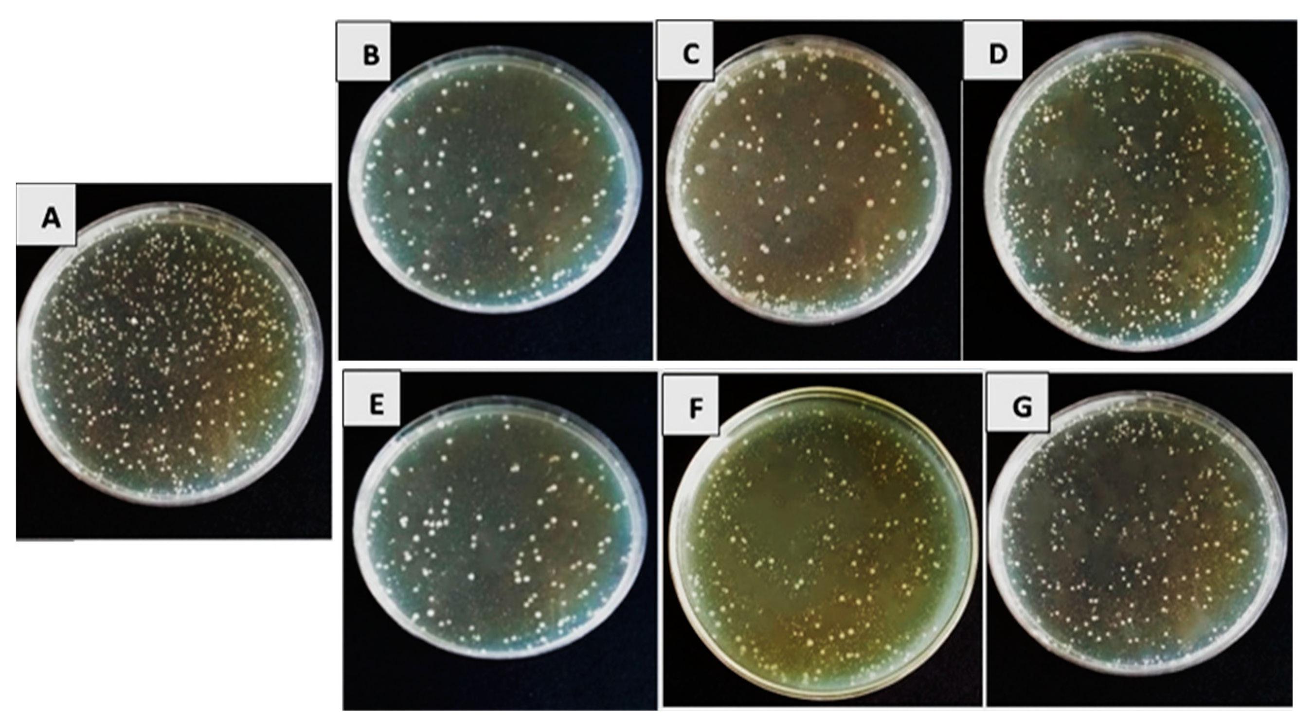
3.1. In Vitro Efficacy of Bioagents’ Filtrates on Bacterial Pathogens’ Growth Reduction

The data presented in Table 1 and Table 2, as well as Figure 1 and Figure 2, illustrate the impact of antagonistic products derived from the secondary metabolism of tested bacterial and fungal bioagents on the percentage reduction in growth of the targeted bacterial pathogens. The results clearly showed that the tested bioagents significantly slowed down the growth of pathogenic strains of Xanthomonas spp. and Pseudomonas spp. compared to the control treatment. The recorded data represent the percentage reduction in growth achieved.
At the highest concentration of 100%, the filtrates of B. subtilis demonstrated remarkable inhibition, resulting in the highest percentage reduction in bacterial pathogen growth (71.8%, 63.4%, 71.1%, and 66.7% for the tested pathogens Xcv1, Xe2, Pst1, and Pst2, respectively). Following closely, P. fluorescens exhibited notable inhibition as well, with reductions of 69.1%, 63.4%, 68.8%, and 65.1% against the respective pathogens. Conversely, S. marcescens, even at higher concentrations, showed comparatively lower effectiveness, achieving reductions of 43.8%, 36.7%, 47.1%, and 43.3% against the tested pathogens.
Table 2 also shows the outcomes of antagonistic culture filtrates made from secondary metabolic products from T. viride, T. harzianum, and T. album. We observed significant growth inhibition of Xanthomonas spp. and Pseudomonas sp. strains compared to the control treatment, where no reduction was evident. Trichoderma viride demonstrated the highest percentage growth reduction against all tested pathogens at the highest concentration (100%), with values of 73.7%, 71.0%, 68.0%, and 59.6%, respectively. The effectiveness followed a descending order of T. viride, T. harzianum, and T. album. We noted the differences in concentrations and selected 100% to evaluate their impact on tomato seedlings under greenhouse conditions. Conversely, the limited effectiveness of S. marcescens in inhibiting the growth of pathogenic bacteria suggests that it is unsuitable for use in greenhouse conditions. Figure 1 shows the In vitro effectiveness of various culture filtrates of bioagents against Xcv1 isolate growth. Similarly, Figure 2 illustrates the In vitro efficacy of the same culture filtrates against Pst1 isolate growth.

3.2. In Vivo Assessment

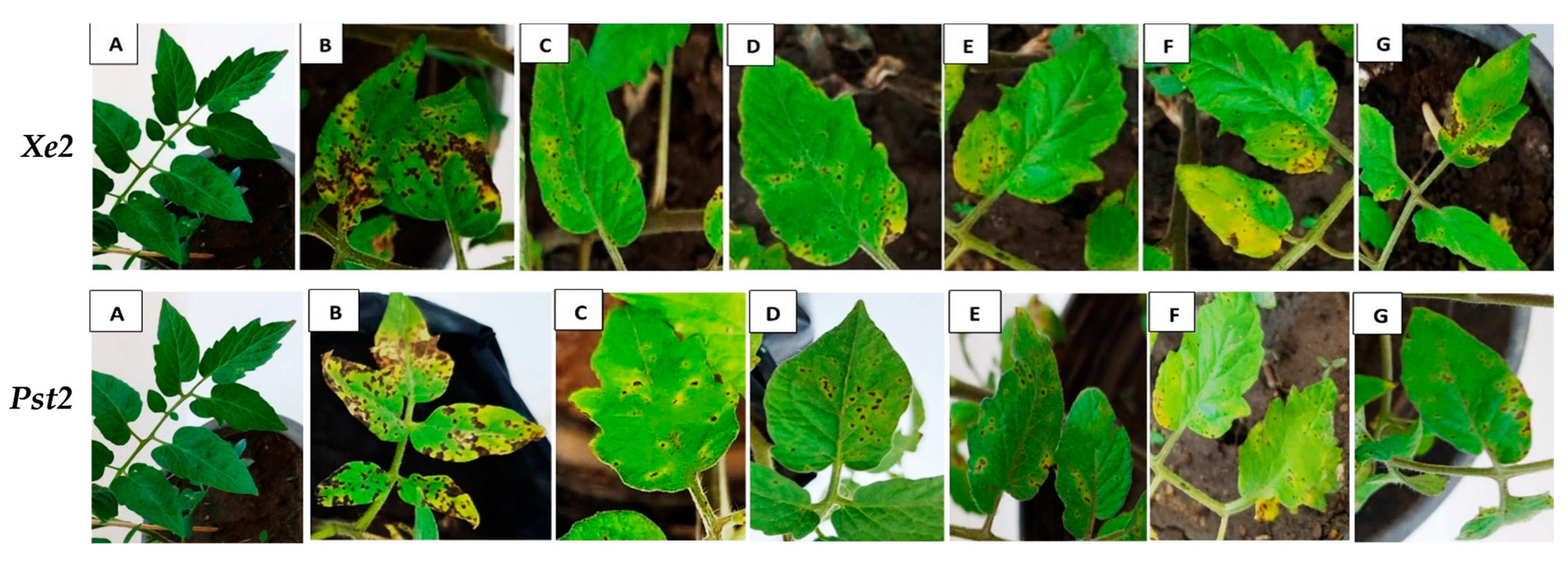
3.2.1. Efficacy of Bioagents’ Culture Filtrates on Leaf Spot and Speck Diseases in Tomato Plants

The In vivo efficacy of tested secondary metabolites (100% culture filtrates) from bacterial and fungal bioagents on disease incidence and severity is analyzed in Table 3 and Figure 3. In most cases studied, B. subtilis emerged as the most effective inhibiting bioagent, resulting in a significant percentage decrease in disease incidence compared to P. fluorescens. Specifically, B. subtilis achieved reductions of 56.9%, 54.8%, 41.7%, and 34.9% in disease incidence caused by Xcv1, Xe2, Pst1, and Pst2 bacterial pathogens, respectively, compared to the positive infected control. Meanwhile, P. fluorescens achieved reductions of 43.0%, 39.7%, 34.9%, and 33.4% against the respective pathogens.
Similarly, in terms of disease severity, B. subtilis demonstrated significant reductions of 65.4%, 60.0%, 56.6%, and 51.5% for Xcv1, Xe2, Pst1, and Pst2, respectively. Interestingly, disease severity was more affected by the tested bacterial bioagents compared to disease incidence. Among the Trichoderma spp. bioagents, T. viride was the most effective, achieving reductions of 53.8%, 50.8%, 47.7%, and 39.5% in disease incidence for Xcv1, Xe2, Pst1, and Pst2, respectively. Its effect on disease severity was also notable, recording reductions of 58.3%, 52.3%, 51.1%, and 41.5% for Xcv1, Xe2, Pst1, and Pst2, respectively, compared to the positive control. The effectiveness of T. viride was followed by T. harzianum and T. album in descending order.

3.2.2. In Vivo Changes in Phenolic Compounds

The results obtained in Table 4 indicate that P. fluorescens had a more significant impact on increasing phenol contents in the leaves, including total phenols, free phenols, and bound phenols, compared to B. subtilis. Similarly, among the Trichoderma spp., T. viride exhibited the highest efficacy in enhancing phenol contents (total, free, and bound), followed by T. harzianum and T. album, in comparison with the untreated control. It is noteworthy that the phenol contents in infected tomato seedlings treated with the tested bioagents exhibited higher enzymatic activity values compared to untreated infected tomato seedlings. This suggests that the application of bioagents resulted in increased activity of enzymes associated with phenol metabolism in response to infection.

3.2.3. In Vivo Changes in Antioxidant Enzymes

The results presented in Table 5 show that B. subtilis exhibited the highest increase in the enzyme activities of chitinase, peroxidase, and polyphenol oxidase in tomato plants infected by the bacterial pathogens Xcv1, Xe2, Pst1, and Pst2, compared to P. fluorescens. In turn, T. viride was had most effective fungal bioagent on activity of chitinase, peroxidase, and polyphenol oxidase, followed by T. harizianum and T. album, compared with the untreated infected control. Notable variations in enzyme activity were observed across treatments. The most effective bioagents, ranked based on their impact on enzyme activity, were B. subtilis, T. viride, P. fluorescens, T. harzianum, and T. album. These bioagents exhibited significant differences (p < 0.05) in enzyme activity compared to the infected control group.

3.2.4. Changes in Chlorophyll a, Chlorophyll b and Carotenoids

The data obtained in Table 6 revealed that B. subtilis exhibited the most pronounced inhibition against tomato bacterial spot and speck diseases, leading to a significant increase in Chl.a, Chl.b, and Carto., followed by P. fluorescens. Similarly, among the Trichoderma spp., T. viride demonstrated the most significant effectiveness in enhancing Chl.a, Chl.b, and Carto., followed by T. harzianum and T. album at a concentration of 100%, when compared with the untreated control.

4. Discussion

Managing bacterial spots or specks on tomatoes poses challenges due to the limited availability of effective chemical control measures and commercially resistant varieties. Breeding tomatoes for resistance to bacterial spots and specks faces complexity due to various factors, including the emergence of new pathogen species capable of overcoming existing resistance traits [58]. In recent decades, various strategies have been proposed to encourage the application of copper-based agrochemicals in the treatment of diseases caused by various Xanthomonas species. These agrochemicals partially or completely mitigate the disease caused by P. syringe pv. tomato, reducing the harmful effects of chemical use on consumers and the environment. Among these alternatives, biocontrol stands out as an effective, environmentally friendly, and economical solution [59].
The In vitro study showed that the culture filtrate supernatant (CFS) from the secondary metabolites of B. subtilis significantly inhibited the growth of pathogenic Xanthomonas spp. (Xcv1 and Xe2) and Pseudomonas spp. strains (Pst1 and Pst2) in Petri plates. The highest inhibition of growth was observed at the maximum CFS concentrations from B. subtilis and P. fluorescens. In contrast, S. marcescens was the least effective bioagent against the pathogenic bacteria, so it was not included in further tests on tomato seedlings under In vivo conditions. Among the Trichoderma species, T. viride showed the greatest inhibition effect, followed by T. harzianum and T. album. Studies suggest that these variations in the In vitro efficacy might result from differences in the composition and concentration of cell-free supernatants, including antibiotics [60], lytic enzymes, and siderophores [61].
Culture filtrates (CF) from biocontrol agents have also shown effectiveness in reducing plant pathogens in a range of other crops. For example, culture filtrates from Bacillus amyloliquefaciens RS-25, B. subtilis Z-14, B. licheniformis MG-4, and B. subtilis Pnf-4 have been found to suppress gray mold caused by Botrytis cinerea on postharvest fruits, including tomato, grapefruit, and strawberry [62]. Culture filtrates of Trichoderma species, including T. harzianum, T. longibrachiatum, and T. atroviride, were shown to inhibit Alternaria solani in tomato plants (variety: doucen), demonstrating potential for reducing early blight symptoms under greenhouse and field conditions. The CF-treated plants showed higher antioxidant enzymes, higher flavonoids, and increased phenolic compounds [63]. Moreover, Trichoderma virens and Bacillus velezensis culture filtrates have exhibited antimicrobial activity against Ralstonia solanacearum in tomato plants, reducing the severity of bacterial wilt [64].
Upon reaching the surface of tomato leaves, bacterial cells commence colonization of the phyllosphere, a process previously observed via confocal laser detection and scanning microscopy [65]. These bacteria can inhabit various epidermal cell structures, like stomata, trichomes, lenticels, and hydathodes, through which they penetrate the host plant. Pathogens responsible for bacterial spots or specks typically enter the leaf apoplast via stomata, multiplying in the intercellular and substomatal spaces. As the disease progresses, invasion by bacterial pathogens and the subsequent degradation of chlorophylls (Chl.a, Chl.b, and Carto.) within plant cells lead to widespread necrosis, resulting in the loss of phyllosphere individuals. Application of culture filtrates (CFSs) as a bioactive agent has been shown to mitigate these symptoms in infected plants.
The In vivo current data revealed that sprayed B. subtilis CFSs significantly decreased the percentage of disease incidence and severity, followed by P. fluorescence, compared with control treatment. Among the Trichoderma spp., T. viride exhibited the highest efficacy in reducing both disease incidence and severity in sprayed infected tomato plants, followed by T. harzianum and T. album. The success of our study in preventing tomato spot infections may be attributed to various scientific theories elucidated by numerous investigations. According to [66], there has been a notable rise in the utilization and prevalence of Bacillus spp. with antagonistic properties for disease management. Bacillus spp. possess distinctive attributes, including rapid replication rates, resilience to adverse environmental conditions, and a wide-ranging capacity for biocontrol. Certain Bacillus species possess an exceptional capacity to withstand unfavorable environmental circumstances and reproduce quickly, giving them a wide range of biocontrol capabilities. Bacillus enhances plant resistance by producing antimicrobial antibiotics, toxins, hydrolytic enzymes, and lipopeptides [67]. Many antibiotics produced by B. subtilis have been proven to be broad-spectrum antibiotics, such as glycopeptides, which play a role in stimulating plant growth [68]. Specific strains of B. subtilis were documented to efficiently suppress Ralstonia wilt in particular plant hosts at specified concentrations [69]. Though B. subtilis is known for its production of various antimicrobial agents, including a wide array of lipopeptides, it also functions as a potent biosurfactant [70]. Pseudomonas spp. are strong candidates for biocontrol due to their fast growth, adaptability to various environments, and broad antibiotic synthesis capability [71]. These studies corroborate and align with the findings of our study.
Another explanation provided in this study categorizes the biocontrol bacteria utilized as members of a group known as (PGPB) or plant-promoting rhizobacteria (PGPR), primarily as a large number of them come from the rhizosphere [72]. Rhizobacterial communities often include Pseudomonas and Bacillus strains, which can encourage the development of plants and act as efficient biocontrol agents. Plants can develop systemic resistance to Bacillus species, which can also compete for habitats and hinder phytopathogens [73]. The primary biocontrol mechanism involves producing secondary metabolites that inhibit pathogens [74]. Commonly used biological control agents include multiple strains of Bacillus and Pseudomonas [71]. These bacteria directly combat plant pathogens by releasing bioactive compounds. Alternatively, they can act indirectly by inducing plant defense responses, as seen in the B. subtilis group [75]. Additionally, they form beneficial connections with plant roots, releasing biomolecules with antibiotic properties [76]. Pseudomonas spp., typically found in soil and belonging to the P. putida group [77], produce Xantholycine, a secondary metabolite with anti-iron oxidation activity that inhibits the growth of various pathogenic bacteria [77].
Trichoderma, a soil fungus used as a bioagent in this study, exhibits antagonistic properties against plant pathogens [78]. Its mechanisms include competition, mycoparasitism, and antibiosis [79]. Antibiosis involves releasing chemical products that inhibit pathogen growth, often through extracellular enzymes and antibiotics [80]. Trichoderma spp. also produces secondary metabolites like polyketides, pyrones, and terpenes with antifungal and antibacterial properties [81]. Crude extracts of T. harzianum have been shown to effectively inhibit various plant pathogenic bacteria at different concentrations [80].
Our findings demonstrate that the bacteria employed In vitro effectively inhibited the growth of Xcv1 and Xe2, as well as Pst1 and Pst2, effectively controlling tomato bacterial leaf spot and speck when their culture filtrates were applied to infected tomato seedlings. This effect may be attributed to the production of siderophores, amylases, and proteases. Siderophores and lytic enzymes contribute to competitive colonization and biological control within plant tissues. Additionally, siderophores may induce both systemic and local host resistance [82]. The ability of fluorescent dyes to create proteases and siderophores could contribute to their efficacy against bacterial pathogens responsible for spot formation [83].
The timing of biological agent application is critical for effective plant disease control. Pre-inoculation studies, like those by Ons et al. [84] and Scortichini [85], demonstrated that applying biocontrol agents a few days before pathogen exposure can reduce disease severity by promoting early colonization and resistance. Post-inoculation applications, as investigated by Wei et al. [86], showed that treating plants after infection can suppress disease progression by enhancing plant defense mechanisms. Simultaneous applications, as evidenced by Ajuna et al. [87], indicated that introducing biological agents at the same time as pathogens can reduce disease symptoms through direct competition and disruption of pathogen establishment.
Using crude secondary metabolites or culture filtrates (CFS) offers advantages over using the microorganisms that produce them, as they do not require the same level of competition and colonization in the plant’s environment. However, there are concerns regarding the limited representation of the genetic diversity available for synthesizing bioactive compounds, particularly with antibiotics from bacteria like B. amyloliquefaciens. While some antibiotics may reach efficient concentrations in the rhizosphere, others may not, posing challenges. Moreover, high concentrations of secondary metabolites may pose risks to crops and the environment, necessitating the identification of safe compounds or those effective at lower concentrations. Selecting the optimal application strategy is also crucial for success [88].
Microbial filtrates, especially those from strains like P. fluorescens and B. subtilis, are gaining attention for their role as biostimulants in agriculture [89]. These filtrates contain a variety of biologically active compounds, including siderophores, which are crucial for iron chelation, enhancing plant growth, and improving resistance to environmental stresses [89]. Siderophores facilitate iron uptake in plants, an essential micronutrient that plays a critical role in cellular processes and chlorophyll synthesis [90]. Recent studies have shown that the application of microbial filtrates not only boosts nutrient uptake but also activates soil and plant-associated beneficial microbiota [90]. This dual action enhances plant resilience against pathogens and abiotic stresses, promoting a more sustainable agricultural practice by reducing the reliance on chemical fertilizers and pesticides [91].
The cultural filtrates of the tested bioagents increased total phenols, chitinase, PO, and PPO activities in tomato leaves infected by Xanthomonas spp. and Pseudomonas strains compared to untreated infected plants. These findings suggest that spraying bioagent culture filtrates can enhance key defense mechanisms in tomato plants. Treated plants exhibited higher enzymatic activity, indicating an improved ability to combat plant pathogens. Oxidative enzymes like PO and PPO play a critical role in forming lignin and reinforcing cell structure, thus aiding in plant defense [92].
Phenolic compounds, which are among the most abundant secondary metabolites in plants, regulate products governing disease resistance in crops [93]. Studies show a positive correlation between higher phenolic content and increased plant disease resistance. Phenolic compounds, present ubiquitously in plants, are essential in various aspects of plant life, particularly in interactions with the environment [94]. Certain phenolics trigger plant defense responses against pathogens.
Infected pathogens significantly affect chloroplasts within plant cells, leading to visible symptoms on tomato leaves, such as bacterial spots or specks. These symptoms typically appear as dark, irregular, or circular water-soaked spots with chlorotic halos, as observed in the unsprayed control plants. Lesion growth exacerbates pigment degradation, reducing green photosynthetic tissue. Leaves from untreated infected plants showed increased yellowing and merging lesions, indicating reduced concentrations of key pigments like (Chl.a, Chl.b, Carto.), which are critical for light absorption and photochemical reactions [95]. The presence of chlorotic halos, a sign of chloroplast degradation, suggests that infections by Xanthomonas spp. and Pseudomonas strains might damage the thylakoid network, impairing CO2 fixation and electron transport [96].
Applying culture filtrates from the tested bacteria and fungi, which contain antimicrobial substances like extracellular antibiotics, polyketides, pyrones, and terpenes, to tomato seedling leaves two days before infection inhibits bacterial growth. These substances act by preventing bacterial pathogens from entering through stomatal openings, which reduces their ability to proliferate in the intercellular spaces, where they could infect leaf cells and degrade pigments such as carotenoids and chlorophyll. As a result, the treated plants showed higher concentrations of (Chl.a, Chl.b, and Carto.) compared to plants that were infected but not treated with culture filtrates. The improved pigment levels in treated plants suggest that the bioagents effectively prevent infection by blocking bacterial entry through the stomata.

5. Conclusions

This study highlights the significant threat posed by bacterial spot diseases to tomato production, emphasizing the urgent need for effective management strategies to safeguard yield and fruit quality. Among the microbial groups tested, T. viride stood out as the most effective fungal bioagent, primarily due to its robust antipathogenic activity and ability to significantly enhance plant defense enzymes. Its superiority could be attributed to its aggressive colonization and higher competitive saprophytic ability, which makes it an excellent candidate for biocontrol applications. For the bacterial bioagents, B. subtilis emerged as the most potent, possibly due to its ability to produce a wide range of antibiotics and induce systemic resistance within the plant. This bacterial species showed not only high efficacy in reducing disease severity but also enhanced the biochemical pathways involved in plant defense, as evidenced by increased levels of peroxidase and polyphenol oxidase activities. The results suggest that preemptive application of these biocontrol agents—specifically, two days prior to potential pathogen attack—provides an optimal window for the agents to establish themselves and activate plant defense mechanisms. This timing is crucial for ensuring that the plants are sufficiently primed against incoming pathogens, thereby reducing the incidence and severity of the diseases. Overall, our study not only highlights the potential of using Trichoderma and Bacillus species as biocontrol agents but also provides a foundation for further research into their practical applications in sustainable agriculture. This approach promises not only to control disease but also to enhance the overall health and productivity of tomato plants, paving the way for more resilient agricultural practices.

Author Contributions

Conceptualization, M.R.A.T. and M.A.S.A.; methodology, A.H.A.; software, A.S.M.E. and A.M.K.; validation, M.A.S.A. and A.S.M.E.; formal analysis, A.H.A. and A.M.K.; investigation, A.H.A., M.A.S.A., A.M.K. and A.S.M.E.; resources, A.H.A. and A.S.M.E.; data curation, H.E.A., H.A.R., F.A.J. and A.S.M.E.; writing—original draft preparation, M.R.A.T., H.E.A. and H.A.R.; writing—review and editing, A.S.M.E., A.M.K. and M.A.S.A.; visualization, H.E.A. and H.A.R.; supervision, M.R.A.T.; project administration, F.A.J.; funding acquisition, H.E.A., H.A.R. and F.A.J. All authors have read and agreed to the published version of the manuscript.

Funding

Princess Nourah bint Abdulrahman University Researchers Supporting Project (number PNURSP2024R400), Princess Nourah bint Abdulrahman University, Riyadh, Saudi Arabia.

Institutional Review Board Statement

Not applicable.

Informed Consent Statement

Not applicable.

Data Availability Statement

The data presented in this study are available upon request from the corresponding author.

Acknowledgments

The authors acknowledge Princess Nourah bint Abdulrahman University, Riyadh, Saudi Arabia for funding this work.

Conflicts of Interest

The authors declare no conflicts of interest.

References

  1. FAO. Crops and Livestock Products. Available online: http://www.fao.org/FAOSTAT/en/ (accessed on 1 November 2020).
  2. Pohronezny, K.; Volin, R. The Effect of Bacterial Spot on Yield and Quality of Fresh Market Tomatoes1. HortScience 1983, 18, 69–70. [Google Scholar] [CrossRef]
  3. Perez, K.; Froikin-Gordon, J.S.; Abdourhamane, I.K.; Levasseur, V.; Alfari, A.A.; Mensah, A.; Bonsu, O.; Habsatou, B.; Assogba-Komlan, F.; Mbaye, A.A. Connecting smallholder tomato producers to improved seed in West Africa. Agric. Food Secur. 2017, 6, 42. [Google Scholar] [CrossRef]
  4. Raymaekers, K.; Ponet, L.; Holtappels, D.; Berckmans, B.; Cammue, B.P. Screening for novel biocontrol agents applicable in plant disease management—A review. Biol. Control 2020, 144, 104240. [Google Scholar] [CrossRef]
  5. Potnis, N. Harnessing Eco-Evolutionary dynamics of Xanthomonads on tomato and pepper to tackle new problems of an old disease. Annu. Rev. Phytopathol. 2021, 59, 289–310. [Google Scholar] [CrossRef]
  6. Savary, S.; Willocquet, L.; Pethybridge, S.J.; Esker, P.; McRoberts, N.; Nelson, A. The global burden of pathogens and pests on major food crops. Nat. Ecol. Evol. 2019, 3, 430–439. [Google Scholar] [CrossRef] [PubMed]
  7. Wai, K.P.P.; Siddique, M.I.; Mo, H.-S.; Yoo, H.J.; Byeon, S.-E.; Jegal, Y.; Mekuriaw, A.A.; Kim, B.-S. Pathotypes of bacterial spot pathogen infecting Capsicum peppers in Korea. Plant Pathol. J. 2015, 31, 428. [Google Scholar] [CrossRef]
  8. Jones, J.B.; Zitter, T.A.; Momol, T.M.; Miller, S.A. Compendium of Tomato Diseases and Pests; APS Press: College Park, MD, USA, 2014. [Google Scholar]
  9. Farias, G.A.; Olmedilla, A.; Gallegos, M.T. Visualization and characterization of Pseudomonas syringae pv. tomato DC 3000 pellicles. Microb. Biotechnol. 2019, 12, 688–702. [Google Scholar] [CrossRef] [PubMed]
  10. Abrahamian, P.; Jones, J.B.; Vallad, G.E. Efficacy of copper and copper alternatives for management of bacterial spot on tomato under transplant and field production. Crop Prot. 2019, 126, 104919. [Google Scholar] [CrossRef]
  11. Šević, M.; Gašić, K.; Ignjatov, M.; Mijatović, M.; Prokić, A.; Obradović, A. Integration of biological and conventional treatments in control of pepper bacterial spot. Crop Prot. 2019, 119, 46–51. [Google Scholar] [CrossRef]
  12. Herbert, A.; Hancock, C.N.; Cox, B.; Schnabel, G.; Moreno, D.; Carvalho, R.; Jones, J.; Paret, M.; Geng, X.; Wang, H. Oxytetracycline and streptomycin resistance genes in Xanthomonas arboricola pv. pruni, the causal agent of bacterial spot in peach. Front. Microbiol. 2022, 13, 821808. [Google Scholar] [CrossRef]
  13. Gikas, G.D.; Parlakidis, P.; Mavropoulos, T.; Vryzas, Z. Particularities of fungicides and factors affecting their fate and removal efficacy: A review. Sustainability 2022, 14, 4056. [Google Scholar] [CrossRef]
  14. El-Gremi, S.M.; Draz, I.S.; Youssef, W.A.-E. Biological control of pathogens associated with kernel black point disease of wheat. Crop Prot. 2017, 91, 13–19. [Google Scholar] [CrossRef]
  15. Sharma, V.; Salwan, R.; Sharma, P. The comparative mechanistic aspects of Trichoderma and probiotics: Scope for future research. Physiol. Mol. Plant Pathol. 2017, 100, 84–96. [Google Scholar] [CrossRef]
  16. Segarra, G.; Casanova, E.; Avilés, M.; Trillas, I. Trichoderma asperellum strain T34 controls Fusarium wilt disease in tomato plants in soilless culture through competition for iron. Microb. Ecol. 2010, 59, 141–149. [Google Scholar] [CrossRef] [PubMed]
  17. Marzano, M.; Gallo, A.; Altomare, C. Improvement of biocontrol efficacy of Trichoderma harzianum vs. Fusarium oxysporum f. sp. lycopersici through UV-induced tolerance to fusaric acid. Biol. Control 2013, 67, 397–408. [Google Scholar]
  18. Thayer, P.; Stall, R. The survey of Xanthomonas vesicatoria resistance to streptomycin. Proc. Fla. State Hortic. Soc. 1962, 75, 163–165. [Google Scholar]
  19. Vallad, G.E.; Pernezny, K.L.; Balogh, B.; Wen, A.; Figueiredo, J.F.L.; Jones, J.B.; Momol, T.; Muchovej, R.M.; Havranek, N.; Abdallah, N. Comparison of kasugamycin to traditional bactericides for the management of bacterial spot on tomato. HortScience 2010, 45, 1834–1840. [Google Scholar] [CrossRef]
  20. Narayanasamy, P. Biological Management of Diseases of Crops; Springer: Berlin/Heidelberg, Germany, 2013; Volume 673. [Google Scholar]
  21. Dimkić, I.; Janakiev, T.; Petrović, M.; Degrassi, G.; Fira, D. Plant-associated Bacillus and Pseudomonas antimicrobial activities in plant disease suppression via biological control mechanisms—A review. Physiol. Mol. Plant Pathol. 2022, 117, 101754. [Google Scholar] [CrossRef]
  22. Byrne, J.; Dianese, A.; Ji, P.; Campbell, H.; Cuppels, D.; Louws, F.; Miller, S.; Jones, J.; Wilson, M. Biological control of bacterial spot of tomato under field conditions at several locations in North America. Biol. Control 2005, 32, 408–418. [Google Scholar] [CrossRef]
  23. Ji, P.; Campbell, H.; Kloepper, J.; Jones, J.; Suslow, T.; Wilson, M. Integrated biological control of bacterial speck and spot of tomato under field conditions using foliar biological control agents and plant growth-promoting rhizobacteria. Biol. Control 2006, 36, 358–367. [Google Scholar] [CrossRef]
  24. Hemlata Chauhan, H.C.; Bagyaraj, D.; Selvakumar, G.; Sundaram, S. Novel plant growth promoting rhizobacteria-prospects and potential. Appl. Soil Ecol. 2015, 95, 38–53. [Google Scholar] [CrossRef]
  25. Wu, B.; Wang, X.; Yang, L.; Yang, H.; Zeng, H.; Qiu, Y.; Wang, C.; Yu, J.; Li, J.; Xu, D. Effects of Bacillus amyloliquefaciens ZM9 on bacterial wilt and rhizosphere microbial communities of tobacco. Appl. Soil Ecol. 2016, 103, 1–12. [Google Scholar] [CrossRef]
  26. Chandrasekaran, M.; Paramasivan, M.; Chun, S.-C. Bacillus subtilis CBR05 induces Vitamin B6 biosynthesis in tomato through the de novo pathway in contributing disease resistance against Xanthomonas campestris pv. vesicatoria. Sci. Rep. 2019, 9, 6495. [Google Scholar] [CrossRef] [PubMed]
  27. Kavitha, R.; Umesha, S. Regulation of defense-related enzymes associated with bacterial spot resistance in tomato. Phytoparasitica 2008, 36, 144–159. [Google Scholar] [CrossRef]
  28. Lattanzio, V.; Lattanzio, V.M.; Cardinali, A. Role of phenolics in the resistance mechanisms of plants against fungal pathogens and insects. Phytochem. Adv. Res. 2006, 661, 23–67. [Google Scholar]
  29. Dicko, M.H.; Gruppen, H.; Traoré, A.S.; Voragen, A.G.; van Berkel, W.J. Phenolic compounds and related enzymes as determinants of sorghum for food use. Biotechnol. Mol. Biol. Rev. 2006, 1, 20–37. [Google Scholar]
  30. Umesha, S. Occurrence of bacterial canker in tomato fields of Karnataka and effect of biological seed treatment on disease incidence. Crop Prot. 2006, 25, 375–381. [Google Scholar] [CrossRef]
  31. Backer, R.; Rokem, J.S.; Ilangumaran, G.; Lamont, J.; Praslickova, D.; Ricci, E.; Subramanian, S.; Smith, D.L. Plant growth-promoting rhizobacteria: Context, mechanisms of action, and roadmap to commercialization of biostimulants for sustainable agriculture. Front. Plant Sci. 2018, 9, 402666. [Google Scholar] [CrossRef]
  32. Saini, S.; Lohani, S.; Khati, P.; Rani, V. PGPR-mediated mitigation of biotic and abiotic stress in plants. In Advanced Microbial Technology for Sustainable Agriculture and Environment; Elsevier: Amsterdam, The Netherlands, 2023; pp. 199–227. [Google Scholar]
  33. El-Fatah, B.E.A.; Imran, M.; Abo-Elyousr, K.A.; Mahmoud, A.F. Isolation of Pseudomonas syringae pv. Tomato strains causing bacterial speck disease of tomato and marker-based monitoring for their virulence. Mol. Biol. Rep. 2023, 50, 4917–4930. [Google Scholar] [CrossRef]
  34. Kyeon, M.-S.; Son, S.-H.; Noh, Y.-H.; Kim, Y.-E.; Lee, H.-I.; Cha, J.-S. Xanthomonas euvesicatoria causes bacterial spot disease on pepper plant in Korea. Plant Pathol. J. 2016, 32, 431. [Google Scholar] [CrossRef]
  35. Khan, Z.; Kim, S.; Jeon, Y.; Khan, H.; Son, S.; Kim, Y. A plant growth promoting rhizobacterium, Paenibacillus polymyxa strain GBR-1, suppresses root-knot nematode. Bioresour. Technol. 2008, 99, 3016–3023. [Google Scholar] [CrossRef]
  36. Brian, P.; Hemming, H. Gliotoxin, a fungistatic metabolic product of Trichoderma viride. Ann. Appl. Biol. 1945, 32, 214–220. [Google Scholar] [CrossRef]
  37. Bonilla, N.; Vida, C.; Martínez-Alonso, M.; Landa, B.B.; Gaju, N.; Cazorla, F.M.; De Vicente, A. Organic amendments to avocado crops induce suppressiveness and influence the composition and activity of soil microbial communities. Appl. Environ. Microbiol. 2015, 81, 3405–3418. [Google Scholar] [CrossRef] [PubMed]
  38. Breitling, R.; Ceniceros, A.; Jankevics, A.; Takano, E. Metabolomics for secondary metabolite research. Metabolites 2013, 3, 1076–1083. [Google Scholar] [CrossRef]
  39. Ghorbanpour, M.; Omidvari, M.; Abbaszadeh-Dahaji, P.; Omidvar, R.; Kariman, K. Mechanisms underlying the protective effects of beneficial fungi against plant diseases. Biol. Control 2018, 117, 147–157. [Google Scholar] [CrossRef]
  40. Ghoneem, K.M.; Khalil, A.A.; Rashad, E.M.; Ahmed, M.I.; Mahmoud, M.S. Granular bioactive formulation of Trichoderma viride and Arbuscular mycorrhizal fungi for biological control of Cumin wilt disease. Egypt. J. Phytopathol. 2019, 47, 175–197. [Google Scholar] [CrossRef]
  41. da Silva, R.S.; Moutinho, B.L.; dos Santos, D.R.; Vasconcelo-Rodrigues, I.; Talamini, V.; Fernandes, M.F.; Fernandes, R.P. Using antagonistic soil bacteria and their cell-free filtrates to control the black rot pathogen Xanthomonas campestris pv. campestris. J. Phytopathol. 2018, 166, 494–501. [Google Scholar] [CrossRef]
  42. Moustaine, M.; El Kahkahi, R.; Benbouazza, A.; Benkirane, R. Potential of biological treatments for control of bacterial canker of tomato incited by Clavibacter michiganensis spp michiganensis in Morocco. EurAsian J. BioSciences 2019, 13, 1481–1488. [Google Scholar]
  43. Romano, N.; Lignola, G.; Brigante, M.; Bosso, L.; Chirico, G.B. Residual life and degradation assessment of wood elements used in soil bioengineering structures for slope protection. Ecol. Eng. 2016, 90, 498–509. [Google Scholar] [CrossRef]
  44. Sallam, N.; Ali, E.F.; Seleim, M.A.; Khalil Bagy, H.M. Endophytic fungi associated with soybean plants and their antagonistic activity against Rhizoctonia solani. Egypt. J. Biol. Pest Control 2021, 31, 1–9. [Google Scholar] [CrossRef]
  45. Suresh, S.; Mathiyazhalagan, N.; Selvam, R. Evaluation of bioagents and bactericides for controlling the Bacterial spot Disease in tomato. South Asian J. Eng. Technol. 2015, 1, 1–10. [Google Scholar]
  46. Gwa, V.; Ekefan, E. Fungal organisms isolated from rotted white yam (Dioscorea rotundata) tubers and antagonistic potential of Trichoderma harzianum against Colletotrichum species. Agric. Res. Tech. Open Access J. 2017, 10, 555787. [Google Scholar]
  47. Hang, N.T.T.; Oh, S.O.; Kim, G.H.; Hur, J.S.; Koh, Y.J. Bacillus subtilis S1-0210 as a biocontrol agent against Botrytis cinerea in strawberries. Plant Pathol. J. 2005, 21, 59–63. [Google Scholar] [CrossRef]
  48. Vitória, A.P.; Lea, P.J.; Azevedo, R.A. Antioxidant enzymes responses to cadmium in radish tissues. Phytochemistry 2001, 57, 701–710. [Google Scholar] [CrossRef] [PubMed]
  49. Mergawy, M.M.; Metwaly, H.A.; Shoeip, A.M. Evaluation of the efficacy of some bioagents accompanied with Bio-and Mineral Fertilizers in controlling early blight of tomato and improvement yield. Egypt. J. Phytopathol. 2022, 50, 31–50. [Google Scholar] [CrossRef]
  50. Lisker, N.; Cohen, L.; Chalutz, E.; Fuchs, Y. Fungal infections suppress ethylene-induced phenylalanine ammonia-lyase activity in grapefruits. Physiol. Plant Pathol. 1983, 22, 331–338. [Google Scholar] [CrossRef]
  51. Boller, T.; Gehri, A.; Mauch, F.; Vögeli, U. Chitinase in bean leaves: Induction by ethylene, purification, properties, and possible function. Planta 1983, 157, 22–31. [Google Scholar] [CrossRef]
  52. Legrand, M.; Kauffmann, S.; Geoffroy, P.; Fritig, B. Biological function of pathogenesis-related proteins: Four tobacco pathogenesis-related proteins are chitinases. Proc. Natl. Acad. Sci. USA 1987, 84, 6750–6754. [Google Scholar] [CrossRef]
  53. Reissig, J.L.; Strominger, J.L.; Leloir, L.F. A modified colorimetric method for the estimation of N-acetylamino sugars. J. Biol. Chem. 1955, 217, 959–966. [Google Scholar] [CrossRef]
  54. Boller, T.; Metraux, J. Extracellular localization of chitinase in cucumber. Physiol. Mol. Plant Pathol. 1988, 33, 11–16. [Google Scholar] [CrossRef]
  55. Snell, F.D.; Snell, C.T. Colorimetric Methods of Analysis; D. Van Nostrand Company Inc.: Princeton, NJ, USA; Toronto, ON, Canada; New York, NY, USA; London, UK, 1959; 606p. [Google Scholar]
  56. Fadeel, A. Assimilation of carbon dioxide by chlorophyllous roots. Physiol. Plant. 1963, 16, 870–888. [Google Scholar] [CrossRef]
  57. Ibrahim, S.; Abo-ELwafa, T. Alleviation harmful impacts of salinity using some antioxidants substances on Thompson seedless grapevines seedlings. J. Plant Prod. 2018, 9, 39–49. [Google Scholar] [CrossRef]
  58. Adhikari, P.; Adhikari, T.B.; Louws, F.J.; Panthee, D.R. Advances and challenges in bacterial spot resistance breeding in tomato (Solanum lycopersicum L.). Int. J. Mol. Sci. 2020, 21, 1734. [Google Scholar] [CrossRef] [PubMed]
  59. Peng, Y.; Li, S.J.; Yan, J.; Tang, Y.; Cheng, J.P.; Gao, A.J.; Yao, X.; Ruan, J.J.; Xu, B.L. Research progress on phytopathogenic fungi and their role as biocontrol agents. Front. Microbiol. 2021, 12, 670135. [Google Scholar] [CrossRef] [PubMed]
  60. Défago, G.; Haas, D. Pseudomonads as antagonists of soilborne plant pathogens: Modes of action and genetic analysis. In Soil Biochemistry; Routledge: London, UK, 2017; pp. 249–292. [Google Scholar]
  61. Bakker, P.; Peer, R.V.; Schippers, B. Specificity of siderophores and siderophore receptors and biocontrol by Pseudomonas spp. Biol. Control Soil-Borne Plant Pathogens 1990, 131–142. [Google Scholar]
  62. Chen, X.; Wang, Y.; Gao, Y.; Gao, T.; Zhang, D. Inhibitory abilities of Bacillus isolates and their culture filtrates against the gray mold caused by Botrytis cinerea on postharvest fruit. Plant Pathol. J. 2019, 35, 425. [Google Scholar] [CrossRef] [PubMed]
  63. Imran, M.; Abo-Elyousr, K.A.M.; Mousa, M.A.A.; Saad, M.M. Use of Trichoderma culture filtrates as a sustainable approach to mitigate early blight disease of tomato and their influence on plant biomarkers and antioxidants production. Front. Plant Sci. 2023, 14, 1192818. [Google Scholar] [CrossRef]
  64. Zhou, Y.; Yang, L.; Wang, J.; Guo, L.; Huang, J. Synergistic Effect between Trichoderma virens and Bacillus velezensis on the Control of Tomato Bacterial Wilt Disease. Horticulturae 2021, 7, 439. [Google Scholar] [CrossRef]
  65. Zhang, Y.; Callaway, E.M.; Jones, J.B.; Wilson, M. Visualisation of hrp gene expression in Xanthomonas euvesicatoria in the tomato phyllosphere. Eur. J. Plant Pathol. 2009, 124, 379–390. [Google Scholar] [CrossRef]
  66. Shafi, J.; Tian, H.; Ji, M. Bacillus species as versatile weapons for plant pathogens: A review. Biotechnol. Biotechnol. Equip. 2017, 31, 446–459. [Google Scholar] [CrossRef]
  67. Bardin, M.; Ajouz, S.; Comby, M.; Lopez-Ferber, M.; Graillot, B.; Siegwart, M.; Nicot, P.C. Is the efficacy of biological control against plant diseases likely to be more durable than that of chemical pesticides? Front. Plant Sci. 2015, 6, 566. [Google Scholar] [CrossRef]
  68. Khan, M.A.; Zhao, Y.; Korban, S.S. Molecular mechanisms of pathogenesis and resistance to the bacterial pathogen Erwinia amylovora, causal agent of fire blight disease in Rosaceae. Plant Mol. Biol. Report. 2012, 30, 247–260. [Google Scholar] [CrossRef]
  69. Aliye, N.; Fininsa, C.; Hiskias, Y. Evaluation of rhizosphere bacterial antagonists for their potential to bioprotect potato (Solanum tuberosum) against bacterial wilt (Ralstonia solanacearum). Biol. Control 2008, 47, 282–288. [Google Scholar] [CrossRef]
  70. Peypoux, F.; Bonmatin, J.; Wallach, J. Recent trends in the biochemistry of surfactin. Appl. Microbiol. Biotechnol. 1999, 51, 553–563. [Google Scholar] [CrossRef] [PubMed]
  71. Santoyo, G.; Orozco-Mosqueda, M.d.C.; Govindappa, M. Mechanisms of biocontrol and plant growth-promoting activity in soil bacterial species of Bacillus and Pseudomonas: A review. Biocontrol Sci. Technol. 2012, 22, 855–872. [Google Scholar] [CrossRef]
  72. Labuschagne, N.; Pretorius, T.; Idris, A. Plant growth promoting rhizobacteria as biocontrol agents against soil-borne plant diseases. Plant Growth Health Promot. Bact. 2011, 211–230. [Google Scholar]
  73. Fira, D.; Dimkić, I.; Berić, T.; Lozo, J.; Stanković, S. Biological control of plant pathogens by Bacillus species. J. Biotechnol. 2018, 285, 44–55. [Google Scholar] [CrossRef]
  74. Bacon, C.W.; Palencia, E.R.; Hinton, D.M. Abiotic and biotic plant stress-tolerant and beneficial secondary metabolites produced by endophytic Bacillus species. In Plant Microbes Symbiosis: Applied Facets; Springer: Berlin/Heidelberg, Germany, 2014; pp. 163–177. [Google Scholar]
  75. Glick, B.R.; Glick, B.R. Biocontrol of bacteria and fungi. Benef. Plant-Bact. Interact. 2020, 181–230. [Google Scholar]
  76. Rabbee, M.F.; Ali, M.S.; Choi, J.; Hwang, B.S.; Jeong, S.C.; Baek, K.-H. Bacillus velezensis: A valuable member of bioactive molecules within plant microbiomes. Molecules 2019, 24, 1046. [Google Scholar] [CrossRef]
  77. Pascual, J.; García-López, M.; Carmona, C.; Sousa, T.d.S.; de Pedro, N.; Cautain, B.; Martín, J.; Vicente, F.; Reyes, F.; Bills, G.F. Pseudomonas soli sp. nov., a novel producer of xantholysin congeners. Syst. Appl. Microbiol. 2014, 37, 412–416. [Google Scholar] [CrossRef]
  78. Cook, R.J.; Baker, K.F. The Nature and Practice of Biological Control of Plant Pathogens; American Phytopathological Society: St. Paul, MN, USA, 1983. [Google Scholar]
  79. Howell, C. Mechanisms employed by Trichoderma species in the biological control of plant diseases: The history and evolution of current concepts. Plant Dis. 2003, 87, 4–10. [Google Scholar] [CrossRef] [PubMed]
  80. Leelavathi, M.; Vani, L.; Reena, P. Antimicrobial activity of Trichoderma harzianum against bacteria and fungi. Int. J. Curr. Microbiol. Appl. Sci 2014, 3, 96–103. [Google Scholar]
  81. Naher, L.; Yusuf, U.K.; Ismail, A.; Hossain, K. Trichoderma spp.: A biocontrol agent for sustainable management of plant diseases. Pak. J. Bot 2014, 46, 1489–1493. [Google Scholar]
  82. Aznar, A.; Dellagi, A. New insights into the role of siderophores as triggers of plant immunity: What can we learn from animals? J. Exp. Bot. 2015, 66, 3001–3010. [Google Scholar] [CrossRef]
  83. Liu, K.; Garrett, C.; Fadamiro, H.; Kloepper, J.W. Induction of systemic resistance in Chinese cabbage against black rot by plant growth-promoting rhizobacteria. Biol. Control 2016, 99, 8–13. [Google Scholar] [CrossRef]
  84. Ons, L.; Bylemans, D.; Thevissen, K.; Cammue, B.P. Combining biocontrol agents with chemical fungicides for integrated plant fungal disease control. Microorganisms 2020, 8, 1930. [Google Scholar] [CrossRef] [PubMed]
  85. Scortichini, M. Sustainable management of diseases in horticulture: Conventional and new options. Horticulturae 2022, 8, 517. [Google Scholar] [CrossRef]
  86. Wei, L.; Wang, D.; Gupta, R.; Kim, S.T.; Wang, Y. A proteomics insight into advancements in the rice–microbe interaction. Plants 2023, 12, 1079. [Google Scholar] [CrossRef]
  87. Ajuna, H.B.; Lim, H.-I.; Moon, J.-H.; Won, S.-J.; Choub, V.; Choi, S.-I.; Yun, J.-Y.; Ahn, Y.S. The Prospect of Hydrolytic Enzymes from Bacillus Species in the Biological Control of Pests and Diseases in Forest and Fruit Tree Production. Int. J. Mol. Sci. 2023, 24, 16889. [Google Scholar] [CrossRef]
  88. Debois, D.; Jourdan, E.; Smargiasso, N.; Thonart, P.; De Pauw, E.; Ongena, M. Spatiotemporal monitoring of the antibiome secreted by Bacillus biofilms on plant roots using MALDI mass spectrometry imaging. Anal. Chem. 2014, 86, 4431–4438. [Google Scholar] [CrossRef]
  89. Morcillo, R.J.L.; Baroja-Fernández, E.; López-Serrano, L.; Leal-López, J.; Muñoz, F.J.; Bahaji, A.; Férez-Gómez, A.; Pozueta-Romero, J. Cell-free microbial culture filtrates as candidate biostimulants to enhance plant growth and yield and activate soil- and plant-associated beneficial microbiota. Front. Plant Sci. 2022, 13, 1040515. [Google Scholar] [CrossRef] [PubMed]
  90. Nithyapriya, S.; Lalitha, S.; Sayyed, R.Z.; Reddy, M.S.; Dailin, D.J.; El Enshasy, H.A.; Luh Suriani, N.; Herlambang, S. Production, Purification, and Characterization of Bacillibactin Siderophore of Bacillus subtilis and Its Application for Improvement in Plant Growth and Oil Content in Sesame. Sustainability 2021, 13, 5394. [Google Scholar] [CrossRef]
  91. Ansari, M.; Devi, B.M.; Sarkar, A.; Chattopadhyay, A.; Satnami, L.; Balu, P.; Choudhary, M.; Shahid, M.A.; Jailani, A.A.K. Microbial Exudates as Biostimulants: Role in Plant Growth Promotion and Stress Mitigation. J. Xenobiotics 2023, 13, 572–603. [Google Scholar] [CrossRef]
  92. Avdiushko, S.; Ye, X.; Kuc, J. Detection of several enzymatic activities in leaf prints of cucumber plants. Physiol. Mol. Plant Pathol. 1993, 42, 441–454. [Google Scholar] [CrossRef]
  93. Fan, S.; Tian, F.; Li, J.; Hutchins, W.; Chen, H.; Yang, F.; Yuan, X.; Cui, Z.; Yang, C.H.; He, C. Identification of phenolic compounds that suppress the virulence of Xanthomonas oryzae on rice via the type III secretion system. Mol. Plant Pathol. 2017, 18, 555–568. [Google Scholar] [CrossRef]
  94. Sharifi-Rad, J.; Quispe, C.; Zam, W.; Kumar, M.; Cardoso, S.M.; Pereira, O.R.; Ademiluyi, A.O.; Adeleke, O.; Moreira, A.C.; Živković, J. Phenolic bioactives as antiplatelet aggregation factors: The pivotal ingredients in maintaining cardiovascular health. Oxidative Med. Cell. Longev. 2021, 2021, 2195902. [Google Scholar] [CrossRef] [PubMed]
  95. Bassanezi, R.; Yamamoto, P.; Gimenes-Fernandes, N. Progresso dos sintomas de “morte súbita” em pomares de laranjeiras ‘Valência’e ‘Pêra’. Summa Phytopathol. 2002, 28, 23. [Google Scholar]
  96. Han, S.-W.; Choi, M.-S.; Lee, S.-H.; Hwang, D.-J.; Hwang, B.-K.; Heu, S.-G. Characterization of a novel necrotic response of Glycine max line’PI96188’to Xanthomonas axonopodis pv. glycines. Plant Pathol. J. 2007, 23, 193–202. [Google Scholar] [CrossRef]
Figure 1. In vitro efficacy of different tested culture filtrates of tested bioagents at 100% concentration against Xanthomonas spp. isolate X. campestris pv. vesicatoria (Xcv1) growth: (A) Control; (B) B. subtilis; (C) P. fluorescens; (D) Serratia marcescens; (E) T. viride; (F) T. harzianum; (G) T. album.
Biology 13 00369 g001
Figure 2. In vitro efficacy of different tested culture filtrates of tested bioagents at 100% concentration against Pseudomonas spp. isolate P. syringae pv. tomato (Pst1) growth: (A) control; (B) B. subtilis; (C) P. fluorescens; (D) Serratia marcescens; (E) T. viride; (F) T. harzianum; (G) T. album.
Biology 13 00369 g002
Figure 3. In vivo efficacy of different tested secondary metabolites (culture filtrate) of bacteria at 100% against X. euvesicatoria (Xe2) and P. syringe pv. tomato (Pst2). Plants were treated with biological products two days before infection. The negative control was treated with Streptrol fungicide 21.3% at the same time, while the healthy control received no treatment and was not infected. (A) Healthy control; (B) infected control; (C) infected treated with B. subtilis; (D) infected treated with P. fluorescens; (E) infected treated with T. viride; (F) infected treated with T. harzianum; (G) infected treated with T. album.
Biology 13 00369 g003
Table 1. In vitro efficacy of different bacterial bioagent culture filtrates and their concentrations against X. campestris pv. vesicatoria (Xcv1), X. euvesicatoria (Xe2), and two strains of P. syringae pv. tomato (Pst1 and Pst2), measured as percentage growth reduction.
PathogenBioagent% Reduction
25%50%100%
X. campestris pv. vesicatoria
(Xcv1)
B. subtilis41.1 l56.3 gh71.8 a
P. fluorescens27.3 pqr54.5 h69.1 bc
S. marcescens25.7 r36.6 m43.8 k
X. euvesicatoria
(Xe2)
B. subtilis31.6 n55.0 h63.4 f
P. fluorescens17.9 t42.0 kl63.4 f
S. marcescens20.5 s28.6 pq36.7 m
P. syringae pv. tomato
(Pst1)
B. subtilis27.4 pqr64.1 f71.1 ab
P. fluorescens29.3 op58.3 g68.8 cd
S. marcescens26.4 qr31.0 no47.1 j
P. syringae pv. tomato
(Pst2)
B. subtilis21.6 s51.0 i66.7 de
P. fluorescens14.2 u63.7 f65.1 ef
S. marcescens21.8 s25.4 r43.3 k
LSDBacteria2.84
Bioagent1.96
bacteria × bioagent1.81
The assigned letters represent Duncan’s multiple range test outcomes at p < 0.05 significance. Different letters signify significant treatment differences, while similar ones denote non significance. ‘×’ indicates variable interaction.
Table 2. In vitro efficacy of different Trichoderma spp. culture filtrates and their concentrations against X. campestris pv. vesicatoria (Xcv1), X. euvesicatoria (Xe2), and two strains of P. syringae pv. tomato (Pst1 and Pst2), measured as percentage growth reduction.
PathogenBioagent%Reduction
25%50%100%
X. campestris pv. vesicatoria
(Xcv1)
T. viride43.1 i52.9 g73.7 a
T. harzianum25.7 m30.4 kl59.3 e
T. album20.3 n29.3 l41.9 i
X. euvesicatoria
(Xe2)
T. viride55.4 f64.5 d71.0 b
T. harzianum21.6 n32.5 k46.3 h
T. album16.9 o28.6 l36.6 j
P. syringae pv. tomato
(Pst1)
T. viride46.6 h51.2 g68.0 c
T. harzianum30.4 kl46.8 h55.9 f
T. album28.9 l37.7 j42.5 i
P. syringae pv. tomato
(Pst2)
T. viride28.9 l45.6 h59.6 e
T. harzianum17.2 o24.6 m43.1 i
T. album17.3 o20.1 n31.9 k
LSDBacteria3.34
Bioagent2.31
bacteria × bioagent2.13
The letters denote the results of the Duncan’s multiple range test with a significance level of p < 0.05. Distinct letters indicate significant variations in treatment, whereas similar letters indicate negligible relevance. ‘×’ indicates variable interaction.
Table 3. In vivo efficacy of different tested secondary metabolites (culture filtrates) of different bacteria and fungi bioagents at 100% concentration on disease incidence (DI) and disease severity (DS) of leaf spot and speck disease in tomato plants.
PathogenBioagent%Reduction
DI%DS%
X. campestris pv. vesicatoria
(Xcv1)
B. subtilis56.9 gh65.4 j
P. fluorescens43.0 b–f46.5 c–g
T. viride53.8 fgh58.3 hi
T. harzianum48.8 def55.0 f–i
T. album43.1 b46.8 ghi
Infected100.0 a90.5 ab
Healthy0.0 i0.0 k
X. euvesicatoria
(Xe2)
B. subtilis54.8 d–h60.0 hij
P. fluorescens39.7 bc43.6 cd
T. viride50.8 d–g52.3 e–i
T. harzianum45.4 bcde52.7 e–i
T. album36.7 bcd41.4 d–h
Infected100.0 a95.4 a
Healthy0.0 i0.0 k
P. syringae pv. tomato
(Pst1)
B.subtilis41.7 fgh56.6 ij
P. fluorescens34.9 c–f39.5 c–f
T. viride47.7 h51.1 ghi
T. harzianum43.5 fgh53.3 hi
T. album40.7 e–h44.8 g–h
Infected95.0 a80.0 b
Healthy0.0 i0.0 k
P. syringae pv. tomato
(Pst2)
B. subtilis34.9 bcde51.5 d–h
P. fluorescens33.4 bcd37.1 c
T. viride39.5 b–f41.5 cd
T. harzianum50.0 def40.5 d–h
T. album38.4 b–f39.8 cd
Infected100.0a85.5 ab
Healthy0.0 i0.0 k
LSDBacteria4.283.99
Bioagent5.675.27
bacteria × bioagent11.3310.55
The letters refer to Duncan’s multiple range test outcomes at p < 0.05 significance. Dissimilar letters indicate significant differences, whereas similar ones indicate non significance. ‘×’ denotes the interaction between variables.
Table 4. Activity of total phenol contents measured as mg/g fresh weight in leaves of infected tomato plant cv 102 by X. campestris pv. vesicatoria (Xcv1), X. euvesicatoria (Xe2), and two strains of P. syringae pv. tomato (Pst1) and (Pst2) on treated plants with bacteria and fungi bioagents culture filtrates at 100% concentration.
BioagentPathogenFree PhenolBound PhenolTotal Phenol
Infected controlHealthy control1.25 o0.50 v1.75 s
X.campestris pv. vesicatoria (Xcv1)1.55 gh0.68 s2.23 mn
X. euvesicatoria (Xe2) 1.51 ij0.65 tu2.17 o
P. syringae pv. tomato (Pst1)1.52 hij0.66 st2.18 no
P. syringae pv. tomato (Pst2)1.51 ijk0.66 tu2.16 o
B. subtilisHealthy control1.25 no0.64 u1.90 r
X. campestris pv. vesicatoria (Xcv1)1.46 l0.78 o2.24 m
X. euvesicatoria (Xe2) 1.48 kl0.80 n2.27 lm
P. syringae pv. tomato (Pst1)1.49 jkl0.81 mn2.30 kl
P. syringae pv. tomato (Pst2)1.50 ijk0.82 m2.32 jk
P. fluorescensHealthy control1.27 mn0.70 r1.98 q
X. campestris pv. vesicatoria (Xcv1)1.55 gh0.88 i2.43 gh
X. euvesicatoria (Xe2) 1.55 fgh0.89 hi2.44 g
P. syringae pv. tomato (Pst1)1.57 efg0.90 gh2.47 fg
P. syringae pv. tomato (Pst2)1.58 efg0.91 g2.49 ef
T. virideHealthy control1.26 no0.66 st1.92 r
X. campestris pv. vesicatoria (Xcv1)1.51 ij0.84 kl2.35 ij
X. euvesicatoria (Xe1) 1.50 ijk0.84 k2.34 ijk
P. syringae pv. tomato (Pst1)1.53 hi0.86 j2.38 hi
P. syringae pv. tomato (Pst2)1.53 hij0.86 j2.38 i
T. harzianumHealthy control1.31 m0.73 q2.04 cd
X. campestris pv. vesicatoria (Xcv1)1.59 de0.93 f2.52 de
X. euvesicatoria (Xe2) 1.59 def0.93 df2.52 de
P. syringae pv. tomato (Pst1)1.60 b-e0.95 de2.55 cd
P. syringae pv. tomato (Pst2)1.60 cde0.95 d2.55 cd
T. albumHealthy control1.33 m0.76 p2.09 s
X. campestris pv. vesicatoria (Xcv1)1.61 bcd0.97 c2.58 bc
X. euvesicatoria (Xe2) 1.64 ab0.99 b2.62 ab
P. syringae pv. tomato (Pst1)1.63 abc0.99 b2.62 ab
P. syringae pv. tomato (Pst2)1.66 a1.01 a2.66 a
LSDBacteria0.016.250.01
Bioagent0.016.510.02
bacteria × bioagent0.040.020.05
The letters indicate Duncan’s multiple range test outcomes at p < 0.05 significance. Similar letters represent non significance among treatments, while different letters imply significant differences. The symbol ‘×’ indicates the interaction between variables.
Table 5. Effect of sprayed different bioagents culture filtrates at 100% concentration on enzymes activity of infected tomato plants cultivars (102) by Xanthomonas campestris pv. vesicatoria (Xcv1), Xanthomonas euvesicatoria (Xe2), and two strains of Pseudomonas syringae pv. tomato (Pst1 and Pst2) measured as Mg/g leaves.
BioagentPathogenChitinasePolyphenol OxidasePeroxidase
Infected controlHealthy control0.06 t30.63 b49.0 n–q
X. campestris pv. vesicatoria (Xcv1)0.07 st32.39 c51.8 pq
X. euvesicatoria (Xe2) 0.08 rs34.52 ab55.2 l–p
P. syringae pv. tomato (Pst1)0.08 r35.21 d56.3 q
P. syringae pv. tomato (Pst2)0.09 q39.54 a63.3 l–p
B. subtilisHealthy control0.21 mn0.25 abc64.5 n–q
X. campestris pv. vesicatoria (Xcv1)0.31 j0.31 e65.8 b–f
X. euvesicatoria (Xe2) 0.35 h0.35 e66.5 ab
P. syringae pv. tomato (Pst1)0.46 cd0.46 e71.8 g–k
P. syringae pv. tomato (Pst2)0.51 a0.51 e78.1 abc
P.fluorescensHealthy control0.24 o0.24 abc60.4 n–q
X. campestris pv. vesicatoria (Xcv1)0.28 j0.28 e61.8 f–j
X. euvesicatoria (Xe2) 0.31 i0.31 e62.5 b–f
P. syringae pv. tomato (Pst1)0.45 e0.45 e67.4 k–o
P. syringae pv. tomato (Pst2)0.45 de0.45 e73.2 c–g
T.virideHealthy control0.28 k0.28 abc62.6 n–q
X. campestris pv. vesicatoria (Xcv1)0.30 h0.30 e63.6 d–h
X. euvesicatoria (Xe2) 0.46 bc0.46 e64.6 a–d
P. syringae pv. tomato (Pst1)0.355 h0.35 e69.7 l–m
P. syringae pv. tomato (Pst2)0.47 b0.47 e75.5 a–e
T. harzianumHealthy control0.25 n0.25 abc58.4 n–q
X. campestris pv. vesicatoria (Xcv1)0.26 lm0.26 e59.3 h–l
X. euvesicatoria (Xe2) 0.26 l0.26 e60.1 e–h
P. syringae pv. tomato (Pst1)0.44 f0.44 e64.7 m–p
P. syringae pv. tomato (Pst2)0.45 e0.45 e 71.0 g e–i
T. albumHealthy control0.21 p0.21 abc56.2 n–q
X. campestris pv. vesicatoria (Xcv1)0.24 o0.24 e57.7 j–n
X. euvesicatoria (Xe2) 0.26 l0.26 e58.3 g–j
P. syringae pv. tomato (Pst1)0.41 g0.41 e62.8 o–q
P. syringae pv. tomato (Pst2)0.43 f0.43 e68.4 jg–k
LSDbacteria2.470.271.33
bioagent2.990.801.14
bacteria × bioagent8.270.904.47
The assigned letters (‘a’, ‘b’, ‘c’, ‘d’, …) represent Duncan’s multiple range test outcomes at p < 0.05 significance. Different letters imply significant differences, while similar ones denote non significance. The ‘×’ indicates the interaction between variables.
Table 6. Effect of different bioagents on chlorophyll a, b, and carotenoids contents in infected tomato plants by X. campestris pv. vesicatoria (Xcv), X. euvesicatoria (Xe), and two strains of P. syringae pv. tomato (Pst1 and Pst2) measured as Mg/g leaves.
BioagentPathogenChlorophyll a
Mg/g Leaves
Chlorophyll b
Mg/g Leaves
Carotenoids
Mg/g Leaves
Infected controlHealthy control1.51 j0.80 c–g1.07 h
X. campestris pv. vesicatoria (Xcv1)1.62 hij0.85 c–g1.15 fgh
X. euvesicatoria (Xe2) 1.61 hij0.85 d–g1.15 fgh
P. syringae pv. tomato (Pst1)1.57 hij0.85 fg1.14 fgh
P. syringae pv. tomato (Pst2)1.56 j0.81 fg1.13 gh
B. subtilisHealthy control1.62 fgh0.89 b–g1.21 def
X. campestris pv. vesicatoria (Xcv1)2.05 a1.06 a1.43 a
X. euvesicatoria (Xe2) 2.05 a1.05 ab1.42 a
P. syringae pv. tomato (Pst1)2.04 ab1.05 ab1.42 a
P. syringae pv. tomato (Pst2)1.99 abc1.04 abc1.41 a
P. fluorescensHealthy control1.68 ghi0.88 b–g1.19 def
X. campestris pv. vesicatoria (Xcv1)1.96 a–d1.03 a–d1.37 ab
X. euvesicatoria (Xe2) 1.95 a–d1.02 a–d1.36 ab
P. syringae pv. tomato (Pst1)1.95 a–d1.02 a–d1.37 ab
P. syringae pv. tomato (Pst2)1.95 a–d1.02 a–e1.36 ab
T. virdiHealthy control1.69 fgh0.88 b–g1.20 def
X. campestris pv. vesicatoria (Xcv1)1.98 abc1.04 abc1.40 a
X. euvesicatoria (Xe2) 1.97 abc1.03 a–d1.40 a
P. syringae pv. tomato (Pst1)1.97 abc1.03 a–d1.38 a
P. syringae pv. tomato (Pst2)1.97 abc1.03 a–d1.37 a
T. harzianumHealthy control1.68 hij0.87 efg1.19 e–h
X. campestris pv. vesicatoria (Xcv1)1.93 a–e1.02 a–e1.36 ab
X. euvesicatoria (Xe2) 1.93 a–d1.02 a–d1.35 ab
P. syringae pv. tomato (Pst1)1.93a–d1.01 a–e1.35 ab
P. syringae pv. tomato (Pst2)1.93 efg0.99 a–g1.35 cde
T. albumHealthy control1.67 hij0.87 efg1.19 fgh
X. campestris pv. vesicatoria (Xcv1)1.92b–e0.99 a–e1.35 abc
X. euvesicatoria (Xe2) 1.92 b–e0.99 a–f1.34 abc
P. syringae pv. tomato (Pst1)1.91 cde0.99 a–d1.33 ab
P. syringae pv. tomato (Pst2)1.90 def0.99 a–g1.33 bcd
LSD bacteria0.050.030.04
bioagent0.070.170.06
bacteria × bioagent0.130.060.09
The letters denote Duncan’s multiple range test outcomes at p < 0.05 significance. Similar letters indicate non-significant treatment, while different ones denote non significance among treatments. The ‘×’ symbol refers to interaction between variables.
Disclaimer/Publisher’s Note: The statements, opinions and data contained in all publications are solely those of the individual author(s) and contributor(s) and not of MDPI and/or the editor(s). MDPI and/or the editor(s) disclaim responsibility for any injury to people or property resulting from any ideas, methods, instructions or products referred to in the content.

