Article

Endolithic Fungal Diversity in Antarctic Oligocene Rock Samples Explored Using DNA Metabarcoding

by
Natana G. Rabelo
1,
Vívian N. Gonçalves
1,
Marcelo A. Carvalho
2,
Sandro M. Scheffler
2,
Gustavo Santiago
2,
Paula A. Sucerquia
3,
Fabio S. Oliveira
4,
Larissa P. Campos
4,
Fabyano A. C. Lopes
5,
Karita C. R. Santos
5,
Micheline C. Silva
6,
Peter Convey
7,8,9,10,
Paulo E. A. S. Câmara
6,11 and
Luiz H. Rosa
1,*
1
Departamento de Microbiologia, Universidade Federal de Minas Gerais, Belo Horizonte 31270-901, Brazil
2
Departamento de Geologia e Paleontologia, Museu Nacional, Universidade Federal do Rio de Janeiro, Rio de Janeiro 20940-040, Brazil
3
Departmento de Geologia, Universidade Federal de Pernambuco, Recife 50740-540, Brazil
4
Departamento de Geografia, Universidade Federal de Minas Gerais, Belo Horizonte 31270-901, Brazil
5
Laboratório de Microbiologia, Universidade Federal do Tocantins, Porto Nacional 77500-000, Brazil
6
Departamento de Botânica, Universidade de Brasília, Brasília 70297-400, Brazil
7
British Antarctic Survey, NERC, High Cross, Madingley Road, Cambridge CB3 0ET, UK
8
Department of Zoology, University of Johannesburg, Auckland Park 2006, South Africa
9
Millennium Institute Biodiversity of Antarctic and Subantarctic Ecosystems (BASE), Las Palmeras 3425, Santiago 8320000, Chile
10
Cape Horn International Center (CHIC), Puerto Williams 6350000, Chile
11
Programa de Pós-Graduação em Fungos, Algas e Plantas, Universidade Federal de Santa Catarina, Florianópolis 88040-900, Brazil
*
Author to whom correspondence should be addressed.
Biology 2024, 13(6), 414; https://doi.org/10.3390/biology13060414
Submission received: 18 April 2024 / Revised: 15 May 2024 / Accepted: 22 May 2024 / Published: 5 June 2024
(This article belongs to the Topic Extreme Environments: Microbial and Biochemical Diversity)

Abstract

:

Simple Summary

We investigated the environmental DNA fungal diversity present in cores of Oligocene rocks from Antarctica using a DNA metabarcoding approach. After eDNA extraction and analysis, we identified 198 amplicon sequence variants (ASVs) representing the phyla Ascomycota, Basidiomycota, Mortierellomycota, Chytridiomycota, Mucoromycota, Rozellomycota, Blastocladiomycota, Monoblepharomycota, Zoopagomycota, Aphelidiomycota (Fungi), and the fungal-like Oomycota (Stramenopila). The dominant taxa detected were Pseudogymnoascus pannorum, Penicillium sp., Aspergillus sp., Cladosporium sp., Aspergillaceae sp., and Diaporthaceae sp. We also detected 22 fungal ASVs as intermediate and 170 as minor components of the assigned fungal diversity. The samples displayed a high fungal diversity; however, rarefaction analysis suggested that further diversity remains to be detected. The endolithic fungal community detected contains a rich and complex mycobiome comprising taxa with different lifestyles, comparable to the diversity reported in other Antarctic habitats. Our results suggest the need for further research to develop strategies for isolating these fungi into culture for evolutionary, physiological, and biogeochemical studies, and to assess their potential roles in biotechnological applications.

Abstract

In this study, we evaluated the fungal diversity present associated with cores of Oligocene rocks using a DNA metabarcoding approach. We detected 940,969 DNA reads grouped into 198 amplicon sequence variants (ASVs) representing the phyla Ascomycota, Basidiomycota, Mortierellomycota, Chytridiomycota, Mucoromycota, Rozellomycota, Blastocladiomycota, Monoblepharomycota, Zoopagomycota, Aphelidiomycota (Fungi) and the fungal-like Oomycota (Stramenopila), in rank abundance order. Pseudogymnoascus pannorum, Penicillium sp., Aspergillus sp., Cladosporium sp., Aspergillaceae sp. and Diaporthaceae sp. were assessed to be dominant taxa, with 22 fungal ASVs displaying intermediate abundance and 170 being minor components of the assigned fungal diversity. The data obtained displayed high diversity indices, while rarefaction indicated that the majority of the diversity was detected. However, the diversity indices varied between the cores analysed. The endolithic fungal community detected using a metabarcoding approach in the Oligocene rock samples examined contains a rich and complex mycobiome comprising taxa with different lifestyles, comparable with the diversity reported in recent studies of a range of Antarctic habitats. Due to the high fungal diversity detected, our results suggest the necessity of further research to develop strategies to isolate these fungi in culture for evolutionary, physiological, and biogeochemical studies, and to assess their potential role in biotechnological applications.

1. Introduction

Antarctic ecosystems experience polyextremophilic environmental stresses and offer unique opportunities to discover and study extremophilic organisms [1]. While more than 99.6% of the Antarctic continent is currently permanently covered by ice and snow, the remaining snow-/ice-free area provides diverse habitats including soils and sediments, fresh waters, vegetation, and rocks, hosting a diversity of organisms [2]. These are characterized by the presence of complex microbial communities, including fungi and their allies, that are present in virtually all Antarctic ecosystems, where they display multiple ecological roles [3,4,5].
Fungal diversity is high in many ecosystems, where they play important ecological roles. Meanwhile, these ecosystems are also facing the impacts of contemporary climatic and other environmental changes [5,6,7,8]. Antarctic fungal communities include taxa highly adapted to survival in habitats that experience rapid temperature fluctuations, along with prolonged desiccation, high levels of solar radiation and lack of nutrients [5]. Rocks can provide a range of habitats that are occupied by different fungal groups, which can live directly on the exposed surfaces of rocks, as well as forming hypolithic communities on the more protected undersurfaces of translucent rocks (e.g., quartz, gypsum) [3]. Fungi living within rocks are known as endoliths, taking advantage of small and even microscopic fissures within the rocks (chasmoendoliths) or, at the most extreme, the spaces between the individual crystals which comprise the rock (cryptoendoliths) [3,7,9,10,11,12,13,14,15].
Despite their importance, few studies of endolithic fungal diversity in Antarctica have been reported until recently, most using traditional culturing methods [16]. Only the studies published by [15,17] used a high-throughput sequencing (HTS) approach to analyse the unculturable portion of the Antarctic endolithic fungal community. However, there are differences in the fungal diversity profiles between culturable and non-culturable fungi. The use of traditional culturing methods revealed the presence of a limited diversity of taxa dominated by the phyla Ascomycota and Basidiomycota [18]; in contrast, metabarcoding studies have shown high taxa richness again dominated by Ascomycota and Basidiomycota, but also with the presence of the phyla Mortierellomycota and Mucoromycota [15,17].
The South Shetland Islands archipelago is composed of Mesozoic and Cenozoic volcanic and volcaniclastic rocks. These rocks were formed during the Paleogene and Neogene periods (~23 to 66 Mya). The Oligocene age, in particular, is represented by numerous lithostratigraphic units (formations) [18], including the study site utilized here, the Polonez Cove Formation (33–25.7 Mya) [19]. The Oligocene (~34–23 Mya) was a period marked by significant climatic and evolutionary changes in the Antarctic region, with the continent undergoing cooling, eventually resulting in the formation of extensive continental ice sheets which reached their maximum extent in the Miocene and remain to the present day [20,21,22,23,24,25]. These considerable environmental changes were accompanied by a drastic reduction in Antarctic terrestrial diversity [21,22]. The sediments of the Polonez Cove Formation provide evidence of a reduced vegetation characterized by a sparse tundra flora vegetation, with limited herbaceous plants and rare podocarp conifers and southern beech (Nothofagus) [24].
In recent years, Antarctic fungal diversity studies have taken advantage of the development of environmental DNA (eDNA) metabarcoding approaches, which have revealed the potential presence of previously unrecognized and complex communities present in different substrates and habitats, as indicated by high levels of assigned sequence diversity [26,27,28,29]. Although the importance of endolithic habitats in the search for microbial diversity at the edge of life has been recognized, few studies on the fungal diversity of Antarctic endolithic ecosystems have been reported until recently, as noted above. To date, no studies of the endolithic fungal diversity present in ancient Antarctic rocks have been conducted. Here, we used metabarcoding to detect and characterize the endolithic fungal community in Oligocene rocks obtained from King George Island, South Shetland Islands, Antarctica.

2. Materials and Methods

2.1. Sample Strategy and Processing
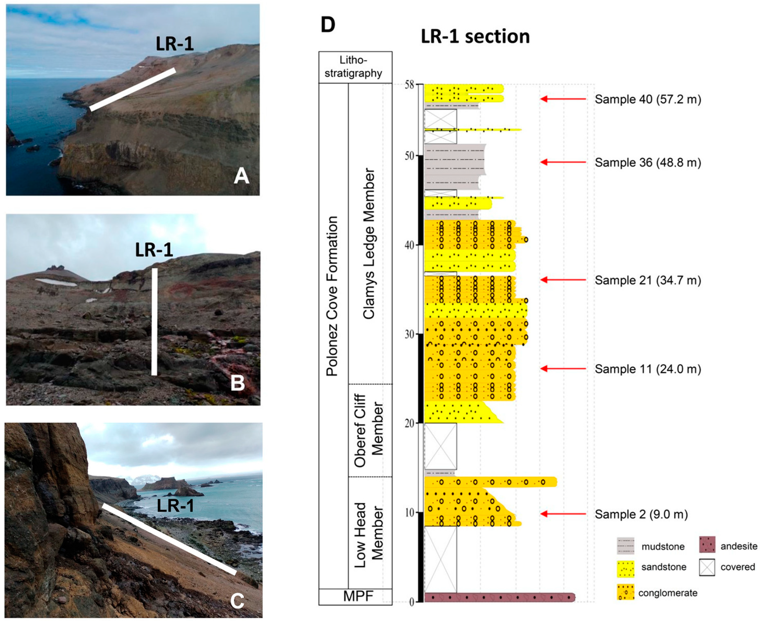
The rock material examined in this study derives from a section coded as LR-1 = Lions Rump-1 located in the Mazurek Point Formation [30] at Lions Rump region (62°08′30.81″ S 58°07′34.25″ W), King George Island, South Shetland Islands, during the austral summer of 2021/22 (Figure 1), which, according to paleobotanic studies, represents rock from the Oligocene epoch. Section LR-1 has a thickness of ~58 m and five rock samples were obtained at 9 m (sample-S2), 24 m (S11), 34.7 m (S21), 48.8 m (S36) and 57.2 m (S40) using a geological hammer. The rock samples (Figure 2) were stored in sterile Whirl Pack bags (Nasco, Ft. Atkinson, NH, USA) and frozen at −20 °C until processing for eDNA at the Federal University of Minas Gerais, Brazil. There, the samples were first fragmented into approximately 1 cm3 using a miniature drill device (Storm Feel-0001, Gumroad, Sumaré, Brazil) with diamond drills, one for each sample, and autoclaved before extraction [25]. After that, these fragments were pulverized with the aid of a previously sterilized agate mortar. The entire process was carried out in a laminar flow cabinet to avoid external contamination.

2.2. Stratigraphy

The Polonez Cove Formation is a continuous 2 km-long cliff section between Low Head and Lions Rump. The formation consists of a basal unit dominated by glacigenic diamictite, overlain by a series of basalt-sourced sedimentary and dacitic volcanic units [31]. The Polonez Cove Formation has been divided into six members [23]: Krakowiak Glacier; Low Head; Bayview; Siklawa; Oberek Cliff; and Chlamys Ledge. These members have been interpreted as: (1) sparsely fossiliferous, planar-bedded, generally fine-grained sediments (mainly siltstones and fine sandstones) formed in relatively deep water; (2) locally fossiliferous coarse epiclastic fan deltas sourced in recently active volcanism (subunits of the Low Head and Oberek Cliff members); and (3) gravelly conglomeratic sediments with prominent channels and cross-stratification, typically highly fossiliferous and characteristic of deposition under relatively shallow-water high-energy conditions (other subunits of the Low Head and Oberek Cliff members) [24].

2.3. Geochemical Analysis

Analysis of chemical element concentrations in the LR-1 bulk sediment rock samples (n = 1) was performed using X-ray fluorescence (XRF) at the National Museum (Rio de Janeiro, Brazil), utilizing a Bruker Tracer 5i Handheld XRF instrument (Bruker, Billerica, MA, USA). The data obtained were interpreted in the context of elemental ratios, specifically the Fe/Ca ratio, to deduce the presence of terrigenous inputs. This is because iron is more prevalent in terrestrial materials such as soils and rocks, whereas calcium tends to be the dominant element in marine environments, owing to the abundance of calcium-rich minerals such as calcite and aragonite. The Sr/Ca ratio was used to estimate relative sample temperatures, where higher values indicate warmer waters (higher Sr concentration) and lower values suggest colder waters (higher Ca concentration).

2.4. Petrographic Analyses

Thin sections were made with the undisturbed samples, and optical microscopic investigations were carried out using a Zeiss trinocular optical microscope (Axiophot model) with an integrated digital camera, to identify the types of rocks and their features, in particular any crack/fissure patterns. Rock porosity is an important part of the formation of habitats suitable for microbial colonization [13,31]. We follow [9] for void system description. Samples 11, 36, and 40 (n = 1), which displayed the best structural conditions for petrographic analysis, were selected to analyse the relationship between the degree of rock porosity and its chemical alteration, in order to identify whether the rocks showed any evidence of recent colonization by fungi, as some fungal species (particularly those that can become lichenized) are known to exude chemicals that can alter the rock minerals present [32]. For this, we used a scanning electron microscope (SEM, QUANTA FEI 3D) coupled with an energy dispersive system (EDS), operated with an acceleration potential of 15 kV and a current of 20 nA. The following elements were determined in the analysis: Na, K, Mg, Ca, Fe, Al and Si. The thin sections were metalized with carbon films, and this element was used as an indicator of porosity in the microchemical maps produced.

2.5. DNA Extraction, Illumina Library Construction and Sequencing

One sub-sample (approximately 100 g) of each of sample S2, S11, S21, S36 and S40 (five for each sample) was pulverized as described by [31]. Total DNA was extracted from these using the FastDNA Spin Kit for Soil (MPBIO, Solon, OH, USA) under strict contamination control conditions, following the manufacturer’s instructions. DNA quality was analysed using agarose gel electrophoresis (1% agarose in 1 × Trisborate-EDTA) and then quantified using the Quanti- iT™ Pico Green dsDNA Assay (Invitrogen, Carlsbad, CA, USA). Extracted DNA was used as template for generating PCR amplicons.
The internal transcribed spacer 2 (ITS2) of the nuclear ribosomal DNA was used as a DNA barcode for molecular species identification [27,33,34,35]. PCR amplicons generated using the universal primers ITS3 and ITS4 [36] were sequenced commercially by Macrogen Inc. (Seoul, Republic of Korea) using high-throughput paired-end sequencing (2 × 300 bp) on a MiSeq System (Illumina, San Diego, CA, USA), using the MiSeq Reagent Kit v3 (600 cycles). Library construction and DNA amplification were performed using the Library kit Herculase II Fusion DNA Polymerase Nextera XT Index Kit V2 (Illumina, San Diego, CA, USA), following Illumina 16S Metagenomic Sequencing Library Preparation Part #15044223 Rev. B protocol.

2.6. Data Analysis and Fungal Identification

Quality analysis was carried out using BBDuk v. 38.87 in BBmap software [37] with the following parameters: Illumina adapters removing (Illumina artefacts and the PhiX Control v3 Library); ktrim = l; k = 23; mink = 11; hdist = 1; minlen = 50; tpe; tbo; qtrim = rl; trimq = 20; ftm = 5; maq = 20. The remaining sequences were imported to QIIME2 version 2021.4 (https://qiime2.org/, accessed on 28 February 2024) for bioinformatics analyses [38]. The qiime2-dada2 plugin was used for filtering, dereplication, turn paired-end fastq files into merged, and to remove chimeras, using default parameters [39]. Taxonomic assignments were determined for amplicon sequence variants (ASVs) in three steps. First, ASVs were classified using the qiime2-feature-classifier [40] classify-sklearn against the UNITE Eukaryotes ITS database version 8.3 [41]. Second, remaining unclassified ASVs were filtered and aligned against the filtered NCBI non-redundant nucleotide sequences (nt) database (October 2021) using BLASTn [42] with default parameters; the nt database was filtered using the following keywords: “ITS1”, “ITS2”, “Internal transcribed spacer”, and “internal transcribed spacer”. Third, output files from BLASTn [42] were imported to MEGAN6 [43] and taxonomic assignments were performed using the “megan-nucl-Jan2021.db” mapping file with default parameters and trained with Naive Bayes classifier and a confidence threshold of 98.5%. Taxonomic profiles were plotted using the Krona [44]. The heatmap of ASV abundance and clustering analysis were performed using Heatmapper [45]; clustering analysis was performed using the following parameters: average linkage, Spearman’s rank correlation, and Z-score among samples for each ASV. Sequences have been submitted to GenBank under the accession numbers SAMN37305760-SAMN37305772.
Many factors, including extraction, PCR and primer bias, can affect the number of reads obtained [46], and thus lead to misinterpretation of absolute abundances [47]. However, ref. [48] concluded that such biases did not affect the proportionality between reads and cell abundance, implying that more reads are linked with higher abundance [49,50]. Therefore, for comparative purposes, we used the number of reads as a proxy for relative abundance. Fungal classification followed [51,52], MycoBank (http://www.mycobank.org, accessed on 28 February 2024) and the Index Fungorum (http://www.indexfungorum.org, accessed on 28 February 2024).

2.7. Fungal Diversity

The relative abundances of the ASVs were used to quantify the fungal taxa present in the samples; fungal ASVs with relative abundance > 10% were considered dominant, those between 1% and 10% as intermediate, and those with <1% as minor (rare) components of the fungal community [27]. The relative abundances were used to quantify taxon diversity, richness, and dominance, using the following indices: (i) Fisher’s α, (ii) Margalef’s and (iii) Simpson’s, respectively. In addition, species accumulation rarefaction curves were obtained using the Mao Tao index. All results were obtained with 95% confidence, and bootstrap values were calculated from 1000 replicates using the PAST 1.90 [53]. Venn diagrams were prepared following [54] to visualise the fungal assemblages present in the different sampling areas.

3. Results

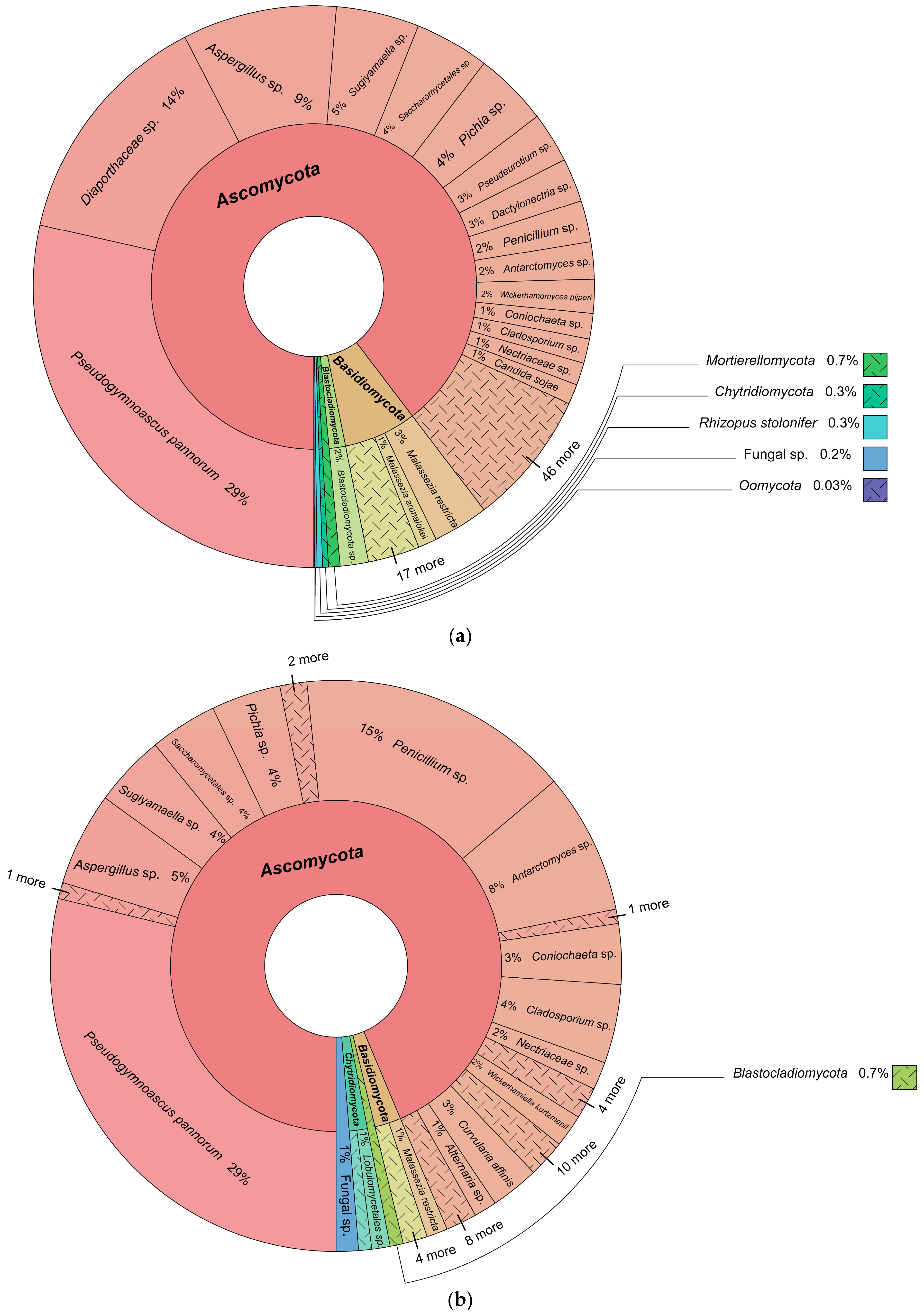
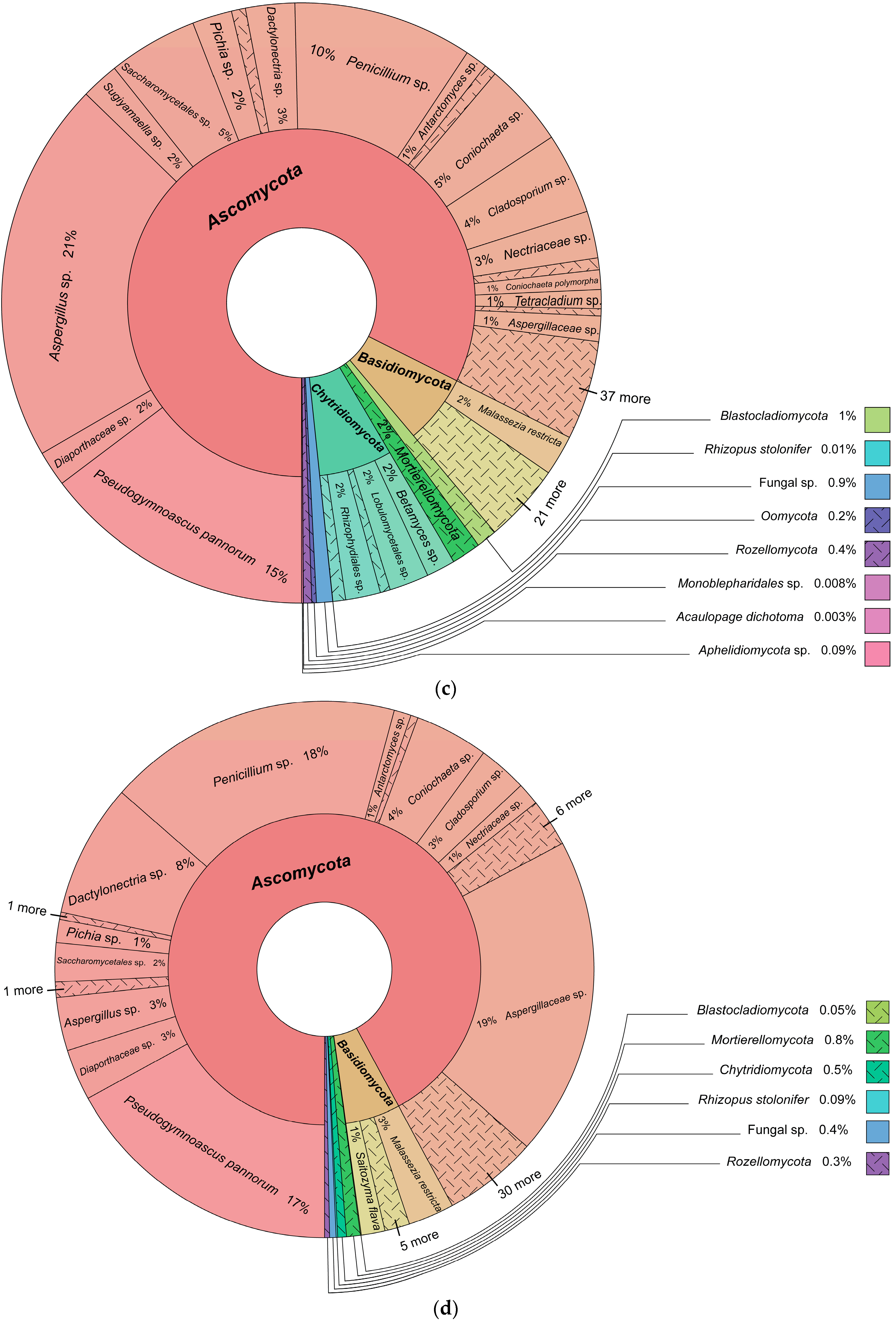
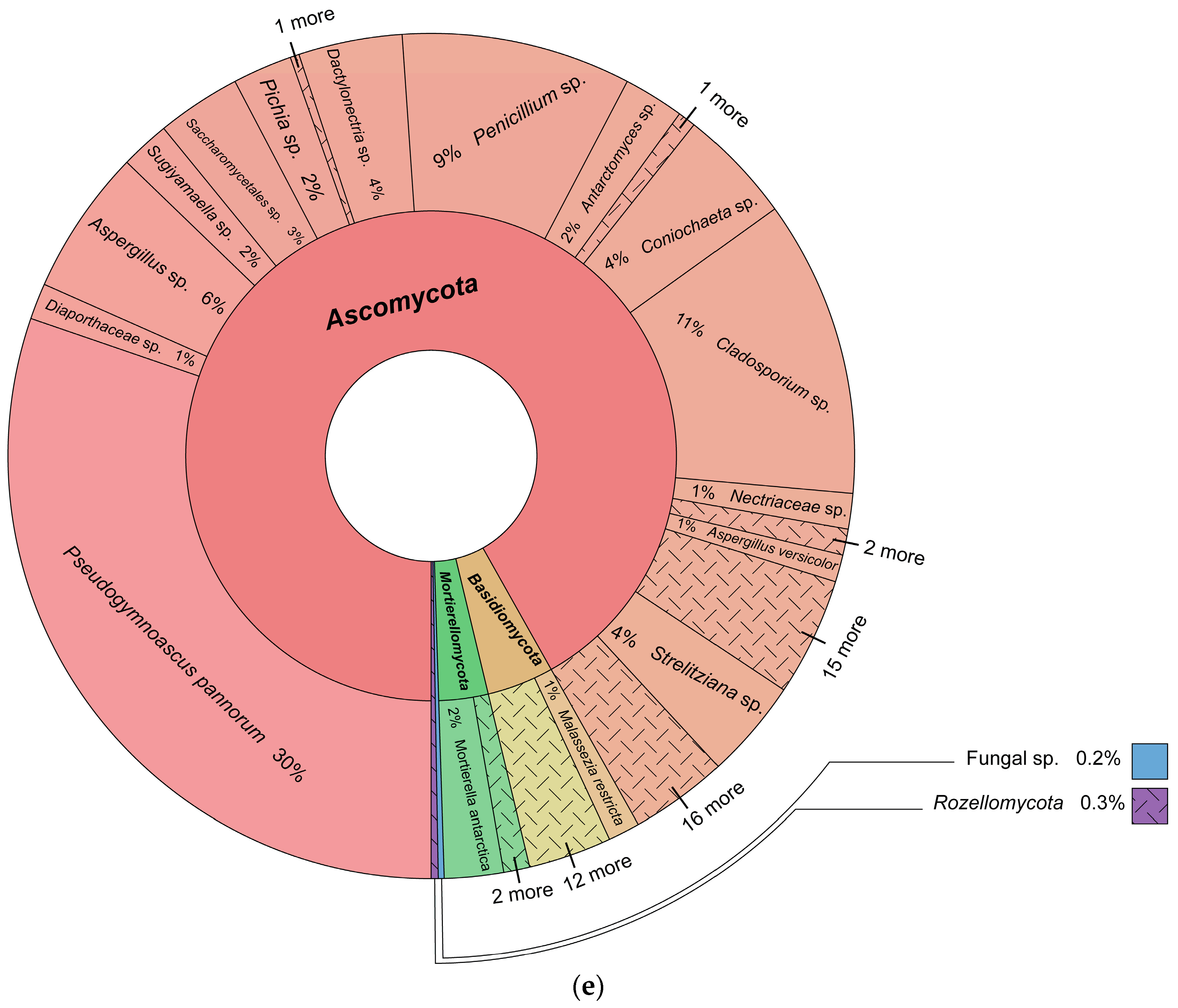
3.1. Fungal Identification

A total of 940,969 fungal DNA reads were obtained, which were assigned to 198 amplicon sequence variants (ASVs) (Figure 3, Supplementary Table S1). Ascomycota was the dominant phylum, followed by Basidiomycota, Mortierellomycota, Chytridiomycota, Mucoromycota, Rozellomycota, Blastocladiomycota, Monoblepharomycota, Zoopagomycota, Aphelidiomycota (Fungi) and the fungal-like Oomycota (Stramenopila), in rank order (Supplementary Figure S1). Pseudogymnoascus pannorum, Penicillium sp., Aspergillus sp., Cladosporium sp., Aspergillaceae sp. and Diaporthaceae sp. were the most abundant taxa (relative abundance ≥ 10%). In addition, 22 fungal ASVs displayed intermediate abundance, and 170 formed minor components of the assigned fungal community.

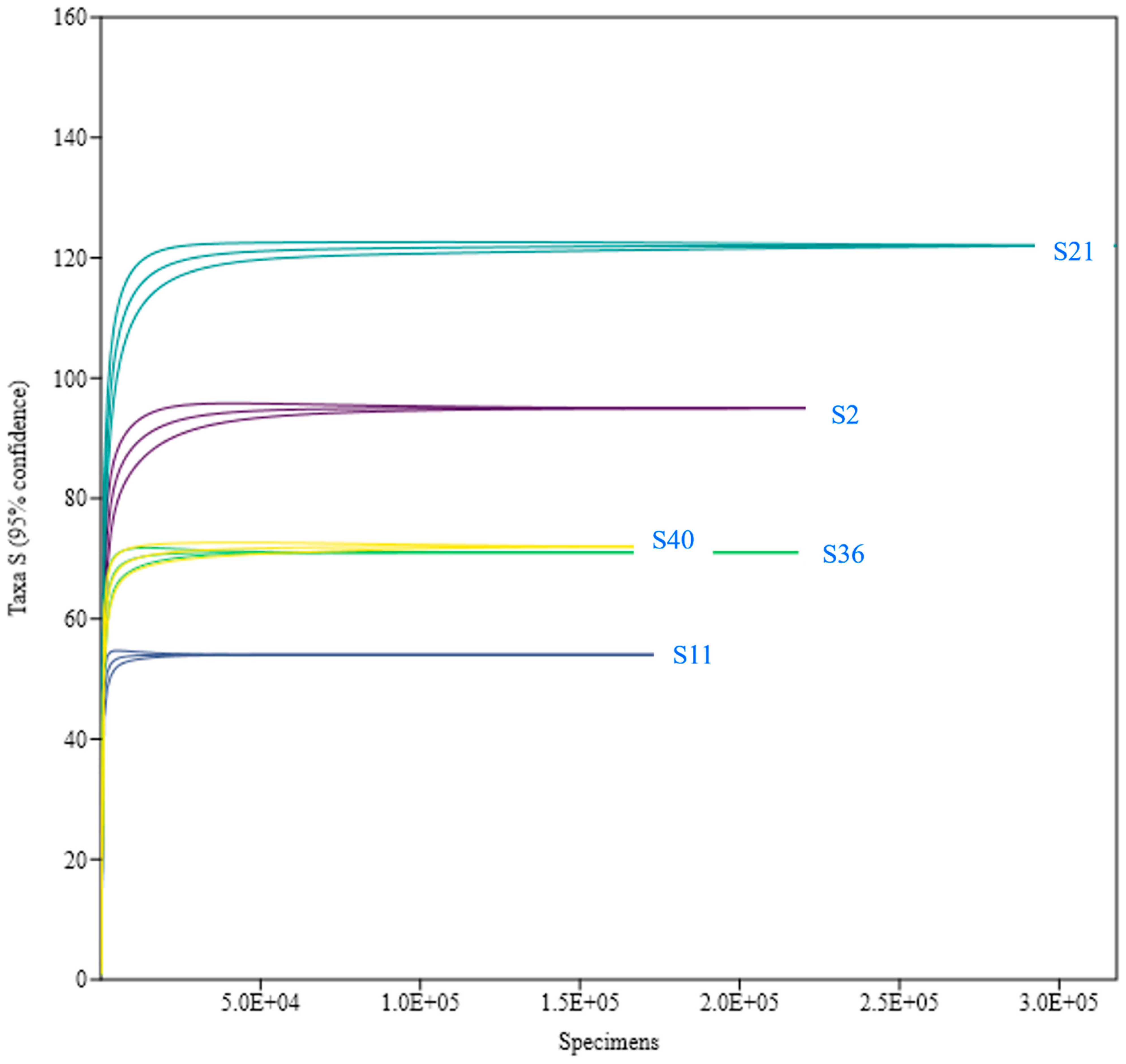
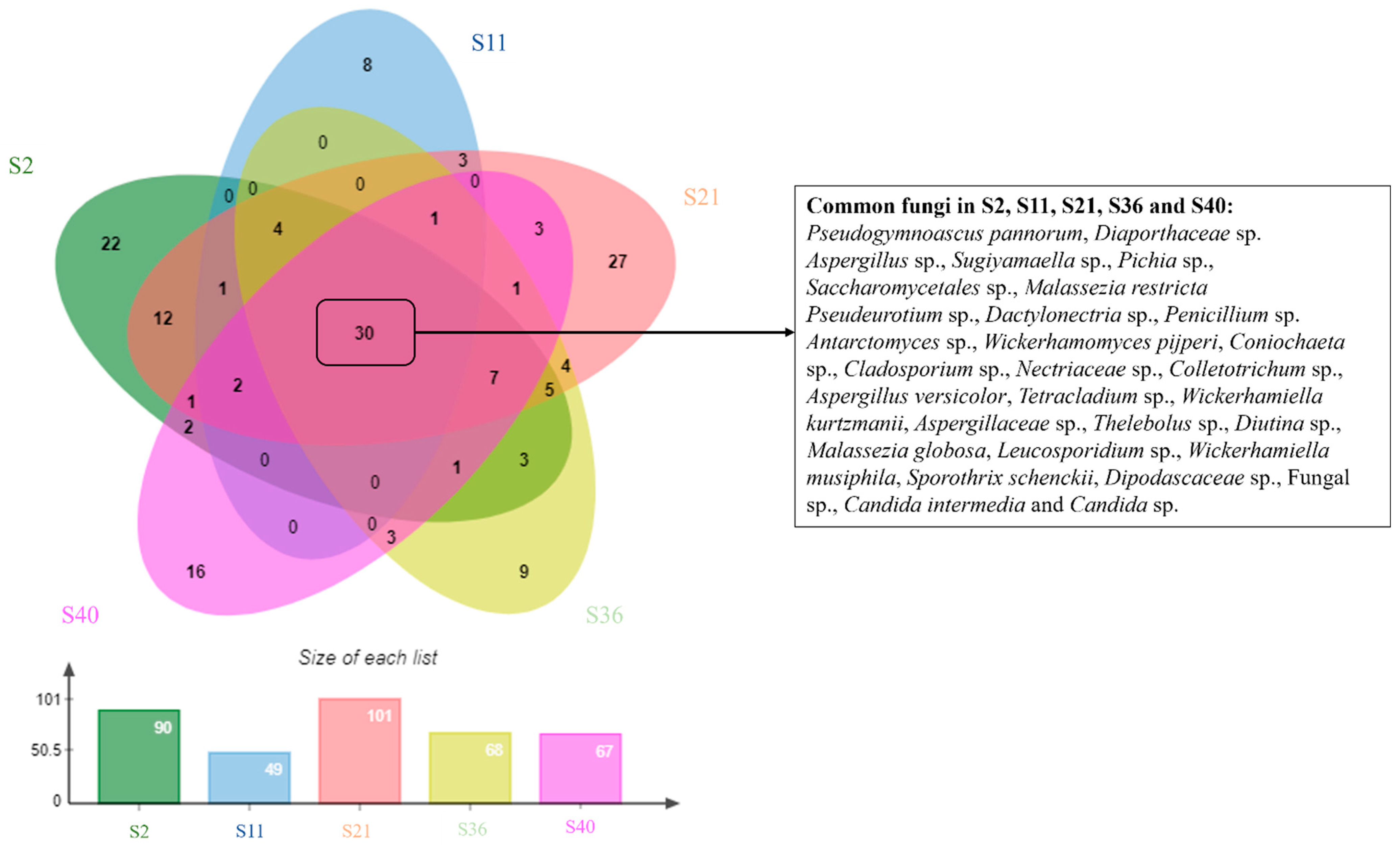
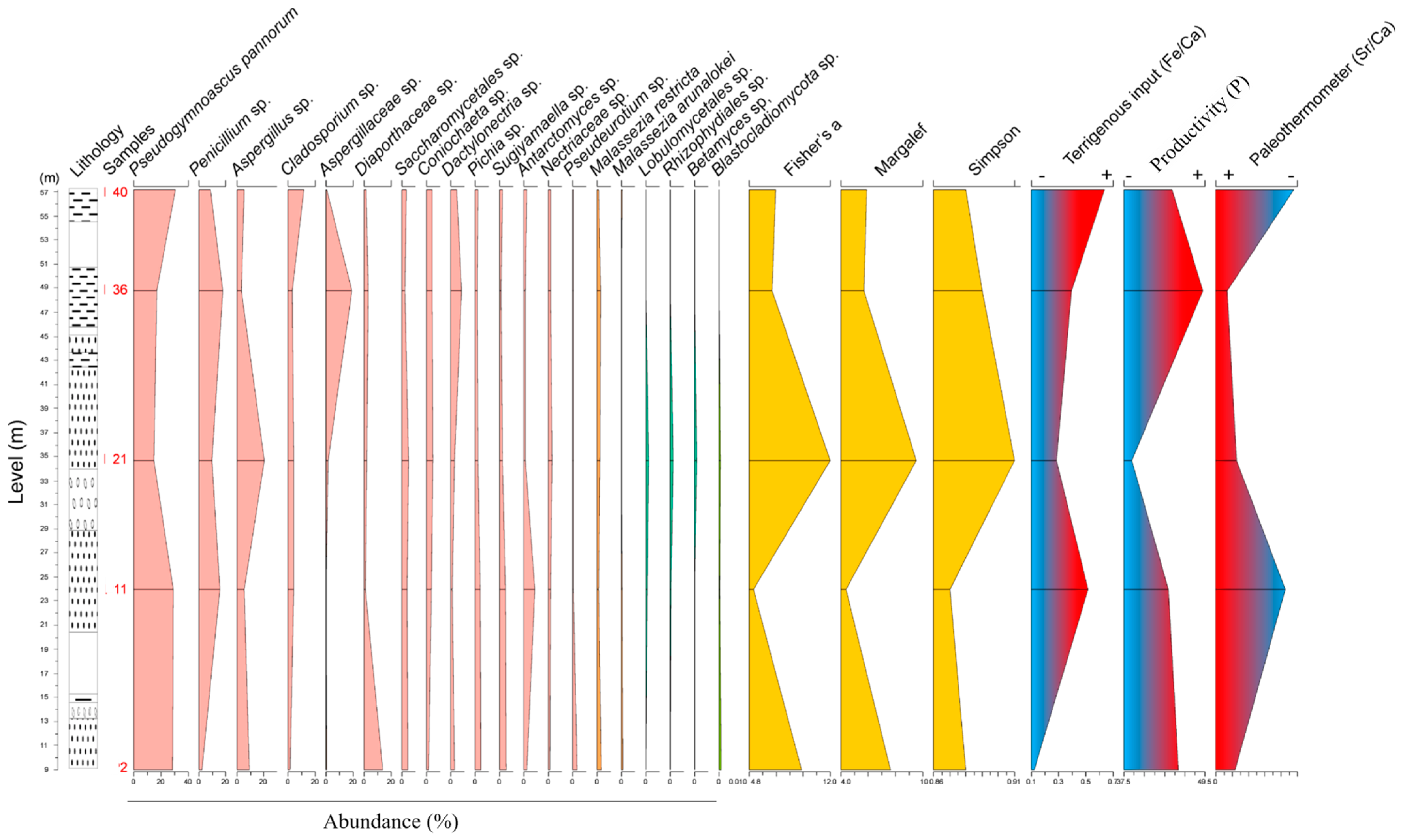
3.2. Fungal Diversity and Distribution

The Mao Tao rarefaction curves reached asymptote for all fungal assemblages from the different sites, indicating that the majority of the diversity was detected (Figure 4). The diversity indices varied among the fungal assemblages for the five sites sampled (Table 1). The highest diversity assemblages were detected in S21 and S2, whilst those from S11, S36 and S40 displayed the lowest values. Among the 198 fungal ASVs assigned, 30 (15%) were detected in all five samples (Figure 5).

3.3. Geochemical Results

X-ray fluorescence results (Table 1) revealed differences in the chemical compositions of the five samples. The major chemical elements were Si (with an average of 42.8%), Fe (37.2%) and Al (7.3%). Other elements were also present in minor proportions in the samples, including Mg, P, and Ti (Table 1). The curves of the Fe/Ca and Sr/Ca ratios exhibited similar patterns, with the lowest concentrations found in sample S2 and the highest in sample S40. Meanwhile, the phosphorus (P) concentration, indicative of productivity, exhibited its lowest levels in sample S21 and highest in S36 (Figure 6).
The stratigraphic distribution of the relevant fungal ASVs revealed a correlation between high diversity (S21) in conjunction with rocks formed during a period of reduced terrigenous input and higher temperatures. Conversely, lower diversity values (S11) were associated with rocks formed during periods of moderate to high terrigenous input and lower temperatures. (Figure 6). Specifically, the abundance curve of P. pannorum followed the same pattern as the curve of terrigenous input (Figure 6), suggesting a connection between this species and rocks whose formation and properties are related to terrigenous input. Conversely, the species Aspergillus sp. and Penicillium sp. were associated with rocks formed during periods of higher temperatures.

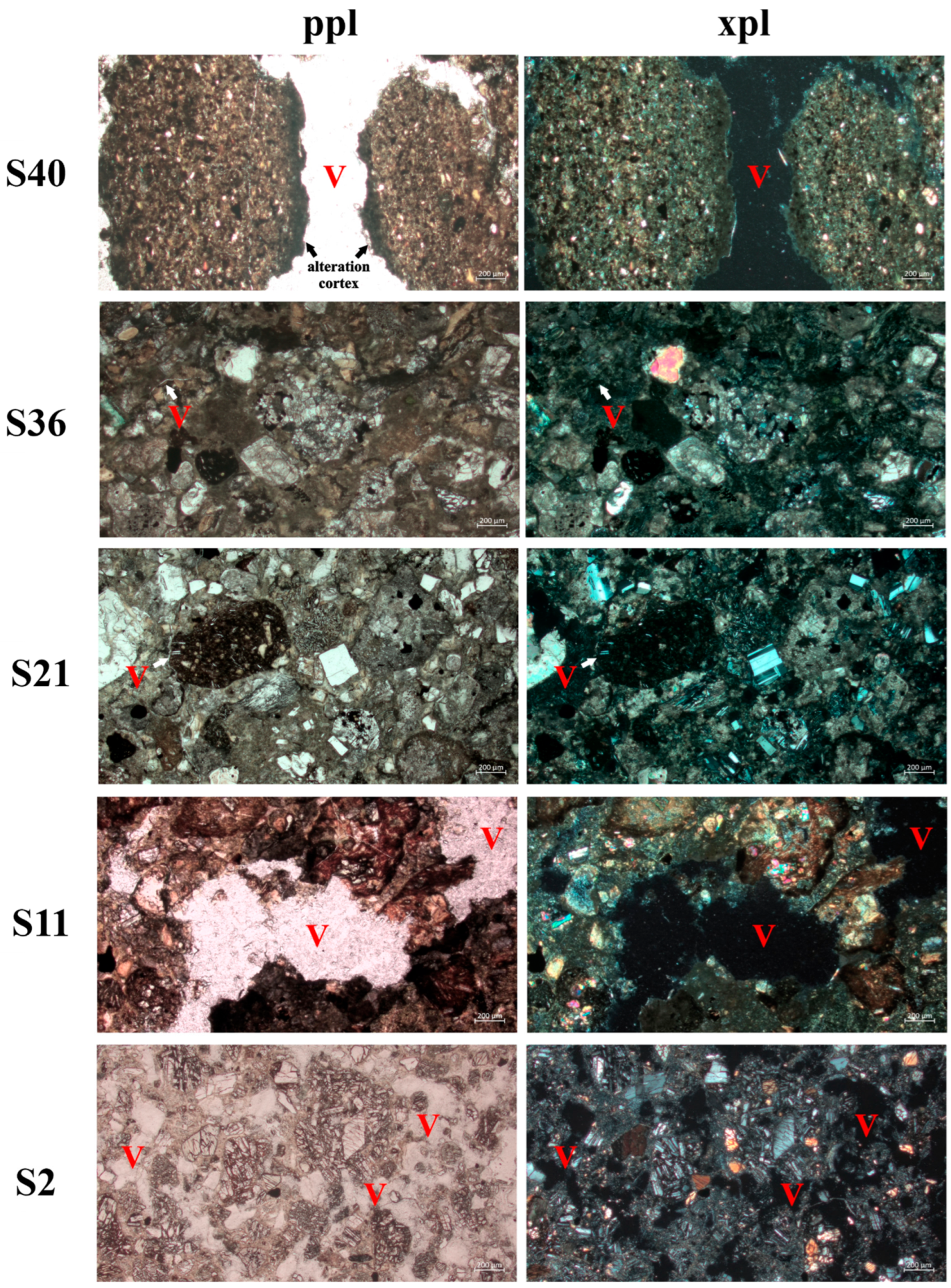
3.4. Petrographic Analyses

The photomicrographs show that the samples had different degrees and patterns of micro-fissure formation (Figure 7). Samples S2, S11, and S40 were the most fragmented, with voids that varied from flat and curved planes to a simple packing void system. In conglomerates (S2 and S11), fissure formation had occurred through the fragmentation of the matrix and the release of rounded clasts. The sandstone rock (S40) was also fractured, with some rotated clasts. Samples S21 and S36 showed the lowest degree of fragmentation, with some areas with poorly connected flat voids. The less fragmented samples were those that presented the highest fungal diversity values (Figure 7).
Our analyses identified little evidence of chemical weathering, with the presence of an alteration cortex observed under optical microscopy only in sample S2 (sandstone). In the conglomerate rocks, backscattered electron microscopy images and microchemical maps (Supplementary Figures S2–S4) indicated that zones of geochemical alteration were not evident in the void systems present. Comparing samples S11, S36, and S40, there were differences in void size, but no evidence of chemical alteration. The specific location of carbon indicates the presence of pores, as the thin sections were metallized with graphite, with the numbers increasing from samples S36 and S40 to S11, respectively 20.33%, 25.66%, and 44.05% carbon (Supplementary Figures S2d, S3d, and S4d).

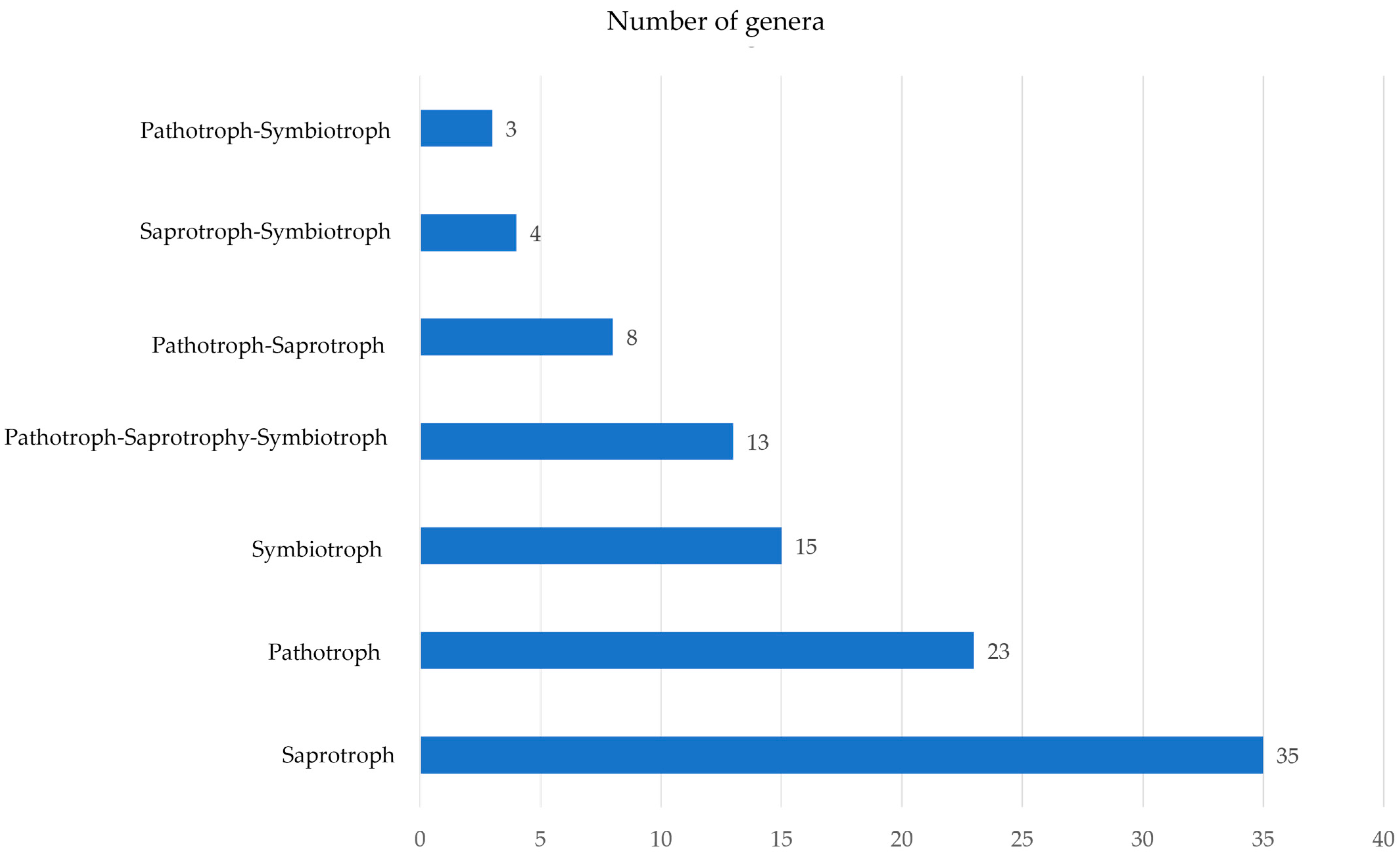
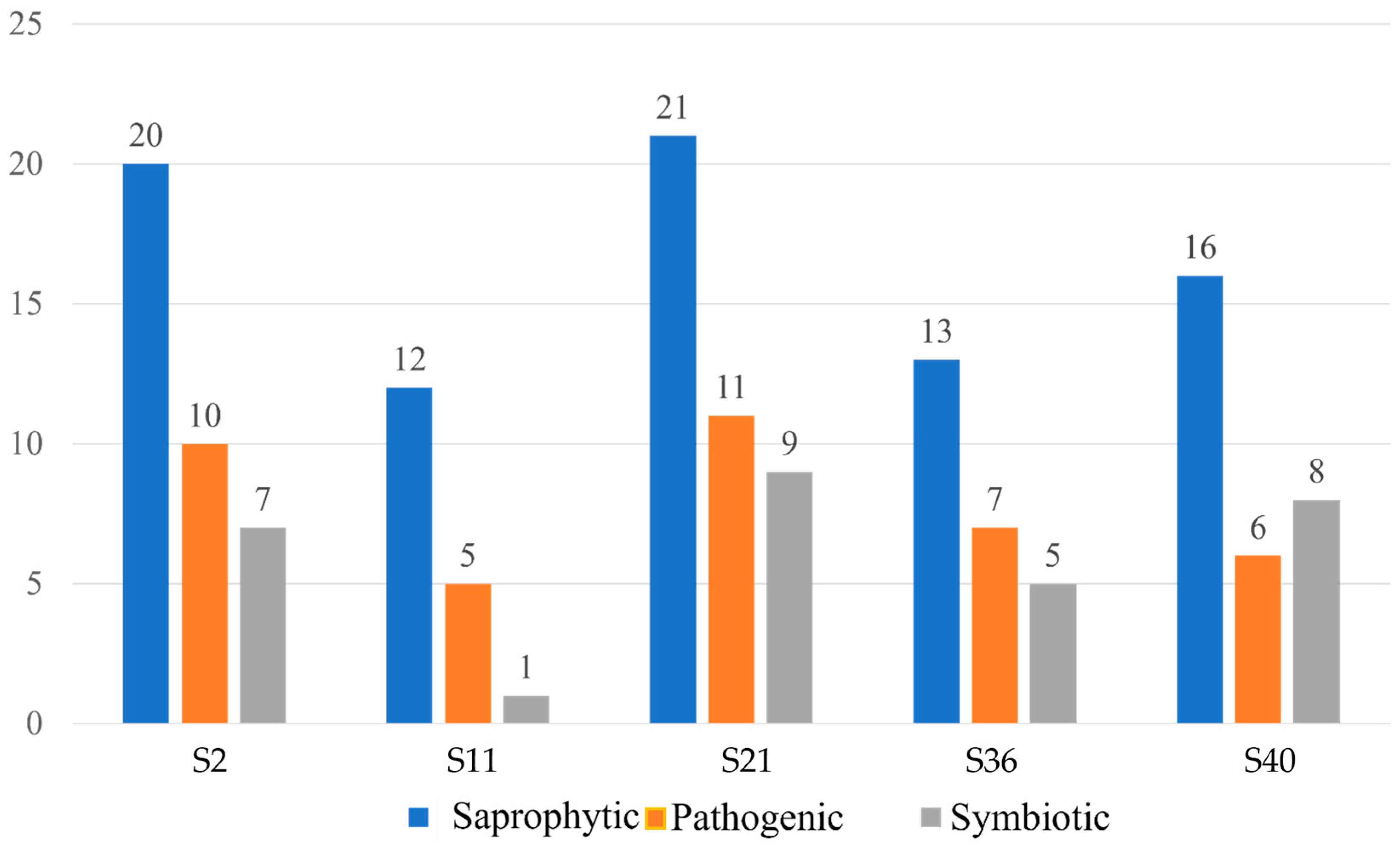
3.5. Assignment to Fungal Lifestyles

The 101 fungal ASVs genera detected were assigned to fungal lifestyles using FunGuild (Supplementary Table S2). The majority of assignments were to saprotrophic, pathogenic, and symbiotic taxa, with some displaying multiple ecological roles (Figure 8). The fungal genera included animal and plant pathogens, soil, wood, litter and undefined saprophytic taxa, endophytes, and lichenized fungi. Considering only ASVs classified to a single ecological status (Figure 9), in four of the samples saprophytic taxa dominated, followed by pathogenic and symbiotic taxa, while, in sample S40, symbiotic taxa were more diverse than pathogenic taxa.

4. Discussion

Fungal Taxonomy, Fungal Diversity, and Fungal Lifestyle

Existing studies of endolithic fungal communities present in Antarctic rocks have involved their direct observation in the substrate, cultivation techniques, or culture-independent methods [13]. Rocks from Antarctic can be considered a unique microhabitat for life forms, providing a natural habitat for rock-inhabiting microbes, including fungal taxa [7,13,17]. In the extreme conditions across Antarctica, endolithic fungi present within rocks are dominated by the phyla Ascomycota and Basidiomycota, as revealed by studies using traditional culturing methods [13].
The use in this study of metabarcoding to detect fungal eDNA associated with Antarctic Oligocene rocks revealed the presence of eDNA assigned to the widespread and common phyla Ascomycota and Basidiomycota. It also detected members of the less commonly reported Mortierellomycota, Chytridiomycota, and Mucoromycota, as well as cryptic fungi from the phyla Rozellomycota, Blastocladiomycota, Monoblepharomycota, Zoopagomycota, Aphelidiomycota, and fungal-like Oomycota (Stramenopila). Among the ASVs assigned, 74 were resolved only at higher hierarchical taxonomic levels (family, class, order, and phylum), suggesting that they might represent undescribed fungi or taxa not currently included in publicly accessible databases, which deserve further taxonomic studies.
In a study carried out in a polar desert region of continental Antarctica, rocks were analysed using DNA metabarcoding, reporting 262,268 fungal DNA reads grouped into 517 fungal ASVs dominated by the phyla Ascomycota, Basidiomycota, Mortierellomycota, and Mucoromycota, in abundance order [17]. These results showed similarity to those obtained in our study [55,56,57], in which Ascomycota and Basidiomycota represented the dominant phyla, with Mortierellomycota and Mucoromycota as minor components. However, in the current study, we detected taxa from further phyla including Chytridiomycota, Rozellomycota, Blastocladiomycota, Monoblepharomycota, Zoopagomycota, Aphelidiomycota, and the fungal-like Oomycota (Stramenopila). The dominant genera detected in the current study (Pseudogymnoascus, Penicillium, Aspergillus, and Cladosporium) were similar to those reported in [17].
Endolithic microbes, including those present in Antarctica, can exist in a dormant form alongside non-viable cells that retain their morphological integrity [58]. It is therefore important to recognize that the metabarcoding approach can detected only the presence of DNA and does not confirm viability. The petrographic analysis confirms the presence of microchannels within the rocks, and we assume that spores, hyphal fragments, or resistant structures might be recovered from such structures, as proposed by [13].
Amongst the taxa assigned, Pseudogymnoascus pannorum and representatives of the genera Penicillium, Aspergillus, and Cladosporium were the most abundant. The genus Pseudogymnoascus (syn. Geomyces) includes many psychrophilic and psychrotolerant species commonly present in Antarctic terrestrial and marine habitats [5]. Although the genus is widely distributed in cold environments globally [59,60], it is particularly well represented in Antarctica, with reports from habitats including soils [59,61,62], in association with plants [63], marine macroalgae [64,65], lichens [66], sponges [67] and in freshwater lakes [1,68]. Pseudogymnoascus pannorum was reported to produce high levels of protease at low temperatures [69], which may contribute to its occurrence, dominance, and capability to survive in the extreme conditions of Antarctica.
Representatives of the genera Penicillium, Aspergillus, and Cladosporium are found globally, including some truly cosmopolitan species. They have been reported from multiple Antarctic habitats, including soils [62,70] and permafrost [71,72]. Taxa of Penicillium, Aspergillus, and Cladosporium have been reported as endolithic microbes in rocks across various environments. Within Antarctic rocks, taxa from these genera have been identified as dominant [13,17,73]. Recent metabarcoding studies in Antarctica have reported eDNA assignments to members of Pseudogymnoascus, Penicillium, Aspergillus, and Cladosporium from habitats including mosses, soils [26,72,74], snow [75], rock surfaces [55], marine environments [76], and lake sediments [29].
The diversity indices and similarity among the fungal assemblages varied. The highest diversity values were detected in the fungal assemblages of S21 and S2, followed by those of S11, S36, and S40. These results might be explained by differences in the formation of the rock samples. Rock fragmentation in periglacial environments occurs primarily through cryoclastic processes [77,78] and is influenced by the type of rock, its position in the relief, exposure time, and the intensity of freezing and thawing cycles [79,80,81]. In the South Shetland Islands, some studies have shown that sedimentary rocks are more easily fragmented than igneous rocks [82]. This is consistent with the high degree of fragmentation observed in the samples studied here, which included conglomerates and sandstones. The latter are also more affected by cryoturbation, because of their greater compositional homogeneity. As they are progressively fragmented, the rocks can become chemically altered along the fissures, or fragments may be physically removed by water or ice activity. However, chemical modification was not evident in the rock samples analysed here (Supplementary Figures S2–S4). Therefore, the chemical change might be promoted by biological action as proposed by [31,83,84,85].
Although this study is based on a small sample size, samples S2, S11, and S40 were the most fragmented rocks, while samples S21 and S36 displayed the lowest degree of fragmentation. We therefore expected that samples S21 and S36 would present low diversity indices and the most fragmented samples would have the highest diversity indices. However, fragmentation did not show a correlation with eDNA fungal assemblage diversity. Rather, our data suggest a possible link between (undefined) rock properties developed at their time of formation under the influence of different environmental temperatures (as indicated by Sr/Ca ratio) and their subsequent, much more recent, colonization by endolithic fungi.
The ecological functional group assignments in the current study are similar to our recent metabarcoding studies of fungal communities in various Antarctic habitats [26,28,29,72,74,86,87]. Our ASV assignments suggest the presence of a diverse fungal community associated with the Oligocene rock samples analysed. The dominance of saprophytic fungi is consistent with the studies of [57,88], which suggest that fungi can colonize and establish in cold environments, slowly degrading the limited organic matter available, releasing nutrients, and making them available to other organisms.

5. Conclusions

The use of a metabarcoding approach led to detection of a diverse endolithic fungal community associated with the Oligocene rock samples examined. The endolithic fungal community detected contains a rich and complex mycobiome comprising taxa with different lifestyles, comparable with the diversity reported in recent studies of a range of Antarctic habitats. Due to the high fungal diversity detected, our results highlight the need for further research to develop strategies to isolate these fungi in culture for evolutionary, physiological, and biogeochemical studies, and to assess their potential role in biotechnological applications.

Supplementary Materials

Please add this part: The following supporting information can be downloaded at: https://www.mdpi.com/article/10.3390/biology13060414/s1. Refs. [89,90,91,92,93,94,95,96,97,98,99,100,101,102,103,104,105,106,107,108,109,110,111,112,113,114,115,116,117,118,119] are cited in Supplementary Materials.

Author Contributions

N.G.R.; V.N.G.; M.A.C.; S.M.S.; G.S.; P.A.S.; P.E.A.S.C. and L.H.R. conceived the study. M.A.C.; S.M.S.; G.S. and P.A.S. collected the samples. N.G.R.; P.E.A.S.C. and L.H.R. performed DNA extraction from samples. F.A.C.L. and K.C.R.S. performed the metabarcoding analysis. F.S.O. and L.P.C. performed the samples physicochemical analysis. N.G.R.; M.A.C.; S.M.S.; G.S.; P.A.S.; F.S.O.; L.P.C.; F.A.C.L.; K.C.R.S.; M.C.S.; P.C.; P.E.A.S.C. and L.H.R. analysed the results and wrote the manuscript. All authors have read and agreed to the published version of the manuscript.

Funding

This study received financial support from CNPq, CAPES, PROANTAR, Pró-Reitoria de Pesquisa da Universidade Federal de Minas Gerais (PRPq-UFMG). For open access purposes, the authors have assigned the Creative Commons CC BY license to any accepted version of the article.

Institutional Review Board Statement

Not applicable as the study did not involve human or animal subjects. The collections and studies performed in Antarctic Peninsula were authorized by the Secretariat of the Antarctic Treaty and by PROANTAR.

Informed Consent Statement

Not applicable.

Data Availability Statement

The datasets generated and/or analysed during the current study are available in the NCBI repository under the codes SAMN37305760-SAMN37305772, which can be accessed in https://www.ncbi.nlm.nih.gov/. Data are provided in the article.

Acknowledgments

CNPq-PROANTAR, CAPES, Pró-Reitoria de Pesquisa da Universidade Federal de Minas Gerais (PRPq-UFMG). P. Convey is supported by NERC core funding to the British Antarctic Survey’s ‘Biodiversity, Evolution and Adaptation’ Team. F.A.C. Lopes was supported by FAPT.

Conflicts of Interest

The authors declare that they have no conflicts of interest.

References

  1. Gonçalves, V.N.; Vaz, A.B.M.; Rosa, C.A.; Rosa, L.H. Diversity and distribution of fungal communities in lakes of Antarctica. FEMS Microbiol. Ecol. 2012, 82, 459–471. [Google Scholar] [CrossRef]
  2. Convey, P. Antarctic ecosystems. In Reference Module in Life Sciences; Elsevier: Amsterdam, The Netherlands, 2017. [Google Scholar]
  3. Cowan, D.A. Diverse hypolithic refuge communities in Antarctic Dry Valleys. Antarct. Sci. 2010, 22, 714–720. [Google Scholar] [CrossRef]
  4. Cavicchioli, R.; Ripple, W.J.; Timmis, K.N.; Azam, F.; Bakken, L.R.; Baylis, M.; Behrenfeld, M.J.; Boetius, A.; Boyd, P.W.; Aimée, T.; et al. Scientists’ warning to humanity: Microorganisms and climate change. Nat. Rev. Microbiol. 2019, 17, 569–586. [Google Scholar] [CrossRef]
  5. Rosa, L.H. Fungi in Antarctica: Diversity, Ecology, Effects of Climate Change, and Bioprospection for Bioactive Compounds. In Fungi of Antarctica: Diversity, Ecology and Biotechnological Applications; Rosa, L.H., Ed.; Springer: Cham, Switzerland, 2019; pp. 1–18. [Google Scholar]
  6. Vincent, W.F. Evolutionary origins of Antarctic microbiota: Invasion, selection and endemism. Antarct. Sci. 2000, 12, 374–385. [Google Scholar] [CrossRef]
  7. Onofri, S.; Fenice, M.; Cicalini, A.R.; Tosi, S.; Magrino, A.; Pagano, S.; Selbmann, L.; Zucconi, L.; Vishniac, H.S.; Ocampo-Friedmann, R.; et al. Ecology and biology of microfungi from Antarctic rocks and soil. Ital. J. Zool. 2000, 67, 163–168. [Google Scholar] [CrossRef]
  8. Arenz, B.E.; Blanchette, R.A. Distribution and abundance of soil fungi in Antarctica at sites on the Peninsula, Ross Sea Region and McMurdo Dry Valleys. Soil Biol. Biochem. 2011, 43, 308–315. [Google Scholar] [CrossRef]
  9. Stoops, G. Guidelines for the Analysis and Description of Soil and Regolith Thin Sections; SSSA: Madison, WI, USA, 2003. [Google Scholar]
  10. Friedmann, E.I. Endolithic microorganisms in the Antarctic cold desert. Science 1982, 215, 1045–1053. [Google Scholar] [CrossRef]
  11. Hughes, K.A.; Lawley, B. A novel Antarctic microbial endolithic community within gypsum crusts. Environ. Microbiol. 2003, 5, 555–565. [Google Scholar] [CrossRef] [PubMed]
  12. Selbmann, L.; de Hoog, G.S.; Mazzaglia, A.; Friedmann, E.I.; Onofri, S. Fungi at the edge of life: Cryptoendolithic black fungi from Antarctic desert. Stud. Mycol. 2005, 51, 1–32. [Google Scholar]
  13. Gonçalves, V.N.; Oliveira, F.S.; Carvalho, C.R.; Schaefer, C.E.G.R.; Rosa, C.A.; Rosa, L.H. Antarctic rocks from continental Antarctica as source of potential human opportunistic fungi. Extremophiles 2017, 21, 851–860. [Google Scholar] [CrossRef]
  14. Alves, I.M.S.; Gonçalves, V.N.; Oliveira, F.S.; Schaefer, C.E.G.R.; Rosa, C.A.; Rosa, L.H. Diversity, distribution, and pathogenic potential of fungi present in rocks of the South Shetlands Archipelago, Maritime Antarctica. Extremophiles 2019, 23, 327–336. [Google Scholar] [CrossRef] [PubMed]
  15. Coleine, C.; Pombubpa, N.; Zucconi, L.; Onofri, S.; Turchetti, B.; Buzzini, P.; Stajich, J.E.; Selbmann, L. Uncovered microbial diversity in Antarctic cryptoendolithic communities sampling three representative locations of the Victoria Land. Microorganisms 2020, 8, 942. [Google Scholar] [CrossRef] [PubMed]
  16. Gonçalves, V.N.; Alves, I.M.S.; de Oliveira, F.S.; Schaefer, C.E.G.R.; Turbay, C.V.G.; Rosa, C.A.; Rosa, L.H. Rock-inhabiting fungi in Antarctica: New frontiers of the edge of life. In Fungi of Antarctica: Diversity, Ecology and Biotechnological Applications, 1st ed.; Rosa, L.H., Ed.; Springer: Cham, Switzerland, 2019; pp. 99–126. [Google Scholar]
  17. de Menezes, G.C.A.; Câmara, P.E.A.S.; Pinto, O.H.B.; Carvalho-Silva, M.; Oliveira, F.O.; Souza, C.D.; Schaefer, C.E.R.; Convey, P.; Rosa, C.A.; Rosa, L.H. Fungal diversity present on rocks from a polar desert in continental Antarctica assessed using DNA metabarcoding. Extremophiles 2021, 25, 193–202. [Google Scholar] [CrossRef] [PubMed]
  18. Smelie, J.L.; McIntosh, W.C.; Whittle, R.; Troedson, A.; Hunt, R.J. A lithostratigraphical and chronological study of Oligocene-Miocene sequences on eastern King George Island, South Shetland Islands (Antarctica), and correlation of glacial episodes with global isotope events. Antarct. Sci. 2021, 33, 502–532. [Google Scholar] [CrossRef]
  19. Birkenmajer, K. A guide to Tertiary geochronology of King George Island, West Antarctica. Pol. Polar Res. 1989, 10, 555–579. [Google Scholar]
  20. Zachos, J.C.; Berggren, W.A.; Aubry, M.P.; Mackensen, A. Stable isotope record and trace element ratios of Eocene and Oligocene foraminifers from ODP Site 120-748, Pangea, Kerguelen Plateau. In Ocean Drilling Program; Wise, S.W., Schlich, R., Eds.; PANGAEA: College Station, TX, USA, 1992; Volume 120, pp. 839–854. [Google Scholar]
  21. Cooper, A.; Cooper, R.A. The Oligocene Bottleneck and New Zealand Biota: Genetic record of a past environmental crisis. PRS Lond. B 1995, 261, 293–302. [Google Scholar]
  22. Convey, P.; Bowman, V.; Chown, S.; Francis, J.; Fraser, C.; Smellie, J.L.; Storey, B.; Terauds, A. Ice bound Antarctica: Biotic consequences of the shift from a temperate to a polar climate. In Mountains, Climate, and Biodiversity; Hoorn, C., Perrigo, A., Antonelli, A., Eds.; Wiley: Oxford, UK, 2018; ISBN 978-1-119-15987-2. [Google Scholar]
  23. Warny, S.; Kymes, C.M.; Askin, R.A.; Krajewski, K. Terrestrial and marine floral response to latest Eocene and Oligocene events on the Antarctic Peninsula. Palynology 2019, 43, 1–18. [Google Scholar] [CrossRef]
  24. Smellie, J.L.; Caça, R.J.; Mcintosh, W.C.; Esser, R.P. Lithostratigraphy, age and distribution of Eocene volcanic sequences on eastern King George Island, South Shetland Islands, Antarctica. Antarc. Sci. 2021, 33, 373–401. [Google Scholar] [CrossRef]
  25. Light, J.J.; Passchier, S. Eocene to Oligocene cooling and ice growth based on the geochemistry of interglacial mudstones from the East Antarctic continental shelf. Antarct. Sci. 2023, 35, 270–282. [Google Scholar] [CrossRef]
  26. Rosa, L.H.; Pinto, O.H.B.; Santl-Temkiv, T.; Convey, P.; Carvalho-Silva, M.; Rosa, C.A.; Câmara, P.E.A.S. DNA metabarcoding of fungal diversity in air and snow of Livingston Island, South Shetland Islands, Antarctica. Sci. Rep. 2020, 10, 21793. [Google Scholar] [CrossRef]
  27. Rosa, L.H.; Pinto, O.H.B.; Convey, P.; Carvalho-Silva, M.; Rosa, C.A.; Câmara, P.E.A.S. DNA metabarcoding to assess the diversity of airborne fungi present over Keller Peninsula, King George Island, Antarctica. Microb. Ecol. 2021, 82, 165–172. [Google Scholar] [CrossRef] [PubMed]
  28. de Souza, L.M.D.; Ogaki, M.B.; Câmara, P.E.A.S.; Pinto, O.H.B.; Convey, P.; Carvalho-Silva, M.; Rosa, C.A.; Rosa, L.H. Assessment of fungal diversity present in lakes of maritime Antarctica using DNA metabarcoding: A temporal microcosm experiment. Extremophiles 2021, 25, 77–84. [Google Scholar] [CrossRef] [PubMed]
  29. de Souza, L.M.D.; Lirio, J.M.; Coria, S.H.; Lopes, F.A.C.; Convey, P.; Carvalho-Silva, M.; Oliveira, F.S.; Rosa, C.A.; Câmara, P.E.A.S.; Rosa, L.H. Diversity, distribution and ecology of fungal communities present in Antarctic lake sediments uncovered by DNA metabarcoding. Sci. Rep. 2022, 12, 8407. [Google Scholar] [CrossRef] [PubMed]
  30. Troedson, A.L.; Smellie, J.L. The Polonez Cove Formation of King George Island, Antarctic: Stratigraphy, facies and implications for mid-Cenozoic cryosphere development. Sedimentology 2002, 49, 277–301. [Google Scholar] [CrossRef]
  31. Gonçalves, V.N.; Cantrell, C.L.; Wedge, D.E.; Ferreira, M.C.; Soares, M.A.; Jacob, M.R.; Fabio, S.O.; Galante, D.; Rodrigues, F.; Alves, T.M.A.; et al. Fungi associated with rocks of the Atacama Desert: Taxonomy, distribution, diversity, ecology and bioprospection for bioactive compounds. Environ. Microbiol. 2016, 18, 232–245. [Google Scholar] [CrossRef] [PubMed]
  32. Purvis, O.W.; Convey, P.; Flowerdew, M.J.; Peat, H.J.; Najorka, J.; Kearsley, A. Iron localization in Acarospora colonizing schist following glacial retreat on Signy Island. Antarct. Sci. 2013, 25, 24–30. [Google Scholar] [CrossRef]
  33. Chen, S.; Yao, H.; Han, J.; Liu, C.; Song, J.; Shi, L.; Zhu, Y.; Ma, X.; Gao, T.; Pang, X.; et al. Validation of the ITS2 region as a novel DNA barcode for identifying medicinal plant species. PLoS ONE 2010, 5, e8613. [Google Scholar] [CrossRef] [PubMed]
  34. Câmara, P.; Convey, P.; Rangel, S.B.; Konrath, M.; Barreto, C.C.; Pinto, O.H.B.; Silva, M.C.; Henriques, D.D.; Oliveira, H.C.; Rosa, L.H. The largest moss carpet transplant in Antarctica and its bryosphere cryptic biodiversity. Extremophiles 2021, 25, 369–384. [Google Scholar] [CrossRef]
  35. Câmara, P.; Souza, L.M.D.; Pinto, O.H.B.; Convey, P.; Amorim, E.T.; Carvalho-Silva, M.; Rosa, L.H. Periphyton diversity in two different Antarctic lakes assessed using metabarcoding. Antarct. Sci. 2021, 33, 596–604. [Google Scholar] [CrossRef]
  36. White, T.J.; Bruns, T.D.; Lee, S.B.; Taylor, J.W. Amplification and direct sequencing of fungal ribosomal RNA genes for phylogenetics. In PCR Protocols: A Guide to Methods and Applications; Innis, M.A., Gelfand, D.H., Sninsky, J.J., White, T.J., Eds.; Academic Press: New York, NY, USA, 1990; pp. 315–322. [Google Scholar]
  37. Bushnell, B. BBMap: A Fast, Accurate, Splice-Aware Aligner; Lawrence Berkeley National Lab. (LBNL): Berkeley, CA, USA, 2014. [Google Scholar]
  38. Bolyen, E.; Rideout, J.R.; Dillon, M.R.; Bokulich, N.A.; Abnet, C.C.; Al-Ghalith, G.A.; Alexander, H.; Alm, E.J.; Arumugam, M.; Asnicar, F.; et al. Reproducible, interactive, scalable and extensible microbiome data science using QIIME 2. Nat. Biotechnol. 2019, 37, 852–857. [Google Scholar] [CrossRef]
  39. Callahan, B.; McMurdie, P.J.; Rosen, M.J.; Han, A.W.; Johnson, A.J.A.; Holmes, S.P. DADA2: High-resolution sample inference from Illumina amplicon data. Nat. Methods 2016, 13, 581–583. [Google Scholar] [CrossRef] [PubMed]
  40. Bokulich, N.A.; Kaehler, B.D.; Rideout, J.R.; Dillon, M.; Bolyen, E.; Knight, R.; Huttley, G.A.; Caporaso, J.G. Optimizing taxonomic classification of marker-gene amplicon sequences with QIIME 2’s q2-feature-classifier plugin. Microbiome 2018, 6, 90–107. [Google Scholar] [CrossRef] [PubMed]
  41. Camacho, C.; Coulouris, G.; Avagyan, V.; Ma, N.; Papadopoulos, J.; Bealer, K.; Madden, T.L. BLAST+: Architecture and applications. BMC Bioinform. 2019, 10, 421. [Google Scholar] [CrossRef] [PubMed]
  42. Huson, D.H.; Beier, S.; Flade, I.; Górska, A.; El-Hadidi, M.; Mitra, S.; Ruscheweyh, H.-J.; Tappu, R. MEGAN community edition-interactive exploration and analysis of large-scale microbiome sequencing data. PLoS Comput. Biol. 2016, 12, 1004957. [Google Scholar] [CrossRef] [PubMed]
  43. Ondov, B.D.; Bergman, N.H.; Phillippy, A.M. Interactive metagenomic visualization in a web browser. BMC Bioinform. 2011, 12, 385. [Google Scholar] [CrossRef]
  44. Abarenkov, K.; Allan, Z.; Timo, P.; Raivo, P.; Filipp, I.; Nilsson, H.R.; Urmas, K. UNITE QIIME Release for Fungi, Version 04.02.2020; UNITE Community: Salt Lake City, UT, USA, 2020. [Google Scholar]
  45. Babicki, S.; Arndt, D.; Marcu, A.; Liang, Y.; Grant, J.R.; Maciejewski, A.; Wishart, D. Heatmapper: Web-enabled heat mapping for all. Nucl. Acids Res. 2016, 44, 147–153. [Google Scholar] [CrossRef] [PubMed]
  46. Medinger, R.; Nolte, V.; Pandey, R.V.; Jost, S.; Ottenwalder, B.; Schlotterer, C.; Boenigk, J. Diversity in a hidden world: Potential and limitation of next-generation sequencing for surveys of molecular diversity of eukaryotic microorganisms. Mol. Ecol. 2010, 19, 32–40. [Google Scholar] [CrossRef] [PubMed]
  47. Weber, A.A.; Pawlowski, J. Can abundance of protists be inferred from sequence data: A case study of Foraminifera. PLoS ONE 2013, 8, 56739. [Google Scholar] [CrossRef] [PubMed]
  48. Giner, C.R.; Forn, I.; Romac, S.; Logares, R.; De Vargas, C.; Massana, R. Environmental sequencing provides reasonable estimates of the relative abundance of specific picoeukaryotes. Appl. Environ. Microbiol. 2016, 82, 4757–4766. [Google Scholar] [CrossRef]
  49. Deiner, K.; Bik, H.M.; Machler, E.; Seymour, M.; Lacoursière-Roussel, A.; Altermatt, F.; Creer, S.; Bista, I.; Lodge, D.M.; de Vere, N.; et al. Environmental DNA metabarcoding: Transforming how we survey animal and plant communities. Mol. Ecol. 2017, 26, 5872–5895. [Google Scholar] [CrossRef]
  50. Hering, D.; Borja, A.; Jones, J.I.; Pont, D.; Boets, P.; Bouchez, A.; Bruce, K.; Drakare, S.; Hanfling, B.; Kahlert, M.; et al. Implementation options for DNA-based identification into ecological status assessment under the European Water Framework Directive. Water Res. 2018, 138, 192–205. [Google Scholar] [CrossRef]
  51. Kirk, P.M.; Cannon, P.; Stalpers, J.; Minter, D.W. Dictionary of the Fung; CAB International: Wallingford, UK, 2011. [Google Scholar]
  52. Tedersoo, L.; Sánchez-Ramírez, S.; Kõljalg, U.; Bahram, M.; Doring, M.; Schigel, D.; May, T.; Ryberg, M.; Abarenkov, K. High-level classification of the Fungi and a tool for evolutionary ecological analyses. Fungal Div. 2018, 90, 135–159. [Google Scholar] [CrossRef]
  53. Hammer, Ø.; Harper, D.A.T.; Ryan, P.D. PAST: Paleontological statistics software package for education and data analysis. Palaeontol. Electron. 2001, 4, 1–9. [Google Scholar]
  54. Bardou, P.; Mariette, J.; Escudié, F.; Djemiel, C.; Klopp, C. An interactive Venn diagram viewer. BMC Bioinform. 2014, 15, 293. [Google Scholar] [CrossRef] [PubMed]
  55. de Menezes, G.C.A.; Camara, P.; Pinto, O.; Convey, P.; Carvalho-Silva, M.; Simoes, J.C.; Rosa, C.A.; Rosa, L.H. Fungi in the Antarctic Cryosphere: Using DNA Metabarcoding to Reveal Fungal Diversity in Glacial Ice from the Antarctic Peninsula Region. Microb. Ecol. 2022, 83, 647–657. [Google Scholar] [CrossRef] [PubMed]
  56. Gonçalves, V.N.; de Souza, L.M.D.; Lirio, J.M.; Coria, S.H.; Lopes, F.A.C.; Convey, P.; Carvalho-Silva, M.; de Oliveira, F.S.; Câmara, P.E.A.S.; Rosa, L.H. Diversity and ecology of fungal assemblages present in lake sediments at Clearwater Mesa, James Ross Island, Antarctica, assessed using metabarcoding of environmental DNA. Fungal Biol. 2022, 126, 640–647. [Google Scholar] [CrossRef] [PubMed]
  57. Gonçalves, V.N.; Lirio, J.M.; Coria, S.H.; Lopes, F.A.C.; Convey, P.; de Oliveira, F.S.; Carvalho-Silva, M.; Câmara, P.E.A.S.; Rosa, L.H. Soil fungal diversity and ecology assessed using DNA metabarcoding along a deglaciated chronosequence at Clearwater Mesa, James Ross Island, Antarctic Peninsula. Biology 2023, 12, 275. [Google Scholar] [CrossRef] [PubMed]
  58. Wierzchos, J.; de Los Ríos, A.; Sancho, L.G.; Ascaso, C. Viability of endolithic microorganisms in rocks from the McMurdo Dry Valleys of Antarctica established by confocal and fluorescence microscopy. J. Microsc. 2004, 216, 57–61. [Google Scholar] [CrossRef] [PubMed]
  59. Gomes, E.C.; Godinho, V.M.; Silva, D.A.S.; de Paula, M.T.R.; Vitoreli, G.A.; Zani, C.L.; Alves, T.M.A.; Junior, P.A.S.; Murta, S.M.F.; Barbosa, E.C.; et al. Cultivable fungi present in Antarctic soils: Taxonomy, phylogeny, diversity, and bioprospecting of antiparasitic and herbicidal metabolites. Extremophiles 2018, 22, 381–393. [Google Scholar] [CrossRef]
  60. Villanueva, P. Description of the first four species of the genus Pseudogymnoascus from Antarctica. Front. Microbiol. 2021, 12, 713189. [Google Scholar] [CrossRef]
  61. Mercantini, R. Keratinophilic fungi isolated from Antarctic soil. Mycopathologia 1989, 106, 47–52. [Google Scholar] [CrossRef] [PubMed]
  62. Godinho, V.M.; Gonçalves, V.N.; Santiago, I.F.; Figueredo, H.M.; Vitoeli, G.A.; Schaefer, C.E.R.; Barbosa, E.C.; Oliveira, J.G.; Alves, T.M.A.; Zani, C.L.; et al. Diversity and bioprospection of fungal community present in oligotrophic soil of continental Antarctica. Extremophiles 2015, 19, 585–596. [Google Scholar] [CrossRef] [PubMed]
  63. Santiago, I.F.; Alvez, T.M.A.; Rabello, A.; Junior, P.A.S.; Romanha, A.; Zani, C.L.; Rosa, C.A.; Rosa, L.H. Leishmanicidal and antitumoral activities of endophytic fungi associated with the Antarctic angiosperms Deschampsia antarctica Desv. and Colobanthus quitensis (kunth) Bartl. Extremophiles 2012, 16, 95–103. [Google Scholar]
  64. Godinho, V.M.; Furbino, L.E.; Santiago, I.F.; Pellizzari, F.M.; Yokoya, N.S.; Pupo, D.; Alves, T.M.A.; Junior, P.A.S.; Romanha, A.J.; Zani, C.L.; et al. Diversity and bioprospecting of fungal communities associated with endemic and cold-adapted macroalgae in Antarctica. ISME J. 2013, 7, 1434–1451. [Google Scholar] [CrossRef] [PubMed]
  65. Furbino, L.E.; Godinho, V.M.; Santiago, I.F.; Pellizari, F.M.; Alves, T.M.A.; Zani, C.L.; Junior, P.A.S.; Romanha, A.J.; Carvalho, A.G.O.; Gil, L.H.V.G.; et al. Diversity patterns, ecology and biological activities of fungal communities associated with the endemic macroalgae across the Antarctic Peninsula. Microb. Ecol. 2014, 67, 775–787. [Google Scholar] [CrossRef] [PubMed]
  66. Santiago, I.F.; Soares, M.A.; Rosa, C.A.; Rosa, L.H. Lichenosphere: A protected natural microhabitat of the non-lichenized fungal communities living in extreme environments of Antarctica. Extremophiles 2015, 19, 1087–1097. [Google Scholar] [CrossRef] [PubMed]
  67. Henríquez, M.; Vergana, K.; Norambuena, J.; Beiza, A.; Maza, F.; Ubilla, P.; Araya, I.; Chávez, R.; San-Martín, A.; Darias, J.; et al. Diversity of cultivable fungi associated with Antarctic marine sponges and screening for their antimicrobial, antitumoral and antioxidant potential. World J. Microbiol. Biotechnol. 2014, 30, 65–76. [Google Scholar] [CrossRef] [PubMed]
  68. Ogaki, M.B.; Teixeira, D.R.; Vieira, R.; Lírio, J.M.; Felizardo, J.P.S.; Abuchacra, R.C.; Cardoso, R.P.; Zani, C.L.; Alves, T.M.A.; Junior, P.A.S.; et al. Diversity and bioprospecting of cultivable fungal assemblages in sediments of lakes in the Antarctic Peninsula. Fungal Biol. 2020, 124, 601–611. [Google Scholar] [CrossRef] [PubMed]
  69. Furhan, J. Adaptation, production, and biotechnological potential of cold-adapted proteases from psychrophiles and psy-chrotrophs: Recent overview. J. Genet. Eng. Biotechnol. 2020, 18, 36. [Google Scholar] [CrossRef] [PubMed]
  70. McRae, C.F.; Hocking, A.D.; Seppelt, R.D. Penicillium species from terrestrial habitats in the Windmill Islands, east Antarctica, including a new species, Penicillium antarcticum. Polar Biol. 1999, 21, 97–111. [Google Scholar] [CrossRef]
  71. Zucconi, L.; Selbmann, L.; Buzzini, P.; Turchetti, B.; Guglielmin, M.; Frisvad, J.C.; Onofri, S. Searching for eukaryotic life preserved in Antarctic permafrost. Polar Biol. 2012, 35, 749–757. [Google Scholar] [CrossRef]
  72. da Silva, T.H.; Silva, D.A.S.; Oliveira, F.S.; Schaefer, C.E.G.; Rosa, C.A.; Rosa, L.H. Diversity, distribution, and ecology of viable fungi in permafrost and active layer of Maritime Antarctica. Extremophiles 2020, 24, 565–576. [Google Scholar] [CrossRef] [PubMed]
  73. Canini, F.; Borruso, L.; Newsham, K.K.; D’Alò, F.; D’Acqui, L.P.; Zucconi, L. Wide divergence of fungal communities inhabiting rocks and soils in a hyper-arid Antarctic desert. Environ. Microbiol. 2023, 25, 3671–3682. [Google Scholar] [CrossRef] [PubMed]
  74. Rosa, L.H.; Da Silva, T.H.; Ogaki, M.B.; Pinto, O.H.B.; Stech, M.; Convey, P.; Carvalho-Silva, M.; Rosa, C.A.; Câmara, E.A.S. DNA metabarcoding uncovers fungal diversity in soils of protected and non-protected areas on Deception Island, Antarctica. Sci. Rep. 2020, 10, 21986. [Google Scholar] [CrossRef] [PubMed]
  75. Rosa, L.H.; de Menezes, G.C.A.; Pinto, O.H.B.; Convey, P.; Carvalho-Silva, M.; Simoes, J.C.; Rosa, C.A.; Cama, P. Fungal diversity in seasonal snow of Martel Inlet, King George Island, South Shetland Islands, assessed using DNA metabarcoding. Polar Biol. 2022, 45, 627–636. [Google Scholar] [CrossRef]
  76. Ogaki, M.B.; Pinto, O.H.B.; Vieira, R.; Neto, A.A.; Convey, P.; Carvalho-Silva, M.; Rosa, C.A.; Câmara, P.E.A.S.; Rosa, L.H. Fungi present in Antarctic deep-sea sediments assessed using DNA metabarcoding. Microb. Ecol. 2021, 82, 157–164. [Google Scholar] [CrossRef] [PubMed]
  77. Bockheim, J.G.; Tarnocai, C. Recognition of cryoturbation for classifying permafrost-affected soils. Geoderma 1998, 81, 281–293. [Google Scholar] [CrossRef]
  78. Tarnocai, C.; Broll, G.; Blume, H.P. Classification of permafrost-affected soils in the WRB. In Cryosols: Permafrost-Affected Soils; Kimble, J.M., Ed.; Springer: Berlin/Heidelberg, Germany, 2004; pp. 637–656. [Google Scholar]
  79. Campbell, I.B.; Claridge, G.G.C. Antarctica: Soils, Weathering Processes an Environment; Elsevier Science Publishers: Amsterdam, The Netherlands, 1987; 368p. [Google Scholar]
  80. Bockheim, J.G. Properties and classification of cold desert soils from Antarctica. Soil Sci. Soc. Am. J. 1997, 61, 224–231. [Google Scholar] [CrossRef]
  81. Ugolini, F.C.; Bockheim, J.G. Antartic soil and soil formation in a changing environment: A review. Geoderma 2008, 144, 1–8. [Google Scholar] [CrossRef]
  82. Lopes, D.; Oliveira, F.S.; Pereira, T.T.C.; Schaefer, C.E.G.R. Pedogeomorphology and weathering at Snow Island, Maritime Antarctica. Catena 2022, 217, 106515. [Google Scholar] [CrossRef]
  83. Schaefer, C.E.G.R.; Simas, F.N.B.; Gilkes, R.J.; Mathison, C.; da Costa, L.M.; Albuquerque, M.A. Micromorphology and microchemistry of selected cryosols from maritime Antarctica. Geoderma 2008, 144, 104–115. [Google Scholar] [CrossRef]
  84. Pereira, T.T.C.; Schaefer, C.E.G.R.; Ker, J.C.; Almeida, C.C.; Almeida, I.C.C.; Pereira, A.B. Genesis, mineralogy and ecological significance of ornithogenic soils from a semi-desert polar landscape at Hope Bay, Antarctic Peninsula. Geoderma 2013, 209–210, 98–109. [Google Scholar] [CrossRef]
  85. Fortes, W.; Oliveira, F.S.; Schaefer, C.E.G.R.; Leite, M.G.P.; Pavinato, P. Phosphatization under birds’ activity: Ornithogenesis at different scales on Antarctic Soilscapes. Geoderma 2021, 391, 114950. [Google Scholar]
  86. Rosa, L.H.; Coelho, L.C.; Pinto, O.H.B.; Carvalho-Silva, M.; Convey, P.; Rosa, C.A.; Câmara, P.E.A.S. Ecological succession of fungal and bacterial communities in Antarctic mosses affected by a fairy ring disease. Extremophiles 2021, 25, 471–481. [Google Scholar] [CrossRef] [PubMed]
  87. da Silva, M.K.; de Souza, L.M.D.; Vieira, R.; Neto, A.A.; Lopes, F.A.C.; de Oliveira, F.S.; Convey, P.; Carvalho-Silva, M.; Duarte, A.W.F.; Câmara, P.E.A.S.; et al. Fungal and fungal-like diversity in marine sediments from the maritime Antarctic assessed using DNA Metabarcoding. Sci. Rep. 2022, 6, 21044. [Google Scholar] [CrossRef] [PubMed]
  88. Schutte, C.A.; Samarkin, V.A.; Peters, B.; Madigan, M.T.; Boeles, M.; Morgan-Kiss, R.; Casciotti, K.; Joye, S. Vertical stratification and stability of biogeochemical processes in the deep saline waters of Lake Vanda, Antarctic. Limnol. Oceanogr. 2020, 65, 569–581. [Google Scholar] [CrossRef]
  89. de Hoog, G.S.; Guarro, J.; Gen JéFigueras, M.J. Atlas of Clinical Fungi; Centraalbureau voor Schimmel-cultures: Utrecht, The Netherlands, 2000; p. 1126. [Google Scholar]
  90. Sung, G.-H.; Hywel-Jones, N.L.; Sung, J.-M.; Luangsa-Ard, J.J.; Shrestha, B.; Spatafora, J.W. Phylogenetic classification of Cordyceps and the clavicipitaceous fungi. Stud. Mycol. 2007, 57, 5–59. [Google Scholar] [CrossRef] [PubMed]
  91. Schoutteten, N.; Yurkov, A.; Leroux, O.; Haelewaters, D.; Straeten, D.V.D.; Miettinen, O.; Boekhout, T.; Begerow, D.; Verbeken, A. Diversity of colacosome-interacting mycoparasites expands the understanding of the evolution and ecology of Microbotryomycetes. Stud. Mycol. 2023, 106, 41–94. [Google Scholar]
  92. Ludwig, A.; de Jesus, F.; Dutra, V.; Cândido, S.; Alves, S.; Santurio, J. Susceptibility profile of Candida rugosa (Diutina rugosa) against antifungals and compounds of essential oils. J. Med Mycol. 2019, 29, 154–157. [Google Scholar] [CrossRef]
  93. Bisby, F.A.; Roskov, Y.R.; Orrell, T.M.; Nicolson, D.; Paglinawan, L.E.; Bailly, N.; Kirk, P.M.; Bourgoin, T.; Baillargeon, G.; Ouvrard, D. Species 2000 & ITIS Catalogue of Life: 2011 Annual Checklis; 2011 Annual Checklist, DVD; Species 2000: Reading, UK, 2011. [Google Scholar]
  94. Zhang, J.; Huang, X.; Zhang, X.; Zhu, Y.; Liao, K.; Ma, J.; Wang, G.; Guo, Y.; Xie, C. Coinfection of disseminated Talaromyces marneffei and Mycobacteria kansasii in a patient with papillary thyroid cancer. Medicine 2017, 96, e9072. [Google Scholar] [CrossRef]
  95. De Andrade, G.A.K.; Cañón, E.R.P.; Alves, R.P.; Schmitz, D.; Schünemann, A.L.; De Albuquerque, M.P.; Putzke, J.; Pereira, A.B.; Victoria, F.D.C. First Record of Juncaceicola as Endophytic Fungi Associated with Deschampsia antarctica Desv. Diversity 2018, 10, 107. [Google Scholar] [CrossRef]
  96. Lachance, M.-A.; Starmer, W.T. Kurtzmaniella gen. nov. and description of the heterothallic, haplontic yeast species Kurtzmaniella cleridarum sp. nov., the teleomorph of Candida cleridarum. Int. J. Syst. Evol. Microbiol. 2008, 58, 520–524. [Google Scholar] [CrossRef]
  97. Targino, H.M.d.L.; Silva, V.S.L.; Escobar, I.E.C.; Ribeiro, P.R.d.A.; Gava, C.A.T.; Fernandes-Júnior, P.I. Maize-associated Meyerozyma from the Brazilian semiarid region are effective plant growth-promoting yeasts. Rhizosphere 2022, 22. [Google Scholar] [CrossRef]
  98. Huang, S.; Xia, J.; Zhang, X.; Sun, W.; Li, Z. Two new species of Microdochium from Indocalamus longiauritus in south-western China. MycoKeys 2020, 72, 93–108. [Google Scholar] [CrossRef]
  99. Rocha, F.P.; Machado, J.L. O gênero Myzocytiopsis (Oomycota) no estado do Piauí: Novos registros para o Brasil. Gaia Sci. 2017, 11. [Google Scholar] [CrossRef]
  100. Corsaro, D.; Walochnik, J.; Venditti, D.; Steinmann, J.; Müller, K.-D.; Michel, R. Microsporidia-like parasites of amoebae belong to the early fungal lineage Rozellomycota. Parasitol. Res. 2014, 113, 1909–1918. [Google Scholar] [CrossRef]
  101. Thomé, P.C.; Wolinska, J.; Wyngaert, S.V.D.; Reñé, A.; Ilicic, D.; Agha, R.; Grossart, H.-P.; Garcés, E.; Monaghan, M.T.; Strassert, J.F.H. Phylogenomics including new sequence data of phytoplankton-infecting chytrids reveals multiple independent lifestyle transitions across the phylum. bioRxiv 2023. [Google Scholar] [CrossRef]
  102. Crous, P.W.; Luangsa-Ard, J.J.; Wingfield, M.J.; Carnegie, A.J.; Hernández-Restrepo, M.; Lombard, L.; Roux, J.; Barreto, R.W.; Baseia, I.G.; Cano-Lira, J.F.; et al. Fungal Planet description sheets: 785–867. Persoonia 2018, 41, 238–417. [Google Scholar] [CrossRef]
  103. de Errasti, A.; Novas, M.V.; Carmarán, C.C. Plant-fungal association in trees: Insights into changes in ecological strategies of Peroneutypa scoparia (Diatrypaceae). Flora 2014, 209, 704–710. [Google Scholar] [CrossRef]
  104. Ferreira, E.M.S.; de Sousa, F.M.P.; Rosa, L.H.; Pimenta, R.S. Taxonomy and richness of yeasts associated with angiosperms, bryophytes, and meltwater biofilms collected in the Antarctic Peninsula. Extremophiles 2018, 23, 151–159. [Google Scholar] [CrossRef]
  105. Martinez, D.; Challacombe, J.; Morgenstern, I.; Hibbett, D.; Schmoll, M.; Kubicek, C.P.; Ferreira, P.; Ruiz-Duenas, F.J.; Martinez, A.T.; Kersten, P.; et al. Genome, transcriptome, and secretome analysis of wood decay fungus Postia placenta supports unique mechanisms of lignocellulose conversion. Proc. Natl. Acad. Sci. USA 2009, 106, 1954–1959. [Google Scholar] [CrossRef]
  106. Hawker, L.E. Fungi: Na Introduction; Hutchinson University Library: London, UK, 1966; p. 216. [Google Scholar]
  107. Adhikari, B.N.; Hamilton, J.P.; Zerillo, M.M.; Tisserat, N.; Lévesque, C.A.; Buell, C.R. Comparative Genomics Reveals Insight into Virulence Strategies of Plant Pathogenic Oomycetes. PLoS ONE 2013, 8, e75072. [Google Scholar] [CrossRef] [PubMed]
  108. Li, T.; Li, D.; Chen, X.; Gérard, F.; Li, M.; Wu, J.; Gao, J. Compartments of roots and mature leaves are key hubs in the connectivity of tea-plant mycobiomes and are influenced by environmental factors and host age. Sci. Total Environ. 2023, 893, 164827. [Google Scholar] [CrossRef] [PubMed]
  109. Jia, R.-R.; Lv, S.-L.; Chai, C.-Y.; Hui, F.-L. Three new Scheffersomyces species associated with insects and rotting wood in China. MycoKeys 2020, 71, 87–99. [Google Scholar] [CrossRef] [PubMed]
  110. Lopes, D.D.; Cibulski, S.P.; Mayer, F.Q.; Siqueira, F.M.; Rosa, C.A.; Hector, R.E.; Ayub, M.A.Z. Draft Genome Sequence of the d -Xylose-Fermenting Yeast Spathaspora xylofermentans UFMG-HMD23.3. Genome Announc. 2017, 5. [Google Scholar] [CrossRef] [PubMed]
  111. Péter, G.; Dlauchy, D.; Tornai-Lehoczki, J.; Suzuki, M.; Kurtzman, C.P. Spencermartinsiella europaea gen. nov., sp. nov., a new member of the family Trichomonascaceae. Int. J. Syst. Evol. Microbiol. 2011, 61, 993–1000. [Google Scholar] [CrossRef]
  112. Rosa, C.A.; Lachance, M.-A. The yeast genus Starmerella gen. nov. and Starmerella bombicola sp. nov., the teleomorph of Candida bombicola (Spencer, Gorin & Tullock) Meyer & Yarrow. Int. J. Syst. Evol. Microbiol. 1998, 48, 1413–1417. [Google Scholar] [CrossRef] [PubMed]
  113. Souza, G.F.L.; Barros, K.O.; Alvarenga, F.B.M.; Santos, A.R.O.; Fonseca, C.R.V.; Abegg, M.A.; Lachance, M.-A.; Rosa, C.A. Sugiyamaella bielyi f. a., sp. nov. and Sugiyamaella amazoniana f. a., sp. nov., two yeast species isolated from passalid beetles and rotting wood in Amazonia. Int. J. Syst. Evol. Microbiol. 2023, 73, 005839. [Google Scholar] [CrossRef] [PubMed]
  114. Shi, C.-F.; Zhang, K.-H.; Chai, C.-Y.; Yan, Z.-L.; Hui, F.-L. Diversity of the genus Sugiyamaella and description of two new species from rotting wood in China. MycoKeys 2021, 77, 27–39. [Google Scholar] [CrossRef] [PubMed]
  115. Niu, J.; Li, X.; Zhang, S.; Yao, Y.; Zhang, Y.; Liu, Y.; Peng, X.; Huang, J.; Peng, F. Identification and functional studies of microbial volatile organic compounds produced by Arctic flower yeasts. Front. Plant Sci. 2023, 13, 941929. [Google Scholar] [CrossRef]
  116. Rosa, M.M.; Tauk-Tornisielo, S.M.; Rampazzo, P.E.; Ceccato-Antonini, S.R. Evaluation of the biological control by the yeast Torulaspora globosa against Colletotrichum sublineolum in sorghum. World J. Microbiol. Biotechnol. 2010, 26, 1491–1502. [Google Scholar] [CrossRef]
  117. Chang, C.-F.; Liu, Y.-R.; Naumov, G.I.; Naumova, E.S.; Lee, C.-F. Taxonomy of the yeast genus Vanderwaltozyma and proposal of Vanderwaltozyma meishanica sp. nov., Vanderwaltozyma huisunica sp. nov., and Vanderwaltozyma molinica sp. nov. Antonie Van Leeuwenhoek 2020, 113, 663–676. [Google Scholar] [CrossRef] [PubMed]
  118. Sanders, W.B.; Moe, R.L.; Ascaso, C. The intertidal marine lichen formed by the pyrenomycete fungus Verrucaria tavaresiae (Ascomycotina) and the brown alga Petroderma maculiforme (Phaeophyceae): Thallus organization and symbiont interaction. Am. J. Bot. 2004, 91, 511–522. [Google Scholar] [CrossRef] [PubMed]
  119. Lumbsch, H.T.; Huhndorf, S.M. Myconet Volume 14. Part One. Outline of Ascomycota—2009. Part Two. Notes on Ascomycete Systematics. Nos. 4751–5113. Fieldiana Life Earth Sci. 2010, 1, 1–64. [Google Scholar] [CrossRef]
Figure 1. Satellite images of the study region from which the rock samples analysed were obtained (https://www.google.com.br/maps/, accessed on 28 February 2024). (A) Antarctica, with the Antarctic Peninsula indicated within the red rectangle, (B) South Shetland Islands indicated within red rectangle and King George Island indicated by yellow arrow, (C) King George Island with the Lions Rump region highlighted by the red circle. Images obtained from Google Earth Pro, 2019 (https://earth.google.com, accessed on 28 February 2024).
Biology 13 00414 g001
Figure 2. Lithostratigraphic scheme of the Mazurek Point Formation at Lions Rump, King George Island. (AC): sections where the samples were obtained. Photographs of the limits (base and top), where the samples were collected. (D): Geological interpretation of the Formation; MPF = Mazurek Point Formation. Images by Gustavo Santiago. Samples 9.0 m (sample 2), 24.0 m (sample 11), 34.7 m (sample 21), 48.8 m (sample 36) and 57.2 m (sample 40).
Biology 13 00414 g002
Figure 3. Krona charts of the fungal assemblages detected from the five different rock samples obtained at Lions Rump, King George Island, South Shetland Islands. (a) Sample 2, (b) sample 11, (c) sample 21, (d) sample 36, and (e) sample 40, each referring to a specific fungal community. ‘More’ indicates the presence of other different taxa.
Biology 13 00414 g003aBiology 13 00414 g003bBiology 13 00414 g003c
Figure 4. Rarefaction curves (Mao Tao index) for fungal assemblages detected from each of the five rock samples. S2 (sample 2), S11 (sample 11), S21 (sample 21), S36 (sample 36) and S40 (sample 40). Results obtained using the PAST computer program 1.90.
Biology 13 00414 g004
Figure 5. Venn diagram illustrating fungal diversity across the five samples.
Biology 13 00414 g005
Figure 6. Comparison across the lithology of the most abundant fungal taxa, diversity indices, terrigenous input (Fe/Ca), productivity (P) and paleothermometer (Sr/Ca) data detected in samples 2, 11, 21, 36 and 40.
Biology 13 00414 g006
Figure 7. Petrographic microscope photomicrographs in plane polarized light (ppl) and crossed polarized light (xpl) of the samples in section LR-1. V = voids.
Biology 13 00414 g007
Figure 8. Assignment to fungal lifestyles of the ASVs detected in Oligocene rock samples obtained from the South Shetland Islands, maritime Antarctic.
Biology 13 00414 g008
Figure 9. Diversity of saprophytic, pathogenic and symbiotic fungal taxa in the different Oligocene rock samples analysed from the South Shetland Islands, maritime Antarctic.
Biology 13 00414 g009
Table 1. Chemical elements present in the rock samples analysed and diversity indices of fungal assemblages detected associated with the different rock samples.
Sample
Chemical Elements *S2S11S21S36S40
Mg4.134.893.40<LOD<LOD
Ca4.1418.0714.927.972.76
Al5.877.898.168.837.88
Si44.0645.6238.6849.1444.61
P0.142.541.130.450.13
S0.752.901.640.660.704
Ti1.810.792.151.962.43
V0.05<LOD0.05<LOD0.0858
Cr0.210.150.2150.270.11
Mn0.642.230.591.172.15
Fe40.0329.7241.8334.2738.25
Co0.12<LOD0.14<LOD0.0794
Ni0.520.310.220.330.21
Cu0.10.10.110.020.09
Zn0.050.050.060.040.06
Zr0.340.580.450.190.38
Pd1.112.061.160.871.08
Altitude above sea level (m)9.024.034.748.857.2
Diversity indices
Number of amplicon sequence variants (ASVs)90491016867
Number of assigned ASVs225177,098319,448219,426172,971
Fisher’s α9.425.1711.976.847.13
Margalef7.634.399.555.695.89
Simpson0.880.870.910.890.88
* The concentration measured in percentage. LOD = limit of detection, ASV = amplicon sequence variant.
Disclaimer/Publisher’s Note: The statements, opinions and data contained in all publications are solely those of the individual author(s) and contributor(s) and not of MDPI and/or the editor(s). MDPI and/or the editor(s) disclaim responsibility for any injury to people or property resulting from any ideas, methods, instructions or products referred to in the content.

