Simple Summary
The increase in the average life expectancy of people, the deterioration of the environment, and the stress of various etiologies, all these reasons contribute to the rising prevalence of Parkinson’s disease. With this, the relevance of developing drugs for the prevention and treatment of it increases. Parkinson’s disease is accompanied by a disruption of the protein structure and inflammatory processes. One of the approaches to combating this disease is the use of pharmabiotics based on bacteria that are able to produce enzymes that help maintain the native structure of proteins, i.e., possessing chaperone activity. Previously, L. fermentum U-21 has demonstrated the potential for use as a pharmabiotic. This study focuses on one of the proteins secreted by these bacteria, whose activity can help maintain the native state of proteins in patient tissues. We demonstrated the chaperone activity of the mixture of proteins secreted by L. fermentum U-21 cells and the presence of ClpL among these proteins. In vivo and in vitro tests showed the chaperone activity of ClpL. These findings indicate an involvement of the ClpL chaperone in the disaggregase activity and pharmabiotic properties of L. fermentum U-21.
Abstract
The L. fermentum U-21 strain, known for secreting chaperones into the extracellular milieu, emerges as a promising candidate for the development of novel therapeutics termed disaggregases for Parkinson’s disease. Our study focuses on characterizing the secreted protein encoded by the C0965_000195 locus in the genome of this strain. Through sequence analysis and structural predictions, the protein encoded by C0965_000195 is identified as ClpL, homologs of which are known for their chaperone functions. The chaperone activity of ClpL from L. fermentum U-21 is investigated in vivo by assessing the refolding of luciferases with varying thermostabilities from Aliivibrio fischeri and Photorhabdus luminescens within Escherichia coli cells. The results indicate that the clpL gene from L. fermentum U-21 can compensate for the absence of the clpB gene, enhancing the refolding capacity of thermodenatured proteins in clpB-deficient cells. In vitro experiments demonstrate that both spent culture medium containing proteins secreted by L. fermentum U-21 cells, including ClpL, and purified heterologically expressed ClpL partially prevent the thermodenaturation of luciferases. The findings suggest that the ClpL protein from L. fermentum U-21, exhibiting disaggregase properties against aggregating proteins, may represent a key component contributing to the pharmabiotic attributes of this strain.
1. Introduction
ClpB protein, despite its name being derived from caseinolytic protease [1,2], is not a protease but rather an AAA ATPase chaperone. In E. coli cells, ClpB belongs to the HSP100 family of heat shock proteins, along with ClpA and ClpX [3]. While ClpA and ClpX are known chaperones [4,5], they can also form a complex with the protease subunit ClpP, determining substrate specificity in the proteolysis of cellular proteins by ClpAP and ClpXP complexes [6,7,8]. In contrast, ClpB does not interact with ClpP. ClpB is characterized by its interaction with the DnaKJ/GrpE chaperone complex [9]. Notably, a knockout of clpB results in a significant decrease in the DnaK-dependent refolding of bacterial luciferases [10]. Additionally, the Clp family is presented in Gram-positive bacteria by the ClpE, ClpC, and ClpL proteins, which differ in certain structural elements [11,12].
The bacterial proteins of the Clp family often play an important role in the interaction of bacterial cells with eukaryotic organisms. In particular, elevated plasma concentrations of bacterial ClpB protein have been observed in patients with eating disorders [13]. Studies have suggested that the bacterial protein ClpB acts as a conformational mimetic of α-MSH and may play a role in the transmission of satiety signals [14,15]. L. fermentum is a species commonly isolated from the gastrointestinal tract. Several L. fermentum strains possessing immunomodulatory, anti-inflammatory, antiviral, and anti-oxidative effects have shown potential as pharmabiotics in in vitro models, preclinical studies, and human trials [16,17].
The L. fermentum U-21 strain, isolated in the Laboratory of Microbial Genetics, Vavilov Institute of General Genetics RAS, has demonstrated high antioxidant activity in in vitro models using the oxidative stress inducer paraquat. In vivo experiments on animal models of Parkinson’s disease have shown that the administration of L. fermentum U-21 improves movement coordination, prevents the degradation of dopaminergic brain neurons, increases survival rates, preserves tyrosine hydroxylase-positive nerve fibers, and enhances the number of goblet cells in enteric nerve plexuses [18,19]. High biological activity was observed both in the 1-methyl-4-phenyl-1,2,3,6-tetrahydropyridine (MPTP)-induced and paraquat-induced Parkinsonism models. Taken together, these data demonstrate the unique antioxidant activity properties of this strain, allowing us to position it as a promising candidate for the development of pharmaceuticals aimed at preventing and treating various inflammatory conditions, such as Parkinsonism.
Previous research [20] has shown that the L. fermentum U-21 strain secretes a protein encoded by the clp-family gene in the locus C0965_000195, which is absent in L. fermentum strains 103 and 279, which lack antioxidant properties. This secreted protein, initially denoted as ClpB, was hypothesized to contribute to the pharmabiotic properties of U-21 by refolding proteins misfolded due to oxidative stress in various tissues of the animal body.
The present work aims to study the chaperone properties of this Clp protein in order to characterize it as a component of the disaggregase activity of L. fermentum U-21. The annotation of the gene located at locus C0965_000195 has been refined as clpL. The ability of clpL to accelerate the refolding of thermodenatured proteins was investigated in vivo using a luciferase model in E. coli clpB− cells, which exhibit a reduced refolding capacity. Additionally, in vitro experiments with purified P. luminescens luciferase explored the effect of the compounds and proteins from the spent L. fermentum U-21 culture medium (SCM) and separately purified ClpL on the thermodenaturation of luciferase.
2. Materials and Methods
2.1. Bacterial Strains and Plasmids
The following strains were used in the study:
- L. fermentum U-21 (collection number VKPM V-12075, NCBI Genome assembly ASM286982v2);
- E. coli SG20250 (ΔlacU169 araD flbB relA clpB+), and its insertion derivative SG22100 clpB::kan− (kindly provided by S. Gottesman) [21];
- E. coli XL1-Blue (recA1 endA1 gyrA96 thi-1 hsdR17 supE44 relA1 lac [F’proAB lacIqZΔM15 Tn10 (Tetr)]) (Stratagene, La Jolla, CA, USA);
- E. coli BL21-Gold (DE3) (F− ompT dcm+ TetR gal lon hsdSB(rB− mB−) λ(DE3 [lacI lacUV5-T7p07 ind1sam7 nin5]) used for the biosynthesis of luciferase LuxAB and NADH-FMN oxidoreductases LuxG (obtained from VKPM);
- E. coli Nico21(DE3) can::CBD fhuA2 [lon] ompT gal (λ DE3) [dcm] arnA::CBD slyD::CBD glmS6Ala ∆hsdS λ DE3 = λ sBamHIo ∆EcoRI-B int::(lacI::PlacUV5::T7 gene1) i21 ∆nin5 (NEB strain catalog no. C2529H);
- E. coli TG1 (thi relA supE44 hsdR17 hsdM Δ(lacproAB) [F’traD36 proAB lacIqZ ΔM15]) for plasmid preparation (obtained from VKPM).
A description of the plasmids used in this work is given in Table 1.

Table 1.
Description of plasmids.
2.2. Cultivation Conditions
The L. fermentum U-21 strain was grown on the Man–Rogosa–Sharpe (MRS) medium (HiMedia) at 37 °C under partially anaerobic conditions (in a desiccator where oxygen was burned up by burning a candle). The E. coli strains were cultivated in Lysogeny Broth (LB) or on LB agar plates (1.5% (w/v) agar) [25]. The medium was supplemented with ampicillin (150 mg/L), tetracycline (10 mg/L), or chloramphenicol (10 mg/L) as needed. For blue–white screening during pUC19:clpL cloning, 50 µg/mL IPTG and X-gal were added into the medium.
2.3. DNA Manipulations
Genomic DNA of L. fermentum U-21 strain was obtained using the GenElute bacterial genomic DNA kit (Sigma Aldrich Inc., St. Louis, MO, USA). Plasmid DNA isolation, preparation of competent E. coli cells, and transformation were performed using standard methods [25]. The DNA fragment encoding ClpL, which was previously annotated as ATP-dependent Clp protease ATP-binding subunit ClpB [20], was amplified from the L. fermentum U-21 genomic DNA. The amplification was carried out using a Tersus Plus PCR kit (Evrogen, Moscow, Russia) with oligonucleotides ClpL-N and ClpL-C on a PTC-0150 minicycler (MJ Research Inc., Watertown, MA, USA). The amplified DNA fragment was then cloned into a pUC19 plasmid at the HindIII and XbaI restriction sites. To screen for recombinant clones containing the final plasmid pUC19:clpL, PCR was performed using M13dirShort and M13rev primers (Evrogen, Moscow, Russia).
For the preparation of the p15FisAB plasmid, based on p15 replicon, the A. fischeri luxAB genes were amplified from plasmid pF6. The resulting DNA fragment was then ligated with EcoRI-linearized p15Tc-lac vector using Gibson Assembly [26].
Similarly, p15XenAB plasmid based on p15 replicon was obtained for the expression of P. luminescens luxAB genes. The lux operon of P. luminescens from plasmid pXen7 was used as source of genes.
To construct the pABX-T7 plasmid, the luxAB fragment from pXen7 was amplified and cloned in BamHI-NcoI sites of the pET15b vector. This resulted in placing the luxAB genes downstream of the T7 promoter with the addition of the 6× His-tag at the N-terminus of LuxA.
pLuxG-T7 plasmid was prepared by linearizing the pET15b vector by NdeI restriction endonuclease, amplifying the luxG gene from the genomic DNA of Vibrio aquamarinus [27], and ligating the resulting fragments using Gibson Assembly.
For the creation of pET16b.clpL plasmid, the pET16b vector was linearized by NdeI and XhoI endonucleases. The clpL gene from pUC19:clpL was then amplified and ligated into the linearized pET16b vector.
The primers used in the study are presented in Table S1.
2.4. Expression of the ClpL Gene from L. fermentum U-21 Strain in E. coli
Transformant clones of E. coli XL1-Blue strain containing recombinant plasmid pUC19:clpL were seeded in liquid LB medium supplemented with ampicillin and grown in a shaker-incubator for 18 h at 37 °C, 250 rpm. Then, the overnight culture was diluted 1:100 with fresh LB and grown in a shaker-incubator at 37 °C, 250 rpm. Probes were collected at specific time points: 2, 3, 4, 5, and 24 h of incubation. The culture’s optical density (OD) was measured at a wavelength of 600 nm using a spectrophotometer.
To investigate clpL gene expression, cells were precipitated by centrifugation and suspended in a sample buffer containing 62.5 mM Tris-HCl (pH 6.8), 5% glycerol, 2% 2-mercaptoethanol, 0.1% SDS, and 0.001% bromphenol blue. The suspended cells were heated at 95 °C for 10 min. The proteins were then analyzed by SDS-PAGE. E. coli XL1-Blue cells containing plasmid pUC19 without insertion were used as a negative control.
2.5. In Vivo Luminescence Measurement
Luminescent cells at the early stationary growth phase were suspended in LB medium to achieve a final OD of ~0.01. E. coli cells containing the corresponding genes were incubated in a water bath at 44 °C for 9–15 min for in vivo thermal inactivation of A. fischeri luciferase, or at 47 °C for 20 min for inactivation of P. luminescens luciferase. To halt the production of luciferase and heat shock proteins, chloramphenicol was added to the cell suspension to a final concentration of 167 μg/mL. The cells were then transferred to a lower temperature of 22 °C to allow for subsequent refolding. Activation of luciferase reaction was carried out by adding 0.001% decanal. Luminescence measurements in Relative Light Units (RLUs) were performed using a Biotox-7BM luminometer (BioPhysTech, Dolgoprudny, Russia).
2.6. Biosynthesis, Isolation, and Purification of P. luminescens Luciferase, V. aquamarinus LuxG, and L. fermentum U-21 ClpL
The expression of P. luminescens luciferase LuxAB in E. coli BL21-Gold (DE3) and L. fermentum U-21 ClpL in E. coli Nico21(DE3) with subsequent purification was carried out using the following method. Cells were cultured in baffled shake flasks containing LB medium supplemented with 150 mg/L ampicillin. Protein expression was induced by adding 1 mM IPTG (Isopropyl β-D-1-thiogalactopyranoside) and continued for 22 h at 20 °C for LuxAB or 1.5 h at 26 °C for ClpL. Harvested cells were resuspended in phosphate-buffered saline with the addition of 1 mM phenylmethylsulfonyl fluoride (PMSF) as a protease inhibitor. Cell disruption was performed using an M-110P Lab Homogenizer (Microfluidics, Newton, MA, USA) at a pressure of 25,000 psi. The lysate was clarified by removing the cell membrane fraction through ultracentrifugation at 40,000× g for 1 h at 10 °C. The supernatant obtained after ultracentrifugation was incubated with Ni-nitrilotriacetic acid (Ni-NTA) resin (Qiagen, Hilden, Germany) on a rocker for 1 h at 4 °C. The Ni-NTA resin and supernatant were loaded onto a gravity flow column and washed with buffer containing 150 mM NaCl and 50 mM Tris-HCl (pH 8.0), supplemented with 20 mM imidazole. The proteins of interest were then eluted from the column using a buffer containing 50–500 mM imidazole, 150 mM NaCl, and 50 mM Tris-HCl (pH 8.0). To remove imidazole from the eluted fractions, centrifugation at 3300× g was performed using 10 kDa pore concentrators. The eluted proteins were then transferred to fresh buffer without imidazole. The purity of the obtained ClpL was assessed by SDS-PAGE as shown in Figure S3, and it was about 90% according to ImageJ IJ 1.46r analysis.
For the purification of LuxG, a similar method was employed with some modifications:
LuxG protein was predominantly in an aggregated state, and, hence, after the cell lysis, it was dissolved in 2M urea. The dissolved protein was then centrifuged at 15,000× g, and the supernatant obtained was applied to the Ni-NTA resin. The column was washed as described previously. The resulting LuxG protein, with a concentration of approximately 0.2 mg/mL and exhibiting enzymatic activity, was further used for the restoration of FMN (Flavin Mononucleotide) in the luciferase reaction.
2.7. Spent L. fermentum U-21 Culture Medium
To prepare a spent culture medium (SCM), L. fermentum U-21 cells underwent a 24 h cultivation process, followed by centrifugation at 7000 rpm and filtration through a 0.22 µm filter. To prepare the SCM with inactivated proteins, the SCM obtained at the previous step was heated to 100 °C for 30 min. The method for mass-spectrometry analysis of SCM is described in Supplementary Text S1.
2.8. Measurement of P. luminescens Luciferase Activity In Vitro
The assessment of P. luminescens luciferase activity in vitro involved the addition of purified LuxAB protein to a reaction solution composed of 50 mM Tris-HCl (pH 8.0) and 150 mM NaCl buffer at a final concentration of 40 μg/mL. The reaction mixture contained 20 μM FMN, 0.2 mM NADH, 20 μg/mL LuxG, and 1 mM ATP. This solution was then divided into multiple samples for testing. In the first sample, 10 μL of SCM containing ClpL was added. The second sample incorporated 10 μL of SCM containing ClpL that had been inactivated at 100 °C. The third sample, which served as a control experiment, was without the addition of SCM. Varying concentrations of purified ClpL protein were added to the remaining samples. Each sample was heated at 45 °C for 9–11 min. Following thermoinactivation, refolding was monitored through periodic sampling with luminescence measurements conducted at room temperature. The luminescence reaction was triggered by the addition of the luciferase substrate decanal at a final concentration of 0.001% (v/v). Luminescence measurements were performed using Biotox-72K luminometer (Kalinichenko, Moscow, Russia).
2.9. Phylogenetic and Molecular Evolutionary Analysis
Phylogenetic and molecular evolutionary analyses were carried out using MEGA11 with the MUSCLE algorithm [28]. Phylogenetic tree was built using the neighbor-joining method incorporated in MEGA11. For the phylogeny analysis, reference sequences of Clp proteins from various organisms were retrieved from GenBank, with their respective GenBank IDs within the text (https://www.ncbi.nlm.nih.gov/genbank/, accessed on 7 May 2024).
3. Results
3.1. Phylogenetic Analysis of C0965_000195 L. fermentum U-21
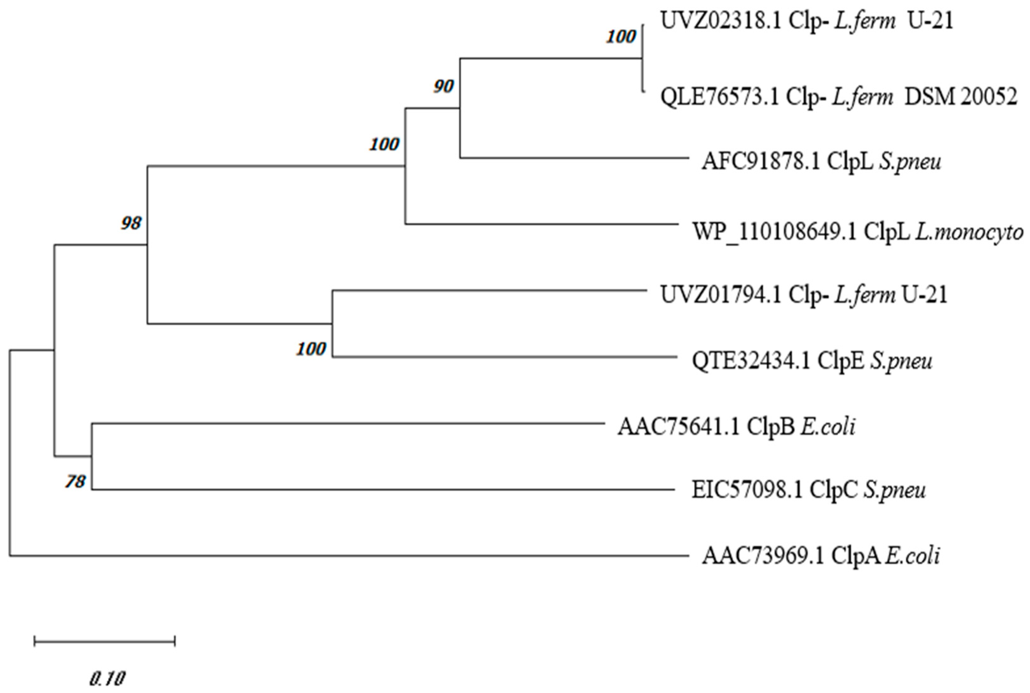
The comparative analysis of the amino acid sequence of the L. fermentum U-21 UVZ02318.1 protein, derived from the gene expression at the C0965_000195 locus, annotated as “ATP-dependent Clp protease ATP-binding subunit or ClpB” [20], revealed almost equal similarities with ClpA and ClpB proteins from E. coli MG1655, exhibiting approximately 39.51% and 41.96% identical amino acids, respectively. Moreover, intra-species comparison between ClpA and ClpB from E. coli demonstrated around 40.1% identical amino acids. Notably, other Clp-family proteins such as ClpC, ClpE, and ClpL were predominantly found in Gram-positive bacteria. A phylogenetic tree constructed using the amino acid sequences of UVZ02318.1 L. fermentum U-21, along with a series of Clp proteins from Streptococcus pneumonia and Listeria monocytogenes whose functions are described in the literature [8,11,12,29,30], is shown in Figure 1. Notably, the currently unannotated sequence QLE76573.1 from the type strain L. fermentum DSM 20052, sharing 99.57% identical amino acids with UVZ02318.1, was added in the tree for reference. Additionally, the sequence UVZ01794.1 of L. fermentum U-21 annotated as ClpA, sharing 50% identical amino acids with the UVZ02318.1, was also depicted in the tree.
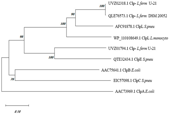
Figure 1.
Phylogenetic tree of Clp proteins from L. fermentum and reference sequences of Clp proteins with a confirmed function from other organisms. The sequences of Clp proteins: L. fermentum U-21 (UVZ02318.1 and UVZ01794.1), L. fermentum DSM 20052 (QLE76573.1), E. coli MG1655 ClpA (AAC73969.1) and ClpB (AAC75641.1), S. pneumoniae ClpC (EIC57098.1), ClpE (QTE32434.1) and ClpL (AFC91878.1), and L. monocytogenes ClpL (WP_110108649.1).
As can be seen from the data in Figure 1, the phylogenetic tree allows one to assign with some certainty UVZ02318.1 and QLE76573.1 proteins from L. fermentum, as ClpL, and UVZ01794.1 protein, as ClpE.
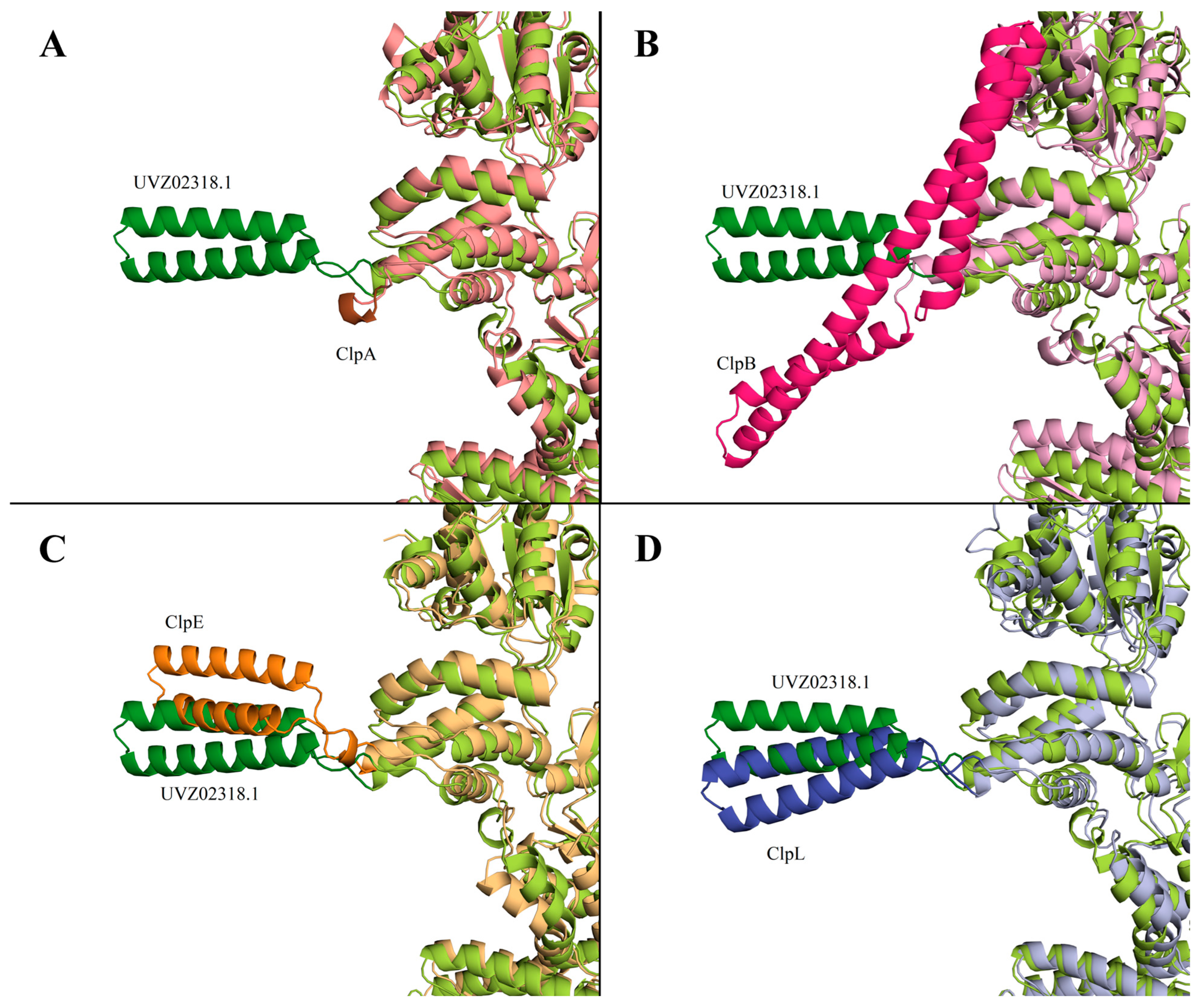
It was described in [31] that a distinctive feature of ClpB protein is the presence of M-domain in the structure (396-512 aa for WP_011228712). No homologous sequence corresponding to the M-domain of ClpB was found in UVZ02318.1. It should be noted that in ClpA, the M-domain is completely absent, while in ClpC, ClpE, and ClpL proteins, this domain is present but has a smaller size, and its structure differs significantly from the ClpB M-domain. Figure 2 shows a comparison of the AlphaFold models of ClpA, ClpB, ClpL, and ClpE proteins with the AlphaFold-predicted structure of Clp-protein UVZ02318.1. The M-domain is highlighted in more intense colors.
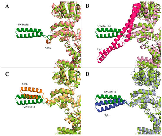
Figure 2.
AlphaFold model of UVZ02318.1 (“limon” color, M-domain highlighted in darker “forest” color) in pairwise comparison with AlphaFold structures of the following proteins: ClpA E. coli (panel (A); color “salmon”, M-domain—“brown”), ClpB E. coli (panel (B); “pink”, “hotpink”), ClpE S. pneumonia (panel (C); “lightorange”, “tv_orange”), and ClpL S. pneumonia (panel (D); “lightblue”, “tv_blue”).
The comparison of the structures of the Clp-family proteins shows a characteristically significant difference in the M-domains with a general similarity of the main part of the protein. Based on the alignment with other Clp-family proteins, UVZ02318.1 is most closely related to ClpL, with some resemblance to ClpE.
In all, the bioinformatic analysis of the UVZ02318.1 protein encoded by the gene at locus C0965_000195 demonstrates its classification within the Clp-protein family as ClpL. Hence, the UVZ02318.1 protein is denoted as L. fermentum U-21 ClpL in our investigation.
3.2. Gene Cloning and Expression and Chaperone Activity Investigation of the L. fermentum U-21 ClpL Protein in E. coli Cells
To investigate the chaperone activity of ClpL from L. fermentum U-21, the clpL gene was cloned and expressed in a heterologous system of E. coli cells. To analyze the expression of the clpL gene in E. coli, the XL1-Blue strain containing recombinant plasmid pUC19:clpL was grown in liquid LB medium. The soluble fraction of proteins was analyzed by SDS-PAGE (Figure S1). During the stationary growth phase and at the beginning of the logarithmic growth phase (after 2 h), an additional protein fraction with a molecular mass of about 77.5 kDa was observed in E. coli cells containing the pUC19:clpL plasmid. This molecular mass corresponds to the calculated molecular mass of the ClpL protein in combination with the molecular mass of the linker protein of the pUC19 plasmid. Mass-spectrometric analysis confirmed that this protein is an “ATP-dependent Clp protease ATP-binding subunit” ClpL of L. fermentum U-21. Furthermore, the presence of a high content of ClpL protein led to a delay in the transition to the exponential growth phase of strain XL-1 pUC19:clpL compared to the control strain XL-1 pUC19 (Figure S2).
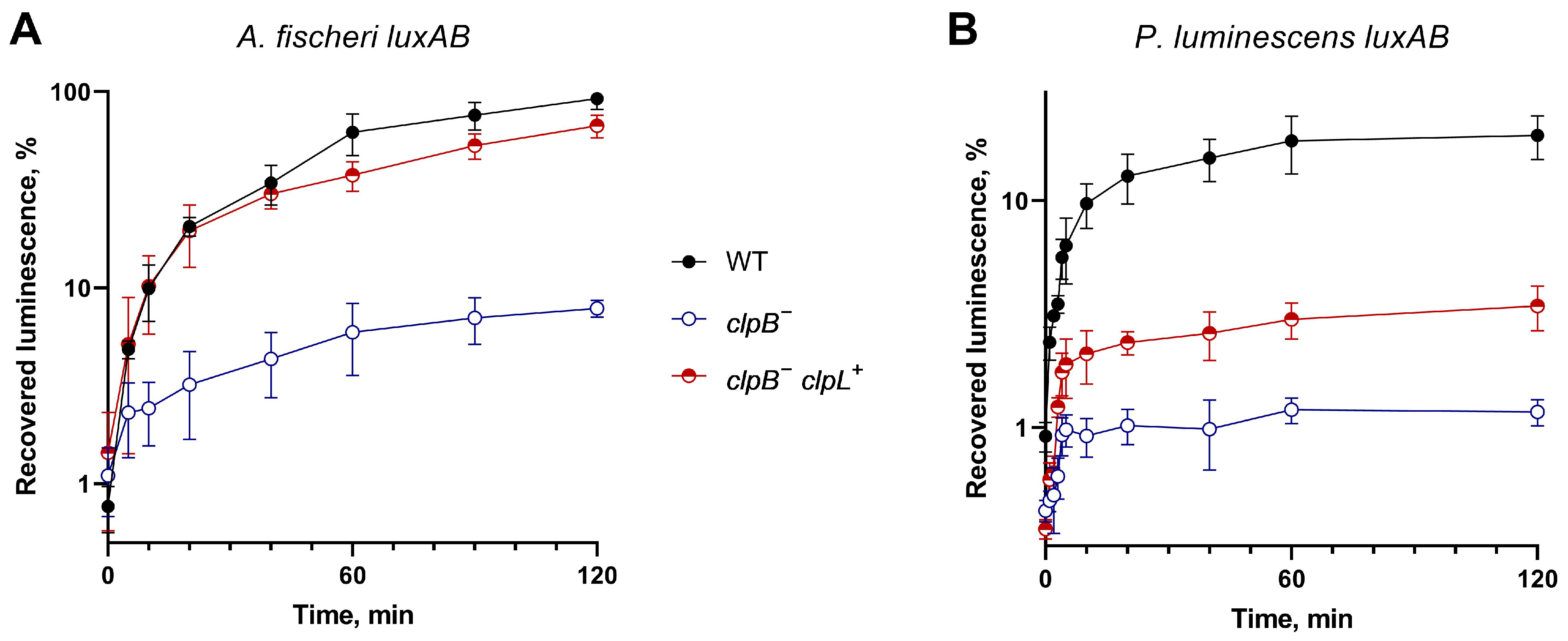
E. coli cells mutant in clpA and clpB genes have a low ability to refold heat-inactivated proteins [10]. Hence, these E. coli strains are ideal model hosts for the investigation of the ability of various heterologous proteins to compensate for the chaperone function. To assess the ability of ClpL to compensate the ClpB absence in refolding denatured proteins, E. coli SG20250 and its insertion derivative SG22100 clpB::kan− were used as hosts. The recovery of luminescence after the heat inactivation of the luciferases was measured in cells harboring p15XenAB or p15FisAB plasmids separately or in combination with pUC19:clpL (Figure 3). pUC19:clpL drives the heterologous expression of clpL from L. fermentum U-21; p15XenAB and p15FisAB are compatible with pUC19:clpL and drive the heterologous expression of luxAB from P. luminescens and A. fischeri, correspondently.

Figure 3.
Effect of the clpB::kan mutation and L. fermentum U-21 clpL gene introduction on the kinetics and level of refolding of heat-inactivated bacterial luciferases from A. fischeri (panel (A)) and P. luminescens (panel (B)) in E. coli cells. Inactivation of LuxAB A. fischeri was conducted by heating to 44 °C for 10 min, and LuxAB P. luminescens were inactivated by heating to 47 °C for 20 min. Ordinate—recovered luciferase activity relative to the initial activity before inactivation; abscissa—time of refolding at 22 °C after inactivation. Means and error bars were calculated from three independent replicates. At 120 min, measurements for “WT”, “clpB−”, and “clpB− clpL+” reliably differ with p-value < 0.01 for all, except “WT” and “clpB− clpL+” A. fischeri, which differ with p-value ~0.02.
The data presented in Figure 3 demonstrate that pUC19:clpL partially compensates for the deletion of clpB during the refolding of luciferases. The compensation is more pronounced for the thermolabile luciferase of A. fischeri and less prominent for the more thermostable luciferase of P. luminescens. LuxAB P. luminescens has poor ability to refold in E. coli in general. These findings are consistent with previous data [10] that indicate the importance of ClpB in DnaK-dependent refolding, with clpB deletion resulting in a 10-fold reduction in refolding efficiency. The ability of clpL from U-21 to compensate for the absence of clpB suggests a potential interaction between L. fermentum U-21 ClpL and the DnaKJE chaperone complex of E. coli.
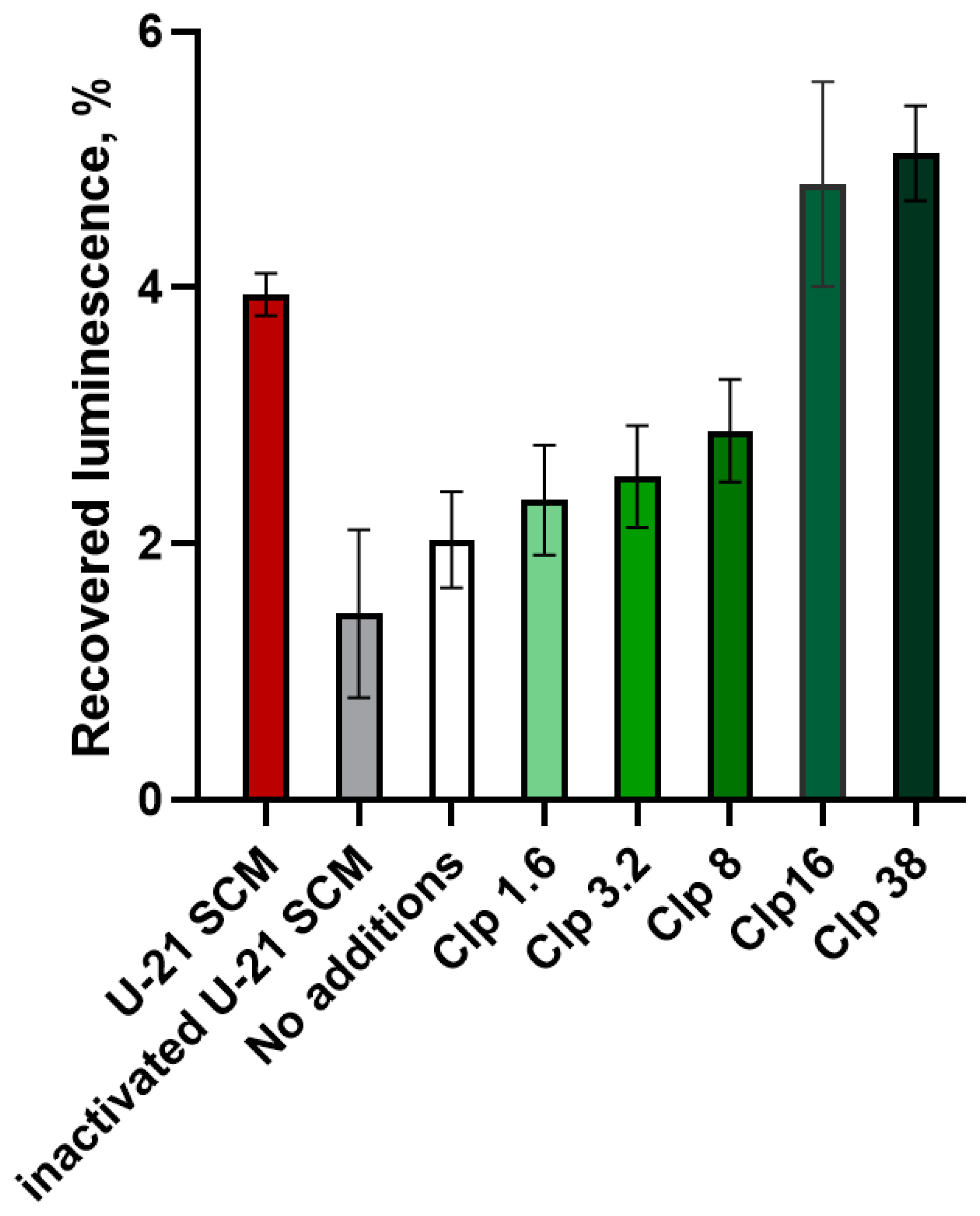
3.3. Investigation of the Chaperone Activity of L. fermentum U-21 SCM In Vitro
The SCM of L. fermentum U-21 cells, known to contain ClpL protein [20], was analyzed for its chaperone activity. Electrophoresis coupled with mass-spectrometry analysis revealed the presence of various proteins, including chaperones ClpL, DnaK, DnaJ, GroEL, GroES, ClpX, and other Clp-family members (Table S2). The effect of the SCM on the activity of the thermally denatured luciferase was tested in vitro using purified P. luminescens LuxAB. NADH, FMN, and LuxG were added to the reaction medium to maintain the pool of reduced FMNH2, which is a co-factor for the luciferase. For the identification of the role of proteins from the SCM in the refolding of luciferase, three types of samples were compared: luciferase reaction mixture without SCM, with the addition of SCM (final concentration of 10%), and with the addition of boiled SCM in the same amount. As a positive control, a purified ClpL protein from L. fermentum U-21, expressed in E. coli cells, was used. After the thermal inactivation of luciferase (45 °C for 9–11 min) and 4 min of refolding at room temperature, the luciferase reaction was triggered by the addition of decanal, and the luminescence was measured. The experiment was conducted in three independent biological replicates. Figure 4 shows the typical results with error bars corresponding to the measurement error (standard deviation) within a single replicate. From one experiment to another, the overall refolding efficacy varies, but each time the SCM and high concentrations of the purified ClpL enhance the luminescence approximately twice, while the boiled SCM does not have a significant effect.

Figure 4.
Recovered activity of thermoinactivated P. luminescens luciferase after refolding in vitro dependent on the presence of the SCM of L. fermentum U-21 or purified ClpL. SCM was added, representing 10% of the final reaction volume, with a ClpL concentration of 1.6–38 µg/mL.
Figure 4 presents the efficacy of luminescence recovery after thermal inactivation and the subsequent refolding of luciferase. The control sample without the addition of SCM showed the recovery of only about 2% of the initial luminescence. The addition of SCM to the sample increased the luminescence level by an average of two times. Boiled SCM had a slight negative effect on luminescence, but this effect was not statistically significant. Furthermore, the addition of purified ClpL protein prevented luciferase thermal inactivation in a dose-dependent manner. The effect became statistically significant when the concentration of ClpL was 38 µg/mL (paired t-test based on independent biological replicates, p-value < 0.01).
4. Discussion
Bioinformatics analysis comparing the amino acid sequence encoded by the gene located at locus “C0965_000195” in the L. fermentum U-21 genome with known Clp-family proteins (ClpA, ClpB, ClpC, ClpE, and ClpL) revealed a high similarity to ClpL (Figure 1 and Figure 2). This, therefore, allows us to assign the locus gene to clpL, which was first described for plasmids containing a transposon-like structure [32]. The presence of a specific M-domain characteristic of the ClpL protein was observed in the predicted protein structure (Figure 2), and the confidence scores obtained from AlphaFold models indicated the reliability of the predicted structures. Presented in Figure 2, AlphaFold-produced fragments have a per-residue confidence score (pLDDT) between 70 and 100 with the link of M-domain to the rest of the protein with pLDDT between 50 and 70. The intermediate confidence scores indicate the flexibility of that link, which could be the reason for the missing M-domains in most of the X-ray structures of Clp proteins. ClpL is known to exhibit chaperone activity [33], lacks the motif for binding to the protease subunit of ClpP [34], and does not require DnaKJ-type auxiliaries for its chaperone function in in vitro experiments [29].
In this study, we successfully cloned, expressed, and functionally characterized the clpL gene from L. fermentum U-21 in E. coli cells. High levels of ClpL were observed during the late exponential and stationary growth phases. The disappearance of the target band on SDS-PAGE between 3 and 5 h of growth (Figure S1) likely resulted from the proteolytic degradation of ClpL within E. coli cells. This hypothesis was supported by the observation of smaller fragments in the fraction of purified ClpL from E. coli NiCo21(DE3) pET16b.clpL cells after prolonged expression. When expression lasted 6 h, a significant degradation of ClpL into smaller fragments was observed (Figure S3, lane 6), whereas 1.5 h of induction allows the isolation of the full-length protein. Moreover, cells expressing ClpL exhibited a significant delay in transition to the exponential growth phase. We demonstrate the in vivo chaperone activity of ClpL by its ability to compensate for the absence of the clpB gene in E. coli cells. At the same time, we found no compensation for refolding in clpA− mutant cells (data not presented), suggesting that ClpL may operate through mechanisms similar to ClpB rather than ClpA.
Our in vitro assays further confirmed the chaperone activity of ClpL. The SCM of L. fermentum U-21, which contains various proteins including ClpL, significantly enhanced P. luminescens luciferase activity after thermal inactivation. The addition of the purified ClpL protein alone also prevented luciferase thermal inactivation in a dose-dependent manner. These findings are consistent with recent research [30] on the chaperone activity of ClpL from L. monocytogenes. The ability of ClpL to prevent protein aggregation and maintain protein activity suggests its potential application in addressing protein misfolding- and aggregation-related disorders, such as amyloid diseases including [30] Alzheimer’s, Parkinson’s, and Huntington’s diseases. Disaggregases are being intensively investigated as potential drugs targeting protein misfolding and aggregation [35,36]. Agents able to disaggregate preformed amyloids have been classified as molecular chaperones (e.g., Hsp70 and Hsp90), chemical chaperones (bile acids, steroid hormones, and trehalose), and pharmacological chaperones (amino acid derivatives, benzophenone, and tetracycline) [37]. The bacterial ATPases of the Clp proteins family have been suggested to be used for such purposes [38].
The compensation of clpB deficit in E. coli by clpL from L. fermentum suggests that ClpL may interact with heterologous environments, including other chaperone systems such as DnaKJ or even eukaryotic chaperones. This potential interaction may be important for the pharmacobiological effects of the proteins secreted by U-21 cells. L. fermentum U-21 was shown to act as pharmabiotic [39], which can reduce pro-inflammatory changes in rat models of Parkinson’s disease [40]. Certain bacterial strains, including Lactobacillus, Bifidobacterium, and Faecalibacterium, have shown the ability to alleviate symptoms of neurodegenerative diseases in animal models [41,42]. Clinical trials also support the potential of pharmabiotic in ameliorating manifestations of nigral dopaminergic neuronal death and motor deficits and regulating dopamine pathways in individuals with Parkinson’s disease [43,44]. However, the specific components of bacterial cells responsible for these effects and the underlying mechanisms remain unclear [39,40].
5. Conclusions
Our study provides the functional characteristics of the ClpL protein from L. fermentum U-21. The cloning, expression, and in vivo and in vitro assays demonstrated its chaperone activity. Our results suggest that the anti-Parkinsonian properties of the L. fermentum U-21 strain are determined, in part, by the ClpL protein and its ability to refold protein aggregates.
Supplementary Materials
The following supporting information can be downloaded at: https://www.mdpi.com/article/10.3390/biology13080592/s1. Table S1: Primers used in the study; Text S1: Fingerprint analysis of the proteins; Table S2: Chaperone proteins in the spent culture medium (SCM) after cultivation of L. fermentum U-21; Figure S1: Electrophoretogram of the soluble protein fraction of E. coli XL1-Blue strains containing pUC19 (lanes 1, 3, 5, 7, 9, and 11) and pUC19:clpL (lanes 2, 4, 6, 8, 10, and 12) plasmids after 2, 3, 4, 5, 18 and 24 h of growth; Figure S2: Growth curves of E. coli XL1-Blue strains containing pUC19 and pUC19:clpL plasmids; Figure S3: Electrophoretogram of the lysated Nico21(DE3) cells containing pET16b.clpL before induction (lane 1) and after induction for 45 and 90 min (lanes 2 and 3). The next lanes present ClpL protein purified by affinity chromatography after 1.5 h of expression (lanes 4 and 5) and after overnight expression (lane 6). Ref. [45] is cited within the Supporting Information.
Author Contributions
Conceptualization, I.V.M. and V.N.D.; methodology, I.V.M. and R.N.A.E.; software, S.V.B.; validation, I.V.M., R.N.A.E. and S.V.B.; formal analysis, I.V.M.; investigation, R.N.A.E., M.G.A., V.V.F., D.A.M., A.A.N. and E.U.P.; resources, I.V.M. and V.N.D.; data curation, S.V.B.; writing—original draft preparation, I.V.M. and R.N.A.E.; writing—review and editing, I.V.M., R.N.A.E., S.V.B. and M.G.A.; visualization, I.V.M. and S.V.B.; supervision, I.V.M. and V.N.D.; project administration, I.V.M.; funding acquisition, I.V.M. and V.N.D. All authors have read and agreed to the published version of the manuscript.
Funding
The measurements of the chaperone activity of ClpL with the use of luciferases in vitro and in vivo were supported by RSF 22-14-00124. The search of the possible applications of L. fermentum U-21 strain was supported by the Ministry of Science and Higher Education of the RF, project FSMF-2023-0010. The L. fermentum clpL gene search and the cloning, as well as the spent L. fermentum culture medium preparation conducted in the Vavilov Institute of General Genetics RAS, were funded within the framework of the state task 122022600163-7. The spent L. fermentum culture medium proteome analysis was supported by a grant from the Ministry of Science and the Higher Education of the Russian Federation for major scientific projects in priority areas of scientific and technological development (project #075-15-2024-638).
Institutional Review Board Statement
Not applicable.
Informed Consent Statement
Not applicable.
Data Availability Statement
Available data are presented in the manuscript and in the Supplementary Materials.
Conflicts of Interest
The authors declare no conflicts of interest.
References
- Katayama, Y.; Gottesman, S.; Pumphrey, J.; Rudikoff, S.; Clark, W.P.; Maurizi, M.R. The Two-Component, ATP-Dependent Clp Protease of Escherichia Coli. Purification, Cloning, and Mutational Analysis of the ATP-Binding Component. J. Biol. Chem. 1988, 263, 15226–15236. [Google Scholar] [CrossRef] [PubMed]
- Kitagawa, M.; Wada, C.; Yoshioka, S.; Yura, T. Expression of ClpB, an Analog of the ATP-Dependent Protease Regulatory Subunit in Escherichia coli, Is Controlled by a Heat Shock Sigma Factor (Sigma 32). J. Bacteriol. 1991, 173, 4247–4253. [Google Scholar] [CrossRef] [PubMed]
- Hoskins, J.R.; Sharma, S.; Sathyanarayana, B.K.; Wickner, S. Clp ATPases and Their Role in Protein Unfolding and Degradation. Adv. Protein Chem. 2001, 59, 413–420. [Google Scholar] [CrossRef] [PubMed]
- Wawrzynow, A.; Wojtkowiak, D.; Marszalek, J.; Banecki, B.; Jonsen, M.; Graves, B.; Georgopoulos, C.; Zylicz, M. The ClpX Heat-Shock Protein of Escherichia coli, the ATP-Dependent Substrate Specificity Component of the ClpP-ClpX Protease, Is a Novel Molecular Chaperone. EMBO J. 1995, 14, 1867–1877. [Google Scholar] [CrossRef] [PubMed]
- Wickner, S.; Gottesman, S.; Skowyra, D.; Hoskins, J.; McKenney, K.; Maurizi, M.R. A Molecular Chaperone, ClpA, Functions like DnaK and DnaJ. Proc. Natl. Acad. Sci. USA 1994, 91, 12218. [Google Scholar] [CrossRef] [PubMed]
- Weber-Ban, E.U.; Reid, B.G.; Miranker, A.D.; Horwich, A.L. Global Unfolding of a Substrate Protein by the Hsp100 Chaperone ClpA. Nature 1999, 401, 90–93. [Google Scholar] [CrossRef] [PubMed]
- Hoskins, J.R.; Singh, S.K.; Maurizi, M.R.; Wickner, S. Protein Binding and Unfolding by the Chaperone ClpA and Degradation by the Protease ClpAP. Proc. Natl. Acad. Sci. USA 2000, 97, 8892–8897. [Google Scholar] [CrossRef] [PubMed]
- Gottesman, S.; Wickner, S.; Maurizi, M.R. Protein Quality Control: Triage by Chaperones and Proteases. Genes Dev. 1997, 11, 815–823. [Google Scholar] [CrossRef] [PubMed]
- Motohashi, K.; Watanabe, Y.; Yohda, M.; Yoshida, M. Heat-Inactivated Proteins Are Rescued by the DnaK.J-GrpE Set and ClpB Chaperones. Proc. Natl. Acad. Sci. USA 1999, 96, 7184–7189. [Google Scholar] [CrossRef]
- Zavilgelsky, G.B.; Kotova, V.Y.; Mazhul’, M.M.; Manukhov, I.V. The Effect of Clp Proteins on DnaK-Dependent Refolding of Bacterial Luciferases. Mol. Biol. 2004, 38, 427–433. [Google Scholar] [CrossRef]
- Kim, G.; Lee, S.G.; Han, S.; Jung, J.; Jeong, H.S.; Hyun, J.k.; Rhee, D.K.; Kim, H.M.; Lee, S. ClpL Is a Functionally Active Tetradecameric AAA+ Chaperone, Distinct from Hexameric/Dodecameric Ones. FASEB J. 2020, 34, 14353–14370. [Google Scholar] [CrossRef] [PubMed]
- Namy, O.; Mock, M.; Fouet, A. Co-Existence of ClpB and ClpC in the Bacillaceae. FEMS Microbiol. Lett. 1999, 173, 297–302. [Google Scholar] [CrossRef] [PubMed]
- Breton, J.; Legrand, R.; Akkermann, K.; Järv, A.; Harro, J.; Déchelotte, P.; Fetissov, S.O. Elevated Plasma Concentrations of Bacterial ClpB Protein in Patients with Eating Disorders. Int. J. Eat. Disord. 2016, 49, 805–808. [Google Scholar] [CrossRef] [PubMed]
- Dominique, M.; Breton, J.; Guérin, C.; Bole-Feysot, C.; Lambert, G.; Déchelotte, P.; Fetissov, S. Effects of Macronutrients on the In Vitro Production of ClpB, a Bacterial Mimetic Protein of α-MSH and Its Possible Role in Satiety Signaling. Nutrients 2019, 11, 2115. [Google Scholar] [CrossRef] [PubMed]
- Tennoune, N.; Chan, P.; Breton, J.; Legrand, R.; Chabane, Y.N.; Akkermann, K.; Järv, A.; Ouelaa, W.; Takagi, K.; Ghouzali, I.; et al. Bacterial ClpB Heat-Shock Protein, an Antigen-Mimetic of the Anorexigenic Peptide α-MSH, at the Origin of Eating Disorders. Transl. Psychiatry 2014, 4, e458. [Google Scholar] [CrossRef] [PubMed]
- Rodríguez-Sojo, M.J.; Ruiz-Malagón, A.J.; Rodríguez-Cabezas, M.E.; Gálvez, J.; Rodríguez-Nogales, A. Limosilactobacillus fermentum CECT5716: Mechanisms and Therapeutic Insights. Nutrients 2021, 13, 1016. [Google Scholar] [CrossRef] [PubMed]
- De Luna Freire, M.O.; Cruz Neto, J.P.R.; de Albuquerque Lemos, D.E.; de Albuquerque, T.M.R.; Garcia, E.F.; de Souza, E.L.; de Brito Alves, J.L. Limosilactobacillus fermentum Strains as Novel Probiotic Candidates to Promote Host Health Benefits and Development of Biotherapeutics: A Comprehensive Review. Probiotics Antimicrob. Proteins 2024, 1–16. [Google Scholar] [CrossRef] [PubMed]
- Marsova, M.; Poluektova, E.; Odorskaya, M.; Ambaryan, A.; Revishchin, A.; Pavlova, G.; Danilenko, V. Protective Effects of Lactobacillus fermentum U-21 against Paraquat-Induced Oxidative Stress in Caenorhabditis elegans and Mouse Models. World J. Microbiol. Biotechnol. 2020, 36, 104. [Google Scholar] [CrossRef]
- Danilenko, V.N.; Stavrovskaya, A.V.; Voronkov, D.N.; Gushchina, A.S.; Marsova, M.V.; Yamshchikova, N.G.; Ol’shansky, A.S.; Ivanov, M.V.; Illarioshkin, S.N. The Use of a Pharmabiotic Based on the Lactobacillus fermentum U-21 Strain to Modulate the Neurodegenerative Process in an Experimental Model of Parkinson Disease. Ann. Clin. Exp. Neurol. 2020, 14, 62–69. [Google Scholar] [CrossRef]
- Poluektova, E.U.; Mavletova, D.A.; Odorskaya, M.V.; Marsova, M.V.; Klimina, K.M.; Koshenko, T.A.; Yunes, R.A.; Danilenko, V.N. Comparative Genomic, Transcriptomic, and Proteomic Analysis of the Limosilactobacillus fermentum U-21 Strain Promising for the Creation of a Pharmabiotic. Russ. J. Genet. 2022, 58, 1079–1090. [Google Scholar] [CrossRef]
- Gottesman, S.; Roche, E.; Zhou, Y.N.; Sauer, R.T. The ClpXP and ClpAP Proteases Degrade Proteins with Carboxy-Terminal Peptide Tails Added by the SsrA-Tagging System. Genes Dev. 1998, 12, 1338–1347. [Google Scholar] [CrossRef]
- Bazhenov, S.; Novoyatlova, U.; Scheglova, E.; Fomin, V.; Khrulnova, S.; Melkina, O.; Chistyakov, V.; Manukhov, I. Influence of the luxR Regulatory Gene Dosage and Expression Level on the Sensitivity of the Whole-Cell Biosensor to Acyl-Homoserine Lactone. Biosensors 2021, 11, 166. [Google Scholar] [CrossRef] [PubMed]
- Zavil’gel’skiĭ, G.B.; Kotova, V.I.; Manukhov, I.V. A Kinetic Method of Determining the Frequency of Homologous Recombination of Plasmids in Escherichia coli Cells. Mol. Biol. (Mosk) 1994, 28, 1299–1307. [Google Scholar]
- Zavilgelsky, G.B.; Zarubina, A.P.; Manukhov, I.V. Sequencing and Comparative Analysis of the lux-Operon of Photorhabdus luminescens Strain ZM1: ERIC Elements as Putative Recombination Spots. Mol. Biol. 2002, 36, 792–804. [Google Scholar] [CrossRef]
- Joseph, S.; Maccallum, P.; William, R.D. Molecular Cloning a Laboratory Manual, Volume 1; CSHL Press: Cold Spring Harbor, NY, USA, 2001. [Google Scholar]
- Gibson, D.G.; Young, L.; Chuang, R.Y.; Venter, J.C.; Hutchison, C.A.; Smith, H.O. Enzymatic Assembly of DNA Molecules up to Several Hundred Kilobases. Nat. Methods 2009, 6, 343–345. [Google Scholar] [CrossRef]
- Nazarov, P.A.; Khrulnova, S.A.; Kessenikh, A.G.; Novoyatlova, U.S.; Kuznetsova, S.B.; Bazhenov, S.V.; Sorochkina, A.I.; Karakozova, M.V.; Manukhov, I.V. Observation of Cytotoxicity of Phosphonium Derivatives Is Explained: Metabolism Inhibition and Adhesion Alteration. Antibiotics 2023, 12, 720. [Google Scholar] [CrossRef]
- Tamura, K.; Stecher, G.; Kumar, S. MEGA11: Molecular Evolutionary Genetics Analysis Version 11. Mol. Biol. Evol. 2021, 38, 3022–3027. [Google Scholar] [CrossRef]
- Park, S.S.; Kwon, H.Y.; Tran, T.D.H.; Choi, M.H.; Jung, S.H.; Lee, S.; Briles, D.E.; Rhee, D.K. ClpL Is a Chaperone without Auxiliary Factors. FEBS J. 2015, 282, 1352–1367. [Google Scholar] [CrossRef] [PubMed]
- Bohl, V.; Hollmann, N.M.; Melzer, T.; Katikaridis, P.; Meins, L.; Simon, B.; Flemming, D.; Sinning, I.; Hennig, J.; Mogk, A. The Listeria Monocytogenes Persistence Factor ClpL Is a Potent Stand-Alone Disaggregase. eLife 2024, 12, RP92746. [Google Scholar] [CrossRef]
- Rotanova, T.V.; Andrianova, A.G.; Kudzhaev, A.M.; Li, M.; Botos, I.; Wlodawer, A.; Gustchina, A. New Insights into Structural and Functional Relationships between LonA Proteases and ClpB Chaperones. FEBS Open Bio 2019, 9, 1536. [Google Scholar] [CrossRef]
- Huang, D.C.; Huang, X.F.; Novel, G.; Novel, M. Two Genes Present on a Transposon-like Structure in Lactococcus Lactis Are Involved in a Clp-Family Proteolytic Activity. Mol. Microbiol. 1993, 7, 957–965. [Google Scholar] [CrossRef] [PubMed]
- Kwon, H.Y.; Kim, S.W.; Choi, M.H.; Ogunniyi, A.D.; Paton, J.C.; Park, S.H.; Pyo, S.N.; Rhee, D.K. Effect of Heat Shock and Mutations in ClpL and ClpP on Virulence Gene Expression in Streptococcus pneumoniae. Infect. Immun. 2003, 71, 3757–3765. [Google Scholar] [CrossRef] [PubMed]
- Tao, L.; Biswas, I. ClpL Is Required for Folding of CtsR in Streptococcus Mutans. J. Bacteriol. 2013, 195, 576. [Google Scholar] [CrossRef] [PubMed]
- Khan, A.N.; Khan, R.H. Protein Misfolding and Related Human Diseases: A Comprehensive Review of Toxicity, Proteins Involved, and Current Therapeutic Strategies. Int. J. Biol. Macromol. 2022, 223, 143–160. [Google Scholar] [CrossRef]
- Lo, A.C.; Callaerts-Vegh, Z.; Nunes, A.F.; Rodrigues, C.M.P.; D’Hooge, R. Tauroursodeoxycholic Acid (TUDCA) Supplementation Prevents Cognitive Impairment and Amyloid Deposition in APP/PS1 Mice. Neurobiol. Dis. 2013, 50, 21–29. [Google Scholar] [CrossRef]
- Almeida, Z.L.; Brito, R.M.M. Amyloid Disassembly: What Can We Learn from Chaperones? Biomedicines 2022, 10, 3276. [Google Scholar] [CrossRef]
- Auburger, G.; Key, J.; Gispert, S. The Bacterial ClpXP-ClpB Family Is Enriched with RNA-Binding Protein Complexes. Cells 2022, 11, 2370. [Google Scholar] [CrossRef]
- Odorskaya, M.V.; Mavletova, D.A.; Nesterov, A.A.; Tikhonova, O.V.; Soloveva, N.A.; Reznikova, D.A.; Galanova, O.O.; Vatlin, A.A.; Slynko, N.M.; Vasilieva, A.R.; et al. The Use of Omics Technologies in Creating LBP and Postbiotics Based on the Limosilactobacillus fermentum U-21. Front. Microbiol. 2024, 15, 1416688. [Google Scholar] [CrossRef]
- Stavrovskaya, A.V.; Voronkov, D.N.; Marsova, M.V.; Olshansky, A.S.; Gushchina, A.S.; Danilenko, V.N.; Illarioshkin, S.N. Effects of the Pharmabiotic U-21 in a Combined Neuroinflammatory Model of Parkinson’s Disease in Rats. Bull. Exp. Biol. Med. 2024, 177, 193–199. [Google Scholar] [CrossRef]
- Ojha, S.; Patil, N.; Jain, M.; Kole, C.; Kaushik, P. Probiotics for Neurodegenerative Diseases: A Systemic Review. Microorganisms 2023, 11, 1083. [Google Scholar] [CrossRef]
- Dhyani, P.; Goyal, C.; Dhull, S.B.; Chauhan, A.K.; Singh Saharan, B.; Harshita; Duhan, J.S.; Goksen, G. Psychobiotics for Mitigation of Neuro-Degenerative Diseases: Recent Advancements. Mol. Nutr. Food Res. 2023, 68, 2300461. [Google Scholar] [CrossRef]
- Peña-Díaz, S.; García-Pardo, J.; Ventura, S. Development of Small Molecules Targeting α-Synuclein Aggregation: A Promising Strategy to Treat Parkinson’s Disease. Pharmaceutics 2023, 15, 839. [Google Scholar] [CrossRef]
- Chu, C.; Yu, L.; Li, Y.; Guo, H.; Zhai, Q.; Chen, W.; Tian, F. Meta-Analysis of Randomized Controlled Trials of the Effects of Probiotics in Parkinson’s Disease. Food Funct. 2023, 14, 3406–3422. [Google Scholar] [CrossRef]
- Tyanova, S.; Temu, T.; Cox, J. The MaxQuant Computational Platform for Mass Spectrometry-Based 41 Shotgun Proteomics. Nat. Protoc. 2016, 11, 2301–2319. [Google Scholar] [CrossRef]
Disclaimer/Publisher’s Note: The statements, opinions and data contained in all publications are solely those of the individual author(s) and contributor(s) and not of MDPI and/or the editor(s). MDPI and/or the editor(s) disclaim responsibility for any injury to people or property resulting from any ideas, methods, instructions or products referred to in the content. |
© 2024 by the authors. Licensee MDPI, Basel, Switzerland. This article is an open access article distributed under the terms and conditions of the Creative Commons Attribution (CC BY) license (https://creativecommons.org/licenses/by/4.0/).