Article

Bacterial Metal Accumulation as a Strategy for Waste Recycling Management

1
Exobiology Group, CNRS-Centre de Biophysique Moléculaire, University of Orléans, Rue Charles Sadron, CEDEX 2, 45071 Orléans, France
2
Voestalpine Stahl Donawitz GmbH, 8700 Leoben, Austria
3
Graz Centre for Electron Microscopy, 8010 Graz, Austria
*
Author to whom correspondence should be addressed.
Resources 2023, 12(12), 144; https://doi.org/10.3390/resources12120144
Submission received: 16 September 2023 / Revised: 30 November 2023 / Accepted: 6 December 2023 / Published: 8 December 2023

Abstract

:
Sustainable mechanisms for efficient and circular metal recycling have yet to be uncovered. In this study, the metal recycling potential of seven metal-resistant bacterial species (Deinococcus radiodurans, Deinococcus aerius, Bacillus coagulans, Pseudomonas putida, Staphylococcus rimosus, Streptomyces xylosus and Acidocella aluminiidurans) was investigated in a multi-step strategy, which comprises bioleaching of industrial waste products and subsequent biosorption/bioaccumulation studies. Each species was subjected to an acidic, multi-metal bioleachate solution and screened for potential experimental implementation. Bacterial growth and metal acquisition were examined using scanning transmission electron microscopy coupled to electron dispersive X-ray spectroscopy (STEM-EDS). Two of the seven screened species, D. aerius and A. aluminiidurans, propagated in a highly acidic and metal-laden environment. Both accumulated iron and copper compounds during cultivation on a multi-metallic bioleachate. Our findings suggest that extremotolerant bacteria should be considered for waste recycling operations due to their inherent polyextremophily. Furthermore, STEM-EDS is a promising tool to investigate microbial–metal interactions in the frames of native industrial waste products. To develop further experimental steps, detailed analyses of adsorption/accumulation mechanisms in D. aerius and A. aluminiidurans are required to design a circular metal recycling procedure.

1. Introduction

The development of a sustainable circular economy for the ever-increasing demand of metals for our everyday use is of great importance. As of 2021, European steel plants account for 16.2% of worldwide crude steel production [1], generating a plethora of multi-metallic by-products [2], which cannot be completely fed into existing circular economy yet. Various technologies for recycling metal elements from high-metal-content waste products are being developed. Innovative ideas range from mathematical models proposing anaerobic dark fermentation to extract metals from electronic waste via leaching [3] to ultrafast electrothermal processes (~3000 °C, ~1 s) [4] and adsorption on a functionalized material with improved sorbent properties [5] for rare earth element recycling to avoid harmful conventional pyrometallurgical and lavish hydrometallurgical treatments. Traditional leaching technology often requires large quantities of leaching solution (EDTA, hydrochloric acid, and organic acids) which are expensive and unsuitable to remove hazardous heavy metals such as hexavalent chromium from sensitive environments [6,7]. Measures to improve traditional leaching via application of electric fields [8] or using more gentle chemicals [9] for metal solubilization are diverse. Recently, we have proposed to harness extreme metallophilic microorganisms such as Acidianus manzaensis to mobilize metals from waste products derived from steel production [10] and potentially feed them into a circular process via selective adsorption processes, providing a gentle and cost-effective alternative to traditional technologies.
Even in traces, metals have a great potential to cause toxicity in humans and animals, consequently leading to cancer and mitochondrial damage [11,12]. Efforts to remove toxic metals from anthropogenic environments (e.g., wastewater, contaminated drinking water, etc.) generated a wide array of different advances. This includes adsorption technology (e.g., carbon composites, minerals, magnets, biosorbents, etc.), membrane treatments (e.g., nanofiltration and osmosis), chemical and electrical processes (e.g., chemical precipitation and reduction) and photocatalysis, and many authors have come to the conclusion that biosorption is one of the most promising methods to gently remove heavy meals from the environment (reviews by [13,14]). Biological sorbents, for instance, exhibit various advances compared to pure physico-chemical sorption methods since they are cost- and energy-effective as well as non-toxic [15]. Biologically derived compounds such as the extracellular polymeric substance (EPS) found in bacteria and archaea have great potential for removing heavy metals from their environment due to their cell surface chemistry. The EPS usually consists of polysaccharides, nucleic acids, proteins and lipids, which can be associated with the cell-surface or can be secreted extracellularly [16]. As a biosorbing agent, immobilized EPS of Pseudomonas spp. can efficiently remove Cd, Pb, Cu, and Zn from contaminated environments, reaching adsorption equilibria quickly [17,18]. Furthermore, Acinetobacter junii isolated from a chromite mining site, is able to adsorb cytotoxic Cr(VI) while reducing it to Cr(V) on the EPS surface [19]. Even though successful removal of heavy metals was achieved via EPS, this naturally derived product does not provide the mechanic stability to apply it in large-scale operations yet [20]. Many types of biosorbents have been characterized, e.g., bacteria, fungi, yeasts, algae, plants, and many more [21]. Indigenous microbial species isolated from metal-polluted or extreme environments demonstrate an inherent heavy metal resistance as well as biosorption abilities [22,23,24], since microbial adaptation to specific metals in their environment seems to be a common phenomenon [25,26].
A variety of factors such as pH, temperature, pollutant concentration, and metal ion properties, influence the success of biosorption or bioaccumulation processes. While biosorption describes a passive process initiated by electrostatic interactions between metals and functional groups on the cell surface, bioaccumulation comprises metal incorporation or adsorption by metabolically active cells [21,22]. The pH of a solution is one of the most crucial aspects of biosorption since it affects the charge and ionization of functional groups on the cell surface [22]. In general, a low pH seems to be favourable for cation binding stability due to the overall negative charge of the cell surface and increased metal solubility under acidic conditions [27,28], but depending on the metal of interest and the biosorbing organism, binding capacity might decrease under highly acidic solution pH [29]. Other factors, which could increase biosorption include elevated temperatures (increases surface activity and kinetic energy), low ionic strength (less competition of binding sites), high agitation speed (enhances pollutant removal rate by minimizing mass transfer resistance), high pollutant load, etc. [30].
Various bacterial and archaeal species either possess an inherent metal resistance, mechanisms to detoxify metals or are metallophilic microorganisms, which require the active use of metals to cover their energetic needs [31,32,33,34]. Bacteria, such as Bacillus spp., Pseudomonas spp., Citrobacter sp., can cope with a metalliferous environment by producing biosurfactants, which actively lower surface tensions by emulsification, thereby increasing metal binding to the surface [35]. Besides the production of compounds, the whole biomass of bacteria, such as Bacillus coagulans isolated from a tannery effluent, can effectively remove cytotoxic hexavalent chromium [36]. Pseudomonas putida cells isolated from a plant rhizosphere can tolerate and adsorb high amounts of copper and zinc while growing actively [37], whereas hexavalent chromium biosorption reaches its equilibrium after 96 h post-incubation on inactivated biomass [38]. In one study, Pseudomonas sp. succeeded over Staphylococcus xylosus in maximum cadmium uptake, and chromium biosorption was more successful for S. xylosus cells. In a binary system where cadmium and chromium ions compete about binding to the cell surface, chromium almost exclusively bound to S. xylosus cells [39]. Promising biosorption processes are mainly accounted for carboxyl, amino/amide, -CH, C-O and -OH functional groups on the cell surface [40,41]. Carboxyl, amide and phosphate groups on D. radiodurans cell surface are suspected to play a major role in uranium sequestration in nuclear waste polluted environments [42].
A correlation between metal pollution and microbial adaptation to specific metals in their environment seems to be a common phenomenon. In our study, seven (heavy) metal-tolerant bacteria (D. radiodurans, D. aerius, B. coagulans, P. putida, S. rimosus, S. xylosus and A. aluminiidurans) were used to implement initial stages a cascaded multi-step approach of metal recycling. In the first step, as previously published, a multi-metallic bioleachate solution was obtained by cultivation of the extremophilic archaeon Acidianus manzaensis [10] on a steel production plant waste product (a secondary dust waste product of the basic oxygen furnace (BOF) steelmaking process, referred to as BOF dust [10,43] (voestalpine Stahl Donawitz GmbH, Leoben, Austria)) to solubilize metals from a solid into a liquid phase. Here, we report on the second step of biosorption/bioaccumulation trials with seven bacterial strains. We show that two strains (D. aerius and A. aluminiidurans) were able to withstand an extreme low-pH and multi-metallic environment. Via transmission electron microscopy analyses (TEM), strain-selective spatial and temporal accumulation of iron and copper can be visualized. This work can contribute to further and deeper investigations of microbial metal adsorption and metal uptake processes to gain detailed knowledge on environmentally friendly metal removal technology.

2. Materials and Methods

2.1. Strains and Media Composition

A. manzaensis (NBRC 100595) [44] was cultured with BOF dust obtained from a steel production plant as previously described [10]. A variety of metals was solubilized from BOF dust into the surrounding medium (bioleachate) by a thermoacidophilic bioleaching setup [10]. Bioleachate was harvested by filtration through a sterile 0.45µm pore-sized membrane (Chromafil, Macherey-Nagel, Düren, Germany). The native pH of filtered bioleachate solution was kept at 1.7 for the following experiments to avoid intense and non-controlled metal precipitation by pH adjustment. Seven bacterial strains, Pseudomonas putida (DSM 100120) [45], Bacillus coagulans (DSM 1) [46], Streptomyces rimosus (DSM 40260) [47], Staphylococcus xylosus (DSM 20266) [48], Deinococcus aerius (DSM 21212) [49], Deinococcus radiodurans (DSM 20539) [50] and Acidocella aluminiidurans (DSM 108313) [51], were grown aerobically in their respective liquid media at strain-specific temperatures and pH (Table 1; media composition was provided by the DSMZ database according to DSM number for each strain mentioned above).

2.2. Cascaded Approach of Metal Waste Bioconversion

As part of a multi-step metal recycling process, initial stages of cascaded waste bioconversion were investigated in this study. A scheme of this cascade waste recycling approach is presented in Figure S1. At first, steelmaking processes generate a variety of metal-containing waste products (A). A multi metallic BOF dust (B) waste (metal composition table in [10]) is generated as a consequence of basic oxygen furnace (BOF) steelmaking processes [43]. BOF dust used in this study was provided by voestalpine Stahl Donawitz GmbH, Leoben, Austria. Next, BOF dust was fed into a liquid Acidianus manzaensis culture (C) to test the bioleaching abilities of this thermoacidophilic archaeon as published previously [10]. At the end of the cultivation of A. manzaensis, the cells were harvested by centrifugation and supernatants were separated as described before [10]. The resulting supernatants were filtered with a 0.44 μm pore size filter (Macherey-Nagel GmbH & Co. KG, Düren, Germany) (D) to obtain bioleachate solution (E) for further biosorption/bioaccumulation studies. At the next stage, seven bacterial strains were cultivated (F) on native bioleachate solution (pH 1.70) and screened for cell growth or decay (G). To test bacterial survival in acidic multi-metallic bioleachate solution, each strain was cultured in a 1:5 mixture of 10-fold concentrated strain-specific medium (Table 1). Cells were harvested by centrifugation (10 min; 3000× g), and ice-cold and sterile PBS was added to wash away residual rich medium. Filtered bioleachate solution produced by A. manzaensis (Table 2) was added to previously washed cells. Bacterial propagation and/or decay on acidic bioleachate solution was monitored via a cell counting chamber (Neubauer improved, Brand GmbH + Co KG, Wertheim, Germany). Two strains (Deinococcus aerius and Acidocella aluminiidurans) were analysed (H) for metal sorption/accumulation capacities via scanning transmission electron microscopy (STEM) coupled to energy dispersive spectroscopy (STEM-EDS).

2.3. Electron Microscopy and Elemental Mapping

2.3.1. Cell Preparation for Electron Microscopy

After survivability and possible propagation of all strains on metal, bioleachate was determined via cell counting; D. aerius and A. aluminiidurans were subjected to further experiments. To monitor bacterial metal accumulation abilities, D. aerius and A. aluminiidurans cells were processed for TEM. Microbial cells cultivated with bioleachate were sampled at different time points of cultivation (maximum 48 h) to examine differences in cellular metal acquisition patterns. Cells were harvested by centrifugation (10 min; 3000× g) and were fixed in glutaraldehyde for 1 h (2.5% in 1 M Na-cacodylate buffer), washed in PHEM buffer (360 mM PIPES, 150 mM HEPES, 60 mM EGTA, 12 mM MgCl2) followed by secondary fixation for 1 h in 1% OsO4, finalizing fixation by washing in PHEM buffer. Pellets were dehydrated in a graded ethanol series (30%, 50%, 70%, 90%, abs.). Before embedding, samples were dehydrated in dried acetone for resin infiltration and finally embedded in epoxy resin (Agar Low Viscosity Resin, Agar Scientific, Essex, UK). Samples embedded in resin were polymerized at 60 °C for 48 h, followed by ultrathin sectioning (Reichert-Jung Ultracut E, Reichert, Wels, Austria).

2.3.2. STEM Imaging and EDS Investigations

STEM investigations were performed at the Graz Centre for Electron Microscopy (FELMI-ZFE, Graz, Austria) on a probe-corrected FEI Titan G2 60–300 (S/TEM) microscope equipped with an X-FEG Schottky field-emission electron source operated at 60 and 300 kV (current of 150 pA, beam diameter of 1 Å). Elemental maps and spectra were processed with Gatan Digital Micrograph software 3.5 (Gatan Inc., Pleasanton, CA, USA). Element quantification for EDS spectra was performed by using the k-factor method.

3. Results and Discussion

3.1. Growth on Extremely Acidic Bioleachate Solution

Seven bacterial strains (Table 1) were incubated on a multi-metallic and extremely acidic bioleachate solution (pH 1.7) to assess the individual microbial potential for a cascade metal recycling approach (Figure S1). Properties such as cell growth or decay in an acidic multi-metal solution, as well as the observation of intra- or extracellular metal accumulation via electron microscopy were examined. Each strain was incubated on previously obtained bioleaching solution produced by the thermoacidophilic archaeon Acidianus manzaensis [10]. A. manzaensis was cultivated with a metallic steel plant waste product (BOF dust) under aerobic conditions. Major metal components of the bioleachate solution (Mg, Ca, Fe, Zn, Mn and Al as well as Sr, Cu, Cr, Ni, Co, Ti, V, Cd, Mo, As, Pb, and Ba in trace amounts) were detected by ICP-MS (Table 2). These results are in concordance with the o.verall native BOF dust composition where Fe, Ca and Mg are the most abundant metals (table in [10]). All seven strains were cultured for 48 h on the aforementioned acidic bioleachate solution. Cell density at the time point of inoculation (0 h) was compared to cell densities at 24 h and 48 h after inoculation to investigate potential pH and metal resistance of each strain (Table 3). Overall observation shows a decrease in cells for B. coagulans, P. putida and S. xylosus (from 7.98 × 10⁶ to 8.03 × 10⁵, 9.92 × 10⁶ to 1.54 × 10⁶ and from 1.7 × 10⁷ to 2.50 × 10⁴ cells/mL, respectively).
Upon contact with bioleachate solution, S. rimosus cultures formed large pellets and agglomerated cellular debris (Figure S2) with no detectable planktonic cells. D. radiodurans cell density remained stable (from 2.76 × 10⁶ to 3.36 × 10⁶ cells/mL) throughout 48 h of cultivation. Interestingly, D. aerius cells were able to propagate under these conditions (from 6.96 × 10⁵ to 5.60 × 10⁶ cells/mL) while keeping an intact cellular morphology as observed by electron microscopy (Figure 1A–C and Figure S3A–C). Similarly, A. aluminiidurans cell density increased by an order of magnitude in course of 24 h (from 1.06 × 10⁶ to 3.20 × 10⁷ cells/mL) (Table 3) while keeping a rod-shaped structure and preserved cellular integrity (Figure 2A–C and Figure S4A,B).
Strain-dependent differences in resistance to a low pH, multi-metallic solution may be attributed to a harsh shift away from pH and nutritional optima. Since the optimal pH for most of the species used in this study is neutral (pH 7.0–7.2), the drastic decrease in pH of the surrounding environment (pH 1.7) most likely impaired cell viability and initiated cell lysis rather than cell division. Cells dealing with acid stress can employ several mechanisms to avoid the drop of intracellular pH [52], since a high concentration of protons inside the cell is deleterious to proteins and nucleic acids. Bacteria such as D. radiodurans and D. aerius are potent reactive oxygen species (ROS) scavengers and possess highly efficient DNA repair mechanisms [53,54], which may allow cells to maintain viability and morphology in a very acidic and metalliferous environment. Cells of acidophilic microorganisms can be very well equipped with a powerful proton pump mechanism to activate ATP-dependent H+ extrusion and/or other effective pH homeostasis mechanisms such as chemical and enzymatic proton sequestration [55,56,57]. For this study, viable biomass was subjected to high metal stress and toxicity (which is not relevant for dead biomass). Most likely, a high metal variety and concentration exceeded levels where individual strains could still employ efficient detoxification mechanisms [58]. A rather prolonged exposure to a low-key metalliferous environment seems to be necessary for microorganisms to establish resistance mechanisms than a sudden exposure to high concentrations of metals [59]. Acidophilic and metal-tolerating bacteria such as D. aerius and A. aluminiidurans can also be considered to recover metals in other multi-metallic systems such as asteroid biomining operations. Analogously to our approach described in this manuscript, the initial bioleaching process step is carried out on thermoacidophilic archaea grown on extraterrestrial materials to solubilize metals from the mineral into the liquid phase [60,61]. In the second step, metals are recovered by biosorption or bioaccumulation processes for further technological implementation.
For the acidophilic bacterium A. aluminiidurans, the pH range for cellular growth is 3.0–7.0, while optimum growth occurs at around 4.0–5.0 [51]. Acidocella spp. exhibit, among other acidophilic bacteria, the ability to dissimilatory ferric iron reduction [62], which may have facilitated the bioleachate solution to promote cell propagation (bulk elemental composition of BOF dust [45], Table 2). Furthermore, the supplementation with concentrated strain-specific medium might play a role in keeping cells in a viable state as long as nutrients and/or energy sources for propagation are still available.

3.2. Microbial Metal Incorporation Abilities

Biosorption describes the natural, metabolically passive process of rapid and reversible binding of charged ions onto a biological surface, whereas naturally occurring bioaccumulation is a metabolically active process performed by living cells [63,64]. To investigate possible metal biosorption/bioaccumulation of D. aerius and A. aluminiidurans from the bioleachate solution, cells were examined via elemental ultrastructure analysis coupled to electron dispersive X-ray spectroscopy (STEM-EDS).
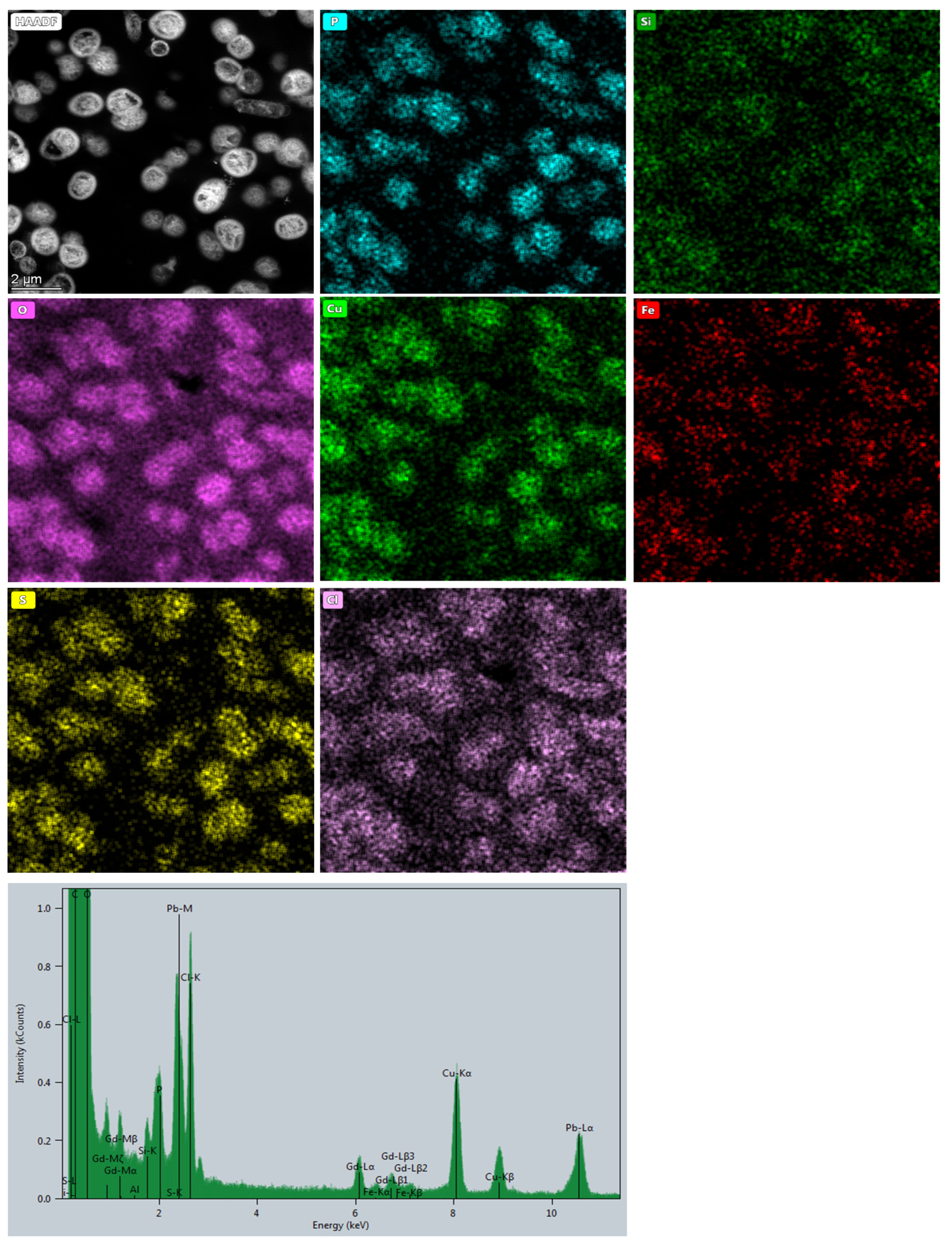
Overall, the TEM images of D. aerius and A. aluminiidurans show cellular association with inorganic electron-dense structures forming granules and precipitated morphologies (Figure 1A–C, Figure S3A–C, Figure 2A–C and Figure S4A,B). Elemental mapping of D. aerius after 24 h of cultivation on bioleachate solution displays a refined intracellular distribution of elements such as Fe, Cu, S and P (Figure 3) with strong corresponding spectral signals (Figure S5). Cu, S and P are equally distributed in the cells, whereas Fe shows diffused distribution and punctured deposition (Figure 3). Si and Cl are also associated with intracellular content of D. aerius cells (Figure 3). Interestingly, a close relative of D. aerius, D. radiodurans, can adsorb uranium-containing compounds onto its functional groups on the cell surface (–C=O, –NH2, –OH), forming uranium phosphates by biomineralization [65], which very likely possesses similar biosorptive abilities of D. aerius. Whether or not D. aerius cells can act as a nucleation site for biominerals has to be elucidated in future experiments. The observed metal incorporation in D. aerius cultivated on multi-metallic bioleachate solution may be accounted to the electrostatic interactions between cell surface of D. aerius and metal cations binding in terms of passive biosorption. Adsorption effects and metal binding affinity depend on factors such as solution pH, temperature, metal dosage, electronegativity and metal ion radius [20]. Heavy metals with large cation sizes are prone to bind more effectively to bacterial surfaces [66], an observation which is in accordance with metal binding to D. aerius. Inherent polyextremophily (e.g., radiation, desiccation, and nuclear waste resistance) as in the case of D. radiodurans [53] might be considered as a prerequisite to examine more intrinsic features of the Deinococcus genus in regard to biosorption.
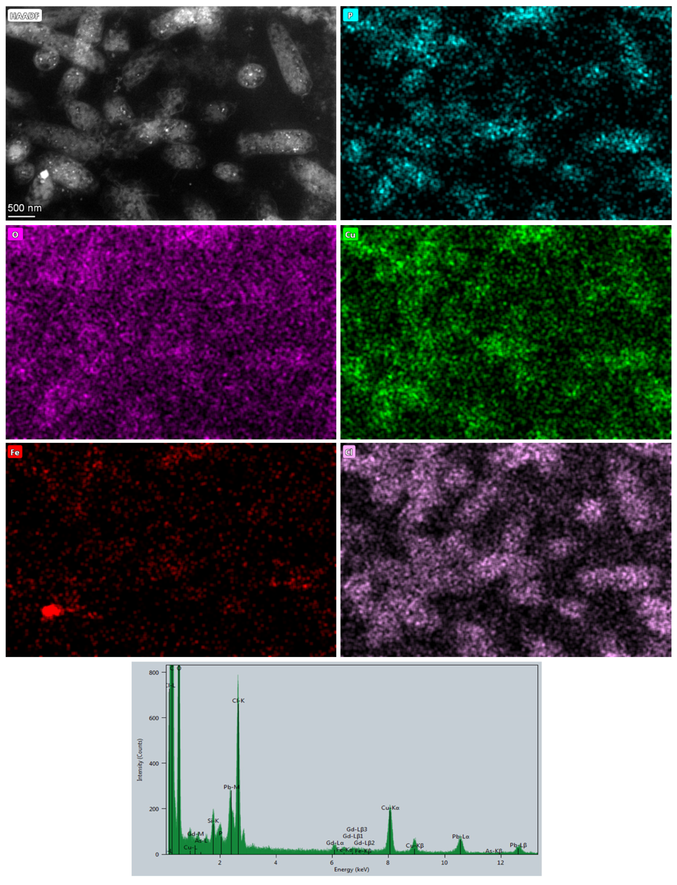
A. aluminiidurans cells kept their characteristic rod-shaped appearance while propagating in the multi-metallic solution (Figure 2A–C and Figure S4, Table 2). Dark granules and smaller deposited electron-dense structures are present and ubiquitously dispersed throughout the cells of A. aluminiidurans (Figure 2A–C and Figure S4). STEM-EDS analysis reveals intracellular accumulations of Cu, P, and Fe (Figure 4 and Figure S6). Cl also shows intracellular distribution in A. aluminiidurans cells (Figure 4, corresponding elemental map). A study by Rizki et al. [67] shows that Acidocella sp. cells are able to rapidly incorporate gold particles into the cell in a matter of minutes, which is consistent with our results of metal incorporation by this microorganism. While the Fe signal is less distinct but visibly concentrated in A. aluminiidurans cells, Cu ions produce a stronger EDS response (Figure 4, corresponding elemental maps).
The phosphorus signal was intensively distributed inside the cells as well. Such pronounced phosphorus accumulation observed in both D. aerius and A. aluminiidurans may serve as a sequestration of heavy metals and represent an effective detoxifying mechanism commonly employed by metal-tolerant microorganisms [68,69]. Since A. aluminiidurans was isolated from an acidic, sulphate-containing swamp (pH 3) [51], survival in bioleachate solution seems coherent. Close phylogenetic relatives (A. aminolytica and A. facilis) were isolated from acid mine drainage sites [70,71,72], thereby suggesting an inherent metal resistance in the Acidocella genus. Other factors that could influence biosorption include pH, temperature (increased surface activity and kinetic energy), low ionic strength (less competition for binding sites), high agitation speed (enhanced pollutant removal rate by minimizing mass transfer resistance), and high pollutant loads [30].
The results on metal incorporation obtained for D. aerius and A. aluminiidurans provide a promising perspective to gain in-depth knowledge about their interactions with a metalliferous waste product derived from a steelmaking plant. To develop the next steps for cascade bioprocessing of waste products, functional groups involved in biosorption, binding chemistry of metals to the cell surface and the potential microbial detoxification mechanisms need to be characterized. Information about individual molecular mechanisms activated when dealing with bioleachate solution could enhance our understanding of genetic modification to make future operations more effective or even selective in terms of metal separation. Quantitative measurements and/or modelling of biosorption efficiency under the controlled initial microbial cell densities need to be carried out in future experiments to extend insights into microbe–metal interactions. Screening for novel metabolic traits (e.g., the ability of D. aerius, D. radiodurans, and A. aluminiidurans to withstand acidic and multi-metallic systems) can contribute to interdisciplinary studies and broaden our understanding of microbial adaptability in extreme environments.

4. Conclusions

Seven bacterial strains (D. radiodurans, D. aerius, B. coagulans, P. putida, S. rimosus, S. xylosus and A. aluminiidurans) have been subjected to acidic and metalliferous bioleachate solution obtained from a previous thermoacidophilic bioleaching operation to find candidates for potential metal recycling strategies. Two out of seven tested strains, D. aerius and A. aluminiidurans, were able to propagate, maintain their characteristic morphology and preserve cellular integrity during cultivation on bioleachate. Both strains were investigated concerning metal acquisition via STEM-EDS analysis. Metals such as iron and copper could be detected in D. aerius as well as A. aluminiidurans cells, indicating a preference in binding affinity of these metals compared to other metals in the bioleachate. These results emphasize the ability of acidophilic bacteria and other extremophiles to survive harsh conditions of low pH and high metal load. Furthermore, microbial adsorption and/or accumulation of metals detected in this study can be used further for their subsequent recycling and re-introduction into an industrial process. Future works need to focus on clarifying the mechanisms employed by bacteria (biosorption and/or bioaccumulation), selective metal affinity, and subsequent separation of metals of interest for their successful recovery.

Supplementary Materials

The following supporting information can be downloaded at https://www.mdpi.com/article/10.3390/resources12120144/s1. Figure S1: Scheme of a cascade waste recycling approach; Figure S2: S. rimosus culture; Figures S3 and S4: additional TEM images; Figures S5 and S6: additional STEM-EDS data.

Author Contributions

D.K., A.M., H.S., M.A. and T.M. performed experiments. D.K., A.M. and T.M. planned and executed the cultivation of all bacterial strains, as well as performance and interpretation of SEM-EDS experiments. M.A. and T.M. performed and analyzed STEM-EDS results. D.K., A.M., H.S., D.W., G.K., M.A. and T.M. made substantial contributions to the acquisition, analysis, and interpretation of the data described in this study. All authors have read and agreed to the published version of the manuscript.

Funding

This research received no external funding.

Data Availability Statement

The original contributions presented in the study are included in the article/Supplementary Materials; further inquiries can be directed to the corresponding author.

Acknowledgments

We thank N. Cyran (the Core Facility Cell Imaging and Ultrastructure Research, University of Vienna—member of the Vienna Life-Science Instruments (VLSI)) and Claudia Mayrhofer (Graz Centre for Electron Microscopy) for helping with TEM cell preparation and analysis.

Conflicts of Interest

G.K., H.S., and D.W. were employed by the company voestalpine Stahl Donawitz GmbH, Leoben, Austria. The remaining authors declare that the research was conducted in the absence of any commercial or financial relationships that could be construed as a potential conflict of interest.

References

  1. EUROFER. European Steel in Figures. 2022. Available online: https://www.eurofer.eu/publications/brochures-booklets-and-factsheets/european-steel-in-figures-2022 (accessed on 1 July 2022).
  2. EUROFER. Annual Report. 2020. Available online: https://www.eurofer.eu/publications/archive/annual-report-2020 (accessed on 27 November 2021).
  3. Russo, F.; Luongo, V.; Mattei, M.R.; Frunzo, L. Mathematical Modeling of Metal Recovery from E-Waste Using a Dark-Fermentation-Leaching Process. Sci. Rep. 2022, 12, 4274. [Google Scholar] [CrossRef] [PubMed]
  4. Deng, B.; Wang, X.; Luong, D.X.; Carter, R.A.; Wang, Z.; Tomson, M.B.; Tour, J.M. Rare Earth Elements from Waste. Sci. Adv. 2022, 8, 3132. [Google Scholar] [CrossRef] [PubMed]
  5. Gabor, A.E.; Davidescu, C.M.; Negrea, A.; Ciopec, M.; Butnariu, M.; Ianasi, C.; Muntean, C.; Negrea, P. Lanthanum Separation from Aqueous Solutions Using Magnesium Silicate Functionalized with Tetrabutylammonium Dihydrogen Phosphate. J. Chem. Eng. Data 2016, 61, 535–542. [Google Scholar] [CrossRef]
  6. Qiao, J.; Sun, H.; Luo, X.; Zhang, W.; Mathews, S.; Yin, X. EDTA-Assisted Leaching of Pb and Cd from Contaminated Soil. Chemosphere 2017, 167, 422–428. [Google Scholar] [CrossRef] [PubMed]
  7. Zhu, G.; Guo, Q.; Yang, J.; Zhang, H.; Wei, R.; Wang, C.; Peters, M.; Zhou, X.; Yang, J. Washing out Heavy Metals from Contaminated Soils from an Iron and Steel Smelting Site. Front. Environ. Sci. Eng. 2015, 9, 634–641. [Google Scholar] [CrossRef]
  8. Hu, L.; Zhang, T.; Zhang, D.; Jiang, M.; Tan, J.; Li, J.; Lin, Z.; Li, Z. Electro-Enhanced Leaching Method for the Mobilization of Cr(VI) in Contaminated Groundwater Aquifer. Sci. Rep. 2020, 10, 5297. [Google Scholar] [CrossRef] [PubMed]
  9. Das, D.; Mukherjee, S.; Chaudhuri, M.G. Studies on Leaching Characteristics of Electronic Waste for Metal Recovery Using Inorganic and Organic Acids and Base. Waste Manag. Res. 2021, 39, 242–249. [Google Scholar] [CrossRef]
  10. Kölbl, D.; Memic, A.; Schnideritsch, H.; Wohlmuth, D.; Klösch, G.; Albu, M.; Giester, G.; Bujdoš, M.; Milojevic, T. Thermoacidophilic Bioleaching of Industrial Metallic Steel Waste Product. Front. Microbiol. 2022, 13, 864411. [Google Scholar] [CrossRef]
  11. Sun, Q.; Li, Y.; Shi, L.; Hussain, R.; Mehmood, K.; Tang, Z.; Zhang, H. Heavy Metals Induced Mitochondrial Dysfunction in Animals: Molecular Mechanism of Toxicity. Toxicology 2022, 469, 153136. [Google Scholar] [CrossRef]
  12. Gianì, F.; Masto, R.; Trovato, M.A.; Malandrino, P.; Russo, M.; Pellegriti, G.; Vigneri, P.; Vigneri, R. Heavy Metals in the Environment and Thyroid Cancer. Cancers 2021, 13, 4052. [Google Scholar] [CrossRef]
  13. Qasem, N.A.A.; Mohammed, R.H.; Lawal, D.U. Removal of Heavy Metal Ions from Wastewater: A Comprehensive and Critical Review. NPJ Clean Water 2021, 4, 36. [Google Scholar] [CrossRef]
  14. Kurniawan, T.A.; Lo, W.; Chan, G.Y. Physico-Chemical Treatments for Removal of Recalcitrant Contaminants from Landfill Leachate. J. Hazard. Mater. 2006, 129, 80–100. [Google Scholar] [CrossRef] [PubMed]
  15. Filote, C.; Roșca, M.; Hlihor, R.M.; Cozma, P.; Simion, I.M.; Apostol, M.; Gavrilescu, M. Sustainable Application of Biosorption and Bioaccumulation of Persistent Pollutants in Wastewater Treatment: Current Practice. Processes 2021, 9, 1696. [Google Scholar] [CrossRef]
  16. Karygianni, L.; Ren, Z.; Koo, H.; Thurnheer, T. Biofilm Matrixome: Extracellular Components in Structured Microbial Communities. Trends Microbiol. 2020, 28, 668–681. [Google Scholar] [CrossRef] [PubMed]
  17. Pardo, R.; Herguedas, M.; Barrado, E.; Vega, M. Biosorption of Cadmium, Copper, Lead and Zinc by Inactive Biomass of Pseudomonas putida. Anal. Bioanal. Chem. 2003, 376, 26–32. [Google Scholar] [CrossRef] [PubMed]
  18. Kumari, S.; Mahapatra, S.; Das, S. Ca-Alginate as a Support Matrix for Pb(II) Biosorption with Immobilized Biofilm Associated Extracellular Polymeric Substances of Pseudomonas aeruginosa N6P6. J. Chem. Eng. 2017, 328, 556–566. [Google Scholar] [CrossRef]
  19. Paul, M.L.; Samuel, J.; Chandrasekaran, N.; Mukherjee, A. Comparative Kinetics, Equilibrium, Thermodynamic and Mechanistic Studies on Biosorption of Hexavalent Chromium by Live and Heat Killed Biomass of Acinetobacter junii VITSUKMW2, an Indigenous Chromite Mine Isolate. J. Chem. Eng. 2012, 187, 104–113. [Google Scholar] [CrossRef]
  20. Li, C.; Yu, Y.; Fang, A.; Feng, D.; Du, M.; Tang, A.; Chen, S.; Li, A. Insight into Biosorption of Heavy Metals by Extracellular Polymer Substances and the Improvement of the Efficacy: A Review. Lett. Appl. Microbiol. 2021, 75, 1064–1073. [Google Scholar] [CrossRef]
  21. Aryal, M.A. Comprehensive Study on the Bacterial Biosorption of Heavy Metals: Materials, Performances, Mechanisms, and Mathematical Modellings. Rev. Chem. Eng. 2021, 37, 715–754. [Google Scholar] [CrossRef]
  22. Pham, V.H.T.; Kim, J.; Chang, S.; Chung, W. Bacterial Biosorbents, an Efficient Heavy Metals Green Clean-Up Strategy: Prospects, Challenges, and Opportunities. Microorganisms 2022, 10, 610. [Google Scholar] [CrossRef]
  23. Mukherjee, A.; Wheaton, G.H.; Blum, P.H.; Kelly, R.M. Uranium Extremophily Is an Adaptive, Rather Than Intrinsic, Feature for Extremely Thermoacidophilic Metallosphaera Species. Proc. Natl. Acad. Sci. USA 2012, 109, 16702–16707. [Google Scholar] [CrossRef]
  24. Giovanella, P.; Vieira, G.A.L.; Ramos Otero, I.V.; Pais Pellizzer, E.; de Jesus Fontes, B.; Sette, L.D. Metal and Organic Pollutants Bioremediation by Extremophile Microorganisms. J. Hazard. Mater. 2020, 382, 121024. [Google Scholar] [CrossRef]
  25. de Vicente, A.; Avilés, M.; Codina, J.C.; Borrego, J.J.; Romero, P. Resistance to Antibiotics and Heavy Metals of Pseudontonas aeruginosa Isolated from Natural Waters. J. Appl. Bacteriol. 1990, 68, 625–632. [Google Scholar] [CrossRef] [PubMed]
  26. Kalita, D.; Joshi, S.R. Study on Bioremediation of Lead by Exopolysaccharide Producing Metallophilic Bacterium Isolated from Extreme Habitat. Biotechnol. Rep. 2017, 16, 48–57. [Google Scholar] [CrossRef] [PubMed]
  27. Blázquez, G.; Hernáinz, F.; Calero, M.; Martín-Lara, M.A.; Tenorio, G. The Effect of pH on the Biosorption of Cr (III) and Cr (VI) with Olive Stone. J. Chem. Eng. 2009, 148, 473–479. [Google Scholar] [CrossRef]
  28. Chuan, M.C.; Shu, G.Y.; Liu, J.C. Solubility of Heavy Metals in a Contaminated Soil: Effects of Redox Potential and pH. Water Air Soil Poll. 1996, 90, 543–556. [Google Scholar] [CrossRef]
  29. Liang, C.; Shen, J. Removal of Yttrium from Rare-Earth Wastewater by Serratia marcescens: Biosorption Optimization and Mechanisms Studies. Sci. Rep. 2022, 12, 4861. [Google Scholar] [CrossRef] [PubMed]
  30. Park, D.; Yun, Y.-S.; Park, J.M. The Past, Present, and Future Trends of Biosorption. Biotechnol. Bioproc. E 2010, 15, 86–102. [Google Scholar] [CrossRef]
  31. Auernik, K.S.; Kelly, R.M. Physiological Versatility of the Extremely Thermoacidophilic Archaeon Metallosphaera sedula Supported by Transcriptomic Analysis of Heterotrophic, Autotrophic, and Mixotrophic Growth. Appl. Environ. Microbiol. 2010, 76, 931–935. [Google Scholar] [CrossRef]
  32. Milojevic, T.; Kölbl, D.; Ferrière, L.; Albu, M.; Kish, A.; Flemming, R.L.; Koeberl, C.; Blazevic, A.; Zebec, Z.; Rittmann, S.K.-M.R. Exploring the Microbial Biotransformation of Extraterrestrial Material on Nanometer Scale. Sci. Rep. 2019, 9, 18028. [Google Scholar] [CrossRef]
  33. Tobita, M.; Yokozeki, M.; Nishikawa, N.; Kawakami, Y. Pyrite Oxidation by Sulfolobus acidocaldarius. Biosci. Biotechnol. Biochem. 1994, 58, 771–772. [Google Scholar] [CrossRef]
  34. Valdés, J.; Pedroso, I.; Quatrini, R.; Dodson, R.J.; Tettelin, H.; Blake, R.; Eisen, J.A.; Holmes, D.S. Acidithiobacillus ferrooxidans Metabolism: From Genome Sequence to Industrial Applications. BMC Genom. 2008, 9, 597. [Google Scholar] [CrossRef] [PubMed]
  35. Mishra, S.; Lin, Z.; Pang, S.; Zhang, Y.; Bhatt, P.; Chen, S. Biosurfactant Is a Powerful Tool for the Bioremediation of Heavy Metals from Contaminated Soils. J. Hazard. Mater. 2021, 418, 126253. [Google Scholar] [CrossRef] [PubMed]
  36. Srinath, T.; Verma, T.; Ramteke, P.W.; Garg, S.K. Chromium (VI) Biosorption and Bioaccumulation by Chromate Resistant Bacteria. Chemosphere 2002, 48, 427–435. [Google Scholar] [CrossRef] [PubMed]
  37. Chen, X.; Shi, J.; Chen, Y.; Xu, X.; Xu, S.; Wang, Y. Tolerance and Biosorption of Copper and Zinc by Pseudomonas putida CZ1 Isolated from Metal-Polluted Soil. Can. J. Microbiol. 2006, 52, 308–316. [Google Scholar] [CrossRef] [PubMed]
  38. Kaur, H.; Kumar, A.; Kaur, H. Bioremediation of hexavalent Chromium in Wastewater Effluent by Pseudomonas putida (MTCC 102). Int. J. Sci. Knowl. 2014, 1, 7. [Google Scholar]
  39. Ziagova, M.; Dimitriadis, G.; Aslanidou, D.; Papaioannou, X.; Litopoulou Tzannetaki, E.; Liakopoulou-Kyriakides, M. Comparative Study of Cd(II) and Cr(VI) Biosorption on Staphylococcus xylosus and Pseudomonas sp. in Single and Binary Mixtures. Bioresour. Technol. 2007, 98, 2859–2865. [Google Scholar] [CrossRef]
  40. Sahmoune, M.N.; Louhab, K.; Boukhiar, A. Studies of Chromium Removal from Tannery Effluents by Dead Streptomyces rimosus. Chem. Prod. Process Model. 2008, 3, 1. [Google Scholar] [CrossRef]
  41. Sahmoune, M.N. Performance of Streptomyces rimosus Biomass in Biosorption of Heavy Metals from Aqueous Solutions. Microchem. J. 2018, 141, 87–95. [Google Scholar] [CrossRef]
  42. Manobala, T.; Shukla, S.K.; Rao, T.S.; Kumar, M.D. A New Uranium Bioremediation Approach Using Radio-Tolerant Deinocoocus radiodurans Biofilm. J. Biosci. 2018, 5, 503896. [Google Scholar]
  43. Gara, S.; Schrimpf, S. Behandlung von Reststoffen und Abfällen in der Eisen- und Stahlindustrie; Bundesministerium für Umwelt, Jugend und Familie: Wien, Austria, 1998.
  44. Yoshida, N.; Nakasato, M.; Ohmura, N.; Ando, A.; Saiki, H.; Ishii, M.; Igarashi, Y. Acidianus manzaensis sp. nov., a Novel Thermoacidophilic Archaeon Growing Autotrophically by the Oxidation of H2 with the Reduction of Fe3+. Curr. Microbiol. 2006, 53, 406–411. [Google Scholar] [CrossRef]
  45. Felux, A.-K.; Franchini, P.; Schleheck, D. Permanent Draft Genome Sequence of Sulfoquinovose-Degrading Pseudomonas putida Strain SQ1. Stand. Genom. Sci. 2015, 10, 42. [Google Scholar] [CrossRef] [PubMed]
  46. Hammer, B.W. Bacteriological Studies on the Coagulation of Evaporated Milk. Res. Bull. Iowa Agric. Exp. Stn. 1918, 19, 119–131. [Google Scholar]
  47. Maleki, H.; Dehnad, A.; Hanifian, S.; Khani, S. Isolation and Molecular Identification of Streptomyces spp. with Antibacterial Activity from Northwest of Iran. Bioimpacts 2013, 3, 129–134. [Google Scholar]
  48. Schleifer, K.H.; Kloos, W.E. Isolation and Characterization of Staphylococci from Human Skin. I. Amended Descriptions of Staphylococcus epidermidis and Staphylococcus saprophyticus, and Descriptions of Three New Species: Staphylococcus cohnii, Staphylococcus haemolyticus, and Staphylococcus xylosus. Int. J. Syst. Bacteriol. 1975, 25, 50–61. [Google Scholar]
  49. Yang, Y.; Itoh, T.; Yokobori, S.; Itahashi, S.; Shimada, H.; Satoh, K.; Ohba, H.; Narumi, I.; Yamagishi, A. Deinococcus aerius sp. nov., Isolated from the High Atmosphere. Int. J. Syst. Evol. Microbiol. 2009, 59, 1862–1866. [Google Scholar] [CrossRef] [PubMed]
  50. Brooks, B.W.; Murray, R.G.E.; Johnson, J.L.; Stackebrandt, E.; Woese, C.R.; Fox, G.E. Red-Pigmented Micrococci: A Basis for Taxonomy. Int. J. Syst. Bacteriol. 1980, 30, 627–646. [Google Scholar] [CrossRef]
  51. Kimoto, K.; Aizawa, T.; Urai, M.; Bao Ve, N.; Suzuki, K.; Nakajima, M.; Sunairi, M. Acidocella aluminiidurans sp. nov., an Aluminium-Tolerant Bacterium Isolated from Panicum Repens Grown in a Highly Acidic Swamp in Actual Acid Sulfate Soil Area of Vietnam. Int. J. Syst. Evol. Microbiol. 2010, 60, 764–768. [Google Scholar] [CrossRef]
  52. Lund, P.A.; De Biase, D.; Liran, O.; Scheler, O.; Mira, N.P.; Cetecioglu, Z.; Fernández, E.N.; Bover-Cid, S.; Hall, R.; Sauer, M.; et al. Understanding How Microorganisms Respond to Acid PH Is Central to Their Control and Successful Exploitation. Front. Microbiol. 2020, 11, 556140. [Google Scholar] [CrossRef]
  53. Slade, D.; Radman, M. Oxidative Stress Resistance in Deinococcus radiodurans. Microbiol. Mol. Biol. Rev. 2011, 75, 133–191. [Google Scholar] [CrossRef]
  54. Satoh, K.; Arai, H.; Sanzen, T.; Kawaguchi, Y.; Hayashi, H.; Yokobori, S.; Yamagishi, A.; Oono, Y.; Narumi, I. Draft Genome Sequence of the Radioresistant Bacterium Deinococcus aerius TR0125, Isolated from the High Atmosphere above Japan. Genome Announc. 2018, 6, e00080-18. [Google Scholar] [CrossRef] [PubMed]
  55. Milojevic, T.; Treiman, A.H.; Limaye, S.S. Phosphorus in the Clouds of Venus: Potential for Bioavailability. Astrobiology 2021, 21, 1250–1263. [Google Scholar] [CrossRef] [PubMed]
  56. Baker-Austin, C.; Dopson, M. Life in Acid: PH Homeostasis in Acidophiles. Trends Microbiol. 2007, 15, 165–171. [Google Scholar] [CrossRef] [PubMed]
  57. Krulwich, T.A.; Sachs, G.; Padan, E. Molecular Aspects of Bacterial PH Sensing and Homeostasis. Nat. Rev. Microbiol. 2011, 9, 330–343. [Google Scholar] [CrossRef] [PubMed]
  58. Yin, K.; Wang, Q.; Lv, M.; Chen, L. Microorganism Remediation Strategies towards Heavy Metals. Chem. Eng. J. 2019, 360, 1553–1563. [Google Scholar] [CrossRef]
  59. Malik, A. Metal Bioremediation through Growing Cells. Enrivon. Int. 2004, 30, 261–278. [Google Scholar] [CrossRef]
  60. Kölbl, D.; Pignitter, M.; Somoza, V.; Schimak, M.P.; Strbak, O.; Blazevic, A.; Milojevic, T. Exploring Fingerprints of the Extreme Thermoacidophile Metallosphaera Sedula Grown on Synthetic Martian Regolith Materials as the Sole Energy Sources. Front. Microbiol. 2017, 8, 1918. [Google Scholar] [CrossRef] [PubMed]
  61. Milojevic, T.; Albu, M.; Kölbl, D.; Kothleitner, G.; Bruner, R.; Morgan, M.L. Chemolithotrophy on the Noachian Martian Breccia NWA 7034 via Experimental Microbial Biotransformation. Commun. Earth Environ. 2021, 2, 39. [Google Scholar] [CrossRef]
  62. Coupland, C.; Johnson, D.B. Evidence that the Potential for Dissimilatory Ferric Iron Reduction is Widespread among Acidophilic Heterotrophic Bacteria. FEMS Microbiol. Lett. 2008, 279, 30–35. [Google Scholar] [CrossRef] [PubMed]
  63. Michalak, I.; Chojnacka, K.; Witek-Krowiak, A. State of the Art for the Biosorption Process—A Review. Appl. Biochem. Biotechnol. 2013, 170, 1389–1416. [Google Scholar] [CrossRef]
  64. Chojnacka, K. Biosorption and Bioaccumulation—The Prospects for Practical Applications. Environ. Int. 2010, 36, 299–307. [Google Scholar] [CrossRef]
  65. Nie, X.; Dong, F.; Liu, M.; Cheng, W.; Ding, C.; Bian, L.; Sun, S. Bonding Behavior and Mechanism of U(VI) by Chemically Modified Deinococcus Radiodurans. Minerals 2021, 11, 1108. [Google Scholar] [CrossRef]
  66. Fathollahi, A.; Khasteganan, N.; Coupe, S.J.; Newman, A.P. A Meta-Analysis of Metal Biosorption by Suspended Bacteria from Three Phyla. Chemosphere 2021, 268, 129290. [Google Scholar] [CrossRef]
  67. Rizki, I.; Okibe, N. Size-Controlled Production of Gold Bionanoparticles Using the Extremely Acidophilic Fe(III)-Reducing Bacterium, Acidocella Aromatica. Minerals 2018, 8, 81. [Google Scholar] [CrossRef]
  68. Merroun, M.L. Interactions between metals and bacteria: Fundamental and applied research. Biometals 2007, 16, 331–339. [Google Scholar] [CrossRef]
  69. Gadd, G.M. Metals, Minerals and Microbes: Geomicrobiology and bioremediation. Microbiology 2010, 156, 609–643. [Google Scholar] [CrossRef]
  70. Wichlacz, P.; Unz, R.F.; Langworthy, T.A. Acidiphilium angustum sp. nov., Acidiphilium facilis sp. nov., and Acidiphilium rubrum sp. nov.: Acidophilic Heterotrophic Bacteria Isolated from Acidic Coal Mine Drainage. Int. J. Syst. Evol. Microbiol. 1986, 36, 197–201. [Google Scholar] [CrossRef]
  71. Kishimoto, N.; Kosako, Y.; Wakao, N.; Tano, T.; Hiraishi, A. Transfer of Acidiphilium facilis and Acidiphilium aminolytica to the Genus Acidocella gen. nov., and Emendation of the Genus Acidiphilium. Syst. Appl. Microbiol. 1995, 18, 85–91. [Google Scholar] [CrossRef]
  72. Kishimoto, N.; Kosako, Y.; Tano, T. Acidiphilium aminolytica sp. nov.: An Acidophilic Chemoorganotrophic Bacterium Isolated from Acidic Mineral Environment. Curr. Microbiol. 1993, 27, 131–136. [Google Scholar] [CrossRef] [PubMed]
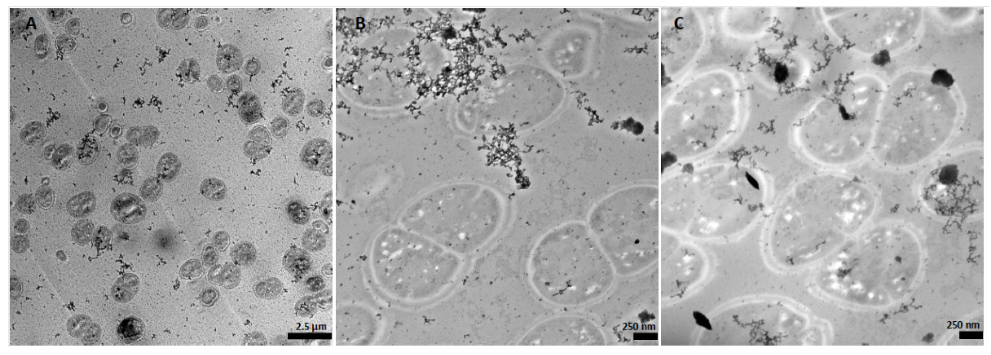
Figure 1. TEM images of D. aerius cells after cultivation for 48 h on acidic bioleachate solution. (A) 10 min of cultivation; (B,C) 48 h of cultivation. The typical diplococci morphology of D. aerius is preserved. Electron dense granules (black) and filamentous structures are associated with cells.
Resources 12 00144 g001
Figure 2. TEM images of A. aluminiidurans cells after cultivation on acidic bioleachate solution for 48 h. A. aluminiidurans typical rod-shaped morphology is not altered throughout cultivation. (A) 10 min of cultivation; (B,C) 48 h of cultivation. Electron dense granules (black) are associated with cells.
Resources 12 00144 g002
Figure 3. Elemental ultrastructural analysis of D. aerius cells cultivated on bioleachate for 24 h. The high-angle annular dark-field (HAADF) STEM image of D. aerius cells used for the EDS spectrum image acquisition and corresponding elemental distribution maps of phosphorus (P), silica (Si), oxygen (O), copper (Cu), iron (Fe), sulphur (S) and chlorine (Cl) is shown. Representative EDS spectra acquired from the area in HAADF STEM image are shown in the bottom panel. Gadolinium (Gd) and Lead (Pb) peaks in EDS spectra occur due to the sample staining procedure for TEM.
Resources 12 00144 g003
Figure 4. Elemental ultrastructural analysis of A. aluminiidurans cells cultivated on bioleachate for 24 h. The high-angle annular dark-field (HAADF) STEM image of A. aluminiidurans cells used for the EDS spectrum image acquisition and corresponding elemental distribution maps of phosphorus (P), oxygen (O), copper (Cu), iron (Fe) and chlorine (Cl) is shown. Representative EDS spectra acquired from the area in HAADF STEM image are shown in the bottom panel. Gadolinium (Gd) and Lead (Pb) peaks in EDS spectra are due to the sample staining procedure for TEM.
Resources 12 00144 g004
Table 1. Composition of the media used for the cultivation of bacterial strains prior to cultivation on acidic bioleachate solution. Metal-accumulating bacterial strains and their respective media composition, cultivation temperature (T (°C)) and pH are presented.
StrainMedium Components (w/v)T (°C)pH
P. putida0.5% peptone; 0.3% meat extract287.0
B. coagulans0.5% peptone; 0.3% meat extract407.0
S. xylosus0.5% peptone; 0.3% meat extract; 0.3% yeast extract377.2
D. aerius1% tryptone; 0.6% meat extract; 0.2% glucose287.0
D. radiodurans1% tryptone; 0.6% meat extract; 0.2% glucose287.0
A. aluminiidurans0.2% (NH4)2SO4; 0.01 KCl; 0.05% K2HPO4; 0.05% MgSO4 × 7H2O; 0.03 yeast extract; 0.1% glucose; 0.1% trypticase soy broth374.0
S. rimosus0.4% glucose; 0.4% yeast extract; 1% malt extract; 0.2% CaCO3287.2
Table 2. Metal concentrations of the acidic bioleachate solution (pH 1.7) measured via Inductively Coupled Plasma Mass Spectrometry (ICP-MS) [10]. Multiple solubilized elements were detected in the extremely acidic bioleachate solution.
mg/L mg/L
Mg778Ni0.185
Ca618Co0.17
Fe145Ti0.169
Zn111V0.144
Mn85.5Cd0.143
Al36.6Mo0.068
Sr0.906As0.037
Cu0.757Pb0.021
Cr0.695Ba0.003
Table 3. Cell densities (cells/mL) of seven bacterial strains grown on acidic (pH 1.7) and metalliferous bioleachate solution for a maximum of 48 h. * Streptomyces rimosus planktonic cells could not be detected due to precipitation, production of big filamentous pellets and formation of flocculation (N.D corresponds to not detected).
Strain0 h24 h48 h
B. coagulans7.98 × 106 ± 1.18 × 1064.69 × 106 ± 1.27 × 1068.03 × 105 ± 1.31 × 10⁵
S. xylosus1.17 × 107 ± 4.35 × 1055.60 × 106 ± 8.57 × 1052.50 × 104 ± 5.00 × 10³
P. putida9.92 × 106 ± 4.17 × 1057.09 × 106 ± 5.28 × 1051.54 × 106 ± 3.80 × 10⁵
D. radiodurans2.76 × 106 ± 2.38 × 1053.15 × 106 ± 1.14 × 1063.36 × 106 ± 4.71 × 10⁵
D. aerius6.96 × 105 ± 9.98 × 1041.01 × 106 ± 6.40 × 1055.60 × 106 ± 6.00 × 10⁵
A. aluminiidurans1.06 × 106 ± 1.24 × 1054.00 × 106 ± 4.80 × 1053.20 × 107 ± 6.40 × 10⁵
S. rimosusN.D. *N.D. *N.D. *
Disclaimer/Publisher’s Note: The statements, opinions and data contained in all publications are solely those of the individual author(s) and contributor(s) and not of MDPI and/or the editor(s). MDPI and/or the editor(s) disclaim responsibility for any injury to people or property resulting from any ideas, methods, instructions or products referred to in the content.