Article

A Cistus incanus Extract Blocks Psychological Stress Signaling and Reduces Neurogenic Inflammation and Signs of Aging in Skin, as Shown in In-Vitro Models and a Randomized Clinical Trial

1
R&D, International Flavors and Fragrances—Lucas Meyer Cosmetics, Yavne 8122503, Israel
2
Marketing, International Flavors and Fragrances—Lucas Meyer Cosmetics, Yavne 8122503, Israel
3
International Flavors and Fragrances, New York, NY 10019, USA
4
R&D, International Flavors and Fragrances—Lucas Meyer Cosmetics, 31036 Toulouse, France
*
Author to whom correspondence should be addressed.
Cosmetics 2023, 10(1), 4; https://doi.org/10.3390/cosmetics10010004
Submission received: 6 November 2022 / Revised: 4 December 2022 / Accepted: 20 December 2022 / Published: 26 December 2022

Abstract

:
Psychological stress exerts its effects mainly through the release of corticotropin releasing hormone (CRH), which activates inflammatory pathways in skin (inter alia), resulting in redness, extracellular matrix degradation, loss of skin elasticity and firmness, and the appearance of wrinkles—namely, accelerated skin aging. In order to propose a solution to this neurogenic aging phenomenon, we report here on studies using a myricitrin-rich extract of Cistus incanus, a Mediterranean shrub used in traditional medicine for the treatment of inflammatory and other diseases. These studies include a CRH receptor (CRH-R1) blocking assay; in vitro inflammatory cytokine reduction under CRH stimulation, and ex vivo NF-kB inhibition; and a double-blind clinical trial performed on highly stressed panelists, evaluating skin inflammation and wrinkling (active formulation vs. placebo control, applied split-face following a computer-generated randomization scheme; 36 subjects recruited and randomized, 30 analyzed; no adverse effects recorded; EMA/INFARMED registration #118505, internally funded). The results show that this extract can effectively block the CRH-R1 receptor, preventing NF-κB activation and the production of related pro-inflammatory cytokines. In a clinical setting, this same extract delivered significant anti-inflammatory and anti-aging effects. Taken together, these results demonstrate the value of this extract as a cosmetic active to counter neurogenic inflammation and skin aging.

Graphical Abstract

1. Introduction

The link between anxiety, psychological stress, and aging (as neurogenic aging, resulting from chronic neurogenic inflammation) is now well documented, though some underlying mechanisms are not yet fully understood. Evidence suggests that chronic psychological stress stimulates the hypothalamic–pituitary–adrenal (HPA) axis, inter alia, when the body attempts to resolve perceived threats. Prolonged activation of these pathways can result in chronic immune dysfunction, chronic inflammation, increased production of reactive oxygen species, and DNA damage, which contribute to the accelerated aging of skin and other tissues [1].
Anxiety and stress are conditions characteristic of modern life, characterized by feelings of emotional or physical tension, fear, worry, or unease. A wide range has been reported for the prevalence of anxiety in the general population, partly due to variations in methodology. Baxter et al. [2] quote a prevalence of 7.3% among the general population, and 10.4% among Western nations. Remes et al. [3] quote a range of up to 25% in the general population, and emphasize subgroups including women, young adults, people with chronic disease, and Western cultures. Bryant et al. [4], focusing on adults aged over 60, quotes a prevalence of up to 15%, but notes that the prevalence of anxiety symptoms is up to 52%.
Recent research has confirmed the skin as both a target of stress mediators and a local source for these factors. Stress conditions exert their effects on skin mainly through the HPA axis. Neurons in the hypothalamus secrete corticotropin-releasing hormone (CRH), which is transported to the pituitary gland, where it binds to the CRH receptor type-1 (CRH-R1) and stimulates the secretion of adrenocorticotropin (ACTH) (Figure 1). In turn, ACTH travels to the outer layer of the adrenal cortex and stimulates the production of cortisol. Cortisol is the primary stress hormone in humans and regulates a wide range of stress responses, inducing various immune and inflammation responses. Cortisol circulates through the blood stream to the skin and activates skin mast cells, which produce stress hormones locally, including CRH. This leads to a stress-induced inflammatory cascade [5,6]. CRH produced by activated mast cells binds to CRH-R1-expressing keratinocytes, inducing NF-kB activation [7,8]. The transcription factor NF-κB regulates multiple aspects of innate and adaptive immune functions, serving as a pivotal inflammatory response mediator. NF-κB induces the expression of pro-inflammatory genes, including those encoding cytokines and chemokines such as IL1, IL6, and TNF-α [9]. The inflammatory response increases blood flow to the inflamed site, to supply more nutrients and immune cells. Blood vessels dilate, resulting in redness and edema [10]. In addition, chronic inflammation is recognized as an important factor in the aging process (sometimes referred to as “inflammaging”). One of the features of this process is the release of matrix metalloproteinases (MMPs) that degrade the extracellular matrix, resulting in accelerated loss of skin elasticity and firmness, and the appearance of wrinkles [11]. Adverse effects of psychological stress on skin have been documented in rats, where immobilization stress resulted in skin mast cell degranulation [12]; and, in humans, where stress (Trier Social Stress Test) was shown to correlate with delayed skin barrier recovery [13].
The above suggests that blocking the CRH-R1 receptor locally in the skin should alleviate the effects of chronic states of stress on skin, by preventing NF-κB activation and related pro-inflammatory cytokine production—thus potentially representing a valuable strategy to address neurogenic skin inflammation and aging.
Cistus incanus is a Mediterranean shrub, rich in polyphenols and representing a source of valuable bioactive compounds. Secondary sources report that Cistus incanus has been used in traditional medicine, inter alia as an anti-inflammatory agent, to encourage wound healing, and in the treatment of some skin conditions [14,15,16,17,18,19]. Furthermore, Cistus incanus extracts containing polyphenolic compounds have been reported to possess antioxidant [20], antimicrobial [21], and antiviral [22,23] properties. Recently, aqueous extracts of the aerial parts of this plant have been demonstrated to possess in vivo antioxidant capacities, possibly attributable to their high polyphenol content [24]. Cistus species produce flavonoids, a class of polyphenolic secondary metabolites, and particularly flavanols (quercetin, kaempferol, and myricetin derivatives) [16,20,25,26,27,28]. In the extract reported herein, we identified the myricetin glycoside myricitrin, originally isolated from the bark of Myrica rubra (Lour.) [29], but previously unreported in aqueous Cistus incanus extract fractions.
Myricitrin is a 3-O-α-L-rhamnopyranoside of myricetin, reported to have anxiolytic effects [30]. The underlying mode of action is unclear. Myricitrin has also been shown to block activation of the NF-κB signaling pathway, decrease the production of pro-inflammatory factors including IL1-β, IL6, and TNFα [29,31], downregulate the activation of Janus kinases (as well as that of downstream transcription factor STAT1), and reduce ROS production (in a NOX2-dependent way). These precedents suggested that a Cistus incanus extract containing myricitrin may be an interesting candidate for an attempt to mitigate psychological stress-induced neurogenic inflammation. We therefore set out to test the efficacy of such an extract in in vitro, ex vivo, and in vivo (clinical) models, focusing on effects in skin tissue. Given the crucial role of the CRH signaling pathway in neurogenic inflammation, we also planned to look into the extract’s ability to interfere with said pathway. The present work reports on these studies and their results, aiming to demonstrate a solution to premature neurogenic skin aging driven by psychological stress using an extract of Cistus incanus in topical application.

2. Materials and Methods

2.1. Material: Cistus Incanus Extract

The studies described herein were performed using an aqueous extract of Cistus incanus aerial parts (IBR-Chill™, IFF—Lucas Meyer Cosmetics, Yavne, Israel). Aerial parts of Cistus incanus were harvested by hand in Southern Israel (by IFF—Lucas Meyer Cosmetics staff members), on the basis of unambiguous phenotypic identification. No permit was necessary for this collection. The biomass was dried (at ambient temperature, in the shade), ground, and stored at room temperature for several months. The ground biomass was then extracted in heated water. Solids were separated, and the resulting liquid was filtered, yielding a crude extract which was used as-is for in vitro studies, and diluted 1:1 in glycerin (Interaxion C50380681, Deyme, France) and preserved (1% Sharomix HP, Sharon Laboratories, Ashdod, Israel) for use in clinical studies. The final extract’s color is light brown to brown, with a pH of 4.5–6.5.
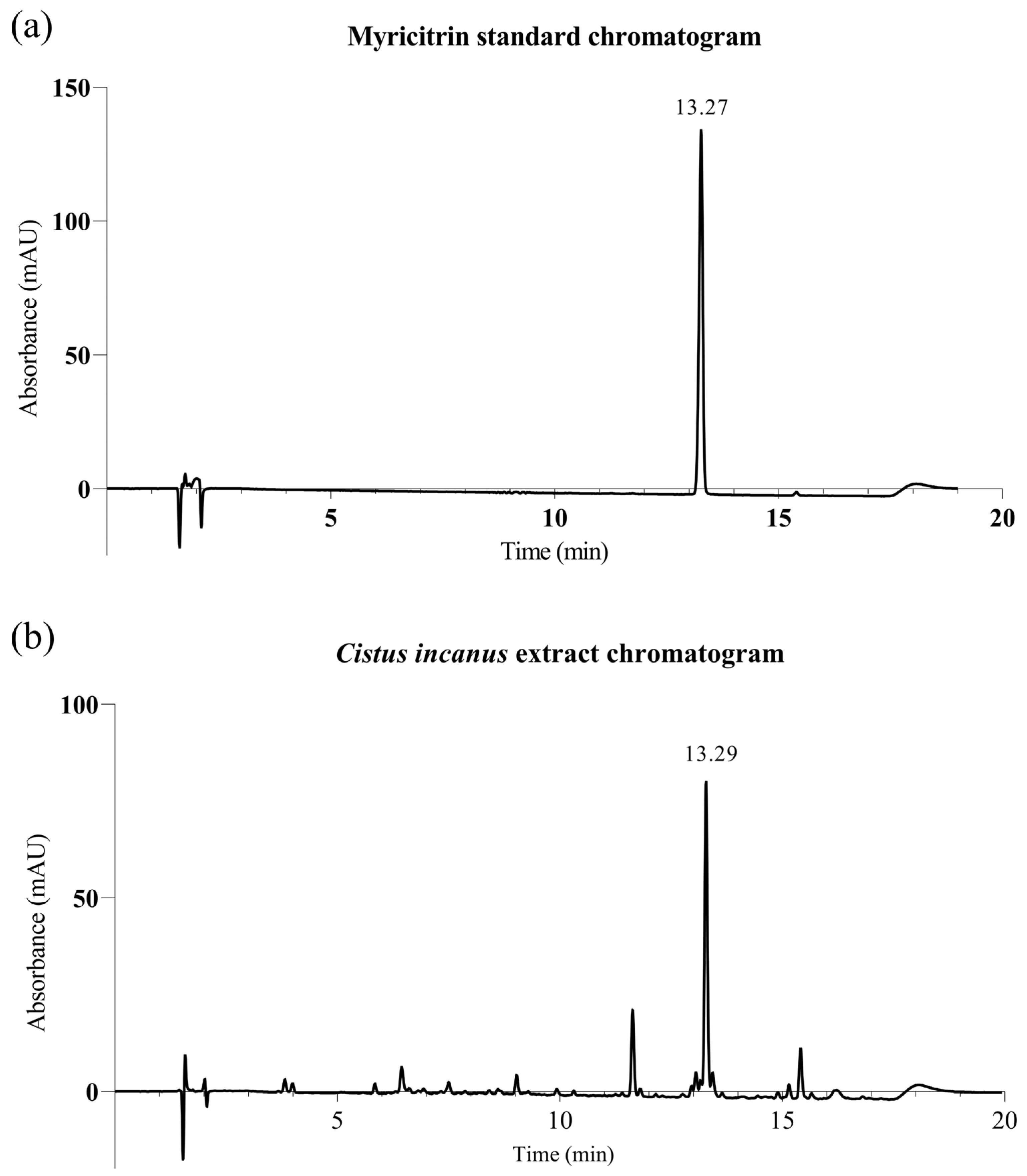
The extract’s content of the flavonoid glycoside myricitrin was titrated by HPLC chromatography (Agilent, Santa Clara, CA, USA: 1100 series with G1315A diode array detector and Poroshell Phenyl Hexyl 150 x 4.6 mm, 2.7 µm column). The extract was diluted x10 in HPLC grade water (deionized and irradiated with Direct-Q 5 UV, Merck-Millipore, Burlington, MA, USA), and filtered (0.22 μm) before injection. A myricitrin standard (Sigma-Aldrich 67268, St Louis, Mo, USA) was prepared and diluted with methanol (J.T. Baker 8402, Avantor, Radnor, PA, USA) and filtered (0.22 μm) before injection. Injection volume: 10 μL; flow: 1.0 mL/min; column temperature: 30 °C; detection wavelength: 355 nm. Eluent: 25% of a 0.1% solution of trifluoroacetic acid (TFA, Sigma-Aldrich 302031, St. Louis, MO, USA) in HPLC grade water (v/v)/75 % of a 0.1% solution of TFA in Acetonitrile (J.T. Baker 9017, Avantor, Radnor, PA, USA) (v/v). Figure 2 shows a representative HPLC trace. Finally, the extract was analyzed by an independent laboratory for traces of heavy metals and pesticides (none detected above the respective quantification thresholds).

2.2. Testing Protocols

2.2.1. CRH-R1 Receptor Blocking Functional Assay

The ability of Cistus incanus extract to interrupt the stress signaling cascade by blocking the CRH-R1 receptor was tested in a receptor functional assay. Recombinant Chinese Hamster Ovary (CHO) cells expressing the human CRH-R1 receptor (clone 36, Eurofins Cerep SA, Celle L’Evescault, France) [32] were incubated in presence of Cistus incanus extract for 30 min at 37 ˚C, and the antagonist effect of the extract was assessed via the measurement of the response to a control agonist (30 nM Ovine CRH, Bachem H2445, Bubendorf, Switzerland). Said response was evaluated via measurement of the cAMP signal, by homogenous time-resolved fluorescence (RubyStar, BMG Labtech, Ortenberg, Germany).

2.2.2. Reduction of Inflammatory Cytokine Markers under CRH Stimulation

The effect of Cistus incanus extract against neurogenic inflammation was confirmed in a downstream model, showing the inhibition of the expression of inflammatory cytokines under induction by CRH. A culture of primary human keratinocytes from a 62-year-old donor was cultivated in monolayer until confluency, and then starved for 24 h. The culture was then incubated for 1 h in the absence (control) or presence of the extract at 0.05%; 0.1%; and 0.2% v/v (a preliminary cytotoxicity study having established that the extract possesses negligible cytotoxicity in such a culture up to 0.3% v/v). Then 100 nM CRH (Tocris 1151, Bio-Techne, Bristol, UK) was added, and cells were incubated for a further 24 h. IL1-β, IL-6, and TNF-alpha were quantified in culture medium by ELISA (Victor V, Perkin-Elmer, Waltham, MA, USA; R&D system Duoset kits, IL1-β: DY201; IL6: DY206; TNF-α: DY210, Bio-Techne, Minneapolis, MN, USA).

2.2.3. Ex Vivo Inhibition of the NF-kB Master Inflammation Regulator

In order to further confirm the effect of this Cistus incanus extract against neurogenic inflammation, an ex vivo study was carried out aiming to show the extract’s effect on the expression of the NF-kB master regulator of inflammation, in explants under induction by CRH. Fresh human skin explants from a 26-year-old donor (sourced in France in full observance of local regulatory and ethical requirements) were maintained in explant culture medium (CurioBiotech CB-EM-SKN, Visp, Switzerland), at 37 °C and 5% CO2. After overnight incubation, explants were processed in the following groups: Untreated control; induction with 1 µM CRH (Tocris 1151, Bio-Techne, Bristol, UK) for 72 h, otherwise untreated; exposure to 0.125% and 0.25% extract for 24 h, followed by continued exposure to extract, for a further 48 h, with simultaneous Induction with 1 µM CRH. Applications of CRH and extract were performed topically, in triplicate. NF-kB expression was analyzed by immunohistochemistry on fixed tissue samples, using the DAB (3,3′-diaminobenzidine, BOND Polymer Refine Detection Kit DS980, Leica Biosystems, Buffalo Grove, IL, USA) staining method. High-resolution light microscopy images were captured (Realux BDS400, Advilab, Allauch, France; ODC 825, VIS (OXM 901), Kern & Sohn, Balingen, Germany). NF-kB expression was further quantified by image analysis (ImageJ: https://imagej.net/software/imagej/, accessed on 6 September 2020), converting images from RGB to CIELAB color space (expressing color as values along 3 axes: L* for lightness (black to white); a* from green to red; and b* from blue to yellow). Signal quantification calculated the total amount of red-orange intensities as the sum of pixel intensities (total intensity), and mean intensity as a ratio between total intensity and size of the stained region, along each color axis. The a* (redness) values were retained as most representative of the intensity of the visual red-orange stain.

2.2.4. Clinical Trial on a Highly Stressed Population Sample

This study aimed at demonstrating the ability of the Cistus incanus extract to alleviate stress-induced signs of aging and reduce inflammation in the context of a highly stressed population sample. For this purpose, the extract was incorporated at 1% in a simple gel-cream formulation (Table 1), opposite a corresponding placebo wherein the extract was replaced with appropriate amounts of glycerin, water, and FD&C colorants.
The study was performed according to the Declaration of Helsinki principles and subsequent amendments, and in the spirit of Good Clinical Practice Guidelines and general principles of Portugal Law 46/2004 of August 19th. The protocol and test conditions were reviewed by an Internal Review Board (opinion nº 5580/2020 and 5581/2020) and the standard protocol was submitted to the PhD Trials Ethical Commission (25/10/2019). Study participants gave written informed consent as well as consent for the use of photographic images taken during the study. The trial was registered in the RNEC registry, as required for cosmetic clinical studies by local (Portuguese) regulations (EMA/INFARMED), under registration number 118505.
General design: The study was carried out as a double-blind, placebo-controlled (randomized split-face) trial. Thirty female panelists were included, aged 35–60 years old, with phototype (Fitzpatrick) II to IV, of all skin types, and presenting signs of aging (wrinkles or fine lines). Panelists were selected for high stress levels, on the basis of a psychological questionnaire as well as cortisol levels measurement. Cortisol levels were checked again at the study’s end, and showed no evolution over the duration of the study, holding steady at amounts considered typical of highly stressed subjects but not pathological. Therefore, we may conclude that the psychological stress state of the participants remained consistent throughout the study.
A formulation containing 1% preserved Cistus incanus extract was used opposite a placebo, in which the active ingredient was replaced with water and glycerin (to account for glycerin content in the preserved extract).
Measurements were taken before application on D0 and after 14 and 28 days of twice-a-day application.
Skin microcirculation: Skin microcirculation was assessed by Laser Doppler Flowmetry (LDF) using a 780 nm monochromatic low energy laser (Periflux LDPM PF5000 with a laser channel PF 5010, Perimed, Jarfalla, Sweden). The results are expressed in arbitrary units (AU). Basal values were obtained without any stimulation. In order to evaluate the anti-inflammatory effects of the tested products, the skin was heated to 42 °C using the instrument’s self-heating probe, and microcirculation was measured over 9 min. The difference between basal microcirculation and microcirculation upon thermal induction was termed ‘induced microcirculation’, representing the component of microcirculation induced by heating the skin.
Red spots by Visia-CA: Standardized photographic images were obtained with cross polarized lighting using the VISIA-CA (Canfield, Parsippany, NJ, USA) system, in order to quantify the evolution of red spots. The VISIA-CA system was also used to obtain full-spectrum illustrative images.
Determination of skin redness by ChromaMeter: Skin color was measured using a tristimulus color analyzer measuring reflected color (Chromameter CR-400, Minolta, Osaka, Japan). The system provides data for the L* (Luminance), a* (red-green) and b* (blue-yellow) color distribution.
SLS inflammation recovery: The anti-inflammatory effect of the extract was further evaluated based on treated skin’s recovery from an insult with 2% sodium lauryl sulfate (SLS) in deionized water. Erythema and skin microcirculation were measured using the chromameter (as a*) and laser doppler flowmetry, as described above, immediately before SLS insult and every 24 h thereafter, over a period of 7 days. The measuring area was the forearm, in the following areas: treated with the active product; treated with the placebo product; and untreated, unchallenged area (‘blank’).
Anti-wrinkle effect by image analysis (AEVA 3D): 3D images of the skin topography were obtained using a stereo camera with a fringe projection system (AEVA-HE, Eotech, Marcoussis, France). The 3D effect is calculated by the deflection in the fringes in the stereo image combination, representing the skin profile.
Skin elasticity by Cutometer: Skin biomechanical evaluation was performed using a Cutometer® dual MPA 580 with a 2 mm probe (Courage & Khazaka, Koln, Germany). Firmness is evaluated based on the initial deformation observed upon mechanical deformation, while elasticity is evaluated as the skin’s ability to return into its original position following deformation.

2.2.5. Statistics

General: Data analysis was performed using GraphPad Prism 9. The statistical significance threshold was set at p = 0.05, following general practice in the art. Statistical tests were selected on the basis of normality tests: a parametric test (paired Student t-test) was applied when the normality was positive, and a non-parametric test (Wilcoxon test) was used when the normality was negative. For in-vitro studies, we used a 2-way ANOVA analysis and a Holm–Sidak test.

3. Results

3.1. Results of In-Vitro Studies

3.1.1. CRH-R1 Receptor Blocking Functional Assay

The ability of Cistus incanus extract to interrupt the stress signaling cascade was tested in a CRH-R1 functional receptor assay with CRH stimulation. The extract displayed significant, dose-dependent CRH-R1 blocking behavior, reaching near-total (88%, p = 0.0080) receptor blockage at a concentration of 1% extract in medium (Figure 3).

3.1.2. In-Vitro Reduction of Inflammatory Cytokine Expression under CRH Stimulation

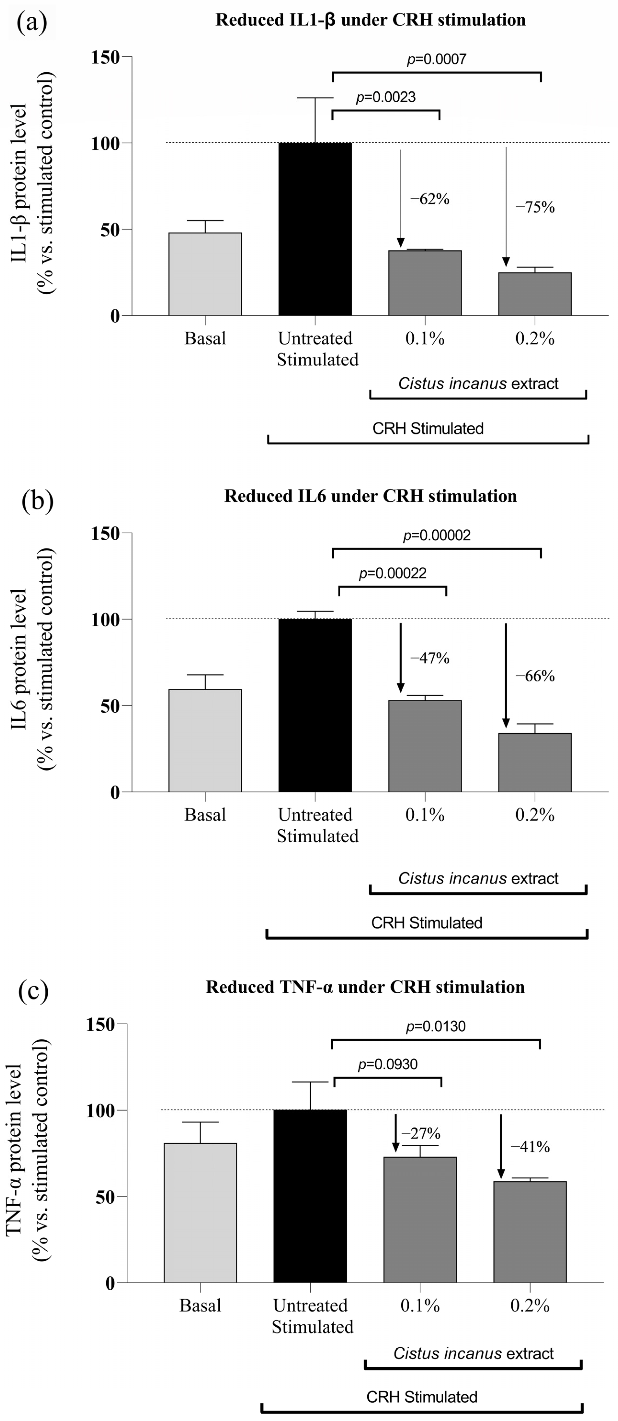
The effect of Cistus incanus extract against neurogenic inflammation was confirmed in a keratinocyte cell culture, under stimulation by CRH, in which expression of key inflammatory cytokines was measured by ELISA. Under these conditions, stimulation by CRH significantly increased production of IL1-β, IL6, and TNF-α (Figure 4a–c, respectively). In turn, concurrent addition of Cistus incanus extract significantly decreased the levels of these same markers, in a dose-dependent and statistically significant manner (respectively, up to: −75%, p = 0.007; −66%, p = 0.00002; −41%, p = 0.0130).

3.1.3. Ex-Vivo Inhibition of the NF-kB Master Inflammation Regulator

The effect of Cistus incanus extract against neurogenic inflammation was further confirmed in an ex-vivo model aiming to show the extract’s effect on the expression of the NF-kB master regulator of inflammation, in explants under CRH induction. Immunohistochemical staining for NF-kB showed a clear increase in levels of expression in CRH-induced explants, confirming induction of inflammation with CRH, in comparison to negative, uninduced controls. Explants exposed to the extract and CRH induction showed a decrease in NF-kB expression compared to the negative control, CRH-induced explants (Figure 5). Visual examination of immunohistochemistry images (Figure 5a) suggests NF-kB levels in explants exposed to the extract were comparable to those in unexposed, unstimulated control explants. Quantification of the stain coloration by image analysis (Figure 5b) supports this observation, showing a significant increase (ca. 30%, p = 0.0797) in NF-kB expression upon stimulation with CRH, and a very strong decrease in NF-kB expression upon addition of extract (by ca. 67% with 0.125% extract, p = 0.00007; and 90% with 0.25% extract, p = 0.00002, vs. CRH induction alone).

3.2. Clinical Trial Results

3.2.1. Anti-Inflammatory Effects

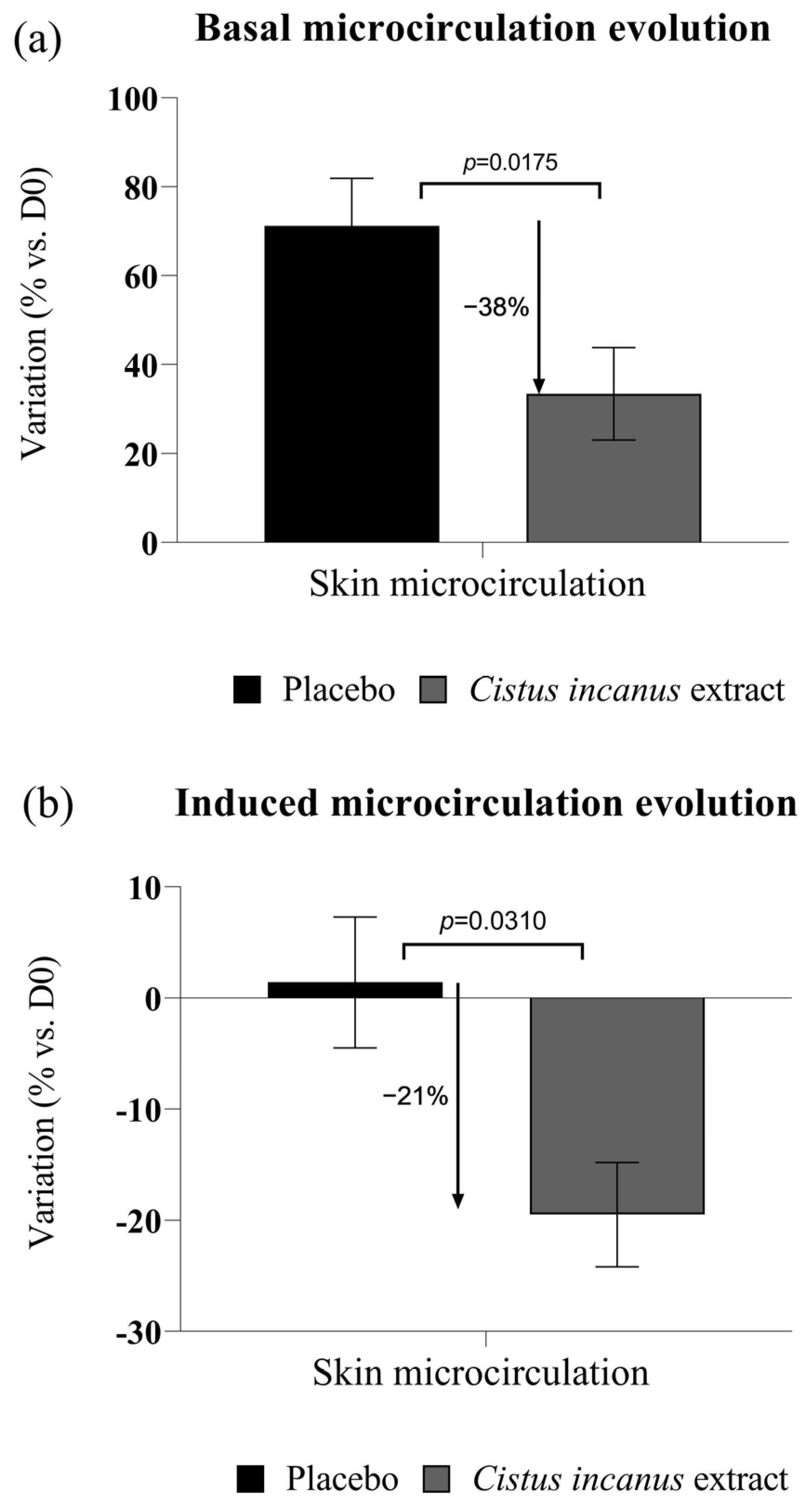
After 28 days of treatment, 1% Cistus incanus extract in formula displayed a significant anti-inflammatory effect, as manifested in a strong reduction of basal microcirculation (−38% vs. placebo, p = 0.0175) (Figure 6a). We also observed a strong reduction (21% vs. placebo, p = 0.0310) in induced microcirculation (being the difference in microcirculation measured by Laser Doppler Flowmetry before and after thermal induction) (Figure 6b).
In the chemical (SLS) insult model, we observed a significant improvement in resistance to and recovery from chemical (SLS) insult, with lower microcirculation (Laser Doppler) and redness (chromameter a*) values throughout the measurement period in the area treated with the active product vs. the area treated with the placebo, while the unchallenged area showed essentially no change throughout, as expected (Figure 7). The 1% extract in formulation showed a statistically significant advantage in terms of peak irritation (measured at 24 h from SLS challenge; p = 0.0049 for the erythema measurement) and recovery time (time required for a return to the initial, pre-challenge state; p = 0.000002 for the erythema measurement, p = 0.00003 for the Laser Doppler measurement).

3.2.2. Skin Redness, Red Spots

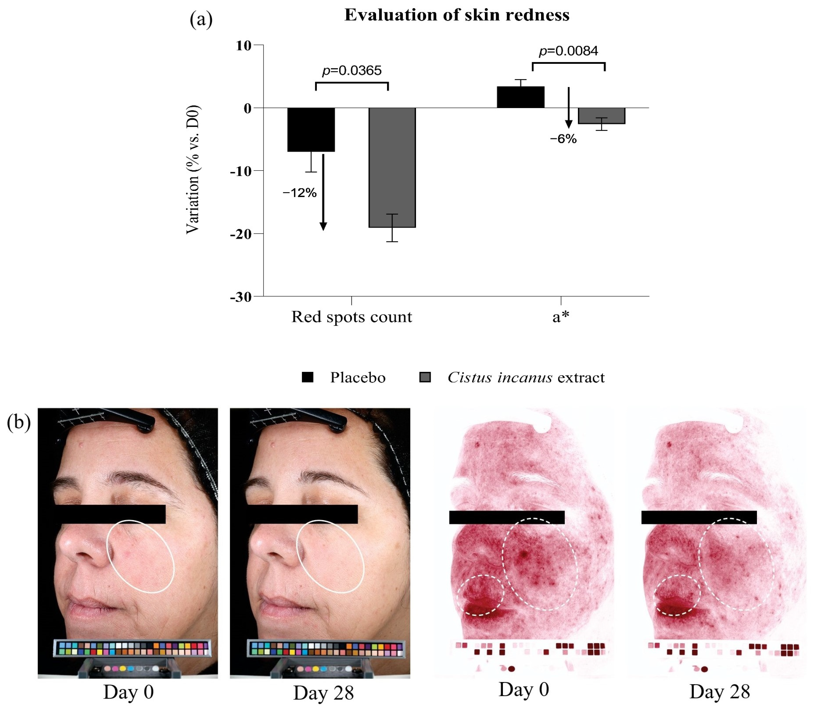
After 28 days of treatment, 1% extract in formula delivered a modest but statistically significant reduction in redness (−6% a* vs. placebo, p = 0.0084) and a significant reduction in red spots counts (−12% vs. placebo, p = 0.0365) (Figure 8).

3.2.3. Anti-Aging Effects

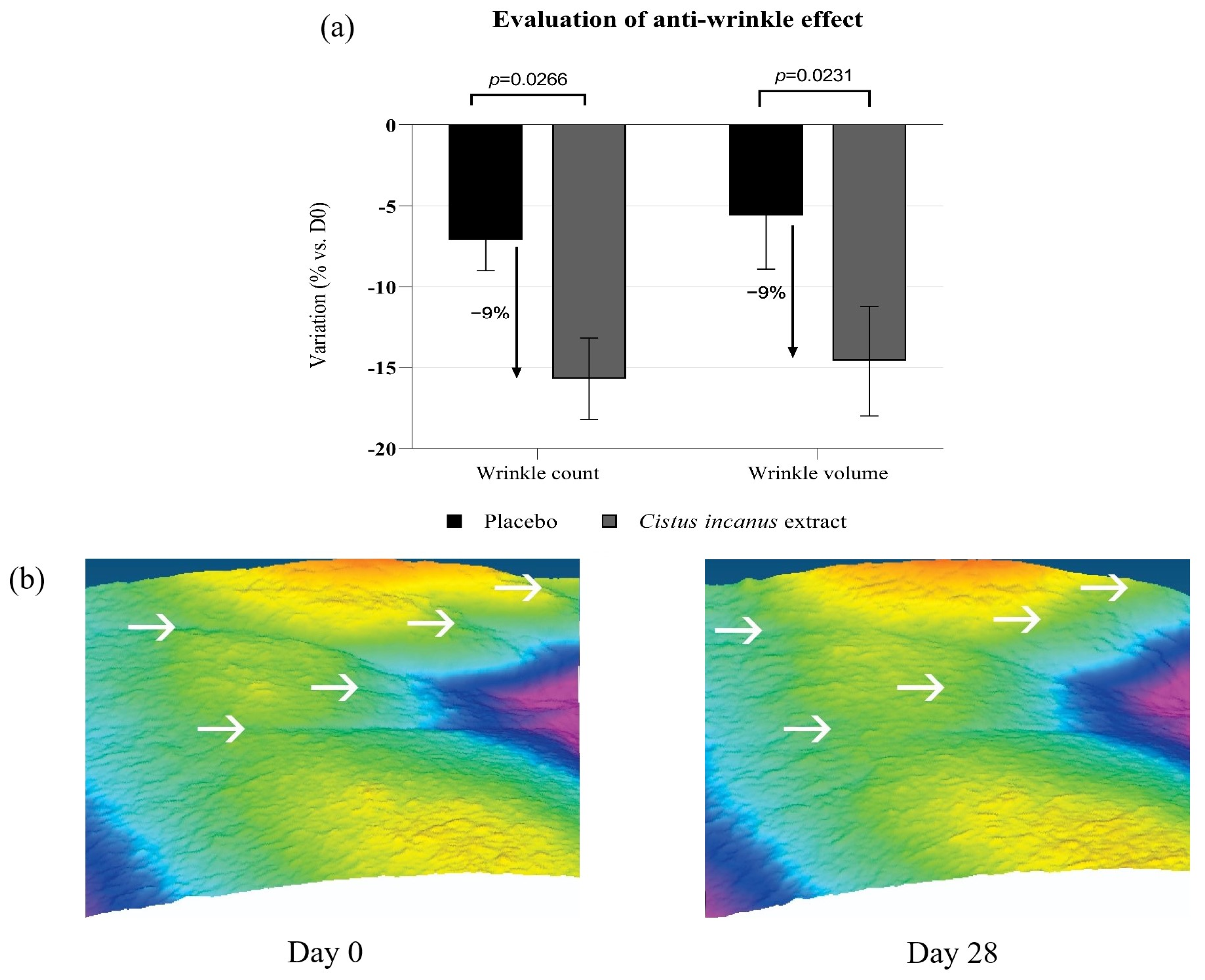
After 28 days of treatment, 1% extract in formula delivered a significant anti-aging effect, as manifested in: a significant reduction in wrinkle and fine line counts and volume (9% advantage vs. placebo in both cases, as evaluated by AEVA-3D image analysis, with p = 0.0266 and p = 0.0231 respectively) (Figure 9); and significant improvements in skin firmness and elasticity (respectively, 7% and 12% advantages vs. placebo, as evaluated by Cutometer), with statistical significance vs. Day 0 and placebo at Day 28.

4. Discussion

In a functional receptor assay, we demonstrated that a Cistus incanus extract is an effective antagonist of the CRH-R1 receptor, indicating that the extract may have the capacity to interrupt the neurogenic inflammation signaling chain at this stage and thus potentially blocking the stress response chain.
This was corroborated in an ex-vivo skin explant model, stimulated with CRH. In this model, CRH stimulation significantly increased expression of the master inflammation regulator NF-kB, as expected. On the other hand, addition of Cistus incanus extract was able to block the effects of the CRH stimulation and effectively decrease NF-kB levels in the explants. This result seems to confirm that the Cistus incanus extract is able to suppress the CRH-induced activation of an inflammation signaling cascade, and thus may offer some protection from psychogenic stress-induced inflammation processes.
These indications are further supported by results in a human keratinocyte culture under stimulation by CRH. In this model, the CRH stimulation increased the expression of inflammation markers IL1-β, IL6, and TNF-α, also as expected. Conversely, addition of Cistus incanus extract effectively decreased the expression of the same markers, negating the effects of the CRH stimulation. Here also, these results reinforce the implication that the extract is able to block downstream inflammatory processes resulting from CRH signaling, and therefore may effectively block stress-induced inflammation.
The ability of the extract to inhibit downstream effects of CRH signaling, and therefore also prevent some of the eventual damage caused by psychological stress on living tissue, in this case skin, was finally confirmed in vivo in a double-blind, placebo-controlled clinical trial in a population sample selected for high levels of psychological stress. These volunteers were recruited on the basis of a dual evaluation, featuring a screening questionnaire as well as saliva cortisol measurements. In this trial, we were able to observe that the extract delivered significant baseline anti-inflammatory effects (observable through microcirculation measurements and analysis of cross-polarized visible light photographs). We observed significantly improved skin resilience to inflammation-inducing provocations, as evaluated by following the evolution of skin redness and microcirculation after a chemical insult with model irritant SLS, which showed that treatment with the extract significantly reduced peak irritation and increased the speed of recovery from insult. Finally, and perhaps most valuable where the ultimate objective is to reduce the signs of premature aging induced by chronic neurogenic inflammation, we observed that treatment with the extract resulted in significantly reduced skin wrinkling after 28 days, with a statistically significant advantage over the placebo product.
The potential for use of Cistus incanus extracts in cosmetic, skin care, or personal care applications has been reported, in particular by Gawel–Beben et al. [33], who report on these extracts’ contents of phenolic and flavonoid species, their activity as tyrosinase inhibitors (an effect linked to possible reduction of pigmentation in the skin), and in particular on their antioxidant potential, as evaluated by DPPH radical scavenging. A DNA-protective effect has also been reported for Cistus extracts by Vanella et al. [34], most likely resulting from the same antioxidant properties. Vanella et al. show that the Cistus extracts can scavenge oxidative species including the DPPH radical and the superoxide anion, inhibit lipoperoxidation in rat liver microsomes, and protect DNA from damage caused by hydroxyl radicals. These antioxidant effects could be seen as likely contributors to the anti-inflammatory and anti-aging benefits observed in our own work.
Nevertheless, to the best of our knowledge, the work described herein constitutes the first direct, controlled, in-vivo demonstration of the anti-inflammatory and anti-aging effects of such an extract to be published in the scientific literature. What is more, to the best of our knowledge no work has been published regarding the effect of a Cistus extract (or indeed, any other similar botanical extract) on the interruption of psychological stress signaling or neurogenic inflammation, especially via blockage of the CRH-R1 receptor.
As noted above, the flavonoid myricitrin, identified in the extract, has been associated with similar effects, including antioxidant and anti-inflammatory effects. Zhang et al. reported that myricitrin reduced the production of inflammatory markers including IL6 and TNF-α, as well as nitric oxide (NO) and the enzyme catalyzing its production (iNOS), in mouse macrophages [32]. Du et al. [29] also reported on anti-inflammatory effects, showing decreased inflammatory mediators including IL1-β, IL6, TNFα, as well as lowered COX-2 and iNOS expression in the nigrostriatum neurons of an LPS-stimulated mouse model of neuroinflammation. Of particular interest in context of our own results, the same authors also showed that myricitrin could block the activation of NF-kB (as well as TLR4 and MyD88, upstream regulators of the NF-kB signaling pathway), and concluded that this was a main pathway responsible for the neuroprotective effect against LPS-induced inflammation and injury observed in their work.
While other compounds present in the extract likely also play a role, the presence of myricitrin in our extract, in light of the pathways on which myricitrin has been shown to have an influence and their possible association to the effects we observe in our studies, seems to suggest that this compound may be linked to some or all these same effects.
Conversely, it is difficult to unequivocally attribute the entirety of the effects observed, especially those observed in vivo, to the single mechanism of CRH-R1 blockage. Other effects and biological mechanisms may be in play. For example, the extract’s anti-oxidant potential [33,34], remarked upon above, may play a direct role on improving tissue inflammation states, independently of the CRH stress signaling chain—and may also be behind part of the benefits observed in vivo. One could also imagine that other compounds in the extract could have an independent anti-inflammatory effect, once again bypassing neurogenic stress signaling or affecting a different level of the signaling chain. Nevertheless, blockage of the CRH receptor by the extract is clearly demonstrated here, as are several expected downstream effects—suggesting that this mechanism is indeed behind at least part of the extract’s observed efficacy.
Some further limitations of the work presented herein include the need for a better understanding of the mechanisms of the effects demonstrated here, in particular regarding their dose- and time-dependency, especially in connection with the influence on NF-kB and resulting downstream effects. This understanding could be expanded through further studies, using similar ex-vivo and/or clinical models. Additionally, our investigations have been limited to effects on skin, while effects on skin appendages and/or other types of tissue affected by neurogenic inflammation may also be of scientific and industrial interest.

5. Conclusions

We have demonstrated that an extract of Cistus incanus aerial parts may interrupt the psychological stress signaling cascade at the stage where this signaling chain is relayed by CRH, through blockage of the CRH-R1 receptor. We have also shown that this same extract can deliver significant anti-inflammatory effects, preventing inflammation triggered by CRH and thereby preventing or alleviating at least part of the inflammatory state triggered by psychological stress. Finally, we were able to confirm these effects in vivo, on a group of healthy volunteers selected for a high level of psychological stress; in these volunteers, we showed the anti-inflammatory benefits of treatment with the extract, as well as the extension of these effects into improvements in skin resilience to chemical (SLS) insult and improvements in aging signs (wrinkles).
Taken together, these results effectively indicate that an extract of Cistus incanus aerial parts may mitigate the effects of neurogenic stress on skin and therefore has significant potential for application as an active ingredient in a broad range of skin care applications, especially applications aimed at delivering anti-inflammatory and/or anti-aging benefits in populations suffering from chronic psychological states of stress.
It may be valuable to expand on the data presented herein through further studies, in order to shed further light on the dose- and time-dependency of the effects described, as well as on effects on other tissue types (e.g., hair and other skin appendages, but possibly also internal tissues such as soft joint tissues, cardiac muscle tissue, and more), and under different types of psychological stresses.

Author Contributions

Conceptualization, F.H., S.K., E.L. and J.A.-V.; methodology, F.H., J.A.-V. and E.L.; formal analysis, M.C. and F.H.; investigation, F.H., M.C. and J.A.-V.; resources, J.A.-V. and E.L.; data curation, M.C. and F.H.; writing—original draft preparation, F.H.; writing—review and editing, F.H., J.A.-V. and S.K.; supervision, J.A.-V. and E.L.; project administration, F.H. All authors have read and agreed to the published version of the manuscript.

Funding

This research was funded by International Flavors and Fragrances (IFF—Lucas Meyer Cosmetics), and received no external funding.

Institutional Review Board Statement

The human study reported above was conducted in accordance with the guidelines of the Declaration of Helsinki, and the protocol was approved by the Ethics Committee of Clinica Dr. Carlos Ramos of Portugal (opinion nº 5580/2020 and 5581/2020; approval date: 25 October 2019).

Informed Consent Statement

Informed consent was obtained from all subjects involved in the study.

Data Availability Statement

The data presented in this study are available on request from the corresponding author.

Conflicts of Interest

The authors are employees of International Flavors and Fragrances and its affiliates, and declare no conflict of interest. Beyond funding our work, the company as such had no role in the design of the study; in the collection, analyses, or interpretation of data; in the writing or editing of the manuscript; or in the decision to publish the results.

References

  1. Dunn, J.H.; Koo, J. Psychological stress and skin aging: A review of possible mechanisms and potential therapies. Dermatol. Online J. 2013, 19, 18561. [Google Scholar] [CrossRef] [PubMed]
  2. Baxter, A.; Scott, K.; Vos, T.; Whiteford, H. Global prevalence of anxiety disorders: A systematic review and meta-regression. Psychol. Med. 2013, 43, 897–910. [Google Scholar] [CrossRef] [PubMed]
  3. Remes, O.; Brayne, C.; van der Linde, R.; Lafortune, L. A systematic review of reviews on the prevalence of anxiety disorders in adult populations. Brain Behav. 2016, 6, e00497. [Google Scholar] [CrossRef]
  4. Bryant, C.; Jackson, H.; Ames, D. The prevalence of anxiety in older adults: Methodological issues and a review of the literature. J. Affect. Disord. 2008, 109, 233–250. [Google Scholar] [CrossRef] [PubMed]
  5. Chen, Y.; Lyga, J. Brain-skin connection: Stress, inflammation and skin aging. Inflamm. Allergy Drug Targets 2014, 13, 177–190. [Google Scholar] [CrossRef] [Green Version]
  6. Pondeljak, N.; Lugovic-Mihic, L. Stress-induced Interaction of Skin Immune Cells, Hormones, and Neurotransmitters. Clin. Ther. 2020, 42, 757–770. [Google Scholar] [CrossRef]
  7. Zbytek, B.; Pfeffer, L.M.; Slominski, A.T. Corticotropin-releasing hormone stimulates NF-kappaB in human epidermal keratinocytes. J. Endocrinol. 2004, 181, R1–R7. [Google Scholar] [CrossRef] [Green Version]
  8. Smith, E.M.; Gregg, M.; Hashemi, F.; Schott, L.; Hughes, T.K. Corticotropin Releasing Factor (CRF) Activation of NF-κB-Directed Transcription in Leukocytes. Cell. Mol. Neurobiol. 2006, 26, 1019–1034. [Google Scholar] [CrossRef]
  9. Liu, T.; Zhang, L.; Joo, D.; Sun, S.C. NF-κB signaling in inflammation. Signal Transduct. Target. Ther. 2017, 2, 17023. [Google Scholar] [CrossRef] [Green Version]
  10. Pober, J.S.; Sessa, W.C. Inflammation and the blood microvascular system. Cold Spring Harb. Perspect Biol. 2014, 7, a016345. [Google Scholar] [CrossRef]
  11. Zhang, S.; Duan, E. Fighting against Skin Aging: The Way from Bench to Bedside. Cell. Transplant. 2018, 27, 729–738. [Google Scholar] [CrossRef] [PubMed]
  12. Singh, L.K.; Pang, X.; Alexacos, N.; Letourneau, R.; Theoharides, T.C. Acute immobilization stress triggers skin mast cell degranulation via corticotropin releasing hormone, neurotensin, and substance P: A link to neurogenic skin disorders. Brain. Behav. Immun. 1999, 13, 225–239. [Google Scholar] [CrossRef] [PubMed] [Green Version]
  13. Robles, T.F. Stress, social support, and delayed skin barrier recovery. Psychosom. Med. 2007, 69, 807–815. [Google Scholar] [CrossRef] [PubMed] [Green Version]
  14. Lukas, B.; Bragagna, L.; Starzyk, K.; Labedz, K.; Stolze, K.; Novak, J. Polyphenol diversity and antioxidant activity of European Cistus creticus L. (cistaceae) compared to six further, partly sympatric Cistus species. Plants 2021, 10, 615. [Google Scholar] [CrossRef] [PubMed]
  15. Riehle, P.; Rusche, N.; Saake, B.; Rohn, S. Influence of the Leaf Content and Herbal Particle Size on the Presence and Extractability of Quantitated Phenolic Compounds in Cistus incanus Herbal Teas. J. Agric. Food Chem. 2014, 62, 10978–10988. [Google Scholar] [CrossRef]
  16. Barrajón-Catalán, E.; Fernández-Arroyo, S.; Roldán, C.; Guillén, E.; Saura, D.; Segura-Carretero, A.; Micol, V. A systematic study of the polyphenolic composition of aqueous extracts deriving from several Cistus genus species: Evolutionary relationship. Phytochem. Anal. 2011, 22, 303–312. [Google Scholar] [CrossRef]
  17. Lardos, A.; Prieto-Garcia, J.; Heinrich, M. Resins and gums in historical iatrosophia texts from Cyprus—A botanical and medico-pharmacological approach. Front. Pharmacol. 2011, 2, 32. [Google Scholar] [CrossRef] [Green Version]
  18. Tuzlacı, E.; Aymaz, P.E. Turkish folk medicinal plants. Part IV: Gönen (Balıkesir). Fitoterapia 2001, 72, 323–343. [Google Scholar] [CrossRef]
  19. Petereit, F.; Kolodziej, H.; Nahrstedt, A. Flavan-3-ols and proanthocyanidins from Cistus incanus. Phytochemistry 1991, 30, 981–985. [Google Scholar] [CrossRef]
  20. Bernacka, K.; Bednarska, K.; Starzec, A.; Mazurek, S.; Fecka, I. Antioxidant and Antiglycation Effects of Cistus x incanus Water Infusion, Its Phenolic Components, and Respective Metabolites. Molecules 2022, 27, 2432. [Google Scholar] [CrossRef]
  21. Zalegh, I.; Akssira, M.; Bourhia, M.; Mellouki, F.; Rhallabi, N.; Salamatullah, A.M.; Alkaltham, M.S.; Khalil Alyahya, H.; Mhand, R.A. A Review on Cistus sp.: Phytochemical and Antimicrobial Activities. Plants 2021, 10, 1214. [Google Scholar] [CrossRef] [PubMed]
  22. Saifulazmi, N.F.; Rohani, E.R.; Harun, S.; Bunawan, H.; Hamezah, H.S.; Nor Muhammad, N.A.; Azizan, K.A.; Ahmed, Q.U.; Fakurazi, S.; Mediani, A.; et al. A Review with Updated Perspectives on the Antiviral Potentials of Traditional Medicinal Plants and Their Prospects in Antiviral Therapy. Life 2022, 12, 1287. [Google Scholar] [CrossRef] [PubMed]
  23. Rebensburg, S.; Helfer, M.; Schneider, M.; Koppensteiner, H.; Eberle, J.; Schindler, M.; Gurtler, L.; Brack-Werner, R. Potent in vitro antiviral activity of Cistus incanus extract against HIV and Filoviruses targets viral envelope proteins. Sci. Rep. 2016, 6, 20394. [Google Scholar] [CrossRef] [PubMed] [Green Version]
  24. Kuchta, A.; Konopacka, A.; Waleron, K.; Viapiana, A.; Wesołowski, M.; Dąbkowski, K.; Ćwiklińska, A.; Mickiewicz, A.; Śledzińska, A.; Wieczorek, E.; et al. The effect of Cistus incanus herbal tea supplementation on oxidative stress markers and lipid profile in healthy adults. Cardiol. J. 2021, 28, 534–542. [Google Scholar] [CrossRef] [PubMed] [Green Version]
  25. Mocan, A.; Fernandes, Â.; Calhelha, R.C.; Gavrilas, L.; Ferreira, I.C.F.R.; Ivanov, M.; Sokovic, M.; Barros, L.; Babota, M. Bioactive Compounds and Functional Properties of Herbal Preparations of Cystus creticus L. Collected from Rhodes Island. Front. Nutr. 2022, 9, 881210. [Google Scholar] [CrossRef]
  26. Dimcheva, V.; Karsheva, M. Cistus incanus from Strandja Mountain as a Source of Bioactive Antioxidants. Plants 2018, 7, 8. [Google Scholar] [CrossRef] [Green Version]
  27. Petereit, F.; Kolodziej, H.; Nahrstedt, A. Proanthocyanidins and bio genetically related dihydroflavonols from Cistus incanus L. In Plant Polyphenols: Synthesis, Properties, Significance; Hemingway, R.W., Laks, P.E., Eds.; Springer: Boston, MA, USA, 1992; pp. 729–737. [Google Scholar]
  28. Gori, A.; Ferrini, F.; Marzano, M.C.; Tattini, M.; Centritto, M.; Baratto, M.C.; Pogni, R.; Brunetti, C. Characterization and Antioxidant Activity of Crude Extract and Polyphenolic Rich Fractions from C. incanus Leaves. Int. J. Mol. Sci. 2016, 17, 1344. [Google Scholar] [CrossRef]
  29. Yang, Y.L.; Liu, M.; Cheng, X.; Li, W.H.; Zhang, S.S.; Wang, Y.H.; Du, G.H. Myricitrin blocks activation of NF-κB and MAPK signaling pathways to protect nigrostriatum neuron in LPS-stimulated mice. J. Neuroimmunol. 2019, 337, 577049. [Google Scholar] [CrossRef] [Green Version]
  30. Fernandez, S.P.; Nguyen, M.; Yow, T.T.; Chu, C.; Johnston, G.A.R.; Hanrahan, J.R.; Chebib, M. The flavonoid glycosides, myricitrin, gossypin and naringin exert anxiolytic action in mice. Neurochem. Res. 2009, 34, 1867–1875. [Google Scholar] [CrossRef]
  31. Qi, S.; Feng, Z.; Li, Q.; Qi, Z.; Zhang, Y. Myricitrin Modulates NADPH Oxidase-Dependent ROS Production to Inhibit Endotoxin-Mediated Inflammation by Blocking the JAK/STAT1 and NOX2/p47phox Pathways. Oxidative Med. Cell. Longev. 2017, 2017, 9738745. [Google Scholar] [CrossRef]
  32. Chen, R.; Lewis, K.A.; Perrin, M.H.; Vale, W.W. Expression cloning of a human corticotropin-releasing-factor receptor. Proc. Natl. Acad. Sci. USA 1993, 90, 8967–8971. [Google Scholar] [CrossRef] [PubMed] [Green Version]
  33. Gaweł-Bęben, K.; Kukula-Koch, W.; Hoian, U.; Czop, M.; Strzępek-Gomółka, M.; Antosiewicz, B. Characterization of Cistus × incanus L. and Cistus ladanifer L. Extracts as Potential Multifunctional Antioxidant Ingredients for Skin Protecting Cosmetics. Antioxidants 2020, 9, 202. [Google Scholar] [CrossRef] [PubMed] [Green Version]
  34. Attaguile, G.; Russo, A.; Campisi, A.; Savoca, F.; Acquaviva, R.; Ragusa, N.; Vanella, A. Antioxidant activity and protective effect on DNA cleavage of extracts from Cistus incanus L. and Cistus monspeliensis L. Cell Biol. Toxicol. 2000, 16, 83–90. [Google Scholar] [CrossRef] [PubMed]
Figure 1. Neurogenic inflammation. Left: central stress signaling axis. Right: stress signaling cascade in skin. (1) cortisol delivered to the skin; (2) cortisol activates skin mast cells; (3) mast cells produce CRH; (4) CRH produced by activated mast cells binds to CRH-R1; (4) activation of NF-kB, expression of inflammatory cytokines; (5) blood vessel dilation; (6) appearance of wrinkles.
Cosmetics 10 00004 g001
Figure 2. HPLC-DAD traces (detection at 355 nm): (a) myricitrin standard; (b) Cistus incanus extract, showing the myricitrin peak (ca. 13.3 min).
Cosmetics 10 00004 g002
Figure 3. Interruption of the corticotropin releasing hormone (CRH) signaling chain: corticotropin releasing hormone receptor 1 (CRH-R1) blocking assay, showing the blocking effect of the extract on CRH-R1, vs. negative, unexposed control.
Cosmetics 10 00004 g003
Figure 4. Anti-inflammatory effect under CRH stimulation: expression of inflammatory cytokines IL1-β (a), IL6 (b), and TNF-α (c) in a keratinocyte culture under stimulation by CRH, in all three cases showing induction by CRH and strongly reduced levels in presence of Cistus incanus extract.
Cosmetics 10 00004 g004
Figure 5. Effect of the extract on NF-kB levels in human skin explants under CRH stimulation. (a) immunohistochemistry images (orange stain indicates NF-kB in the epidermis); and (b) quantification of staining intensity by image analysis—both showing NF-kB expression to be induced of with CRH stimulation, and very strongly reduced upon addition of Cistus incanus extract.
Cosmetics 10 00004 g005
Figure 6. In vivo anti-inflammatory effects in a stressed population: (a) basal microcirculation and (b) thermally induced microcirculation, both showing strong advantages for treatment with 1% Cistus incanus extract vs. placebo, in terms of the values’ evolution from D0 to D28; Standard deviation bars were scaled for readability.
Cosmetics 10 00004 g006
Figure 7. Enhanced skin resilience to chemical insult, in vivo in a stressed population: (a) chromameter and (b) microcirculation evaluation of skin resilience to, and recovery from, chemical (SLS) insult, showing a significant advantage for treatment with 1% Cistus incanus extract vs. placebo in terms of peak induced inflammation (lower with treatment with the extract) and speed of recovery (faster with treatment with the extract).
Cosmetics 10 00004 g007
Figure 8. Skin redness, red spots in vivo in a stressed population: (a) evaluation of skin redness by chromameter (left) and red spots count by Visia (right), showing significantly stronger reductions at D28, compared with D0, after treatment with 1% Cistus incanus extract vs. placebo, in red spots counts and skin redness; (b) illustrative Visia images, full-color (left) and red spots (right)—volunteer #2 (age 41). Ovals indicate regions of interest. Standard deviation bars were scaled for readability.
Cosmetics 10 00004 g008
Figure 9. Anti-wrinkle effect (AEVA 3D image analysis) in vivo in a stressed population. (a) Quantitative wrinkle parameter evolution, showing significantly stronger reductions at D28, compared with D0, after treatment with 1% Cistus incanus extract vs. placebo, in wrinkle counts, volume, and depth; (b) illustrative AEVA 3D raw images showing the comparison of wrinkling at the corner of the eye (crow’s feet) at D0 vs. D28 (Volunteer #1, age 58). Arrows indicate features of interest (wrinkles). Standard deviation bars were scaled for readability.
Cosmetics 10 00004 g009
Table 1. Clinical trial formulations.
INCI/Chemical Name% in Formula—Active% in Formula—Placebo
Water84.3584.85
Butylene glycol4.004.00
Dipropylene glycol1.001.00
Hexylene glycol1.001.00
Polysorbate 201.001.00
Hydrogenated polydecene1.501.50
Cyclomethicone4.004.00
Cistus incanus extract1.000.00
Glycerol0.000.50
Carbomer0.800.80
Triethanolamine0.700.70
Phenoxyethanol0.400.40
Methyl paraben0.150.15
EDTA0.100.10
FD&C Blue 10.00000000.0000010
FD&C red 400.00000000.0000075
FD&C yellow 50.00000000.0000250
TOTAL100.00100.00
Disclaimer/Publisher’s Note: The statements, opinions and data contained in all publications are solely those of the individual author(s) and contributor(s) and not of MDPI and/or the editor(s). MDPI and/or the editor(s) disclaim responsibility for any injury to people or property resulting from any ideas, methods, instructions or products referred to in the content.