Article

Exploring Stearic-Acid-Based Nanoparticles for Skin Applications—Focusing on Stability and Cosmetic Benefits

by
Catarina Pereira-Leite
1,2,*,
Mariana Bom
3,†,
Andria Ribeiro
3,†,
Cíntia Almeida
1,4 and
Catarina Rosado
1,*
1
CBIOS—Universidade Lusófona’s Research Center for Biosciences & Health Technologies, Campo Grande 376, 1749-024 Lisbon, Portugal
2
LAQV, REQUIMTE, Departamento de Ciências Químicas, Faculdade de Farmácia, Universidade do Porto, Rua de Jorge Viterbo Ferreira 228, 4050-313 Porto, Portugal
3
School of Health Sciences and Technologies, Universidade Lusófona, Campo Grande 376, 1749-024 Lisbon, Portugal
4
Department of Biomedical Sciences, University of Alcalá, Ctra. Madrid-Barcelona Km. 33.600, 28871 Alcalá de Henares, Madrid, Spain
*
Authors to whom correspondence should be addressed.
These authors contributed equally to this work.
Cosmetics 2023, 10(4), 99; https://doi.org/10.3390/cosmetics10040099
Submission received: 9 May 2023 / Revised: 29 June 2023 / Accepted: 4 July 2023 / Published: 5 July 2023
(This article belongs to the Special Issue Nanoparticles for Cosmetic Use and Their Application)

Abstract

:
The outermost layer of the epidermis, the stratum corneum (SC), ensures protection against harmful xenobiotics, and alterations in its lipidic matrix composition are related to several cutaneous dysfunctions. The skin barrier function is usually attributed to ceramides, but the role of free fatty acids, such as stearic acid, has been increasingly acknowledged. This research work aimed to develop solid lipid nanoparticles (SLN) and nanostructured lipid carriers (NLC) based on stearic acid and glyceryl distearate, in order to explore the potential of these materials as the basis of lipid nanoparticles. Different blends of stearic acid, Precirol® ATO 5, Capryol® 90 and Tween® 80 were probed to prepare SLN and NLC. These lipid nanoparticles were further characterised according to particle size, polydispersity index (PDI), pH, and viscosity. Accelerated and long-term stability tests were also performed for 90 days, as well as in vivo assays to evaluate safety and efficacy. Overall, most nanoparticles showed interesting properties for topical application if they had sizes less than 300 nm, PDI below 0.3, pH compatible with skin and viscosity lower than 5 mPa.s. In long-term stability studies, the SLN_2 and NLC_2 formulations stood out, as they remained stable over time. In vivo biocompatibility tests conducted in human volunteers showed no negative impact of the formulations when applied openly or under occlusion. Efficacy studies with the most stable nanoparticles made of Precirol® ATO 5 showed an increase in skin hydration. The nanoparticles developed in this study have shown potential to be used for cosmetic purposes, and the blend of lipids provided good biocompatibility and moisturising properties.

1. Introduction

The skin is a highly complex organ with specific characteristics that allow it to interact dynamically with the surrounding environment, performing several vital functions. First and foremost, it forms a physical and chemical barrier against the entry of harmful external compounds and prevents water loss [1,2,3].
The superficial layer of the skin—the epidermis—is formed by four different strata, however, the skin barrier function relies fundamentally in its outermost layer, the stratum corneum (SC), mainly due to the composition and organisation of its lipid extracellular matrix containing ceramides, fatty acids, and cholesterol [4,5].
Depletions or disturbances in the SC lipids may occur due to different intrinsic, environmental, occupational and/or physical factors, such as age, humidity and temperature conditions, or contact with detergents. In this context, a direct relationship between the severity of a skin condition and the degree of lipid imbalance in the SC has been established, and the dysregulation of the epidermal barrier function can lead to the development of various skin diseases, which have a severe impact on the quality of life of the population, and are thus considered a progressively emerging public health problem [5,6,7,8].
Since the SC, the main skin barrier, often hinders the efficacy of pharmaceuticals and cosmetics, a promising strategy to improve the penetration of bioactive molecules is the use of lipid nanoparticles, since their hydrophobic nature facilitates their passage through this skin layer. Additionally, the application of lipid-based formulations has been found to improve their safety profile when applied topically [9,10].
Lipid nanoparticles are being increasingly used in the cosmetics industry, assuming a prominent role due to their broad spectrum of properties, on the one hand, and controlled release of active substances on the skin surface, on the other. The release of the molecules of interest encapsulated in these nanosystems depends essentially on the type of matrix and their location within that matrix. However, factors such as the surfactant concentration, the lipid composition and parameters related to its production (temperature and agitation speed) can also influence the delivery of the bioactive molecule [10,11].
Two types of lipid nanoparticles have gained particular prominence: solid lipid nanoparticles (SLN), formed by solid lipids and a surfactant; and nanostructured lipid carriers (NLC), containing solid and liquid lipids and a surfactant. Their most distinctive properties are (1) enabling the transport of hydrophilic and lipophilic substances, (2) having a biocompatible and biodegradable nature, (3) increasing the solubility of the incorporated compounds, (4) displaying low toxicity, (5) allowing a controlled release of the actives and (6) being relatively easy for large-scale production [12,13,14]. These advantages underlie the current use of these types of nanoparticles in cosmetic products [15,16].
In spite of the wide set of advantages already achieved with these delivery systems, nanoparticle design can still be streamlined, especially to reduce formulation costs, boost stability and widen cosmetic applicability. Lipid nanoparticles can be more than carriers of bioactive compounds, becoming an interesting strategy to supply lipids to the skin surface [17]. Moreover, a protective hydrophobic layer can be formed by these lipids, reducing water loss and thus increasing skin hydration [18].
It is noteworthy that the most abundant free fatty acids (FFA) found in the human SC are long-chain FFA (C24:0 > C26:0 > C25:0~C28:0), followed by medium-chain FFA (C18:0 > C16:0) [19]. Considering the high fusion temperatures of long-chain FFA, hampering the preparation methods, lipid nanoparticles are more easily produced using the most abundant medium-chain FFA found in the human SC, stearic acid. The use of stearic acid in SLN and NLC seems to increase the flexibility of the lipid matrix, creating an opportunity to incorporate a large quantity of actives [20]. This inexpensive lipid could provide additional benefits, replenishing SC fatty acids to promote skin barrier preservation [4]. However, the polymorphic behaviour of this FFA increases the risk of active expulsion from the lipid matrix and poses challenges in the long-term stability of the nanoparticles [21]. The use of glyceryl distearate (Precirol® ATO 5) presents itself as a viable alternative to stearic acid for several reasons. Firstly, the hydrolysis of glyceryl distearate results in the release of stearic acid, thereby providing a source of stearic acid to the skin barrier. Additionally, the melting range of this material is lower than that of stearic acid, thereby facilitating the preparation method of lipid nanoparticles. Furthermore, glyceryl distearate has been extensively investigated for its potential to encapsulate various active compounds for skin applications, demonstrating its versatility [22,23]. However, Precirol® ATO 5 is also prone to polymorphic transitions [24]. Thus, it becomes crucial to explore the stability of lipid nanoparticles prepared using stearic acid and/or its derivatives, allowing researchers to make informed decisions.
The research work developed herein had as a primary goal the development of SLN and NLC based on stearic acid and its derivative (Precirol® ATO 5) and further explore the potential of these materials as the basis of lipid nanoparticles for cosmetic purposes. The nanoparticles were produced and then characterised by analysing several critical parameters, namely, particle size, polydispersity index (PDI), pH and viscosity. Accelerated and long-term stability studies were also performed. Furthermore, in order to study the skin compatibility of the formulations, in vivo assays were performed under in-use and occlusion conditions. Finally, the moisturizing capacity of the developed SLN and NLC formulations was assessed in vivo.

2. Materials and Methods

2.1. Materials

Stearic acid and Tween® 80 were obtained from Sigma-Aldrich™ (St. Louis, MO, USA). Capryol® 90 and Precirol® ATO 5 were kindly provided by Gattefossé SAS (Saint-Priest, France).

2.2. Preparation of Lipid Nanoparticles

To prepare SLN, a mixture of solid lipids (Precirol® ATO 5 and/or stearic acid) and surfactant (Tween® 80) was heated in a water bath (TERMOFIN, JP Selecta, Spain) at 75 °C, simultaneously with double-distilled water. Subsequently, the aqueous phase (10 mL) was added to the lipid phase, which was at the same temperature. The emulsion obtained was sonicated using an ultrasonic sonicator (Q125 Sonicator, Qsonica Sonicators, Newtown, CT, USA) at 70% amplitude for 5 min. The resulting nanoemulsion was then cooled to room temperature, enabling the nanoparticles to solidify. For the production of NLC, the experimental procedure was identical to the one described for SLN, except that the liquid lipid (Capryol® 90) was added to the lipid phase before heating at 75 °C. A total of 6 formulations were prepared in triplicate as shown in Table 1.

2.3. Characterization of Lipid Nanoparticles

Particle size and PDI were analysed using dynamic light scattering using the Delsa Nano C Particle Analyzer (Beckman Coulter, Brea, CA, USA) equipped with a dual 30 mW laser and a scattering angle of 165°. Prior to the analysis, the nanoparticles were diluted 50× with distilled water to avoid interference at the level of multiple dispersion and viscosity effects caused by the concentrated composition of the formulations. Each formulation was analysed throughout 70 cycles and in triplicate.
The pH of the formulations under study was determined using a pH meter (744 pH Meter, Metrohm AG, Herisau, Switzerland) after calibration at room temperature.
Viscosity was evaluated using a rotational viscometer attached to concentric cylinders (DV3T Rheometer, AMETEK Brookfield, Middleborough, MA, USA). Specifically, measurements were performed for 30 s using spindle 18 and 250 rpm for SLN formulations, and with spindle 18 and 250 rpm or spindle 34 and 30 rpm for NLC formulations, depending on the resistance (i.e., fluid friction) offered by the formulation particles to cylinder-torque rotation. All measurements were performed at controlled temperature (25 °C) and each formulation was analysed in triplicate.

2.4. Stability Studies

2.4.1. Accelerated Stability

The accelerated stability studies have as their main goal the simulation of stress environments to the formulations, in order to evaluate their physicochemical profile. In this work, two distinct approaches were applied, the first named the “Centrifugation Test”, which consisted in centrifuging the formulations (Z323K centrifuge, Hermle, Gosheim, Germany), in triplicate, at 3000 rpm for 30 min, to evaluate possible signs of physical instability, i.e., phase separation. The second approach, called the “Gradual Temperature Increase Test”, consisted in the gradual heating of the formulations, in triplicate, in a water bath, starting at 40 °C and increasing 10 °C every 30 min until a final temperature of 80 °C was reached. After cooling down the formulations to 25 °C, the following parameters were characterised: organoleptic characteristics, pH, viscosity and thermal instability (visual analysis).

2.4.2. Long-Term Stability

To assess the stability of the formulations over time, the formulations, in triplicate, were stored after production in amber glass vials at room temperature (25 ± 2 °C). Subsequently, their physicochemical characterisation was performed, including size, PDI, pH and viscosity analysis as described in Section 2.3, as well as visual analysis (evaluation of thermal instability: precipitation of components and/or phase separation and microbiological contamination). The study was carried out over 90 days of storage and the measurements occurred on days 7, 15, 30, 60 and 90.

2.5. In Vivo Tests

2.5.1. Skin Compatibility

Twelve female volunteers participated in the skin compatibility tests, aged between 21 and 55 years (mean age 27 ± 11 years), after being informed of the study objectives and all procedures involved. The study was approved by the Ethics Committee of the School of Health Sciences and Technologies of the Universidade Lusófona and fully complied with the principles of the Declaration of Helsinki.
Seven sites were marked on the volar forearms of each volunteer: (a) SLN_1 formulation; (b) SLN_2 formulation; (c) SLN_3 formulation; (d) NLC_1 formulation; (e) NLC_2 formulation; (f) NLC_3 formulation; and (g) control (untreated area). The formulations were applied once daily for 2 consecutive days (2 mg/cm2), following a randomization scheme, as detailed in Wagemaker et. al. [25]. Skin measurements were performed using the following bioengineering equipment: skin colour (L and a* parameters of the CIE LAB system)—Chromameter CR-300 (Minolta, Osaka, Japan); transepidermal water loss (TEWL)—Tewameter® TM300; SC hydration—Corneometer CM825 (Courage Khazaka, Cologne, Germany). Baseline values were determined on the first day of the study (D0), and the remaining measurements were made 24 h (D1) and 48 h (D2) after application of the formulations.
A single application patch test was also performed. Each of the six formulations (20 μL) described above was applied under occlusion using Finn Chambers® on Scanpor (Epitest Ltd. Oy, Tuusula, Finland). An additional site containing distilled water was used as the negative control. The patch was applied to the lower backs of the volunteers for 24 h and, after removal, a visual scoring was conducted. The scoring was performed by the same researcher to avoid biases in the results.

2.5.2. Efficacy Assessment

Twenty healthy male and female volunteers aged between 19 and 48 years (mean age 26 ± 9 years) were included in the efficacy studies.
In this case, 3 areas were marked on the volar forearm of each volunteer, following a randomization scheme: (i) untreated control area; (ii) area with application of SLN_2 and (iii) area with application of NLC_2 formulation. The formulations in question (200 μL) were applied for 4 h using an occlusive patch (Hill Top Chamber®, Cliantha, Saint Petersburg, FL, USA), due to their low viscosity. SC hydration was measured before and after application using the Corneometer CM825 (Courage & Khazaka, Cologne, Germany).

2.6. Statistical Analysis

All results are presented as mean ± standard deviation and were statistically analysed using GraphPad Prism 8.0.2 (GraphPad Software Inc., San Diego, CA, USA). One-way ANOVA followed by Tukey’s multiple comparison test was performed for the lipid nanoparticle characterization and efficacy results, or followed by Sidak’s multiple comparison test for the accelerated stability studies. The data obtained from the skin compatibility tests and long-term stability were statistically analysed with two-way analysis of variance (Two-way ANOVA) followed by Dunnett’s multiple comparison test. Differences were considered significant if p-values were lower than 0.05 (p < 0.05).

3. Results

3.1. Characterization of Lipid Nanoparticles

The characterization of the lipid nanoparticles was assessed using several physicochemical parameters, namely particle size, PDI, pH, and viscosity (Figure 1A–D and Table S1 in Supplementary Materials), in order to analyse the impact of the solid lipid(s) incorporated in the formulations, namely stearic acid (SLN_1 and NLC_1), Precirol® ATO 5 (SLN_2 and NLC_2) or the mixture of both (stearic acid:Precirol® ATO 5 1:1, SLN_3 and NLC_3).
Regarding particle size (Figure 1A), it was possible to observe that the SLN and NLC presented sizes between 200 and 400 nm. SLN_2 and NLC_2 formulations (composed only of Precirol® ATO 5) exhibited the smallest size: 198 ± 35 nm and 202 ± 30 nm, respectively. On the other hand, formulation NLC_3 was the one that presented the highest size (340 ± 49 nm), due to the combination of three lipids (stearic acid, Precirol® ATO 5, and Capryol® 90).
Concerning PDI (Figure 1B), it was observed for both types of lipid nanoparticles that stearic acid (SLN_1 and NLC_1) yields nanoparticles with better size uniformity (lower PDI values), followed by those prepared with Precirol® ATO 5 (SLN_2 and NLC_2). The nanoparticles prepared by mixing both solid lipids (SLN_3 and NLC_3) displayed PDI values of around 0.3, indicating that the size homogeneity of these nanosystems is not ideal.
Phase-contrast microscopy (Figure S1 in Supplementary Materials) provided preliminary insights into the morphology of the studied lipid nanoparticles. NLC and SLN composed of stearic acid and/or Precirol® ATO 5 exhibited the expected spherical shapes. However, NLC_3 demonstrated a higher tendency to form clusters, consistent with the reported particle size and PDI data.
The pH of the prepared nanoparticles was also evaluated, as shown in Figure 1C. Overall, the pH of the developed formulations was around 4, but slightly lower values were observed for NLC_3 (3.81 ± 0.11) when compared to SLN_3 (4.16 ± 0.05).
From the analysis of Figure 1D, which displays the viscosity of the nanoparticles, it can be observed that all formulations presented viscosity values between 1 and 5 mPa.s, except for NLC_3 (1730 ± 70 mPa.s) that presented a viscosity about 1000 times higher than all the other formulations.

3.2. Stability Studies

3.2.1. Accelerated Stability

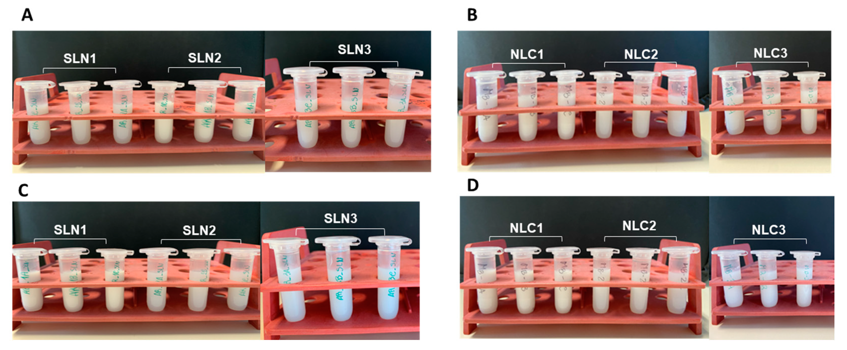
The macroscopic organoleptic characteristics of each of the formulations (SLN_1-3 and NLC_1-3) are shown in Figure 2. Through the “Centrifugation Test” it was possible to observe that after the induction of physical stress, none of the formulations under study showed any signs of phase separation, maintaining a homogeneous and milky appearance with white coloration.
Regarding the results obtained during the gradual temperature increase test (Table 2 and Figure 3), the pH remained stable except for the SLN_3 formulation, which showed a significant decrease in the pH value after gradual heating. As for the viscosity, only formulation NLC_3 showed statistically significant differences after the heat stress test.
As for the macroscopic organoleptic characteristics analysed after the gradual heating of the nanoparticles, it was found that all formulations maintained a milky and homogeneous appearance, not displaying visual signs of instability (Figure 3).

3.2.2. Long-Term Stability

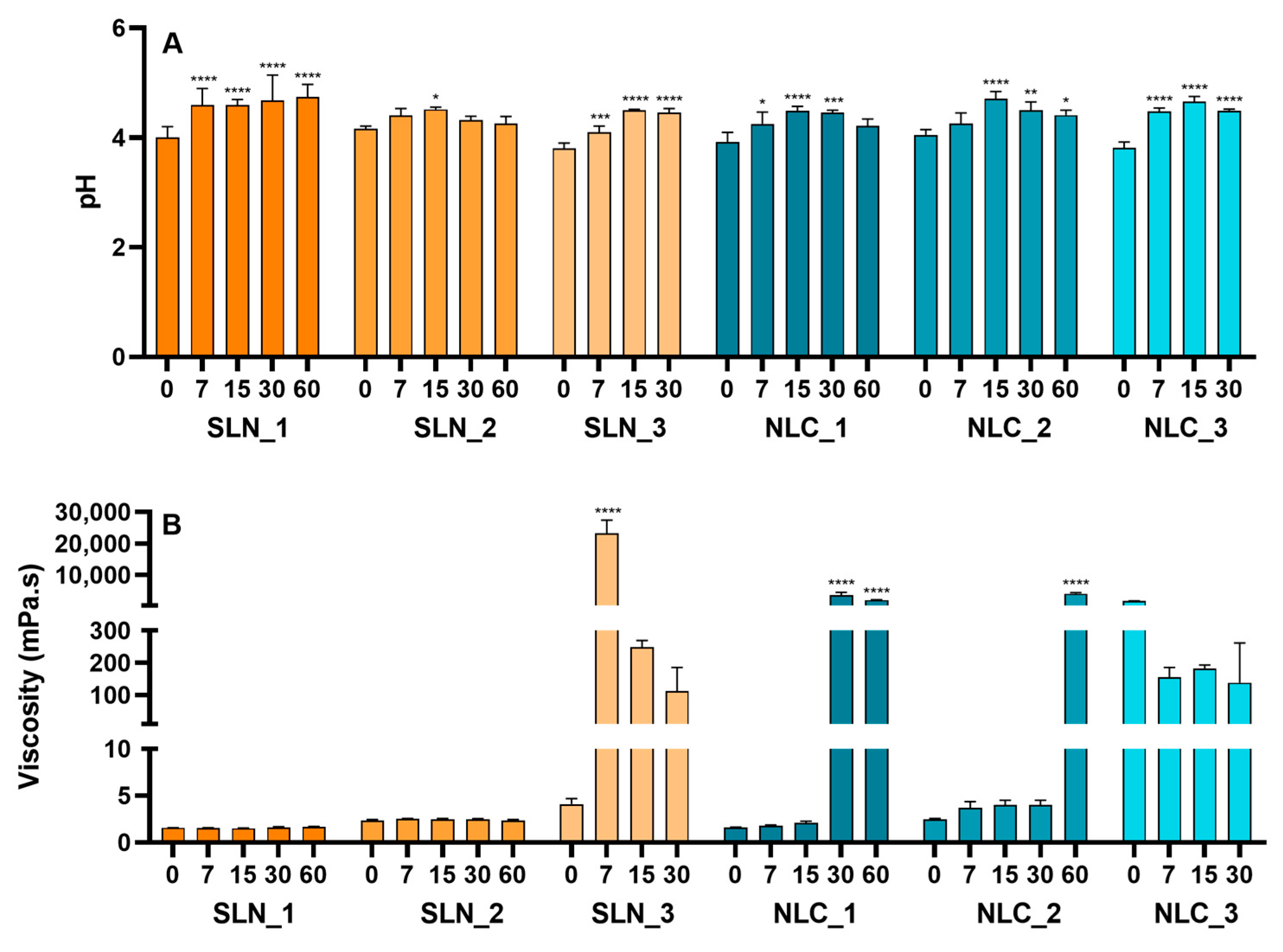
Long-term stability studies were performed over a period of three months, and particle size, PDI, pH and viscosity at days 7, 15, 30, 60 and 90 were evaluated as detailed in Figure 4 and Figure 5.
In terms of particle size analysis (Figure 4A), it was possible to observe an increase in this parameter as a function of time for all formulations under study, except for formulation SLN_2 (only composed of Precirol® ATO 5), where the values remained constant throughout the 90 days of the study. Specifically, formulations SLN_1 and NLC_1 showed a significant increase in values as of day 7. Formulation NLC_2 kept the size values constant until day 30; however, from day 60 on, it showed a significant increase (ca. 6-fold). Formulations SLN_3 and NLC_3 showed a prominent increase from day 7. It should be noted that in this case, the measurements were made only until day 30, since at this point the formulation was already unstable, considering its initial properties.
The PDI data (Figure 4B) showed that only SLN_2 did not exhibit any significant variations in this parameter over the entire 90-day period of the study. However, SLN_1 showed a statistically significant increase from day 7, but since PDI remained below 0.3 throughout the study period, the size homogeneity was reasonably maintained. NLC_1 and NLC_2 displayed stable values of PDI for 15 days, but from day 30, a significant increase in the values was observed. SLN_3 and NLC_3 showed high PDI values and a statistically significant increase as of day 7.
Considering the pH (Figure 5A), although results showed statistically significant differences for all formulations compared to the first day of the study, the pH values ranged between 4 and 5.
Finally, regarding the viscosity (Figure 5B), no significant changes were observed in the values of SLN_1 and SLN_2 throughout the study, and the values remained between 1 and 3 mPA.s. NLC_1 and NLC_2 showed a statistically significant increase of values starting on day 30 and day 60, respectively. For SLN_3, there was a statistically significant increase in viscosity on day 7, and the values obtained for NLC_3 were high during the 30 days.

3.3. In Vivo Tests

3.3.1. Skin Compatibility

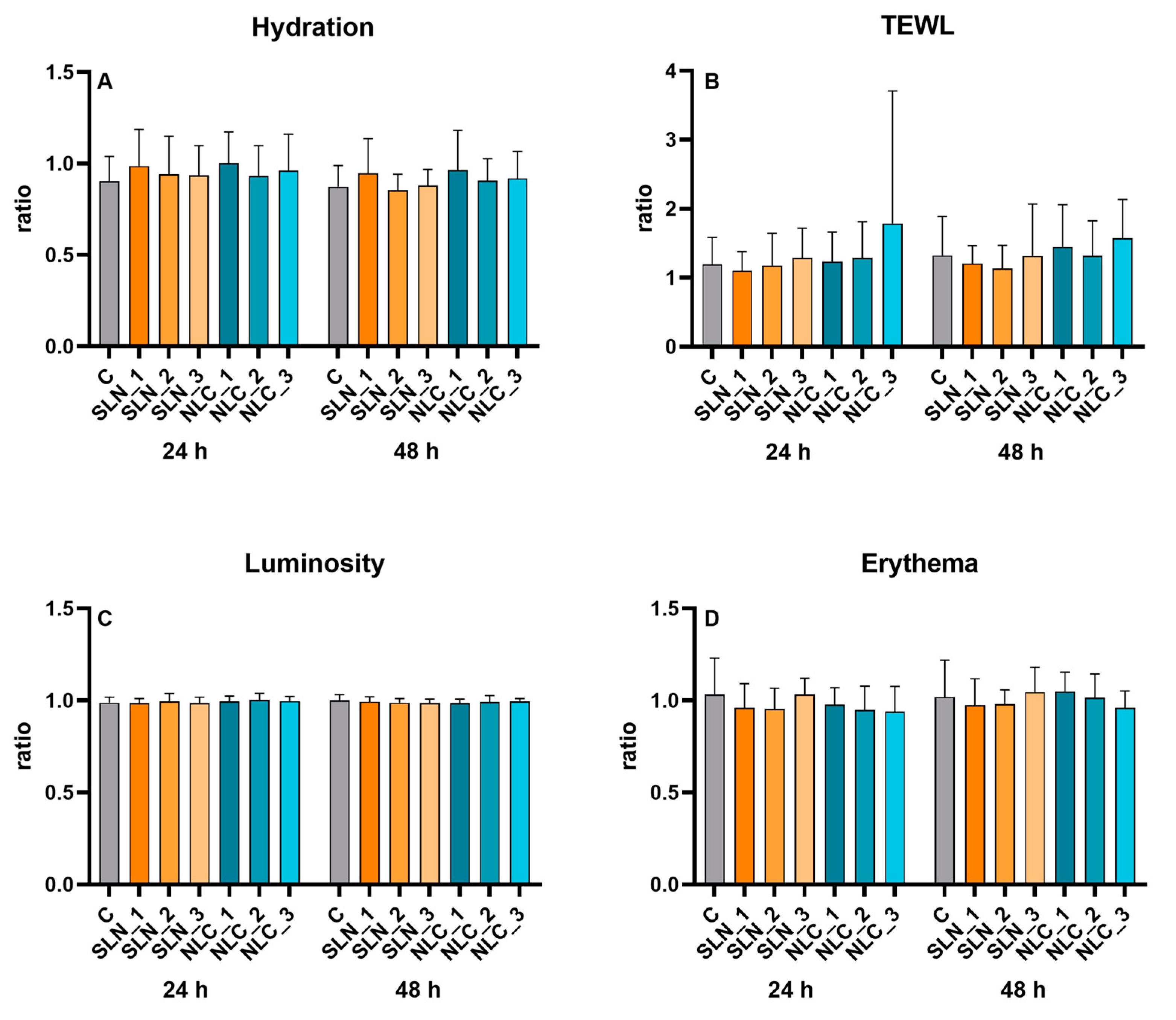
In the first part of the in vivo study to evaluate the skin compatibility, a repeated open application protocol was conducted. To reduce the impact of inter-individual variability, the data were analysed as the ratio between the results obtained with 24 h and 48 h application and the baseline values.
The results were close to 1, which indicates that none of the lipid nanoparticles caused significant perturbations in TEWL and hydration (Figure 6A,B). Additionally, no statistically significant differences were observed between the sites treated with the formulations and the untreated control.
The measurements with the Chromameter CR-300 (Minolta, Osaka, Japan) provided results in two variables of the CIE LAB system: L, which is an indicator of color changes in the black and white axis (skin luminosity); and a*, an erythema indicator because it represents the intensity of the red coloration. Once again, the results showed that the formulations did not significantly affect skin colour and luminosity over time (Figure 6C,D).
A further in vivo compatibility test was performed using an application under occlusion protocol. No redness or other skin changes were observed in any of the test sites, and none of the volunteers reported any discomfort.

3.3.2. Skin Hydration

To assess the efficacy of the SLN and NLC developed, the impact on skin hydration after application under occlusion was studied. It should be noted that, taking into account the stability results, only the SLN_2 and NLC_2 formulations were selected for further analysis. The formulations were applied under occlusion for 4 h, and the results were also analysed as the ratio of hydration obtained after and before treatment.
At the control site, the results were close to 1, whereas values of 2.5 ± 0.7 for the SLN_2 formulation and values of 2.7 ± 0.7 for the NLC_2 formulation were obtained in the treated sites. Thus, a significant increase (almost 3-fold) in the hydration parameter was observed (p < 0.0001).

4. Discussion

To understand the effect of incorporating stearic acid (SLN_1 and NLC_1), Precirol® ATO 5 (SLN_2 and NLC_2) or a mixture of both (SLN_3 and NLC_3) in lipid nanoparticles (SLN and NLC), various physicochemical parameters were assessed over time (particle size, PDI, pH, and viscosity). These parameters correspond to important properties that reflect a strong influence on the stability, biological performance and release rate of nanosystems [26,27,28,29]. These properties are dependent not only on the nanoparticles’ composition but also on the production method. When using the hot ultrasonication method as herein, it is key to know the melting range temperature of the lipid phase provided by the manufactures, or, for unknown lipids, as determined by calorimetric studies [30]. To ensure that the lipid phase is properly melted before adding the aqueous phase, it is crucial to select a target temperature higher than the reported melting range and to visually verify this critical process parameter.
To avoid agglomeration during storage, the particle size should remain within a narrow range of values [31,32,33]. Literature data also suggest that it should be lower than 300 nm, as this is considered the most appropriate size for SLN and NLC for skin applications [31]. When analysing the values obtained for particle size, it was possible to observe that all formulations presented a diameter appropriate for the intended cutaneous application. It is noteworthy that formulation NLC_3 showed a larger size compared to SLN_3, which means that although both formulations have the same solid lipid composition, the addition of Capryol® 90 (a liquid lipid) in the case of NLC_3 caused an increase in particle size, as reported in the literature by Zhang et al. [34]. These data suggest that the incorporation of the lipid blend—stearic acid:Precirol® ATO 5 1:1—favoured an increase in particle size, which may be caused by the presence of multiple components forming an imperfect matrix.
The PDI is another important parameter in the physical stability of nanoparticles, as it describes the particle size distribution. Low PDI values (<0.3) indicate a narrow size distribution, and PDI values higher than 0.5 indicate a low homogeneity of particle size and sometimes suggest the presence of particle aggregates [31,35]. In this study, the lipid nanoparticles composed of only one solid lipid displayed particle size uniformity, while the presence of two solid lipids reduced the homogeneity of this parameter.
In the development of topical formulations, pH determination is of paramount importance because products for dermal application must remain within a specific pH range. The skin normally has a pH between 4.6 and 5.8, although it can vary depending on the anatomical region [36]. Literature studies suggest that low pH values in the epidermal extracellular space may also contribute to the regulation of enzyme activity, especially at the level of keratinization and skin barrier regeneration. Results showed that the nanoparticles present pH values that are compatible with the intended topical application. It is noteworthy that the lower pH observed for the NLC_3 formulation compared with that of SLN_3 can be explained by the addition of the liquid lipid Capryol® 90. This finding may be due to the presence of small amounts of caprylic acid in the liquid lipid, leading to a decrease in the pH of the formulation. Similar results were reported by Savić et al. [37].
The rheological properties play a key role In the efficacy of topical formulations. Regarding the analysis of this parameter, it is noteworthy that the lipid nanoparticles had a low viscosity, except for NLC_3. This result may be related to the complexity of the lipid system, being in agreement with the previously discussed particle size values. In future studies, the nanosystems should be included in a semisolid matrix to facilitate topical application and improve consumer acceptance.
Accelerated stability studies are considered an alternative to long-term stability studies and have the primary objective of simulating and inducing extreme stress environments in formulations. In this way, it is possible to evaluate the physicochemical profile of the developed nanoparticles and to predict possible changes in these parameters during storage. These changes may be due to the qualitative composition and/or the selected production process/method. Overall, the results obtained herein revealed that most of the formulations under study remained stable under mechanical and thermal stress. However, NLC_3 showed statistically significant differences in viscosity after induction of heat stress. Comparing the NLC_3 and SLN_3 compositions, it is possible that the incorporation of Capryol® 90 in NLC_3 may contribute to this change in viscosity. As observed by phase-contrast microscopy, NLC_3 particles are more prone to agglomerate. By increasing temperature, the Browning motion also increases, thereby augmenting the probability of particles to form clusters. Ultimately, the formation of these clusters may contribute to the elevated viscosity of the formulation.
Still, long-term stability studies were conducted with the main objective of simulating the behaviour of the formulations under shelf-life conditions during storage. Overall, the results showed that some of the formulations did not remain stable over time. SLN_2 showed to be the most promising nanoparticle, remaining unchanged throughout the 90-day study in all 4 parameters analysed. NLC_2 showed superior stability compared to the other NLC, displaying suitable properties for 30 days. For both types of lipid nanoparticles, the most stable formulations were those composed of Precirol® ATO 5, which can be explained by the fact that it is a hydrophobic solid lipid that has the ability to give the system greater resistance to erosion, thus avoiding surface disintegration and possible problems of physical instability [38]. The stability profile of Precirol® ATO 5-containing SLN and NLC may also be related to a low-rate transition to the more-ordered β polymorph, as previously suggested [39]. The least stable formulations were SLN_3 and NLC_3, again due to possible instability in the synergy between the two solid lipids.
SLN and NLC are usually regarded as delivery systems with a favourable safety profile, but some authors have reported on the potential toxicity of the use of nanoparticles in cosmetic products due to the possibility of systemic absorption of the incorporated ingredients, owing to their nanometric size [40]. Still, numerous studies showed no evidence of a link between toxicity and the reduction in particle size and, further, indicated that nanoparticles remain in the SC and/or upper epidermis after application to the skin and are eliminated without systemic absorption [41]. Nevertheless, even if the lipid-based nanosystems have a favourable toxicity profile, it is of utmost importance to evaluate their suitability for cutaneous applicability. For this purpose, in vivo tolerance assays were conducted, which have confirmed that the nanoparticles have a low potential for irritation, even when applied under occlusion, thus corroborating their good skin compatibility.
Regarding the efficacy studies, the results showed an approximately 3-fold increase in hydration after application of the lipid nanoparticles. This increase may be due to the occlusive properties of the SLN and NLC formulations, which can form a film on the skin surface with the consequent decrease in cutaneous TEWL, thus promoting an increase in the hydration of the SC [42]. Stearic acid is known to diffuse into the SC intercellular lipid domains, potentially contributing to reinforcing the skin barrier [43], unlike occlusive or emollient ingredients that form a film at the SC surface [4]. The moisturising properties of SLN and NLC containing Precirol® ATO 5 have been previously reported as being attributable to its film forming properties [44].
Overall, this study showed the advantages of using glyceryl distearate (Precirol® ATO 5) instead of stearic acid to produce lipid nanoparticles for cutaneous applications with suitable stability, skin compatibility and moisturizing properties. Importantly, this work provides a comprehensive analysis of the stability of the developed nanoparticles over a period of 3 months, filling a critical knowledge gap left by previous studies that either incompletely described stability results or omitted them altogether [22,39,45,46]. Furthermore, this report reveals that a straightforward SLN formulation, comprising Precirol® ATO 5 and a widely available and affordable anionic surfactant, displays suitable physicochemical properties for skin application. Notably, SLN_2 displayed smaller particle size and lower PDI values compared with the SLN made of the same components reported by Butani et al. 39]. Additionally, SLN_2 demonstrated superior stability when compared with both NLC_2 and the NLC formulation described by Espinosa-Olivares et al. [22], utilizing the same solid lipid and surfactant. These findings highlight the feasibility of designing uncomplicated and cost-effective lipid nanoparticles for cosmetic applications, facilitating the potential translation of such products to the market.

5. Conclusions

In the present work, lipid nanoparticles based on stearic acid, one of the fatty acids found in the SC, and its derivative glyceryl distearate, were developed, with the main objective of further exploring their physicochemical stability and cosmetic applicability. The lipid nanoparticles made of Precirol® ATO 5 (SLN_2 and NLC_2) displayed superior physicochemical properties for skin applications, together with a good performance in accelerated and long-term stability studies for at least 1 month. Beyond exhibiting good skin compatibility, even when applied under occlusion, these lipid nanoparticles were also able to improve skin hydration. Therefore, this study suggests that the design of nanoparticles based on these materials may be a valuable strategy with intrinsic cosmetic benefits. Further studies are foreseen using a Quality-by-Design approach to fine-tune the lipid/surfactant blend, to load active compounds into these nanoparticles and to further evaluate their cosmetic benefits.

Supplementary Materials

The following supporting information can be downloaded at: https://www.mdpi.com/article/10.3390/cosmetics10040099/s1, Table S1: Physicochemical properties of solid lipid nanoparticles (SLN) and nanostructured lipid carriers (NLC) according to their solid lipid composition: stearic acid (SLN_1 and NLC_1), Precirol® ATO 5 (SLN_2 and NLC_2) or stearic acid:Precirol® ATO 5 1:1 (SLN_3 and NLC_3). Data are presented as mean ± SD (n = 3). Figure S1: Phase-contrast images of solid lipid nanoparticles (SLN) and nanostructured lipid carriers (NLC) according to their solid lipid composition: stearic acid (SLN_1 and NLC_1), Precirol® ATO 5 (SLN_2 and NLC_2) or stearic acid:Precirol® ATO 5 1:1 (SLN_3 and NLC_3). Lipid nanoparticles were diluted 1:100 prior to image acquisition with a Zeiss Axio Observer microscope (White Plains, NY, USA) with a LD A-Plan x40 objective using ZEN blue software. Scale bar 5 µm.

Author Contributions

Conceptualization, C.P.-L. and C.R.; formal analysis, M.B., A.R. and C.A.; investigation, M.B., A.R. and C.A.; writing—original draft preparation, M.B. and A.R.; writing—review and editing, C.P.-L. and C.R.; supervision, C.P.-L. and C.R.; funding acquisition, C.P.-L. and C.R. All authors have read and agreed to the published version of the manuscript.

Funding

This research was funded by FCT—Foundation for Science and Technology, I.P. through national funds under the UIDB/04567/2020, UIDP/04567/2020, and EXPL/BTM-MAT/0112/2021 projects attributed to CBIOS and by the research grants attributed to C.A. (UI/BD/151423/2021).

Institutional Review Board Statement

The study was conducted in accordance with the Declaration of Helsinki, and approved by the Ethics Committee of the School of Health Sciences and Technologies of the Universidade Lusófona (protocol code CE.ECTS/P01-22 and date of approval 16 November 2022).

Informed Consent Statement

Informed consent was obtained from all subjects involved in the study.

Data Availability Statement

The data presented in this study are available in this manuscript.

Acknowledgments

The authors thank Gattefossé (Saint-Priest, France) for providing the Precirol® ATO 5 sample used in this study. Moreover, we are thankful to Nuno Saraiva for his assistance and support on acquiring phase-contrast microscopy images.

Conflicts of Interest

The authors declare no conflict of interest.

References

  1. Gaboriau, H.P.; Murakami, C.S. Skin Anatomy and Flap Physiology. Otolaryngol. Clin. N. Am. 2001, 34, 555–569. [Google Scholar] [CrossRef] [PubMed]
  2. Honari, G.; Andersen, R.; Maibach, H.L. Sensitive Skin Syndrome, 2nd ed.; CRC Press: Boca Raton, FL, USA, 2017. [Google Scholar]
  3. Nicol, N.H. Anatomy and Physiology of the Skin. Dermatol. Nurs. Dermatol. Nurses’ Assoc. 2005, 17, 62. [Google Scholar] [CrossRef]
  4. Ananthapadmanabhan, K.P.; Mukherjee, S.; Chandar, P. Stratum Corneum Fatty Acids: Their Critical Role in Preserving Barrier Integrity during Cleansing. Int. J. Cosmet. Sci. 2013, 35, 337–345. [Google Scholar] [CrossRef] [PubMed]
  5. Van Smeden, J.; Bouwstra, J.A. Stratum Corneum Lipids: Their Role for the Skin Barrier Function in Healthy Subjects and Atopic Dermatitis Patients. In Skin Barrier Function; Agner, T., Ed.; S. Karger AG: Basel, Switzerland, 2016; Volume 49, pp. 8–26. [Google Scholar]
  6. Sahle, F.F.; Gebre-Mariam, T.; Dobner, B.; Wohlrab, J.; Neubert, R.H.H. Skin Diseases Associated with the Depletion of Stratum Corneum Lipids and Stratum Corneum Lipid Substitution Therapy. Skin. Pharmacol. Physiol. 2015, 28, 42–55. [Google Scholar] [CrossRef] [PubMed]
  7. Harding, C.R. The Stratum Corneum: Structure and Function in Health and Disease. Dermatol. Ther. 2004, 17, 6–15. [Google Scholar] [CrossRef]
  8. Schmuth, M.; Blunder, S.; Dubrac, S.; Gruber, R.; Moosbrugger-Martinz, V. Epidermal Barrier in Hereditary Ichthyoses, Atopic Dermatitis, and Psoriasis. JDDG-J. Ger. Soc. Dermatol. 2015, 13, 1119–1124. [Google Scholar] [CrossRef] [PubMed] [Green Version]
  9. Obeidat, W.M.; Schwabe, K.; Müller, R.H.; Keck, C.M. Preservation of Nanostructured Lipid Carriers (NLC). Eur. J. Pharm. Biopharm. 2010, 76, 56–67. [Google Scholar] [CrossRef]
  10. Souto, E.B.; Müller, R.H. Cosmetic Features and Applications of Lipid Nanoparticles (SLN®, NLC®). Int. J. Cosmet. Sci. 2008, 30, 157–165. [Google Scholar] [CrossRef]
  11. Ghasemiyeh, P.; Mohammadi-Samani, S. Solid Lipid Nanoparticles and Nanostructured Lipid Carriers as Novel Drug Delivery Systems: Applications, Advantages and Disadvantages. Res. Pharm. Sci. 2018, 13, 288–303. [Google Scholar] [CrossRef]
  12. Guimarães, K.L.; Ré, M.I. Lipid Nanoparticles as Carriers for Cosmetic Ingredients: The First (SLN) and the Second Generation (NLC). In Nanocosmetics and Nanomedicines: New Approaches for Skin Care; Springer: Berlin/Heidelberg, Germany, 2011; pp. 101–122. [Google Scholar] [CrossRef]
  13. Dey, S.; Datta, S.; Dasgupta, S.; Mazumder, B.; Pathak, Y.V. Lipid Nanoparticles for Topical Application of Drugs for Skin Diseases; Elsevier Inc.: Amsterdam, The Netherlands, 2016; ISBN 9780323428910. [Google Scholar]
  14. Scioli Montoto, S.; Muraca, G.; Ruiz, M.E. Solid Lipid Nanoparticles for Drug Delivery: Pharmacological and Biopharmaceutical Aspects. Front. Mol. Biosci. 2020, 7, 319. [Google Scholar] [CrossRef]
  15. Dubey, S.K.; Dey, A.; Singhvi, G.; Pandey, M.M.; Singh, V.; Kesharwani, P. Emerging Trends of Nanotechnology in Advanced Cosmetics. Colloids Surf. B Biointerfaces 2022, 214, 112440. [Google Scholar] [CrossRef] [PubMed]
  16. Santos, J.S.; Barradas, T.N.; Tavares, G.D. Advances in Nanotechnology-based Hair Care Products Applied to Hair Shaft and Hair Scalp Disorders. Int. J. Cosmet. Sci. 2022, 44, 320–332. [Google Scholar] [CrossRef] [PubMed]
  17. Moddaresi, M.; Tamburic, S.; Williams, S.; Jones, S.A.; Zhao, Y.; Brown, M.B. Effects of Lipid Nanocarriers on the Performance of Topical Vehicles in Vivo. J. Cosmet. Dermatol. 2009, 8, 136–143. [Google Scholar] [CrossRef]
  18. Akombaetwa, N.; Ilangala, A.B.; Thom, L.; Memvanga, P.B.; Witika, B.A.; Buya, A.B. Current Advances in Lipid Nanosystems Intended for Topical and Transdermal Drug Delivery Applications. Pharmaceutics 2023, 15, 656. [Google Scholar] [CrossRef]
  19. van Smeden, J.; Boiten, W.A.; Hankemeier, T.; Rissmann, R.; Bouwstra, J.A.; Vreeken, R.J. Combined LC/MS-Platform for Analysis of All Major Stratum Corneum Lipids, and the Profiling of Skin Substitutes. Biochim. Biophys. Acta (BBA)-Mol. Cell Biol. Lipids 2014, 1841, 70–79. [Google Scholar] [CrossRef]
  20. Souto, E.B.; Fangueiro, J.F.; Fernandes, A.R.; Cano, A.; Sanchez-Lopez, E.; Garcia, M.L.; Severino, P.; Paganelli, M.O.; Chaud, M.V.; Silva, A.M. Physicochemical and Biopharmaceutical Aspects Influencing Skin Permeation and Role of SLN and NLC for Skin Drug Delivery. Heliyon 2022, 8, e08938. [Google Scholar] [CrossRef]
  21. Severino, P.; Pinho, S.C.; Souto, E.B.; Santana, M.H.A. Polymorphism, Crystallinity and Hydrophilic–Lipophilic Balance of Stearic Acid and Stearic Acid–Capric/Caprylic Triglyceride Matrices for Production of Stable Nanoparticles. Colloids Surf. B Biointerfaces 2011, 86, 125–130. [Google Scholar] [CrossRef]
  22. Espinosa-Olivares, M.A.; Delgado-Buenrostro, N.L.; Chirino, Y.I.; Trejo-Márquez, M.A.; Pascual-Bustamante, S.; Ganem-Rondero, A. Nanostructured Lipid Carriers Loaded with Curcuminoids: Physicochemical Characterization, in Vitro Release, Ex Vivo Skin Penetration, Stability and Antioxidant Activity. Eur. J. Pharm. Sci. 2020, 155, 105533. [Google Scholar] [CrossRef] [PubMed]
  23. Patil, P.V.; Menon, M.D.; Palshetkar, A.D.; Desai, N.D. Topical Delivery of Mupirocin Calcium Nanostructured Lipid Carriers Using a Full-Thickness Excision Wound Healing Model. J. Wound Care 2023, 32, lxiii–lxxiv. [Google Scholar] [CrossRef] [PubMed]
  24. Kasongo, W.A.; Pardeike, J.; Müller, R.H.; Walker, R.B. Selection and Characterization of Suitable Lipid Excipients for Use in the Manufacture of Didanosine-Loaded Solid Lipid Nanoparticles and Nanostructured Lipid Carriers. J. Pharm. Sci. 2011, 100, 5185–5196. [Google Scholar] [CrossRef] [PubMed]
  25. Wagemaker, T.A.L.; Rijo, P.; Rodrigues, L.M.; Maia Campos, P.M.B.G.; Fernandes, A.S.; Rosado, C. Integrated Approach in the Assessment of Skin Compatibility of Cosmetic Formulations with Green Coffee Oil. Int. J. Cosmet. Sci. 2015, 37, 506–510. [Google Scholar] [CrossRef] [PubMed]
  26. Delgado, J.M.F. Preparação e Caracterização de Nanotransportadores (Nanocápsulas, Nanoesferas, Lipossomas e Transportadores Lipídicos Nanoestruturados) sem Substância Ativa; Escola Superior de Tecnologia e Gestão, Instituto Politécnico de Bragança: Bragança, Portugal, 2013. [Google Scholar]
  27. Han, F.; Li, S.; Yin, R.; Liu, H.; Xu, L. Effect of Surfactants on the Formation and Characterization of a New Type of Colloidal Drug Delivery System: Nanostructured Lipid Carriers. Colloids Surf. A 2008, 315, 210–216. [Google Scholar] [CrossRef]
  28. Schäfer-Korting, M.; Mehnert, W.; Korting, H.C. Lipid Nanoparticles for Improved Topical Application of Drugs for Skin Diseases. Adv. Drug Deliv. Rev. 2007, 59, 427–443. [Google Scholar] [CrossRef] [PubMed]
  29. Subramaniam, B.; Siddik, Z.H.; Nagoor, N.H. Optimization of Nanostructured Lipid Carriers: Understanding the Types, Designs, and Parameters in the Process of Formulations. J. Nanopart. Res. 2020, 22, 141. [Google Scholar] [CrossRef]
  30. De Caro, V.; Giannola, L.I.; Di Prima, G. Solid and Semisolid Innovative Formulations Containing Miconazole-Loaded Solid Lipid Microparticles to Promote Drug Entrapment into the Buccal Mucosa. Pharmaceutics 2021, 13, 1361. [Google Scholar] [CrossRef] [PubMed]
  31. Mura, P.; Maestrelli, F.; D’Ambrosio, M.; Luceri, C.; Cirri, M. Evaluation and Comparison of Solid Lipid Nanoparticles (SLNs) and Nanostructured Lipid Carriers (NLCs) as Vectors to Develop Hydrochlorothiazide Effective and Safe Pediatric Oral Liquid Formulations. Pharmaceutics 2021, 13, 437. [Google Scholar] [CrossRef] [PubMed]
  32. Souto, E.B.; Baldim, I.; Oliveira, W.P.; Rao, R.; Yadav, N.; Gama, F.M.; Mahant, S. SLN and NLC for Topical, Dermal, and Transdermal Drug Delivery. Expert. Opin. Drug Deliv. 2020, 17, 357–377. [Google Scholar] [CrossRef]
  33. Souto, E.B.; Almeida, A.J.; Müller, R.H. Lipid Nanoparticles (SLN®, NLC®) for Cutaneous Drug Delivery: Structure, Protection and Skin Effects. J. Biomed. Nanotechnol. 2007, 3, 317–331. [Google Scholar] [CrossRef]
  34. Zhang, C.; Peng, F.; Liu, W.; Wan, J.; Wan, C.; Xu, H.; Lam, C.W.; Yang, X. Nanostructured Lipid Carriers as a Novel Oral Delivery System for Triptolide: Induced Changes in Pharmacokinetics Profile Associated with Reduced Toxicity in Male Rats. Int. J. Nanomed. 2014, 9, 1049–1063. [Google Scholar] [CrossRef] [Green Version]
  35. Iriventi, P.; Gupta, N.V. Topical Delivery of Curcumin and Caffeine Mixture-Loaded Nanostructured Lipid Carriers for Effective Treatment of Psoriasis. Pharmacogn. Mag. 2020, 16, 206–217. [Google Scholar] [CrossRef]
  36. Tichota, D.M.; Silva, A.C.; Sousa Lobo, J.M.; Amaral, M.H. Design, Characterization, and Clinical Evaluation of Argan Oil Nanostructured Lipid Carriers to Improve Skin Hydration. Int. J. Nanomed. 2014, 9, 3855–3864. [Google Scholar] [CrossRef] [Green Version]
  37. Savić, V.; Ilić, T.; Nikolić, I.; Marković, B.; Čalija, B.; Cekić, N.; Savić, S. Tacrolimus-Loaded Lecithin-Based Nanostructured Lipid Carrier and Nanoemulsion with Propylene Glycol Monocaprylate as a Liquid Lipid: Formulation Characterization and Assessment of Dermal Delivery Compared to Referent Ointment. Int. J. Pharm. 2019, 569, 118624. [Google Scholar] [CrossRef] [PubMed]
  38. Jannin, V.; Pochard, E.; Chambin, O. Influence of Poloxamers on the Dissolution Performance and Stability of Controlled-Release Formulations Containing Precirol® ATO 5. Int. J. Pharm. 2006, 309, 6–15. [Google Scholar] [CrossRef]
  39. Butani, D.; Yewale, C.; Misra, A. Topical Amphotericin B Solid Lipid Nanoparticles: Design and Development. Colloids Surf. B Biointerfaces 2016, 139, 17–24. [Google Scholar] [CrossRef] [PubMed]
  40. Nohynek, G.J.; Dufour, E.K. Nano-Sized Cosmetic Formulations or Solid Nanoparticles in Sunscreens: A Risk to Human Health? Arch. Toxicol. 2012, 86, 1063–1075. [Google Scholar] [CrossRef]
  41. Eiras, F.; Amaral, M.H.; Silva, R.; Martins, E.; Lobo, J.M.S.; Silva, A.C. Characterization and Biocompatibility Evaluation of Cutaneous Formulations Containing Lipid Nanoparticles. Int. J. Pharm. 2017, 519, 373–380. [Google Scholar] [CrossRef] [PubMed]
  42. Loo, C.H.; Basri, M.; Ismail, R.; Lau, H.L.N.; Tejo, B.A.; Kanthimathi, M.S.; Hassan, H.A.; Choo, Y.M. Effect of Compositions in Nanostructured Lipid Carriers (NLC) on Skin Hydration and Occlusion. Int. J. Nanomed. 2013, 8, 13–22. [Google Scholar] [CrossRef] [Green Version]
  43. Purnamawati, S.; Indrastuti, N.; Danarti, R.; Saefudin, T. The Role of Moisturizers in Addressing Various Kinds of Dermatitis: A Review. Clin. Med. Res. 2017, 15, 75–87. [Google Scholar] [CrossRef] [Green Version]
  44. Sarhadi, S.; Gholizadeh, M.; Moghadasian, T.; Golmohammadzadeh, S. Moisturizing Effects of Solid Lipid Nanoparticles (SLN) and Nanostructured Lipid Carriers (NLC) Using Deionized and Magnetized Water by In Vivo and In Vitro Methods. Iran. J. Basic. Med. Sci. 2020, 23, 337–343. [Google Scholar] [CrossRef]
  45. Jeon, H.S.; Seo, J.E.; Kim, M.S.; Kang, M.H.; Oh, D.H.; Jeon, S.O.; Jeong, S.H.; Choi, Y.W.; Lee, S. A Retinyl Palmitate-Loaded Solid Lipid Nanoparticle System: Effect of Surface Modification with Dicetyl Phosphate on Skin Permeation In Vitro and Anti-Wrinkle Effect In Vivo. Int. J. Pharm. 2013, 452, 311–320. [Google Scholar] [CrossRef]
  46. El-Housiny, S.; Shams Eldeen, M.A.; El-Attar, Y.A.; Salem, H.A.; Attia, D.; Bendas, E.R.; El-Nabarawi, M.A. Fluconazole-Loaded Solid Lipid Nanoparticles Topical Gel for Treatment of Pityriasis Versicolor: Formulation and Clinical Study. Drug Deliv. 2018, 25, 78–90. [Google Scholar] [CrossRef] [PubMed] [Green Version]
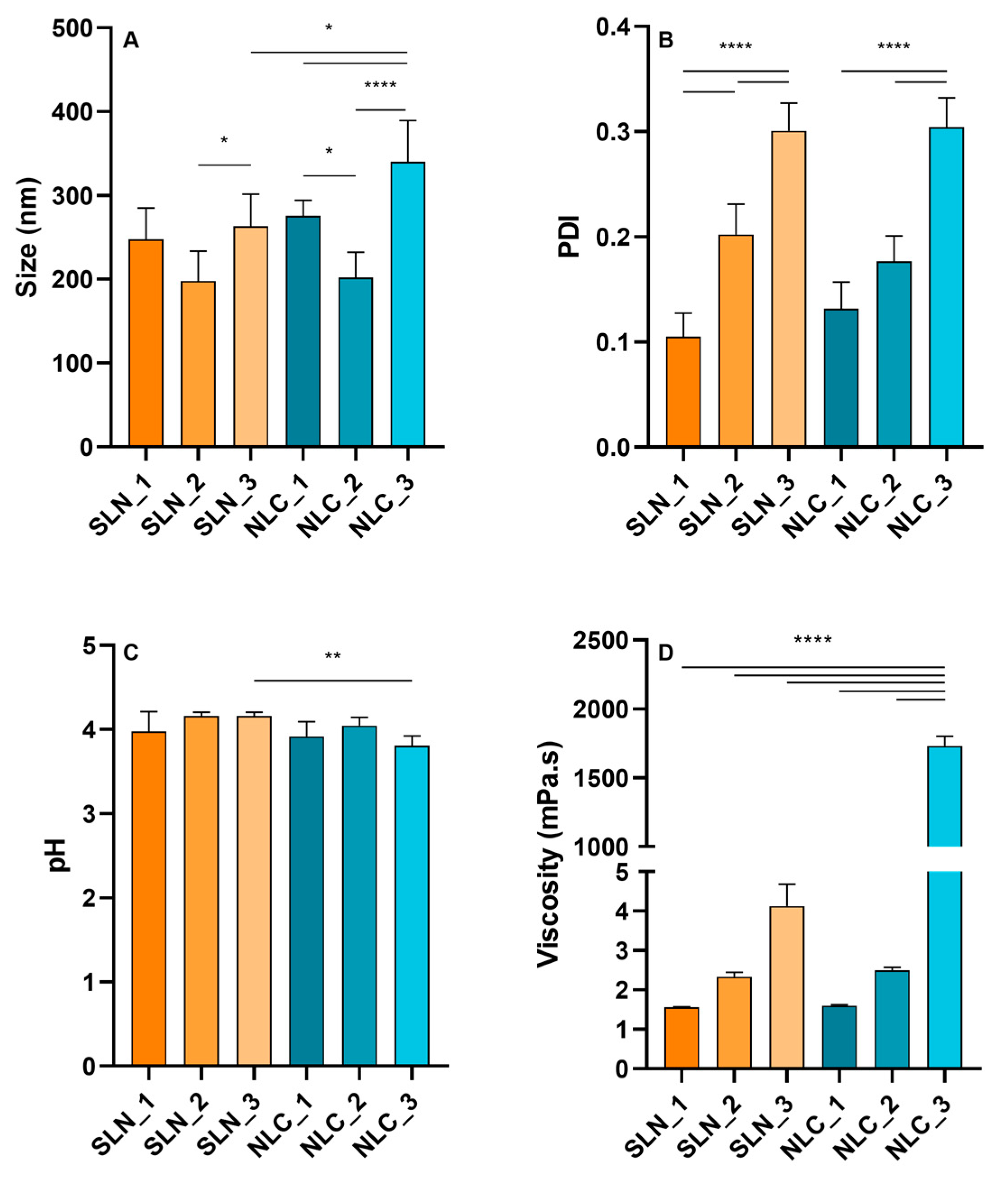
Figure 1. Influence of solid lipid composition: stearic acid (SLN_1 and NLC_1), Precirol® ATO 5 (SLN_2 and NLC_2) or stearic acid:Precirol® ATO 5 1:1 (SLN_3 and NLC_3), on the physicochemical properties of solid lipid nanoparticles (SLN) and nanostructured lipid carriers (NLC). (A) size, (B) polydispersity index (PDI), (C) pH, and (D) viscosity. Data are presented as mean ± SD (n = 3 for each formulation) and data sets with significant differences are labeled * p < 0.05, ** p < 0.01 and **** p < 0.0001.
Cosmetics 10 00099 g001
Figure 2. Accelerated stability study—Results of the centrifugation test. Organoleptic characteristics of lipid nanoparticles before (A,B) and after (C,D) induction of mechanical stress (n = 3 for each type of nanoparticle under study). Lipid composition of nanoparticles: stearic acid (SLN_1 and NLC_1), Precirol® ATO 5 (SLN_2 and NLC_2) or stearic acid:Precirol® ATO 5 1:1 (SLN_3 and NLC_3).
Cosmetics 10 00099 g002
Figure 3. Accelerated stability studies—Results of the gradual temperature increase test. Organoleptic characteristics of lipid nanoparticles before (A,B) and after (C,D) heat stress induction (n = 3 for each type of nanoparticle under study). Lipid composition of nanoparticles: stearic acid (SLN_1 and NLC_1), Precirol® ATO 5 (SLN_2 and NLC_2) or stearic acid:Precirol® ATO 5 1:1 (SLN_3 and NLC_3).
Cosmetics 10 00099 g003
Figure 4. Stability of solid lipid nanoparticles (SLN) and nanostructured lipid carriers (NLC) during storage—90-day period. Analysis of changes in physicochemical characteristics, in terms of size (A) and polydispersity index (PDI, (B)). Data are presented as mean ± SD (n = 3 for each type of nanoparticle) and data sets with significant differences are identified with * p < 0.05, ** p < 0.01 and **** p < 0.0001.
Cosmetics 10 00099 g004
Figure 5. Stability of solid lipid nanoparticles (SLN) and nanostructured lipid carriers (NLC) during storage—60-day period. Analysis of changes in physicochemical characteristics, in terms of pH (A) and viscosity (B). Data are presented as mean ± SD (n = 3 for each type of nanoparticles) and data sets with significant differences are identified with * p < 0.05, ** p < 0.01, *** p < 0.001 and **** p < 0.0001.
Cosmetics 10 00099 g005
Figure 6. Effect of solid lipid composition: stearic acid (SLN_1 and NLC_1), Precirol® ATO 5 (SLN_2 and NLC_2) or stearic acid:Precirol® ATO 5 1:1 (SLN_3 and NLC_3) on the skin compatibility of solid lipid nanoparticles (SLN) and nanostructured lipid carriers (NLC). The results obtained in the untreated area (C) are also presented. (A) Hydration, (B) Transepidermal Water Loss (TEWL), (C) Luminosity, and (D) Erythema. The obtained data (n = 12) are presented as mean ± SD of the ratio between the values obtained at 24 h or 48 h compared to baseline values.
Cosmetics 10 00099 g006
Table 1. Qualitative and quantitative composition of the SLN and NLC formulations.
FormulationsStearic Acid
(% w/v)
Precirol® ATO 5
(% w/v)
Capryol® 90
(% w/v)
Tween® 80
(% w/v)
SLN_15--2
SLN_2-5-2
SLN_32.52.5-2
NLC_14.17-0.832
NLC_2-4.170.832
NLC_32.082.080.832
Table 2. Effect of gradual temperature increase on pH and viscosity of solid lipid nanoparticles (SLN) and nanostructured lipid carriers (NLC) as a function of their solid lipid composition: stearic acid (SLN_1 and NLC_1), Precirol® ATO 5 (SLN_2 and NLC_2) or stearic acid:Precirol® ATO 5 1:1 (SLN_3 and NLC_3). All data are represented as mean ± SD (n = 3) and data with significant differences from the value obtained before gradual heating are identified with * p < 0.05 and **** p < 0.0001.
FormulationBefore Gradual HeatingAfter Gradual Heating
pHViscosity (mPa.s)pHViscosity (mPa.s)
SLN_14.11 ± 0.071.56 ± 0.024.3 ± 0.31.56 ± 0.02
SLN_24.20 ± 0.022.3 ± 0.14.33 ± 0.042.5 ± 0.1
SLN_33.85 ± 0.086 ± 33.6 ± 0.1 *3.1 ± 0.3
NLC_14.07 ± 0.021.60 ± 0.034.2 ± 0.12.05 ± 0.02
NLC_24.11 ± 0.062.49 ± 0.084.3 ± 0.13.9 ± 0.7
NLC_33.7 ± 0.11730 ± 703.99 ± 0.06619 ± 269 ****
Disclaimer/Publisher’s Note: The statements, opinions and data contained in all publications are solely those of the individual author(s) and contributor(s) and not of MDPI and/or the editor(s). MDPI and/or the editor(s) disclaim responsibility for any injury to people or property resulting from any ideas, methods, instructions or products referred to in the content.