Review

Use of Exosomes for Cosmetics Applications

by
Luis Jesús Villarreal-Gómez
1,
Sergio Origel-Lucio
2,
Daniela Alejandra Hernández-Hernández
1 and
Graciela Lizeth Pérez-González
1,*
1
Facultad de Ciencias de la Ingeniería y Tecnología, Universidad Autónoma de Baja California, Unidad Valle de las Palmas, Blvd. Universitario No. 1000, C.P. 22200, Tijuana 22260, Baja California, Mexico
2
Centro de Medicina de Precisión, Calle Germán Gedovius 9506/204, Zona Río, Tijuana 22010, Baja California, Mexico
*
Author to whom correspondence should be addressed.
Cosmetics 2025, 12(1), 9; https://doi.org/10.3390/cosmetics12010009
Submission received: 21 November 2024 / Revised: 20 December 2024 / Accepted: 8 January 2025 / Published: 13 January 2025
(This article belongs to the Section Cosmetic Dermatology)

Abstract

:
This study addresses a significant gap in the clinical validation and standardization of exosome-based applications within the domains of cosmetics and regenerative medicine. Despite offering a thorough overview of the potential therapeutic benefits and underlying mechanisms of exosomes, the manuscript underscores several unresolved challenges, including the paucity of clinical evidence, regulatory barriers, inconsistencies in standardization, and incomplete mechanistic understanding. The primary aim of this research is to conduct a systematic and comprehensive review of existing studies on the applications of exosomes in cosmetics and skincare. To achieve this, a systematic literature review was performed, drawing on the major medical database PubMed. This approach facilitated the collection and meta-analysis of relevant data, ensuring a rigorous assessment of recent advancements in exosome research. The main outcomes of the study highlight the multifaceted roles of exosomes in promoting skin rejuvenation and mitigating signs of aging. Specific applications discussed include pigmentation correction, wound healing, tissue repair, and innovative delivery mechanisms. Moreover, the study examines the emerging potential of exosomes in plastic surgery and regenerative medicine. Challenges and limitations, such as regulatory constraints, standardization hurdles, and the need for further clinical validation, are critically analyzed, providing a framework for future research directions.

Graphical Abstract

1. Introduction

Exosomes have gained interest in the field of cosmetics and skincare due to their potential benefits for cosmetic dermatology [1]. This potential small, membrane-bound vesicle that is naturally released from cells into the extracellular space, contains bioactive molecules such as proteins, lipids, and nucleic acids that play a role in cell communication and tissue repair [2]. Exosomes have attracted considerable interest in the medical and clinical sectors because of their broad potential for therapeutic applications such as regenerative medicine benefits from these structural compounds, as they promote tissue repair and regeneration, offering potential treatments for various conditions [2,3]. In tissue engineering, these cellular derivatives facilitate tissue growth and the integration of bioengineered tissues, supporting their viability and functionality [4,5,6,7], and active ingredients delivery and targeted therapy to leverage the ability of exosomes to be engineered for direct delivery of molecules or proteins to specific cells, enhancing treatment efficacy while minimizing side effects [8].
Cell-based therapies exert their therapeutic effects through soluble growth factors and vesicular entities, positioning exosomes as a prototype for biological therapy in dermal wound healing. Despite this, a comprehensive systematic review has been reported before to provide recommendations on their optimal concentrations for preclinical studies [9], and its clinical validation needs to be further discussed.
The ongoing quest for interventions to combat the effects of aging has led to a growing demand for nonsurgical aesthetic methods. In recent years, exosomes have garnered significant attention as both a topical and injectable option. This is primarily because of their recognized regenerative properties and their potential to impact processes like wound healing, scar improvement, and hair growth. While dermatology and anti-aging medicine have been early advocates of this evolving technology, their adoption in the field of plastic surgery is on the rise. However, it is important to note that the number of published studies in this area remains somewhat limited [10].
While the subcutaneous injection of autologous or allogeneic cells can influence skin regeneration by providing sustained release of paracrine factors and extracellular matrix proteins, challenges arise from the need for cell isolation, cryopreservation, and adherence to lot release criteria, especially in the case of autologous cells produced at the point of care. The prospect of producing freeze-dried exosome products that remain stable at room temperature for extended periods is appealing. However, exosomes alone may not encompass the benefits offered by extracellular proteins and will require extensive evaluation before attaining regulatory approval. It may be necessary to complement exosomes with hydrogels derived from adipose tissue, blood, or cellulose, either as cryopreserved injectable products or lyophilized topical formulations. Whether used individually or as combination products, hydrogels are likely to necessitate randomized, controlled clinical trials before they can be considered for regulatory approval [11].
This review aims to inform readers about the current state of research on exosomes in skincare and to offer insights into their potential as therapeutic agents in dermatology and plastic surgery, while also highlighting areas that require further investigation and clinical validation.

2. Methodology

Search Strategy

With the assistance of a medical bariatrics specialist, we conducted a comprehensive search of available databases using Medical Subject Headings (MeSH) terms related to exosomes, such as “Regenerative dermatology”, “Anti-aging” “Skin hydration”, “Skin pigmentation”, “Wound healing”, “Delivery of biomolecules”, “Customized dermatology”, and “Clinical validation”. The literature was retrieved from PubMed, covering the period of 10 years from 2014 to 2024. The initial search was conducted in 2014, and the most recent update was completed in December 2024, resulting in a total of 37,972 unique peer-reviewed studies on exosomes and their cosmetic applications. Only full-text articles in English investigating the therapeutic potential of exosomes in cosmetic applications were included.
The references were managed using Zotero (Digital Scholar, version 6.0.37), which was also employed to remove duplicates. Following the initial screening, three independent reviewers (G.L.P.G., S.O., L.J.V.G.) manually reviewed the titles and abstracts of the 37,972 studies. Full-text reviews were then conducted to extract both quantitative and qualitative data relevant to cosmetic applications. Disagreements among authors were resolved by the corresponding author. A meta-analysis was subsequently performed using the studies obtained from this systematic search.

3. Results

Meta-Analysis

PubMed, used as the primary database for this study, revealed that exosome research has been extensively published, with 36,173 publications over the past decade. Among cosmetic applications, wound healing emerged as the most studied area, with 1438 publications, while skin hydration was the least explored. Table 1 also highlights the exponential growth of exosome research over the years, particularly in the fields of wound healing and biomolecule delivery.
The analysis of exosome research over the past decade (2014–2024) reveals a significant and exponential increase in publications, reflecting growing scientific and clinical interest. Exosomes, nanoscale extracellular vesicles involved in intercellular communication, have been extensively studied for their regenerative and therapeutic properties. With a total of 36,173 publications over ten years, this surge is driven by advancements in nanotechnology, regenerative medicine, and molecular biology. In 2024 alone, 5382 articles were published, demonstrating how this field has matured and become a focal point of biomedical research.
Among cosmetic applications, wound healing emerged as the most studied area, with 1438 publications. This prominence can be attributed to the critical role of exosomes in tissue repair, as they facilitate cellular communication, promote angiogenesis, and modulate inflammation. Exosomes derived from stem cells, particularly mesenchymal stem cells (MSCs), have shown considerable promise in accelerating wound closure and improving skin regeneration. The ability of exosomes to deliver growth factors, cytokines, and microRNAs to target cells makes them a powerful tool for enhancing wound healing outcomes. As a result, the potential for exosome-based therapies in treating chronic wounds, burns, and surgical scars has driven significant research activity [12].
In contrast, delivery of biomolecules accounted for 187 publications, emphasizing the role of exosomes as natural delivery vehicles for therapeutic agents, including growth factors, peptides, and bioactive molecules. Exosomes’ unique lipid bilayer structure allows for effective cargo encapsulation and targeted delivery, improving the stability and bioavailability of therapeutic compounds. This property is particularly valuable in cosmetic applications, where precision delivery of anti-aging or regenerative agents can yield superior results. The controlled release of bioactive molecules further enhances their efficacy in treating skin-related conditions, making exosomes a promising innovation in cosmetic dermatology [13].
Research on anti-aging (62 publications) and regenerative dermatology (81 publications) highlights the expanding interest in exosome-based interventions for skin rejuvenation and repair. Exosomes have been shown to improve skin elasticity, reduce wrinkles, and stimulate collagen synthesis by transferring growth factors, such as TGF-β, and regulatory RNAs to skin cells. These effects can mitigate aging-related changes and support tissue homeostasis. While studies in this area are growing, there is still substantial opportunity for exploring mechanisms underlying exosome-mediated skin rejuvenation, particularly in clinical settings [14].
Interestingly, skin pigmentation and skin hydration remain the least explored areas, with 24 and 7 publications, respectively. Exosomes have the potential to regulate melanogenesis and skin hydration through the delivery of melanogenic inhibitors, antioxidants, and moisture-retaining biomolecules. However, limited research in these fields suggests untapped opportunities for leveraging exosome-based therapies to treat pigmentation disorders, such as melasma, and address challenges related to dry skin. Future studies exploring these applications could reveal novel pathways and therapeutic strategies for enhancing skin health and aesthetics [15].
This meta-analysis highlights the remarkable expansion of exosome research over the past decade, with a pronounced emphasis on wound healing and biomolecule delivery, which are key drivers of advancements in both cosmetic and therapeutic applications. The intrinsic regenerative properties of exosomes, combined with their capacity for targeted delivery of bioactive molecules, have laid the foundation for innovative strategies in cosmetic dermatology. Despite significant progress, areas such as skin hydration and pigmentation remain largely unexplored, representing promising avenues for future investigations. As research in this field continues to advance, the clinical translation of exosome-based therapies holds substantial potential to enhance skin regeneration, accelerate wound healing, and achieve superior cosmetic outcomes.

4. Exosomes Composition and Biomedical Applications

Exosomes can be obtained through ultracentrifugation of cell cultures derived from adipose tissue biopsies which can contain mesenchymal stem cells (MSCs), keratinocytes, or adipocytes. The isolated and purified exosomes are then lyophilized and administered either in solution or combined with another molecular carrier [3,16] (Figure 1).

Exosome Growth Factors in Biomedical Applications

Exosomes growth factors have emerged as innovative tools in cosmetics, driven by their ability to enhance skin health and rejuvenation. Growth factors are proteins that regulate cell proliferation, differentiation, and survival. Together, they provide synergistic effects for skin regeneration and anti-aging treatments [17].
Growth factors, such as epidermal growth factor (EGF), fibroblast growth factor (FGF), and transforming growth factor-beta (TGF-β), further amplify these effects of tissue regeneration by stimulating cell turnover and enhancing extracellular matrix remodeling [18].
Formulations incorporating exosomes and growth factors are used in serums, creams, and masks, offering non-invasive alternatives to traditional treatments. These products aim to deliver bioactive molecules to the dermis, promoting wound healing, reducing inflammation, and supporting a youthful complexion. However, the clinical efficacy, production scalability, and the safety of such technologies remain areas of ongoing research to ensure their reliability and acceptance in the cosmetic industry [14].
Yoo et al., 2022, explores innovative therapeutic approaches for osteoarthritis (OA), a condition that currently lacks effective treatments beyond joint replacement surgery, which carries significant risks. The authors emphasize the critical role of transforming growth factor-β (TGF-β) in cartilage homeostasis and its potential as a therapeutic target in OA management. They highlight recent advances in stem cell-based therapies, particularly the role of exosomes—nanosized extracellular vesicles secreted by stem cells—in promoting anti-inflammatory and chondroprotective effects. The review discusses the potential of combining TGF-β3, recognized for its chondrogenic properties, with bone morphogenetic protein-6 to enhance cartilage regeneration. This approach leverages the intrinsic chondroprotective functions of exosomes as a delivery platform, presenting a promising and innovative alternative to current OA treatments. The article underscores the growing evidence supporting exosomes as a superior therapeutic option due to their efficiency and regenerative potential [19].
Zhou, X. et al., 2017, investigates the interplay between exosome production and epidermal growth factor receptor (EGFR) activation during kidney repair following injury. Renal tubular epithelial repair involves the reconstitution of its structure and function, with growth factors like EGF playing a crucial role. The study observed that scratch-induced wounding in renal tubular cells triggered EGFR activation, promoting wound healing, which was further enhanced by EGF and inhibited by the EGFR inhibitor gefitinib. Interestingly, scratch wounding also stimulated exosome production, which was reduced by EGF and increased by gefitinib, suggesting that EGFR signaling suppresses exosome release. Inhibitors of exosome release (GW4869 and manumycin A) further activated EGFR and enhanced wound healing, while exosomes derived from wounded cells were found to inhibit wound healing. These findings reveal a dual role for exosomes: while EGFR activation promotes wound healing, the exosomes released may counteract this process, providing insight into the complex regulatory mechanisms of renal epithelial repair [20].
Olejarz, W. et al., 2020, explores the critical role of exosomes in cancer-related angiogenesis and their potential as therapeutic targets. Angiogenesis, the formation of new blood vessels, is vital for tumor growth and metastasis, with exosomes playing a pivotal role by delivering pro-angiogenic factors such as VEGF, MMPs, and microRNAs. Tumor-derived exosomes (TEX) promote angiogenesis by suppressing inhibitors like factor-inhibiting HIF-1 and activating angiogenic signaling in endothelial cells. TEX also mediate cross-talk between mesenchymal stem cells (MSCs) and immune cells, impairing anti-tumor immunity. While antiangiogenic therapies, such as VEGF-specific antibodies like Bevacizumab, have shown promise in slowing tumor progression, resistance remains a significant clinical challenge. The study highlights the potential of combining angiogenesis inhibitors with immunotherapies to improve outcomes, emphasizing the need for continued research to optimize cancer treatment strategies [21].
The study of Park, S. et al., 2023, investigates the use of thermostable basic fibroblast growth factor (TS-bFGF) to improve the production and functionality of exosomes derived from Wharton’s jelly mesenchymal stem cells (WJ-MSCs). Exosomes from WJ-MSCs are known for their regenerative and immune-modulating properties but are limited by low secretion rates. The research demonstrates that MSCs cultured with TS-bFGF exhibit enhanced proliferation, stemness, and colony-forming ability compared to those cultured with wild-type bFGF. Nanoparticle tracking analysis revealed significantly increased exosome production under three-dimensional culture conditions with TS-bFGF. The exosomes produced under these conditions showed enhanced anti-inflammatory and wound-healing properties, as confirmed by nitric oxide and scratch assays. The findings suggest that TS-bFGF can optimize WJ-MSC-derived exosome production and activity, offering improved potential for therapeutic applications in inflammation and tissue regeneration [22].
Heo, J. et al., 2020, explores how exosomal microRNAs (miRNAs) mediate communication between vascular smooth muscle cells (VSMCs) and endothelial cells (ECs) in maintaining vascular homeostasis. It highlights the role of platelet-derived growth factor (PDGF) in altering exosomal miRNA release from VSMCs, specifically reducing miR-1246, miR-182, and miR-486 levels. This decrease correlates with increased EC migration, providing insights into exosome-mediated crosstalk in vascular pathogenesis and revealing potential targets for therapeutic intervention in vascular diseases [23].
Moreover, Yu, L. et al., 2023, investigates the hepatoprotective potential of exosomes derived from adipose mesenchymal stem cells (ADMSCs) overexpressing hepatocyte growth factor (HGF) in a mouse model of liver injury. Exosomes (ADMSC-Exo, ADMSCNC-Exo, and ADMSCHGF-Exo) were isolated and characterized, with their therapeutic efficacy evaluated in mice with liver injury induced by carbon tetrachloride (CCl4). The administration of ADMSCHGF-Exo showed the most pronounced effects, including significant improvement in serum liver function markers, reduction in hepatic fibrosis-related proteins (α-SMA and collagen I), restoration of hepatocyte-specific markers, and reversal of histopathological damage and collagen accumulation. The study concludes that ADMSCHGF-Exo alleviates hepatic fibrosis and restores liver function, highlighting its potential as an advanced therapeutic approach for liver injuries [24].
On the other hand, Ma, K. et al., 2019, explores the potential of neural stem cell (NSC)-derived exosomes as a treatment for spinal cord injury (SCI). Exosomes formed in the presence of insulin-like growth factor-1 (IGF-Exo) were compared to normal exosomes (Nor-Exo) for their therapeutic efficacy in reducing apoptosis and neuroinflammation after SCI. Through microRNA sequencing and qRT-PCR, the study identified miR-219a-2-3p as a key component enriched in IGF-Exo, with significant anti-inflammatory and anti-apoptotic roles. Mechanistically, IGF-Exo upregulates miR-219a-2-3p, which suppresses YY1 expression and inhibits the NF-κB signaling pathway, thereby mitigating neuroinflammation and enhancing neuroprotection. These findings suggest IGF-Exo as a promising therapeutic strategy for promoting neural regeneration and recovery in SCI patients [25].

5. Cosmetic Applications of Exosomes

5.1. Regenerative Dermatology

In recent years, the use of exosomes in skin regeneration has garnered great interest from the cosmetic community as a potential non-surgical and regenerative approach to improving the appearance and health of the skin [3,6,11,15,26,27,28]. As exosomes are secreted by various cells, including stem cells, they contain bioactive molecules that can stimulate cell-to-cell communication and tissue repair [29]. When applied to the skin, exosomes can have several beneficial effects for skin rejuvenation [14].
Tienda-Vazquez, et al. discussed the need for skin repair and regeneration. However, conditions like diabetes can prolong the inflammatory stage, complicating the healing process and sometimes completely inhibiting it, thus increasing susceptibility to infections [15]. The authors explained that exosomes have proven to be effective as anti-inflammatory agents, inducers of macrophage polarization, and facilitators of skin repair and regeneration. This reduces the potential complications associated with inadequate wound healing and prolonged inflammation [15].
Exosomes can contain growth factors and proteins (Table 2) that can stimulate collagen synthesis. Collagen is crucial for maintaining skin elasticity and firmness, and increased collagen production can help reduce the appearance of wrinkles and fine lines [30], and also promotes the production of elastin, a protein that gives skin its elasticity. Enhanced elastin production can help the skin bounce back and appear more youthful [31]. However, the existing evidence on collagen types, their mechanisms of action, time required to achieve desired outcomes, and potential side effects have not been thoroughly reviewed or systematically analyzed. This lack of clarity can lead to controversies regarding the use of collagen for reversing the aging process. Furthermore, it remains uncertain whether oral or topical collagen is more effective in delivering these sought-after anti-aging benefits. Therefore, this study aims to compare and synthesize the effects of oral collagen versus topical collagen in reducing or delaying the signs of aging [32].
Collagen plays a critical role in skin regeneration due to its structural and functional importance in maintaining skin integrity, elasticity, and strength. As the most abundant protein in the extracellular matrix (ECM) of the skin, collagen provides a scaffold that supports cell adhesion, proliferation, and migration, which are essential processes for tissue repair and regeneration (Figure 2) [41].
During skin regeneration, collagen serves as a primary component in wound healing. Fibroblasts are activated to synthesize and deposit collagen, primarily type I and type III, in the ECM to rebuild damaged tissue. Type III collagen is produced early in the healing process, offering a temporary framework, while type I collagen gradually replaces it, strengthening and stabilizing the regenerated skin [42].
Additionally, collagen influences skin cell behavior by interacting with surface receptors, such as integrins, that mediate cellular signaling pathways. These pathways regulate processes like keratinocyte proliferation, fibroblast activity, and angiogenesis, all of which are vital for tissue repair and dermal remodeling. The balance between collagen production and degradation, governed by matrix metalloproteinases (MMPs) and their inhibitors, is essential to ensure effective skin regeneration without scarring or fibrosis [42].
In cosmetic and clinical applications, collagen-based therapies, including topical formulations, oral supplements, and injectable forms, aim to stimulate natural collagen synthesis, promote ECM remodeling, and accelerate skin regeneration. By improving skin hydration, elasticity, and thickness, collagen helps mitigate signs of aging and supports the repair of damaged or aging skin [43].
Ye, C. et al., 2022, have shown that hMSC-exosomes offer benefits in terms of being biocompatible and easily degradable. These protein residues, have the potential to enhance clinical symptoms and skin eruptions in individuals with sensitive skin, suggesting their potential as a novel cell-free therapy for the treatment of conditions related to sensitive skin [29,44,45,46].
The review article reported by Wang, H., 2021, explores the clinical applications of exosomes in regenerative and cosmetic dermatology, highlighting their role as extracellular vesicles capable of transporting bioactive substances for cellular communication and therapeutic use. By analyzing 34 studies from a comprehensive literature search spanning 2010–2023, the authors discuss exosome-derived benefits in wound healing, scar prevention, skin regeneration, photodamage repair, hair loss management, and enhanced graft success, as well as their potential as biomarkers and drug delivery systems. Despite their promising applications, the clinical translation of exosome-based therapies remains limited by high costs, complex isolation techniques, the absence of standardized protocols, concerns about infective potential, and a lack of robust clinical evidence. The review underscores the need for further research and clinical trials to validate the safety, efficacy, and feasibility of exosomes in dermatological treatments [43].
Moreover, the study of Miller, J. et al., 2023, reviews the emerging role of exosomes in regenerative cosmetic dermatology, emphasizing their function as small extracellular vesicles that mediate intercellular communication by transporting proteins, miRNA, nucleic acids, and metabolites. Due to their small size, modifiability, and high membrane permeability, exosomes have shown therapeutic and diagnostic potential across various fields, including oncology, neurodegenerative disorders, and cardiovascular diseases. In cosmetic dermatology, exosomes derived from mesenchymal stem cells and platelets are gaining prominence for their regenerative properties, particularly in skin rejuvenation, postprocedural recovery, and dermal maintenance. Popular applications include treatments for hair restoration, photorejuvenation, anti-aging therapies, and daily skincare maintenance. The review also addresses exosome isolation methods and their biological functionality, highlighting their promising role in advancing therapeutic and aesthetic dermatology [47].
Similarly, Vyas, K.S. et al., 2023, provides a comprehensive review of the role of exosomes in regenerative aesthetics, focusing on their emerging applications in skin rejuvenation and hair restoration. Exosomes, as extracellular vesicles, possess bioactive cargo that facilitate intercellular communication, making them a novel, minimally invasive tool for addressing the root causes of skin aging and tissue degeneration. The novelty of this review lies in its focus on the multifaceted regenerative potential of exosomes, which can improve overall tissue homeostasis while targeting aesthetic concerns at a cellular level. However, the authors highlight significant challenges, including variability in exosome source, purification methods, storage, scalability, and reproducibility. Importantly, the study underscores the need for robust clinical trials to establish long-term safety, efficacy, and regulatory oversight, as no exosome products currently hold FDA approval for aesthetic medical applications. This review contributes to the growing understanding of exosomes’ potential while emphasizing the gaps that must be addressed for their integration into clinical practice [48].
A latest study also explores the potential of exosomes in dermatology, emphasizing their biological significance and therapeutic applications. Exosomes are extracellular nanovesicles secreted by various cell types, containing bioactive substances like proteins, RNA, DNA, and metabolites that facilitate critical cell-to-cell communication. Since their discovery in the 1980s, their role in immune function and tissue regeneration has attracted significant attention, particularly for dermatological treatments such as scar management, skin rejuvenation, and hair regeneration. Despite this promise, the study highlights important gaps, including the need for rigorous clinical trials to validate their efficacy and safety. Additionally, regulatory challenges persist, as exosome-based therapies are not yet fully approved for cosmetic or medical applications in several countries. The authors stress the importance of understanding potential risks and side effects before widespread clinical adoption. Overall, while exosomes represent an innovative frontier in dermatology, further research and regulatory clarity are required to realize their full therapeutic potential [49].
Also, Taub, A.F. et al., 2024, through a mini-review, explores the emerging role of exosomes and stem cells in regenerative topical skincare within the context of regenerative medicine and aesthetics. While stem cells are widely recognized for their regenerative potential, challenges in extraction, cultivation, and maintenance have limited their practical application. Exosomes, extracellular vesicles ranging from 30 to 150 nm, have emerged as crucial mediators of intercellular communication and are increasingly used in aesthetic treatments. Generated from stem cell supernatants, exosomes demonstrate promising preclinical benefits, including enhanced fibroblast function and accelerated wound healing. However, the authors emphasize that despite the excitement surrounding exosome-based treatments, clinical studies in aesthetics remain scarce, and there is a lack of robust scientific evidence supporting their efficacy. This study underscores the need for tempered expectations while advocating for further research to validate the therapeutic benefits of exosomes in skincare and aesthetics [50].
While all the studies agree on the regenerative potential of exosomes in dermatology, they consistently highlight major challenges, including complex isolation, high costs, variability in quality, lack of standardized protocols, and regulatory hurdles. Clinical evidence is limited to preclinical studies, and robust trials are urgently needed to validate the long-term safety, efficacy, and reproducibility of exosome-based treatments. The novelty lies in their ability to act as cell-free therapies that transport bioactive substances to facilitate cellular repair and regeneration. Moving forward, collaborative research that establishes standardized protocols for exosome production and purification, coupled with well-designed clinical trials to ensure safety and efficacy, will be critical. Regulatory bodies must work alongside researchers to provide frameworks for clinical approval. Until such steps are taken, the use of exosomes in dermatology and regenerative aesthetics should remain cautiously optimistic, with applications focused on validated preclinical outcomes [43,44,47,48].

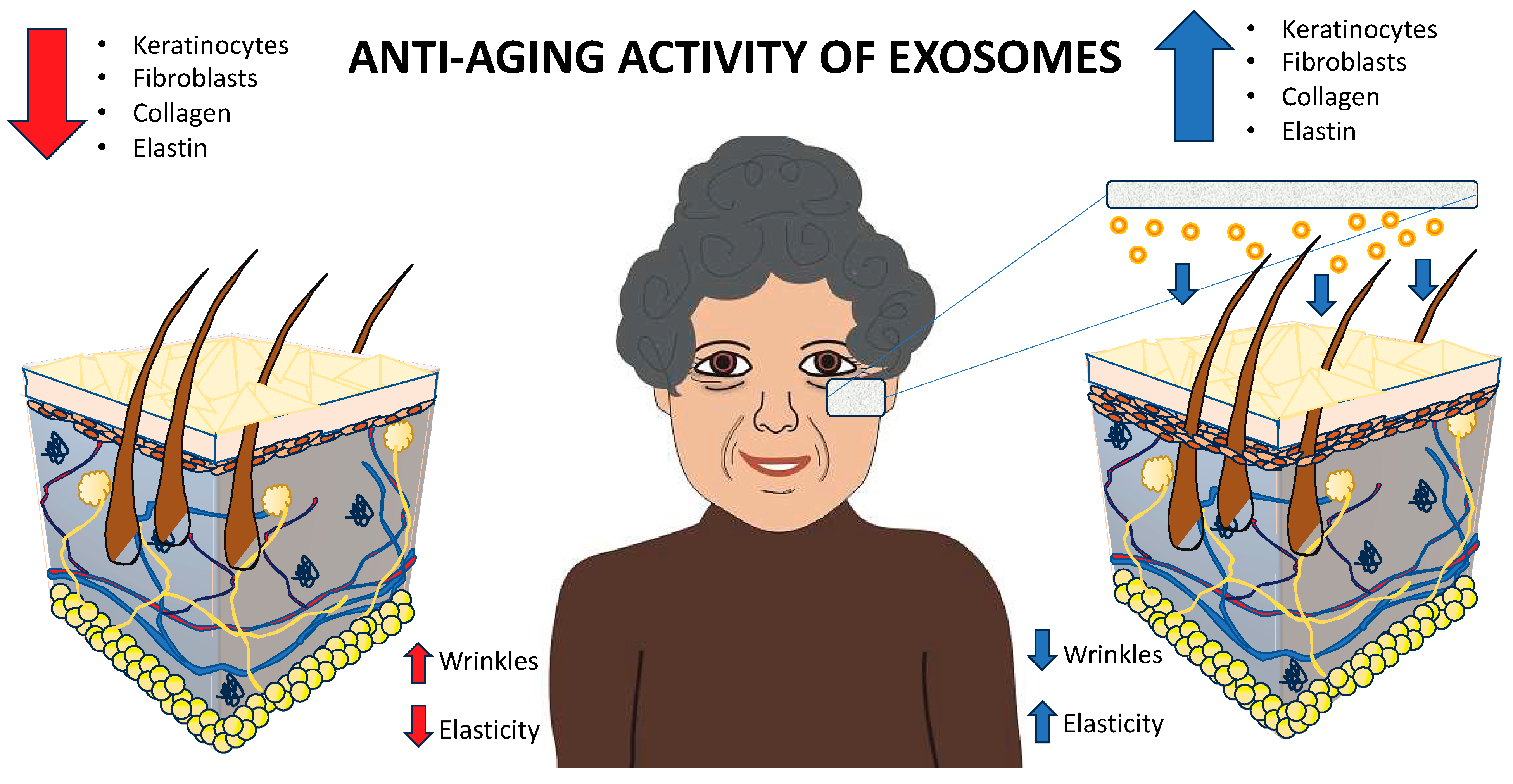
5.1.1. Geroprevention or Geroprotection (Anti-Aging)

Exosomes have the potential to attenuate signs of aging by stimulating cellular turnover and enhancing the skin’s intrinsic repair mechanisms. Additionally, they may alleviate oxidative stress, a major factor contributing to premature aging, by facilitating the delivery of bioactive molecules that promote cellular resilience and homeostasis [5]. As mentioned before, these small vesicular structures stimulate collagen and elastin production have gained attention in the field of skincare and anti-aging [15,51] (Figure 3).
Chen et al. obtained exosomes from mesenchymal stem cells which exhibited functional characteristics that closely resemble those of their parent cells, suggesting their potential to play a crucial role in tissue repair and regeneration. In that experimental approach using lipotransfer as a model, the exosomes were isolated from a conditioned medium of mouse adipose-derived stem cells and thoroughly characterized. Subsequently, minced fat tissue was combined with either exosomes, the source cells themselves (cell-assisted lipotransfer), or saline solution and implanted under the skin on the lower back of C57/BL mice in a bilateral manner (with 16 mice in each group). The transplanted fat tissues were collected and assessed at 3 and 10 weeks following the procedure. At both the 3-week and 10-week marks after the transplantation, the fat grafts in the exosome and cell-assisted lipotransfer groups displayed superior fat tissue integrity, fewer instances of oil cysts, and reduced levels of fibrosis. By the 10-week point, the retention rates of grafts in the cell-assisted lipotransfer group (50.9% ± 2.4%; p = 0.03) and the exosome group (56.4% ± 1.6%; p < 0.001) were significantly higher compared to the saline group (40.7% ± 4.7%). Further investigations into parameters such as macrophage infiltration, inflammatory factors, angiogenic factors, adipogenic factors, and extracellular matrix demonstrated that these cellular derivatives, promoted angiogenesis and boosted early-stage inflammation. In contrast, during the middle to late stages of fat grafting, they exhibited proadipogenic effects and increased collagen synthesis, much like their parent cells. Essentially, adipose-derived stem cell-derived exosomes exhibited effects comparable to their source cells in enhancing graft retention by upregulating early-stage inflammation and augmenting angiogenesis. These properties make exosomes a compelling cell-free alternative in the realm of therapeutic regenerative medicine [52].
Again, collagen is a structural protein that provides firmness and support to the skin. Increased collagen production can help reduce the appearance of wrinkles and fine lines [53]. Also, some of these vesicular structures may contain factors that encourage elastin production. Elastin is responsible for maintaining the skin’s elasticity, allowing it to return to its original shape after stretching. Enhanced elastin production can help the skin appear more supple and youthful [2].
Zhang et al. revealed that human induced pluripotent stem cell-derived mesenchymal stem cells (hiPSC-MSCs) have a positive impact on the development of granulation tissue and the promotion of angiogenesis, both of which are crucial stages in the wound-healing process. Furthermore, that research observed that hiPSC-MSC-Exos played a significant role in improving the outcome of cutaneous wound healing. Those findings indicate that hiPSC-MSC-Exos have the potential to be employed as therapeutic agents in the context of healing skin wounds [54].
On the other hand, exosomes can play a role in tissue repair, including the repair of damaged or aged skin. By facilitating the regeneration of skin cells and tissues, exosomes can contribute to the rejuvenation of the skin [55].
Cui et al. conducted an experiment in which they treated murine superficial flexor tendon cells with exosomes derived from mouse bone marrow-derived macrophages (BMDM-Exos). They observed increased proliferation and migration of these tenocytes in vitro [56].
Shi et al. employed CD146 to identify tendon stem cells (TSCs) and noted an accumulation of CD146+ cells at the injury site after treatment with BMSC-EVs, while this staining was not evident in other groups. Additionally, they found reduced signals of cleaved caspase-3 in the BMSC-EV group, suggesting a decrease in apoptotic cell death within the tendon [57].
One prevalent hypothesis regarding the mechanism behind exosome-induced tenogenesis revolves around TGF-β-dependent signaling [58]. Xu et al. discovered that tenocyte-derived exosomes (tenosomes) could induce tenogenic differentiation in mesenchymal stem cells in a dose-dependent manner and found that tenosomes contained higher levels of TGF-β compared to tenocytes. To support their findings, they applied a TGF-β signaling inhibitor (SB 431542) during exosomal treatment, which abolished the tenogenic effect on MSCs from tenosomes. They also suggested that TGF-β signaling played a crucial role in initiating the expression of scleraxis (SCX), an important tenocyte marker, in MSCs [58].
Consistently, Li et al. demonstrated that exosomes derived from bone marrow mesenchymal stem cells (BMSC-Exos) significantly increased the proliferation and migration of tenocytes. They also noted a substantial upregulation of TGF-β1 in BMSC-Exos compared to normal BM-MSCs. Inhibiting TGF-β1 signaling reversed the effects of enhanced cell proliferation, migration, and levels of tenogenic genes resulting from BMSC-Exo treatment [59].
Another study found that exosomes from tendon stem cells (TSC-Exo) contained a substantial amount of TGF-β and contributed to tendon healing through a TGF-β-dependent pathway. They revealed that TGF-β from exosomes activated the TGF-β-Smad2/3 signaling pathway and the extracellular signal-regulated kinase (ERK)1/2 signaling pathway in TSCs. The former pathway increased the expression of MMP2, while the latter was associated with tenocyte proliferation [60].
Liu et al. also published research demonstrating the activation of the SMAD2/3 signaling pathway by exosomes from adipose-derived stem cells (ADSC-Exos) in promoting tendon healing [61]. Interestingly, in a canine ex vivo model, the use of a purified exosome TISSEEL patch resulted in significantly higher amounts of tenocytes and improved biomechanical properties of the tendon, despite a reduction in the expression of TGF-β [62].
Exosomes that stimulate collagen and elastin production are being explored as a potential component of anti-aging skincare products and treatments. However, it is important to note that the effectiveness and safety of these products may vary, and more research is needed to fully understand their mechanisms and potential side effects [6].
In dermatology, exosomes have been explored for their potential to stimulate collagen and elastin production, which are essential for skin firmness and elasticity. By promoting cellular turnover and extracellular matrix (ECM) remodeling, exosomes may reduce visible signs of aging, such as wrinkles and fine lines. These effects position exosomes as a promising component of anti-aging therapies. However, the majority of evidence remains preliminary, relying on preclinical studies or theoretical mechanisms. Robust clinical trials are required to validate the efficacy, safety, and long-term outcomes of exosome-based dermatological products [14].
Several overarching limitations are evident across the discussed studies. First, most findings rely on in vitro or animal models, which, while informative, may not accurately predict clinical outcomes in humans. Second, the variability in exosome isolation, characterization, and delivery techniques poses significant reproducibility challenges. A standardized protocol is essential to ensure consistency in therapeutic outcomes. Third, while pathways such as TGF-β-Smad2/3 and ERK1/2 have been explored, the precise molecular mechanisms underlying exosome-induced regeneration remain incompletely understood. Fourth, the lack of large-scale clinical trials hinders the validation of exosome therapies, particularly regarding safety, dosage optimization, and long-term effects. Finally, regulatory hurdles surrounding exosome-based therapies delay their clinical adoption, necessitating clearer guidelines for their approval and application [58,59,60,63].
Exosomes exhibit immense promise as therapeutic agents in regenerative medicine, tendon repair, and dermatology, offering cell-free solutions that mimic the functional characteristics of their parent cells. The ability of exosomes to promote angiogenesis, modulate inflammation, and stimulate ECM remodeling highlights their versatility across tissue types. However, addressing the limitations mentioned—particularly standardization, clinical validation, and regulatory frameworks—is critical for advancing exosome-based therapies into clinical practice. Future research should focus on elucidating molecular mechanisms, optimizing isolation techniques, and conducting rigorous clinical trials to ensure safety, efficacy, and scalability of exosome treatments [12].

5.1.2. Hidroregulation (Skin Hydration)

Skin dehydration occurs when the skin lacks sufficient moisture, leading to dryness, flakiness, and discomfort [64]. Likewise, these extracellular vesicles can help the skin retain moisture by improving the skin’s natural barrier function. A healthy skin barrier prevents water loss, keeping the skin hydrated [65].
In the context of skin hydration, exosomes are at the forefront of research due to their ability to regulate skin homeostasis, restore the extracellular matrix (ECM), and modulate cellular communication in the dermal and epidermal layers [14].
The hydration of skin largely depends on the integrity of the ECM, the presence of essential proteins such as collagen, elastin, and hyaluronic acid, and the functional capacity of skin cells such as fibroblasts and keratinocytes [42]. Exosomes contribute to skin hydration through multiple mechanisms. They can stimulate the production of hyaluronic acid (HA), a glycosaminoglycan critical for water retention, by activating pathways such as TGF-β, MAPK/ERK, and PI3K/AKT. Hyaluronic acid binds water molecules to maintain skin moisture and elasticity. Additionally, exosomes promote fibroblast proliferation and migration, encouraging the production of collagen, elastin, and fibronectin. These ECM components play a crucial role in enhancing skin structure, reducing trans-epidermal water loss (TEWL), and improving hydration [15,66].
Bahr et al. revised the capacity of exosomes for facilitating cell-to-cell communication, as they carry a distinct cargo comprising proteins, RNA species, DNAs, and lipids intended for transportation and exchange between cells, both at systemic and local levels. They are integral to regulating normal physiological processes. Exosomes released from stem cells exhibit therapeutic effects similar to those of the cells they originate from. To make clinical use of exosomes feasible, it is necessary to prepare a sufficient quantity of viable, active therapeutic exosomes and establish effective methods for their long-term preservation, thereby expediting their clinical and commercial applications. Cryopreservation is the most commonly employed method for preserving perishable biomaterials [67].
Moreover, exosomes may support the repair and maintenance of the skin barrier, which is essential for preventing moisture from escaping and protecting the skin from external aggressors [6]. The skin’s natural processes for maintaining hydration, helping it achieve and maintain optimal moisture levels, can also be stimulated [68].
Yoo et al. assessed the potential of exosomes derived from dermal fibroblasts (DF) as a promising candidate for repairing skin damage. To investigate the impact of DF exosomes on safeguarding the skin’s permeability barrier, that study analyzed the expression levels of biomarkers associated with the maintenance of the skin’s permeability barrier in keratinocytes exposed to 1-chloro-2,4-dinitrobenzene (DNCB). They obtained exosomes from the conditioned media of fibroblast cells through a process of differential ultracentrifugation. When the fibroblast cells reached approximately 95% confluence in a 150-mm petri dish, cells were subjected to cycloheximide treatment (ranging from 5 to 100 μg/mL) to induce a stress response. The conditioned media collected after this treatment were then processed through differential centrifugation. Subsequently, the pellets containing the harvested exosomes were separated, collected from the supernatant obtained through ultracentrifugation, resuspended in phosphate-buffered saline, and stored at −80 °C for further analysis. The characterization of the exosomes harvested from the conditioned media of fibroblast cell cultures was carried out using nanoparticle tracking analysis (NTA). NTA allowed us to determine both the number and size of the isolated exosomes. The mean particle size of the isolated fibroblast exosomes was measured at 215.4 ± 116.1 nm, with a concentration of 2.33 × 10¹⁰ ± 2.22 × 10⁹ particles/mL [69].
Some exosomes have anti-inflammatory properties, which can help soothe irritated and dehydrated skin, reducing redness and discomfort [70]. While these cell-derived products hold promise for skin hydration, it is essential to understand that the effectiveness of exosome-based skincare products may vary, and more research is needed to fully comprehend their mechanisms and potential side effects. It is recommended that individuals with dehydrated skin ensure any exosome-based skincare products they use comply with local regulatory standards [71].
Another mechanism involves the reduction of oxidative stress, which is a major contributor to skin aging and dehydration. Exosomes contain antioxidant enzymes, such as superoxide dismutase (SOD) and glutathione peroxidase (GPX), as well as bioactive microRNAs (miRNAs) that reduce reactive oxygen species (ROS). By mitigating oxidative stress, exosomes help maintain the functional integrity of skin cells and their hydration. Furthermore, exosomes possess immunomodulatory properties, suppressing pro-inflammatory cytokines like IL-6 and TNF-α while enhancing anti-inflammatory responses. This regulation restores skin barrier function, which is essential for retaining moisture and preventing water loss [72].
In addition to their intrinsic bioactive properties, exosomes can act as carriers for delivering hydration-enhancing molecules such as hyaluronic acid, ceramides, and peptides directly to the skin. This targeted delivery improves hydration and ensures deeper penetration compared to conventional skincare formulations. These findings have led to the exploration of exosome integration into advanced cosmeceuticals, such as creams, serums, and masks, designed to rejuvenate the skin and address dehydration [17].
The most extensively studied sources of exosomes for skin hydration are mesenchymal stem cells (MSCs), particularly those derived from adipose tissue (ADSC-Exos) and bone marrow (BMSC-Exos). Exosomes from ADSCs are rich in growth factors, cytokines, and miRNAs that stimulate dermal fibroblasts to produce hyaluronic acid and collagen while improving skin barrier integrity. BMSC-derived exosomes, on the other hand, are known for their ability to promote collagen synthesis and regulate ECM-related gene expression, enhancing both hydration and elasticity. Recently, epidermal stem cell-derived exosomes have also shown promise by promoting keratinocyte proliferation and differentiation, which is essential for maintaining a strong skin barrier. Emerging research on plant- and algae-derived exosomes suggests they could be a sustainable source of hydration-enhancing molecules, though this remains largely experimental [73].
Despite the promising results, challenges remain in the application of exosomes for skin hydration. One key limitation is the lack of standardization in exosome production, isolation, and characterization, which impacts reproducibility and scalability for clinical and commercial use. Ensuring the efficient delivery of exosomes to deeper skin layers is another challenge, although novel nanocarrier-based systems such as liposomes and hydrogels are being developed to improve penetration. Furthermore, while preclinical data demonstrate significant potential, clinical trials evaluating the long-term safety and efficacy of exosome-based hydration therapies are still limited. Regulatory hurdles also pose barriers to commercialization, as exosomes are classified as biological products that require stringent approval processes [74].
Hence, exosomes, particularly those derived from MSCs, represent a promising solution for improving skin hydration through ECM remodeling, hyaluronic acid synthesis, and oxidative stress reduction. Their incorporation into skincare formulations represents an innovative approach to addressing skin dryness and maintaining hydration. However, further efforts are needed to standardize production, develop efficient delivery systems, and conduct robust clinical trials to validate their safety and efficacy. As research advances, exosomes have the potential to revolutionize dermatology and cosmeceuticals, offering a natural and bioactive solution for hydrated, youthful skin [75].

5.1.3. Facial Dyschromia (Skin Pigmentation)

Exosomes may contain factors that can help reduce skin pigmentation issues, such as dark spots or uneven skin tone. They can promote a more even distribution of melanin and support skin brightening [7]. These vesicles can play a role in addressing pigmentation issues and uneven skin tone. Pigmentation refers to the color of the skin, and when it is uneven, it can result in the appearance of dark spots, freckles, or blemishes [76].
Exosomes have the capacity to regulate melanin, the pigment responsible for skin color. They can potentially mitigate the excessive production of melanin in specific areas, resulting in a more consistent skin tone. Some exosomes contain elements that facilitate skin brightening. Additionally, certain exosomes possess anti-inflammatory properties, which can be soothing for irritated or reddened skin areas, further contributing to a more uniform skin tone. Furthermore, exosomes offer the potential for customization to meet individual skin needs, thereby providing personalized solutions for addressing specific pigmentation concerns [4,76,77].
It is important to note that while exosomes show promise in managing pigmentation and uneven skin tone, more research is needed to fully understand their mechanisms and potential side effects [29,78]. The exploration of exosomes in dermatology, particularly for regulating hyperpigmentation and hypopigmentation, is advancing as researchers elucidate their mechanisms and therapeutic applications [79].
The pigmentation of skin is primarily determined by melanin, a pigment synthesized in melanocytes within the basal layer of the epidermis. Exosomes derived from keratinocytes, fibroblasts, and melanocytes are known to influence melanogenesis through intercellular signaling pathways. For instance, keratinocyte-derived exosomes have been shown to transfer molecules such as prostaglandin E2 (PGE2) and microRNAs (e.g., miR-330-5p), which promote melanin production in melanocytes. These exosomes play a central role in responding to external stimuli like ultraviolet (UV) radiation, which induces the release of exosomes carrying pro-melanogenic factors to regulate pigmentation as part of the skin’s photoprotective response [80].
In contrast, fibroblast-derived exosomes influence melanogenesis through growth factors and cytokines. For example, exosomes from dermal fibroblasts may carry stem cell factor (SCF), TGF-β, and bFGF, which can stimulate melanin synthesis and melanosome transfer by activating pathways such as MITF (microphthalmia-associated transcription factor) and ERK/MAPK signaling. These pathways are critical regulators of melanocyte proliferation and melanogenic enzyme activity, including tyrosinase, which is the rate-limiting enzyme in melanin synthesis. Interestingly, exosomes derived from aged fibroblasts may contribute to skin pigmentation disorders, such as senile lentigines or age spots, due to altered molecular content that dysregulates melanin synthesis [81].
Conversely, exosomes are also being investigated for their depigmenting properties to treat hyperpigmentation disorders like melasma and post-inflammatory hyperpigmentation. For instance, exosomes derived from mesenchymal stem cells (MSCs), particularly those from adipose tissue (ADSC-Exos) and bone marrow (BMSC-Exos), demonstrate anti-melanogenic effects by suppressing melanocyte activity. These exosomes often carry bioactive components, such as miRNAs (e.g., miR-145) and anti-inflammatory cytokines, that downregulate melanin production by inhibiting key melanogenic pathways, including MITF signaling and tyrosinase activity. Studies suggest that MSC-derived exosomes exert antioxidant effects, mitigating oxidative stress caused by UV exposure—a known driver of hyperpigmentation—thereby preventing excessive melanogenesis [82].
In addition to their natural functions, exosomes have been engineered to deliver depigmenting agents, such as kojic acid, arbutin, and other tyrosinase inhibitors, directly to melanocytes. These engineered exosomes enhance therapeutic efficiency by providing targeted delivery and improved bioavailability of depigmenting molecules compared to traditional formulations. This approach is being explored as a novel, minimally invasive alternative for managing pigmentation disorders [83].
Exosome research has also expanded into understanding hypopigmentation disorders such as vitiligo, where melanocyte loss leads to depigmented skin patches. Studies indicate that exosomes derived from melanocytes or stem cells could play a role in restoring pigmentation by promoting melanocyte proliferation, migration, and repopulation of the depigmented areas. Exosomal cargo, such as pro-melanogenic miRNAs and growth factors, helps regenerate melanocyte function and counteract autoimmune responses often associated with vitiligo [84].
Despite promising results, significant challenges remain in applying exosomes for skin pigmentation regulation. A major limitation lies in the heterogeneity of exosome content, which can vary depending on the parent cell type, cell condition, and isolation techniques. Additionally, the mechanisms underlying exosome-mediated pigmentation are complex and not yet fully understood, requiring further elucidation through rigorous molecular studies. Standardization of exosome production, characterization, and delivery is crucial to ensure reproducibility and efficacy. Furthermore, while preclinical studies have demonstrated notable outcomes, clinical trials assessing the safety, stability, and long-term effects of exosome-based therapies for pigmentation disorders are still in early stages [85].
For all the above, exosomes represent an innovative approach for modulating skin pigmentation, offering potential treatments for both hyperpigmentation and hypopigmentation disorders. Their ability to regulate melanogenesis, deliver bioactive molecules, and repair pigmentation-related skin damage highlights their promise in therapeutic dermatology and cosmetic applications. However, to fully harness their potential, ongoing research must address existing challenges related to standardization, delivery, and clinical validation. As advancements continue, exosome-based therapies could provide a transformative solution for managing skin pigmentation disorders in a safe, effective, and targeted manner [86].

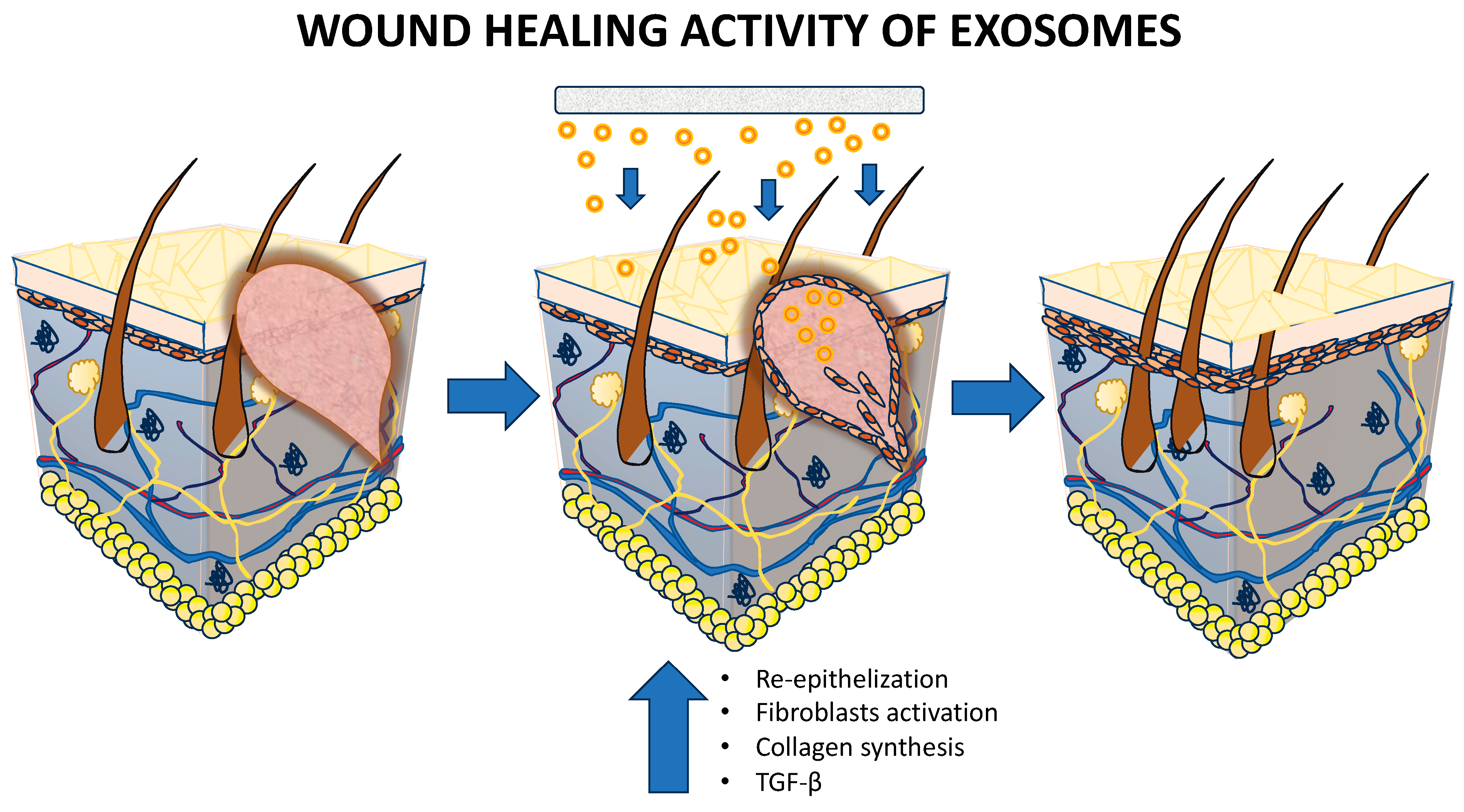
5.1.4. Wound Healing

Exosomes have gained significant attention in the field of wound healing due to their potential therapeutic applications. In cosmetics, they may be used in products aimed at reducing the appearance of scars or promoting the healing of minor skin blemishes [2]. The structural components play a crucial role in intercellular communication and have been investigated for their regenerative properties in wound healing [27] (Figure 4).
Zhao et al. discussed the utilization of mesenchymal stem cell-derived exosomes. These small vesicles constitute the principal bioactive extracellular vesicles responsible for the paracrine influence of mesenchymal stem cells (MSCs). The merits of MSC-exosomes encompass their capability to stimulate angiogenesis, encourage cell proliferation, boost collagen production, modulate inflammatory responses, and ultimately enhance the regenerative potential of tissues [27].
Nevertheless, there are significant impediments to the clinical application of MSC-exosomes, including issues related to their precise targeting and their susceptibility to removal from the wound site. In response to these challenges, the concept of bioengineering technology has been introduced to customize exosomes [27].
Hence, exosomes derived from certain cell types, like mesenchymal stem cells (MSCs), can have anti-inflammatory properties. Inflammation is a natural response to tissue injury, but excessive or prolonged inflammation can hinder the healing process. Also, exosomes can modulate the immune response, reducing inflammation and promoting a more favorable environment for wound healing [87].
Buschow et al. found that exosomes originating from immune cells have the capacity to carry vital molecules required for triggering immune responses, facilitating antigen presentation, and enabling the exchange of membrane or cytosolic components between cells, even when these cells are not physically close to each other (as described in reference [88]). Furthermore, the specific proteins packaged within these cellular derivates can influence their target receptors and the nature of immune cell activation. Recent research has not only underscored the immune-related functions of dendritic cell (DC)-derived exosomes but has also unveiled their potential in exerting an anti-tumor effect in addition to their recognized roles in immune responses [89].
Angiogenesis is another process promoted by the exosomes, which is crucial for supplying oxygen and nutrients to the wounded area. This helps in tissue regeneration and accelerates the healing process [70].
Exosomes can stimulate the proliferation of various cell types involved in wound healing, including fibroblasts, which are responsible for producing collagen and rebuilding tissue at the wound site [9]. Similarly, exosomes can influence the remodeling of the extracellular matrix, which is essential for proper tissue repair. They can modulate the synthesis and degradation of matrix components, such as collagen and elastin [15].
Prasai et al. performed 51 studies involving rodents studying the role of exosomes in dermal wound healing. The results of the analysis indicated that the therapeutic effects of exosomes were most pronounced at the 7-day mark (with an odds ratio of 1.82 and a 95% confidence interval of [0.69, 2.95]) as compared to the 14-day point (with an odds ratio of 2.29 and a 95% confidence interval of [0.01, 4.56]) following administration. It was observed that exosomes played a regulatory role in all stages of skin wound healing, primarily through the actions of circulating microRNA. The findings from this study can serve as valuable guidance for the design of both pre-clinical and clinical studies focusing on the contributions of exosomes to the process of wound healing [9].
On the other hand, exosomes can contribute to wound closure and contraction, which is crucial for healing in chronic or large wounds. They can enhance the migration and contractile ability of cells involved in wound closure, such as keratinocytes and myofibroblasts [90]. Moreover, they can carry specific microRNAs and growth factors that promote tissue regeneration. These factors can stimulate the differentiation of progenitor cells and the repair of damaged tissues [91]. Finally, they have the potential to reduce scar formation by modulating collagen production and the inflammatory response during wound healing [92].
Research into exosome-based therapies for wound healing is ongoing, and clinical trials are being conducted to assess their safety and efficacy. While the therapeutic potential of exosomes in wound healing is promising, there are still challenges to overcome, such as standardizing isolation methods and understanding the best sources of exosomes. Nevertheless, these small vesicles hold great potential for improving the outcomes of various wound healing applications, including chronic wounds, burns, and surgical incisions [93].
When comparing the studies, it becomes clear that exosomes exhibit multifaceted mechanisms of action across different biological systems (Table 3). Zhao et al. and Prasai et al. both focus on exosome-mediated regenerative processes, particularly angiogenesis, extracellular matrix remodeling, and inflammation modulation [9,27]. However, Prasai et al., add a statistical and temporal dimension, providing evidence of the timeline for exosome efficacy in wound healing. On the other hand, Buschow et al., expand the scope by focusing on immune-specific roles, demonstrating that exosomes not only regulate immune responses but also possess anti-tumor capabilities. This highlights the unique contributions of exosomes depending on their cellular origin [88].
All the studies reported in this section emphasize the therapeutic potential of exosomes in regenerative medicine, immune modulation, and anti-tumor applications. While MSC-derived exosomes excel in promoting tissue repair and inflammation resolution, immune cell-derived exosomes showcase broader immunological functions, including antigen presentation and tumor suppression. Prasai et al.’s meta-analysis strengthens the evidence base for exosomes in wound healing, offering pre-clinical insights into their regulatory role across all stages of tissue repair [9]. Despite their promising potential, challenges such as precise targeting, retention, and clinical standardization remain, underscoring the need for further research to unlock the full therapeutic capabilities of exosomes.

5.1.5. Delivery of Biomolecules

Exosomes can serve as delivery vehicles for other active ingredients in skincare products. They can encapsulate and transport bioactive molecules, ensuring their targeted delivery to skin cells [8].
Exosomes have emerged as promising drug delivery systems due to their unique properties and capabilities [13]. Since exosomes are cell-derived and typically well-accepted by the body, they serve as a biocompatible choice for drug delivery [55]. Also, exosomes can be engineered to carry specific drugs or therapeutic molecules. They can also be modified to target particular cell types or tissues, improving the precision of drug delivery [94]. Another advantage is that exosomes help protect their cargo, such as drugs or genetic material, from degradation. This stability is especially valuable for delivering sensitive drugs [87].
Exosomes have a natural ability to home in on specific cell types, making them effective for delivering drugs to particular target sites [13]. Nevertheless, they are less likely to trigger an immune response when compared to synthetic drug delivery systems, reducing the risk of adverse reactions [95]. These cellular derivatives can readily fuse with target cells, enabling efficient delivery of their cargo [96] and the ability to cross biological barriers, such as the blood–brain barrier, which poses a challenge for some drug delivery systems [97].
Exosomes can be isolated from a patient’s own cells, customized to carry specific therapeutic agents, and then reintroduced into the patient. This personalized approach holds promise for tailored treatments [98]. Targeted delivery and minimized off-target effects can reduce the side effects associated with traditional drug delivery methods [99].
Exosomes have shown potential in delivering a wide range of therapeutic agents, including small molecules, RNA, DNA, and proteins, for the treatment of various diseases, including cancer, neurodegenerative disorders, and cardiovascular conditions [100]. On the other hand, exosomes can be loaded in electrospun nanofibers which are excellent drug delivery systems for local drug release [101,102]. While exosomes hold great promise as drug delivery systems, there are still challenges to overcome, such as scalability, standardization of production, and regulatory considerations. Researchers are actively exploring and developing exosome-based therapies, and clinical trials are ongoing to assess their safety and efficacy in various medical applications [103].
The studies together highlight the versatility of exosomes as delivery vehicles for various bioactive molecules and therapeutic agents, underscoring their potential applications across skincare, drug delivery, and localized treatments. In the context of skincare products, exosomes serve as efficient carriers for active ingredients by encapsulating and delivering them directly to skin cells. This targeted delivery improves bioavailability and ensures stability, particularly for bioactive molecules sensitive to environmental degradation. The ability of exosomes to transport their cargo while maintaining its efficacy presents a significant advantage over traditional topical formulations, which often struggle with limited penetration and stability [104].

5.1.6. Customized Dermatology

In the future, personalized skincare could involve the use of exosomes derived from a person’s own cells to address specific skin issues effectively [4], in the past few years regenerative exosomes have been explored for their potential in personalized skincare and the production of various compounds of cosmetic interest [30].
Frazier et al. discussed that both acute and chronic skin wounds resulting from burns, pressure injuries, and trauma present a significant healthcare challenge, especially for older individuals and those with paraplegia or quadriplegia around the world. However, the existing standard of care heavily relies on preventive strategies for pressure injuries, surgical removal of damaged tissue, skin flap procedures, and the use of negative pressure wound vacuums. That study underscores the potential of products derived from adipose tissue, blood, and cellulose, including cells, decellularized matrices and scaffolds, as well as exosomes and secretome factors, to address this unmet medical need [11].
Exosomes can be isolated from specific cell sources or conditioned to carry specific bioactive molecules, making them well-suited for addressing individual skincare concerns. For example, exosomes derived from stem cells can be tailored to promote skin rejuvenation, hydration, or address pigmentation issues [105].
Exosomes have the capability to aid in the transportation of active ingredients into the skin. They can serve as carriers and guardians for a variety of skincare compounds, guaranteeing their penetration into deeper layers of the skin, thereby enhancing their efficacy. Exosomes can also potentially stimulate the production of collagen and foster the regeneration of skin cells, which, in turn, can help diminish the appearance of fine lines and wrinkles, ultimately resulting in a more youthful skin appearance. Furthermore, by encouraging the turnover of skin cells and the removal of older ones, exosomes can play a role in achieving a smoother and more uniform skin texture. Additionally, exosomes can contribute to enhancing skin hydration by supporting the natural barrier function of the skin, which aids in reducing moisture loss and promoting a healthy and plump complexion [30,87,106].
Some exosomes contain anti-inflammatory properties, which can be beneficial for soothing sensitive or irritated skin, reducing redness and discomfort. They can also potentially be customized based on an individual’s unique skin needs, offering a tailored approach to address specific concerns [107].
It is important to note that while exosomes show promise in customized skincare, more research and clinical studies are needed to fully understand their mechanisms, potential side effects, and long-term benefits. Skincare products containing exosomes should meet regulatory standards, and it is advisable to consult with a dermatologist or skincare professional to determine the most suitable products for individual skincare concerns [108].
Hence, exosomes have numerous applications besides cosmetics applications, some examples are given in Table 4.

5.1.7. Clinical Validation

While preclinical studies have shown impressive results, clinical validation remains a critical step to ensure their safety, efficacy, and practical utility in the cosmetic industry [114].
Recent advancements have led to the incorporation of exosomes derived primarily from mesenchymal stem cells (MSCs) and other cell types into skincare products [17]. These exosomes have been evaluated for their anti-aging, anti-inflammatory, and regenerative effects in human clinical studies. For instance, clinical trials involving adipose-derived stem cell exosomes (ADSC-Exos) have demonstrated significant improvements in wrinkle reduction [115], skin hydration [116], and overall skin texture [52]. In one clinical study, a cream containing ADSC-Exos was applied to participants for 4–8 weeks, resulting in increased skin moisture levels, reduced transepidermal water loss (TEWL), and improved elasticity due to enhanced collagen production. These outcomes highlight exosomes’ capacity to promote dermal fibroblast activation and extracellular matrix (ECM) remodeling [117].
Similarly, exosomes derived from human umbilical cord MSCs have shown notable effects in reducing skin aging markers. Clinical evaluations revealed improved skin tone, reduced fine lines, and increased elasticity following topical application. These effects are attributed to the exosomal delivery of growth factors (e.g., VEGF, TGF-β) and anti-oxidative miRNAs, which support skin repair and protect against oxidative stress caused by UV exposure—a significant factor in photoaging [14,118].
One of the most validated applications of exosomes in cosmetics is their role in skin hydration. Exosomes enhance keratinocyte proliferation, improve the function of the skin barrier, and reduce TEWL, which helps retain moisture. Clinical studies utilizing exosome-based serums and creams have demonstrated increased hydration levels and improved barrier integrity within 2–4 weeks of consistent use. This is particularly promising for individuals with dry or sensitive skin, as exosomal treatment can restore hydration without the irritation commonly associated with synthetic skincare additives [54].
Clinical studies have also explored exosomes’ role in controlling hyperpigmentation. Topical formulations containing exosomes derived from MSCs and keratinocytes have shown promise in reducing melanin synthesis and correcting uneven skin tone. For instance, a clinical trial using MSC-derived exosome creams reported a decrease in hyperpigmented lesions and an improvement in skin radiance within 8 weeks. These depigmenting effects are associated with the suppression of MITF signaling and downregulation of melanogenic enzymes like tyrosinase. While encouraging, further trials are needed to validate exosomes’ long-term efficacy and safety for pigmentation disorders [119].
Despite promising results, clinical validation of exosome-based cosmetic products faces several challenges. First, standardization in exosome isolation, characterization, and delivery remains a major hurdle. Variability in exosome quality due to differences in cell sources, isolation methods, and storage conditions can impact clinical outcomes. Reproducibility of results is crucial for regulatory approval, yet studies often lack consistency in exosome dose, duration of treatment, and endpoint evaluations [120].
Second, while early clinical trials demonstrate safety and efficacy, most studies have been conducted on small sample sizes and over relatively short durations (4–12 weeks). Large-scale, double-blind, placebo-controlled clinical trials are essential to confirm the long-term benefits and identify any potential side effects of exosome-based treatments. Additionally, regulatory bodies such as the FDA and EMA currently lack comprehensive guidelines for exosome-based cosmetic products, which can slow their clinical translation [121].
The clinical validation of exosomes in cosmetics also faces regulatory scrutiny due to their biological origin and therapeutic-like properties. Unlike conventional skincare products, exosomes blur the line between cosmetics and biologics, raising concerns regarding their classification, safety testing, and approval processes. Furthermore, ethical issues surrounding the use of human-derived exosomes, particularly from embryonic or fetal tissue sources, require careful consideration and transparency to ensure public acceptance [122].
To advance the clinical validation of exosome-based cosmetics, researchers must focus on improving manufacturing standardization, enhancing delivery systems, and conducting robust clinical trials. The use of engineered exosomes, tailored to deliver specific bioactive molecules for targeted skin benefits, represents a future direction with significant potential. Additionally, the development of plant- or synthetic-derived exosome mimetics offers a promising alternative that may overcome ethical and regulatory concerns while retaining functional efficacy [123].
Hence, exosomes hold immense promise in the cosmetic industry for improving skin hydration, pigmentation, and anti-aging outcomes. While clinical trials have demonstrated encouraging results, particularly for MSC-derived exosome formulations, large-scale, standardized studies are needed to solidify their efficacy and safety. Overcoming challenges related to standardization, regulation, and public acceptance will be critical for the widespread adoption of exosome-based cosmetic therapies, positioning them as a transformative innovation in skincare.

6. Limitations

Ku et al., affirmed that the use of exosomes in the medical field is gaining increasing attention and represents a growing area of interest in the realm of plastic surgery. However, it is important to note that no exosome-based products have received FDA approval, despite some manufacturers indicating “pending status” for approval concerning topical and intravenous infusion-based modalities. There is a clear need for further clinical studies to establish the impact, benefits, effectiveness, outcomes, and safety profile of exosomes in the context of plastic surgery [10].
Some noteworthy limitations that deserve discussion are presented. To begin with, it is crucial to clarify that this is not a systematic review, and thus, it does not adhere to strict inclusion or exclusion criteria. The majority of the studies covered in this review are preclinical in nature, underscoring the limited available literature on exosome application within the plastic surgery community. Clinical reports that have been published are typically based on small groups of patients and often provide anecdotal evidence, likely because of the absence of FDA approval. Furthermore, these clinical studies lack substantial evidence and do not delve into potential or observed adverse effects in depth. The heterogeneity among these clinical studies arises from variations in exosome source cells and different purification methods, making direct comparisons challenging. Lastly, there is currently no published evidence available regarding the long-term outcomes of exosome usage [10,124].
Skin wound healing continues to pose a significant challenge for healthcare systems worldwide. Although most wounds heal promptly and completely with current standard-of-care methods, a significant subgroup of patients with impaired angiogenesis and vascularization remains at elevated risk of developing chronic wounds. These chronic wounds can lead to serious complications, including infection, sepsis, and osteomyelitis. Various approaches, such as adipose tissue and blood-derived cells, exosomes, hydrogels, and plant-derived cellulose, hold promise for improving wound therapy [11].

7. Conclusions

In conclusion, exosomes have emerged as a focal point of interest in the cosmetics and skincare industry due to their remarkable potential for enhancing skin health and rejuvenation. These naturally produced vesicles, originating from skin cells, contain bioactive molecules crucial for cellular communication and tissue repair, thereby offering a wide array of skincare applications. These applications encompass skin rejuvenation, anti-aging, hydration, pigmentation correction, wound healing, and customized skincare, all of which have been explored in recent research. Moreover, exosomes, as a form of cell-based therapy, demonstrate promise in facilitating skin regeneration, with their therapeutic benefits stemming from growth factors and vesicular entities. A comprehensive review aims to establish exosomes as a prototype for the biological treatment of dermal wounds, offering insights into optimal concentrations for pre-clinical investigations. The quest for non-surgical aesthetic solutions has led to the prominence of exosomes in both topical and injectable applications. Their regenerative properties have garnered attention in dermatology, anti-aging medicine, and, more recently, in the field of plastic surgery, even though the number of published studies in this domain is relatively limited. These studies are shedding light on the potential benefits and applications of exosomes in various facets of aesthetics. Challenges, however, persist in the use of exosomes, particularly in the context of their integration into the medical field. These challenges include logistical hurdles associated with the subcutaneous injection of autologous or allogeneic cells, which influence skin regeneration but require careful cell isolation, cryopreservation, and adherence to regulatory criteria. The development of stable, freeze-dried exosome products is an attractive prospect, but it necessitates extensive evaluation, as exosomes alone may not encompass the full range of benefits provided by extracellular proteins. Combining exosomes with hydrogels derived from different sources, whether as cryopreserved injectable products or lyophilized topical formulations, may be necessary. However, the approval of such products, whether used individually or in combination, will likely require randomized, controlled clinical trials. While the potential of exosomes in the field of plastic surgery is gaining attention, it is essential to note that no exosome-based products have secured FDA approval as of yet. Although some manufacturers indicate “pending status” for approval, further clinical studies are warranted to fully elucidate the impact, benefits, effectiveness, outcomes, and safety profile of exosomes in the context of plastic surgery. In the realm of skin wound healing, the challenge persists as a burden on healthcare systems worldwide. While many wounds respond well to standard-of-care approaches, a significant subset of patients with impaired angiogenesis and vascularization faces the risk of chronic wounds and related complications. Promising avenues, including adipose tissue and blood-derived cells, exosomes, hydrogels, and plant-derived cellulose, offer hope for improved wound therapy. Noteworthy limitations in the existing body of research should be acknowledged, including the predominance of preclinical studies and small patient cohorts in clinical reports. The field faces challenges arising from the heterogeneity in exosome source cells and purification methods. Furthermore, there is a dearth of long-term outcome data. Therefore, the continued exploration and rigorous evaluation of exosome-based approaches in various medical and aesthetic contexts are essential to unlock their full potential and realize their benefits for patients and skincare enthusiasts.

Author Contributions

L.J.V.-G.: conceptualization, research, writing—original draft, writing: proofreading and editing, formal analysis, research, project administration. S.O.-L.: Validation, resources, supervision, project administration, acquisition of funds. D.A.H.-H.: research, formal analysis. G.L.P.-G.: Formal analysis, research, conceptualization, research, writing—original draft, writing: proofreading and editing, project administration. All authors have read and agreed to the published version of the manuscript.

Funding

Authors thanks to the administration of the Facultad de Ciencias de la Ingeniería y Tecnología, Universidad Autónoma de Baja California, for the economical support.

Conflicts of Interest

The authors declare no conflict of interest.

Abbreviations

CCR2: CC chemokine receptor 2; CCL2, CC chemokine ligand 2; CCR5, CC chemokine receptor 5; TLC, thin layer chromatography; Exo: Exosomes; GM-CSF: Granulocyte-macrophage colony-stimulating factor; LPS: Lipopolysaccharides.

References

  1. Cho, B.S.; Irvine Duncan, D. Perspective Chapter: Development of Exosomes for Esthetic Use. In Physiology; Saheera, S., Ed.; IntechOpen: London, UK, 2023; Volume 20, ISBN 978-1-83768-949-1. [Google Scholar]
  2. Zhang, Y.; Liu, Y.; Liu, H.; Tang, W.H. Exosomes: Biogenesis, Biologic Function and Clinical Potential. Cell Biosci. 2019, 9, 19. [Google Scholar] [CrossRef]
  3. Ha, D.H.; Kim, H.; Lee, J.; Kwon, H.H.; Park, G.-H.; Yang, S.H.; Jung, J.Y.; Choi, H.; Lee, J.H.; Sung, S.; et al. Mesenchymal Stem/Stromal Cell-Derived Exosomes for Immunomodulatory Therapeutics and Skin Regeneration. Cells 2020, 9, 1157. [Google Scholar] [CrossRef] [PubMed]
  4. Bondhopadhyay, B.; Sisodiya, S.; Alzahrani, F.A.; Bakhrebah, M.A.; Chikara, A.; Kasherwal, V.; Khan, A.; Rani, J.; Dar, S.A.; Akhter, N.; et al. Exosomes: A Forthcoming Era of Breast Cancer Therapeutics. Cancers 2021, 13, 4672. [Google Scholar] [CrossRef]
  5. Li, X.; Zhang, H.; Wang, X.; Lu, M.; Ding, Q.; Chen, A.F.; Xiang, M.; Chen, S. iPSC-Derived Exosomes Promote Angiogenesis in Naturally Aged Mice. Aging 2023, 15, 5854. [Google Scholar] [CrossRef] [PubMed]
  6. Yang, G.H.; Lee, Y.B.; Kang, D.; Choi, E.; Nam, Y.; Lee, K.H.; You, H.-J.; Kang, H.J.; An, S.H.; Jeon, H. Overcome the Barriers of the Skin: Exosome Therapy. Biomater. Res. 2021, 25, 22. [Google Scholar] [CrossRef] [PubMed]
  7. Benito-Martínez, S.; Zhu, Y.; Jani, R.A.; Harper, D.C.; Marks, M.S.; Delevoye, C. Research Techniques Made Simple: Cell Biology Methods for the Analysis of Pigmentation. J. Investig. Dermatol. 2020, 140, 257–268.e8. [Google Scholar] [CrossRef] [PubMed]
  8. Samal, S.; Dash, P.; Dash, M. Drug Delivery to the Bone Microenvironment Mediated by Exosomes: An Axiom or Enigma. Int. J. Nanomed. 2021, 16, 3509–3540. [Google Scholar] [CrossRef] [PubMed]
  9. Prasai, A.; Jay, J.W.; Jupiter, D.; Wolf, S.E.; El Ayadi, A. Role of Exosomes in Dermal Wound Healing: A Systematic Review. J. Investig. Dermatol. 2022, 142, 662–678.e8. [Google Scholar] [CrossRef]
  10. Ku, Y.C.; Omer Sulaiman, H.; Anderson, S.R.; Abtahi, A.R. The Potential Role of Exosomes in Aesthetic Plastic Surgery: A Review of Current Literature. Plast. Reconstr. Surg.-Glob. Open 2023, 11, e5051. [Google Scholar] [CrossRef] [PubMed]
  11. Frazier, T.; Alarcon, A.; Wu, X.; Mohiuddin, O.A.; Motherwell, J.M.; Carlsson, A.H.; Christy, R.J.; Edwards, J.V.; Mackin, R.T.; Prevost, N.; et al. Clinical Translational Potential in Skin Wound Regeneration for Adipose-Derived, Blood-Derived, and Cellulose Materials: Cells, Exosomes, and Hydrogels. Biomolecules 2020, 10, 1373. [Google Scholar] [CrossRef] [PubMed]
  12. Manzoor, T.; Farooq, N.; Sharma, A.; Shiekh, P.A.; Hassan, A.; Dar, L.A.; Nazir, J.; Godha, M.; Sheikh, F.A.; Gugjoo, M.B.; et al. Exosomes in Nanomedicine: A Promising Cell-Free Therapeutic Intervention in Burn Wounds. Stem Cell Res. Ther. 2024, 15, 355. [Google Scholar] [CrossRef] [PubMed]
  13. Butreddy, A.; Kommineni, N.; Dudhipala, N. Exosomes as Naturally Occurring Vehicles for Delivery of Biopharmaceuticals: Insights from Drug Delivery to Clinical Perspectives. Nanomaterials 2021, 11, 1481. [Google Scholar] [CrossRef]
  14. Hajialiasgary Najafabadi, A.; Soheilifar, M.H.; Masoudi-Khoram, N. Exosomes in Skin Photoaging: Biological Functions and Therapeutic Opportunity. Cell Commun. Signal. 2024, 22, 32. [Google Scholar] [CrossRef] [PubMed]
  15. Tienda-Vázquez, M.A.; Hanel, J.M.; Márquez-Arteaga, E.M.; Salgado-Álvarez, A.P.; Scheckhuber, C.Q.; Alanis-Gómez, J.R.; Espinoza-Silva, J.I.; Ramos-Kuri, M.; Hernández-Rosas, F.; Melchor-Martínez, E.M.; et al. Exosomes: A Promising Strategy for Repair, Regeneration and Treatment of Skin Disorders. Cells 2023, 12, 1625. [Google Scholar] [CrossRef] [PubMed]
  16. Lotfy, A.; AboQuella, N.M.; Wang, H. Mesenchymal Stromal/Stem Cell (MSC)-Derived Exosomes in Clinical Trials. Stem Cell Res. Ther. 2023, 14, 66. [Google Scholar] [CrossRef] [PubMed]
  17. Yoo, K.; Thapa, N.; Chwae, Y.; Yoon, S.; Kim, B.; Lee, J.; Jang, Y.; Kim, J. Transforming Growth Factor-β Family and Stem Cell-derived Exosome Therapeutic Treatment in Osteoarthritis (Review). Int. J. Mol. Med. 2022, 49, 62. [Google Scholar] [CrossRef]
  18. Zhou, X.; Zhang, W.; Yao, Q.; Zhang, H.; Dong, G.; Zhang, M.; Liu, Y.; Chen, J.-K.; Dong, Z. Exosome Production and Its Regulation of EGFR during Wound Healing in Renal Tubular Cells. Am. J. Physiol.-Ren. Physiol. 2017, 312, F963–F970. [Google Scholar] [CrossRef] [PubMed]
  19. Olejarz, W.; Kubiak-Tomaszewska, G.; Chrzanowska, A.; Lorenc, T. Exosomes in Angiogenesis and Anti-Angiogenic Therapy in Cancers. Int. J. Mol. Sci. 2020, 21, 5840. [Google Scholar] [CrossRef]
  20. Park, S.; Kim, S.; Lim, K.; Shin, Y.; Song, K.; Kang, G.-H.; Kim, D.Y.; Shin, H.-C.; Cho, S.-G. Thermostable Basic Fibroblast Growth Factor Enhances the Production and Activity of Human Wharton’s Jelly Mesenchymal Stem Cell-Derived Extracellular Vesicles. Int. J. Mol. Sci. 2023, 24, 16460. [Google Scholar] [CrossRef] [PubMed]
  21. Heo, J.; Yang, H.C.; Rhee, W.J.; Kang, H. Vascular Smooth Muscle Cell-Derived Exosomal MicroRNAs Regulate Endothelial Cell Migration Under PDGF Stimulation. Cells 2020, 9, 639. [Google Scholar] [CrossRef] [PubMed]
  22. Yu, L.; Xue, J.; Wu, Y.; Zhou, H. Therapeutic Effect of Exosomes Derived from Hepatocyte-Growth-Factor-Overexpressing Adipose Mesenchymal Stem Cells on Liver Injury. Folia Histochem. Cytobiol. 2023, 61, 160–171. [Google Scholar] [CrossRef]
  23. Ma, K.; Xu, H.; Zhang, J.; Zhao, F.; Liang, H.; Sun, H.; Li, P.; Zhang, S.; Wang, R.; Chen, X. Insulin-like Growth Factor-1 Enhances Neuroprotective Effects of Neural Stem Cell Exosomes after Spinal Cord Injury via an miR-219a-2-3p/YY1 Mechanism. Aging 2019, 11, 12278–12294. [Google Scholar] [CrossRef] [PubMed]
  24. Taha, E.A.; Ono, K.; Eguchi, T. Roles of Extracellular HSPs as Biomarkers in Immune Surveillance and Immune Evasion. Int. J. Mol. Sci. 2019, 20, 4588. [Google Scholar] [CrossRef] [PubMed]
  25. Willms, E.; Johansson, H.J.; Mäger, I.; Lee, Y.; Blomberg, K.E.M.; Sadik, M.; Alaarg, A.; Smith, C.I.E.; Lehtiö, J.; El Andaloussi, S.; et al. Cells Release Subpopulations of Exosomes with Distinct Molecular and Biological Properties. Sci. Rep. 2016, 6, 22519. [Google Scholar] [CrossRef]
  26. Kawamoto, E.; Park, E.J.; Shimaoka, M. Methods to Study Integrin Functions on Exosomes. In The Integrin Interactome; Vicente-Manzanares, M., Ed.; Methods in Molecular Biology; Springer: New York, NY, USA, 2021; Volume 2217, pp. 265–281. ISBN 978-1-07-160961-3. [Google Scholar]
  27. Maji, S.; Chaudhary, P.; Akopova, I.; Nguyen, P.M.; Hare, R.J.; Gryczynski, I.; Vishwanatha, J.K. Exosomal Annexin II Promotes Angiogenesis and Breast Cancer Metastasis. Mol. Cancer Res. 2017, 15, 93–105. [Google Scholar] [CrossRef] [PubMed]
  28. Mathieu, M.; Névo, N.; Jouve, M.; Valenzuela, J.I.; Maurin, M.; Verweij, F.J.; Palmulli, R.; Lankar, D.; Dingli, F.; Loew, D.; et al. Specificities of Exosome versus Small Ectosome Secretion Revealed by Live Intracellular Tracking of CD63 and CD9. Nat. Commun. 2021, 12, 4389. [Google Scholar] [CrossRef]
  29. You, Y.; Shan, Y.; Chen, J.; Yue, H.; You, B.; Shi, S.; Li, X.; Cao, X. Matrix Metalloproteinase 13-containing Exosomes Promote Nasopharyngeal Carcinoma Metastasis. Cancer Sci. 2015, 106, 1669–1677. [Google Scholar] [CrossRef]
  30. Hong, C.S.; Diergaarde, B.; Whiteside, T.L. Small Extracellular Vesicles in Plasma Carry Luminal Cytokines That Remain Undetectable by Antibody-Based Assays in Cancer Patients and Healthy Donors. BJC Rep. 2024, 2, 16. [Google Scholar] [CrossRef] [PubMed]
  31. Sanderson, R.D.; Bandari, S.K.; Vlodavsky, I. Proteases and Glycosidases on the Surface of Exosomes: Newly Discovered Mechanisms for Extracellular Remodeling. Matrix Biol. 2019, 75, 160–169. [Google Scholar] [CrossRef]
  32. Sreeraj, H.; AnuKiruthika, R.; Tamilselvi, K.S.; Subha, D. Exosomes for Skin Treatment: Therapeutic and Cosmetic Applications. Nano TransMed 2024, 3, 100048. [Google Scholar] [CrossRef]
  33. Farooq, M.; Khan, A.W.; Kim, M.S.; Choi, S. The Role of Fibroblast Growth Factor (FGF) Signaling in Tissue Repair and Regeneration. Cells 2021, 10, 3242. [Google Scholar] [CrossRef]
  34. McGraw, I.T.; Wilson, E.E.; Behfar, A.; Paradise, C.R.; Rohrich, R.J.; Wyles, S.P. Evolving Role of Exosomes in Plastic and Reconstructive Surgery and Dermatology. Plast. Reconstr. Surg.-Glob. Open 2024, 12, e6061. [Google Scholar] [CrossRef]
  35. Zhao, H.; Li, Z.; Wang, Y.; Zhou, K.; Li, H.; Bi, S.; Wang, Y.; Wu, W.; Huang, Y.; Peng, B.; et al. Bioengineered MSC-Derived Exosomes in Skin Wound Repair and Regeneration. Front. Cell Dev. Biol. 2023, 11, 1029671. [Google Scholar] [CrossRef] [PubMed]
  36. Proffer, S.L.; Paradise, C.R.; DeGrazia, E.; Halaas, Y.; Durairaj, K.K.; Somenek, M.; Sivly, A.; Boon, A.J.; Behfar, A.; Wyles, S.P. Efficacy and Tolerability of Topical Platelet Exosomes for Skin Rejuvenation: Six-Week Results. Aesthetic Surg. J. 2022, 42, 1185–1193. [Google Scholar] [CrossRef]
  37. Zhang, B.; Gong, J.; He, L.; Khan, A.; Xiong, T.; Shen, H.; Li, Z. Exosomes Based Advancements for Application in Medical Aesthetics. Front. Bioeng. Biotechnol. 2022, 10, 1083640. [Google Scholar] [CrossRef]
  38. Thakur, A.; Shah, D.; Rai, D.; Parra, D.C.; Pathikonda, S.; Kurilova, S.; Cili, A. Therapeutic Values of Exosomes in Cosmetics, Skin Care, Tissue Regeneration, and Dermatological Diseases. Cosmetics 2023, 10, 65. [Google Scholar] [CrossRef]
  39. Baumann, L.; Bernstein, E.F.; Weiss, A.S.; Bates, D.; Humphrey, S.; Silberberg, M.; Daniels, R. Clinical Relevance of Elastin in the Structure and Function of Skin. Aesthetic Surg. J. Open Forum 2021, 3, ojab019. [Google Scholar] [CrossRef]
  40. Al-Atif, H. Collagen Supplements for Aging and Wrinkles: A Paradigm Shift in the Field of Dermatology and Cosmetics. Dermatol. Pract. Concept. 2022, 12, e2022018. [Google Scholar] [CrossRef]
  41. Amirrah, I.N.; Lokanathan, Y.; Zulkiflee, I.; Wee, M.F.M.R.; Motta, A.; Fauzi, M.B. A Comprehensive Review on Collagen Type I Development of Biomaterials for Tissue Engineering: From Biosynthesis to Bioscaffold. Biomedicines 2022, 10, 2307. [Google Scholar] [CrossRef] [PubMed]
  42. Tracy, L.E.; Minasian, R.A.; Caterson, E.J. Extracellular Matrix and Dermal Fibroblast Function in the Healing Wound. Adv. Wound Care 2016, 5, 119–136. [Google Scholar] [CrossRef]
  43. Wang, H. A Review of the Effects of Collagen Treatment in Clinical Studies. Polymers 2021, 13, 3868. [Google Scholar] [CrossRef] [PubMed]
  44. Ye, C.; Zhang, Y.; Su, Z.; Wu, S.; Li, Y.; Yi, J.; Lai, W.; Chen, J.; Zheng, Y. hMSC Exosomes as a Novel Treatment for Female Sensitive Skin: An in Vivo Study. Front. Bioeng. Biotechnol. 2022, 10, 1053679. [Google Scholar] [CrossRef] [PubMed]
  45. Lin, T.-K.; Zhong, L.; Santiago, J. Anti-Inflammatory and Skin Barrier Repair Effects of Topical Application of Some Plant Oils. Int. J. Mol. Sci. 2017, 19, 70. [Google Scholar] [CrossRef]
  46. Muthu, S.; Bapat, A.; Jain, R.; Jeyaraman, N.; Jeyaraman, M. Exosomal Therapy—A New Frontier in Regenerative Medicine. Stem Cell Investig. 2021, 8, 7. [Google Scholar] [CrossRef]
  47. Miller, J.; Chen, G.; Hayag, M.V. A Review of Exosomes in Regenerative Cosmetic Dermatology. Mol. Front. J. 2023, 7, 64–70. [Google Scholar] [CrossRef]
  48. Vyas, K.S.; Kaufman, J.; Munavalli, G.S.; Robertson, K.; Behfar, A.; Wyles, S.P. Exosomes: The Latest in Regenerative Aesthetics. Regen. Med. 2023, 18, 181–194. [Google Scholar] [CrossRef]
  49. Dal’Forno-Dini, T.; Birck, M.S.; Rocha, M.; Bagatin, E. Exploring the Reality of Exosomes in Dermatology. An. Bras. Dermatol. 2024, 100, 121–130. [Google Scholar] [CrossRef]
  50. Taub, A.F. Regenerative Topical Skincare: Stem Cells and Exosomes. Front. Med. 2024, 11, 1443963. [Google Scholar] [CrossRef] [PubMed]
  51. Shi, W.; Ji, Y.; Zhang, X.; Shu, S.; Wu, Z. Characterization of pH- and Thermosensitive Hydrogel as a Vehicle for Controlled Protein Delivery. J. Pharm. Sci. 2011, 100, 886–895. [Google Scholar] [CrossRef] [PubMed]
  52. Chen, B.; Cai, J.; Wei, Y.; Jiang, Z.; Desjardins, H.E.; Adams, A.E.; Li, S.; Kao, H.-K.; Guo, L. Exosomes Are Comparable to Source Adipose Stem Cells in Fat Graft Retention with Up-Regulating Early Inflammation and Angiogenesis. Plast. Reconstr. Surg. 2019, 144, 816e–827e. [Google Scholar] [CrossRef]
  53. Li, K.; Zhou, P.; Guo, Y.; Xu, T.; Lin, S.; Lin, S.; Ji, C. Recent Advances in Exosomal Non-coding RNA-based Therapeutic Approaches for Photoaging. Ski. Res. Technol. 2023, 29, e13463. [Google Scholar] [CrossRef] [PubMed]
  54. Zhang, J.; Guan, J.; Niu, X.; Hu, G.; Guo, S.; Li, Q.; Xie, Z.; Zhang, C.; Wang, Y. Exosomes Released from Human Induced Pluripotent Stem Cells-Derived MSCs Facilitate Cutaneous Wound Healing by Promoting Collagen Synthesis and Angiogenesis. J. Transl. Med. 2015, 13, 49. [Google Scholar] [CrossRef]
  55. Wan, R.; Hussain, A.; Behfar, A.; Moran, S.L.; Zhao, C. The Therapeutic Potential of Exosomes in Soft Tissue Repair and Regeneration. Int. J. Mol. Sci. 2022, 23, 3869. [Google Scholar] [CrossRef] [PubMed]
  56. Cui, H.; He, Y.; Chen, S.; Zhang, D.; Yu, Y.; Fan, C. Macrophage-Derived miRNA-Containing Exosomes Induce Peritendinous Fibrosis after Tendon Injury through the miR-21-5p/Smad7 Pathway. Mol. Ther.-Nucleic Acids 2019, 14, 114–130. [Google Scholar] [CrossRef] [PubMed]
  57. Shi, Z.; Wang, Q.; Jiang, D. Extracellular Vesicles from Bone Marrow-Derived Multipotent Mesenchymal Stromal Cells Regulate Inflammation and Enhance Tendon Healing. J. Transl. Med. 2019, 17, 211. [Google Scholar] [CrossRef] [PubMed]
  58. Xu, T.; Xu, M.; Bai, J.; Lin, J.; Yu, B.; Liu, Y.; Guo, X.; Shen, J.; Sun, H.; Hao, Y.; et al. Tenocyte-Derived Exosomes Induce the Tenogenic Differentiation of Mesenchymal Stem Cells through TGF-β. Cytotechnology 2019, 71, 57–65. [Google Scholar] [CrossRef] [PubMed]
  59. Li, J.; Liu, Z.-P.; Xu, C.; Guo, A. TGF-Β1-Containing Exosomes Derived from Bone Marrow Mesenchymal Stem Cells Promote Proliferation, Migration and Fibrotic Activity in Rotator Cuff Tenocytes. Regen. Ther. 2020, 15, 70–76. [Google Scholar] [CrossRef]
  60. Li, M.; Jia, J.; Li, S.; Cui, B.; Huang, J.; Guo, Z.; Ma, K.; Wang, L.; Cui, C. Exosomes Derived from Tendon Stem Cells Promote Cell Proliferation and Migration through the TGF β Signal Pathway. Biochem. Biophys. Res. Commun. 2021, 536, 88–94. [Google Scholar] [CrossRef] [PubMed]
  61. Liu, H.; Zhang, M.; Shi, M.; Zhang, T.; Lu, W.; Yang, S.; Cui, Q.; Li, Z. Adipose-Derived Mesenchymal Stromal Cell-Derived Exosomes Promote Tendon Healing by Activating Both SMAD1/5/9 and SMAD2/3. Stem Cell Res. Ther. 2021, 12, 338. [Google Scholar] [CrossRef] [PubMed]
  62. Shi, G.; Wang, Y.; Wang, Z.; Thoreson, A.R.; Jacobson, D.S.; Amadio, P.C.; Behfar, A.; Moran, S.L.; Zhao, C. A Novel Engineered Purified Exosome Product Patch for Tendon Healing: An Explant in an Ex Vivo Model. J. Orthop. Res. 2021, 39, 1825–1837. [Google Scholar] [CrossRef]
  63. Dilsiz, N. A Comprehensive Review on Recent Advances in Exosome Isolation and Characterization: Toward Clinical Applications. Transl. Oncol. 2024, 50, 102121. [Google Scholar] [CrossRef] [PubMed]
  64. Harris-Tryon, T.A.; Grice, E.A. Microbiota and Maintenance of Skin Barrier Function. Science 2022, 376, 940–945. [Google Scholar] [CrossRef]
  65. Guillot, A.J.; Martínez-Navarrete, M.; Garrigues, T.M.; Melero, A. Skin Drug Delivery Using Lipid Vesicles: A Starting Guideline for Their Development. J. Control. Release 2023, 355, 624–654. [Google Scholar] [CrossRef] [PubMed]
  66. Papakonstantinou, E.; Roth, M.; Karakiulakis, G. Hyaluronic Acid: A Key Molecule in Skin Aging. Dermato-Endocrinology 2012, 4, 253–258. [Google Scholar] [CrossRef]
  67. Bahr, M.M.; Amer, M.S.; Abo-El-Sooud, K.; Abdallah, A.N.; El-Tookhy, O.S. Preservation Techniques of Stem Cells Extracellular Vesicles: A Gate for Manufacturing of Clinical Grade Therapeutic Extracellular Vesicles and Long-Term Clinical Trials. Int. J. Vet. Sci. Med. 2020, 8, 1–8. [Google Scholar] [CrossRef]
  68. Wang, D.-Q.; Li, X.; Zhang, R.-Y.; Yuan, C.; Yan, B.; Humbert, P.; Quan, Z.-X. Effects of Investigational Moisturizers on the Skin Barrier and Microbiome Following Exposure to Environmental Aggressors: A Randomized Clinical Trial and Ex Vivo Analysis. J. Clin. Med. 2023, 12, 6078. [Google Scholar] [CrossRef]
  69. Yoo, K.; Thapa, N.; Lee, J.; Jang, Y.; Lee, J.O.; Kim, J. Dermal Fibroblast Cell-derived Exosomes for Atopic Dermatitis: In-vitro Test. Ski. Res. Technol. 2023, 29, e13382. [Google Scholar] [CrossRef] [PubMed]
  70. Quiñones-Vico, M.I.; Sanabria-de La Torre, R.; Sánchez-Díaz, M.; Sierra-Sánchez, Á.; Montero-Vílchez, T.; Fernández-González, A.; Arias-Santiago, S. The Role of Exosomes Derived from Mesenchymal Stromal Cells in Dermatology. Front. Cell Dev. Biol. 2021, 9, 647012. [Google Scholar] [CrossRef]
  71. Dong, J.; Wu, B.; Tian, W. How to Maximize the Therapeutic Effect of Exosomes on Skin Wounds in Diabetes Mellitus: Review and Discussion. Front. Endocrinol. 2023, 14, 1146991. [Google Scholar] [CrossRef]
  72. Jomova, K.; Raptova, R.; Alomar, S.Y.; Alwasel, S.H.; Nepovimova, E.; Kuca, K.; Valko, M. Reactive Oxygen Species, Toxicity, Oxidative Stress, and Antioxidants: Chronic Diseases and Aging. Arch. Toxicol 2023, 97, 2499–2574. [Google Scholar] [CrossRef]
  73. Qin, X.; He, J.; Wang, X.; Wang, J.; Yang, R.; Chen, X. The Functions and Clinical Application Potential of Exosomes Derived from Mesenchymal Stem Cells on Wound Repair: A Review of Recent Research Advances. Front. Immunol. 2023, 14, 1256687. [Google Scholar] [CrossRef]
  74. Wang, Z.; Wang, Q.; Qin, F.; Chen, J. Exosomes: A Promising Avenue for Cancer Diagnosis beyond Treatment. Front. Cell Dev. Biol. 2024, 12, 1344705. [Google Scholar] [CrossRef]
  75. Gui, Q.; Ding, N.; Yao, Z.; Wu, M.; Fu, R.; Wang, Y.; Zhao, Y.; Zhu, L. Extracellular Vesicles Derived from Mesenchymal Stem Cells: The Wine in Hebe’s Hands to Treat Skin Aging. Precis. Clin. Med. 2024, 7, pbae004. [Google Scholar] [CrossRef]
  76. Cicero, A.L.; Delevoye, C.; Gilles-Marsens, F.; Loew, D.; Dingli, F.; Guéré, C.; André, N.; Vié, K.; Van Niel, G.; Raposo, G. Exosomes Released by Keratinocytes Modulate Melanocyte Pigmentation. Nat. Commun. 2015, 6, 7506. [Google Scholar] [CrossRef] [PubMed]
  77. Takano, K.; Hachiya, A.; Murase, D.; Tanabe, H.; Kasamatsu, S.; Takahashi, Y.; Moriwaki, S.; Hase, T. Quantitative Changes in the Secretion of Exosomes from Keratinocytes Homeostatically Regulate Skin Pigmentation in a Paracrine Manner. J. Dermatol. 2020, 47, 265–276. [Google Scholar] [CrossRef] [PubMed]
  78. Khan, A.Q.; Akhtar, S.; Prabhu, K.S.; Zarif, L.; Khan, R.; Alam, M.; Buddenkotte, J.; Ahmad, A.; Steinhoff, M.; Uddin, S. Exosomes: Emerging Diagnostic and Therapeutic Targets in Cutaneous Diseases. Int. J. Mol. Sci. 2020, 21, 9264. [Google Scholar] [CrossRef] [PubMed]
  79. Yu, H.; Feng, H.; Zeng, H.; Wu, Y.; Zhang, Q.; Yu, J.; Hou, K.; Wu, M. Exosomes: The Emerging Mechanisms and Potential Clinical Applications in Dermatology. Int. J. Biol. Sci. 2024, 20, 1778–1795. [Google Scholar] [CrossRef]
  80. Bento-Lopes, L.; Cabaço, L.C.; Charneca, J.; Neto, M.V.; Seabra, M.C.; Barral, D.C. Melanin’s Journey from Melanocytes to Keratinocytes: Uncovering the Molecular Mechanisms of Melanin Transfer and Processing. Int. J. Mol. Sci. 2023, 24, 11289. [Google Scholar] [CrossRef] [PubMed]
  81. Upadhyay, P.R.; Ho, T.; Abdel-Malek, Z.A. Participation of Keratinocyte- and Fibroblast-derived Factors in Melanocyte Homeostasis, the Response to UV, and Pigmentary Disorders. Pigment. Cell Melanoma Res. 2021, 34, 762–776. [Google Scholar] [CrossRef]
  82. Wang, T.; Gao, H.; Wang, D.; Zhang, C.; Hu, K.; Zhang, H.; Lin, J.; Chen, X. Stem Cell-derived Exosomes in the Treatment of Melasma and Its Percutaneous Penetration. Lasers Surg. Med. 2023, 55, 178–189. [Google Scholar] [CrossRef] [PubMed]
  83. Tian, J.; Han, Z.; Song, D.; Peng, Y.; Xiong, M.; Chen, Z.; Duan, S.; Zhang, L. Engineered Exosome for Drug Delivery: Recent Development and Clinical Applications. Int. J. Nanomed. 2023, 18, 7923–7940. [Google Scholar] [CrossRef] [PubMed]
  84. Li, W.; Pang, Y.; He, Q.; Song, Z.; Xie, X.; Zeng, J.; Guo, J. Exosome-Derived microRNAs: Emerging Players in Vitiligo. Front. Immunol. 2024, 15, 1419660. [Google Scholar] [CrossRef]
  85. Moghassemi, S.; Dadashzadeh, A.; Sousa, M.J.; Vlieghe, H.; Yang, J.; León-Félix, C.M.; Amorim, C.A. Extracellular Vesicles in Nanomedicine and Regenerative Medicine: A Review over the Last Decade. Bioact. Mater. 2024, 36, 126–156. [Google Scholar] [CrossRef] [PubMed]
  86. Liu, Y.; Wang, H.; Wang, J. Exosomes as a Novel Pathway for Regulating Development and Diseases of the Skin (Review). Biomed. Rep. 2018, 8, 207–214. [Google Scholar] [CrossRef]
  87. Zhang, W.; Wang, T.; Xue, Y.; Zhan, B.; Lai, Z.; Huang, W.; Peng, X.; Zhou, Y. Research Progress of Extracellular Vesicles and Exosomes Derived from Mesenchymal Stem Cells in the Treatment of Oxidative Stress-Related Diseases. Front. Immunol. 2023, 14, 1238789. [Google Scholar] [CrossRef] [PubMed]
  88. Buschow, S.I.; Nolte‘t Hoen, E.N.M.; Van Niel, G.; Pols, M.S.; Ten Broeke, T.; Lauwen, M.; Ossendorp, F.; Melief, C.J.M.; Raposo, G.; Wubbolts, R.; et al. MHC II in Dendritic Cells Is Targeted to Lysosomes or T Cell-Induced Exosomes Via Distinct Multivesicular Body Pathways. Traffic 2009, 10, 1528–1542. [Google Scholar] [CrossRef]
  89. Hussain, M.W.A.; Jahangir, S.; Ghosh, B.; Yesmin, F.; Anis, A.; Satil, S.N.; Anwar, F.; Rashid, M.H. Exosomes for Regulation of Immune Responses and Immunotherapy. J. Nanotheranostics 2022, 3, 55–85. [Google Scholar] [CrossRef]
  90. Tutuianu, R.; Rosca, A.-M.; Iacomi, D.M.; Simionescu, M.; Titorencu, I. Human Mesenchymal Stromal Cell-Derived Exosomes Promote In Vitro Wound Healing by Modulating the Biological Properties of Skin Keratinocytes and Fibroblasts and Stimulating Angiogenesis. Int. J. Mol. Sci. 2021, 22, 6239. [Google Scholar] [CrossRef]
  91. Foo, J.B.; Looi, Q.H.; How, C.W.; Lee, S.H.; Al-Masawa, M.E.; Chong, P.P.; Law, J.X. Mesenchymal Stem Cell-Derived Exosomes and MicroRNAs in Cartilage Regeneration: Biogenesis, Efficacy, miRNA Enrichment and Delivery. Pharmaceuticals 2021, 14, 1093. [Google Scholar] [CrossRef]
  92. Zhong, Y.; Zhang, Y.; Yu, A.; Zhang, Z.; Deng, Z.; Xiong, K.; Wang, Q.; Zhang, J. Therapeutic Role of Exosomes and Conditioned Medium in Keloid and Hypertrophic Scar and Possible Mechanisms. Front. Physiol. 2023, 14, 1247734. [Google Scholar] [CrossRef]
  93. Sousa, P.; Lopes, B.; Sousa, A.C.; Moreira, A.; Coelho, A.; Alvites, R.; Alves, N.; Geuna, S.; Maurício, A.C. Advancements and Insights in Exosome-Based Therapies for Wound Healing: A Comprehensive Systematic Review (2018–June 2023). Biomedicines 2023, 11, 2099. [Google Scholar] [CrossRef] [PubMed]
  94. Liang, H.; Li, J.; He, Y.; Xu, W.; Liu, S.; Li, Y.; Chen, Y.; Li, B. Engineering Multifunctional Films Based on Metal-Phenolic Networks for Rational pH-Responsive Delivery and Cell Imaging. ACS Biomater. Sci. Eng. 2016, 2, 317–325. [Google Scholar] [CrossRef]
  95. Liu, Q.; Li, S.; Dupuy, A.; Mai, H.L.; Sailliet, N.; Logé, C.; Robert, J.-M.H.; Brouard, S. Exosomes as New Biomarkers and Drug Delivery Tools for the Prevention and Treatment of Various Diseases: Current Perspectives. Int. J. Mol. Sci. 2021, 22, 7763. [Google Scholar] [CrossRef]
  96. Zubarev, I.; Vladimirtsev, D.; Vorontsova, M.; Blatov, I.; Shevchenko, K.; Zvereva, S.; Lunev, E.A.; Faizuloev, E.; Barlev, N. Viral Membrane Fusion Proteins and RNA Sorting Mechanisms for the Molecular Delivery by Exosomes. Cells 2021, 10, 3043. [Google Scholar] [CrossRef] [PubMed]
  97. Elliott, R.O.; He, M. Unlocking the Power of Exosomes for Crossing Biological Barriers in Drug Delivery. Pharmaceutics 2021, 13, 122. [Google Scholar] [CrossRef]
  98. Batrakova, E.V.; Kim, M.S. Using Exosomes, Naturally-Equipped Nanocarriers, for Drug Delivery. J. Control. Release 2015, 219, 396–405. [Google Scholar] [CrossRef] [PubMed]
  99. Ezike, T.C.; Okpala, U.S.; Onoja, U.L.; Nwike, C.P.; Ezeako, E.C.; Okpara, O.J.; Okoroafor, C.C.; Eze, S.C.; Kalu, O.L.; Odoh, E.C.; et al. Advances in Drug Delivery Systems, Challenges and Future Directions. Heliyon 2023, 9, e17488. [Google Scholar] [CrossRef] [PubMed]
  100. Kalluri, R.; LeBleu, V.S. The Biology, Function, and Biomedical Applications of Exosomes. Science 2020, 367, eaau6977. [Google Scholar] [CrossRef]
  101. Torres-Martinez, E.J.; Cornejo Bravo, J.M.; Serrano Medina, A.; Pérez González, G.L.; Villarreal Gómez, L.J. A Summary of Electrospun Nanofibers as Drug Delivery System: Drugs Loaded and Biopolymers Used as Matrices. Curr. Drug Deliv. 2018, 15, 1360–1374. [Google Scholar] [CrossRef]
  102. Villarreal-Gómez, L.J.; Cornejo-Bravo, J.M.; Vera-Graziano, R.; Grande, D. Electrospinning as a Powerful Technique for Biomedical Applications: A Critically Selected Survey. J. Biomater. Sci. Polym. Ed. 2016, 27, 157–176. [Google Scholar] [CrossRef] [PubMed]
  103. Rajput, A.; Varshney, A.; Bajaj, R.; Pokharkar, V. Exosomes as New Generation Vehicles for Drug Delivery: Biomedical Applications and Future Perspectives. Molecules 2022, 27, 7289. [Google Scholar] [CrossRef]
  104. Rodriguez, C.; Porcello, A.; Chemali, M.; Raffoul, W.; Marques, C.; Scaletta, C.; Lourenço, K.; Abdel-Sayed, P.; Applegate, L.A.; Pelissier Vatter, F.; et al. Medicalized Aesthetic Uses of Exosomes and Cell Culture-Conditioned Media: Opening an Advanced Care Era for Biologically Inspired Cutaneous Prejuvenation and Rejuvenation. Cosmetics 2024, 11, 154. [Google Scholar] [CrossRef]
  105. Burke, J.; Kolhe, R.; Hunter, M.; Isales, C.; Hamrick, M.; Fulzele, S. Stem Cell-Derived Exosomes: A Potential Alternative Therapeutic Agent in Orthopaedics. Stem Cells Int. 2016, 2016, 5802529. [Google Scholar] [CrossRef] [PubMed]
  106. Yang, K.; Chu, X.; Zhang, X.; Li, X.; Zheng, J.; Li, S.; Li, N.; Sherazi, T.A.; Zhang, S. The Effect of Polymer Backbones and Cation Functional Groups on Properties of Anion Exchange Membranes for Fuel Cells. J. Membr. Sci. 2020, 603, 118025. [Google Scholar] [CrossRef]
  107. Suh, J.H.; Joo, H.S.; Hong, E.B.; Lee, H.J.; Lee, J.M. Therapeutic Application of Exosomes in Inflammatory Diseases. Int. J. Mol. Sci. 2021, 22, 1144. [Google Scholar] [CrossRef]
  108. Tenchov, R.; Sasso, J.M.; Wang, X.; Liaw, W.-S.; Chen, C.-A.; Zhou, Q.A. Exosomes─Nature’s Lipid Nanoparticles, a Rising Star in Drug Delivery and Diagnostics. ACS Nano 2022, 16, 17802–17846. [Google Scholar] [CrossRef]
  109. Besse, B.; Charrier, M.; Lapierre, V.; Dansin, E.; Lantz, O.; Planchard, D.; Le Chevalier, T.; Livartoski, A.; Barlesi, F.; Laplanche, A.; et al. Dendritic Cell-Derived Exosomes as Maintenance Immunotherapy after First Line Chemotherapy in NSCLC. OncoImmunology 2016, 5, e1071008. [Google Scholar] [CrossRef] [PubMed]
  110. Dai, S.; Wei, D.; Wu, Z.; Zhou, X.; Wei, X.; Huang, H.; Li, G. Phase I Clinical Trial of Autologous Ascites-Derived Exosomes Combined With GM-CSF for Colorectal Cancer. Mol. Ther. 2008, 16, 782–790. [Google Scholar] [CrossRef] [PubMed]
  111. Ti, D.; Hao, H.; Tong, C.; Liu, J.; Dong, L.; Zheng, J.; Zhao, Y.; Liu, H.; Fu, X.; Han, W. LPS-Preconditioned Mesenchymal Stromal Cells Modify Macrophage Polarization for Resolution of Chronic Inflammation via Exosome-Shuttled Let-7b. J. Transl. Med. 2015, 13, 308. [Google Scholar] [CrossRef] [PubMed]
  112. Tao, S.-C.; Guo, S.-C.; Li, M.; Ke, Q.-F.; Guo, Y.-P.; Zhang, C.-Q. Chitosan Wound Dressings Incorporating Exosomes Derived from MicroRNA-126-Overexpressing Synovium Mesenchymal Stem Cells Provide Sustained Release of Exosomes and Heal Full-Thickness Skin Defects in a Diabetic Rat Model. Stem Cells Transl. Med. 2017, 6, 736–747. [Google Scholar] [CrossRef]
  113. He, X.; Dong, Z.; Cao, Y.; Wang, H.; Liu, S.; Liao, L.; Jin, Y.; Yuan, L.; Li, B. MSC-Derived Exosome Promotes M2 Polarization and Enhances Cutaneous Wound Healing. Stem Cells Int. 2019, 2019, 7132708. [Google Scholar] [CrossRef]
  114. Barthe, M.; Bavoux, C.; Finot, F.; Mouche, I.; Cuceu-Petrenci, C.; Forreryd, A.; Chérouvrier Hansson, A.; Johansson, H.; Lemkine, G.F.; Thénot, J.-P.; et al. Safety Testing of Cosmetic Products: Overview of Established Methods and New Approach Methodologies (NAMs). Cosmetics 2021, 8, 50. [Google Scholar] [CrossRef]
  115. Norouzi, F.; Aghajani, S.; Vosoughi, N.; Sharif, S.; Ghahremanzadeh, K.; Mokhtari, Z.; Verdi, J. Exosomes Derived Stem Cells as a Modern Therapeutic Approach for Skin Rejuvenation and Hair Regrowth. Regen. Ther. 2024, 26, 1124–1137. [Google Scholar] [CrossRef] [PubMed]
  116. Ren, H.; Su, P.; Zhao, F.; Zhang, Q.; Huang, X.; He, C.; Wu, Q.; Wang, Z.; Ma, J.; Wang, Z. Adipose Mesenchymal Stem Cell-Derived Exosomes Promote Skin Wound Healing in Diabetic Mice by Regulating Epidermal Autophagy. Burn. Trauma 2024, 12, tkae001. [Google Scholar] [CrossRef]
  117. De, A.; Chakraborty, D.; Agarwal, I.; Sarda, A. Present and Future Use of Exosomes in Dermatology. Indian J. Dermatol. 2024, 69, 461–470. [Google Scholar] [CrossRef]
  118. Kim, Y.-J.; Yoo, S.M.; Park, H.H.; Lim, H.J.; Kim, Y.-L.; Lee, S.; Seo, K.-W.; Kang, K.-S. Exosomes Derived from Human Umbilical Cord Blood Mesenchymal Stem Cells Stimulates Rejuvenation of Human Skin. Biochem. Biophys. Res. Commun. 2017, 493, 1102–1108. [Google Scholar] [CrossRef] [PubMed]
  119. Xiong, M.; Zhang, Q.; Hu, W.; Zhao, C.; Lv, W.; Yi, Y.; Wang, Y.; Tang, H.; Wu, M.; Wu, Y. The Novel Mechanisms and Applications of Exosomes in Dermatology and Cutaneous Medical Aesthetics. Pharmacol. Res. 2021, 166, 105490. [Google Scholar] [CrossRef]
  120. Palakurthi, S.S.; Shah, B.; Kapre, S.; Charbe, N.; Immanuel, S.; Pasham, S.; Thalla, M.; Jain, A.; Palakurthi, S. A Comprehensive Review of Challenges and Advances in Exosome-Based Drug Delivery Systems. Nanoscale Adv. 2024, 6, 5803–5826. [Google Scholar] [CrossRef] [PubMed]
  121. McMahon, R.P.; Kelly, D.L.; Boggs, D.L.; Li, L.; Hu, Q.; Davis, J.M.; Carpenter, W.T. Feasibility of Reducing the Duration of Placebo-Controlled Trials in Schizophrenia Research. Schizophr. Bull. 2008, 34, 292–301. [Google Scholar] [CrossRef] [PubMed]
  122. Wang, C.; Tsai, T.; Lee, C. Regulation of Exosomes as Biologic Medicines: Regulatory Challenges Faced in Exosome Development and Manufacturing Processes. Clin. Transl. Sci. 2024, 17, e13904. [Google Scholar] [CrossRef] [PubMed]
  123. Lee, K.W.A.; Chan, L.K.W.; Hung, L.C.; Phoebe, L.K.W.; Park, Y.; Yi, K.-H. Clinical Applications of Exosomes: A Critical Review. Int. J. Mol. Sci. 2024, 25, 7794. [Google Scholar] [CrossRef]
  124. Sasaki, G.H. Plastic Surgery Update on the Mesenchymal Stem-Cell Derived Extracellular Vesicles towards Cell-Free Therapeutic Applications. OALib 2021, 8, 1–24. [Google Scholar] [CrossRef]
Figure 1. Exosome content and a general extraction process.
Cosmetics 12 00009 g001
Figure 2. Mechanism of action of exosome collagen in skin regeneration.
Cosmetics 12 00009 g002
Figure 3. The use of exosomes promotes the production of cells and proteins that delay aging.
Cosmetics 12 00009 g003
Figure 4. Re-epithelization and accelerated wound healing using exosomes.
Cosmetics 12 00009 g004
Table 1. 2014–2024 Published journal articles related to exosomes and exosomes in cosmetic applications.
YearExosomesRegenerative DermatologyAntiagingSkin HydrationSkin PimentationWound HealingDelivery of BiomoleculesTotal (Year)
20244982262527309315382
20235099171124282285443
20225226151112258305543
2021507511411190275309
202042995414145224480
20193281440292153398
20182573130161102649
20172040000141132095
2016156010013641602
2015112610012051153
2014912000042918
Total36,1738162724143818737,972
Table 2. Biomolecule composition of exosomes and its main applications.
Biomolecule TypeMoleculeApplicationReference
Growth FactorsTransforming Growth Factor-β (TGF-β)Promotes wound healing, tissue remodeling, and immune regulation[19]
Epidermal Growth Factor (EGF)Stimulates cell proliferation and skin regeneration[20]
Vascular Endothelial Growth Factor (VEGF)Enhances angiogenesis and vascular repair[21]
Fibroblast Growth Factor (FGF)Supports fibroblast activity, wound healing, and skin elasticity[22]
Platelet-Derived Growth Factor (PDGF)Aids in tissue repair and fibroblast recruitment[23]
Hepatocyte Growth Factor (HGF)Encourages cell motility, proliferation, and tissue repair[24]
Insulin-Like Growth Factor (IGF)Stimulates cell growth and regeneration[25]
ProteinsHeat Shock Proteins (HSPs)Assist in protein folding, stress responses, and cell survival[33]
Alix and TSG101Exosomal marker proteins involved in exosome biogenesis[34]
IntegrinsMediate cell adhesion and signaling, influencing tissue repair and immune responses[35]
AnnexinsFacilitate membrane fusion and trafficking[36]
CD63, CD9, and CD81Surface markers and tetraspanins involved in exosome structure and cell targeting[37]
Collagen and Elastin PrecursorsContribute to skin structure and elasticity[15]
Matrix Metalloproteinases (MMPs)Regulate extracellular matrix remodeling[38]
Other Bioactive MoleculesCytokinesSuch as IL-6 and TNF-α, modulate inflammation and immune responses[39]
EnzymesPlay roles in extracellular matrix remodeling and signaling[40]
Table 3. Comparative analysis of studies that focus the application of exosomes in wound dressings.
Parameter[27][88][9]
FocusMSC-derived exosomesImmune cell-derived exosomesRole of exosomes in rodents
MechanismAngiogenesis, cell proliferation, ECM remodeling, inflammation modulationImmune activation, antigen presentation, anti-tumor effectsmicroRNA signaling, wound closure, scar reduction
Clinical ImplicationsRegenerative medicine for wound healingImmune therapy and anti-tumor potentialStandardized protocols for wound healing studies
ChallengesTargeting and retention issuesDefining precise immune responsesTranslating rodent data to humans
SolutionsBioengineering customizationProtein packaging for immune modulationPre-clinical guidance for clinical studies
Table 4. Some examples of biomedical applications of exosomes.
ExosomesIsolation/PurificationSource Cell-Type/Application RouteSizeAdministrationIn Vitro/In Vivo AssessmentApplicationRef.
MART-1 peptide-loaded exosomesUltrafiltration/UC sucrose cushionDendritic cell EVs derived from monocytes50–150 nmIntradermal injectionIn vitro/
In vivo
Wound healing and regenerative management[109]
Autologous ascites-derived exosomes combined with GM-CSFSucrose/D2O density gradient ultracentrifugationDendritic cells (Dex), tumor cells (Tex), and malignant effusions60–90 nmSubcutaneous immunizationIn vivoColorectal Cancer[110]
LPS-preconditioned MSC-derived exosomes (LPS pre-Exo)Gradient centrifugation methodHuman umbilical cord tissue40–90 nmIntracutaneous injectionIn vitro/
In vivo
Chronic inflammation and wound healing[111]
Chitosan Wound Dressings Incorporating Exosomes Derived from MicroRNA-126-Overexpressing Synovium Mesenchymal Stem CellsGradient centrifugation methodBiopsies of synovial membrane30–150 nmPressure dressingIn vitro/
In vivo
Heal Full-Thickness Skin Defects in a Diabetic[112]
Bone marrow from human jaw and iliac crestGradient centrifugation methodHuman monocytes20–200  nmIntravenous administrationIn vitro/
In vivo
Cutaneous Wound Healing[113]
Platelet-derived exosomeGradient centrifugation methodHuman platelets40–250 nmTopical treatmentIn vivoSkin rejuvenation[28]
Exo: Exosomes; GM-CSF: Granulocyte-macrophage colony-stimulating factor; LPS: Lipopolysaccharides.
Disclaimer/Publisher’s Note: The statements, opinions and data contained in all publications are solely those of the individual author(s) and contributor(s) and not of MDPI and/or the editor(s). MDPI and/or the editor(s) disclaim responsibility for any injury to people or property resulting from any ideas, methods, instructions or products referred to in the content.

