Article

Acnocure, a Synergistic Anti-Microbial and Anti-Inflammatory Combination of Thymol and Curcuma Turmerones, Formulation and Time-Kill Studies Against C. acnes

1
Advanced Research, L’Oréal Research and Innovation India, Bearys Global Research Triangle, Whitefield Ashram Road, Bangalore 560067, India
2
International Microbiology Department, L’Oreal Research and Innovation, Rue Paul-Hochart, 94550 Chevilly-Larue, France
3
L’Oréal India Pvt Ltd., Research & Innovation, 7th Floor, Universal Majestic, Ghatkopar-Mankhurd Link Road, Chembur, Mumbai 40007, India
*
Author to whom correspondence should be addressed.
Cosmetics 2025, 12(2), 37; https://doi.org/10.3390/cosmetics12020037
Submission received: 15 January 2025 / Revised: 24 February 2025 / Accepted: 24 February 2025 / Published: 27 February 2025
(This article belongs to the Section Cosmetic Formulations)

Abstract

:
Today, most anti-acne treatments employ topical and systemic antibiotics such as erythromycin and clindamycin, which induce cutaneous dysbiosis with adverse side effects to the skin’s normal microbiota, consequently leading to the emergence of antimicrobial resistance. In our quest to discover natural anti-acne bioactives as alternatives, we undertook a research program with the aim to identify a new blend of active ingredients based on the monoterpene phenol moiety. Within this program, we evaluated the in vitro anti-acne efficacy of thymol, Curcuma turmerones and their patented combination “Acnocure” in a cosmetic formulation. The minimum inhibitory concentration (MIC) of Acnocure against C. acnes (ATCC 6919), S. aureus (ATCC 6538), S. epidermidis (ATCC 12228) and C. freneyi (CIP 52.16) was determined to be 0.32, 0.26, 0.47 and 0.11 mg/mL, respectively. In the time-kill curve study against C. acnes, Acnocure, containing thymol 0.25% and 0.1% Curcuma turmerone as well as thymol 0.1% and 0.1% Curcuma turmerone in a cosmetic simplex formulation, demonstrated rapid bactericidal activity with a 4.7 log reduction at pH 5.5, occurring within just two hours of the study and lasting for over 24 h. The killing efficacy was similar to our cosmetic reference benchmark, Effaclar DUO serum, used in the same study. Additionally, thymol, Curcuma turmerones and Acnocure were evaluated in an anti-inflammatory efficacy assay in lipopolysaccharide (LPS)-primed U937 macrophages model and demonstrated moderate inhibition of interleukin-1β (IL-1β) at 100 µg/mL and significant inhibition of prostaglandin E-2 (PGE-2) at 1 µg/mL, respectively. Further evidence gathered on thymol and Curcuma turmerones in an IL-1α-stimulated dermal fibroblast model showed >90% inhibition of PGE-2 release between 2 µg/mL and 30 µg/mL concentrations. These promising results position Acnocure as a natural alternative for the replacement of synthetic corticosteroids and antibiotics with potent anti-acne skincare properties.

1. Introduction

Acne vulgaris is a chronic inflammatory skin disorder of the pilosebaceous unit, affecting 67–95% of adolescents worldwide [1,2,3]. The etiology is associated with changes in sebum production, driven primarily by androgen release, an altered keratinization process, and an increased release of inflammatory mediators, thus leading to non-inflammatory (NI) comedones and inflammatory lesions (LA) such as papules, pustules or nodules, mainly on the face, back and chest [4,5,6,7]. In acne, the resident microbiome includes Cutibacterium acnes (formerly called Propionibacterium acnes) and Staphylococcus epidermidis, whereas the transient microbiome includes Staphylococcus aureus [8]. A microbial imbalance or ‘dysbiosis’ between the different C. acnes phylotypes, particularly the predominance of C. acnes phylotype IA1, results in the progression of acne. Moreover, the loss of diversity of C. acnes phylotypes acts as a trigger for innate immune system activation, leading to cutaneous inflammation [9]. Today, most anti-acne treatments employ topical and systemic antibiotics such as erythromycin and clindamycin, which induce cutaneous dysbiosis with adverse side effects to the skin’s normal microbiota, consequently leading to the emergence of antimicrobial resistance [10]. In response to the escalating challenge of resistance to conventional antibiotics, there is a sustained effort in the search for alternative natural antimicrobial agents within the pharmaceutical and personal care segments.
The beauty market, encompassing skincare, makeup, fragrance, and hair care segments, is valued at USD 430 billion in 2022 and is anticipated to reach USD 580 billion by 2027, with a projected annual growth rate of approximately 6%. Within this beauty market landscape, the plant-based skincare product market stands at a value of USD 789.75 million in 2023, and industry experts estimate the market to grow around USD 1.62 billion by the end of 2033 [11]. Today, our planet is home to more than 250,000–500,000 plant species, of which 10% are of medicinal importance [12]. More than 50% of all drug molecules currently in clinical use are derived from natural products, of which 25% are contributed exclusively from higher plants [13]. In line with global trends, there is a noticeable surge in the demand for chemical-free skincare and personal care products. This shift has led to a significant change in consumer attitudes towards organic skincare, contributing to the demand for natural cosmetic ingredients. Plant-based active ingredients continue to play a key role in the development and advancement of modern research by serving as a starting point for the development of novelties in acne discovery. Between 1980 and 2020, numerous plant bioactive metabolites have been reported for their potential anti-acne benefits, including metabolites such as ampelopsin, artonin E, resveratrol, myricitrin, berberine, schisandrins A, B and C, terchebulin, α-mangostin, rhodomyrtone, curcumin, ellagic acid, lupeol, ursolic acid and epigallocatechin 3-gallate [14,15,16]. Various biological mechanisms ranging from anti-inflammatory (through reduction of proinflammatory cytokines, i.e., IL-1ß, IL-6, IL-8, TGF-β, TNF-α, NF-κB), antioxidant (through down-regulation of H2O2, MDA, Reactive Oxygen Species (ROS), and upregulation of Superoxide dismutase (SOD)), and antibacterial (against Propionibacterium acnes and Propionibacterium granulosum) modes of action have been proposed [17,18,19,20,21]. Nevertheless, the current list is not exhaustive, and there remains significant hope in the realm of natural products for identifying bioactive lead compounds. This, in turn, holds potential for the development of new bioactive molecules aimed at treating acne vulgaris.
Thymol, chemically known as 2-isopropyl-5-methylphenol, is a dietary monoterpene phenol and is reported to be widely distributed in the plant kingdom, namely Thymus vulgaris, Trachyspermum ammi, Ocimum gratissimum, Carum copticum, Satureja intermedia and Nigella sativa seeds [22,23,24,25,26,27]. Thymol is reported to possess antibacterial, antifungal, anti-inflammatory and antioxidant properties [28,29,30,31]. Additionally, α,β-curcumenes belong to aromatic sesquiterpene turmerones bearing the chemical name 2-methyl-2-heptene. Incidentally, these aromatic turmerones were first reported in 1978 by McEnroe and Fenical from Pseudopterogorgia rigida with significant antibacterial properties. Ever since its first appearance, α-curcumene, β-curcumene and xanthorrhizol have been reported from several volatile oils, specifically Curcuma Longa, Curcuma xanthorrhiza and Saussurea costus with antibacterial and antifungal properties [32,33,34,35,36]. In our quest for discovering new anti-acne bioactives, we undertook a research program with the aim to identify a new blend of active ingredients based on the monoterpene phenol moiety, comprising carvacrol, thymol and bisabolene sesquiterpenes, α-curcumene (2-methyl-2-heptene), turmerone and xanthorrhizol. Another goal of the current study was to identify within this family a green novel active combination with antiacne killing efficiency against C. acnes as well as anti-inflammatory benefits in a cosmetically designed simplex formulation.

2. Materials and Methods

2.1. Raw Material, Solvents, Chemicals, and Standards

The samples for thymol were procured from Kankor (Mane) (Ref. THY230414; 97% purity) sourced from Kochi, Kerala, India. Curcuma Zedoaria oil was also procured from Kankor (Mane) (Ref. CZO230422) sourced from Warangal, Andhra Pradesh, India. All solvents and reagents utilized in this study were purchased from Sigma-Aldrich (St. Louis, MO, USA). Tetrasodium glutamate diacetate for the formulation of cosmetic simplexes was procured from Akzo Nobel (Nouryon) (Amersfoort, The Netherlands) Niacinamide from DSM Nutritional Products (Basel, Switzerland). Denatured alcohol was obtained from Aroma Holly Limited (Wuhan, China), Hydroxyethylcellulose from Dow Chemicals (Charleston, WV, USA), Propanediol from Activon (Cheongju, Republic of Korea) and PEG/PPG/Polybutylene glycol-8/5/3 Glycerin from NOF Corporation (Kawasaki, Japan) was used. The surfactants Sodium lauryl sulphate was sourced from Galaxy Surfactants (Mumbai, India) and Polysorbate 20 from Evonik Goldschmidt (Essen, Germany). For the in vitro studies, the U937 cell lines were obtained from the American Type Culture Collection (Rockville, MD, USA), while the culture medium RPMI-1640 was obtained from Gibco-Invitrogen (42401-018) (Geel, Belgium). Inactivated Fetal Calf Serum (FCS) was procured from Biowest (S1810-100) (Nuaillé, France) while the L-Glutamine, penicillin, streptomycin, Phorbol 12-myristate 13-acetate (PMA), Lipopolysaccharides (LPS), DMSO, Dexamethasone, Ciprofloxacin and Lactic acid were obtained from Sigma–Aldrich Chemical Co. (St Louis, MO, USA). Alamar Blue® for the cytotoxicity assessment was obtained from Life Technologies (DAL1025) (Eugene, OR, USA), and the PGE-2 high sensitivity ELISA kit was sourced from Enzo Life Science (ADI-931-001) (Albany, NY, USA). The ELISA kits for the quantification of IL-6 and IL-8 along with the Magnetic Luminex® Performance Assay (Human Cytokine Premix Kit A) were obtained from R&D System (DY206, DY208, FCSTM03-03). The 96-well culture plates were procured from Nunc, Roskilde, Denmark.

2.2. In Vitro Studies: High Throughput Anti-Microbial Screening

This antimicrobial test was performed in a 96-well plate and uses Ciprofloxacin as a reference with a dose-response range from 0.01 mg/mL, utilizing 10 doses and a 2-fold dilution factor. The following five strains used in the assay were Staphylococcus aureus (ATCC 6538), Staphylococcus epidermidis (ATCC 12228), Corynebacterium freneyi (CIP 52.16), grown on and Cutibacterium acnes (ATCC 6919), grown on Trypto-casein-soy Agar (TSA) culture medium at 37 °C under anaerobic conditions (See Table 1). All strains were thawed and sub-cultured on agar prior to testing. Thymol, Curcuma turmerones and Acnocure (0.1% thymol % and 0.1% Curcuma turmerones) were dissolved at a concentration of 20 mg/mL in appropriate solvents (water or a 50:50 mixture of DMSO and water). The test solution was prepared 24 h before contact with the bacterial or fungal strains. On the day of contact, each strain suspension was normalized at 0.2 optical density at 620 nm and 75 µL of each strain culture suspension. The predilution was carried out in corresponding Tryptic Soy Broth (TSB) liquid culture medium for S. aureus, S. epidermidis, C. freneyi and C. acnes dispensed in the wells of microtiter plates. All three test compounds were tested in a dose-responsive manner (from 1 mg/mL, 10 doses, 2-fold dilution factor). Four microliters of each test compound solution was dispensed into the 96-well microtiter plates in duplicate. These microtiter plates were incubated for 24 h for S. aureus and S. epidermidis at 37 °C, 48 h for C. freneyi and 7 days for C. acnes at 37 °C, anaerobically. Since C. acnes is a slower-growing microbe in comparison with other microbes, the incubation time for this pathogen was significantly higher [37]. After respective incubations of the strains, absorbance at 620 nm was measured for each well with a spectrophotometer. Growth inhibition percentages were calculated for each well in comparison with wells containing the strain with and without 0.1 mg/mL of Ciprofloxacin and dose-response curves were prepared for each test compound using an internal sigmoidal curve-fitting algorithm. MIC concentrations were determined based on each calculated dose-response curve at 80% growth inhibition.

Time Kill Curve Kinetics of Acnocure Combination Against C. acnes in a Cosmetic Simplex Formula

The activity of Acnocure cosmetic simplex 1 and 2 against C. acnes (IA phylotype) in a cosmetic simplex formula 1 and 2 was evaluated by measuring the reduction in the numbers of CFU per mL over 0 to 24 h. Bacterial suspensions were prepared by inoculating the C. acnes colonies from the TSA agar into the Tryptone salt broth and homogenizing with 10% of the formula (Effaclar, Cosmetic Simplex 1, Cosmetic Simplex 2, and Cosmetic simplex Placebo), which was further incubated for 2 h, 6 h and 24 h based on classic contact time at 35 °C with agitation at 200 rpm. The protocol was adapted for two different pH conditions, native broth pH of 7.2 and the physiological pH of the skin 5.5, so as to have a better in vitro/in vivo alignment to the benchmark, Effaclar serum compared to the control (medium without formula). Neutralization was carried out at each contact time with LT100 supplementary broth prior to the expression of the results. The samples were then diluted and plated on agar for enumeration. The activity was expressed in Log10 reduction of the viable cells in comparison to the control. The inherent variability in microbial counts must be taken into consideration when interpreting the results. Activity was considered significant if the difference compared to the control was ≥0.5 Log10.
Activity = Log [microorganism at × h]formula − Log [microorganism at × h]control

2.3. Anti-Inflammatory Efficacy Assay (AIEA): Modulation of Inflammatory Mediators by PMA and LPS Stimulated Human Macrophage-like U937 Mononuclear Cell Line

The anti-inflammatory efficacy assay was based on the capacity of the samples to modulate the release of five inflammatory mediators: PGE2, IL-6, IL-8, TNF-α, and IL-1β by the Phorbol-12-Myristate-13-Acetate (PMA) and lipopolysaccharide (LPS) stimulated human macrophage-like U937 mononuclear cell line. The U937 cell line was cultured at 37 °C under a humidified 5% CO2/95% air atmosphere in RPMI 1640 supplemented with 100 U/mL penicillin, 100 μg/mL streptomycin, 2 mM L-glutamine and 10% fetal bovine serum (FBS). On the day of experiment, cells were seeded in 96-well culture plates at a density of 106 cells/mL (200 μL/well) with or without test chemicals. The cells were then treated with PMA and LPS for 24 h at 37 °C, 5% CO2 prior to addition of the test samples. The test samples were dissolved in vehicle one day before the addition (either RPMI or DMSO) and added to the cells in the dose range of 1–1000 μg/mL. Cytotoxicity assessment was carried out using the Alamar Blue® Assay after 21 h of treatment with RM and was maintained at 37 °C in dark for further 3 h. The cell plates were then analyzed by fluorimetry by excitation at 530–560 nm and emission at 590 nm. The cell viability needed to be more than 75% to be significant for assessment of mediator expression results. DMSO (0.4%) or RPMI was used as the blank while Lactic acid 200 µg/mL (solubilized in RPMI) was used as the negative control. Dexamethasone 0.01 µg/mL (solubilized in DMSO) was used as the positive control. The supernatants (SN) of wells were collected at the end of the incubation and pooled for the inflammatory mediators’ quantification. Competitive ELISA was used for the quantification of PGE-2, and the Sandwich ELISA was used for the quantification of IL-8 and IL-6. Magnetic Luminex® Performance Assay was used for the quantification of IL-6, TNF-α, and IL-1β.

In Vitro IL-1α Induced Anti-Inflammatory Efficacy in Fibroblasts

Normal human dermal fibroblasts (NHDFs) were seeded in 96 well plates and cultured for 24 h in Dulbecco’s Modified Eagle Medium (DMEM) supplemented with 10% fetal bovine serum (FBS) and 1% penicillin/streptomycin maintained at 37 °C in a humidified atmosphere of 5% CO2. The test samples were dissolved in appropriate solvents (e.g., DMSO, ethanol) at concentrations ranging from 1–1000 µg/mL and pre-incubated with the cells for 24 h. dexamethasone (0.1 µM) was used as the positive reference for this study, while the cells treated with IL-1α only were used as the negative control. The medium was then removed, the cells were treated and the inducer IL-1α (0.1 ng/mL) was added. The cells were then incubated for 24 h. A control without inducer was performed in parallel (non-stimulated control condition). At the end of incubation, the culture supernatants were collected to quantify IL-6 using the Human IL-6 DuoSet, IL-8 using the Human IL-8/CXCL8 and PGE-2 ELISA kit according to the supplier’s instructions. All experimental conditions were performed in triplicates. The percentage of cytokine inhibition by test compounds was compared to the negative control. The IC50 values were determined using non-linear regression analysis. Statistical analysis was performed using one-way ANOVA followed by Dunnett’s multiple comparison test.

2.4. Formulation

The cosmetic skincare formulation chosen for Acnocure was a completely transparent serum format in the form of an O/W emulsion with a transparent visual sensory. Incorporation of the lipophilic anti-acne bioactives blend, which was based on the monoterpene phenol class of compounds comprising thymol and bisabolane sesquiterpenes (α-curcumene (2-methyl-2-heptene), turmerone and xanthorrhizol), turned the formulation opaque and the sensory aspect of the serum was compromised. Therefore, maintaining the transparency of serum was a significant technical challenge. In this article, we report a method to address this challenge while maintaining the efficacy of this bioactive blend.

Formulation Process

The complete emulsion formation process is a cold process without the use of heat at any stage. The process started with the dissolution of Tetrasodium glutamate diacetate and Niacinamide in demineralized water followed by homogenization with VMI Mixer (Montaigu Vendée, France) at approximately 650 rpm. This was the aqueous phase. Denatured alcohol was added to this phase and further homogenized. In a separate container, Hydroxyethylcellulose was swollen for about 10–15 min using Propanediol. Once swollen, it was added to the aqueous phase and homogenized at 800 rpm until the aqueous phase turned translucent. PEG/PPG/Polybutylene glycol-8/5/3 glycerin was added and mixed for 5 min. The formulation appeared to be hazy at this stage. In a separate container, thymol crystals were solubilized with the addition of Curcuma turmerones (lipophilic actives), which formed the oil phase. Sodium lauryl sulphate and Polysorbate 20 were added to the oil phase and mixed thoroughly to obtain a homogeneous mixture. This mixture of lipophilic actives and surfactants was added to the aqueous phase and homogenized at 950 rpm for 30 min. The formulation became completely transparent post the addition of lipophilic active and surfactant mixture. Two simplex formulation systems containing 0.25% thymol and 0.1% Curcuma turmerones and 0.1% thymol and 0.1% Curcuma turmerones were prepared. For comparative purposes, a placebo simplex system was also created without the active substances. Therefore, in all, 3 samples were prepared:
  • Cosmetic simplex placebo: without the actives;
  • Cosmetic simplex 1: 0.25% thymol and 0.1% Curcuma turmerones;
  • Cosmetic simplex 2: 0.1% thymol and 0.1% Curcuma turmerones.

3. Results

3.1. In Vitro Antimicrobial Activity of Thymol, Curcuma Turmerones and Acnocure

The antimicrobial activities of thymol, Curcuma turmerones and Acnocure against related pathogens that include Cutibacterium acnes (ATCC 6919), Staphylococcus aureus (ATCC 6538), Staphylococcus epidermidis (ATCC 12228), and Corynebacterium freneyi (CIP 52.16) were determined using the broth-microdilution method. The anti-microbial MICs of the compounds were evaluated for the determination of their antimicrobial efficacies (see results in Table 2). Thymol (T) and Curcuma turmerones (TU) exhibited significant inhibition against C. freneyi at 0.58 mg/mL and 0.34 mg/mL, respectively. The results suggest that Curcuma turmerones displayed higher antimicrobial activity against C. freneyi than thymol. Additionally, Curcuma turmerones exhibited a much stronger MIC activity against all pathogenic bacteria compared to thymol. The Acnocure combination of thymol 0.1% and Curcuma turmerone 0.1% displayed a significantly lower MIC value compared to individual compounds alone suggesting that the optimized concentration of Acnocure was effective overall against all pathogens tested in the assay. Generally, Gram-positive bacteria are commonly more susceptible to terpenes than Gram-negative ones, as the structure of the Gram-positive bacterial cell wall allows hydrophobic molecules to easily diffuse into the cell wall and the cytoplasm. Additionally, Nazzaro et al. [38] suggested that the cell wall of Gram-positive bacteria encloses a thinner (2 mm) peptidoglycan layer than its Gram-negative cousins. Moreover, Gram-positive bacteria also do not possess an outer membrane, making the Gram-positive bacteria susceptible to peptidoglycan and morphological damage, thereby increasing cytoplasmic membrane permeability. So far, only limited reports have reported the influence of thymol on bacterial and fungal microorganisms. Trombetta et al. [39] reported for the first time the antimicrobial activity of thymol against S. aureus and E. coli with MIC, 0.31 mg/mL and 5.0 mg/mL, respectively, which was further validated in our assay. The study further revealed the mechanism of action of thymol, which is based on the perturbation of the lipid fraction of the bacterial plasma membrane resulting in the leakage of intracellular material [40,41]. Sikkema et al. [41] revealed that the hydrophobic properties of thymol based on log P (3.28) and the presence of a hydroxyl moiety are crucial to play an important role in depolarizing the cell membrane potential. In particular, the hydrophobic nature of thymol was responsible for interfering with the membrane integrity, increased membrane permeability, and ultimately leading to the leakage of protons and potassium that caused loss of membrane potential [42]. One of the Curcuma turmerones, α-Curcumene, has been reported to exhibit antibacterial activities against S. aureus and E. coli with MIC of 13 mg/mL, respectively [43], and further, its mechanism of action suggested the disruption of the bacterial membrane, inhibition of the production of bacterial virulence factors, disruption of biofilm formation, as well as the induction of oxidative stress [42]. Curcuma longa containing bisabol sesquiterpenoids such as α-curcumene and α-turmerone [44,45] and xanthorrhizol [46] have been reported to possess strong antibacterial activities [47]. In particular, xanthorrhizol has been shown to possess marked antibacterial activity against oral pathogens and bactericidal activity against Streptococcus mutans [48]. Nevertheless, anti-acne efficacy of the novel combination “Acnocure” has not been reported against C. acnes and is reported here for the first time. Further studies are planned on expanding our knowledge on other terpenes present in Trachyspermum ammi and Curcuma zedoaria that can help build comprehensive information on the potential antimicrobial properties of other terpenes as well.

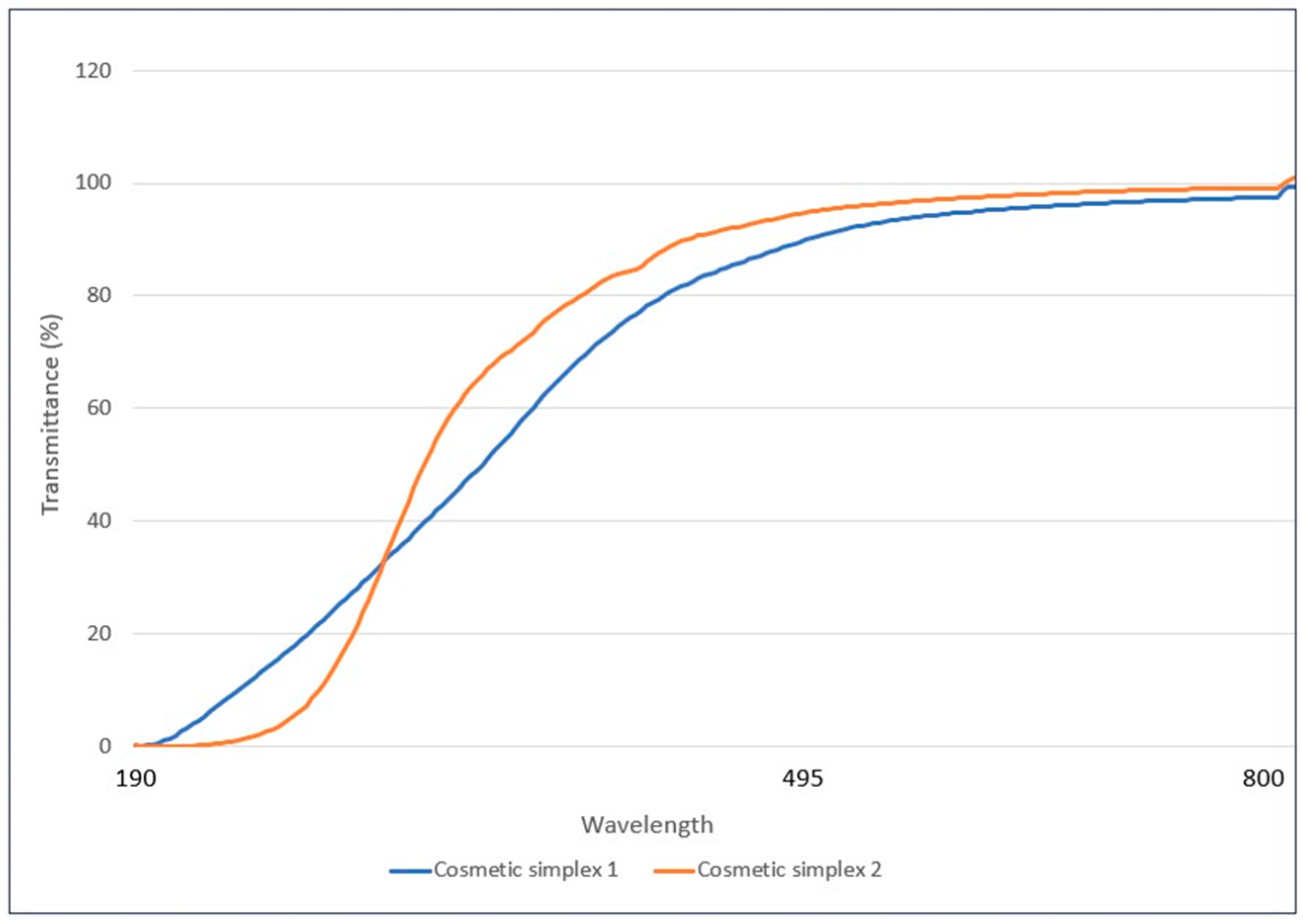
3.2. Assessment of Transparency of Simplex Formulations

A quantitative experiment was conducted with a spectrophotometer to understand the transmittance of cosmetic simplex 1 and cosmetic simplex 2. Transmittance (T) is the fraction of incident light which is transmitted. In other words, it is the amount of light that “successfully” passes through the substance and comes out the other side [49]. The below graph measures transmittance (T%) vs. wavelength (Figure 1 and Table 3). The samples analyzed in this experiment are:
  • Cosmetic simplex 1;
  • Cosmetic simplex 2.

3.3. Time-Kill Curves of Cutibacterium acnes on Exposure to Acnocure Simplex Formulations

In general, time-kill kinetics assays are performed to examine and identify the interactions that occur between the test compound and the microorganism tested. These assays are often conducted to test whether the antimicrobial activity of a test compound or a skincare product is concentration-dependent or time-dependent.
For the current evaluation, the time-kill assay was performed at pH 5.5 (pH of the skin as well as most of the skincare formulations for acne) as well as at 7.2 (native pH of the culture broth for time-kill studies). This was performed to study the effect of pH on the killing activity of Acnocure formulations. On treating C. acnes with Acnocure in a cosmetic simplex formula 1 and 2 at pH 5.5, the time-kill curve suggested a reduction in the number of viable C. acnes count at 2 h and maintained up to 24 h. The time-kill assay for C. acnes is shown in Figure 2. The MICs for Acnocure was 0.3 mg/mL against C. acnes and is shown in Table 2. The chosen test concentrations represent four and eight times the MIC obtained for Acnocure.
The lower detection threshold was 2 × 10² CFU/mL. Although the C. acnes strain was inhibited by cosmetic simplex formula 1 and 2 at pH 5.5, at each time tested, neither simplex formula was bactericidal at 24 h. For instance, cosmetic simplex formula 1 and 2 demonstrated rapid bactericidal activity with a 4.7 log reduction in viability occurring within just two hours of exposure. In contrast, the placebo of the formula did not show a reduction in C. acnes enumeration (Figure 2a). Interestingly, our internal cosmetic reference benchmark, Effaclar DUO, demonstrated a similar log reduction in CFU compared to our cosmetic simplex formula 1 and 2, and the bactericidal effect was sustained over 24 h (4.7 log reduction at 2 h), similar to our internal bench, Effaclar DUO Serum. The time-kill assay demonstrated that Acnocure in simplex formulations 1 and 2 has proven anti-acne efficacy against C. acnes that can last for about 24 h in a concentration-dependent manner.
Next, we decided to investigate Acnocure in a cosmetic simplex formula 1 and 2 at pH 7.2 against C. acnes and observed excellent and maximal activity on C. acnes at 2 h of contact time with cosmetic simplex formula 1 (5.6 log reduction at 2 h), similar to the killing trend with our internal cosmetic benchmark, Effaclar DUO Serum (Figure 2b). Similarly, Acnocure-containing cosmetic simplex formula 2 demonstrated significant inhibition of C. acnes with a 4.0 log reduction at 2 h and maximal activity on C. acnes at 6 h with maintained effect up to 24 h. Therefore, the synergistic effect that occurs between thymol and Curcuma turmerones in Acnocure strongly suggests a significant anti-acne benefit against C. acnes in a simplex formulation and thus plays a role in maintaining a lasting bactericidal effect up to 24 h.

3.4. Anti-Inflammatory Efficacy Assay (AIEA): COX-2 and NF-κB Inflammatory Pathways

Studies over the past decade have demonstrated a central role for inflammation in the development of acne lesions. This knowledge has opened new opportunities for cosmetic interventions [50,51,52]. Given the complex interrelationship between C. acnes, sebaceous lipogenesis, and inflammation, we next tested thymol, Curcuma turmerones and Acnocure for their anti-inflammatory efficacy based on their capacity to modulate the release of pro-inflammatory mediators by phorbol myristate acetate (PMA)- and lipopolysaccharide (LPS)-stimulated human macrophage-like U937 mononuclear cell line [53]. This model has been widely used to investigate both COX-2 and NF-κB inflammatory pathways [54]. In the anti-inflammatory efficacy assay or AIEA test model, thymol, Curcuma turmerones, and Acnocure (0.1% thymol and 0.1% Curcuma turmerones) inhibited the pro-inflammatory cytokines (IL-1β, IL-6, IL-8 and TNF-α) and PGE-2 production (see Table 4). These findings imply that the downregulation of COX-2 expression individually by thymol and Curcuma turmerones as well as by Acnocure at a protein level may be interlinked to the blockage of pro-inflammatory cytokines (IL-6 and IL-1β) and PGE-2 [55]. While thymol inhibited IL-1β at the concentration of 175 µg/mL and IL-6 at <100 µg/mL, both terpenes, thymol and Curcuma turmerones, displayed very strong inhibition of PGE-2 at <1 µg/mL, respectively, signifying that the terpenes were involved in a significant inflammatory response by monocytes/macrophages, and thereby chemical antagonists of COX-2 may be potential agents for the suppression of inflammatory responses.
Multiple studies highlight the anti-inflammatory properties of thymol [56,57,58]. In a study utilizing Staphylococcus aureus membrane vesicles and keratinocytes, thymol reduced expression levels of IL-1β, IL-6, TNF-α, IL-8, and MCP-1 [56]. Wang Q et al. [57] showed that thymol reduced phosphorylation of multiple mediators involved in the NFκB inflammatory pathway. Thymol reduced phosphorylation of IKK, IκBα, and NFκBp65 and resulted in reduction of MCP-1, TNF-α, and IL-6. Additionally, thymol (100 μM) has been reported to alter prostaglandin-catalyzed biosynthesis by inhibiting both isoforms of cyclooxygenase (COX), with the most active being against COX-1 with an IC50 value of 0.2 μM [59,60].

In Vitro IL-1α-Induced Anti-Inflammatory Study in Fibroblasts

In the present investigation, we investigated IL-1α (0.1 ng/mL) induced cutaneous inflammation by measuring the release of IL-6, IL-8 and PGE-2 using an IL-1α-stimulated dermal fibroblasts model. Cordier-Dirikoc et al. [61] publication suggests that dermal fibroblasts are by far the most IL-1-responsive cells compared to keratinocytes, melanocytes and endothelial cells.
Indeed, this study confirmed that fibroblasts have the property to respond to very low concentrations of IL-1α (from 10 fg/mL), even in the presence of 100-fold higher concentrations of IL-1RA, by increasing their expression of chemokines such as IL-8 for neutrophil recruitment. Thus, dermal fibroblasts are key relay cells in the chain of inflammation induced after epidermal lesion [61]. Under these conditions, the reference, dexamethasone was tested at 0.1 µM and significantly inhibited IL-1α stimulated IL-6, IL-8 and PGE-2 release with 81%, 66%, and 90%, respectively (Figure 3). Thymol and Curcuma turmerones were tested between 2 µg/mL to 30 µg/mL and significantly inhibited PGE-2 release by IL-1α-stimulated dermal fibroblasts (Figure 3). In addition, slight inhibition of IL-6 and IL-8 release was observed for Curcuma turmerones but to a lesser extent and at higher tested concentrations of 2.2 µg/mL and 6.7 µg/mL. According to recent studies, PGE-2 was found to significantly induce the expression of IL-6 and IL-8 in nasal polyp-derived fibroblasts due to an inflammatory loop involving PGE-2 via E prostanoid receptors, EP2 and EP4 [62,63]. Additionally, evidence points out that PGE-2 could be responsible for the activation of Akt and NF-κB signal pathways for both IL-6 and IL-8 expression [64,65] and therefore inhibition of PGE-2 could be henceforth accounted for suppression of IL-6 and IL-8. Previously, thymol was reported along with chlorhexidine to reduce the levels of prostaglandin E2 (PGE-2) within 8 days among volunteers suffering from gingival inflammation [59] confirming its anti-inflammatory efficacy through COX pathway. Similarly, xanthorrhizol, one of the Curcuma turmerones is also well known to inhibit inflammation by suppressing the production of cytokines, proinflammatory enzyme mediators cyclooxygenase-2 (COX-2) and inducible nitric oxide synthase (iNOS), and transcription factors [66]. These results indicate the COX inhibitory effect of thymol and Curcuma turmerones in a 2D dermal fibroblast model. The IL-1α-induced anti-inflammatory efficacy for Acnocure has not been communicated here and has been planned in the next study campaign.

4. Discussion

“Acnocure”, our patented combination of thymol and Curcuma turmerones, demonstrated strong antimicrobial efficacy against a wide range of microorganisms and anti-inflammatory activity in our in vitro screening. The present study showcases Acnocure as a promising cosmetic active that can be used to reduce the incidence of antimicrobial resistance in acne due to the increasing emergence of C. acnes resistant strains that is, as a result of excessive antibiotic treatment for acne. Epidemiological data shows that there are higher rates of clindamycin and erythromycin resistance that can also lead to other resistant bacterial pathogens, thereby compromising the effectiveness of current anti-acne treatments. This calls for international guidelines for the treatment of acne, recommending limiting antibiotic use and paving the way to select non-antibiotic treatments, such as natural active ingredients. On the other hand, future research is warranted to examine Acnocure’s potential for its anti-hyperseborrhea properties, as sebocyte hypersensitivity is influenced by androgen release and further regulated by C. acnes. Therefore, new studies could be directed at the investigation of Acnocure to control the overproduction of sebum and also the increased differentiation process in hyperkeratosis. Acnocure merits an in vivo clinical study in individuals with mild to moderate acne.

5. Conclusions

In conclusion, thymol and Curcuma turmerones and their combination as Acnocure in cosmetic simplex formulations have demonstrated potent anti-acne properties against C. acnes and also their ability to inhibit inflammation by suppressing the release of proinflammatory enzyme mediators, cyclooxygenase-2 (COX-2). These promising results position them as natural alternatives for the replacement of synthetic corticosteroids and antibiotics with potent skincare properties. Looking ahead, the shift towards natural individualized acne therapies is arguably driven by advancements in fields like microbiome research and personalized medicine and promises to revolutionize acne treatment and minimize side effects like antibiotic resistance and microbiome disruption while maintaining overall skin health. New alternative treatments involving microbiome-modifying therapies represent a paradigm shift in acne treatment and will form the next generation of ecobiological anti-inflammatory treatments to restore balance and skin health to the skin.

6. Patents

a. A novel combination of thymol and Curcuma zedoaria oil for enhanced activity against C. acnes, Patent No. 202411105169, filed in 2024.
b. Use of surfactant combinations for achieving transparency in a skincare serum for incorporating lipophilic actives (thymol and Curcuma zedoaria root oil), Patent No. 202411105170, filed in 2024.

Author Contributions

Conceptualization, S.T.P. and S.J.; methodology, A.P., S.P. and R.S.; software, A.P.; validation, A.K., A.B. and R.A.; formal analysis, C.M., S.P., S.B. and A.S.; investigation, A.P.; resources, A.P., K.C. and S.P.; data curation, A.P.; writing—original draft preparation, S.T.P.; writing—review and editing, S.T.P.; visualization, A.S.; supervision, S.T.P.; project administration, S.J., K.P. and V.S.; funding acquisition, S.J. and D.R. All authors have read and agreed to the published version of the manuscript.

Funding

This research work was sponsored by the ACNE Bioactives and Biologics platform, L’Oreal, D3I, Advanced research (Project Orchestra code: 2019-2316/B).

Institutional Review Board Statement

Not applicable.

Informed Consent Statement

Not applicable.

Data Availability Statement

Data are contained within the article.

Acknowledgments

We acknowledge the analytical (Ratnadeep Paul Choudhury) and discovery domains departments (Anne Potter and Carine Ballihaut) at L’Oreal, especially the High throughput laboratory headed by Ludwig Baux.

Conflicts of Interest

The authors are employees of L’Oréal India Ltd. The authors declare that the research was conducted in the absence of any commercial or financial relationships that could be construed as a potential conflict of interest. The funders had no role in the design of the study; in the collection, analyses or interpretation of data; in the writing of the manuscript; or in the decision to publish the results.

References

  1. Tuchayi, M.S.; Makrantonaki, E.; Ganceviciene, R.; Dessinioti, C.; Feldman, S.R.; Zouboulis, C.C. Acne vulgaris. Nat. Rev. Dis. Primers 2015, 1, 15029. [Google Scholar] [CrossRef] [PubMed]
  2. Yentzer, B.A.; Hick, J.; Reese, E.L.; Uhas, A.; Feldman, S.R.; Balkrishnan, R. Acne vulgaris in the United States: Descriptive epidemiology. Cutis 2010, 86, 94. [Google Scholar] [PubMed]
  3. McConnell, R.C.; Fleischer, A.B.; Williford, P.M.; Feldman, S.R. Most topical tretinoin treatment is for acne vulgaris through the age of 44 years: An analysis of the National Ambulatory Medical Care Survey. J. Am. Acad. Dermatol. 1998, 38, 221. [Google Scholar] [CrossRef] [PubMed]
  4. Bernales, S.A. Acne vulgaris: Role of the immune system. Int. J. Dermatol. 2021, 60, 1076. [Google Scholar] [CrossRef]
  5. Cong, T.X.; Hao, D.; Wen, X.; Li, X.; He, G.; Jiang, X. From pathogenesis of acne vulgaris to anti-acne agents. Arch. Dermatol. Res. 2019, 311, 337. [Google Scholar] [CrossRef] [PubMed]
  6. Barnard, E.; Shi, B.; Kang, D.; Craft, N.; Li, H. The balance of metagenomic elements shapes the skin microbiome in acne and health. Sci. Rep. 2016, 6, 39491. [Google Scholar] [CrossRef] [PubMed]
  7. Suh, D.H.; Kwon, H.H. What’s new in the physiopathology of acne? Br. J. Dermatol. 2015, 172, 13. [Google Scholar] [CrossRef] [PubMed]
  8. Lee, Y.B.; Byun, E.J.; Kim, H.S. Potential role of the microbiome in acne: A comprehensive review. J. Clin. Med. 2019, 8, 987. [Google Scholar] [CrossRef] [PubMed]
  9. Dréno, B.; Dagnelie, M.A.; Khammari, A.; Corvec, S. The Skin Microbiome: A New Actor in Inflammatory Acne. Am. J. Clin. Dermatol. 2020, 21, 18. [Google Scholar] [CrossRef] [PubMed]
  10. Zaenglein, A.L.; Pathy, A.L.; Schlosser, B.J.; Alikhan, A.; Baldwin, H.E.; Berson, D.S.; Bhushan, R. Guidelines of care for the management of acne vulgaris. J. Am. Acad. Dermatol. 2016, 74, 945. [Google Scholar] [CrossRef]
  11. Amed, I.; Berg, A.; Hudson, S.; Weaver, K.K.; Pacchia, M.L. The Beauty Market in 2023: A Special State of Fashion Report. Available online: https://www.mckinsey.com/industries/retail/our-insights/the-beauty-market-in-2023-a-special-state-of-fashion-report (accessed on 22 December 2024).
  12. Borris, R.P. Natural products research: Perspectives from a major pharmaceutical company. J. Ethnopharmacol. 1996, 51, 29. [Google Scholar] [CrossRef] [PubMed]
  13. Wright, C.W. Plant derived antimalarial agents: New leads and challenges. Phytochem. Rev. 2005, 4, 55. [Google Scholar] [CrossRef]
  14. Marquez-Valadez, B.; Maldonado, P.D.; Galvan-Arzate, S.; Méndez-Cuesta, L.A.; Pérez-De La Cruz, V.; Pedraza-Chaverrí, J.; Chánez-Cárdenas, M.E.; Santamaría, A. Alpha-mangostin induces changes in glutathione levels associated with glutathione peroxidase activity in rat brain synaptosomes. Nutr. Neurosci. 2012, 15, 13. [Google Scholar] [CrossRef] [PubMed]
  15. Qi, S.; Xin, Y.; Guo, Y.; Diao, Y.; Kou, X.; Luo, L.; Yin, Z. Ampelopsin reduces endotoxic infammation via repressing ROS-mediated activation of PI3K/Akt/NF-κB signaling pathways. Int. Immunopharmacol. 2012, 12, 278. [Google Scholar] [CrossRef]
  16. Etti, I.C.; Abdullah, R.; Kadir, A.; Hashim, N.M.; Yeap, S.K.; Imam, M.U.; Ramli, F.; Malami, I.; Lam, K.L.; Etti, U.; et al. The molecular mechanism of the anticancer effect of Artonin E in MDA-MB 231 triple negative breast cancer cells. PLoS ONE 2017, 12, e0182357. [Google Scholar] [CrossRef]
  17. Li, C.G.; Yan, L.; Jing, Y.Y.; Xu, L.H.; Liang, Y.D.; Wei, H.X.; Hu, B.; Pan, H.; Zha, Q.B.; Ouyang, D.Y.; et al. Berberine augments ATP induced infammasome activation in macrophages by enhancing AMPK signaling. Oncotarget 2017, 8, 95. [Google Scholar] [CrossRef]
  18. Farzaei, M.H.; Zobeiri, M.; Parvizi, F.; El-Senduny, F.F.; Marmouzi, I.; Coy-Barrera, E.; Naseri, R.; Nabavi, S.M.; Rahimi, R.; Abdollahi, M. Curcumin in liver diseases: A systematic review of the cellular mechanisms of oxidative stress and clinical perspective. Nutrients 2018, 10, 7. [Google Scholar] [CrossRef] [PubMed]
  19. Umesalma, S.; Sudhandiran, G. Differential inhibitory effects of the polyphenol ellagic acid on infammatory mediators NF-κB, iNOS, COX-2, TNF-α, and IL-6 in 1, 2-dimethylhydrazine-induced rat colon carcinogenesis. Basic Clin. Pharmacol. Toxicol. 2010, 107, 650. [Google Scholar] [CrossRef] [PubMed]
  20. Yang, N.; Zhang, H.; Cai, X.; Shang, Y. Epigallocatechin3-gallate inhibits infammation and epithelialmesenchymal transition through the PI3K/AKT pathway via upregulation of PTEN in asthma. Int. J. Mol. Med. 2018, 41, 818. [Google Scholar]
  21. Soleymani, S.; Farzaei, M.H.; Zargaran, A.; Niknam, S.; Rahimi, R. Promising plant-derived secondary metabolites for treatment of acne vulgaris: A mechanistic review. Arch. Dermatol. Res. 2020, 312, 5. [Google Scholar] [CrossRef]
  22. Amiri, H. Essential oils composition and antioxidant properties of three Thymus species. Evid. Based Complement. Alternat. Med. 2012, 2012, 728065. [Google Scholar] [CrossRef] [PubMed]
  23. Bairwa, R.; Sodha, R.S.; Rajawat, B.S. Trachyspermum ammi. Pharmacogn. Rev. 2012, 6, 56. [Google Scholar] [CrossRef] [PubMed]
  24. Yousefzadi, M.; Riahi-Madvar, A.; Hadian, J.; Rezaee, F.; Rafiee, R. In vitro cytotoxic and antimicrobial activity of essential oil from Satureja sahendica. Toxicol. Environ. Chem. 2012, 94, 1735. [Google Scholar] [CrossRef]
  25. Markovic, P.; Chatzopoulou, P.; Siljegovic, J.; Nikolic, M.; Glamoclija, J.; Ciric, A.; Soković, M. Chemical analysis and antimicrobial activities of the essential oils of Satureja thymbra L. and Thymbra spicata L. and their main components. Arch. Biol. Sci. 2011, 63, 457. [Google Scholar] [CrossRef]
  26. Khajeh, M.; Yamini, Y.; Sefidkon, F.; Bahramifar, N. Comparison of essential oil composition of Carum copticum obtained by supercritical carbon dioxide extraction and hydrodistillation methods. Food Chem. 2004, 86, 587. [Google Scholar] [CrossRef]
  27. Ghosheh, O.A.; Houdi, A.A.; Crooks, P.A. High performance liquid chromatographic analysis of the pharmacologically active quinones and related compounds in the oil of the black seed (Nigella sativa L.). J. Pharm. Biomed. Anal. 1999, 19, 757. [Google Scholar] [CrossRef] [PubMed]
  28. Didri, N.; Dubreuil, L.; Pinkas, M. Activity of thymol, carvacrol, cinnamaldehyde and eugenol on oral bacteria. Pharm. Acta Helv. 1994, 69, 25. [Google Scholar] [CrossRef]
  29. Aeschbach, R.; Loliger, J.; Scott, B.C.; Murcia, A.; Butler, J.; Halliwell, M.; Aruoma, O.I. Antioxidant actions of thymol, carvacrol, 6-gingerol, zingerone and hydroxytyrosol. Food Chem. Toxicol. 1994, 32, 31. [Google Scholar] [CrossRef] [PubMed]
  30. Mahmoud, A.L. Antifungal action and antiaflatoxigenic properties of some essential oil constituents. Lett. Appl. Microbiol. 1994, 19, 110. [Google Scholar] [CrossRef] [PubMed]
  31. Yanishlieva, N.V.; Marinovaa, E.M.; Gordon, M.H.; Raneva, V.G. Antioxidant activity and mechanism of action of thymol and carvacrol in two lipid systems. Food Chem. 1999, 64, 59. [Google Scholar] [CrossRef]
  32. Frank, J.; McEnroe, W.F. Structures and synthesis of some new antibacterial sesquiterpenoids from the gorgonian coral Pseudopterogorgia rigida. Tetrahedron 1978, 34, 1661. [Google Scholar]
  33. Itokawa, H.; Shi, Q.; Akiyama, T.; Morris-Natschke, S.L.; Lee, K.H. Recent advances in the investigation of curcuminoids. Chin. Med. 2008, 17, 11. [Google Scholar] [CrossRef]
  34. Hwang, J.K.; Sim, J.S.; Baek, N.I.; Pyun, Y.R. Xanthorrhizol: A potential antibacterial agent from Curcuma xanthorrhiza against Streptococcus mutans. Planta Med. 2000, 66, 196. [Google Scholar] [CrossRef]
  35. Chun, K.S.; Sohn, Y.; Kim, H.S.; Kim, O.H.; Park, K.K.; Lee, J.M.; Lee, J.; Lee, J.Y.; Moon, A.; Lee, S.S.; et al. Anti-tumor promoting potential of naturally occurring diarylheptanoids structurally related to curcumin. Mutat. Res. 1999, 428, 49. [Google Scholar] [CrossRef] [PubMed]
  36. Imtiyaz, A.S.; Venkatesalu, V. Botany, traditional uses, phytochemistry and pharmacological properties of Saussurea costus—An endangered plant from Himalaya—A review. Phytochem. Lett. 2022, 47, 140. [Google Scholar]
  37. Bokshan, S.L.; Gomez, J.R.; Chapin, K.C.; Green, A.; Paxton, E.S. Reduced Time to Positive Cutibacterium acnes Culture Utilizing a Novel Incubation Technique: A Retrospective Cohort Study. J. Shoulder Elb. Arthroplast. 2019, 3, 2471549219840823. [Google Scholar] [CrossRef] [PubMed]
  38. Nazzaro, F.; Fratianni, F.; De Martino, L.; Coppola, R.; De Feo, V. Effect of Essential Oils on Pathogenic Bacteria. Pharmaceuticals 2013, 6, 1451. [Google Scholar] [CrossRef] [PubMed]
  39. Trombetta, D.; Castelli, F.; Sarpietro, M.G.; Venuti, V.; Cristani, M.; Daniele, C.; Saija, A.; Mazzanti, G.; Bisignano, G. Mechanisms of antibacterial action of three monoterpenes. Antimicrob. Agents Chemother. 2005, 49, 2474. [Google Scholar] [CrossRef] [PubMed]
  40. Khameneh, B.; Eskin, N.A.M.; Iranshahy, M.; Fazly, B.B.S. Phytochemicals: A Promising Weapon in the Arsenal against Antibiotic-Resistant Bacteria. Antibiotics 2021, 10, 1044. [Google Scholar] [CrossRef] [PubMed]
  41. Sikkema, J.; De Bont, J.A.M.; Poolman, B. Mechanisms of membrane toxicity of hydrocarbons. Microbiol. Rev. 1995, 59, 201. [Google Scholar] [CrossRef] [PubMed]
  42. Yousfi, F.; Abrigach, F.; Petrovic, J.D.; Sokovic, M.; Ramdani, M. Phytochemical screening and evaluation of the antioxidant and antibacterial potential of Zingiber officinale extracts. S. Afr. J. Bot. 2021, 142, 433. [Google Scholar] [CrossRef]
  43. Xu, J.; Zhou, F.; Ji, B.P.; Pei, R.S.; Xu, N. The antibacterial mechanism of carvacrol and thymol against Escherichia coli. Lett. Appl. Microbiol. 2008, 47, 174. [Google Scholar] [CrossRef] [PubMed]
  44. Poudel, D.K.; Ojha, P.K.; Rokaya, A.; Satyal, R.; Satyal, P.; Setzer, W.N. Analysis of Volatile Constituents in Curcuma Species, viz. C. aeruginosa, C. zedoaria, and C. longa, from Nepal. Plants 2022, 26, 1932. [Google Scholar]
  45. Yasni, S.; Imaizumi, K.; Sin, K.; Sugano, M.; Nonaka, G.; Sidik, P. Identification of an active principle in essential oils and hexane-soluble fractions of Curcuma xanthorrhiza Roxb. showing triglyceride-lowering action in rats. Food Chem. Toxicol. 1994, 32, 273. [Google Scholar] [CrossRef] [PubMed]
  46. Uehara, S.; Yasuda, I.; Takeya, K.; Itokawa, H. Terpenoids and curcuminoids of the rhizoma of Curcuma xanthorrhiza roxb. J. Pharm. Soc. Jpn. 1992, 112, 817. [Google Scholar] [CrossRef] [PubMed]
  47. Hong, C.H.; Kim, Y.; Lee, S.K. Sesquiterpenoids from the rhizome of Curcuma zedoaria. Arch. Pharm. Res. 2001, 24, 424. [Google Scholar] [CrossRef]
  48. Martins, A.P.; Salgueiro, L.; Goncalves, M.J.; da Cunha, A.P.; Vila, R.; Canigueral, S.; Mazzoni, V.; Tomi, F.; Casanova, J. Essential oil composition and antimicrobial activity of three Zingiberaceae from S.Tome E principe. Planta Med. 2001, 67, 580. [Google Scholar] [CrossRef] [PubMed]
  49. Ciosek, J. Understanding of transmittance. Proc. SPIE-Int. Soc. Opt. Eng. 2003, 5064, 287–294. [Google Scholar]
  50. Hwang, J.K.; Shim, J.S.; Pyun, Y.R. Antibacterial activity of xanthorrhizol from Curcuma xanthorrhiza against oral pathogens. Fitoterapia 2000, 71, 321. [Google Scholar] [CrossRef]
  51. Stein Gold, L.F. What’s new in acne and inflammation. J. Drugs Dermatol. 2013, 12, s67. [Google Scholar]
  52. Weiss, J.S. Messages from molecules: Deciphering the code. J. Drugs Dermatol. 2013, 12, s70. [Google Scholar]
  53. Bellew, S.; Thiboutot, D.; Del Rosso, J.Q. Pathogenesis of acne vulgaris: What’s new, what’s interesting and what may be clinically relevant. J. Drugs Dermatol. 2011, 10, 582. [Google Scholar]
  54. Del Bufalo, A.; Bernad, J.; Dardenne, C.; Verda, D.; Meunier, J.R.; Rousset, F.; Martinozzi-Teissier, S.; Pipy, B. Contact sensitizers modulate the arachidonic acid metabolism of PMA-differentiated U-937 monocytic cells activated by LPS. Toxicol. Appl. Pharmacol. 2011, 256, 35. [Google Scholar] [CrossRef] [PubMed]
  55. Zhao, Y.-L.; Lu, Z.-Y.; Zhang, X.; Liu, W.-W.; Yao, G.-D.; Liu, X.-L.; Liu, W.; Wu, Q.-J.; Hayashi, T.; Yamato, M.; et al. Gelatin promotes cell aggregation and pro-inflammatory cytokine production in PMA-stimulated U937 cells by augmenting endocytosis-autophagy pathway. Int. J. Biochem. Cell Biol. 2018, 95, 132. [Google Scholar] [CrossRef]
  56. Vassiliou, E.; Awoleye, O.; Davis, A.; Mishra, S. Anti-Inflammatory and Antimicrobial Properties of Thyme Oil and Its Main Constituents. Int. J. Mol. Sci. 2023, 24, 6936. [Google Scholar] [CrossRef]
  57. Wang, Q.; Cheng, F.; Xu, Y.; Zhang, J.; Qi, J.; Liu, X.; Wang, R. Thymol alleviates lipopolysaccharide-stimulated inflammatory response via downregulation of RhoA-mediated NF-κB signalling pathway in human peritoneal mesothelial cells. Eur. J. Pharmacol. 2018, 833, 210. [Google Scholar] [CrossRef]
  58. Kwon, H.I.; Jeong, N.H.; Kim, S.Y.; Kim, M.H.; Son, J.H.; Jun, S.H.; Kim, S.; Jeon, H.; Kang, S.C.; Kim, S.H.; et al. Inhibitory effects of thymol on the cytotoxicity and inflammatory responses induced by Staphylococcus aureus extracellular vesicles in cultured keratinocytes. Microb. Pathog. 2019, 134, 103603. [Google Scholar] [CrossRef] [PubMed]
  59. Sköld, K.; Twetman, S.; Hallgren, A.; Yucel-Lindberg, T.; Modéer, T. Effect of a chlorhexidine/thymol-containing varnish on prostaglandin E2 levels in gingival crevicular fluid. Eur. J. Oral Sci. 1998, 106, 571. [Google Scholar] [CrossRef] [PubMed]
  60. Marsik, P.; Kokoska, L.; Landa, P.; Nepovim, A.; Soudek, P.; and Vanek, T. In vitro inhibitory effects of thymol and quinones of Nigella sativa seeds on cyclooxygenase-1- and -2-catalyzed prostaglandin E2 biosyntheses. Planta Med. 2005, 71, 739. [Google Scholar] [CrossRef]
  61. Cordier-Dirikoc, S.; Pedretti, N.; Garnier, J.; Clarhaut-Charreau, S.; Ryffel, B.; Morel, F.; Bernard, F.X.; Hamon de Almeida, V.; Lecron, J.C.; Jégou, J.F. Dermal fibroblasts are the key sensors of aseptic skin inflammation through interleukin 1 release by lesioned keratinocytes. Front. Immunol. 2022, 13, 984045. [Google Scholar] [CrossRef] [PubMed]
  62. Kupper, T.S.; Ballard, D.W.; Chua, A.O.; McGuire, J.S.; Flood, P.M.; Horowitz, M.C.; Langdon, R.; Lightfoot, L.; Gubler, U. Human keratinocytes contain mRNA indistinguishable from monocyte interleukin 1 alpha and beta mRNA. Keratinocyte epidermal cell-derived thymocyte-activating factor is identical to interleukin 1. J. Exp. Med. 1986, 164, 2095. [Google Scholar] [CrossRef] [PubMed]
  63. Mohan, S.; Ahmad, A.S.; Glushakov, A.V.; Chambers, C.; Doré, S. Putative role of prostaglandin receptor in intracerebral hemorrhage. Front. Neurol. 2012, 3, 145. [Google Scholar] [CrossRef] [PubMed]
  64. Cho, J.S.; Han, I.H.; Lee, H.R.; Lee, H.M. Prostaglandin E2 Induces IL-6 and IL-8 Production by the EP Receptors/Akt/NF-κB Pathways in Nasal Polyp-Derived Fibroblasts. Allergy Asthma Immunol. Res. 2014, 6, 449. [Google Scholar] [CrossRef] [PubMed]
  65. Wang, P.; Zhu, F.; Konstantopoulos, K. Prostaglandin E2 induces interleukin-6 expression in human chondrocytes via cAMP/protein kinase A- and phosphatidylinositol 3-kinase-dependent NF-kappaB activation. Am. J. Physiol. Cell Physiol. 2010, 298, C1445. [Google Scholar] [CrossRef] [PubMed]
  66. Lee, S.K.; Hong, C.H.; Huh, S.K.; Kim, S.S.; Oh, O.J.; Min, H.Y.; Park, K.K.; Choong, W.Y.; Hwang, J.K. Suppressive effect of natural sesquiterpenoids on inducible cyclooxygenase (COX-2) and nitric oxide synthase (iNOS) activity in mouse macrophage cells. J. Environ. Pathol. Toxicol. Oncol. 2002, 21, 141. [Google Scholar] [CrossRef]
Figure 1. Transmittance (T%) of Cosmetic Simplex 1 and 2 at 190nm, 495 and 800 nm.
Cosmetics 12 00037 g001
Figure 2. Time-kill curves of Acnocure against C. acnes ATCC 6919 cultured in Tryptic Soy Broth (TSB) broth buffered at (a) pH 5.5 and (b) pH 7.2 with exposure time at concentrations below in-use levels. At each contact time, cultures were sampled and neutralized before enumeration of viable cells. The lower detection threshold was 2.102 CFU/ml.
Cosmetics 12 00037 g002
Figure 3. PGE-2 inhibition of compounds in IL-1α-induced fibroblasts.
Cosmetics 12 00037 g003
Table 1. Bacterial Strains.
Strains (Culture Collection Number)Targeted DisorderCulture/Enumeration MediaDiluent for Inoculum PreparationTest Media
Cutibacterium acnes ATCC 6919AcneTrypto-casein-soy agar (TSA) Tryptone salt brothTrypto-casein-soy broth (TSB)
Staphylococcus aureus (ATCC 6538)AtopyTSA agarTryptone salt brothTrypto-casein-soy broth (TSB)
Staphylococcus epidermidis (ATCC 12228)AcneTSA agarTryptone salt brothTrypto-casein-soy broth (TSB)
Corynebacterium freneyi (CIP 52.16)Armpit
odor
TSA agarTryptone salt brothTrypto-casein-soy broth (TSB)
Table 2. Antimicrobial activities of thymol, Curcuma turmerones and Acnocure against skin-related bacteria.
CompoundsC. acnes
ATCC 6919
S. aureus CIP 4.83S. epidermidis CIP 68.21Corynebacterium freneyi
ATCC 7711
MIC (mg/mL)MIC
(mg/mL)
MIC
(mg/mL)
MIC
(mg/mL)
Thymol0.430.290.470.58
Curcuma turmerones0.370.280.510.34
Acnocure0.320.260.470.11
C. acnes: Cutibacterium acnes; S. aureus: Staphylococcus aureus; S. epidermidis: Staphylococcus epidermidis; MIC: minimum inhibitory concentration; Aconocure: 0.1% thymol and 0.1% Curcuma turmerones. The values are from triplicate experiments.
Table 3. Formulation assessment for transmittance at maximum wavelength.
Name of SampleMaximum Transmittance (%)Wavelength (nm)
Cosmetic simplex 1 100 753
Cosmetic simplex 2 99.634 740
Table 4. Anti-inflammatory efficacy assay in human macrophage-like U937 mononuclear cell line model (AIEA).
CompoundsAnti-Inflammatory IC5O µg/mL
1L-1βIL-6IL-8TNF-αPGE-2
Thymol175<100NANA<1
TurmeronesNANANANA<1
AcnocureNANANANA<1
IC5O: inhibitory concentration at 50%; NA: not active; Acnocure: 0.1% thymol and 0.1% Curcuma turmerones. The values are from multiple experiments.
Disclaimer/Publisher’s Note: The statements, opinions and data contained in all publications are solely those of the individual author(s) and contributor(s) and not of MDPI and/or the editor(s). MDPI and/or the editor(s) disclaim responsibility for any injury to people or property resulting from any ideas, methods, instructions or products referred to in the content.

