
Low-Dose COVID-19 CT Image Denoising Using Batch Normalization and Convolution Neural Network

 , , , ,
  ,
,  and
and 
Abstract
1. Introduction
2. Literature Review
3. Materials and Methods
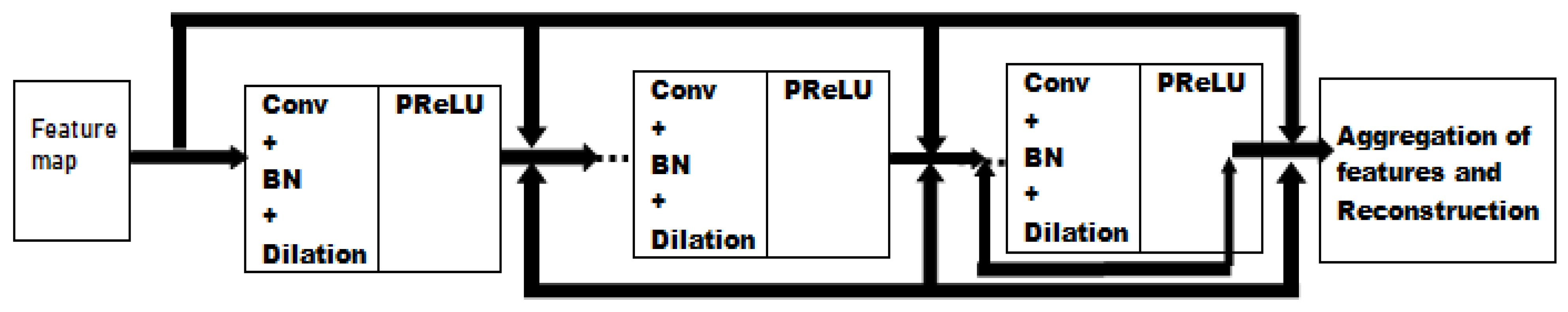
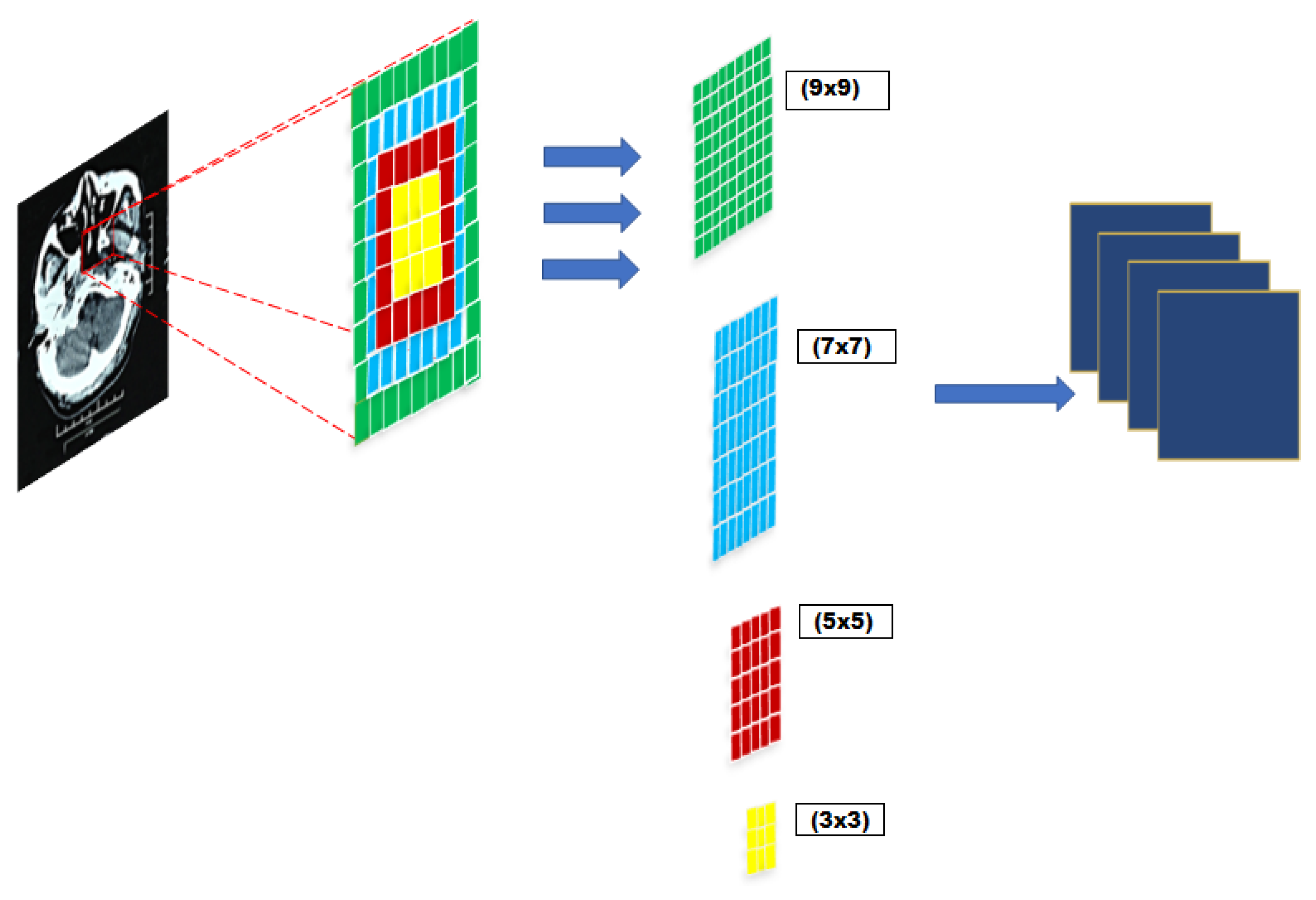
3.1. Network Architecture
3.2. Loss Function
3.3. Batch Normalization and Residual Learning
3.4. Significance of Proposed Model
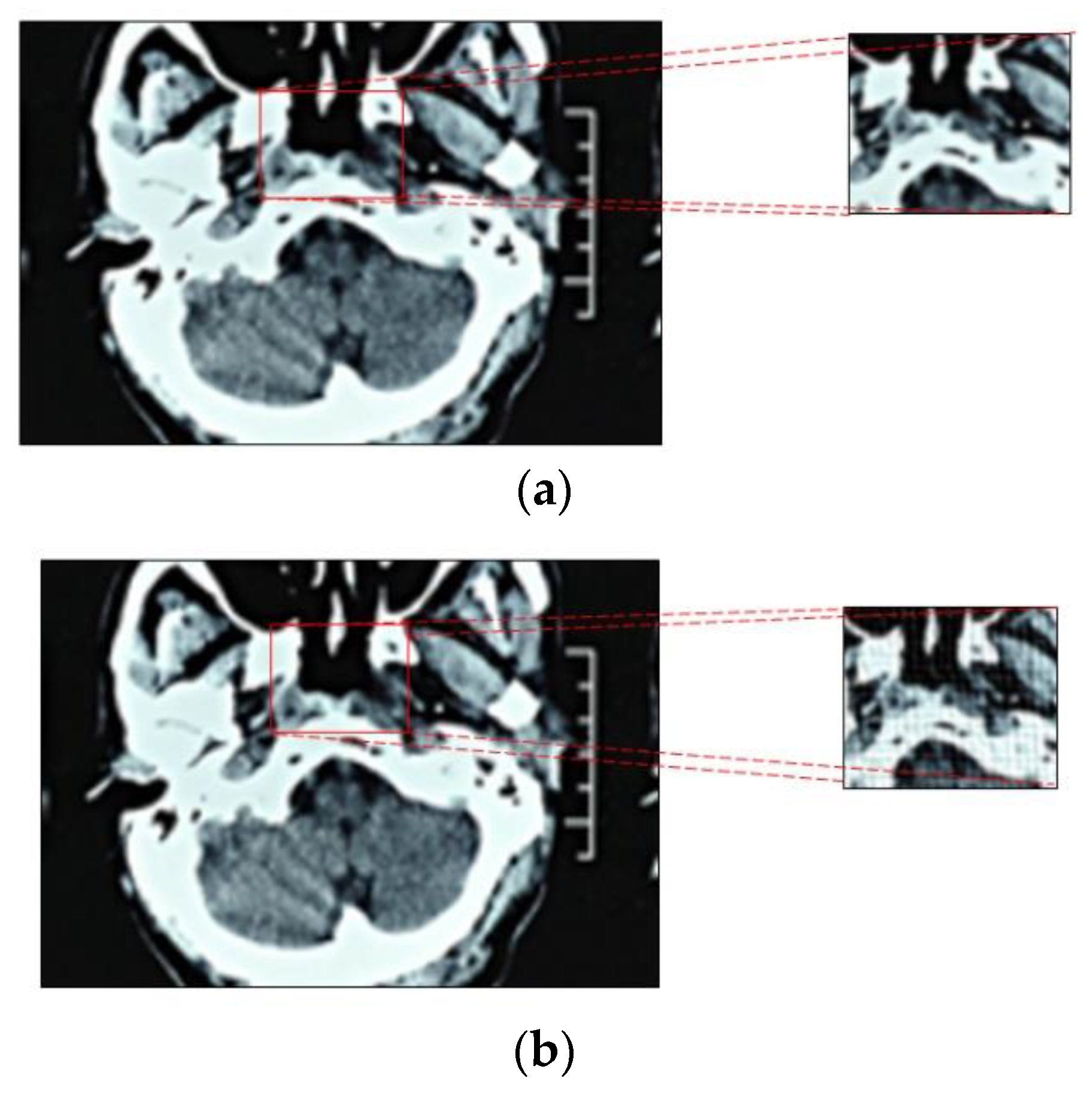
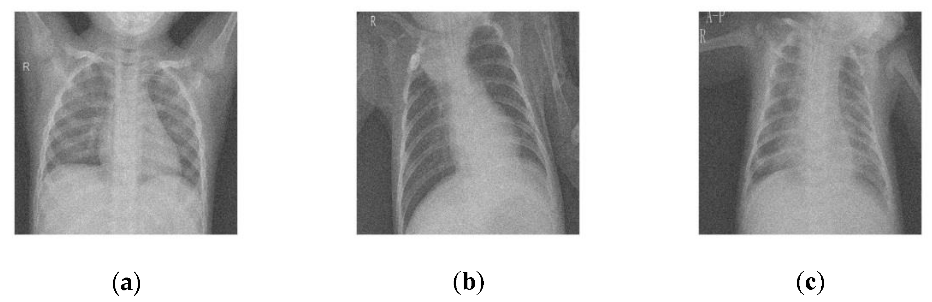
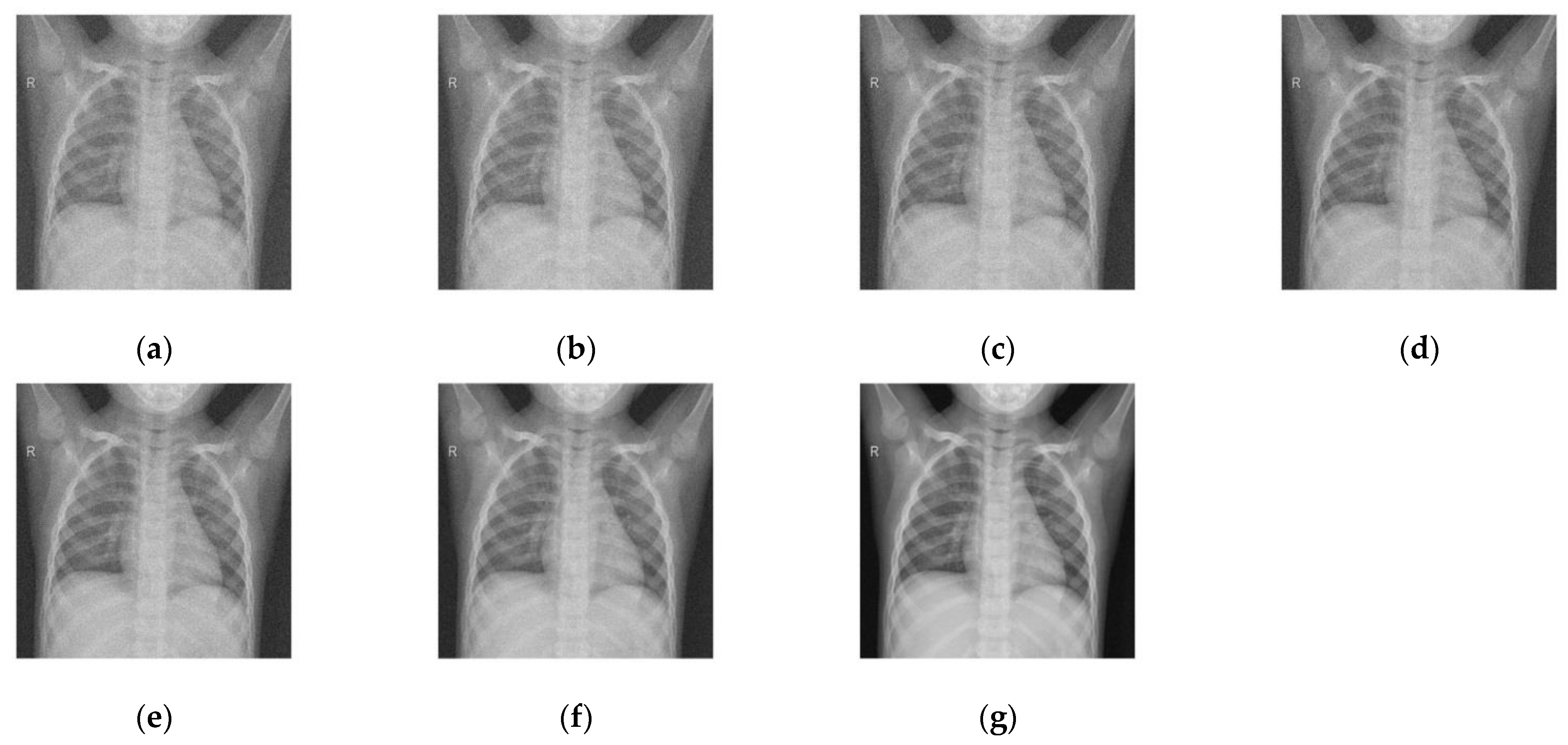
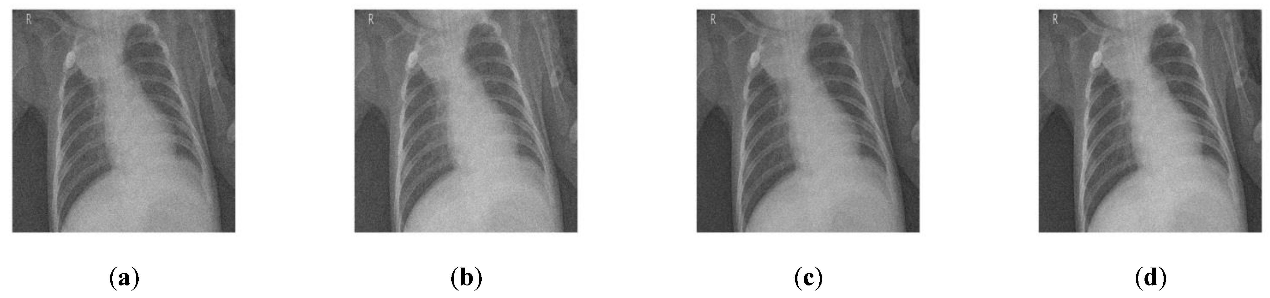
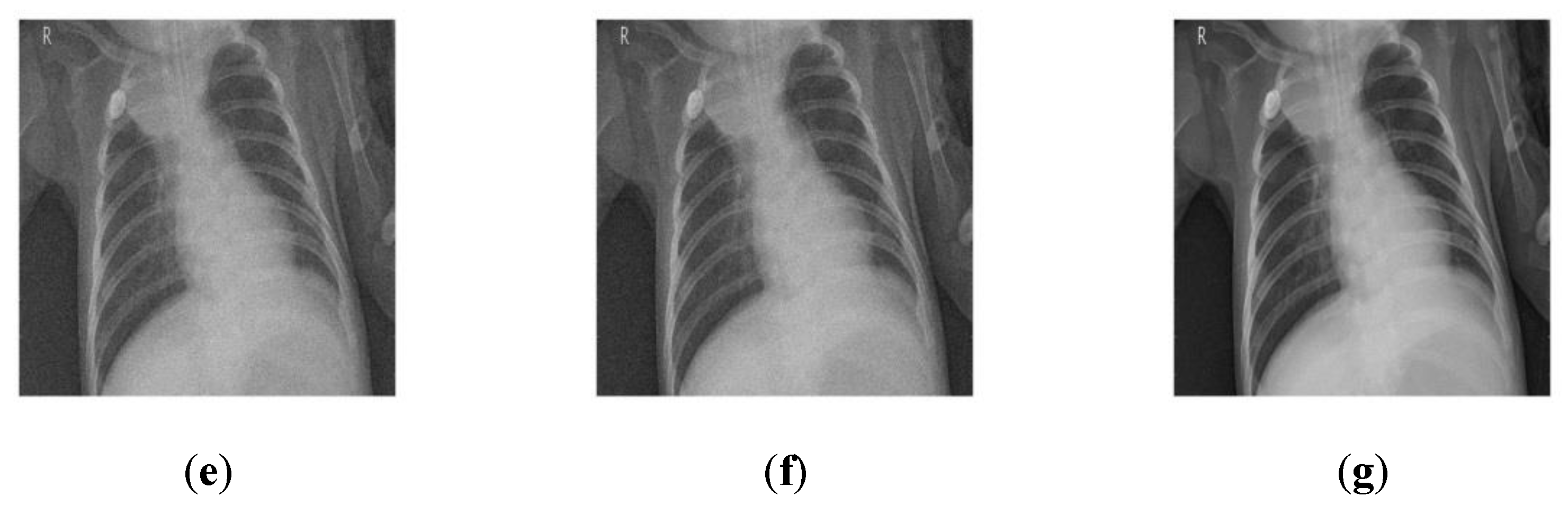
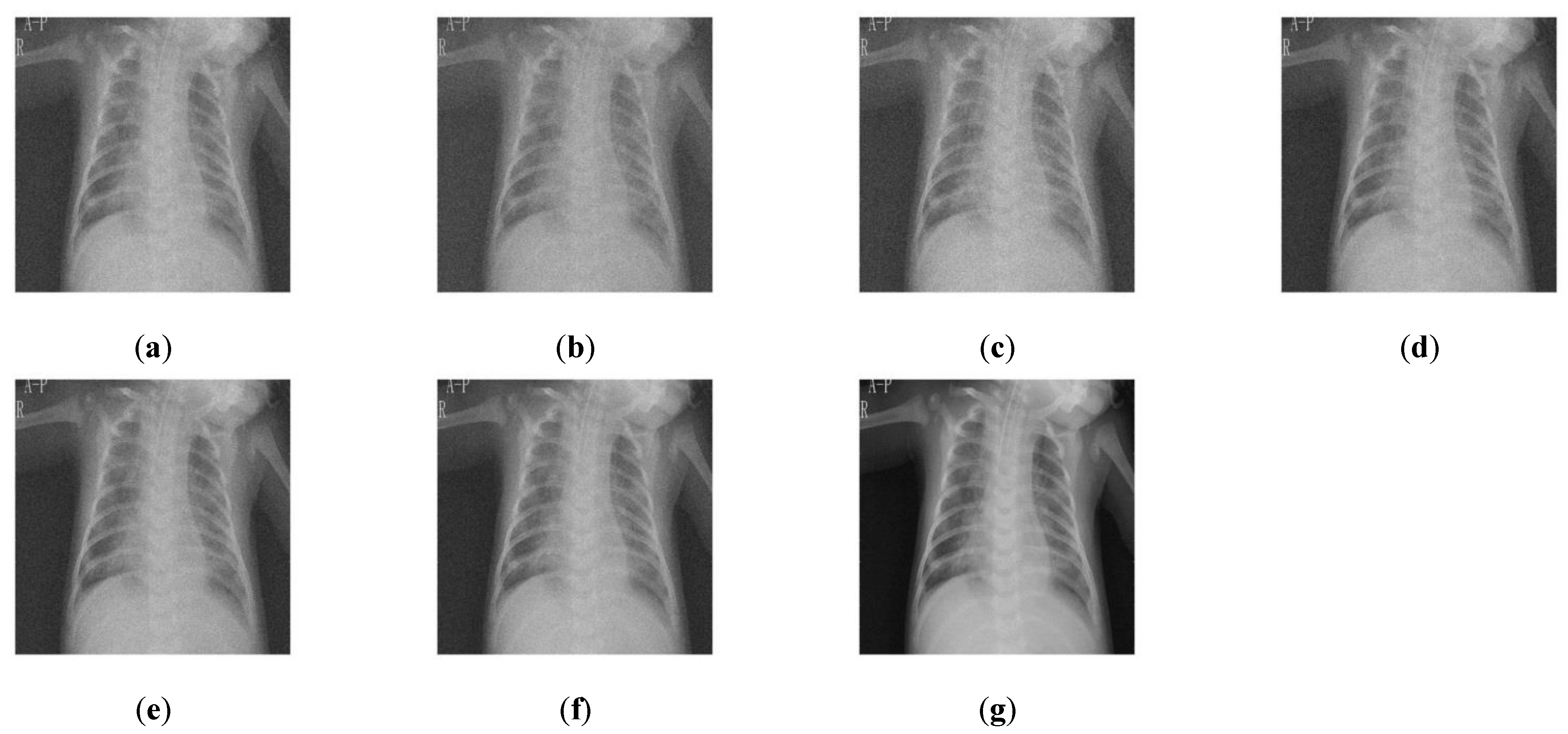
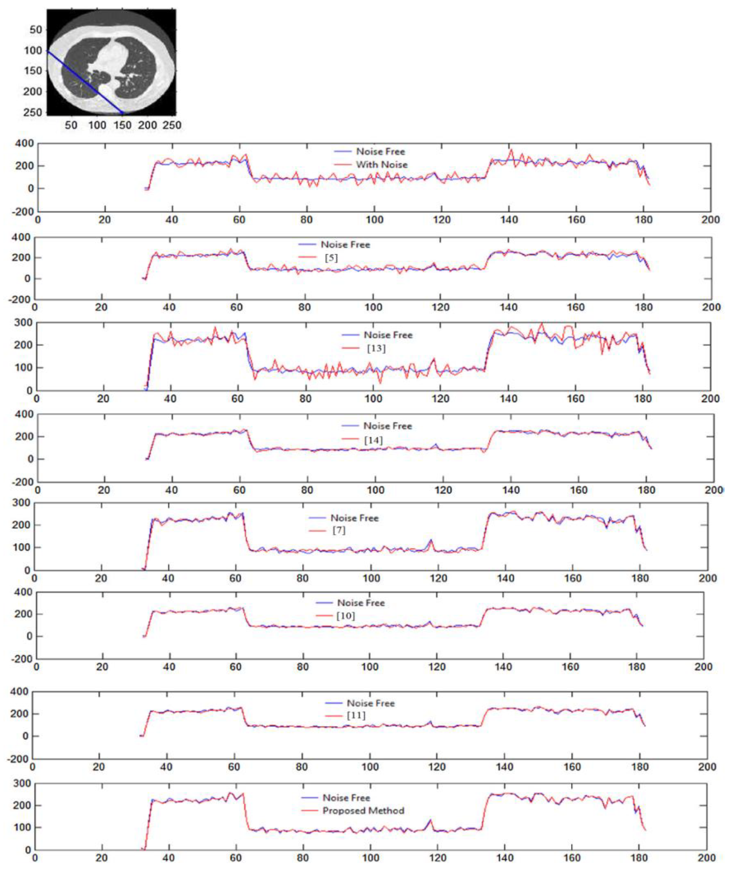
4. Results and Discussion
5. Conclusions
Author Contributions
Funding
Conflicts of Interest
References
- Ali, S.H.; Sukanesh, R. An efficient algorithm for denoising MR and CT images using digital curvelet transform. Adv. Exp. Med. Biol. 2011, 696, 471–480. [Google Scholar]
- Boone, J.; Geraghty, E.M.; Seibert, J.A.; Wootton-Gorges, S.L. Dose reduction in pediatric CT: A rational approach. J. Radiol. 2003, 228, 352–360. [Google Scholar] [CrossRef] [PubMed]
- Borsdorf, A.; Raupach, R.; Flohr, T.; Hornegger, J. Wavelet based noise reduction in CT-images using correlation analysis. IEEE Trans. Med. Imaging 2008, 27, 1685–1703. [Google Scholar] [CrossRef] [PubMed]
- Borsdorf, A.; Raupach, R.; Hornegger, J. Multiple CT-reconstructions for locally adaptive anisotropic wavelet denoising. Int. J. CARS 2008, 2, 255–264. [Google Scholar] [CrossRef]
- Mingliang, X.; Pei, L.; Mingyuan, L.; Hao, F.; Hongling, Z.; Bing, Z.; Yusong, L.; Liwei, Z. Medical image denoising by parallel non-local means. Neurocomputing 2016, 195, 117–122. [Google Scholar] [CrossRef]
- Chang, S.G.; Yu, B.; Vetterli, M. Spatially adaptive thresholding with context modeling for image denoising. IEEE Trans. Image Process. 2000, 9, 1522–1531. [Google Scholar] [CrossRef]
- Kuppusamy, P.G.; Joseph, J.; Jayaraman, S. A customized nonlocal restoration schemes with adaptive strength of smoothening for magnetic resonance images. Biomed. Signal Process. Control. 2019, 49, 160–172. [Google Scholar]
- Diwakar, M.; Kumar, M. CT image denoising using NLM and correlation-based wavelet packet thresholding. IET Image Process. 2018, 12, 708–715. [Google Scholar] [CrossRef]
- Diwakar, M.; Kumar, M. A review on CT image noise and its denoising. Biomed. Signal Process. Control 2018, 42, 73–88. [Google Scholar] [CrossRef]
- Cheng, Y.; Bu, Z.; Xu, Q.; Ye, M.; Zhang, J.; Zhou, J. Shearlet and guided filter based despeckling method for medical ultrasound images. Ultrasound Med. Biol. 2019, 45, S85. [Google Scholar] [CrossRef]
- Zhao, L.; Bai, H.; Liang, J.; Wang, A.; Zeng, B.; Zhao, Y. Local activity-driven structural-preserving filtering for noise removal and image smoothing. Signal Process. 2019, 157, 62–72. [Google Scholar] [CrossRef]
- Wu, H.; Zhang, W.; Gao, D.; Yin, X.; Chen, Y.; Wang, W. Fast CT image processing using parallelized non-local means. J. Med. Biol. Eng. 2011, 31, 437–441. [Google Scholar] [CrossRef]
- Jomaa, H.; Mabrouk, R.; Khlifa, N.; Morain-Nicolier, F. Denoising of dynamic PET images using a multi-scale transform and non-local means filter. Biomed. Signal Process. Control. 2018, 41, 69–80. [Google Scholar] [CrossRef]
- Manoj, D.; Singh, P. CT image denoising using multivariate model and its method noise thresholding in non-subsampled shearlet domain. Biomed. Signal Process. Control. 2020, 57, 101754. [Google Scholar]
- Wang, Y.; Shao, Y.; Zhang, Q.; Liu, Y.; Chen, Y.; Chen, W.; Gui, Z. Noise Removal of Low-Dose CT Images Using Modified Smooth Patch Ordering. IEEE Access 2017, 5, 26092–26103. [Google Scholar] [CrossRef]
- You, C.; Yang, Q.; Gjesteby, L.; Li, G.; Ju, S.; Zhang, Z.; Zhao, Z.; Zhang, Y.; Cong, W.; Wang, G. Structurally sensitive multi-scale deep neural network for low-dose CT denoising. IEEE Access 2018, 6, 41839–41855. [Google Scholar] [CrossRef]
- Diwakar, M.; Kumar, P.; Singh, A.K. CT image denoising using NLM and its method noise thresholding. Multimed. Tools Appl. 2018, 79, 14449–14464. [Google Scholar] [CrossRef]
- Yang, Q.; Yan, P.; Zhang, Y.; Yu, H.; Shi, Y.; Mou, X.; Kalra, M.K.; Zhang, Y.; Sun, L.; Wang, G. Low-dose CT image denoising using a generative adversarial network with Wasserstein distance and perceptual loss. IEEE Trans. Med. Imaging 2018, 37, 1348–1357. [Google Scholar] [CrossRef]
- Hasan, A.M.; Melli, A.; Wahid, K.A.; Babyn, P. Denoising low-dose CT images using multi-frame blind source separation and block matching filter. IEEE Trans. Radiat. Plasma Med. Sci. 2018, 2, 279–287. [Google Scholar] [CrossRef]
- Diwakar, M.; Kumar, M. Edge preservation-based CT image denoising using Wiener filtering and thresholding in wavelet domain. In Proceedings of the 2016 4th International Conference on Parallel, Distributed and Grid Computing (PDGC), Waknaghat, India, 22–24 December 2016; pp. 332–336. [Google Scholar]
- Liu, Y.; Castro, M.; Lederlin, M.; Shu, H.; Kaladji, A.; Haigron, P. Edge-preserving denoising for intra-operative cone beam CT in endovascular aneurysm repair. Comput. Med. Imaging Graph. 2017, 56, 49–59. [Google Scholar] [CrossRef]
- Kolb, M.; Storz, C.; Kim, J.H.; Weiss, J.; Afat, S.; Nikolaou, K.; Bamberg, F.; Othman, A.E. Effect of a novel denoising technique on image quality and diagnostic accuracy in low-dose CT in patients with suspected appendicitis. Eur. J. Radiol. 2019, 116, 198–204. [Google Scholar] [CrossRef] [PubMed]
- Horry, M.J.; Chakraborty, S.; Paul, M.; Ulhaq, A.; Pradhan, B.; Saha, M.; Shukla, N. COVID-19 detection through transfer learning using multimodal imaging data. IEEE Access 2020, 8, 149808–149824. [Google Scholar] [CrossRef] [PubMed]
- Momeny, M.; Neshat, A.A.; Hussain, M.A.; Kia, S.; Marhamati, M.; Jahanbakhshi, A.; Hamarneh, G. Learning-to-augment strategy using noisy and denoised data: Improving generalizability of deep CNN for the detection of COVID-19 in X-ray images. Comput. Biol. Med. 2021, 136, 104704. [Google Scholar] [CrossRef] [PubMed]
- Xiao, C.; Stoel, B.C.; Bakker, M.E.; Peng, Y.; Stolk, J.; Staring, M. Pulmonary fissure detection in CT images using a derivative of stick filter. IEEE Trans. Med. Imaging 2016, 35, 1488–1500. [Google Scholar] [CrossRef]
- Wadhwa, P.; Tripathi, A.; Singh, P.; Diwakar, M.; Kumar, N. Predicting the time period of extension of lockdown due to increase in rate of COVID-19 cases in India using machine learning. Mater. Today Proc. 2021, 37, 2617–2622. [Google Scholar] [CrossRef]
- Iborra, A.; Rodríguez-Álvarez, M.J.; Soriano, A.; Sánchez, F.; Bellido, P.; Conde, P.; Crespo, E.; González, A.J.; Moliner, L.; Rigla, J.P.; et al. Noise analysis in computed tomography (CT) image reconstruction using QR-Decomposition algorithm. IEEE Trans. Nucl. Sci. 2015, 62, 869–875. [Google Scholar] [CrossRef]
- Dabov, K.; Foi, A.; Katkovnik, V.; Egiazarian, K. Image denoising by sparse 3-D transform-domain collaborative filtering. IEEE Trans. Image Process. 2007, 16, 2080–2095. [Google Scholar] [CrossRef]
- Wieclawek, W.; Pietka, E. Granular filter in medical image noise suppression and edge preservation. Biocybern. Biomed. Eng. 2019, 39, 1–16. [Google Scholar] [CrossRef]
- Hashemi, S.M.; Paul, N.S.; Beheshti, S.; Cobbold, R.S.C. Adaptively tuned iterative low dose ct image denoising. Comput. Math. Methods Med. 2015, 2015, 638568. [Google Scholar] [CrossRef]
- Shreyamsha Kumar, B.K. Image denoising based on non-local means filter and its method noise thresholding. Springer J. Signal Image Video Process. 2013, 7, 1211–1227. [Google Scholar] [CrossRef]
- Geraldo, R.J.; Cura, L.M.; Cruvinel, P.E.; Mascarenhas, N.D. Low dose CT filtering in the image domain using MAP algorithms. IEEE Trans. Radiat. Plasma Med. Sci. 2016, 1, 56–67. [Google Scholar] [CrossRef]
- Liu, H.; Fang, L.; Li, J.; Zhang, T.; Wang, D.; Lan, W. Clinical and CT imaging features of the COVID-19 pneumonia: Focus on pregnant women and children. J. Infect. 2020, 80, e7–e13. [Google Scholar] [CrossRef]
- He, X.; Yang, X.; Zhang, S.; Zhao, J.; Zhang, Y.; Xing, E.; Xie, P. Sample-Efficient Deep Learning for COVID-19 Diagnosis Based on CT Scans. medRxiv 2020, 1, 20063941. [Google Scholar]
- He, K.; Sun, J. Convolutional neural networks at constrained time cost. In Proceedings of the IEEE Conference on Computer Vision and Pattern Recognition, Boston, MA, USA, 7–12 June 2015. [Google Scholar]
- Barina, D. Real-time wavelet transform for infinite image strips. J. Real-Time Image Process. 2021, 18, 585–591. [Google Scholar] [CrossRef]
- Ahn, B.; Nam, I.C. Block-matching convolutional neural network for image denoising. arXiv 2017, arXiv:1704.00524. [Google Scholar]
- Zuo, W.; Zhang, K.; Zhang, L. Convolutional Neural Networks for Image Denoising and Restoration. In Denoising of Photographic Images and Video; Springer: Berlin/Heidelberg, Germany, 2018; pp. 93–123. [Google Scholar]
- Shahdoosti, H.R.; Zahra, R. Edge-preserving image denoising using a deep convolutional neural network. Signal Process. 2019, 159, 20–32. [Google Scholar] [CrossRef]
- Haque, K.N.; Mohammad, A.Y.; Rajib, R. Image denoising and restoration with CNN-LSTM Encoder Decoder with Direct Attention. arXiv 2018, arXiv:1801.05141. [Google Scholar]
- Valsesia, D.; Giulia, F.; Enrico, M. Image denoising with graph-convolutional neural networks. In Proceedings of the 2019 IEEE International Conference on Image Processing (ICIP), Taipei, Taiwan, 22–25 September 2019. [Google Scholar]
- Islam, M.T.; Rahman, S.M.; Ahmad, M.O.; Swamy, M.N.S. Mixed Gaussian-impulse noise reduction from images using convolutional neural network. Signal Process. Image Commun. 2018, 68, 26–41. [Google Scholar] [CrossRef]
- Elhoseny, M.; Shankar, K. Optimal bilateral filter and convolutional neural network based denoising method of medical image measurements. Measurement 2019, 143, 125–135. [Google Scholar] [CrossRef]
- Tian, C.; Xu, Y.; Fei, L.; Wang, J.; Wen, J.; Luo, N. Enhanced CNN for image denoising. CAAI Trans. Intell. Technol. 2019, 4, 17–23. [Google Scholar] [CrossRef]
- Gong, K.; Guan, J.; Liu, C.C.; Qi, J. PET image denoising using a deep neural network through fine tuning. IEEE Trans. Radiat. Plasma Med. Sci. 2018, 3, 153–161. [Google Scholar] [CrossRef]
- Zhang, K.; Zuo, W.; Zhang, L. FFDNet: Toward a fast and flexible solution for CNN-based Image Denoising. IEEE Trans. Image Process. 2018, 27, 4608–4622. [Google Scholar] [CrossRef]
- Gondara, L. Medical image denoising using convolutional denoising autoencoders. In Proceedings of the 2016 IEEE 16th International Conference on Data Mining Workshops (ICDMW), Barcelona, Spain, 15 December 2016. [Google Scholar]
- Tian, C.; Xu, Y.; Li, Z.; Zuo, W.; Fei, L.; Liu, H. Attention-guided CNN for image denoising. Neural Netw. 2020, 124, 117–129. [Google Scholar] [CrossRef]
- Yu, S.; Park, B.; Jeong, J. Deep iterative down-up CNN for image denoising. In Proceedings of the IEEE Conference on Computer Vision and Pattern Recognition Workshops, Long Beach, CA, USA, 16–17 June 2019. [Google Scholar]
- Tian, C.; Xu, Y.; Zuo, W. Image denoising using deep CNN with batch renormalization. Neural Netw. 2020, 121, 461–473. [Google Scholar] [CrossRef]
- Zhang, K.; Zuo, W.; Chen, Y.; Meng, D.; Zhang, L. Beyond a gaussian denoiser: Residual learning of deep CNN for image denoising. IEEE Trans. Image Process. 2017, 26, 3142–3155. [Google Scholar] [CrossRef]
- Zhang, J.; Zhou, H.; Niu, Y.; Lv, J.; Chen, J.; Cheng, Y. CNN and multi-feature extraction based denoising of CT images. Biomed. Signal Process. Control. 2021, 67, 102545. [Google Scholar] [CrossRef]
- Available online: https://bmcresnotes.biomedcentral.com/articles/10.1186/s13104-021-05592-x (accessed on 10 July 2022).


| Reference | Objectives | Methods | Merit | Demerit |
|---|---|---|---|---|
| Byeongyong et al. (2017) [37] | Proposes novel approaches to combine NSS and CNN for image denotation, thereby functioning reliably in all types of images | First, 3D block is created by aggregating similar images. Then, after applying current denoising approach, block matching is done by pilot signal; the denoising function is structured by CNN | Efficient for both irregular images and repeating patterns. Considers local and global image characteristics | Multiple iterations are required, and denoising function creation is cumbersome via CNN. |
| Wangmeng et al. (2018) [38] | Understanding whether CNN can be successful; its excellence can cause fast, flexible, and non-blind denotation. Could it help restore? | Survey paper | Reveals how additive white noise may give CNN image denotation unsatisfactory results | Does not include new techniques such as genetic algorithms |
| Hamid et al. (2019) [39] | Preserves image edges while removing noise by CNN method | The canny technique is first used to draw the corners, and then the unsampled shearlet converts noisy images into low-frequency sub bands and 2D band stacking obtains 3D blocks and then maintains a denotation of non-subsampled shearlet transform (NSST) coefficients that is the same as CNN | Edges are preserved | Computing complexity and time will grow; NSST constants are identified as long |
| Haque et al. (2018) [40] | Uses encoder–decoder to denoise and restore image | Encoder = CNN and decoder | Better than auto decoder | Encoder functions and decoder functions can be used differently for future to check performance |
| Diego et al. (2019) [41] | Uses graph methods to treat to local attributes of images that primitive CNN misses out. | Graphing algorithms map local noise via proposed architecture | Local attributes identified | Selecting best graphing method is long trial-and-error process |
| Islam et al. (2018) [42] | Uses end-to-end to handle mixed noise robustly | Uses end-to-end mapping for every noisy entity so handles Gaussian impulse mixed noise | Lightweight structure, thus quick computing and easy to install | Not very suitable for high-end processing due to lightweight structure |
| Elhoseny et al. (2019) [43] | Bio-optimization-based filters are used to improve the PSNR ratio | Swarm-based optimization is carried out utilizing Dragonfly (DF) and modified firefly bilateral filters and algorithms | Very robust in medical images | Application may or may not extend beyond medical images |
| Tian et al. (2019) [44] | Makes the CNN network more trained and efficient with less time and samples | Utilize batch normalization and residual learning | It is effective and can be used in medical images | Application may or may not extend beyond medical images |
| Gong et al. (2018) [45] | Preserves features in PET scan while denoising image | Employs the training loss function to maintain image features, utilizing current data to train layers of last | Very practical and good results with real patient data | The app may or may not expand beyond PET scans, such as X-rays, sonography, etc. |
| Zhang et al. (2018) [46] | Real-world noisy images or spatial variant noise handling | Includes a tunable input noise map | More quickly handles wide variety of sounds and special noise | Not compared to other conventional and nonconventional denoising methods |
| Gondara et al. (2016) [47] | Uses autodecoders using deep learning networks on small-size images for denoising | Boot size image is created by combination of heterogeneous images | Handles high-cost computational issues and huge training sets | Re-dimensioning images decrease their quality of resolution, and the study is data-specific, with no suitable architecture to reuse the method |
| Tian et al. (2020) [48] | It addresses CNN networks that take long time to train and suffer from saturation of performance | Amalgamates two frameworks: renormalization of batch and BRDNet | Fixes the issue of internal shifting covariates and tiny mini-batch issues | This refers to a multi-method that may take more time and space than other alternatives |
| Yu et al. (2019) [49] | Denoises both level and multi-level noise with sequential reduction and escalation using CNN and U-net frameworks | Downscaling and upscaling layers of CNN-based U-net is modified to handle multiple parameters | Handles multiple parameters; less GPU capacity required | Continuous upscaling and downscaling reduces image resolution quality |
| Noise Level | Before Denoising | After Denoising | |||||||
|---|---|---|---|---|---|---|---|---|---|
| [5] | [13] | [14] | [7] | [10] | [11] | Proposed Method | |||
| CT 1 image | 10 | 24.6 | 32.14 | 32.12 | 32.12 | 33.25 | 31.50 | 31.20 | 33.39 |
| 15 | 23.77 | 30.95 | 30.91 | 30.25 | 31.45 | 29.96 | 29.26 | 31.44 | |
| 20 | 21.61 | 29.45 | 29.42 | 29.35 | 30.10 | 28.21 | 28.11 | 30.05 | |
| 25 | 19.97 | 27.98 | 27.92 | 27.38 | 29.68 | 28.01 | 28.11 | 29.85 | |
| 30 | 18.12 | 26.31 | 26.31 | 26.21 | 28.47 | 27.25 | 27.21 | 28.54 | |
| 35 | 16.95 | 25.26 | 25.22 | 25.36 | 26.19 | 25.31 | 25.11 | 26.88 | |
| CT 2 image | 10 | 23.93 | 31.54 | 31.51 | 31.14 | 32.12 | 30.98 | 30.28 | 32.47 |
| 15 | 23.18 | 30.87 | 30.17 | 30.17 | 30.64 | 29.42 | 29.22 | 31.05 | |
| 20 | 21.05 | 28.95 | 28.25 | 28.25 | 29.08 | 28.47 | 28.27 | 29.53 | |
| 25 | 20.53 | 28.48 | 28.28 | 28.18 | 28.64 | 27.26 | 27.16 | 28.96 | |
| 30 | 19.65 | 27.69 | 27.29 | 27.19 | 28.03 | 26.17 | 26.12 | 28.11 | |
| 35 | 17.58 | 25.83 | 25.33 | 25.13 | 26.96 | 25.34 | 25.24 | 26.97 | |
| CT 3 image | 10 | 24.81 | 32.33 | 32.43 | 32.13 | 33.19 | 31.98 | 31.28 | 33.89 |
| 15 | 23.65 | 31.29 | 31.29 | 31.19 | 31.25 | 30.67 | 30.37 | 31.87 | |
| 20 | 22.04 | 29.84 | 29.14 | 29.14 | 30.98 | 28.68 | 28.38 | 30.91 | |
| 25 | 19.05 | 27.15 | 27.25 | 27.25 | 29.27 | 28.34 | 28.14 | 29.31 | |
| 30 | 18.1 | 26.29 | 26.22 | 26.19 | 28.54 | 27.52 | 27.12 | 28.67 | |
| 35 | 15.95 | 24.36 | 24.36 | 24.16 | 26.65 | 24.64 | 24.14 | 26.73 | |
| CT 4 image | 10 | 25.16 | 32.65 | 32.45 | 32.15 | 33.65 | 31.63 | 31.13 | 33.79 |
| 15 | 23.72 | 31.35 | 31.33 | 31.15 | 31.24 | 29.26 | 29.12 | 31.35 | |
| 20 | 21.82 | 29.64 | 29.62 | 29.24 | 30.19 | 28.31 | 28.11 | 30.61 | |
| 25 | 19.38 | 27.45 | 27.15 | 27.25 | 29.34 | 28.72 | 28.11 | 29.36 | |
| 30 | 18.48 | 26.64 | 26.14 | 26.24 | 28.21 | 27.37 | 27.31 | 28.42 | |
| 35 | 17.1 | 25.39 | 25.29 | 25.19 | 26.94 | 25.61 | 25.21 | 26.61 | |
| Noise Level | [5] | [13] | [14] | [7] | [10] | [11] | Proposed Method | |
|---|---|---|---|---|---|---|---|---|
| CT 1 image | 10 | 0.9931 | 0.9911 | 0.9912 | 0.9911 | 0.9924 | 0.9914 | 0.9976 |
| 15 | 0.9534 | 0.9514 | 0.9856 | 0.9826 | 0.9762 | 0.9712 | 0.9865 | |
| 20 | 0.9312 | 0.9312 | 0.9541 | 0.9531 | 0.9365 | 0.9315 | 0.9597 | |
| 25 | 0.8972 | 0.8922 | 0.9165 | 0.9135 | 0.9174 | 0.9114 | 0.9248 | |
| 30 | 0.8903 | 0.8913 | 0.8954 | 0.8934 | 0.8832 | 0.8822 | 0.8962 | |
| 35 | 0.8894 | 0.8814 | 0.8762 | 0.8732 | 0.8614 | 0.8611 | 0.8747 | |
| CT 2 image | 10 | 0.9817 | 0.9814 | 0.9828 | 0.9818 | 0.9751 | 0.9721 | 0.9889 |
| 15 | 0.9789 | 0.9782 | 0.9794 | 0.9744 | 0.9745 | 0.9725 | 0.9831 | |
| 20 | 0.9421 | 0.9411 | 0.9654 | 0.9634 | 0.9241 | 0.9221 | 0.9521 | |
| 25 | 0.8452 | 0.8451 | 0.8684 | 0.8654 | 0.8922 | 0.8912 | 0.9047 | |
| 30 | 0.8364 | 0.8361 | 0.8361 | 0.8351 | 0.8632 | 0.8612 | 0.8740 | |
| 35 | 0.8189 | 0.8129 | 0.8314 | 0.8214 | 0.8614 | 0.8611 | 0.8694 | |
| CT 3 image | 10 | 0.9874 | 0.9872 | 0.9812 | 0.9811 | 0.9914 | 0.9911 | 0.9965 |
| 15 | 0.9514 | 0.9512 | 0.9614 | 0.9611 | 0.9762 | 0.9732 | 0.9893 | |
| 20 | 0.9423 | 0.9421 | 0.9591 | 0.9571 | 0.9432 | 0.9422 | 0.9614 | |
| 25 | 0.9102 | 0.9101 | 0.9241 | 0.9231 | 0.9397 | 0.9391 | 0.9235 | |
| 30 | 0.8964 | 0.8962 | 0.8931 | 0.8921 | 0.8942 | 0.8941 | 0.9131 | |
| 35 | 0.8831 | 0.8830 | 0.8894 | 0.8884 | 0.8913 | 0.8911 | 0.8941 | |
| CT 4 image | 10 | 0.9871 | 0.9821 | 0.9974 | 0.9964 | 0.9954 | 0.9944 | 0.9979 |
| 15 | 0.9642 | 0.9632 | 0.9831 | 0.9821 | 0.9645 | 0.9641 | 0.9846 | |
| 20 | 0.9409 | 0.9309 | 0.9641 | 0.9621 | 0.9469 | 0.9461 | 0.9698 | |
| 25 | 0.9123 | 0.9113 | 0.9352 | 0.9322 | 0.9231 | 0.9221 | 0.9411 | |
| 30 | 0.8991 | 0.8951 | 0.8978 | 0.8958 | 0.8945 | 0.8941 | 0.9006 | |
| 35 | 0.8647 | 0.8637 | 0.8649 | 0.8629 | 0.8791 | 0.8790 | 0.8771 |
Publisher’s Note: MDPI stays neutral with regard to jurisdictional claims in published maps and institutional affiliations. |
© 2022 by the authors. Licensee MDPI, Basel, Switzerland. This article is an open access article distributed under the terms and conditions of the Creative Commons Attribution (CC BY) license (https://creativecommons.org/licenses/by/4.0/).
Share and Cite
Diwakar, M.; Singh, P.; Karetla, G.R.; Narooka, P.; Yadav, A.; Maurya, R.K.; Gupta, R.; Arias-Gonzáles, J.L.; Singh, M.P.; Shetty, D.K.; et al. Low-Dose COVID-19 CT Image Denoising Using Batch Normalization and Convolution Neural Network. Electronics 2022, 11, 3375. https://doi.org/10.3390/electronics11203375
Diwakar M, Singh P, Karetla GR, Narooka P, Yadav A, Maurya RK, Gupta R, Arias-Gonzáles JL, Singh MP, Shetty DK, et al. Low-Dose COVID-19 CT Image Denoising Using Batch Normalization and Convolution Neural Network. Electronics. 2022; 11(20):3375. https://doi.org/10.3390/electronics11203375
Chicago/Turabian StyleDiwakar, Manoj, Prabhishek Singh, Girija Rani Karetla, Preeti Narooka, Arvind Yadav, Rajesh Kumar Maurya, Reena Gupta, José Luis Arias-Gonzáles, Mukund Pratap Singh, Dasharathraj K. Shetty, and et al. 2022. "Low-Dose COVID-19 CT Image Denoising Using Batch Normalization and Convolution Neural Network" Electronics 11, no. 20: 3375. https://doi.org/10.3390/electronics11203375
APA StyleDiwakar, M., Singh, P., Karetla, G. R., Narooka, P., Yadav, A., Maurya, R. K., Gupta, R., Arias-Gonzáles, J. L., Singh, M. P., Shetty, D. K., Paul, R., & Naik, N. (2022). Low-Dose COVID-19 CT Image Denoising Using Batch Normalization and Convolution Neural Network. Electronics, 11(20), 3375. https://doi.org/10.3390/electronics11203375