

Multi-Label Diagnosis of Arrhythmias Based on a Modified Two-Category Cross-Entropy Loss Function

Abstract
:1. Introduction
2. Proposed Method
2.1. Deep Learning Model
2.1.1. Feature Extraction
2.1.2. LSTM
2.1.3. Attention Mechanism
2.1.4. Fully Connected Layer
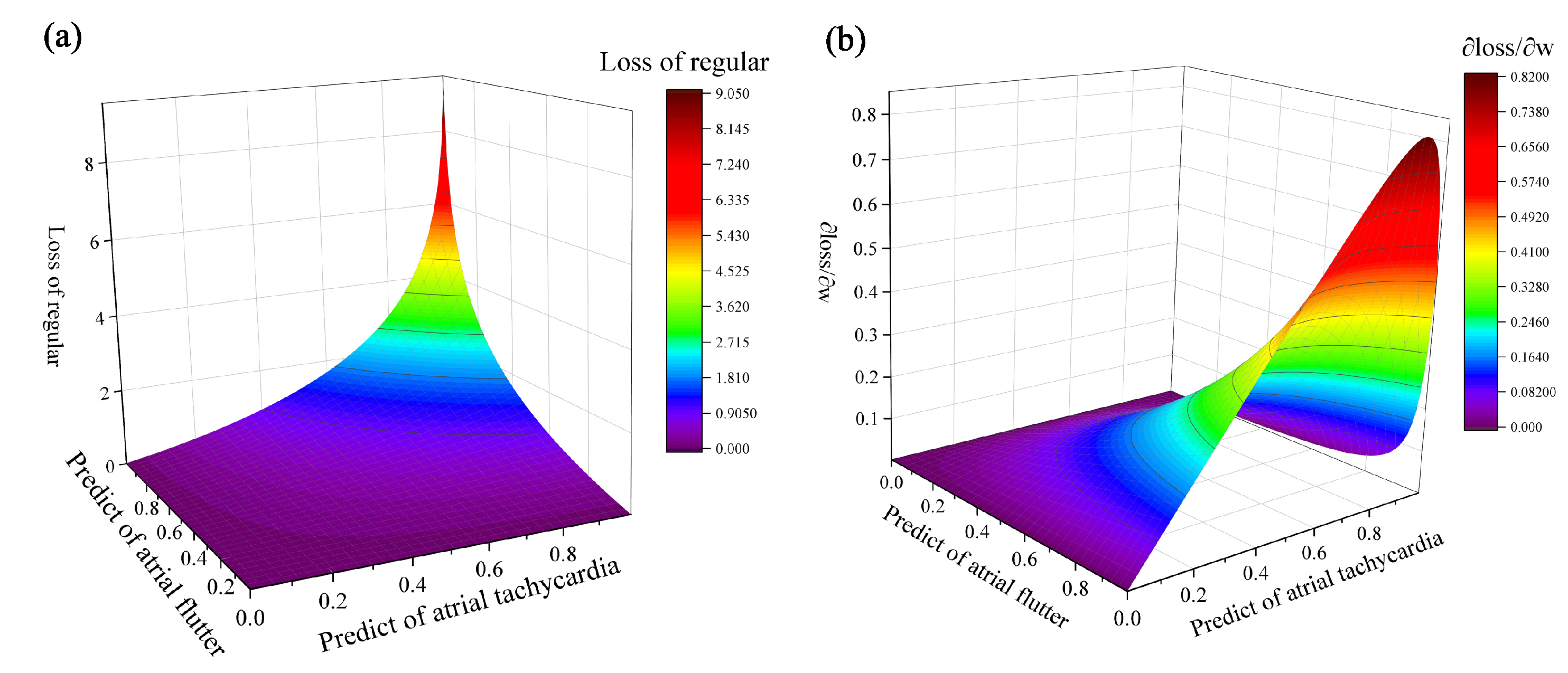
2.2. The Modified Cross-Entropy Loss Function
3. ECG Database
4. Experimental Setup and Analysis
4.1. Experimental Setup
4.2. Parameter Setting
4.2.1. The Deep Learning Model
4.2.2. The Modified Cross-Entropy Loss Function
4.3. Evaluation Indicators
4.4. Performance after Adding LSTM + ATTENTION
4.5. Performance of the Traditional Cross-Entropy Loss Function
4.6. Effectiveness of the Modified Cross-Entropy Loss Function
5. Discussion
6. Conclusions
Author Contributions
Funding
Data Availability Statement
Conflicts of Interest
References
- Geddes, J.S.; Warner, H.R. A PVC detection program. Comput. Biomed. Res. 1971, 4, 493–508. [Google Scholar] [CrossRef] [PubMed]
- Kezdi, P.; Naylor, W.S.; Rambousek, R.; Stanley, E. A practical heart rate and ectopic beat detector. J. Electrocardiol. 1968, 1, 213–219. [Google Scholar] [CrossRef]
- Arafat, M.A.; Chowdhury, A.W.; Hasan, M.K. A simple time domain algorithm for the detection of ventricular fibrillation in electrocardiogram. Signal Image Video Process. 2011, 5, 1–10. [Google Scholar] [CrossRef]
- Thakor, N.V.; Zhu, Y.S.; Pan, K.Y. Ventricular tachycardia and fibrillation detection by a sequential hypothesis testing algorithm. IEEE Trans. Biomed. Eng. 1990, 37, 837–843. [Google Scholar] [CrossRef] [PubMed]
- Anas, E.M.A.; Lee, S.Y.; Hasan, M.K. Sequential algorithm for life threatening cardiac pathologies detection based on mean signal strength and EMD functions. Biomed. Eng. Online 2010, 9, 1–22. [Google Scholar] [CrossRef] [PubMed]
- Jekova, I.; Krasteva, V. Real time detection of ventricular fibrillation and tachycardia. Physiol. Meas. 2004, 25, 1167. [Google Scholar] [CrossRef] [PubMed]
- Barro, S.; Ruiz, R.; Cabello, D.; Mira, J. Algorithmic sequential decision-making in the frequency domain for life threatening ventricular arrhythmias and imitative artefacts: A diagnostic system. J. Biomed. Eng. 1989, 11, 320–328. [Google Scholar] [CrossRef]
- Kuo, S. Computer detection of ventricular fibrillation. In Computers in Cardiology; IEEE Comupter Society: Washington, DC, USA, 1978; pp. 347–349. [Google Scholar]
- Zhang, X.S.; Zhu, Y.S.; Thakor, N.V.; Wang, Z.Z. Detecting ventricular tachycardia and fibrillation by complexity measure. IEEE Trans. Biomed. Eng. 1999, 46, 548–555. [Google Scholar] [CrossRef]
- Amann, A.; Tratnig, R.; Unterkofler, K. Detecting ventricular fibrillation by time-delay methods. IEEE Trans. Biomed. Eng. 2006, 54, 174–177. [Google Scholar] [CrossRef]
- Amann, A.; Tratnig, R.; Unterkofler, K. A new ventricular fibrillation detection algorithm for automated external defibrillators. In Computers in Cardiology; IEEE: New York, NY, USA, 2005; pp. 559–562. [Google Scholar]
- Li, H.; Han, W.; Hu, C.; Meng, M.Q.H. Detecting ventricular fibrillation by fast algorithm of dynamic sample entropy. In Proceedings of the 2009 IEEE International Conference on Robotics and Biomimetics (ROBIO), Guilin, China, 19–23 December 2009; pp. 1105–1110. [Google Scholar]
- Martis, R.J.; Acharya, U.R.; Mandana, K.M.; Ray, A.K.; Chakraborty, C. Cardiac decision making using higher order spectra. Biomed. Signal Process. Control 2013, 8, 193–203. [Google Scholar] [CrossRef]
- Roza, V.C.C.; de Almeida, A.M.; Postolache, O.A. Design of an artificial neural network and feature extraction to identify arrhythmias from ECG. In Proceedings of the 2017 IEEE International Symposium on Medical Measurements and Applications (MeMeA), Rochester, MN, USA, 7–10 May 2017; pp. 391–396. [Google Scholar]
- Yang, W.; Si, Y.; Wang, D.; Guo, B. Automatic recognition of arrhythmia based on principal component analysis network and linear support vector machine. Comput. Biol. Med. 2018, 101, 22–32. [Google Scholar] [CrossRef]
- Houssein, E.H.; Ibrahim, I.E.; Neggaz, N.; Hassaballah, M.; Wazery, Y.M. An efficient ECG arrhythmia classification method based on Manta ray foraging optimization. Expert Syst. Appl. 2021, 181, 115131. [Google Scholar] [CrossRef]
- Singh, A.K.; Krishnan, S. ECG Signal Feature Extraction Trends in Methods and Applications; BioMed Central: London, UK, 2023; Volume 22, pp. 1–36. [Google Scholar]
- Feng, N.; Xu, S.; Liang, Y.; Liu, K. A probabilistic process neural network and its application in ECG classification. IEEE Access 2019, 7, 50431–50439. [Google Scholar] [CrossRef]
- Yıldırım, Ö.; Pławiak, P.; Tan, R.S.; Acharya, U.R. Arrhythmia detection using deep convolutional neural network with long duration ECG signals. Comput. Biol. Med. 2018, 102, 411–420. [Google Scholar] [CrossRef] [PubMed]
- Luo, X.; Yang, L.; Cai, H.; Tang, R.; Chen, Y.; Li, W. Multi-classification of arrhythmias using a HCRNet on imbalanced ECG datasets. Comput. Methods Programs Biomed. 2021, 208, 106258. [Google Scholar] [CrossRef] [PubMed]
- Yao, Q.; Wang, R.; Fan, X.; Liu, J.; Li, Y. Multi-class arrhythmia detection from 12-lead varied-length ECG using attention-based time-incremental convolutional neural network. Inf. Fusion 2020, 53, 174–182. [Google Scholar] [CrossRef]
- Li, Y.; Zhang, Z.; Zhou, F.; Xing, Y.; Li, J.; Liu, C. Multi-Label Classification of Arrhythmia for Long-Term Electrocardiogram Signals with Feature Learning. IEEE Trans. Instrum. Meas. 2021, 70, 1–11. [Google Scholar] [CrossRef]
- Ran, S.; Li, X.; Zhao, B.; Jiang, Y.; Yang, X.; Cheng, C. Label correlation embedding guided network for multi-label ECG arrhythmia diagnosis. Knowl.-Based Syst. 2023, 270, 110545. [Google Scholar] [CrossRef]
- Yang, J.; Li, J.; Lan, K.; Wei, A.; Wang, H.; Huang, S.; Fong, S. Multi-Label Attribute Selection of Arrhythmia for Electrocardiogram Signals with Fusion Learning. Bioengineering 2022, 9, 268. [Google Scholar] [CrossRef]
- Yoo, J.; Jun, T.J.; Kim, Y.H. xECGNet: Fine-tuning attention map within convolutional neural network to improve detection and explainability of concurrent cardiac arrhythmias. Comput. Methods Programs Biomed. 2021, 208, 106281. [Google Scholar] [CrossRef]
- Yang, X.; Zhang, X.; Yang, M.; Zhang, L. 12-Lead ECG arrhythmia classification using cascaded convolutional neural network and expert feature. J. Electrocardiol. 2021, 67, 56–62. [Google Scholar] [CrossRef] [PubMed]
- Ge, Z.; Jiang, X.; Tong, Z.; Feng, P.; Zhou, B.; Xu, M.; Wang, Z.; Pang, Y. Multi-label correlation guided feature fusion network for abnormal ECG diagnosis. Knowl.-Based Syst. 2021, 233, 107508. [Google Scholar] [CrossRef]
- LeCun, Y.; Boser, B.; Denker, J.S.; Henderson, D.; Howard, R.E.; Hubbard, W.; Jackel, L.D. Backpropagation applied to handwritten zip code recognition. Neural Comput. 1989, 1, 541–551. [Google Scholar] [CrossRef]
- Hochreiter, S.; Schmidhuber, J. Long short-term memory. Neural Comput. 1997, 9, 1735–1780. [Google Scholar] [CrossRef] [PubMed]
- Yang, Z.; Yang, D.; Dyer, C.; He, X.; Smola, A.; Hovy, E. Hierarchical attention networks for document classification. In Proceedings of the 2016 Conference of the North American Chapter of the Association for Computational Linguistics: Human Language Technologies, 2016; pp. 1480–1489. [Google Scholar]
- He, T.; Zhang, L.; Guo, J.; Yi, Z. Multilabel classification by exploiting data-driven pair-wise label dependence. Int. J. Intell. Syst. 2020, 35, 1375–1396. [Google Scholar] [CrossRef]
- Gao, W.; Zhou, Z.H. On the consistency of multi-label learning. Artif. Intell. 2013, 199–200, 22–44. [Google Scholar] [CrossRef]
- Kobayashi, T. Two-Way Multi-Label Loss. In Proceedings of the IEEE Computer Society Conference on Computer Vision and Pattern Recognition, Vancouver, BC, Canada, 18–22 June 2023; pp. 7476–7485. [Google Scholar]
- Andreotti, F.; Carr, O.; Pimentel, M.A.F.; Mahdi, A.; De Vos, M. Comparing feature-based classifiers and convolutional neural networks to detect arrhythmia from short segments of ECG. In Proceedings of the IEEE 2017 Computing in Cardiology (CinC), Rennes, France, 24–27 September 2017; pp. 1–4. [Google Scholar]
- Jiang, Z.; Lai, Y.; Zhang, J.; Zhao, H.; Mao, Z. Multi-factor operating condition recognition using 1D convolutional long short-term network. Sensors 2019, 19, 5488. [Google Scholar] [CrossRef]
- He, K.; Zhang, X.; Ren, S.; Sun, J. Deep residual learning for image recognition. In Proceedings of the IEEE Computer Society Conference on Computer Vision and Pattern Recognition, Las Vegas, NV, USA, 27–30 June 2016; pp. 770–778. [Google Scholar]
- Simonyan, K.; Zisserman, A. Very deep convolutional networks for large-scale image recognition. In Proceedings of the 3rd International Conference on Learning Representations, ICLR 2015—Conference Track Proceedings, San Diego, CA, USA, 7–9 May 2015; pp. 1–14. [Google Scholar]
- Pirjatullah; Kartini, D.; Nugrahadi, D.T.; Muliadi; Farmadi, A. Hyperparameter Tuning using GridsearchCV on the Comparison of the Activation Function of the ELM Method to the Classification of Pneumonia in Toddlers. In Proceedings of the 2021 4th International Conference on Computer and Informatics Engineering: IT-Based Digital Industrial Innovation for the Welfare of Society, IC2IE 2021, Depok, Indonesia, 14–15 September 2021; pp. 390–395. [Google Scholar]


| Total | Normal | Sinus Tachycardia | Sinus Bradycardia | Atrial Flutter | Atrial Tachycardia | PVC | |
|---|---|---|---|---|---|---|---|
| Training | 23,322 | 9653 | 4462 | 4650 | 4557 | 2568 | 4194 |
| Validation | 2591 | 1057 | 524 | 555 | 455 | 292 | 458 |
| Test | 13,156 | 3728 | 2431 | 4816 | 2181 | 250 | 1692 |
| VGG16 + LSTM + ATTENTION | Layer Name | Kernel | Parameter | Output Size | Connected from |
| 1D VGG16 [34] | × | 3,713,648 | 44 × 512 | INPUT | |
| LSTM | 60 | 137,520 | 22 × 60 | 1D VGG16 | |
| Attention | × | 3720 | 1 × 60 | LSTM | |
| Dense | 64 | 3904 | 1 × 64 | Attention | |
| Dense | 6 | 390 | 1 × 6 | Dense | |
| Total Parameter: 3,859,182 | |||||
| ResNet34 + LSTM + ATTENTION | Layer Name | Kernel | Parameter | Output Size | Connected from |
| 1D ResNet34 [35] | × | 7,598,916 | 22 × 512 | INPUT | |
| LSTM | 60 | 137,520 | 22 × 60 | 1D ResNet34 | |
| Attention | × | 3720 | 1 × 60 | LSTM | |
| Dense | 64 | 3904 | 1 × 64 | Attention | |
| Dense | 6 | 390 | 1 × 6 | Dense | |
| Total Parameter: 8,320,266 | |||||
| ResNet50 + LSTM + ATTENTION | Layer Name | Kernel | Parameter | Output Size | Connected from |
| 1D ResNet50 [35] | × | 21,945,280 | 22 × 2048 | INPUT | |
| LSTM | 60 | 137,520 | 22 × 60 | 1D ResNet34 | |
| Attention | × | 3720 | 1 × 60 | LSTM | |
| Dense | 64 | 3904 | 1 × 64 | Attention | |
| Dense | 6 | 390 | 1 × 6 | Dense | |
| Total Parameter: 22,090,814 | |||||
| Model | VGG16 [34] | ResNet34 [35] | ResNet50 [35] | |||
|---|---|---|---|---|---|---|
| Add LSTM + ATTENTION | No | Yes | No | Yes | No | Yes |
| Error Num (Num) | 18 | 12 | 41 | 30 | 5 | 4 |
| Hamming Loss | 0.0200 | 0.0187 | 0.0250 | 0.0240 | 0.0252 | 0.0230 |
| Subset Accuracy | 0.9265 | 0.9392 | 0.9188 | 0.9245 | 0.9213 | 0.9269 |
| Jaccard Index | 0.9441 | 0.9521 | 0.9343 | 0.9397 | 0.9383 | 0.9417 |
| Precision | 0.9564 | 0.9561 | 0.9424 | 0.9435 | 0.9452 | 0.9495 |
| Recall | 0.9409 | 0.9491 | 0.9327 | 0.9380 | 0.9338 | 0.9363 |
| F1 score | 0.9474 | 0.9521 | 0.9364 | 0.9398 | 0.9352 | 0.9416 |
| Model | VGG16 + LSTM + ATTENTION | ResNet34 + LSTM + ATTENTION | ResNet50 + LSTM + ATTENTION | |||
|---|---|---|---|---|---|---|
| Loss function | Traditional | Modified | Traditional | Modified | Traditional | Modified |
| Error Num (Num) | 12 | 0 | 30 | 0 | 4 | 0 |
| Hamming Loss | 0.0187 | 0.0197 | 0.0240 | 0.0234 | 0.0230 | 0.0208 |
| Subset Accuracy | 0.9392 | 0.9287 | 0.9245 | 0.9179 | 0.9269 | 0.9242 |
| Jaccard Index | 0.9521 | 0.9419 | 0.9397 | 0.9313 | 0.9417 | 0.9392 |
| Precision | 0.9561 | 0.9595 | 0.9435 | 0.9499 | 0.9495 | 0.9549 |
| Recall | 0.9491 | 0.9360 | 0.9380 | 0.9283 | 0.9363 | 0.9340 |
| F1 score | 0.9521 | 0.9474 | 0.9398 | 0.9382 | 0.9416 | 0.9440 |
| Model | VGG16 + LSTM + ATTENTION | ResNet34 + LSTM + ATTENTION | ResNet50 + LSTM + ATTENTION | |||
|---|---|---|---|---|---|---|
| Loss function | Traditional | Modified | Traditional | Modified | Traditional | Modified |
| Normal | 0.9670 | 0.9707 | 0.9652 | 0.9676 | 0.9615 | 0.9678 |
| Sinus tachycardia | 0.9823 | 0.9826 | 0.9854 | 0.9879 | 0.9822 | 0.9825 |
| Sinus bradycardia | 0.9856 | 0.9853 | 0.9870 | 0.9649 | 0.9861 | 0.9752 |
| Atrial flutter | 0.9744 | 0.9786 | 0.9569 | 0.9769 | 0.9742 | 0.9770 |
| Atrial tachycardia | 0.9776 | 0.9870 | 0.9749 | 0.9806 | 0.9764 | 0.9869 |
| PVC | 0.9806 | 0.9785 | 0.9830 | 0.9405 | 0.9786 | 0.9481 |
Disclaimer/Publisher’s Note: The statements, opinions and data contained in all publications are solely those of the individual author(s) and contributor(s) and not of MDPI and/or the editor(s). MDPI and/or the editor(s) disclaim responsibility for any injury to people or property resulting from any ideas, methods, instructions or products referred to in the content. |
© 2023 by the authors. Licensee MDPI, Basel, Switzerland. This article is an open access article distributed under the terms and conditions of the Creative Commons Attribution (CC BY) license (https://creativecommons.org/licenses/by/4.0/).
Share and Cite
Zhu, J.; Ma, C.; Zhang, Y.; Huang, H.; Kong, D.; Ni, W. Multi-Label Diagnosis of Arrhythmias Based on a Modified Two-Category Cross-Entropy Loss Function. Electronics 2023, 12, 4976. https://doi.org/10.3390/electronics12244976
Zhu J, Ma C, Zhang Y, Huang H, Kong D, Ni W. Multi-Label Diagnosis of Arrhythmias Based on a Modified Two-Category Cross-Entropy Loss Function. Electronics. 2023; 12(24):4976. https://doi.org/10.3390/electronics12244976
Chicago/Turabian StyleZhu, Junjiang, Cheng Ma, Yihui Zhang, Hao Huang, Dongdong Kong, and Wangjin Ni. 2023. "Multi-Label Diagnosis of Arrhythmias Based on a Modified Two-Category Cross-Entropy Loss Function" Electronics 12, no. 24: 4976. https://doi.org/10.3390/electronics12244976