Review

Hybrid Quantum Dot as Promising Tools for Theranostic Application in Cancer

1
Department of Pharmaceutics, College of Pharmacy, Najran University, Najran 11001, Saudi Arabia
2
Institute of Pharmaceutical Research, GLA University, Mathura 281406, India
3
College of Pharmacy, Al-Dawadmi Campus, Shaqra University, Shaqra 11961, Saudi Arabia
4
BBS Institute of Pharmaceutical & Allied Science, Greater Noida 203201, India
5
Department of Pharmacology and Toxicology, National Institute of Pharmaceutical Education and Research (NIPER), Guwahati 781101, India
*
Author to whom correspondence should be addressed.
Electronics 2023, 12(4), 972; https://doi.org/10.3390/electronics12040972
Submission received: 31 December 2022 / Revised: 7 February 2023 / Accepted: 8 February 2023 / Published: 15 February 2023
(This article belongs to the Special Issue Quantum and Optoelectronic Devices, Circuits and Systems)

Abstract

:
Cancer is one of the leading causes of death worldwide. In the last few decades, cancer treatment has come a long way, but multidrug resistance (MDR) in cancer still has low survival rates. It means that much research is required for an accurate diagnosis and effective therapy. The new era of cancer research could include theranostic approaches and targeted delivery of chemotherapeutic agents utilizing the nanoparticulate system. Recently, there has been much interest gained among researchers for carbon-based and graphene-based quantum dots due to their higher biocompatibility and ease of biofunctionalization compared to conventional heavy metal quantum dots. Moreover, these quantum dots have various interesting utilities, including bioimaging, biosensing, quantum dots-mediated drug delivery, and their role in photodynamic therapy (PDT) and photothermal therapy (PTT). The current review highlighted the utility of hybrid quantum dots as a theranostic system in different cancers and discussed the various bio-molecules conjugated hybrid quantum dots investigated for diagnostic/therapeutic applications in cancer. The influence of conjugation of different biomolecules, such as folic acid, PEG, etc., with hybrid quantum dots on their biopharmaceutical attributes (such as aqueous solubility, tumor penetrability, stability of loaded therapeutics in the tumor microenvironment), delivery of drugs specifically to tumor tissues, and its therapeutic outcome in different cancer has also been discussed.

Graphical Abstract

1. Introduction

Cancer is the leading cause of death worldwide, accounting for nearly 10 million deaths in 2020 [1]. The correct diagnosis is crucial for accurate and effective treatment because all tumors need specific treatments such as surgery, radiotherapy, and chemotherapy. Novel therapeutic interventions and early diagnosis of cancer are major concerns for scientists, physicians, and healthcare professionals. In the last few decades, nanotechnology has been at the forefront of most cutting-edge research in many fields, such as medicine, diagnostics, bioimaging, and other biomedical applications [2]. Among the delivery approach of medicines in different cancers, various nanoparticles (NPs) are the most exploited carrier system for drug delivery in different cancer [3,4]. These carrier systems have different structures and dimensions, which may vary in size range from 1 to 100 nm, particularly for drug delivery in cancer. This gives them different physical and chemical properties that can be exploited for various purposes in cancer. The specific physicochemical properties of nanomaterials are exploited for focused ultrasonic heating therapy [5,6,7], radiofrequency (RF) ablation [8,9,10], magnetic fluid hyperthermia [11,12,13], and magnetic particle imaging [14,15] in cancer. NPs are widely exploited for bioimaging [16], drug delivery systems [17], therapeutic agents for photodynamic therapy (PDT) [18], photothermal therapy (PTT) [19], regenerative medicine [20], and smart biomaterials [21]. Furthermore, metallic NPs, especially gold [22], silver [23], platinum [24], and palladium [25], are the most investigated biocompatible NPs, for the diagnosis and treatment of cancer. Along with imaging agents to treat a wide range of diseases, including different carcinomas, the delivery of synthetic drugs, therapeutic peptides, and genes has raised interest in the theranostic approach, which can be simultaneously utilized for both diagnosis and treatment through a single system. Different nanomaterials have high penetrating efficiency to biological membranes, good biocompatibility, and can perform multiple functions due to their small size and ability to functionalization. This makes the utilization of nanomaterials a good candidate for theranostic application in different cancers [26].
Quantum dots (QDs) are a very small nanoparticulate system of organic (such as carbon-QDs, graphene-QDs) and inorganic nature (such as zinc sulfide–QDs, cadmium telluride-QDs, cadmium selenide-QDs) and vary in size ranges from 2 to 10 nm [27]. The small nano dimension is helpful to impart specific optical (high brightness, high quantum yield, high extinction coefficient, intermittent fluorescence signals, high stability against photobleaching) [28] and electronic properties that are exploited in different biomedical applications [27,28,29]. Its nanocrystal is distinguished by an energy band gap, required to excite an electron from one electronic band, i.e., a lower energy level, into another band, i.e., a higher energy level. Because they are so small, these nanocarrier systems of semiconductor origin can easily move electrons to a higher energy state, even when they are exposed to UV light. These properties of QDs are used in the diagnosis and treatment of various diseases, including cancer. This excitation scheme ultimately creates an electron-hole pair known as an exciton. The exciton gives off energy in the form of a fluorescent photon when it goes back to its ground state [30]. The 2–10 nm sized semiconducting nanocrystal started to act like the bulk Bohr exciton radius, and the particle’s electrical and optical properties changed [31]. The inverse relationship between nanocrystal size and energy band gap is well-documented and understood. The inverse property says that as the size of the nanocrystal decreases, the energy band gap increases, and the corresponding excitation/emission wavelengths decrease. The size of the particle can be changed to change the color of the light that the QDs give off when they are exposed to UV light. By adjusting the particle size and size distribution of the QDs, a wide absorbance range with highly symmetric and narrow emission spectra can be achieved [32]. The different types of QDs are prepared by the bottom-up approach, which involves the assembling of their precursor in the molecular state into nanocrystals [33]. The promising techniques for the preparation of QDs are categorized into four basic approaches, which include biotemplate-based synthesis, colloidal synthesis, biogenic synthesis, and electrochemical assembly [27,33]. These methods for the preparation of QDs are illustrated in Figure 1.
Carbon nanomaterials (specifically carbon nanotubes, carbon dots, graphene, and graphene derivatives) and other nanomaterials of organic and inorganic origin are popular for their extraordinary composition and excellent inherent properties for diverse applications such as fluorescent, fingerprinting, photocatalysis, electromagnetic shielding, and electric applications [3,34]. These nanomaterials have also made a pragmatic intervention in the field of biomedical engineering, contributing to research in tissue engineering, drug delivery, biosensing, bioimaging, and cancer theranostic [35]. It is functionalized with QDs as a hybrid nanoparticulate system called hybrid QDs, which have very small dimensions and theranostic utility in cancer. These hybrid systems are utilized to deliver drugs of synthetic/natural/biological origin and act as an imaging agent simultaneously, which are likely to increase their accumulation, specifically in the cancerous or tumor tissue. Thus, these versatile nanoparticulate systems as hybrid QDs have very wide utility in cancer detection/imaging and site-specific delivery of different types of therapeutics to the cancerous tissues (Illustrated in Figure 2).
The low drug efficacy in cancer may increase by improving the EPR (enhanced permeability and retention) effect and overcoming the tumor heterogeneity challenges through designing targeted hybrid QDs of a stealth nature [36]. RBC-camouflaged, light-responsive, carbon-based porous particles may be helpful in targeting and penetrating tumor tissues. Protein- and RBC membrane-targeted nanosponges improve targeting and circulation half-life. Porous carbon/silica and graphene QDs as hybrid systems are photoresponsive and tumor-penetrating drug carrier systems for theranostic application [37]. Cyclodextrins (CDs) are natural, water-soluble cyclic oligosaccharides with hydrophilic exteriors and hydrophobic interiors that are known for their utility in drug delivery applications. Their primary and secondary hydroxyl groups on the outside are easy to modify, and their lipophilic inner cavities can be filled with lipophilic moiety by the formation of an inclusion complex. These types of carriers are utilized to improve the aqueous solubility of water-insoluble therapeutics/imaging agents and serve as a promising carrier or drug delivery system in cancer management through hybridization with QDs for theranostic application due to the presence of numerous hydroxyl groups on their surface [38].
The present manuscript provides a detailed discussion and recent advancements in QDs for their utilization to improve the efficacy of loaded therapeutics and imaging applications in the effective management of cancer. It provided a discussion on hybrid quantum dots as a cancer theranostics and emphasized the recent research development (mainly in the last 5 years) in the area of cancer theranostics utilizing hybrid quantum dots.

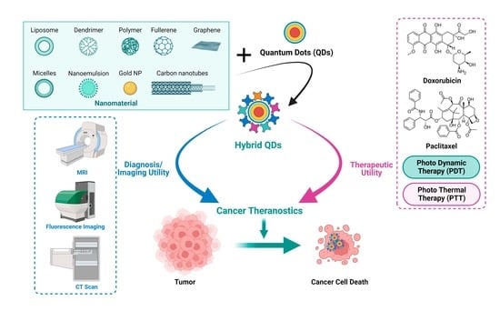
2. Significance of Hybrid Quantum Dot as Cancer Theranostics

The significance of the theranostic approach in cancer treatment may utilize the simultaneous delivery of chemotherapeutics and photolytic agents for deep tumor penetration, which effectively damages and inhibits the tumor when treated with single irradiation. It may be utilized for tracking of progress of cancer therapy using incorporated imaging agents [39]. Hybrid QDs have been widely explored for their theranostic application in different types of cancers. However, toxicity issues of QDs due to their composition (heavy or inorganic materials) and nature (ROS generation and strong surface responses) raised concern for their modification/functionalization for biomedical applications. Hence, strategies have been conceptualized to minimize their toxicity and improve their biocompatibility through hybridization/functionalization with other moieties (such as polymers, lipids, polysaccharides, proteins, etc.), providing efficient accumulation in tumor tissues in addition to preventing their accumulation in healthy tissues [40,41]. Biological molecules are attached to QDs using hydrophilic surfactant shells with reactive groups such as COOH, NH2, or SH. Attachments are made using different methods, such as adsorption, covalent bonding, electrostatic interaction, etc. [39,40,41]. It has been reported to be conjugated with a wide range of biological molecules, including biotin [42], folic acid [43,44], antibodies [45], and peptides [46]. Silanization, which coats QDs with silica, is a good covalent coating method for hydroxyl-rich material surfaces. Silanization makes ligand molecules strongly cross-linked and chemically stable. The end terminal groups of the silane shell can expose thiol, phosphonate, or methyl terminal ends for subsequent QD coating and also make the material more biocompatible. Silanization is favored because it is less toxic than other ligands [47]. Silica shell thickness could control QD light responsiveness. The silica-coated QDs were modified with amino, carboxyl, and epoxy groups and stabilized with PEG segments to assess their applicability. These modified QDs efficiently conjugated with antibodies and were used as fluorescent labels in immunoassay detection [48]. An in vivo study has shown that emissive Si-QDs biodegrade quickly and produce non-toxic silicic acid that may be eliminated by urine [49].
The perspectives of hybrid QDs for their utility in cancer diagnosis/imaging and delivery of payload specifically to tumor tissues are discussed in the subsequent section.

2.1. Perspectives of QDs for Diagnostic/Imaging Utility

QDs in drug delivery may be utilized as therapeutic/imaging cargo that has photothermal and photodynamic features, making them excellent for bioimaging. Many clinically used photosensitizers (PSs) are not tumor-targeted; hence, they are treated with spatially controlled irradiation. After phototherapies, PS can increase reactive oxygen species (ROS) formation in healthy cells; hence, light exposure should be avoided to reduce skin photosensitivity. The hybrid QDs possess low toxicity and good biocompatibility, coupled with stable photoluminescence (PL), and therefore these are ideal candidates for both in vitro and in vivo bioimaging [40,41]. QDs by themselves are not as efficient as molecular PSs, but QDs can be used as antennae to improve light harvesting and energy transfer to molecular PSs because they absorb much light. NPs are commonly utilized for bioimaging, but their toxicity limits their utility. Because fluorescence imaging is very sensitive and has a good temporal and spatial resolution, hybrid QDs are a good choice for sensing and imaging cell targets. Hybrid QDs are chemically inert, dissolve well in water, are photostable, have a relationship between their optoelectronic properties and their shape and size, have fluorescence resonance energy transfer, high stability in physiological conditions, specific accumulation at target sites, are easy to modify on the surface [50], and have a high absorption coefficient because of hybridized C–C bonds. Therefore, these are good phototherapy chromophores.
Carbon QDs (C-QDs) synthesized and dispersed with excellent fluorescence, photostability, photobleaching resistance, and simple coupling with biological species [51]. C-QDs can carry Ce6 and generate ROS. Using a 639 nm laser, water splitting produced oxygen and hydrogen in vivo. Increased oxygen yielded 1O2 to improve PDT. C-QDs with specific cell targets can particularly detect malignant cells in different investigations. C-QDs conjugated with folic acid (FA) (C-dots-FA) to distinguish folate receptor (FR)-positive cancer cells from normal cells (FR-negative) by growing and analyzing NIH-3T3 and HeLa cells. Pheophytin (a natural, low-toxicity Mg-free chlorophyll derivative) was employed as a raw carbon source to synthesize C-QDs using a microwave technique [52]. QDs containing sulfur and nitrogen are used as PTT (photothermal therapy causes ease of cell death by protein denaturation and loosening of the cellular membrane by heating the tumor tissue exploiting irradiation of radiofrequency, ultrasound, microwaves, and magnetic fields, etc.) and PDT (photodynamic therapy utilizes a photosensitizer that absorbs light of a particular wavelength and produces oxygen-based molecular species to induce a cytotoxic effect) for cancers in animals [53,54]. PL and photoacoustic imaging [55,56,57,58] benefited from high photon conversion efficiencies. Passive targeting of QDs around cancer cells destroyed the tumor. Co-doped C-QDs had a strong photothermal conversion, optical and photoacoustic performance, and renal excretion [53,59]. N–O-CQDs with significant NIR absorption. Combining the biocompatible N-doped carbon dots (N-CDs) with folic acid, which possess a wide range of high-targeting capabilities (26 types of tumor cell lines) and alters the cellular metabolism leading to autophagy, results in a new targeted tumor therapy based on autophagy regulation [60]. Similarly, maleimide-terminated TTA1 aptamers complexed with CDs (TTA1–CDs), which is substantially expressed in HeLa and C6 (rat glioma cell line) but not in normal healthy CHO cells, exhibit a strong fluorescence along cancer cell membranes and minimal uptake in healthy cells [61].
Graphene quantum dots (GQDs) are one type of nanocarrier that has been seen in physics and chemical research due to their ultrasmall size, varied photoluminescence, and mechanical features [62]. Ultra-tiny GQDs exploiting imaging agents and labeling cell membranes are promising agents for drug transportation in cancer therapy because of their outstanding optical properties and transmembrane capabilities. The innate immune system and tumor heterogeneity continue to pose challenges to efficient tumor targeting and penetration; however, NIR irradiation, the energy created by photothermal conversion, can not only release therapeutic cargo but also burst the vesicle to suppress the tumor [63]. When NPs first enter the circulatory system, the innate immune system quickly recognizes them as foreign bodies and gets rid of them through the reticuloendothelial system and the mononuclear phagocyte system. This results in poor delivery efficiency. Because the tumor is a strong physiological barrier, only a small part of the dose injected gets to the deep tumor through the increased EPR effect, which helps particle accumulation. The high interstitial fluid pressure (IFP) and cancer-associated fibroblasts in tumors make it hard for therapeutic drugs to reach the perivascular cells of tumors [64,65]. Thus, in order to improve tumor therapy, it is crucial to create stealthy and permeable drug delivery systems for the efficient transportation of therapeutic agents.
For imaging or diagnostic applications, theranostic nanoplatforms must be robust enough to support them, have a superior cargo-loading and -releasing profile, and be able to do so. Hybridization between distinct NPs is a promising strategy because it can result in the accumulation of a wide range of chemical, physical, and biological properties inside a single complex. Because of their exceptional physical and chemical properties [32,66], GQDs have been put to use in a variety of biomedical settings. If GQD fluorescence could be made stable, it would greatly improve the efficiency of imaging in the life sciences. Due to their unique chemical, physical, and biological properties, graphene quantum dots (GQDs) and magnetic nanoparticles (MNPs) are two promising choices for use in these hybrids. Both magnetic resonance imaging (MRI) and computed tomography (CT) use contrast agents made of magnetic nanoparticles [67]. In addition to its use in biosensing and magnetic separation, this nanoparticle may also be put to use in hyperthermia therapy, thermo-ablation, targeted drug administration, and even bio-sensing. For example, magnetic nanoparticles could be added to GQDs to make even more complexes that could be used in medicine. The most commonly used magnetic nanoparticles are iron oxide NPs (usually Fe2O3 or Fe3O4), which can be classified as a pure metal, metal oxide, or magnetic nanocomposites [68]. Combining GQDs with other NPs, such as magnetic nanoparticles, could make them even better for use in biology.
Carbon quantum dots (CQDs), a novel kind of fluorescent carbon nanomaterial possessing the unique advantages of high stability, remarkable biocompatibility, easy synthesis and surface functionalization, and comparable optical characteristics, have been extensively studied, especially for bioimaging applications due to their tunable strong fluorescence emission property [69]. In light of the drawbacks of conventional chemotherapy, PTT has emerged as a viable option for treating cancer. Tumors can be heated from the inside out by injecting photothermal substances into the affected area or by targeting the tumor with other agents. These photothermal agents are designed to stimulate near-infrared (NIR) radiation and generate heat upon relaxation, killing cancer cells. Researchers have been investigating several facets of theranostic nanosystems [66] since it has been postulated that these systems, which meet both diagnostic and therapeutic needs, could be utilized to effectively eradicate cancer cells. It has been found that a wide range of organic, inorganic, organo-inorganic, and combinations can act as photothermal agents. Carbon nanomaterials, including carbon nanotubes, graphene, and graphene derivatives, have garnered significant attention due to their potential applications in fields as diverse as fingerprinting, photocatalysis, electromagnetic shielding, and electrics [70,71]. Due to their luminescence, versatile surface chemistry, easy cellular internalization, and high biocompatibility, CQDs are particularly promising in drug delivery. Additionally, the nano-formulation system also offers the possibility to increase drug solubility, bioavailability, and half-life. Although doxorubicin (DOX) is widely used for cancer treatment, it has many disadvantages, including a low EPR effect, low cellular internalization, and cytotoxicity to normal cells [72]. One approach to bypassing these problems is to use a multifunctional nanocarrier system for tumor-targeted drug delivery, which has the advantage of accumulating at the tumor site due to the increased EPR effect.

2.2. Perspectives of QDs for Therapeutic Utility

Different QD-based therapeutic systems for anticancer application have been widely explored in recent years [73,74]. Various studies have been conducted to investigate the potential of QDs for targeted drug delivery, PDT, PTT, and gene delivery in cancer treatment [75,76]. The QDs for cancer therapy have been investigated both at in vitro and in vivo levels. PDT is one of the most promising non-invasive cancer treatment approaches with limited side effects. It can be used alone or in combination with surgery, chemotherapy, or ionizing radiation to destroy undetected cancerous cells at the margins of resection. PDT uses photosensitizing drugs that are pharmacologically inactive until a particular light wavelength irradiates them in the presence of oxygen, which generates reactive oxygen species and induces cell death and tissue necrosis [77,78]. Graphene oxide (GO), an oxidized version of graphite, has received considerable attention during the past decade. GO, on the other hand, can be dispersed in water, which makes it a good candidate to investigate in a biological system. On the other hand, graphenes need to be surface functionalized to make them dispersible in water and safer for the biological environment [79,80]. Their synthesis and surface tailoring entail hazardous and toxic reagents, traces of which may remain with the material to demonstrate further toxicity in vitro/in vivo systems due to GO sheets having intrinsic toxicity. By delivering medications and energy in two different locations at the same time, a hybrid system of nano dimension may be able to lessen the adverse effects of cancer treatment and improve the distinctive properties required for precision medicine. The hybrid carrier systems are frequently eliminated from blood circulation very quickly, and piled-up tumors at the periphery close to the blood arteries. It has a short elimination half-life in blood and high tumor penetration. The membrane of a red blood cell (RBC) was given the appearance of a sponge by being composed of carbon composite. When it is exposed to light, it functions as both a “stealth agent” and a “photolytic carrier”. It means that it transfers “tumor-penetrating agents” (such as graphene QDs and docetaxel) as well as heat. When compared to the nanosponge, the RBC-membrane-enveloped nanosponge demonstrates an eight-fold increase in accumulation in tumor tissue. This is because the RBC-membrane-enveloped nanosponge will be integrated with a specific protein that accumulates in tumor spheroids through high lateral bilayer fluidity [81]. The delivery of graphene QDs to tumor areas is accomplished by passing near-infrared light through a structure that is only one atom thick. This makes it much simpler for its therapeutic utilization to penetrate the cancerous tissue and improves the prognosis of cancer therapy utilizing the theranostic approach.
The pathway of accumulation and removal of QDs and hybrid QDs in in vivo systems are depicted in Figure 3.
Different types of hybrid QD-based theranostic systems were explored for cancer applications to improve the biopharmaceutical attributes of this nanoparticulate system (such as aqueous solubility, tumor penetrability, and stability of loaded therapeutics in the tumor microenvironment) and delivery of drug specifically to tumor tissues. Recent contemporary research conducted in this field is discussed in the subsequent section.

3. Hybrid Quantum Dot as Cancer Theranostics: Contemporary Research

3.1. Diagnostic Application

Pei et al. developed fluorescent hyper-cross-linked-cyclodextrin–carbon quantum dot (CD-CQD) hybrid nanosponges with outstanding biocompatibility and intense bright blue fluorescence excited at 365 nm with a PLQY of 38.0% [82]. These hybrid QDs systems were generated by simple condensation polymerization of carbon quantum dots (CQDs) with cyclodextrin (CD) at a 1:5 feeding ratio for theranostic applications, specifically in malignancies. In another investigation, Fateh et al. made a hybrid nanostructure of graphene quantum dots (GQDs) and magnetic nanoparticles (MNP) by using hydrophobic interactions between long carbon chains on the surface of GQDs around the edges and MNP in the middle [83]. Pyrolysis was used to create GQDs, which were then changed using cetyl alcohol (CA) to produce surfactant-modified GQDs (CA-GQDs). Moreover, an oleate-iron complex has been utilized to make iron-oxide nanoparticles (IONP) as MNP. After that, CA-GQDs and IONP are mixed to make a structure with IONP in the middle and CA-GQDs all around it (IONP@CA-GQD). IONP@CA-GQD possesses both fluorescence and magnetic characteristics. At room temperature, IONP and IONP@CA-GQD have been tested for magnetization hysteresis loops in a moving magnetic field. There have been no observations of coercivity or remanence, indicating super-paramagnetism. The computed MS values for IONP and IONP@CA-GQD are 34.1 emu/g and 37.8 emu/g, respectively. Because of GQDs are fluorescent in nature, this hybrid structure could also be used for bioimaging [84,85].
A stable compound of graphene oxide (GO) and graphene quantum dots (GQD) was created by Kumavat et al. by electrostatic layer-by-layer assembly using a polyethylene imine bridge (GO-PEI-GQDs) [86]. In addition, various applications of the mono-equivalents of the GO-PEI-GQDs complex were compared, including cell imaging (diagnostics), photothermal, and oxidative stress responses in MDA-MB-231 breast cancer cells. When exposed to an 808 nm laser for 5 min at a concentration of up to 50 μg/mL, GO-PEI-GQDs displayed an outstanding photothermal response (44–49 °C). According to the study, GO-PEI-GQDs had synergistic effects on cancer cells. It has stable fluorescence imaging, improved photothermal effects, and cytotoxic actions. Composite materials made of GO and GQDs combine many different properties, which makes it possible to improve certain therapeutic systems, such as cancer theranostics [86].
Hyaluronic acid and QDs together have been proven to be useful tools for improving intracellular transport into liver cells. This is accomplished by interacting with CD44-receptors, which allows for in vivo real-time imaging [87,88]. The anionic polysaccharide chondroitin sulfate was employed to coat the positively charged oily core of the cadmium telluride (CdTe) QDs as cancer theranostic nanocapsules [89], which were also loaded with rapamycin and celecoxib as anticancer therapeutics [90]. Chondroitin sulfate nanocapsules have an exterior coating of cationic gelatin-coupled QDs placed on them to prevent non-specific uptake by healthy cells. Matrix metalloproteinase (MMPs) dissolved the gelatin at the tumor location, releasing therapeutic nanocapsules and QDs into cancer cells for therapeutic and imaging action as a cancer therapeutics. An ON–OFF effect, where the fluorescence of QDs was first quenched by energy transfer and then restored after bond cleavage in tumor cells, was seen in a study that substituted lactoferrin for gelatin [90]. Thus, using QDs fluorescence, the in vitro and in vivo localization of nanocapsules into breast tumors was observed.
Recent research related to hybrid QDs utilized for their diagnostic/imaging applicability in different types of cancers is summarized in Table 1.

3.2. Therapeutic Application

Pei et al. formulated doxorubicin (DOX) loaded-fluorescent hyper-cross-linked-cyclodextrin–carbon quantum dot (CD-CQD) hybrid nanosponges (DOX-β-CD-CQD) with a size of around 300 nm with a DOX loading capacity of 39.5% through host−guest complexation [82]. This is because of the supramolecular complexation of DOX with the CD units in the CD-CQD nanosponges. The developed DOX-CD-CQD nanosponges demonstrated pH-responsive controlled release in the simulated tumor microenvironment. The loaded DOX molecules in the surface layer of the DOX-CD-CQD were released in the first 30 h, similar to the pH 7.4 medium. Due to the higher solubility of DOX in acidic media attributed to its protonation, the supramolecular complexation of DOX with β-CD units had a lower inclusion constant and a greater release ratio than in pH 7.4 conditions. Due to the high formation constant, they took longer to get out of the loaded DOX inner layer. Protonated DOX diffusion was prevented by hydrophobic DOX-complexed CD. After 12 h of DOX release, with an accumulative release of approximately 50%, hydrophilic outer shells formed, facilitating protonated DOX diffusion out of the theranostic system. After 24 h of incubation, the DOX concentration gradient climbed to 1.7 μg/mL with the DOX-β-CD-CQD theranostic system concentration of 20 μg/mL. Cell viability (29%) was comparable to free DOX at 10 μg/mL. In terms of antitumor efficacy, the DOX-CD-CQD outperformed free DOX. The DOX-β-CD-CQD had an IC50 of 5.00 μg/mL (equivalent to 0.425 μg/mL), compared to 2.26 μg/mL for free DOX. DOX-CD-CQD was internalized by HepG2 cells and accumulated in their nuclei, exhibiting better anticancer activity than the free drug [82]. In another investigation, Fateh et al. developed a hybrid nanostructure of cetyl alcohol-modified graphene quantum dots (CA-GQDs) and conjugated them with iron-oxide nanoparticles (IONP@CA-GQD) [44]. The study indicated no effect on the normal architecture of the liver and cardiac tissues after administration of these hybrid QDs at a dose of 3 mg/kg for 7 days in mice. Hence, IONP@CA-GQD can be offered as a potential drug delivery system for cancer theranostics. Moreover, IONP@CA-GQD was found to be more toxic for the tumor cells as compared to normal cells in the study [39,83]. It is due to the hydrophobic nature of the carbon chains of cetyl alcohol and oleic acid in the middle of IONP@CA-GQD, hydrophobic drugs can be loaded in this space [83,104]. Furthermore, the magnetic properties of IONP@CA-GQD would make cancer targeting feasible through this theranostic system. Similarly, Kim et al. examined QD-labeled hyaluronic acid (HA) derivatives for liver-targeted intracellular drug delivery. EDC activation of HA’s carboxyl group and conjugation to ADH’s amine group produced HA-ADH conjugates. After EDC and sufo-NHS activation of QD carboxyl groups, HA-ADH conjugates were tagged with QDs via amide bond formation. HA binds CD44 via its three carboxyl groups [88]. HA-QD conjugates were endocytosed via HA receptor-mediated endocytosis, as seen in the confocal microscopic images of B16F1 cells. HA receptors such as CD44 are significantly expressed in B16F1 cells. In the case of HEK293 cells without HA receptors, the cellular uptake of HA conjugates and QDs conjugates was noticeably reduced (as shown in Figure 4).
In order to deliver targeted anticancer drugs, Chen et al. created a core-shell structured multifunctional nanocarrier system (ZnO-Au-PLA-GPPS-FA) consisting of ZnO-quantum dots-conjugated AuNPs as the core and folic acid (FA)-conjugated amphiphilic hyperbranched block copolymers as the shell. ZnO-quantum dots-conjugated AuNPs could be employed for photothermal therapy to kill tumor cells and fluorescent labeling, respectively. The outer hydrophilic block (GPPS-FA) and the inner hydrophobic block (PLA) were both biocompatible and biodegradable in in vivo system. The cancer cells may be targeted by an FA-conjugated multifunctional nanocarrier system, which may then be absorbed by the target cell through receptor-mediated endocytosis. Additionally, the presence of GPPS on the surface of multifunctional nanocarrier systems resulted in some of their anticancer effects [105]. In another investigation, Sung et al. made a targeted RBC-membrane-encased nanosponge (RBC-NS) that combines stealth and huge payloads of functional molecules to avoid the low EPR effect and the different types of tumors. This biocompatible, light-sensitive, carbon-based, porous particle looks like red blood cells (RBCs) and targets and gets into tumors well [39]. The targeted nanosponge, made of protein/RBC membranes (targeting and stealth properties), porous carbon/silica (hydrophobic, therapeutic agent transport), and graphene QDs (GQDs)/drug (photoresponsive, tumor-penetrating), were injected into a mouse model to deliver docetaxel (DTX) and GQDs to tumors (Figure 5). Moreover, Cetuximab (Ct), which can target tumors, is attached to the RBC layer to make it easier for particles to gather around tumors. The nanosponge delivers high amounts of GQD/DTX to the tumor as a photo-penetrative and photolytic agent. The Ct-RBC-GQD/NS-treated tumor can be heated to 68 °C for thermal tumor ablation. Ct-RBC-NS and Ct-NS elevated tumor temperatures to 62 and 53 °C, respectively. Irradiated saline-treated mice showed no temperature increase. Ct-RBC-GQD/stronger NS’s improved photothermal conversion may be explained by the accumulation and photothermal combination effects. The localized heat of the NS releases GQD/DTX during NIR exposure, damaging the tumor and improving therapeutic drug penetration (as shown in Figure 5).
Contemporary research related to hybrid QDs utilized to improve the therapeutic performance of loaded drugs for anticancer activity in different types of cancers are summarized in Table 2.

4. Conclusions

The review concludes that hybrid QDs could be multi-modeled to treat different cancers, and therapeutic progress could be monitored in real-time. These QDs combined with different types of nanoparticulate systems (such as NPs of polymeric, lipid, and inorganic origin) to develop a theranostic system for cancer, particularly to improve the therapy outcome in MDR cancer. Further, this review concluded that carbon-based and graphene-based QDs had been extensively explored to conjugate them with different bio-molecules to overcome the challenges associated with conventional QDs. Several preclinical studies showed that hybrid QDs could be successfully used as a theranostic system in cancer, bringing them closer to investigating its clinical utility. However, the literature review reveals that the clinical performance of hybrid QDs as cancer theranostics has not been addressed in detail as yet. Furthermore, the safety perspectives of the hybrid QDs in cancer should also be addressed systematically in future investigations as they may be accumulated in the healthy tissues due to failure of tumor-specific delivery that may increase the risks of untoward events. Although numerous in vivo studies have examined the distribution, accumulation, excretion, and toxic consequences of QDs, no consensus has been established. Moreover, due to the complexity of in vivo models, the replication of pharmacokinetics is difficult. However, certain in-vitro studies eased our basic understanding of mechanisms and possible adverse effects of various QDs. The type of QDs has an impact on their distribution within cells and clearance rate, which is directly related to their cytotoxicity. Based on the local accumulation and biological half-life, the possible cytotoxic potential of QDs can be anticipated. Thus, systematic investigation of the safety and efficacy of hybrid QDs should be of prime concern.

Author Contributions

J.A.: conceptualization, investigation, formal analysis, and validation; writing—original draft preparation, writing—review and editing, supervision, funding acquisition, and project administration; A.G.: investigation, formal analysis, validation, writing—original draft preparation; G.M.: investigation, formal analysis, validation, writing—original draft preparation; M.Z.A.: formal analysis, writing—review and editing, funding acquisition, project administration; M.A.: investigation, formal analysis, validation, writing—review and editing; A.M.: formal analysis, validation, writing—review and editing; All authors have read and agreed to the published version of the manuscript.

Funding

This work was funded by the Deanship of Scientific Research at Najran University, Saudi Arabia, under the National Research Priorities funding program grant code (NU/NRP/MRC/11/5).

Institutional Review Board Statement

Not applicable.

Data Availability Statement

Not applicable.

Acknowledgments

The authors are thankful to the Deanship of Scientific Research at Najran University for funding this work under the National Research Priorities funding program grant code (NU/NRP/MRC/11/5).

Conflicts of Interest

The authors declare no conflict of interest.

References

  1. Available online: https://www.who.int/news-room/fact-sheets/detail/cancer (accessed on 5 December 2022).
  2. Kemp, J.A.; Kwon, Y.J. Cancer nanotechnology: Current status and perspectives. Nano Converg. 2021, 8, 34. [Google Scholar] [CrossRef]
  3. Singh, R.; Deshmukh, R. Carbon nanotube as an emerging theranostic tool for oncology. J. Drug Deliv. Sci. Technol. 2022, 74, 103586. [Google Scholar] [CrossRef]
  4. Siddique, S.; Chow, J.C. Recent advances in functionalized nanoparticles in cancer theranostics. Nanomaterials 2022, 12, 2826. [Google Scholar] [CrossRef]
  5. Bernard, V.; Zobač, O.; Sopoušek, J.; Mornstein, V. AgCu bimetallic nanoparticles under effect of low intensity ultrasound: The cell viability study in vitro. J. Cancer Res. 2014, 2014, 971769. [Google Scholar] [CrossRef] [Green Version]
  6. Zhao, Y.; Zhu, Y.; Fu, J.; Wang, L. Effective Cancer Cell Killing by Hydrophobic Nanovoid-Enhanced Cavitation under Safe Low-Energy Ultrasound. Chem. Asian J. 2014, 9, 790–796. [Google Scholar] [CrossRef] [PubMed]
  7. Miller, D.L.; Smith, N.B.; Bailey, M.R.; Czarnota, G.J.; Hynynen, K.; Makin, I.R.S. Bioeffects Committee of the American Institute of Ultrasound in Medicine. (2012). Overview of therapeutic ultrasound applications and safety considerations. J. Ultrasound Med. 2012, 31, 623–634. [Google Scholar] [CrossRef] [PubMed] [Green Version]
  8. Ashikbayeva, Z.; Aitkulov, A.; Atabaev, T.S.; Blanc, W.; Inglezakis, V.J.; Tosi, D. Green-Synthesized Silver Nanoparticle–Assisted Radiofrequency Ablation for Improved Thermal Treatment Distribution. Nanomaterials 2022, 12, 426. [Google Scholar] [CrossRef]
  9. Glazer, E.S.; Curley, S.A. Non-invasive radiofrequency ablation of malignancies mediated by quantum dots, gold nanoparticles and carbon nanotubes. Ther. Deliv. 2011, 2, 1325–1330. [Google Scholar] [CrossRef] [Green Version]
  10. Nguyen, D.T.; Tzou, W.S.; Zheng, L.; Barham, W.; Schuller, J.L.; Shillinglaw, B.; Sauer, W.H. Enhanced radiofrequency ablation with magnetically directed metallic nanoparticles. Circ. Arrhythm. Electrophysiol. 2016, 9, e003820. [Google Scholar] [CrossRef]
  11. Ranoo, S.; Lahiri, B.B.; Nandy, M.; Philip, J. Enhanced magnetic heating efficiency at acidic pH for magnetic nanoemulsions stabilized with a weak polyelectrolyte. J. Colloid Interface Sci. 2020, 579, 582–597. [Google Scholar] [CrossRef]
  12. Ranoo, S.; Lahiri, B.B.; Damodaran, S.P.; Philip, J. Tuning magnetic heating efficiency of colloidal dispersions of iron oxide nano-clusters by varying the surfactant concentration during solvothermal synthesis. J. Mol. Liq. 2022, 360, 119444. [Google Scholar] [CrossRef]
  13. Dutz, S.; Buske, N.; Landers, J.; Gräfe, C.; Wende, H.; Clement, J.H. Biocompatible magnetic fluids of co-doped iron oxide nanoparticles with tunable magnetic properties. Nanomaterials 2020, 10, 1019. [Google Scholar] [CrossRef]
  14. Du, Y.; Liu, X.; Liang, Q.; Liang, X.J.; Tian, J. Optimization and design of magnetic ferrite nanoparticles with uniform tumor distribution for highly sensitive MRI/MPI performance and improved magnetic hyperthermia therapy. Nano Lett. 2019, 19, 3618–3626. [Google Scholar] [CrossRef] [PubMed]
  15. Dadfar, S.M.; Camozzi, D.; Darguzyte, M.; Roemhild, K.; Varvarà, P.; Metselaar, J.; Lammers, T. Size-isolation of superparamagnetic iron oxide nanoparticles improves MRI, MPI and hyperthermia performance. J. Nanobiotechnol. 2020, 18, 22. [Google Scholar] [CrossRef]
  16. Sharma, P.; Brown, S.; Walter, G.; Santra, S.; Moudgil, B. Nanoparticles for bioimaging. Adv. Colloid Interface Sci. 2006, 123, 471–485. [Google Scholar] [CrossRef]
  17. Couvreur, P. Nanoparticles in drug delivery: Past, present and future. Adv. Drug Deliv. Rev. 2013, 65, 21–23. [Google Scholar] [CrossRef]
  18. Lucky, S.S.; Soo, K.C.; Zhang, Y. Nanoparticles in photodynamic therapy. Chem. Rev. 2015, 115, 1990–2042. [Google Scholar] [CrossRef]
  19. Jaque, D.; Maestro, L.M.; del Rosal, B.; Haro-Gonzalez, P.; Benayas, A.; Plaza, J.L.; Martin Rodriguez, E.; Solé, J.G. Nanoparticles for photothermal therapies. Nanoscale 2014, 6, 9494–9530. [Google Scholar] [CrossRef]
  20. Calabrese, G.; Petralia, S.; Franco, D.; Nocito, G.; Fabbi, C.; Forte, L.; Guglielmino, S.; Squarzoni, S.; Traina, F.; Conoci, S. A new Ag-nanostructured hydroxyapatite porous scaffold: Antibacterial effect and cytotoxicity study. Mater. Sci. Eng. C 2021, 118, 111394. [Google Scholar] [CrossRef] [PubMed]
  21. Mele, E. Introduction: Smart Materials in Biomedicine. In Smart Nanoparticles for Biomedicine; Elsevier: Amsterdam, The Netherlands, 2008; pp. 1–13. [Google Scholar]
  22. Dreaden, E.C.; Alkilany, A.M.; Huang, X.; Murphy, C.J.; El-Sayed, M.A. The golden age: Gold nanoparticles for biomedicine. Chem. Soc. Rev. 2012, 41, 2740–2779. [Google Scholar] [CrossRef]
  23. Eckhardt, S.; Brunetto, P.S.; Gagnon, J.; Priebe, M.; Giese, B.; Fromm, K.M. Nanobio Silver: Its Interactions with Peptides and Bacteria, and Its Uses in Medicine. Chem. Rev. 2013, 113, 4708–4754. [Google Scholar] [CrossRef] [Green Version]
  24. Jeyaraj, M.; Gurunathan, S.; Qasim, M.; Kang, M.H.; Kim, J.H. A Comprehensive Review on the Synthesis, Characterization, and Biomedical Application of Platinum Nanoparticles. Nanomaterials 2019, 9, 1719. [Google Scholar] [CrossRef] [PubMed] [Green Version]
  25. Phan, T.T.V.; Huynh, T.C.; Manivasagan, P.; Mondal, S.; Oh, J. An Up-To-Date Review on Biomedical Applications of Palladium Nanoparticles. Nanomaterials 2020, 10, 66. [Google Scholar] [CrossRef] [Green Version]
  26. Jain, A.; Tiwari, A.; Verma, A.; Saraf, S.; Jain, S.K. Combination cancer therapy using multifunctional liposomes. Crit. Rev.™ Ther. Drug Carr. Syst. 2020, 37, 105–134. [Google Scholar] [CrossRef] [PubMed]
  27. Abdellatif, A.A.; Younis, M.A.; Alsharidah, M.; Al Rugaie, O.; Tawfeek, H.M. Biomedical applications of quantum dots: Overview, challenges, and clinical potential. Int. J. Nanomed. 2022, 17, 1951–1970. [Google Scholar] [CrossRef]
  28. Wagner, A.M.; Knipe, J.M.; Orive, G.; Peppas, N.A. Quantum dots in biomedical applications. Acta Biomater. 2019, 94, 44–63. [Google Scholar] [CrossRef]
  29. Bajwa, N.; Mehra, N.K.; Jain, K.; Jain, N.K. Pharmaceutical and biomedical applications of quantum dots. Artif. Cells Nanomed. Biotechnol. 2016, 44, 758–768. [Google Scholar] [CrossRef]
  30. Liu, M.; Yazdani, N.; Yarema, M.; Jansen, M.; Wood, V.; Sargent, E.H. Colloidal quantum dot electronics. Nat. Electron. 2021, 4, 548–558. [Google Scholar] [CrossRef]
  31. Smith, A.M.; Duan, H.; Mohs, A.M.; Nie, S. Bioconjugated quantum dots for in vivo molecular and cellular imaging. Adv. Drug Deliv. Rev. 2008, 60, 1226–1240. [Google Scholar] [CrossRef] [Green Version]
  32. Xu, Q.; Gao, J.; Wang, S.; Wang, Y.; Liu, D.; Wang, J. Quantum dots in cell imaging and their safety issues. J. Mater. Chem. B 2021, 9, 5765–5779. [Google Scholar] [CrossRef]
  33. Devi, S.; Kumar, M.; Tiwari, A.; Tiwari, V.; Kaushik, D.; Verma, R.; Batiha, G.E.S. Quantum dots: An emerging approach for cancer therapy. Front. Mater. 2022, 8, 585. [Google Scholar] [CrossRef]
  34. Rahman, M.A.; Abul Barkat, H.; Harwansh, R.K.; Deshmukh, R. Carbon-based Nanomaterials: Carbon Nanotubes, Graphene, and Fullerenes for the Control of Burn Infections and Wound Healing. Curr. Pharm. Biotechnol. 2022, 23, 1483–1496. [Google Scholar] [CrossRef] [PubMed]
  35. Kanthi Gudimella, K.; Gedda, G.; Kumar, P.S.; Babu, B.K.; Yamajala, B.; Rao, B.V.; Singh, P.P.; Kumar, D.; Sharma, A. Novel synthesis of fluorescent carbon dots from bio-based Carica Papaya Leaves: Optical and structural properties with antioxidant and anti-inflammatory activities. Environ. Res. 2022, 204, 111854. [Google Scholar] [CrossRef]
  36. Zhang, H.; Jin, Y.; Chi, C.; Han, G.; Jiang, W.; Wang, Z.; Cheng, H.; Zhang, C.; Wang, G.; Sun, C.; et al. Sponge particulates for biomedical applications: Biofunctionalization, multi-drug shielding, and theranostic applications. Biomaterials 2021, 273, 120824. [Google Scholar] [CrossRef]
  37. Ghaffarkhah, A.; Hosseini, E.; Kamkar, M.; Sehat, A.A.; Dordanihaghighi, S.; Allahbakhsh, A.; van der Kuur, C.I.; Arjmand, M. Synthesis, applications, and prospects of graphene quantum dots: A comprehensive review. Small 2022, 18, 2102683. [Google Scholar] [CrossRef]
  38. Paduraru, D.N.; Niculescu, A.G.; Bolocan, A.; Andronic, O.; Grumezescu, A.M.; Bîrlă, R. An Updated overview of cyclodextrin-based drug delivery systems for cancer therapy. Pharmaceutics 2022, 14, 1748. [Google Scholar] [CrossRef] [PubMed]
  39. Sung, S.Y.; Su, Y.L.; Cheng, W.; Hu, P.F.; Chiang, C.S.; Chen, W.T.; Hu, S.H. Graphene quantum dots-mediated theranostic penetrative delivery of drug and photolytics in deep tumors by targeted biomimetic nanosponges. Nano Lett. 2019, 19, 69–81. [Google Scholar] [CrossRef]
  40. Yang, K.; Wan, J.; Zhang, S.; Zhang, Y.; Lee, S.-T.; Liu, Z. In Vivo Pharmacokinetics, Long-Term Biodistribution, and Toxicology of PEGylated Graphene in Mice. ACS Nano 2011, 5, 516–522. [Google Scholar] [CrossRef] [PubMed]
  41. Zhang, S.; Yang, K.; Feng, L.; Liu, Z. In vitro and in vivo behaviors of dextran functionalized graphene. Carbon 2011, 49, 4040–4049. [Google Scholar] [CrossRef]
  42. Lv, C.; Lin, Y.; Liu, A.A.; Hong, Z.Y.; Wen, L.; Zhang, Z.; Pang, D.W. Labeling viral envelope lipids with quantum dots by harnessing the biotinylated lipid-self-inserted cellular membrane. Biomaterials 2016, 106, 69–77. [Google Scholar] [CrossRef]
  43. Hashemkhani, M.; Muti, A.; Sennaroğlu, A.; Acar, H.Y. Multimodal image-guided folic acid targeted Ag-based quantum dots for the combination of selective methotrexate delivery and photothermal therapy. J. Photochem. Photobiol. B Biol. 2020, 213, 112082. [Google Scholar] [CrossRef] [PubMed]
  44. Singh, G.; Kumar, M.; Soni, U.; Arora, V.; Bansal, V.; Gupta, D.; Singh, H. Cancer cell targeting using folic acid/anti-HER2 antibody conjugated fluorescent CdSe/CdS/ZnS-Mercaptopropionic acid and CdTe-Mercaptosuccinic acid quantum dots. J. Nanosci. Nanotechnol. 2016, 16, 130–143. [Google Scholar] [CrossRef] [PubMed]
  45. Sahoo, S.L.; Liu, C.H.; Kumari, M.; Wu, W.C.; Wang, C.C. Biocompatible quantum dot-antibody conjugate for cell imaging, targeting and fluorometric immunoassay: Crosslinking, characterization and applications. RSC Adv. 2019, 9, 32791–32803. [Google Scholar] [CrossRef] [Green Version]
  46. Huang, H.; Bai, Y.L.; Yang, K.; Tang, H.; Wang, Y.W. Optical imaging of head and neck squamous cell carcinoma in vivo using arginine-glycine-aspartic acid peptide conjugated near-infrared quantum dots. OncoTargets Ther. 2013, 6, 1779–1787. [Google Scholar]
  47. Lu, J.; Liong, M.; Li, Z.; Zink, J.I.; Tamanoi, F. Biocompatibility, biodistribution, and drug-delivery efficiency of mesoporous silica nanoparticles for cancer therapy in animals. Small 2010, 6, 1794–1805. [Google Scholar] [CrossRef] [PubMed] [Green Version]
  48. Li, C.; Zou, Z.; Liu, H.; Jin, Y.; Li, G.; Yuan, C.; Jin, M. Synthesis of polystyrene-based fluorescent quantum dots nanolabel and its performance in H5N1 virus and SARS-CoV-2 antibody sensing. Talanta 2021, 225, 122064. [Google Scholar] [CrossRef]
  49. Park, J.H.; Gu, L.; Von Maltzahn, G.; Ruoslahti, E.; Bhatia, S.N.; Sailor, M.J. Biodegradable luminescent porous silicon nanoparticles for in vivo applications. Nat. Mater. 2009, 8, 331–336. [Google Scholar] [CrossRef]
  50. Zayed, D.G.; AbdElhamid, A.S.; Freag, M.S.; Elzoghby, A.O. Hybrid quantum dot-based theranostic nanomedicines for tumor-targeted drug delivery and cancer imaging. Nanomedicine 2019, 14, 225–228. [Google Scholar] [CrossRef] [PubMed] [Green Version]
  51. Liu, W.; Li, C.; Ren, Y.; Sun, X.; Pan, W.; Li, Y.; Wang, J.; Wang, W. Carbon dots: Surface engineering and applications. J. Mater. Chem. B 2016, 4, 5772–5788. [Google Scholar] [CrossRef] [PubMed]
  52. Li, B.; Lin, L.; Lin, H.; Wilson, B.C. Photosensitized singlet oxygen generation and detection: Recent advances and future perspectives in cancer photodynamic therapy. J. Biophotonics 2016, 9, 1314–1325. [Google Scholar] [CrossRef] [PubMed] [Green Version]
  53. Das, R.K.; Panda, S.; Bhol, C.S.; Bhutia, S.K.; Mohapatra, S. N-doped carbon quantum dot (NCQD)-Deposited carbon capsules for synergistic fluorescence imaging and photothermal therapy of oral cancer. Langmuir 2019, 35, 15320–15329. [Google Scholar] [CrossRef] [PubMed]
  54. Sekar, R.; Basavegowda, N.; Jena, S.; Jayakodi, S.; Elumalai, P.; Chaitanyakumar, A.; Baek, K.H. Recent Developments in Heteroatom/Metal-Doped Carbon Dot-Based Image-Guided Photodynamic Therapy for Cancer. Pharmaceutics 2022, 14, 1869. [Google Scholar] [CrossRef]
  55. Proskurnin, M.A.; Khabibullin, V.R.; Usoltseva, L.O.; Vyrko, E.A.; Mikheev, I.V.; Volkov, D.S. Photothermal and optoacoustic spectroscopy: State of the art and prospects. Phys.-Uspekhi 2022, 65, 270. [Google Scholar] [CrossRef]
  56. Walter, M.; Schubert, L.; Heberle, J.; Schlesinger, R.; Losi, A. Time-resolved photoacoustics of channelrhodopsins: Early energetics and light-driven volume changes. Photochem. Photobiol. Sci. 2022, 1–10. [Google Scholar] [CrossRef]
  57. He, Z.; Zhang, C.Y.; Lei, Y.; Song, G.; Yao, Y. Plasmonic nanomaterials: A versatile phototheranostic platform of cancers. Materials Today 2022. [Google Scholar] [CrossRef]
  58. Gellini, C.; Feis, A. Optothermal properties of plasmonic inorganic nanoparticles for photoacoustic applications. Photoacoustics 2021, 23, 100281. [Google Scholar] [CrossRef]
  59. Song, Z.; Quan, F.; Xu, Y.; Liu, M.; Cui, L.; Liu, J. Multifunctional N, S co-doped carbon quantum dots with pH-and thermo-dependent switchable fluorescent properties and highly selective detection of glutathione. Carbon 2016, 104, 169–178. [Google Scholar] [CrossRef]
  60. Liu, B.; Wei, S.; Liu, E.; Zhang, H.; Lu, P.; Wang, J.; Sun, G. Nitrogen-doped carbon dots as a fluorescent probe for folic acid detection and live cell imaging. Spectrochim. Acta Part A Mol. Biomol. Spectrosc. 2022, 268, 120661. [Google Scholar] [CrossRef]
  61. Lee, C.H.; Rajendran, R.; Jeong, M.S.; Ko, H.Y.; Joo, J.Y.; Cho, S.; Chang, Y.W.; Kim, S. Bioimaging of targeting cancers using aptamer-conjugated carbon nanodots. Chem. Commun. 2013, 49, 6543–6545. [Google Scholar] [CrossRef] [PubMed]
  62. Bacon, M.; Bradley, S.J.; Nann, T. Graphene quantum dots. Part. Part. Syst. Charact. 2014, 31, 415–428. [Google Scholar] [CrossRef]
  63. Liu, H.; Li, C.; Qian, Y.; Hu, L.; Fang, J.; Tong, W.; Wang, H. Magnetic-induced graphene quantum dots for imaging-guided photothermal therapy in the second near-infrared window. Biomaterials 2020, 232, 119700. [Google Scholar] [CrossRef]
  64. Sun, B.; Luo, C.; Yu, H.; Zhang, X.; Chen, Q.; Yang, W.; Wang, M.; Kan, Q.; Zhang, H.; Wang, Y.; et al. Disulfide bond-driven oxidation-and reduction-responsive prodrug nanoassemblies for cancer therapy. Nano Lett. 2018, 18, 3643–3650. [Google Scholar] [CrossRef]
  65. Zhu, L.; Zhao, H.; Zhou, Z.; Xia, Y.; Wang, Z.; Ran, H.; Li, P.; Ren, J. Peptide-functionalized phase-transformation nanoparticles for low intensity focused ultrasound-assisted tumor imaging and therapy. Nano Lett. 2018, 18, 1831–1841. [Google Scholar] [CrossRef] [PubMed]
  66. Chen, H.; Zhang, W.; Zhu, G.; Xie, J.; Chen, X. Rethinking cancer nanotheranostics. Nat. Rev. Mater. 2017, 2, 17024. [Google Scholar] [CrossRef] [PubMed] [Green Version]
  67. Avasthi, A.; Caro, C.; Pozo-Torres, E.; Leal, M.P.; García-Martín, M.L. Magnetic nanoparticles as MRI contrast agents. Top. Curr. Chem. 2020, 378, 40. [Google Scholar] [CrossRef]
  68. Pourmadadi, M.; Rahmani, E.; Shamsabadipour, A.; Mahtabian, S.; Ahmadi, M.; Rahdar, A.; Díez-Pascual, A.M. Role of Iron Oxide (Fe2O3) Nanocomposites in Advanced Biomedical Applications: A State-of-the-Art Review. Nanomaterials 2022, 12, 3873. [Google Scholar] [CrossRef] [PubMed]
  69. Jana, P.; Dev, A. Carbon quantum dots: A promising nanocarrier for bioimaging and drug delivery in cancer. Mater. Today Commun. 2022, 32, 104068. [Google Scholar] [CrossRef]
  70. Sasidharan, S.; Bahadur, D.; Srivastava, R. Protein-poly (amino acid) nanocore–shell mediated synthesis of branched gold nanostructures for computed tomographic imaging and photothermal therapy of cancer. ACS Appl. Mater. Interfaces 2016, 8, 15889–15903. [Google Scholar] [CrossRef]
  71. Zhu, Y.; Murali, S.; Cai, W.; Li, X.; Suk, J.W.; Potts, J.R.; Ruoff, R.S. Graphene and graphene oxide: Synthesis, properties, and applications. Adv. Mater. 2010, 22, 3906–3924. [Google Scholar] [CrossRef] [PubMed]
  72. Navya, P.N.; Kaphle, A.; Srinivas, S.P.; Bhargava, S.K.; Rotello, V.M.; Daima, H.K. Current trends and challenges in cancer management and therapy using designer nanomaterials. Nano Converg. 2019, 6, 23. [Google Scholar] [CrossRef] [Green Version]
  73. Luo, G.; Long, J.; Zhang, B.; Liu, C.; Ji, S.; Xu, J.; Ni, Q. Quantum dots in cancer therapy. Expert Opin. Drug Deliv. 2012, 9, 47–58. [Google Scholar] [CrossRef] [PubMed]
  74. Kumar, A.; Singh, K.R.; Ghate, M.D.; Lalhlenmawia, H.; Kumar, D.; Singh, J. Bioinspired quantum dots for cancer therapy: A mini-review. Mater. Lett. 2022, 313, 131742. [Google Scholar] [CrossRef]
  75. Zhang, H.; Yee, D.; Wang, C. Quantum dots for cancer diagnosis and therapy: Biological and clinical perspectives. Nanomedicine 2008, 3, 83–91. [Google Scholar] [CrossRef] [PubMed]
  76. Taghavi, S.; Abnous, K.; Taghdisi, S.M.; Ramezani, M.; Alibolandi, M. Hybrid carbon-based materials for gene delivery in cancer therapy. J. Control Release 2020, 318, 158–175. [Google Scholar] [CrossRef]
  77. Robertson, C.A.; Evans, D.H.; Abrahamse, H. Photodynamic therapy (PDT): A short review on cellular mechanisms and cancer research applications for PDT. J. Photochem. Photobiol. B Biol. 2009, 96, 1–8. [Google Scholar] [CrossRef]
  78. Thomsen, H.; Marino, N.; Conoci, S.; Sortino, S.; Ericson, M.B. Confined photo-release of nitric oxide with simultaneous two-photon fluorescence tracking in a cellular system. Sci. Rep. 2018, 8, 9753. [Google Scholar] [CrossRef] [Green Version]
  79. Zhao, X.; Wei, Z.; Zhao, Z.; Miao, Y.; Qiu, Y.; Yang, W.; Jia, X.; Liu, Z.; Hou, H. Design and development of graphene oxide nanoparticle/chitosan hybrids showing pH-sensitive surface charge-reversible ability for efficient intracellular doxorubicin delivery. ACS Appl. Mater. Interfaces 2018, 10, 6608–6617. [Google Scholar] [CrossRef]
  80. Zhuang, W.; He, L.; Wang, K.; Ma, B.; Ge, L.; Wang, Z.; Huang, J.; Wu, J.; Zhang, Q.; Ying, H. Combined adsorption and covalent linking of paclitaxel on functionalized nano-graphene oxide for inhibiting cancer cells. ACS Omega 2018, 3, 2396–2405. [Google Scholar] [CrossRef]
  81. Zhao, C.; Song, X.; Liu, Y.; Fu, Y.; Ye, L.; Wang, N.; Wang, F.; Li, L.; Mohammadniaei, M.; Zhang, M.; et al. Synthesis of graphene quantum dots and their applications in drug delivery. J. Nanobiotechnol. 2020, 18, 1–32. [Google Scholar] [CrossRef]
  82. Pei, M.; Pai, J.Y.; Du, P.; Liu, P. Facile synthesis of fluorescent hyper-cross-linked β-cyclodextrin-carbon quantum dot hybrid nanosponges for tumor theranostic application with enhanced antitumor efficacy. Mol. Pharm. 2018, 15, 4084–4091. [Google Scholar] [CrossRef]
  83. Fateh, S.T.; Kamalabadi, M.A.; Aliakbarniya, A.; Jafarinejad-Farsangi, S.; Koohi, M.; Jafari, E.; Karam, Z.M.; Keyhanfar, F.; Dezfuli, A.S. Hydrophobic@ amphiphilic hybrid nanostructure of iron-oxide and graphene quantum dot surfactant as a theranostic platform. OpenNano 2022, 6, 100037. [Google Scholar] [CrossRef]
  84. Schroeder, K.L.; Goreham, R.V.; Nann, T. Graphene quantum dots for theranostics and bioimaging. Pharm. Res. 2016, 33, 2337–2357. [Google Scholar] [CrossRef]
  85. Dezfuli, A.S.; Kohan, E.; Fateh, S.T.; Alimirzaei, N.; Arzaghi, H.; Hamblin, M.R. Organic dots (O-dots) for theranostic applications: Preparation and surface engineering. RSC Adv. 2021, 11, 2253–2291. [Google Scholar] [CrossRef]
  86. Kumawat, M.K.; Thakur, M.; Bahadur, R.; Kaku, T.; Prabhuraj, R.S.; Ninawe, A.; Srivastava, R. Preparation of gra-phene oxide-graphene quantum dots hybrid and its application in cancer theranostics. Mater. Sci. Eng. C 2019, 103, 109774. [Google Scholar] [CrossRef] [PubMed]
  87. Kim, K.S.; Hur, W.; Park, S.J.; Hong, S.W.; Choi, J.E.; Goh, E.J.; Yoon, S.K.; Hahn, S.K. Bioimaging for targeted deliv-ery of hyaluronic acid derivatives to the livers in cirrhotic mice using quantum dots. ACS Nano 2010, 4, 3005–3014. [Google Scholar] [CrossRef]
  88. Kim, K.S.; Kim, S.; Beack, S.; Yang, J.A.; Yun, S.H.; Hahn, S.K. In vivo real-time confocal microscopy for tar-get-specific delivery of hyaluronic acid-quantum dot conjugates. Nanomed. Nanotechnol. Biol. Med. 2012, 8, 1070–1073. [Google Scholar] [CrossRef]
  89. AbdElhamid, A.S.; Helmy, M.W.; Ebrahim, S.M.; Bahey-El-Din, M.; Zayed, D.G.; Zein El Dein, E.A.; El-Gizawy, S.A.; El-zoghby, A.O. Layer-by-layer gelatin/chondroitin quantum dots-based nanotheranostics: Combined rapamy-cin/celecoxib delivery and cancer imaging. Nanomedicine 2018, 13, 1707–1730. [Google Scholar] [CrossRef] [PubMed]
  90. AbdElhamid, A.S.; Zayed, D.G.; Helmy, M.W.; Ebrahim, S.M.; Bahey-El-Din, M.; Zein-El-Dein, E.A.; El-Gizawy, S.A.; El-zoghby, A.O. Lactoferrin-tagged quantum dots-based theranostic nanocapsules for combined COX-2 inhibi-tor/herbal therapy of breast cancer. Nanomedicine 2018, 13, 2637–2656. [Google Scholar] [CrossRef] [PubMed]
  91. Chowdhury, A.D.; Ganganboina, A.B.; Tsai, Y.C.; Chiu, H.C.; Doong, R.A. Multifunctional GQDs-Concanavalin A@ Fe3O4 nanocomposites for cancer cells detection and targeted drug delivery. Anal. Chim. Acta 2018, 1027, 109–120. [Google Scholar] [CrossRef]
  92. Su, X.; Chan, C.; Shi, J.; Tsang, M.K.; Pan, Y.; Cheng, C.; Gerile, O.; Yang, M. A graphene quantum dot@ Fe3O4@ SiO2 based nanoprobe for drug delivery sensing and dual-modal fluorescence and MRI imaging in cancer cells. Biosens. Bi-Oelectron. 2017, 92, 489–495. [Google Scholar] [CrossRef]
  93. Bao, X.; Yuan, Y.; Chen, J.; Zhang, B.; Li, D.; Zhou, D.; Jing, P.; Xu, G.; Wang, Y.; Holá, K.; et al. In vivo theranostics with near-infrared-emitting carbon dots—Highly efficient photothermal therapy based on passive targeting after intravenous administration. Light Sci. Appl. 2018, 7, 91. [Google Scholar] [CrossRef] [PubMed]
  94. Bhunia, S.K.; Saha, A.; Maity, A.R.; Ray, S.C.; Jana, N.R. Carbon nanoparticle-based fluorescent bioimaging probes. Sci. Rep. 2013, 3, 1473. [Google Scholar] [CrossRef] [Green Version]
  95. Huang, X.; Zhang, F.; Zhu, L.; Choi, K.Y.; Guo, N.; Guo, J.; Tackett, K.; Anilkumar, P.; Liu, G.; Quan, Q.; et al. Effect of injection routes on the biodistribution, clearance, and tumor uptake of carbon dots. ACS Nano 2013, 7, 5684–5693. [Google Scholar] [CrossRef] [Green Version]
  96. Liu, C.; Zhang, P.; Zhai, X.; Tian, F.; Li, W.; Yang, J.; Liu, Y.; Wang, H.; Wang, W.; Liu, W. Nano-carrier for gene de-livery and bioimaging based on carbon dots with PEI-passivation enhanced fluorescence. Biomaterials 2012, 33, 3604–3613. [Google Scholar] [CrossRef]
  97. Zhang, M.; Wang, W.; Cui, Y.; Chu, X.; Sun, B.; Zhou, N.; Shen, J. Magnetofluorescent Fe3O4/carbon quantum dots coated single-walled carbon nanotubes as dual-modal targeted imaging and chemo/photodynamic/photothermal tri-ple-modal therapeutic agents. Chem. Eng. J. 2018, 338, 526–538. [Google Scholar] [CrossRef]
  98. Yang, X.Q.; Chen, C.; Peng, C.W.; Hou, J.X.; Liu, S.P.; Qi, C.B.; Gong, Y.P.; Zhu, X.B.; Pang, D.W.; Li, Y. Quantum dot-based quantitative immunofluorescence detection and spectrum analysis of epidermal growth factor receptor in breast cancer tissue arrays. Int. J. Nanomed. 2011, 6, 2265. [Google Scholar]
  99. Martins, C.S.; LaGrow, A.P.; Prior, J.A. Quantum Dots for Cancer-Related miRNA Monitoring. ACS Sens. 2022, 7, 1269–1299. [Google Scholar] [CrossRef]
  100. Lv, S.; Chen, F.; Chen, C.; Chen, X.; Gong, H.; Cai, C. A novel CdTe quantum dots probe amplified resonance light scattering signals to detect microRNA-122. Talanta 2017, 165, 659–663. [Google Scholar] [CrossRef] [PubMed]
  101. Volsi, A.L.; Fiorica, C.; D’Amico, M.; Scialabba, C.; Palumbo, F.S.; Giammona, G.; Licciardi, M. Hybrid Gold/Silica/Quantum-Dots supramolecular-nanostructures encapsulated in polymeric micelles as potential theranostic tool for targeted cancer therapy. Eur. Polym. J. 2018, 105, 38–47. [Google Scholar] [CrossRef]
  102. Jabeen, G.; Ahmad, M.H.; Aslam, M.; Riaz, S.; Hayat, A.; Nawaz, M.H. N-Doped graphene quantum dots (N-GQDs) as fluorescent probes for detection of UV induced DNA damage. RSC Adv. 2022, 12, 22458–22464. [Google Scholar] [CrossRef]
  103. Kwon, J.; Jun, S.W.; Choi, S.I.; Mao, X.; Kim, J.; Koh, E.K.; Kim, Y.H.; Kim, S.K.; Hwang, D.Y.; Kim, C.S.; et al. FeSe quantum dots for in vivo multiphoton biomedical imaging. Sci. Adv. 2019, 5, eaay0044. [Google Scholar] [CrossRef] [PubMed] [Green Version]
  104. Hsiao, M.H.; Mu, Q.; Stephen, Z.R.; Fang, C.; Zhang, M. Hexanoyl-chitosan-PEG copolymer coated iron oxide nano-particles for hydrophobic drug delivery. ACS Macro Lett. 2015, 4, 403–407. [Google Scholar] [CrossRef]
  105. Chen, T.; Zhao, T.; Wei, D.; Wei, Y.; Li, Y.; Zhang, H. Core–shell nanocarriers with ZnO quantum dots-conjugated Au nanoparticle for tumor-targeted drug delivery. Carbohydr. Polym. 2013, 92, 1124–1132. [Google Scholar] [CrossRef] [PubMed]
  106. Zheng, S.; Jin, Z.; Han, C.; Li, J.; Xu, H.; Park, S.; Park, J.O.; Choi, E.; Xu, K. Graphene quantum dots-decorated hol-low copper sulfide nanoparticles for controlled intracellular drug release and enhanced photothermal-chemotherapy. J. Mater. Sci. 2020, 55, 1184–1197. [Google Scholar] [CrossRef]
  107. Yang, L.; Wang, Z.; Wang, J.; Jiang, W.; Jiang, X.; Bai, Z.; He, Y.; Jiang, J.; Wang, D.; Yang, L. Doxorubicin conjugated functionalizable carbon dots for nucleus targeted delivery and enhanced therapeutic efficacy. Nanoscale 2016, 8, 6801–6809. [Google Scholar] [CrossRef] [Green Version]
  108. Ahmad, J.; Wahab, R.; Siddiqui, M.A.; Musarrat, J.; Al-Khedhairy, A.A. Zinc oxide quantum dots: A potential candi-date to detain liver cancer cells. Bioprocess Biosyst. Eng. 2015, 38, 155–163. [Google Scholar] [CrossRef] [PubMed]
  109. Rahman, M.M.; Opo, F.A.; Asiri, A.M. Cytotoxicity Study of Cadmium-Selenium Quantum Dots (Cdse QDs) for De-stroying the Human HepG2 Liver Cancer Cell. J. Biomed. Nanotechnol. 2021, 17, 2153–2164. [Google Scholar] [CrossRef]
  110. Fakhri, A.; Tahami, S.; Nejad, P.A. Preparation and characterization of Fe3O4-Ag2O quantum dots decorated cellu-lose nanofibers as a carrier of anticancer drugs for skin cancer. J. Photochem. Photobiol. B Biol. 2017, 175, 83–88. [Google Scholar] [CrossRef] [PubMed]
  111. Chu, M.; Pan, X.; Zhang, D.; Wu, Q.; Peng, J.; Hai, W. The therapeutic efficacy of CdTe and CdSe quantum dots for photothermal cancer therapy. Biomaterials 2012, 33, 7071–7083. [Google Scholar] [CrossRef]
  112. Prasad, R.; Jain, N.K.; Yadav, A.S.; Jadhav, M.; Radharani, N.N.V.; Gorain, M.; Srivastava, R. Ultrahigh Penetration and Retention of Graphene Quantum Dot Mesoporous Silica Nanohybrids for Image Guided Tumor Regression. ACS Appl. Bio Mater. 2021, 4, 1693–1703. [Google Scholar] [CrossRef]
  113. Ghafary, S.M.; Rahimjazi, E.; Hamzehil, H.; Mousavi, S.M.M.; Nikkhah, M.; Hosseinkhani, S. Design and preparation of a theranostic peptideticle for targeted cancer therapy: Peptide-based codelivery of doxorubicin/curcumin and graphene quantum dots. Nanomed. Nanotechnol. Biol. Med. 2022, 42, 102544. [Google Scholar] [CrossRef] [PubMed]
  114. Wang, Y.; Chen, J.; Tian, J.; Wang, G.; Luo, W.; Huang, Z.; Fan, X. Tryptophan-sorbitol based carbon quantum dots for theranostics against hepatocellular carcinoma. J. Nanobiotechnol. 2022, 20, 78. [Google Scholar] [CrossRef] [PubMed]
  115. Li, Y.; Zhang, P.; Tang, W.; McHugh, K.J.; Kershaw, S.V.; Jiao, M.; Han, B. Bright, magnetic NIR-II quantum dot probe for sensitive dual-modality imaging and intensive combination therapy of cancer. ACS Nano 2022, 16, 8076–8094. [Google Scholar] [CrossRef] [PubMed]
Figure 1. Schematic illustration of commonly used preparation approaches for QDs: (A) Colloidal synthesis approach. (B) Biotemplate-based synthesis approach. (C) Electrochemical assembly approach. (D) Biogenic synthesis approach. Reproduced from Abdellatif et al. [27], Dove Medical Press, 2022.
Electronics 12 00972 g001
Figure 2. Illustration highlighting the utility of hybrid QDs as cancer theranostics for various applications.
Electronics 12 00972 g002
Figure 3. Schematic illustration highlights the accumulation and removal of QDs versus hybrid QDs. QDs are more prone to RES clearance and renal clearance compared to hybrid QDs of a stealth nature. The more targeted delivery of hybrid QDs compared to QDs resulted in major accumulation in tumor tissues due to receptor-mediated endocytosis and the EPR effect, leading to improved therapeutic outcomes. “Image created with BioRender.com”.
Electronics 12 00972 g003
Figure 4. Illustration shows the cellular uptake of hyaluronic acid (HA) conjugated hybrid QDs after 2 h incubation in B16F1 cells—a high expression of HA receptors (A); HEK293 cells—without HA receptors (B). Confocal microscopic images reveal low cellular uptake of developed hybrid QDs system in HEK293 cells. Reproduced from Kim et al. [88], Elsevier, 2012.
Electronics 12 00972 g004
Figure 5. Schematic illustration highlights the penetration and accumulation of hybrid QDs (graphene quantum dots—GQDs) for RBC-membrane enveloped nanosponge-mediated targeted delivery in a tumor: (A) After the application of near-infrared (NIR) irradiation, generated heat leads to the penetration and accumulation of developed theranostic systems (GQDs with DTX) to deep tumors and the release of drug (DTX) into tumor cells ultimately causes cancer cell death. (B) Cellular uptake of RBC-membrane enveloped nanosponge with cetuximab (Ct-RBC@NS) and without cetuximab conjugation (RBC@NS) upon incubation for 2 h in A549 cancer cells and control RAW 264.7 cells. (a) Developed system conjugated with cetuximab (Ct-RBC@NS) in A549 cancer cells. It is monitored in the cytoplasm (green) and nuclei (blue). (b) Developed system without conjugation of cetuximab (RBC@NS) in A549 cancer cells. It is monitored in the cytoplasm (green) and nuclei (blue). (c) Developed system conjugated with cetuximab (Ct-RBC@NS) in control RAW 264.7 cells. (d) Developed system without conjugation of cetuximab (RBC@NS) in control RAW 264.7 cells. Reprinted with permission from Sung et al. [39], Copyright 2018, American Chemical Society.
Electronics 12 00972 g005
Table 1. Summary of contemporary research carried out utilizing hybrid quantum dots for diagnostic/imaging applications in cancer.
Type of QDsType of CancerDiagnostic/Imaging TechniqueOutcomeRefs.
Lactoferrin QDsBreast cancerFluorescence imagingIntracellular uptake of QDs showed fluorescence fluorescent due to mercaptopropionic acid-capped cadmium telluride and was successfully used as theranostic[89]
(P:2018)
Gelatin/chondroitin QDs Breast cancer Fluorescence imagingMatrix metalloproteinase layer enabled tracing their internalization into cancer cells and strong non-immunogenic response used as diagnostic[90]
(P:2018)
Magnetic graphene-QDsCancer cellsElectrochemical detection imagingImages show high fluorescence in HeLa cells[91]
(P:2018)
Graphene-QDsCancer cellsMRI and fluorescence imagingMRI and fluorescence imaging of living Hela cells and monitored intracellular drug release[92]
(P:2017)
Carbon-QDsTumor cellsPhotoluminescence and photoacoustic imagingAccumulation of C-QDs around the cancer cells via passive targeting with no active targeting species with fluorescence imaging[93]
(P:2018)
Carbon-QDsCervical cancerFluorescence imagingTAT functionalization enhanced cell labeling and uptake, and that folate selectively tagged tumor cells[94]
(P:2013)
Carbon-QDs doped with Fluorine and NitrogenSquamous cell carcinomaNear-infrared fluorescence (NIRF) and PET imagingCarbon-QDs rapidly uptake by the tumor when administered subcutaneously as compared to intramuscular and intravenous[95]
(P:2013)
Carbon QDs doped with
polyethyleneimine
Hepatocellular carcinomaBioimagingInternalized QDs exhibit fluorescent emission authenticating their potential application for gene delivery and bioimaging[96]
(P:2012)
Magneton-fluorescence carbon-QDs conjugated with cDNA
aptamer
Cervical cancerFluorescence and magnetic resonance (MR) imagingDNA aptamer, which specifically recognizes the receptor tyrosine-protein kinase-like 7 (also known as colon carcinoma kinase 4, CCK4) for targeted dual mode fluorescence/magnetic resonance (MR) imaging[97]
(P:2018)
QDs-conjugated streptavidin
probe
Breast cancerDiagnosisQDs-based immunohistochemistry demonstrates the prognostic value of EGFR area in the HER2-positive and lymph node-positive subtype of invasive breast cancer[98]
(P:2011)
Carboxyl-modified CdTe-QDsHeLa and MCF-7 cellsBioimagingSensing probes for cancer- biosensors was the detection of miRNA-21 on lysates of HeLa and MCF-7 cells and other biomarkers. [99]
(P:2022)
CdTe-QDs functionalized with single-stranded DNANon-specific cellsFluorescence DiagnosisQDs detect miRNA-122 within 40 min with enhanced intensity in proportion with miRNA-122 concentrations range 0.16–4.80 nM and has a low detection limit of 9.4 pM[100]
(P:2017)
Au-SiO2-QDsBreast cancerImagingPhotothermal effect provides real-time imaging capability, which makes it appealing as a potential theranostic tool for cancer treatment.[101]
(P:2018)
Graphene-QDs doped nitrogenSkin cancerImaging and diagnosisFluorescence intensity of N-GQDs was quenched by the static quenching of UV-damaged DNA through the formation of an N-GQD/UV-damaged DNA complex[102]
(P:2022)
Iron selenide-QDsSkin CancerBioimagingSynthesized QDs exhibit two bands of photon excitation property and high quantum yield which are suitable for second-window imaging[103]
(P:2019)
Table 2. Summary of contemporary research carried out utilizing hybrid quantum dots for therapeutic/drug delivery applications in cancer.
Type of QDsType of CancerIn Vitro/In Vivo ModelOutcomeRefs.
Lactoferrin-QDsBreast cancerIn vitro cancer cell line and in vivo tumor modelEnhanced cytotoxicity of breast cancer cells and in vivo antitumor efficacy[89]
(P:2018)
Gelatin/chondroitin-QDs Breast cancerIn vitro cell line and in vivo modelTargeted internalization into cancer cells and enhanced cytotoxicity against breast cancer cells were demonstrated[90]
(P:2018)
Magnetic graphene-QDsCancerous cellsIn vitro Hela cell lineG-QD susceptibility of cancerous HeLa cells to DOX is 13% higher and a promising material for cancer cell detection and targeted Dox[91]
(P:2018)
Graphene-QDsCancerous cellsIn vitro Hela cell lineCell viability study demonstrated the high cytotoxicity[92]
(P:2017)
Carbon-QDs doped with
nitrogen and oxygen
Tumor cellsIn vivo antitumor modelNitrogen and oxygen co-doped C-QDs (N–O-CQDs) with strong absorbance in the NIR region leading to photothermal-based destruction of cancerous cells[93]
(P:2018)
Carbon QDs doped with
polyethyleneimine
Hepatocellular carcinomaIn vitro COS-7 cells and HepG2 cellsFacilitate gene transfection in COS-7 and HepG2 cells with lower cytotoxicity[96]
(P:2012)
Magneton-fluorescence carbon-QDs conjugated with cDNA aptamerCervical cancer In vitro cell line and In vivo tumor modelTargeted synergistic killing of lung cancer cells via PDT, PTT, and rapid release of DOX under simultaneous NIR laser irradiation[97]
(P:2018)
Au-SiO2-QDsBreast cancerMCF-7 human breast cancer cellsA targeted synergistic anticancer effect that induced by DOX delivery and efficient heat generation by exploiting the photothermal effect of QDs-gold NPs.[101]
(P:2018)
ZnO-QDsCancerous cellsHela cells Studies showed that cytotoxicity by both blank and drug-loaded QDs provided high anticancer activity against Hela cells with folate targeting [105]
(P:2018)
Graphene-QDs on the surface of hollow Cu2S NPsBreast cancerMDA-MB-231 cells lineFlow cytometry showed a significant level of NIR-triggered Dox release inside MDA-MB-231 cells[106]
(P:2020)
Carbon-QDs with nuclear localization signal peptideLung cancerHuman lung carcinoma cellsNucleus-targeted drug delivery of therapeutics functionalized with nuclear signal peptide to improve its antitumor activity[107]
(P:2016)
ZnO-QDsLiver cancerIn vitro HepG2 cellsQDs significantly upregulated mRNA expressions, whereas the anti-apoptotic gene (Bcl-2) was down-regulated[108]
(P:2015)
CdSe-QDsHepatocellular carcinomaIn vitro HepG2 cancer cell QDs successfully induced shrinkage and rupture of the membrane, and expression of an apoptotic gene (Bcl2) was positively comparing the untreated HepG2 cell line.[109]
(P:2021)
Fe3O4-Ag2O QDs/Cellulose fibers nanocompositesSkin CancerIn vitro cell line studyMagnetic QDs showed that the targeted cytotoxicity of the drug was increased when loaded on nanocomposites, compared to pure Fe3O4-Ag2O quantum dots/cellulose fibers nanocomposites[110]
(P:2017)
CdTe-QDs and CdSe-QDsMelanoma tumorsIn vivo antitumor modelResult indicated CdTe and CdSe QDs irradiation-induced photothermal therapy shared great potential in the treatment of cancer [111]
(P:2012)
Graphene quantum dot
mesoporous silica nanohybrids
Breast cancer4T1 cancer cell line; 4T1 tumor in Balb/c miceResults indicate that developed hybrid QDs as powerful cancer theranostic for deep tumor localization and regression[112]
(P:2021)
Peptide-based graphene QDsBreast cancerHUVEC Cell line; 4T1 tumor-bearing Balb/c miceSuccessfully demonstrated multifunctional theranostic peptideticles for targeted drug delivery and tracking in αv integrin overexpressed tumor model[113]
(P:2022)
Tryptophan–sorbitol-based carbon QDsLiver cancerHuh7 cell line; Huh7 cells bearing Balb/c micePromising cancer nanotheranostic system utilized for diagnosis, targeting, and PDT therapy in hepatocellular carcinoma[114]
(P:2022)
Mn-doped ZnS QDsBreast cancer4T1 cancer cell line; 4T1 tumor in Balb/c miceTheranostic system for image-guided therapy in breast tumor utilizing NIR-II fluorescence and magnetic resonance imaging[115]
(P:2022)
Disclaimer/Publisher’s Note: The statements, opinions and data contained in all publications are solely those of the individual author(s) and contributor(s) and not of MDPI and/or the editor(s). MDPI and/or the editor(s) disclaim responsibility for any injury to people or property resulting from any ideas, methods, instructions or products referred to in the content.

