Article

In Silico Identification of Dysregulated miRNAs Targeting KRAS Gene in Pancreatic Cancer

by
Asbiel Felipe Garibaldi-Ríos
1,2,
Luis E. Figuera
1,2,
Guillermo Moisés Zúñiga-González
3,
Belinda Claudia Gómez-Meda
4,
Patricia Montserrat García-Verdín
1,2,
Irving Alejandro Carrillo-Dávila
1,2,
Itzae Adonai Gutiérrez-Hurtado
4,
Blanca Miriam Torres-Mendoza
5,6 and
Martha Patricia Gallegos-Arreola
1,*
1
División de Genética, Centro de Investigación Biomédica de Occidente (CIBO), Centro Médico Nacional de Occidente (CMNO), Instituto Mexicano del Seguro Social (IMSS), Guadalajara 44340, Jalisco, Mexico
2
Doctorado en Genética Humana, Centro Universitario de Ciencias de la Salud (CUCS), Universidad de Guadalajara (UdeG), Guadalajara 44340, Jalisco, Mexico
3
División de Medicina Molecular, Centro de Investigación Biomédica de Occidente (CIBO), Centro Médico Nacional de Occidente (CMNO), Instituto Mexicano del Seguro Social (IMSS), Sierra Mojada 800, Col. Independencia, Guadalajara 44340, Jalisco, Mexico
4
Instituto de Genética Humana “Dr. Enrique Corona Rivera”, Departamento de Biología Molecular y Genómica, Centro Universitario de Ciencias de la Salud (CUCS), Universidad de Guadalajara (UdeG), Guadalajara 44340, Jalisco, Mexico
5
Laboratorio de Inmunodeficiencias Humanas y Retrovirus, División de Neurociencias, Centro de Investigación Biomédica de Occidente (CIBO), Centro Médico Nacional de Occidente (CMNO), Instituto Mexicano del Seguro Social (IMSS), Guadalajara 44340, Jalisco, Mexico
6
Departamento de Disciplinas Filosófico-Metodológicas, Centro Universitario de Ciencias de la Salud (CUCS), Universidad de Guadalajara (UdeG), Guadalajara 44340, Jalisco, Mexico
*
Author to whom correspondence should be addressed.
Diseases 2024, 12(7), 152; https://doi.org/10.3390/diseases12070152
Submission received: 7 June 2024 / Revised: 10 July 2024 / Accepted: 10 July 2024 / Published: 12 July 2024
(This article belongs to the Topic miRNAs in Pathophysiology of Disease)

Abstract

:
Pancreatic cancer (PC) is highly lethal, with KRAS mutations in up to 95% of cases. miRNAs inversely correlate with KRAS expression, indicating potential as biomarkers. This study identified miRNAs targeting KRAS and their impact on PC characteristics using in silico methods. dbDEMC identified dysregulated miRNAs in PC; TargetScan, miRDB, and PolymiRTS 3.0 identified miRNAs specific for the KRAS gene; and OncomiR evaluated the association of miRNAs with clinical characteristics and survival in PC. The correlation between miRNAs and KRAS was analysed using ENCORI/starBase. A total of 210 deregulated miRNAs were identified in PC (116 overexpressed and 94 underexpressed). In total, 16 of them were involved in the regulation of KRAS expression and 9 of these (hsa-miR-222-3p, hsa-miR-30a-5p, hsa-miR-30b-5p, hsa-miR-30e-5p, hsa-miR-377-3p, hsa-miR-495-3p, hsa-miR-654-3p, hsa-miR-877-5p and hsa-miR-885-5p) were associated with the clinical characteristics of the PC. Specifically, the overexpression of hsa-miR-30a-5p was associated with PC mortality, and hsa-miR-30b-5p, hsa-miR-377-3p, hsa-miR-495-3p, and hsa-miR-885-5p were associated with survival. Correlation analysis revealed that the expression of 10 miRNAs is correlated with KRAS expression. The dysregulated miRNAs identified in PC may regulate KRAS and some are associated with clinically relevant features, highlighting their potential as biomarkers and therapeutic targets in PC treatment. However, experimental validation is required for confirmation.

1. Introduction

Pancreatic cancer (PC) is one of the most aggressive cancers and a leading cause of death worldwide [1,2]. According to the most recent data published by the Global Cancer Observatory (GLOBOCAN), in 2022, 510,992 new cases of the disease were diagnosed and 467,409 deaths were reported [3]. Moreover, it is expected that the incidence and mortality of this disease will increase, surpassing other neoplasms, including colon cancer, in the coming years [4].
The etiologic factors contributing to the development of this neoplasm are multifactorial. Lifestyle-related risk factors include alcohol and tobacco consumption as well as obesity and diabetes [1], while the genetic predisposition to PC is undeniable. Pathogenic variants and germline mutations in genes, such as KRAS, TP53, and CDKN2A, among others, play significant roles [5]. However, a key feature of PC is mutations in several oncogenes. Among these, KRAS, a member of the RAS oncogene family, is mutated in up to 95% of PC cases [6,7].
The KRAS gene encodes the KRAS protein, a key player in a variety of cellular activities, functioning as a switch in cellular signal transduction. In its inactive state, KRAS remains bound to guanosine diphosphate (GDP). However, upon receiving a mitotic signal upstream, several guanine nucleotide exchange proteins (GEFs), such as SOS and GRB2, facilitate KRAS activation and guanosine triphosphate (GTP) loading. When bound to GTP, KRAS undergoes conformational changes in regions known as “SWITCH I and II”, adopting an open and active conformation and binding to its effector proteins. This cascade transduces signals that lead to cell proliferation, differentiation, metabolism, and migration [8,9,10,11,12].
Approximately 95% of oncogenic KRAS mutations are changes located in codons 12 (G12), 13 (G13), or 61 (Q61), which are precisely located in the Switch I and II domains and lead to increased KRAS activity and, consequently, to the continued activation of effector proteins [13]. Even the KRASG12D mutation is associated with poor patient prognosis [7].
According to the cBioPortal platform [14], in PC, the KRAS gene has 119 types of missense mutations, which can affect both gene expression and the clinical features of the disease. Furthermore, according to GEPIA [15], statistically significant differences in KRAS expression are observed between PC patients and healthy individuals, with expressions of 3.79 TPM (transcripts per million) and 2.14 TPM, respectively.
Furthermore, GEPIA shows that the group with high KRAS gene expression has an increased risk of death compared to the low expression group (Logrank p = 0.013; HR(high) = 1.7; p(HR) = 0.014; n(high) = 88; n(low) = 88). It has also been suggested that high KRAS expression is associated with worse disease-free survival compared to low KRAS expression (Logrank p = 0.0065; HR(high) = 1.8; p(HR) = 0.0076; n(high) = 88; n(low) = 88).
Although the role of KRAS and its point mutations in PC have been well studied, recent evidence suggests that KRAS, due to its large 3′UTR region, may be significantly regulated by miRNAs [16]. miRNAs are short (18–25nt) single-stranded RNAs that associate with other proteins, such as AGO2, to form the RNA-induced silencing complex (RISC). This complex regulates gene expression post-transcriptionally by binding in a complementary manner to the 3′UTR region of target genes, thereby modulating their expression [17,18].
Several studies have elucidated the role of miRNAs in KRAS regulation, including those belonging to the lethal 7 (Let-7) family. Comprehensive analyses of miRNA expression profiles across various tumour types have been performed and compared with KRAS expression. The results indicate that certain miRNAs exhibit a significant negative correlation with KRAS expression. Moreover, in vitro models have demonstrated that the overexpression of these miRNAs leads to a significant decrease in KRAS levels. These findings suggest the direct regulation of KRAS by these miRNAs [19,20].
It has recently been proposed that oncogenic KRAS promotes the dysregulation of miRNAs by promoting the formation of “KRAS-induced granules” (KG), which are composed of accumulations of pro-tumorigenic transcripts caused by the global dysregulation of miRNAs [21]; this occurs due to the suppression of several proteins, leading to a reduction in AGO2 activity [22].
Additionally, reports suggest that KRAS can be indirectly regulated by miRNAs, such as through the modulation of effector genes or KRAS regulators [19].
Although it is well known that KRAS is widely regulated by miRNAs, there are currently no studies that reveal the global expression of miRNAs regulating KRAS in PC. In this study, dysregulated miRNAs in PC and their binding or interaction with KRAS were analysed in silico.

2. Materials and Methods

2.1. Differential Expression of miRNAs in PCs

The expression profiles of dysregulated miRNAs in PC were analysed using the dbDEMC (Database of Differentially Expressed MiRNAs in Human Cancers) platform (https://www.biosino.org/dbDEMC/index [accessed on 5 January 2024] [23]. dbDEMC serves as a comprehensive data repository that aggregates information on miRNA expression across diverse human neoplasms. This tool integrates expression data from repositories such as GEO (Gene Expression Omnibus), Sequence Read Archive (SRA), ArrayExpress, and The Cancer Genome Atlas (TCGA). For the analysis of the miRNA expression profiles in PC, the experiment “EXP00134” (PMID: 21953293) [24] was used, whose source is the GEO platform. In this experiment, the expression profiles of PC cells were compared with those of normal pancreatic ductal cells (NPDCs), which were used as controls. The instrument used for this experiment was the Agilent-019118 Human miRNA Microarray (Agilent technologies, Santa Clara, CA, USA).
microRNAs with a Log Fold Change (LogFC) of ±0.05, along with significant p-values < 0.05 and adjusted p-values (p.adj) < 0.05, were considered dysregulated, categorized as overexpressed (up) or underexpressed (down).

2.2. Identification of KRAS-Targeted miRNAs

To identify miRNAs dysregulated in PC with potential regulatory effects on the KRAS gene through complementarity, we used TargetScan (https://www.targetscan.org/vert_80/ [accessed on 6 January 2024]) [25], miRDB (https://mirdb.org [accessed on 6 January 2024]) [26], and PolymiRTS 3.0 (https://compbio.uthsc.edu/miRSNP/search.php [accessed on 6 January 2024]) [27]. In addition, the last platform was used to determine the precise location in the gene where these miRNAs could bind, based on their complementarity with KRAS.
TargetScan uses a variety of algorithms to identify base complementarity between the seed region of miRNAs and the 3′UTR regions of mRNAs. On the other hand, miRDB relies on the results of high-throughput sequencing experiments to predict miRNA targets. PolymiRTS stores numerous genetic variations, both in the seed region of miRNAs and in the 3′UTR region of the targeted genes, thus allowing, through various algorithms and genomic alignments, the comparison and prediction of miRNA-mRNA target sites.

2.3. Differential Expression of KRAS-Targeted miRNAs in PCs

After identifying the dysregulated miRNAs in PC targeting the KRAS gene, their expression profiles were compared. To accomplish this, the dbDEMC meta-profiling tool was utilised, which integrates expression data from multiple experiments and assesses changes in logFC. Subsequently, to ascertain the expression profiles of each miRNA in the comparison between PC and NPDC, the same tool was employed, utilising the EXP00134 [24] experiment. Dysregulated miRNAs were determined based on logFC with p-values < 0.05 and p.adj < 0.05.
The OncomiR tool (https://oncomir.org [accessed on 8 January 2024]) [28] was used to examine how the expression profiles of each KRAS-targeted miRNA correlated with various clinical features of PC, as well as to compare the expression levels to patient survival or mortality.
OncomiR is a comprehensive platform that uses TCGA data to perform statistical analyses and detect dysregulated microRNAs in human neoplasms and explore their association with the clinicopathologic features of these diseases.
To investigate the association between miRNA expression and the clinical features of PC, as well as its impact on patient survival or mortality, statistical thresholds were established with a significance level of p < 0.05.

2.4. miRNA- KRAS mRNA Co-Expression Analysis

Utilising the ENCORI (Encyclopedia of RNA Interactomes) tool/StarBase (https://rnasysu.com/encori/ [accessed on 8 January 2024]) [29], a co-expression analysis was conducted to elucidate the interplay between miRNA and KRAS mRNA expression. ENCORI/StarBase is a platform that integrates high-throughput sequencing data to facilitate such analyses.
The data were evaluated on a logarithmic scale (log2), and a statistical significance threshold of p < 0.05 was set.

3. Results

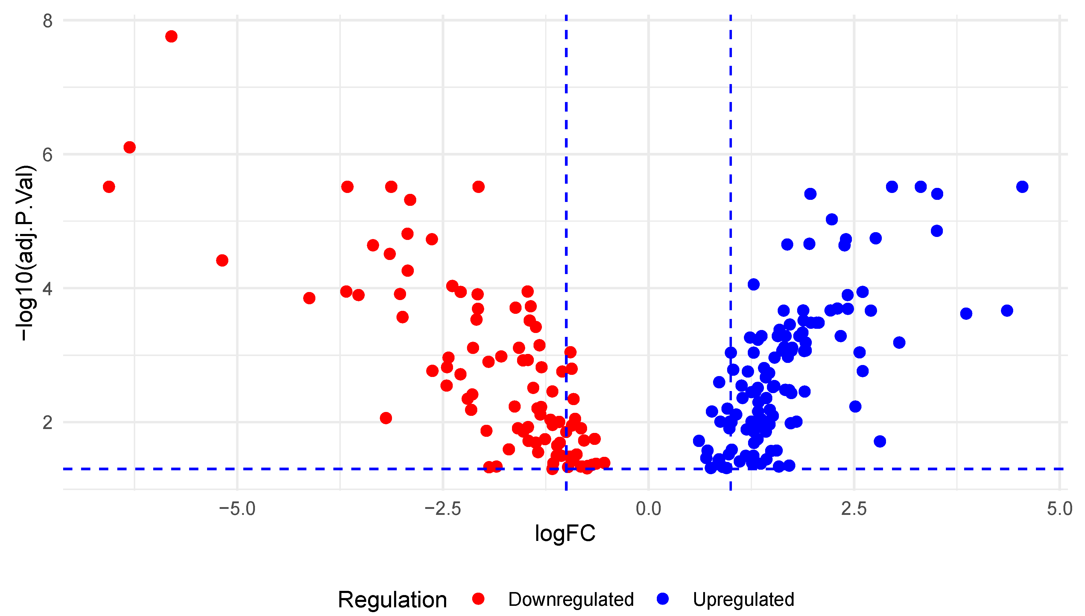
3.1. Differential Expression of miRNAs in PC

An analysis of the EXP00134 [24] experiment using dbDEMC revealed the dysregulation of 210 miRNAs in PC, with 116 upregulated and 94 downregulated miRNAs (Figure 1) (Table S1).

3.2. Identification of KRAS-Targeted miRNAs

Using the TargetScan and miRDB tools, it was identified that out of the 210 dysregulated miRNAs in PC, 16 of them specifically targeted the KRAS gene. A further analysis with PolyMIRTS revealed that these miRNAs interacted with KRAS in regions harbouring several single nucleotide variants (SNVs) (Table 1).

3.3. Differential Expression of KRAS-Targeting miRNAs in PC

A meta-profiling analysis conducted in dbDEMC revealed the significant dysregulation of miRNAs targeting the KRAS gene in PC. Changes in the expression levels (logFC) were observed with a range varying from 5 to −10 (Figure 2A).
In the differential expression analysis of miRNAs from experiment EXP00134 [24], these miRNAs were found to be downregulated in PC compared to NPDC, with a logFC ranging from −5.18 (hsa-miR 217; p = 0.00000173; p.adj = 0.0000385) to 1.71 (hsa-miR-877-5p; p = 0.0164, p.adj = 0.0443) with p and p.adj < 0.005 (Figure 2B, Table S2).
Additionally, the experimental validation data for these miRNAs were analysed on the same platform. It was observed that the deregulation of hsa-miR-885-5p, hsa-miR-326, hsa-miR-30b-5p, hsa-miR-30a-5p, hsa-miR-222-3p, and hsa-miR-217 has been experimentally validated in several studies that investigated their expression in different types of cancer (Table 2).
On the OncomiR platform, the expression of the miRNAs hsa-miR-222-3p, hsa-miR-30a-5p, hsa-miR-30b-5p, hsa-miR-30e-5p, hsa-miR-377-3p, hsa-miR-495-3p, hsa-miR-654-3p, hsa-miR-877-5p and hsa-miR-885-5p was found to be associated with the clinical features of PC (Figure 3, Table S3).
In addition, significant differences were observed between the expression of several miRNAs and the clinical outcomes in terms of patient mortality and survival.
The overexpression of hsa-miR-30a-5p was observed to be higher in deceased patients, whereas the expression of hsa-miR-30b-5p, hsa-miR-377-3p, hsa-miR-495-3p, and hsa-miR-885-5p was higher in disease survivors (Table 3).

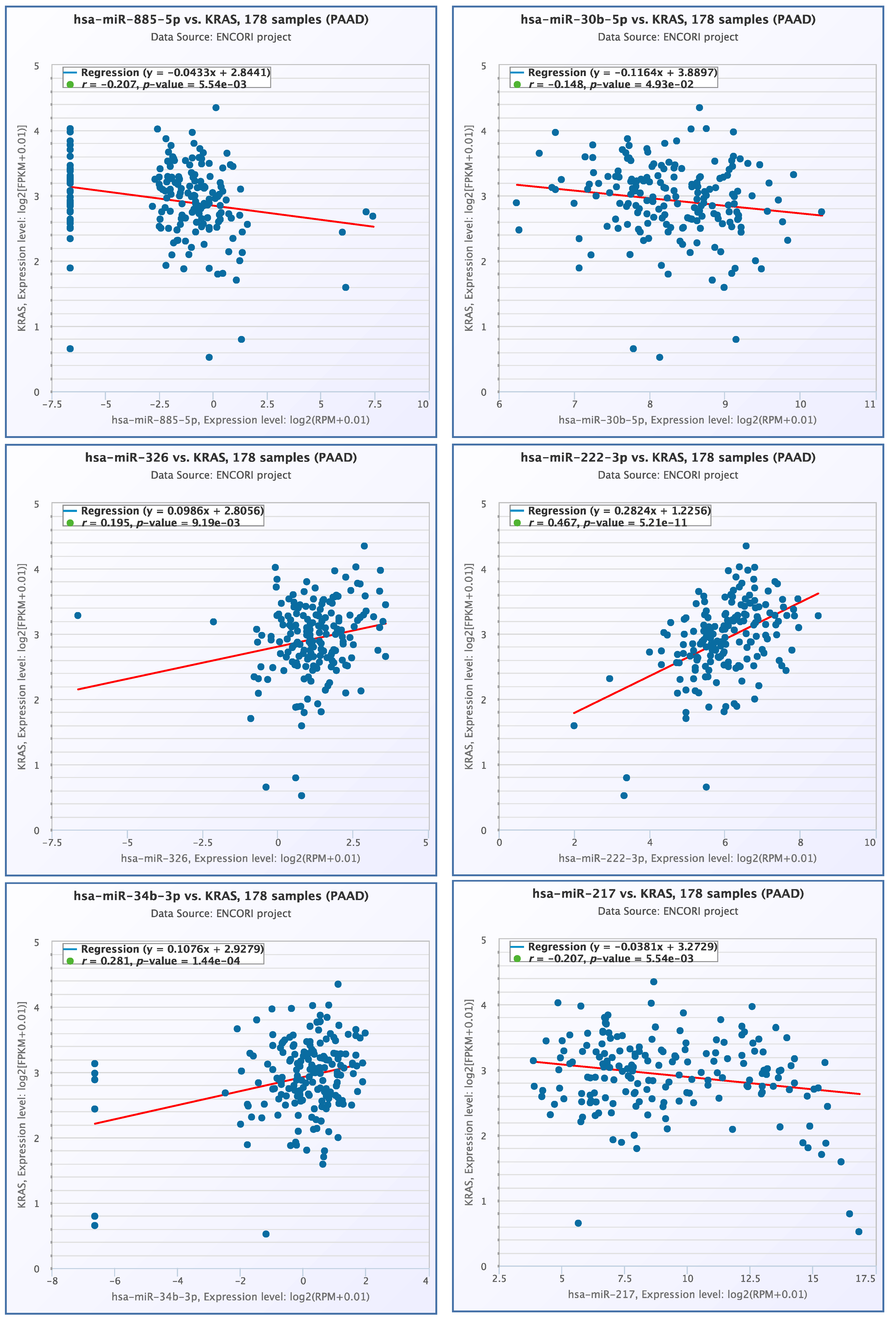
3.4. miRNA- KRAS mRNA Co-Expression Analysis

The co-expression profiles of miRNAs and KRAS mRNA were examined, revealing a significant correlation between the expression profiles of six miRNAs and KRAS mRNA. Notably, a positive correlation was observed between hsa-miR-222-3p and KRAS mRNA (r = 0.467, p = 5.21× 10⁻¹¹), as well as between hsa-miR-34b-3p and KRAS mRNA (r = 0.281, p = 1.44× 10⁻⁴) (Figure 4).

3.5. Molecular Interaction

Using the above observations obtained from the in silico data, we can infer the miRNA-mediated regulation pathway of KRAS and its role in PC (Figure 5).

4. Discussion

Currently, PC remains one of the most deadly cancers worldwide [2]. In addition, the genetic factors of this pathology are undeniable, as the KRAS gene is mutated in up to 95% of cases of this pathology [6]. It has been shown that KRAS has a remarkably large 3′UTR region, suggesting that its regulation is post-transcriptionally mediated by several miRNAs [43]. Therefore, the study of this miRNA-KRAS regulation could lead to the identification of miRNAs useful as molecular markers for early diagnosis, disease prognosis, and the development of more effective, targeted therapies. Recently, miRNAs have gained significant importance in cancer therapies due to their ability to regulate the gene expression involved in tumour progression and metastasis. miRNA-based therapeutic strategies include the use of miRNA mimetics to restore the function of tumour suppressor miRNAs and antagomirs to inhibit the action of oncogenic miRNAs [44].
The present study computationally identified the expression profiles of miRNAs in PC, as well as their relationship with KRAS.
In our study, we identified 210 miRNAs dysregulated in PC, of which 116 were overexpressed and 94 were underexpressed. Of these 210 miRNAs dysregulated in the disease, with predictions performed in TargetScan, miRDB, and PolymiRTS 3.0, 16 were found to target specific loci in the 3′UTR region of KRAS, regions that are also polymorphic. In this sense, it has been reported that variants in the 3′UTR region of the KRAS gene could modify the miRNA-binding sites that could regulate it [19,44,45], and that some of these variants have also been associated with different types of neoplasms [46,47,48,49,50,51].
One of the miRNAs dysregulated in PC and targeting KRAS is hsa-miR-885-5p, which has previously been reported [52] as a tumour suppressor in hepatocellular carcinoma by regulating AEG1 and as an inhibitor of invasion and metastasis in gastric cancer by regulating ME1 [53]. Furthermore, in this study, we observed that, in KRAS, this miRNA targets the rs8720 locus, an SNV that has previously been associated with colorectal cancer [49,54].
Furthermore, we observed that hsa-miR-885-5p is overexpressed in PC with a logFC of 1.41, and it has been experimentally demonstrated that it is also overexpressed in pheochromocytoma [30]. In OncomiR, we observed that the expression of this miRNA is also significantly correlated with the histologic grade, pathologic N stage, and pathologic T stage of PC, and its overexpression is also significantly associated with patient survival. Thus, in addition to regulating KRAS, AEG1 and ME1, this miRNA might coordinate PC progression by regulating other important genes in this pathology. Furthermore, a negative correlation between the expression of this miRNA and KRAS was observed in the co-expression analysis, suggesting that when miRNA expression increases, KRAS expression decreases. These findings could indicate that this miRNA may play a critical role as a prognostic marker of disease progression and survival by regulating the expression of KRAS or other oncogenes, thereby inhibiting cell invasion and impeding cancer progression.
Another miRNA dysregulated in PC and targeting KRAS is hsa-miR-877-5p, which has previously been reported as a marker of bone metastasis in lung cancer [55] and is overexpressed in hepatocellular carcinoma [56]. In this study, we observed that this miRNA targets the rs712 region of KRAS, an SNV that has been associated with colorectal cancer [57], breast cancer [58], and breast cancer metastasis [59]. Therefore, this variant could affect the regulation of KRAS by miRNAs and could possibly promote the neoplastic process. This suggests that this variant could influence the regulation of KRAS by miRNAs and potentially promote the neoplastic process. hsa-miR-877-5p was also found to be overexpressed in PC (logFC 1.71), which is consistent with previously reported studies [56], and that it is associated with the pathological state of the disease.
We found that hsa-miR-654-3p is underexpressed in PC with a logFC −1.59, and that it is also associated with the histologic grade and pathologic status of the disease, so this miRNA may regulate other genes that are also important in the progression of PC.
On the other hand, hsa-miR-647, which was found to be overexpressed in PC patients (logFC 1.49), has also been reported as an inhibitor of hepatocellular carcinoma progression by possibly regulating PTPRF [60], as a regulator of glioma progression and invasion by directly regulating HOXA9 [61], and conversely as a tumour promoter in gastric cancer by regulating the TP73 gene [62]. This suggests that miRNAs may play a critical role as regulators of tumour suppressor genes or oncogenes, influencing the suppression or promotion of carcinogenesis by modulating the expression of key genes in these diseases.
Another miRNA found to be underexpressed in PC (logFC-1.33) is hsa-miR-548c-5p, which has previously been associated with disease-free survival in breast cancer [63]. The above suggests that the silencing of this miRNA may facilitate the overexpression or dysregulation of critical genes in tumour progression.
hsa-miR-513b-5p was also found to be overexpressed in PC (logFC, 1.47), and similar data were found in a previous study [64] where the overexpression of this miRNA suppressed testicular embryonal carcinoma cell proliferation in vitro and induced apoptosis. On the other hand, the silencing of this miRNA reversed this effect by upregulating IRF2 expression. Therefore, it was observed that the overexpression of this miRNA could induce IRF2 silencing and, subsequently, TP53 expression. This could indicate that this miRNA could interact in various cell signalling networks, which could in turn influence cell growth.
In this study, hsa-miR-495-3p was found to be underexpressed in PC with a logFC of −2.13; in a previous study, it was shown that this molecule can regulate TWIST1 and inhibit metastasis in gastric cancer [65]. Furthermore, it has been reported that the loss of expression in this miRNA could induce the overexpression of epigenetic modifiers, which could in turn modify the expression of key genes in malignancy and the growth of gastric epithelial cells [66]. This is in line with the observations made in this study, where it was shown in silico that the expression of this miRNA is associated with the key features of PC, such as the histologic grade and pathologic state, mainly M and T pathologic states. Furthermore, in this study, the overexpression of this miRNA was associated with PC survival and was negatively correlated with KRAS expression, suggesting that the downregulation of this miRNA may have a significant effect on the overexpression of genes that regulate the tumour characteristics of PC.
A previous study reported that the expression levels of the miRNA hsa-miR-377-3p is lower in melanoma patients and that its overexpression inhibits cell growth by directly regulating ARMC8 [67], which is inconsistent with our findings, as we found an underexpression of this miRNA in patients with PC (logFC −2.39). Furthermore, we observed that this miRNA is associated with the pathological T status and its overexpression with disease survival. This suggests that the underexpression of this miRNA could enable the expression of genes such as KRAS or other oncogenes that promote cellular proliferation and differentiation. In the case of PC, these genes are implicated in the pathological state of the disease. Therefore, if this miRNA is overexpressed, it might suppress the effect of these genes, leading to an increase in the survival of individuals with this pathology.
hsa-miR-34b-3p was found to be overexpressed in PC with a logFC of 1.47, which is consistent with a previous study [68], where this miRNA was also found to be overexpressed in oesophageal squamous cell carcinoma. Additionally, in our study, the co-expression of this miRNA was found to be positively correlated with KRAS expression and could regulate the gene in the genomic regions of the SNV rs61764370 variant, which was previously associated with breast cancer in the Mexican population [69] and lung cancer in a population from Iran [70]. The overexpression of this miRNA in PC and oesophageal squamous cell carcinoma might suggest that this miRNA could also regulate and reduce the expression of tumour suppressor genes. Furthermore, the analysis of the association of KRAS SNVs where this miRNA binds could indicate that subtle changes, such as an SNV in the miRNA-binding sites in the 3’UTR region, might influence the regulation of KRAS by miRNAs. This underscores the intricate regulatory mechanisms involved in cancer development and highlights the potential importance of targeting these regulatory pathways for therapeutic intervention.
In a previous study [71], hsa-miR-326 was found to inhibit the development of non-small cell lung cancer by directly regulating the CCND1 gene. It was also reported that this miRNA is underexpressed in this pathology, which is consistent with the results obtained in this analysis, where it has been experimentally proven that this miRNA is expressed in medulloblastoma [31] and Barrett’s carcinogenesis [32]. However, it contrasts with the results obtained from PC, where this miRNA was observed to be overexpressed (logFC 1.69). Furthermore, it was observed that the expression of this miRNA positively correlates with the expression of KRAS, so this miRNA could play a role in tumour promotion in PC, somehow repressing the expression of genes that suppress cell growth in PC.
In this study, we observed that hsa-miR-30e-5p is downregulated in PC (logFC −0.68) and that its expression is associated with the pathological status T of the disease, which is similar to previously reported results [72], where it was identified that this miRNA participates in regulatory pathways that confer radiation resistance to nasopharyngeal carcinoma cell lines by regulating genes such as MAPK1, SOS1, TGFBR1, TGFBR2, TP53, CASP3, CCNE2, PTEN, and CDK2. In addition, it has been found that this miRNA acts as a tumour suppressor by directly regulating the Cyb561/ROS pathway’s signalling in acute myeloid leukaemia [73], and that this molecule mediates carcinogenic processes in colorectal cancer by directly regulating ITGA6 and ITGB [74] and those in prostate cancer by regulating CTHRC1 [75]. Many of these genes are closely related to KRAS, so it is to be expected that the dysregulation of this miRNA could be involved in PC tumour promotion by suppressing the expression of genes related to cell proliferation or allowing the expression of those that promote tumour progression. In addition to the above-mentioned tumours, this miRNA has also been associated with the occurrence, development and prognosis of several types of cancer including cervical squamous cell carcinoma [76], lung cancer [77], and chronic myeloid leukaemia [78], by directly regulating various genes and molecular pathways in these pathologies.
On the other hand, we observed that hsa-miR-30b-5p is underexpressed in PC (−0.78), and that its expression is associated with the histologic grade and pathologic stage M of the disease. Furthermore, we observed that this molecule has been experimentally reported to be underexpressed in colon cancer [33] and medulloblastoma [31], while this miRNA was reported to be overexpressed in breast cancer [34]; it has also been suggested that by decreasing its expression, it acts as a promoter of metastasis in gastric cancer [79]. This again suggests that this miRNA, like others, can have both tumour suppression and tumour promotion effects, depending on the gene it regulates in each pathology. For example, in this study, it was observed that the overexpression of this miRNA is associated with the survival of PC and that its expression negatively correlates with the expression of KRAS, so that, in PC, this miRNA may possibly play a tumour suppressor role by directly regulating this gene.
The miRNA hsa-miR-30a-5p was previously reported to be a tumour suppressor in lung squamous cell carcinoma by directly targeting the ATG5 gene [80] an ad suppressor of hepatocellular carcinoma tumour migration and invasion by directly regulating SNAIL1 [81]. In addition, it has been described that the inhibition of this miRNA promotes chemoresistance in lung cancer by directly regulating the expression of BECN1 [82]. In addition, downregulation has been reported in lung adenocarcinoma [35] and non-small cell lung cancer [83], which is consistent with this study, where the downregulation of this miRNA was observed in PC (logFC −0.82); we also observed that the expression of this molecule is associated with the M and N pathological state and that its overexpression is associated with PC mortality. This could indicate that when this miRNA is downregulated, it may potentially inhibit the expression of oncogenes, whereas when it is overexpressed, it could downregulate the expression of tumour suppressor genes.
We found that hsa-miR-222-3p is overexpressed in PC (logFC 1.53), and the overexpression of this miRNA was previously reported to be associated with the promotion of metastasis in lung cancer [84]. This is consistent with our study, where we observed that this miRNA correlated with the histological grade and pathological N-stage, while its expression correlated positively with KRAS expression. This may indicate that the overexpression of this miRNA negatively regulates the expression of genes that suppress KRAS activity or the expression of genes that suppress cell proliferation in PC. Furthermore, in this study, we again observed that the deregulation of this miRNA has different effects in different types of tumours; for example, downregulation was experimentally observed in prostate cancer [36], while upregulation was reported in lymphoma [37], cholangiocarcinoma [38], and kidney cancer [40].
Recently, a study reported that hsa-miR-217 expression plays a tumour suppressor role in non-small cell lung cancer cells by targeting SIRT1 [85]. In this study, we observed that this miRNA is significantly downregulated and underexpressed in PC (logFC −5.18). Furthermore, experimentally, this miRNA has been observed to be underexpressed in PC [41] and overexpressed in lung cancer [42]. In this study, we observed that the expression of this molecule correlates negatively with the expression of KRAS. Thus, it fulfils a possible suppressor role in PC tumorigenesis.
In a study [86] aimed at identifying downregulated miRNAs in metastatic colorectal cancer patients resistant to irinotecan-based treatment, hsa-miR-181d-5p was found to be overexpressed in this group of patients. In our study, we observed that this miRNA is overexpressed in PC (logFC 0.76) and that it modulates KRAS within the genomic locus of SNV rs9266, which has been previously reported to be associated with breast cancer in the Mexican population [69] and a sample of the Chinese population [48]. This supports that the variant located in the 3′UTR region of KRAS could modify the miRNA-binding sites that potentially regulate the gene, thereby promoting the process of carcinogenesis.
Given that KRAS can be indirectly regulated by miRNAs through the genes interacting with KRAS [19], it is crucial to highlight the potentially pivotal role of miRNAs in tumour promotion in PC and other cancers through complex regulatory networks. For instance, a recent study revealed that miRNAs also regulate YAP/TAZ in the Hippo pathway, a critical pathway in diverse cellular activities [87]; the activation of YAP/TAZ was also significantly associated with resistance to inhibitors specifically targeting KRAS. In addition, the combined inhibition of the Hippo pathways has been shown to improve the response of tumours to treatment with KRAS G12C-specific inhibitors [88,89], suggesting significant therapeutic potential in cancer treatment and the integration of miRNAs that not only indirectly regulate KRAS, but also all those genes that regulate and are regulated by KRAS.

5. Conclusions

A total of 210 miRNAs dysregulated in PC were identified, of which 16 could potentially regulate KRAS. The miRNAs hsa-miR-222-3p, hsa-miR-30a-5p, hsa-miR-30b-5p, hsa-miR-30e-5p, hsa-miR-377-3p, hsa-miR-495-3p, hsa-miR-654-3p, hsa-miR-877-5p, and hsa-miR-885-5p were associated with the relevant clinical features of the pathology, while the overexpression of hsa-miR-30a-5p was associated with PC mortality, and the overexpression of hsa-miR-30b-5p, hsa-miR-377-3p, hsa-miR-495-3p, and hsa-miR-885-5p was associated with survival. The expression of some miRNAs was found to correlate with KRAS expression, mainly the miRNAs hsa-miR-34b-3p and hsa-miR-222-3p. The miRNAs dysregulated in PC target specific loci within the 3′UTR regions of KRAS, and some of them (rs8720, rs712, rs190084851, rs61764370 and rs9266) have been previously associated with different types of cancer. The above findings highlight the potential of miRNAs as biomarkers and possible therapeutic targets in the treatment of PC; however, experimental validation is needed to confirm these results.

Supplementary Materials

The following supporting information can be downloaded at: https://www.mdpi.com/article/10.3390/diseases12070152/s1, Table S1: miRNAs dysregulated in PC; Table S2: miRNAs dysregulated in PC and targeting KRAS; Table S3: Association of miRNAs and clinical features of PC.

Author Contributions

Study Conception and/or Design: A.F.G.-R. and M.P.G.-A.; Data Processing and Collection: A.F.G.-R., M.P.G.-A. and L.E.F.; Analysis and Interpretation of Results: A.F.G.-R., M.P.G.-A. and L.E.F.; Draft Manuscript Preparation, Visualization: A.F.G.-R., M.P.G.-A., L.E.F., I.A.C.-D., P.M.G.-V., B.M.T.-M. and I.A.G.-H.; Critical Revision or Editing of the Article: A.F.G.-R., M.P.G.-A., L.E.F., G.M.Z.-G. and B.C.G.-M. All authors have read and agreed to the published version of the manuscript.

Funding

This research received no internal or external funding.

Institutional Review Board Statement

Not applicable.

Informed Consent Statement

Not applicable.

Data Availability Statement

Data are contained within the article.

Acknowledgments

We would like to express our gratitude to all the researchers and scientists who developed the platforms and data essential for conducting this study.

Conflicts of Interest

The authors declare no conflicts of interest.

References

  1. Klein, A.P. Pancreatic Cancer Epidemiology: Understanding the Role of Lifestyle and Inherited Risk Factors. Nat. Rev. Gastroenterol. Hepatol. 2021, 18, 493–502. [Google Scholar] [CrossRef] [PubMed]
  2. Kolbeinsson, H.M.; Chandana, S.; Wright, G.P.; Chung, M. Pancreatic Cancer: A Review of Current Treatment and Novel Therapies. J. Investig. Surg. 2023, 36, 2129884. [Google Scholar] [CrossRef] [PubMed]
  3. Global Cancer Observatory. International Agency for Research on Cancer. Available online: https://gco.iarc.fr/ (accessed on 22 September 2023).
  4. Halbrook, C.J.; Lyssiotis, C.A.; Pasca Di Magliano, M.; Maitra, A. Pancreatic Cancer: Advances and Challenges. Cell 2023, 186, 1729–1754. [Google Scholar] [CrossRef] [PubMed]
  5. Zhao, Z.; Liu, W. Pancreatic Cancer: A Review of Risk Factors, Diagnosis, and Treatment. Technol. Cancer Res. Treat. 2020, 19, 1533033820962117. [Google Scholar] [CrossRef] [PubMed]
  6. Luo, J. KRAS Mutation in Pancreatic Cancer. Semin. Oncol. 2021, 48, 10–18. [Google Scholar] [CrossRef] [PubMed]
  7. Zhang, Z.; Zhang, H.; Liao, X.; Tsai, H. KRAS Mutation: The Booster of Pancreatic Ductal Adenocarcinoma Transformation and Progression. Front. Cell Dev. Biol. 2023, 11, 1147676. [Google Scholar] [CrossRef] [PubMed]
  8. Johnson, C.; Burkhart, D.L.; Haigis, K.M. Classification of KRAS-Activating Mutations and the Implications for Therapeutic Intervention. Cancer Discov. 2022, 12, 913–923. [Google Scholar] [CrossRef] [PubMed]
  9. Wang, H.; Chi, L.; Yu, F.; Dai, H.; Gao, C.; Si, X.; Wang, Z.; Liu, L.; Zheng, J.; Shan, L.; et al. Annual Review of KRAS Inhibitors in 2022. Eur. J. Med. Chem. 2023, 249, 115124. [Google Scholar] [CrossRef] [PubMed]
  10. Rathod, L.S.; Dabhade, P.S.; Mokale, S.N. Recent Progress in Targeting KRAS Mutant Cancers with Covalent G12C-Specific Inhibitors. Drug Discov. Today 2023, 28, 103557. [Google Scholar] [CrossRef] [PubMed]
  11. Mukhopadhyay, S.; Vander Heiden, M.G.; McCormick, F. The Metabolic Landscape of RAS-Driven Cancers from Biology to Therapy. Nat. Cancer 2021, 2, 271–283. [Google Scholar] [CrossRef]
  12. Pupo, E.; Avanzato, D.; Middonti, E.; Bussolino, F.; Lanzetti, L. KRAS-Driven Metabolic Rewiring Reveals Novel Actionable Targets in Cancer. Front. Oncol. 2019, 9, 848. [Google Scholar] [CrossRef] [PubMed]
  13. Punekar, S.R.; Velcheti, V.; Neel, B.G.; Wong, K.-K. The Current State of the Art and Future Trends in RAS-Targeted Cancer Therapies. Nat. Rev. Clin. Oncol. 2022, 19, 637–655. [Google Scholar] [CrossRef] [PubMed]
  14. Cerami, E.; Gao, J.; Dogrusoz, U.; Gross, B.E.; Sumer, S.O.; Aksoy, B.A.; Jacobsen, A.; Byrne, C.J.; Heuer, M.L.; Larsson, E.; et al. The cBio Cancer Genomics Portal: An Open Platform for Exploring Multidimensional Cancer Genomics Data. Cancer Discov. 2012, 2, 401–404. [Google Scholar] [CrossRef] [PubMed]
  15. Tang, Z.; Li, C.; Kang, B.; Gao, G.; Li, C.; Zhang, Z. GEPIA: A Web Server for Cancer and Normal Gene Expression Profiling and Interactive Analyses. Nucleic Acids Res. 2017, 45, W98–W102. [Google Scholar] [CrossRef] [PubMed]
  16. Bortoletto, A.S.; Parchem, R.J. KRAS Hijacks the miRNA Regulatory Pathway in Cancer. Cancer Res. 2023, 83, 1563–1572. [Google Scholar] [CrossRef] [PubMed]
  17. Yao, Q.; Chen, Y.; Zhou, X. The Roles of microRNAs in Epigenetic Regulation. Curr. Opin. Chem. Biol. 2019, 51, 11–17. [Google Scholar] [CrossRef] [PubMed]
  18. Iwakawa, H.; Tomari, Y. Life of RISC: Formation, Action, and Degradation of RNA-Induced Silencing Complex. Mol. Cell 2022, 82, 30–43. [Google Scholar] [CrossRef] [PubMed]
  19. Shui, B.; La Rocca, G.; Ventura, A.; Haigis, K.M. Interplay between K-RAS and miRNAs. Trends Cancer 2022, 8, 384–396. [Google Scholar] [CrossRef] [PubMed]
  20. Cuzziol, C.I.; Marzochi, L.L.; Possebon, V.S.; Kawasaki-Oyama, R.S.; Mattos, M.F.; Junior, V.S.; Ferreira, L.A.M.; Pavarino, É.C.; Castanhole-Nunes, M.M.U.; Goloni-Bertollo, E.M. Regulation of VEGFA, KRAS, and NFE2L2 Oncogenes by MicroRNAs in Head and Neck Cancer. Int. J. Mol. Sci. 2022, 23, 7483. [Google Scholar] [CrossRef] [PubMed]
  21. Oncogenic KRAS Promotes Pancreatic Ductal Adenocarcinoma (PDAC) through Post-Transcriptionally Regulated KRAS-Induced Granules (KGs). Available online: https://www.researchsquare.com (accessed on 22 September 2023).
  22. Shui, B.; Beyett, T.S.; Chen, Z.; Li, X.; La Rocca, G.; Gazlay, W.M.; Eck, M.J.; Lau, K.S.; Ventura, A.; Haigis, K.M. Oncogenic K-Ras Suppresses Global miRNA Function. Mol. Cell 2023, 83, 2509–2523.e13. [Google Scholar] [CrossRef] [PubMed]
  23. Xu, F.; Wang, Y.; Ling, Y.; Zhou, C.; Wang, H.; Teschendorff, A.E.; Zhao, Y.; Zhao, H.; He, Y.; Zhang, G.; et al. dbDEMC 3.0: Functional Exploration of Differentially Expressed miRNAs in Cancers of Human and Model Organisms. Genom. Proteom. Bioinform. 2022, 20, 446–454. [Google Scholar] [CrossRef] [PubMed]
  24. Munding, J.B.; Adai, A.T.; Maghnouj, A.; Urbanik, A.; Zöllner, H.; Liffers, S.T.; Chromik, A.M.; Uhl, W.; Szafranska-Schwarzbach, A.E.; Tannapfel, A.; et al. Global microRNA Expression Profiling of Microdissected Tissues Identifies miR-135b as a Novel Biomarker for Pancreatic Ductal Adenocarcinoma. Int. J. Cancer 2012, 131, E86–E95. [Google Scholar] [CrossRef] [PubMed]
  25. McGeary, S.E.; Lin, K.S.; Shi, C.Y.; Pham, T.M.; Bisaria, N.; Kelley, G.M.; Bartel, D.P. The Biochemical Basis of microRNA Targeting Efficacy. Science 2019, 366, eaav1741. [Google Scholar] [CrossRef] [PubMed]
  26. Chen, Y.; Wang, X. miRDB: An Online Database for Prediction of Functional microRNA Targets. Nucleic Acids Res. 2020, 48, D127–D131. [Google Scholar] [CrossRef] [PubMed]
  27. Bhattacharya, A.; Ziebarth, J.D.; Cui, Y. PolymiRTS Database 3.0: Linking Polymorphisms in microRNAs and Their Target Sites with Human Diseases and Biological Pathways. Nucleic Acids Res. 2014, 42, D86–D91. [Google Scholar] [CrossRef] [PubMed]
  28. Wong, N.W.; Chen, Y.; Chen, S.; Wang, X. OncomiR: An Online Resource for Exploring Pan-Cancer microRNA Dysregulation. Bioinformatics 2018, 34, 713–715. [Google Scholar] [CrossRef] [PubMed]
  29. Li, J.H.; Liu, S.; Zhou, H.; Qu, L.H.; Yang, J.H. starBase v2.0: Decoding miRNA-ceRNA, miRNA-ncRNA and Protein-RNA Interaction Networks from Large-Scale CLIP-Seq Data. Nucleic Acids Res. 2014, 42, D92–D97. [Google Scholar] [CrossRef] [PubMed]
  30. de Cubas, A.A.; Leandro-García, L.J.; Schiavi, F.; Mancikova, V.; Comino-Méndez, I.; Inglada-Pérez, L.; Perez-Martinez, M.; Ibarz, N.; Ximénez-Embún, P.; López-Jiménez, E.; et al. Integrative Analysis of miRNA and mRNA Expression Profiles in Pheochromocytoma and Paraganglioma Identifies Genotype-Specific Markers and Potentially Regulated Pathways. Endocr. Relat. Cancer 2013, 20, 477–493. [Google Scholar] [CrossRef] [PubMed]
  31. Ferretti, E.; De Smaele, E.; Po, A.; Di Marcotullio, L.; Tosi, E.; Espinola, M.S.B.; Di Rocco, C.; Riccardi, R.; Giangaspero, F.; Farcomeni, A.; et al. MicroRNA Profiling in Human Medulloblastoma. Int. J. Cancer 2009, 124, 568–577. [Google Scholar] [CrossRef] [PubMed]
  32. Fassan, M.; Volinia, S.; Palatini, J.; Pizzi, M.; Baffa, R.; De Bernard, M.; Battaglia, G.; Parente, P.; Croce, C.M.; Zaninotto, G.; et al. MicroRNA Expression Profiling in Human Barrett’s Carcinogenesis. Int. J. Cancer 2011, 129, 1661–1670. [Google Scholar] [CrossRef] [PubMed]
  33. Arndt, G.M.; Dossey, L.; Cullen, L.M.; Lai, A.; Druker, R.; Eisbacher, M.; Zhang, C.; Tran, N.; Fan, H.; Retzlaff, K.; et al. Characterization of Global microRNA Expression Reveals Oncogenic Potential of miR-145 in Metastatic Colorectal Cancer. BMC Cancer 2009, 9, 374. [Google Scholar] [CrossRef] [PubMed]
  34. Zhang, K.; Wang, Y.-W.; Wang, Y.-Y.; Song, Y.; Zhu, J.; Si, P.-C.; Ma, R. Identification of microRNA Biomarkers in the Blood of Breast Cancer Patients Based on microRNA Profiling. Gene 2017, 619, 10–20. [Google Scholar] [CrossRef] [PubMed]
  35. Patnaik, S.K.; Yendamuri, S.; Kannisto, E.; Kucharczuk, J.C.; Singhal, S.; Vachani, A. MicroRNA Expression Profiles of Whole Blood in Lung Adenocarcinoma. PLoS ONE 2012, 7, e46045. [Google Scholar] [CrossRef] [PubMed]
  36. Schaefer, A.; Jung, M.; Mollenkopf, H.-J.; Wagner, I.; Stephan, C.; Jentzmik, F.; Miller, K.; Lein, M.; Kristiansen, G.; Jung, K. Diagnostic and Prognostic Implications of microRNA Profiling in Prostate Carcinoma. Int. J. Cancer 2010, 126, 1166–1176. [Google Scholar] [CrossRef] [PubMed]
  37. Lionetti, M.; Biasiolo, M.; Agnelli, L.; Todoerti, K.; Mosca, L.; Fabris, S.; Sales, G.; Deliliers, G.L.; Bicciato, S.; Lombardi, L.; et al. Identification of microRNA Expression Patterns and Definition of a microRNA/mRNA Regulatory Network in Distinct Molecular Groups of Multiple Myeloma. Blood 2009, 114, e20–e26. [Google Scholar] [CrossRef] [PubMed]
  38. Plieskatt, J.L.; Rinaldi, G.; Feng, Y.; Peng, J.; Yonglitthipagon, P.; Easley, S.; Laha, T.; Pairojkul, C.; Bhudhisawasdi, V.; Sripa, B.; et al. Distinct miRNA Signatures Associate with Subtypes of Cholangiocarcinoma from Infection with the Tumourigenic Liver Fluke Opisthorchis Viverrini. J. Hepatol. 2014, 61, 850–858. [Google Scholar] [CrossRef]
  39. Manso, R.; Martínez-Magunacelaya, N.; Eraña-Tomás, I.; Monsalvez, V.; Rodríguez-Peralto, J.L.; Ortiz-Romero, P.-L.; Santonja, C.; Cristóbal, I.; Piris, M.A.; Rodríguez-Pinilla, S.M. Mycosis Fungoides Progression Could Be Regulated by microRNAs. PLoS ONE 2018, 13, e0198477. [Google Scholar] [CrossRef] [PubMed]
  40. Zhang, J.; Ye, Y.; Chang, D.W.; Lin, S.-H.; Huang, M.; Tannir, N.M.; Matin, S.; Karam, J.A.; Wood, C.G.; Chen, Z.-N.; et al. Global and Targeted miRNA Expression Profiling in Clear Cell Renal Cell Carcinoma Tissues Potentially Links miR-155-5p and miR-210-3p to Both Tumorigenesis and Recurrence. Am. J. Pathol. 2018, 188, 2487–2496. [Google Scholar] [CrossRef] [PubMed]
  41. Cohen, S.J.; Papoulas, M.; Graubardt, N.; Ovdat, E.; Loewenstein, S.; Kania-Almog, J.; Pasmanik-Chor, M.; Brazowski, E.; Cagnano, E.; Nachmany, I.; et al. Micro-RNA Expression Patterns Predict Metastatic Spread in Solid Pseudopapillary Neoplasms of the Pancreas. Front. Oncol. 2020, 10, 328. [Google Scholar] [CrossRef] [PubMed]
  42. Reis, P.P.; Drigo, S.A.; Carvalho, R.F.; Lopez Lapa, R.M.; Felix, T.F.; Patel, D.; Cheng, D.; Pintilie, M.; Liu, G.; Tsao, M.-S. Circulating miR-16-5p, miR-92a-3p, and miR-451a in Plasma from Lung Cancer Patients: Potential Application in Early Detection and a Regulatory Role in Tumorigenesis Pathways. Cancers 2020, 12, 2071. [Google Scholar] [CrossRef]
  43. Kim, M.; Kogan, N.; Slack, F.J. Cis-Acting Elements in Its 3′ UTR Mediate Post-Transcriptional Regulation of KRAS. Oncotarget 2016, 7, 11770–11784. [Google Scholar] [CrossRef] [PubMed]
  44. Sell, M.C.; Ramlogan-Steel, C.A.; Steel, J.C.; Dhungel, B.P. MicroRNAs in Cancer Metastasis: Biological and Therapeutic Implications. Expert. Rev. Mol. Med. 2023, 25, e14. [Google Scholar] [CrossRef] [PubMed]
  45. Chhichholiya, Y.; Singh, H.V.; Vashistha, R.; Singh, S.; Munshi, A. Deciphering the Role of KRAS Gene in Oncogenesis: Focus on Signaling Pathways, Genetic Alterations in 3’UTR, KRAS Specific miRNAs and Therapeutic Interventions. Crit. Rev. Oncol./Hematol. 2024, 194, 104250. [Google Scholar] [CrossRef] [PubMed]
  46. Yang, M.; Xiao, X.; Xing, X.; Li, X.; Xia, T.; Long, H. KRAS and VEGF Gene 3’-UTR Single Nucleotide Polymorphisms Predicted Susceptibility in Colorectal Cancer. PLoS ONE 2017, 12, e0174140. [Google Scholar] [CrossRef] [PubMed]
  47. Insodaite, R.; Smalinskiene, A.; Liutkevicius, V.; Ulozas, V.; Poceviciute, R.; Bielevicius, A.; Kucinskas, L. Associations of Polymorphisms Localized in the 3′UTR Regions of the KRAS, NRAS, MAPK1 Genes with Laryngeal Squamous Cell Carcinoma. Genes 2021, 12, 1679. [Google Scholar] [CrossRef] [PubMed]
  48. Jin, M.; Lu, F.; Li, X.; Zhou, W.; Li, S.; Jiang, Y.; Wu, H.; Wang, J. Association between KRAS Gene Polymorphisms and Genetic Susceptibility to Breast Cancer in a Chinese Population. J. Clin. Lab. Anal. 2022, 37, e24806. [Google Scholar] [CrossRef] [PubMed]
  49. Gallegos-Arreola, M.P.; Garibaldi-Ríos, A.F.; Cruz-Sánchez, J.I.; Figuera, L.E.; Ronquillo-Carreón, C.A.; Rosales-Reynoso, M.A.; Gómez-Meda, B.C.; Carrillo-Dávila, I.A.; Puebla-Pérez, A.M.; Montoya-Fuentes, H.; et al. Association of the Rs8720 and Rs12587 KRAS Gene Variants with Colorectal Cancer in a Mexican Population and Their Analysis In Silico. Cells 2023, 12, 1941. [Google Scholar] [CrossRef] [PubMed]
  50. Fu, W.; Zhuo, Z.; Hua, R.-X.; Fu, K.; Jia, W.; Zhu, J.; Zhang, J.; Cheng, J.; Zhou, H.; Xia, H.; et al. Association of KRAS and NRAS Gene Polymorphisms with Wilms Tumor Risk: A Four-Center Case-Control Study. Aging 2019, 11, 1551–1563. [Google Scholar] [CrossRef] [PubMed]
  51. Ulusan, M.; Sen, S.; Yilmazer, R.; Dalay, N.; Demokan, S. The Let-7 microRNA Binding Site Variant in KRAS as a Predictive Biomarker for Head and Neck Cancer Patients with Lymph Node Metastasis. Pathol. Res. Pr. 2022, 239, 154147. [Google Scholar] [CrossRef] [PubMed]
  52. Li, C.; Wang, X.; Song, Q. MicroRNA 885-5p Inhibits Hepatocellular Carcinoma Metastasis by Repressing AEG1. Onco Targets Ther. 2020, 13, 981–988. [Google Scholar] [CrossRef] [PubMed]
  53. Jiang, Z.; Cui, H.; Zeng, S.; Li, L. miR-885-5p Inhibits Invasion and Metastasis in Gastric Cancer by Targeting Malic Enzyme 1. DNA Cell Biol. 2021, 40, 694–705. [Google Scholar] [CrossRef] [PubMed]
  54. Liu, X.; Yang, G.; Huang, J.; Chai, L.; Liu, X.; Dai, Q.; Yang, Z. KRAS SNPs Are Related to Colorectal Cancer Susceptibility and Survival in Chinese People. Biomark. Med. 2020, 14, 13–22. [Google Scholar] [CrossRef] [PubMed]
  55. Zhao, K.; Jia, C.; Wang, J.; Shi, W.; Wang, X.; Song, Y.; Peng, C. Exosomal Hsa-miR-151a-3p and Hsa-miR-877-5p Are Potential Novel Biomarkers for Predicting Bone Metastasis in Lung Cancer. Aging 2023, 15, 14864–14888. [Google Scholar] [CrossRef] [PubMed]
  56. Xiong, D.; Dang, Y.; Lin, P.; Wen, D.; He, R.; Luo, D.; Feng, Z.; Chen, G. A circRNA–miRNA–mRNA Network Identification for Exploring Underlying Pathogenesis and Therapy Strategy of Hepatocellular Carcinoma. J. Transl. Med. 2018, 16, 220. [Google Scholar] [CrossRef]
  57. Gallegos-Arreola, M.P.; Zúñiga-González, G.M.; Gómez-Mariscal, K.; Rosales-Reynoso, M.A.; Luis, L.; Puebla-Pérez, A.M.; Pineda-Razo, T. Association of Rs712 Polymorphism in a Let-7 microRNA-Binding Site of KRAS Gene with Colorectal Cancer in a Mexican Population. Iran. J. Basic. Med. Sci. 2019, 22, 324–327. [Google Scholar] [CrossRef]
  58. Al-Haddad, R.H.; Jasim, H.M.; Saleh, W.A. Association between KRAS Rs 712 G>T Polymorphism with the Incidence of Breast Tumors in Iraqi Women. Iraqi J. Biotechnol. 2019, 18, 55–61. [Google Scholar]
  59. Huang, X.; Yang, Y.; Guo, Y.; Cao, Z.L.; Cui, Z.W.; Hu, T.C.; Gao, L.B. Association of a Let-7 KRAS Rs712 Polymorphism with the Risk of Breast Cancer. Genet. Mol. Res. 2015, 14, 16913–16920. [Google Scholar] [CrossRef] [PubMed]
  60. Ye, X.; Qiu, R.; He, X.; Hu, Z.; Zheng, F.; Huang, X.; Xie, X.; Chen, F.; Ou, H.; Lin, G. miR-647 Inhibits Hepatocellular Carcinoma Cell Progression by Targeting Protein Tyrosine Phosphatase Receptor Type F. Bioengineered 2022, 13, 1090–1102. [Google Scholar] [CrossRef] [PubMed]
  61. Qin, K.; Tian, G.; Chen, G.; Zhou, D.; Tang, K. miR-647 Inhibits Glioma Cell Proliferation, Colony Formation and Invasion by Regulating HOXA9. J. Gene Med. 2020, 22, e3153. [Google Scholar] [CrossRef] [PubMed]
  62. Zhang, X.; Zhang, M.; Wang, G.; Tian, Y.; He, X. Tumor Promoter Role of miR-647 in Gastric Cancer via Repression of TP73. Mol. Med. Rep. 2018, 18, 3744–3750. [Google Scholar] [CrossRef]
  63. Block, I.; Burton, M.; Sørensen, K.P.; Andersen, L.; Larsen, M.J.; Bak, M.; Cold, S.; Thomassen, M.; Tan, Q.; Kruse, T.A. Association of miR-548c-5p, miR-7-5p, miR-210-3p, miR-128-3p with Recurrence in Systemically Untreated Breast Cancer. Oncotarget 2018, 9, 9030–9042. [Google Scholar] [CrossRef] [PubMed]
  64. Wang, X.; Zhang, X.; Wang, G.; Wang, L.; Lin, Y.; Sun, F. Hsa-miR-513b-5p Suppresses Cell Proliferation and Promotes P53 Expression by Targeting IRF2 in Testicular Embryonal Carcinoma Cells. Gene 2017, 626, 344–353. [Google Scholar] [CrossRef] [PubMed]
  65. Liu, C.; Jian, M.; Qi, H.; Mao, W.-Z. MicroRNA 495 Inhibits Proliferation and Metastasis and Promotes Apoptosis by Targeting Twist1 in Gastric Cancer Cells. Oncol. Res. 2019, 27, 389. [Google Scholar] [CrossRef] [PubMed]
  66. Eun, J.W.; Kim, H.S.; Shen, Q.; Yang, H.D.; Kim, S.Y.; Yoon, J.H.; Park, W.S.; Lee, J.Y.; Nam, S.W. MicroRNA-495-3p Functions as a Tumor Suppressor by Regulating Multiple Epigenetic Modifiers in Gastric Carcinogenesis. J. Pathol. 2018, 244, 107–119. [Google Scholar] [CrossRef] [PubMed]
  67. Yuan, J.; Jiang, L.; Guo, C. The Micro RNA Hsa-miR-377-3p Inhibits Tumor Growth in Malignant Melanoma. RSC Adv. 2019, 9, 19057–19064. [Google Scholar] [CrossRef] [PubMed]
  68. Wang, J.; Yu, P.; Luo, J.; Sun, Z.; Yu, J.; Wang, J. Transcriptomic and microRNA Expression Profiles Identify Biomarkers for Predicting Neo-Chemoradiotherapy Response in Esophageal Squamous Cell Carcinomas (ESCC). Front. Pharmacol. 2021, 12, 626972. [Google Scholar] [CrossRef] [PubMed]
  69. Gallegos-Arreola, M.P.; García Verdín, P.M.; Magaña-Torres, M.T.; Figuera, L.E.; Zúñiga-González, G.M.; Rosales-Reynoso, M.A.; Gómez-Meda, B.C.; Puebla-Pérez, A.M. Association between Rs61764370, Rs9266, and Rs140080026 Polymorphisms of the KRAS Gene and Breast Cancer Risk in a Mexican Population. Eur. Rev. Med. Pharmacol. Sci. 2021, 25, 6454–6464. [Google Scholar] [CrossRef] [PubMed]
  70. Farokhzad, N.; Hosseini, S.M.; Edalat, H.; Sadeghi, M. Association of Rs61764370 Polymorphism within Let-7 microRNA-Binding Site with Lung Cancer in Iranian Population. Afr. Health Sci. 2020, 20, 1299–1303. [Google Scholar] [CrossRef] [PubMed]
  71. Sun, C.; Huang, C.; Li, S.; Yang, C.; Xi, Y.; Wang, L.; Zhang, F.; Fu, Y.; Li, D. Hsa-miR-326 Targets CCND1 and Inhibits Non-Small Cell Lung Cancer Development. Oncotarget 2016, 7, 8341–8359. [Google Scholar] [CrossRef]
  72. Luo, H.; Zhong, F.; Jing, X.; Lin, H.; Li, Y. miRNA Profiling of Human Nasopharyngeal Carcinoma Cell Lines HONE1 and CNE2 after X-Ray Therapy. Adv. Clin. Exp. Med. 2022, 31, 671–687. [Google Scholar] [CrossRef] [PubMed]
  73. Ge, Y.; Hong, M.; Zhang, Y.; Wang, J.; Li, L.; Zhu, H.; Sheng, Y.; Wu, W.-S.; Zhang, Z. miR-30e-5p Regulates Leukemia Stem Cell Self-Renewal through the Cyb561/ROS Signaling Pathway. Haematologica 2023, 109, 411–421. [Google Scholar] [CrossRef] [PubMed]
  74. Laudato, S.; Patil, N.; Abba, M.L.; Leupold, J.H.; Benner, A.; Gaiser, T.; Marx, A.; Allgayer, H. P53-Induced miR-30e-5p Inhibits Colorectal Cancer Invasion and Metastasis by Targeting ITGA6 and ITGB1. Int. J. Cancer 2017, 141, 1879–1890. [Google Scholar] [CrossRef] [PubMed]
  75. Ma, Z.; Chao, F.; Wang, S.; Song, Z.; Zhuo, Z.; Zhang, J.; Xu, G.; Chen, G. CTHRC1 Affects Malignant Tumor Cell Behavior and Is Regulated by miR-30e-5p in Human Prostate Cancer. Biochem. Biophys. Res. Commun. 2020, 525, 418–424. [Google Scholar] [CrossRef] [PubMed]
  76. Kwon, A.Y.; Jeong, J.Y.; Park, H.; Hwang, S.; Kim, G.; Kang, H.; Heo, J.H.; Lee, H.J.; Kim, T.H.; An, H.J. miR-22-3p and miR-30e-5p Are Associated with Prognosis in Cervical Squamous Cell Carcinoma. Int. J. Mol. Sci. 2022, 23, 5623. [Google Scholar] [CrossRef] [PubMed]
  77. Xu, G.; Cai, J.; Wang, L.; Jiang, L.; Huang, J.; Hu, R.; Ding, F. MicroRNA-30e-5p Suppresses Non-Small Cell Lung Cancer Tumorigenesis by Regulating USP22-Mediated Sirt1/JAK/STAT3 Signaling. Exp. Cell Res. 2018, 362, 268–278. [Google Scholar] [CrossRef] [PubMed]
  78. Dai, H.; Wang, J.; Huang, Z.; Zhang, H.; Wang, X.; Li, Q.; Feng, W. LncRNA OIP5-AS1 Promotes the Autophagy-Related Imatinib Resistance in Chronic Myeloid Leukemia Cells by Regulating miR-30e-5p/ATG12 Axis. Technol. Cancer Res. Treat. 2021, 20, 15330338211052150. [Google Scholar] [CrossRef] [PubMed]
  79. Qiao, F.; Zhang, K.; Gong, P.; Wang, L.; Hu, J.; Lu, S.; Fan, H. Decreased miR-30b-5p Expression by DNMT1 Methylation Regulation Involved in Gastric Cancer Metastasis. Mol. Biol. Rep. 2014, 41, 5693–5700. [Google Scholar] [CrossRef] [PubMed]
  80. Yang, J.; Rao, S.; Cao, R.; Xiao, S.; Cui, X.; Ye, L. miR-30a-5p Suppresses Lung Squamous Cell Carcinoma via ATG5—Mediated Autophagy. Aging 2021, 13, 17462–17472. [Google Scholar] [CrossRef] [PubMed]
  81. Xue, C.; Zhang, X.; Gao, P.; Cui, X.; Zhu, C.; Qin, X. LncRNA Loc339803 Acts as CeRNA of miR-30a-5p to Promote the Migration and Invasion of Hepatocellular Carcinoma Cells. J. Cancer 2021, 12, 1061–1072. [Google Scholar] [CrossRef] [PubMed]
  82. Yang, X.; Bai, F.; Xu, Y.; Chen, Y.; Chen, L. Intensified Beclin-1 Mediated by Low Expression of Mir-30a-5p Promotes Chemoresistance in Human Small Cell Lung Cancer. Cell. Physiol. Biochem. 2017, 43, 1126–1139. [Google Scholar] [CrossRef]
  83. Zhu, J.; Zeng, Y.; Xu, C.; Qin, H.; Lei, Z.; Shen, D.; Liu, Z.; Huang, J.-A. Expression Profile Analysis of microRNAs and Downregulated miR-486-5p and miR-30a-5p in Non-Small Cell Lung Cancer. Oncol. Rep. 2015, 34, 1779–1786. [Google Scholar] [CrossRef] [PubMed]
  84. Tepebaşı, M.Y.; Öztürk, Ö. miR-21, miR-221, and miR-222 Upregulation in Lung Cancer Promotes Metastasis by Reducing Oxidative Stress and Apoptosis. Rev. Assoc. Med. Bras. 2023, 69, e20221688. [Google Scholar] [CrossRef] [PubMed]
  85. Jiang, W.; Hou, L.; Wei, J.; Du, Y.; Zhao, Y.; Deng, X.; Lin, X. Hsa-miR-217 Inhibits the Proliferation, Migration, and Invasion in Non-Small Cell Lung Cancer Cells Via Targeting SIRT1 and P53/KAI1 Signaling. Balk. Med. J. 2020, 37, 208–214. [Google Scholar] [CrossRef] [PubMed]
  86. Pliakou, E.; Lampropoulou, D.I.; Dovrolis, N.; Chrysikos, D.; Filippou, D.; Papadimitriou, C.; Vezakis, A.; Aravantinos, G.; Gazouli, M. Circulating miRNA Expression Profiles and Machine Learning Models in Association with Response to Irinotecan-Based Treatment in Metastatic Colorectal Cancer. Int. J. Mol. Sci. 2022, 24, 46. [Google Scholar] [CrossRef] [PubMed]
  87. Wang, Y.; Xu, X.; Maglic, D.; Dill, M.T.; Mojumdar, K.; Ng, P.K.-S.; Jeong, K.J.; Tsang, Y.H.; Moreno, D.; Bhavana, V.H.; et al. Comprehensive Molecular Characterization of the Hippo Signaling Pathway in Cancer. Cell Rep. 2018, 25, 1304–1317.e5. [Google Scholar] [CrossRef] [PubMed]
  88. Mukhopadhyay, S.; Huang, H.-Y.; Lin, Z.; Ranieri, M.; Li, S.; Sahu, S.; Liu, Y.; Ban, Y.; Guidry, K.; Hu, H.; et al. Genome-Wide CRISPR Screens Identify Multiple Synthetic Lethal Targets That Enhance KRASG12C Inhibitor Efficacy. Cancer Res. 2023, 83, 4095–4111. [Google Scholar] [CrossRef] [PubMed]
  89. Kapoor, A.; Yao, W.; Ying, H.; Hua, S.; Liewen, A.; Wang, Q.; Zhong, Y.; Wu, C.-J.; Sadanandam, A.; Hu, B.; et al. Yap1 Activation Enables Bypass of Oncogenic Kras Addiction in Pancreatic Cancer. Cell 2014, 158, 185–197. [Google Scholar] [CrossRef] [PubMed]
Figure 1. miRNAs dysregulated in PC.
Diseases 12 00152 g001
Figure 2. (A) Meta-profiling of miRNAs dysregulated in PC and targeting KRAS. (B) miRNAs dysregulated in PC (EXP00134 [24]) and targeting KRAS.
Diseases 12 00152 g002
Figure 3. Association between miRNAs and clinical features of PC. As shown in the figure, miRNAs are linked to various clinicopathological features of PC. For instance, the expression of hsa-miR-222-3p is associated with the histologic grade of the disease and pathologic stage N. Similarly, the other miRNAs are also associated with various characteristics.
Diseases 12 00152 g003
Figure 4. miRNAs-KRAS mRNA expression profiles.
Diseases 12 00152 g004
Figure 5. General overview of KRAS regulation by miRNAs. (A) Variants located at the 3′UTR end of the KRAS gene, mainly single-nucleotide variants, can modify the miRNA binding sites that could potentially post-transcriptionally regulate this gene. Thus, as variants exist in this region, miRNAs do not bind to the target region appropriately and inefficient post-transcriptional regulation could occur (B), allowing the full translation of more KRAS molecules. This, in turn, could translate into a constant activation of the pathways where KRAS participates, promoting cell growth and, thus, neoplastic processes (C,D). KRAS could be regulated by miRNAs at different levels: one, through direct regulation by binding to its 3′UTR region, and another via the regulation of its effector proteins, such as Raf. In addition, mutagenic KRAS could also influence the global expression of miRNAs or the regulation of the transcription of factors important for the expression of miRNAs or of the components of the RISC (E) complex. Moreover, some miRNAs were observed to be downregulated in PC and, in turn, some of them correlated positively and negatively with KRAS expression (F). This suggests that when a certain miRNA decreases, the expression of KRAS increases and vice versa (negative correlation), or that when the expression of certain miRNA increases, so does the expression of KRAS, or if that miRNA decreases, so does the expression of KRAS (positive correlation). This is evidence of the important and complex regulation exerted by miRNAs on the expression of a key gene in PC, KRAS. Created with Biorender.com.
Diseases 12 00152 g005
Table 1. miRNAs dysregulated in PC and targeting KRAS-specific SNVs.
miARNKRAS SNVTarget Score miRDB *Context+ Score **
hsa-miR-885-5prs8720<50−0.155
hsa-miR-877-5prs71279−0.357
hsa-miR-654-3prs190084851<500.069
hsa-miR-647rs184195260<50−0.245
hsa-miR-548c-5prs180766260740.133
hsa-miR-513b-5prs114194767−0.066
hsa-miR-495-3prs184169974<500.060
hsa-miR-377-3prs1141947<50−0.106
hsa-miR-34b-3prs61764370<500.122
hsa-miR-326rs15033490472−0.283
hsa-miR-30e-5prs6176437380−0.246
hsa-miR-30b-5prs6176437380−0.246
hsa-miR-30a-5prs6176437380−0.225
hsa-miR-222-3prs189426424<50−0.371
hsa-miR-217rs19226374492−0.175
hsa-miR-181d-5prs926666−0.113
* Target Score provided by miRDB; ** Context score provided by TargetScan and PolyMIRTS.
Table 2. Experimental validation of miRNAs dysregulated in PC and targeting KRAS.
miRNACancer TypeTumor Subtype or Cell LineDesignPlatformStatusReference
hsa-miR-885-5pPheochromocytomaTumors with germline mutations in RETTissue: Cancer vs. NormalqRT-PCRUP[30]
hsa-miR-326Brain cancerMedulloblastomaTissue: Cancer vs. NormalqRT-PCRDOWN[31]
Esophageal cancerBarrett’s carcinogenesisTissue: Cancer vs. NormalqRT-PCRDOWN[32]
hsa-miR-30b-5pColon cancerMetastatic colorectal cancerTissue: Cancer vs. NormalNorthern BlotDOWN[33]
Brain cancerMedulloblastomaTissue: Cancer vs. NormalqRT-PCRDOWN[31]
Breast cancerN/ATissue: Cancer vs. NormalqRT-PCRUP[34]
hsa-miR-30a-5pLung cancerLung adenocarcinomaBlood cells: cancer vs. Normal qRT-PCRDOWN[35]
hsa-miR-222-3pProstate cancerN/ATissue: Cancer vs. NormalqRT-PCRDOWN[36]
LymphomaMultiple myeloma (TC4)Tissue: Subtype1 vs. Subtype2qRT-PCRUP[37]
CholangiocarcinomaN/ATissue: Cancer vs. NormalqPCRUP[38]
LymphomaCutaneous T-cell lymphomaTissue: Cancer vs. NormalqRT-PCRUP[39]
Kidney cancerN/ATissue: Cancer vs. NormalqRT-PCRUP[40]
hsa-miR-217PCSolid pseudopapillary neoplasm of pancreasTissue: Cancer vs. NormalReal-Time PCRDOWN[41]
Lung cancerN/ABloodqRT-PCRUP[42]
Table 3. Comparison of miRNA expression and survival in PC.
miRNAType of AssociationLog2 Average Expression (Deceased)Log2 Average Expression (Survivors)p Value
hsa-miR-30a-5pMortality14.2214.020.012
hsa-miR-30b-5pSurvival8.358.560.046
hsa-miR-377-3pSurvival1.302.100.047
hsa-miR-495-3pSurvival3.654.450.013
hsa-miR-885-5pSurvival0.112.650.000412
Disclaimer/Publisher’s Note: The statements, opinions and data contained in all publications are solely those of the individual author(s) and contributor(s) and not of MDPI and/or the editor(s). MDPI and/or the editor(s) disclaim responsibility for any injury to people or property resulting from any ideas, methods, instructions or products referred to in the content.

