Article

Old Dogs with New Tricks: Antiparasitic Potential of Structurally Diverse 5-Nitrofuran and 5-Nitrothiophene Imines and Acyl Hydrazones

1
Department of Biology, College of Science and Arts, Qassim University, Unaizah 51911, Saudi Arabia
2
Department of Science Laboratories, College of Science and Arts, Qassim University, Ar Rass 51921, Saudi Arabia
3
Department of Clinical Nutrition, College of Applied Health Sciences, Qassim University, Ar Rass 51921, Saudi Arabia
4
Organic Chemistry Laboratory, University Bayreuth, Universitätsstrasse 30, 95440 Bayreuth, Germany
*
Author to whom correspondence should be addressed.
Sci. Pharm. 2023, 91(3), 44; https://doi.org/10.3390/scipharm91030044
Submission received: 5 August 2023 / Revised: 31 August 2023 / Accepted: 18 September 2023 / Published: 19 September 2023

Abstract

:
Miscellaneous imines and acyl hydrazones were prepared from 5-nitrofuraldehyde and 5-nitrothiophene-2-carboxaldehyde. Their activities against Toxoplasma gondii and Leishmania major parasites were evaluated. Promising antiparasitic effects and selectivities were observed for certain acyl hydrazones and imines. Cobalt(II) and copper(II) complexes conserved the high anti-Toxoplasma activities of 3-hydroxy-2-naphthoic carboxyl hydrazone (2a). In addition, sound activities against L. major promastigotes were observed for various analogs of 2a (2b and 2i) and pyrid-2-ylpyrazole-based imines (3g and 3h). Relatively low toxicities to kidney cells and macrophages indicate promising selectivity profiles for these compounds.

1. Introduction

Nitro-functionalized heterocycles play a vital role in the daily fight against various life-threatening infectious diseases. Nifurtimox and benznidazole are currently the therapeutics of choice for patients suffering from Chagas disease [1]. In 2009, nifurtimox–eflornithine combination therapy (NECT) was introduced for the treatment of second-stage gambiense Human African Trypanosomiasis and proved to be an applicable and safe therapy, even for patients in remote African regions [2,3]. Further structurally related and clinically applied antibiotics are the semicarbazone nitrofural, the 4-hydroxybenzhydrazide nifuroxazide, and the 5-nitrothienyl derivative nifurzide (Figure 1) [4,5]. Nifuroxazide has been repurposed for cancer treatment due to its STAT3-inhibitory activity [6]. It also showed considerable in vitro activity against Leishmania donovani promastigotes; however, its 5-nitrothiophene analog was slightly more active [7]. Notably, 3,4-dihalophenyl analogs of nifuroxazide showed distinct antiparasitic effects [7]. Thus, the structure of the aroyl hydrazide contributes significantly to the antiparasitic activity of 5-nitrofuran- and 5-nitrothiophene-based hydrazones and enables a chemical fine-tuning of the nitro-arene drug candidates. In addition to halogenated aroyl moieties, hydroxy-substituted compounds (e.g., 3-hydroxy-2-naphthoyl and salicyl derivatives) and nicotinoyl analogs appear to be promising based on the outcomes of previous biological studies [7,8,9].
Toxoplasmosis is caused by infection with the protozoal apicomplexan parasite Toxoplasma gondii and is considered one of the most common zoonoses, affecting about one third of the world population [10,11]. While immune-competent infected persons usually do not develop symptoms, immune-compromised individuals, such as HIV patients and transplant patients as well as newborn children, can suffer from severe complications, indicating the necessity of efficient medical management of the infection in vulnerable people. Clinically applied anti-Toxoplasma drugs include sulfonamide-based dihydropteroate synthase (DHPS) inhibitors, the dihydrofolate reductase (DHFR) inhibitors pyrimethamine and trimethoprim, as well as the redox-active naphthoquinone atovaquone, and the combination of the sulfonamide sulfadiazine with pyrimethamine is currently the standard therapy for toxoplasmosis [12]. Valuable targets for toxoplasmosis treatment include the apicoplast, the rhoptry organelles and mitochondria, the invasion complex, and tachyzoite–brachyzoite interconversion [13]. Resistance to sulfadiazine, pyrimethamine, and atovaquone treatment was observed in T. gondii strains and clinical isolates, which could pose a severe problem in the future and requires the development of new drugs [14]. Since sulfadiazine treatment is accompanied by nephrotoxicity, new drug candidates should also cause minimal renal damage [15].
Leishmaniasis is another serious infectious disease, occurring clinically as cutaneous leishmaniasis (CL), mucocutaneous leishmaniasis (MCL), and visceral leishmaniasis (VL). CL is the most abundant form (ca. 1 million cases per year) and develops painful and disfiguring skin lesions upon infection with L. major, L. tropica, L. mexicana, or L. amazonensis [16,17]. Leishmaniasis is classified as a neglected tropical disease (NTD) lacking inexpensive and safe treatment options. CL patients are currently treated with pentavalent antimonials, miltefosine, amphotericin B, or pentamidine [16]. Emerging drug resistance and severe side effects limit the application of these drugs, and the identification and development of new drugs is required [18,19].
In this study, several known and new hydrazones and imines of 5-nitrofuraldehyde and 5-nitrothiophene-2-carboxaldehyde were prepared or repurposed and tested for their activities against T. gondii and L. major parasites in order to find new clues in the search for possible antiparasitic compounds suitable for further advanced preclinical and clinical investigations. In addition to the aforementioned aroyl hydrazide compounds, various new imines/Schiff bases of nitrofurans/-thiophenes with halogen-substituted anilines, semi-synthetic 28-aminobetulin, and a pyridyl-substituted aminopyrazole were investigated. Betulins and betulinic acids are prominent anticancer and antiparasitic compounds, and, thus, imines of 28-aminobetulin can also become valuable antiparasitic drugs [20,21]. Derivatives of aminopyrazoles, including ureas and amides, were described as promising antiparasitic compounds by research groups associated with the Drugs for Neglected Diseases initiative (DNDi), and new related imines were designed and investigated in this study [17,22,23].

2. Results

2.1. Chemistry

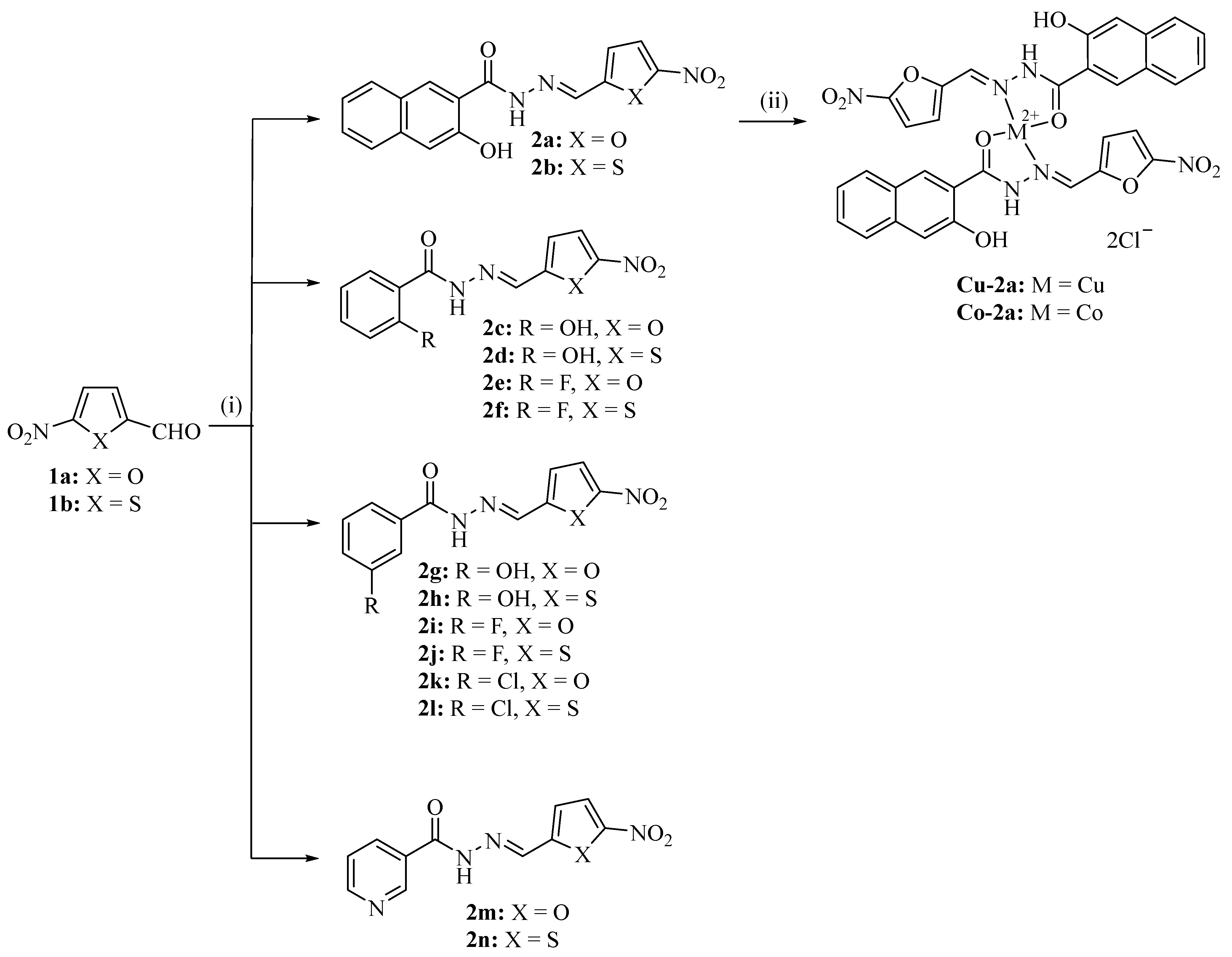
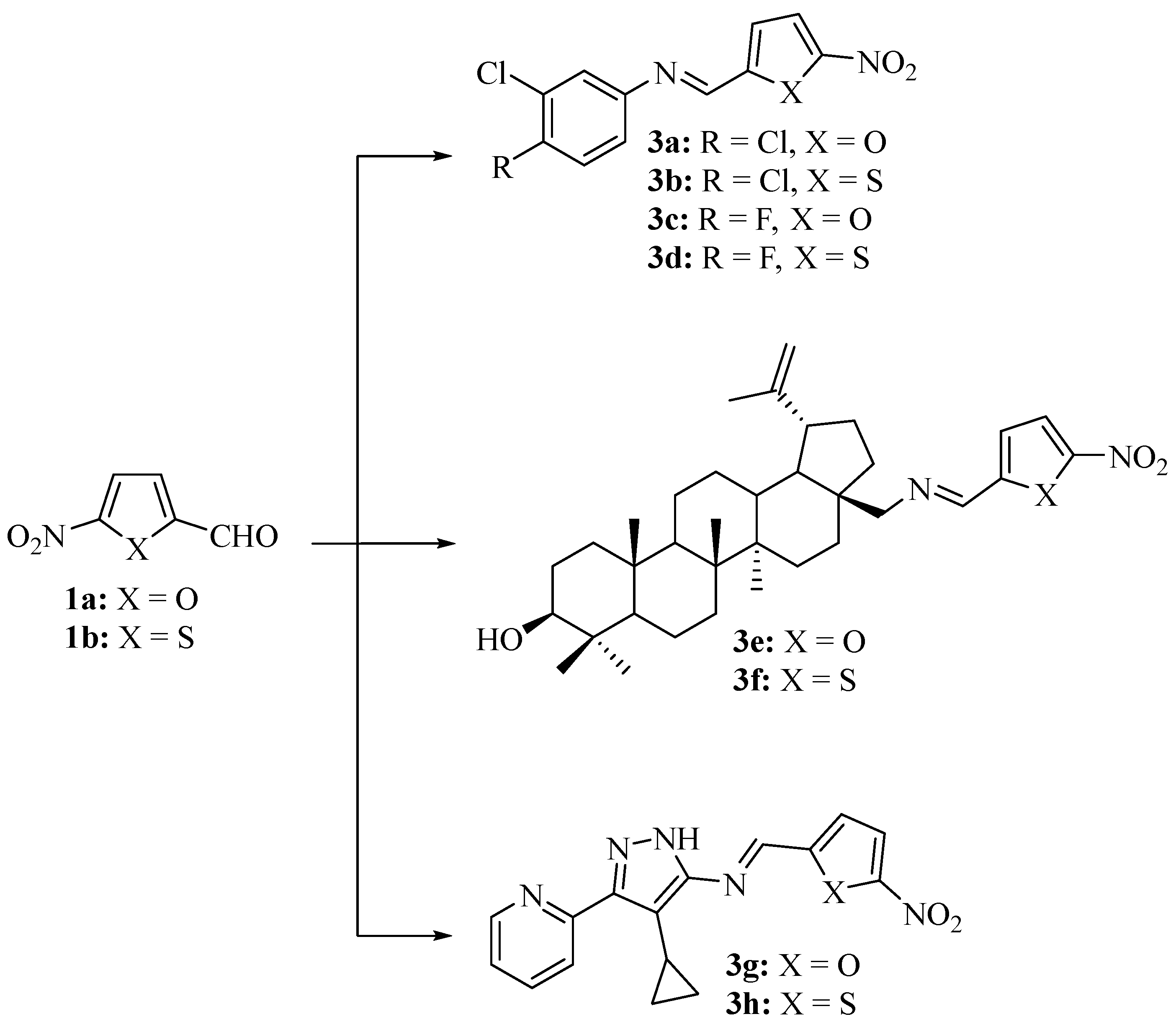
N-Acyl hydrazones 2a–n were prepared from 5-nitrofuraldehyde (1a) or 5-nitrothiophene-2-carboxaldehyde (1b) and the corresponding aroyl hydrazides in hot ethanol (Scheme 1). The 2-fluorophenyl derivatives 2e and 2f are new, while the other compounds (2a–d and 2 g–n) were described before. However, within this compound series, the antileishmanial activities relevant for this study were only reported for compounds 2g and 2h [7]. The new Cu(II) and Co(II) complexes of 2a, which were dubbed Cu-2a and Co-2a, were prepared from the reaction of 2a with the metal chloride salts CuCl2 and CoCl2, respectively [24]. The new imines/Schiff bases 3a–h were synthesized analogously to compounds 2a–n from 1a or 1b and the corresponding amine precursor (Scheme 2). 3,4-Dihalogenated anilines were chosen because this structural motif had previously shown distinct antiparasitic effects [7]. In addition, 28-aminobetulin was selected for condensation with 1a and 1b because of the well-documented antiparasitic activities of betulin derivatives [21,25]. Recently, the globally acting open synthesis network of the DNDi identified 3-amino-4-cyclopropyl-5-(2-pyridyl)-pyrazole as a promising scaffold for the development of antitrypanosomal drug candidates, and it was applied in this study to prepare the new imines 3g and 3h [26].

2.2. Antiparasitic Activity

Compounds 2a–n, Cu-2a, Co-2a, and 3a–h were tested for their activity against Toxoplasma gondii parasites (Table 1). 28-Aminobetulin was added to the panel of test compounds because there are no data for this betulin derivative in terms of antiparasitic activity. Vero kidney epithelial cells and macrophages served as non-malignant cells to evaluate the parasite selectivity and toxic side effects of the test compounds. The 3-hydroxy-2-naphthoic hydrazide derivatives 2a and 2b were the most active compounds, with excellent IC50 values almost reaching the activity of the positive control atovaquone. Compound 2a displayed considerable selectivity for T. gondii when compared with Vero cells, with a selectivity index (SI) of 23. 3-Fluorobenzoic hydrazide 2i also exhibited strong activity against T. gondii (IC50 below 1 µM). However, the selectivity of 2i was lower when compared with the selectivities of 2a and 2b. The salicyl derivative 2c also showed an amenable selectivity profile (SI = 17.7), while its thiophene analog (2d) was only moderately active without any selectivity for T. gondii. However, the other 5-nitrofuran-2-ylidene derivatives (2e, 2g, 2k, and 2m) were much less active than their thiophene-based congeners (2f, 2h, 2l, and 2n). The investigation of the toxicities of the test compounds to macrophages, and their correlation with the found antitoxoplasmal activities revealed an excellent SI value of over 100 (123) for 2a, while 2b reached an SI value of almost 60 (Table 1).
In order to evaluate the scope of the metal compounds based on the highly active ligand 2a, the Cu(II) and Co(II) complexes Cu-2a and Co-2a were prepared and tested for their activities against T. gondii. Both complexes conserved the high activity of their ligand (2a). Complex Co-2a also exhibited considerable selectivity for T. gondii and a toxicity to Vero kidney cells and macrophages similar to ligand 2a. The Cu(II) complex Cu-2a was more toxic to Vero cells than Co-2a, while both complexes showed similar toxicities to macrophages.
Among the new imines 3a–h, derivatives 3b–d were active against T. gondii at concentrations below 5 µM, while the other compounds showed rather moderate activities (IC50 values of 8–16 µM). The SI values calculated with Vero cells were generally low, with a value of three for 3b being the best SI value found in this compound series. Most strikingly, 28-aminobetulin was much more active against T. gondii and toxic to Vero cells than its imines (3e and 3f), indicating that other strategies of 28-aminobetulin modification should be explored in order to conserve its very promising anti-Toxoplasma activity while reducing its kidney cell toxicity. However, 28-aminobetulin revealed a low toxicity to macrophages, and, thus, a considerable SI value of 65.6 was determined with macrophages.
Next, the antileishmanial effects of the test compounds were studied using L. major promastigotes and amastigotes and were compared with their toxic effects on macrophages (Table 2). Compound 2i showed by far the highest activity against L. major promastigotes, distinctly exceeding the activity of amphotericin B by a factor of nearly 10. Moreover, the toxicity of 2i to macrophages was 89 times lower than to promastigotes (SI = 88.7). Compounds 2b, 2j, and 2k also revealed promising activities against L. major promastigotes, while of these compounds only 2j was likewise active against the amastigotes. Compounds 2b and 2i were the only compounds with sound activities against L. major promastigotes and T. gondii parasites, which were accompanied by distinctly lower toxic effects on Vero cells and macrophages. Compound 2k was particularly selective for promastigotes because amastigotes, T. gondii cells, Vero cells, and macrophages were much less sensitive to this compound. In contrast, 2h and 2l displayed reasonable activity against amastigotes (IC50 = 2.75 and 2.91 µM, respectively) while being less active against the promastigotes. Compounds 2h and 2l were less than one third as toxic to macrophages compared to amastigotes. Compound 2a and its metal complexes were only moderately active against L. major promastigotes and amastigotes. Only the cobalt complex Co-2a was slightly more active against promastigotes than ligand 2a. It is noteworthy that the imines 3g and 3h exhibited considerable activities against L. major promastigotes, which were significantly higher than against L. major amastigotes, T. gondii parasites, Vero cells, and macrophages. The betulin derivatives 3e and 3f were also more active against promastigotes than against amastigotes and T. gondii. L. major parasites were generally much less sensitive to their precursor (28-aminobetulin) than T. gondii parasites. However, 5-nitrofuran 3e revealed the highest activity against amastigotes among the imine series of compounds, which was 2.5 times higher than against macrophages. Only 28-aminobetulin and the positive control (amphotericin B) showed higher selectivities for the L. major amastigotes (SI = 6.6 and 17.2, respectively). The chlorophenyl derivatives 3a, 3b, and 3d were active against promastigotes, with IC50 values comparable to the activities of 3e and 3f. Only 3c was distinctly less active.

3. Discussion

The testing of various 5-nitrofuran and 5-nitrothiophene derivatives for their activities against T. gondii and L. major parasites revealed encouraging results. But in addition to antiparasitic activity, the selectivity of test compounds for parasites is a crucial factor for the identification of new drug candidates. The 3-hydroxy-2-naphthoic hydrazide 2a was identified as a promising anti-Toxoplasma compound in terms of activity and selectivity. Nitroaromatic compounds are prodrugs that are activated by cellular nitroreductases, leading to toxic radicals and reactive oxygen species in trypanosomatids [28]. This is an efficient way to kill T. gondii parasites, and nitrofurantoin, for example, exhibited strong in vitro and in vivo anti-toxoplasmal activities [29]. Moreover, the immunomodulatory activities of nitrofuran drugs might be relevant for future applications of 2a and its analogs [30]. The benefits and problems of nitro compounds for the treatment of protozoal parasites were reviewed recently [31]. Aside from toxic effects based on nitroaryl-correlated oxidation mechanisms, additional structure-dependent mechanisms of action likely play a role in the observed differences in the activity of compounds 2a–n. For instance, compound 2j was identified as a protein import blocker in the mitochondria of yeast and mammalian cells that targets a Tim44 interaction with Hsp70 [32]. In addition, dynasore, which is a catechol-based analog of 2a and 2b, is a well-established endocytosis inhibitor that blocks the guanosine triphosphatase (GTPase) dynamin [33]. Dynasore also regulates cellular cholesterol homeostasis and induces actin remodeling. More recently, it was shown that dynasore and its derivatives are strong inhibitors of viral ribonucleases necessary for the processing of RNA molecules [33,34]. Dynamin proteins play crucial roles in the life cycle of T. gondii parasites [35,36,37]. Thus, it is not surprising that dynasore and its analogs exert eminent antiparasitic effects in T. gondii infection models, which include drug-related influences on invasion, migration, and interleukin-10 (IL-10) formation [8,38,39]. The extent that compounds 2a and 2b also act in a dynasore-like manner against protozoal parasites remains to be clarified. However, the 3-hydroxy-2-naphthoic acid moieties of 2a and 2b obviously play a significant role in their activity and might also have a positive impact on the activities of other antiparasitic drugs as well as on the circumvention of drug resistance mechanisms [14,18]. In particular, atovaquone resistance was observed in apicomplexan parasites including T. gondii and Plasmodium falciparum, which necessitates the development of new drugs [14,40]. Moreover, cases of T. gondii infection prophylaxis failure in hematopoietic cell transplant recipients were reported upon treatment with atovaquone [41].
In addition, metal complexes of 2a conserved its activity against T. gondii and, thus, are valuable alternatives to 2a for the treatment of toxoplasmosis that warrant advanced investigations. For instance, the activity of these compounds against other T. gondii-related apicomplexan parasites such as Plasmodium might turn out to be equally promising, and new drugs against malaria are required in order to overcome the shortcomings of currently available therapies. The copper complex Cu-2a was distinctly more toxic to Vero cells than 2a, which might be attributed to the well-documented cytotoxic effects of copper ions and complexes [42,43]. Only a few Cu(II) complexes with considerable anti-Toxoplasma activity were described, while no Co(II) complexes have been published until now showing activity against T. gondii parasites [44]. Antileishmanial Co(II) complexes with comparable activities were recently disclosed, and their mechanism of action involved mitochondrial damage associated with the induction of apoptosis-like parasite death [45]. Both Cu(II) and Co(II) complexes were also reported to target DNA, while Co(II) salts such as CoCl2 are well-established hypoxia inducers [46,47,48].
This study shows that there is no general advantage of 5-nitrofurans over 5-nitrothiophenes. Although the most active compounds 2a and 2i were 5-nitrofurans, the NH2-substituted starting compound and binding partner (i.e., the hydrazide or amine starting compound) often played a crucial role in the antiparasitic effects of the target compounds. 5-Nitrothiophenes were casually more active than their 5-nitrofuran congeners, which matched the results of a published study with compound 2 analogs that were presumably based on a higher reduction rate of nitrothiophenes by nitroreductases because of unoccupied sulfur d orbitals [7]. However, differences sometimes also depended on the tested parasite species and even on certain cell forms within one species, such as the promastigotes and amastigotes of L. major. These differences were clearly less pronounced among the new imine compound series (3a–h). However, the elucidation of the underlying mechanisms is a challenging task, and the observed activities can be explained only in part by the peculiar chemical and physical properties of the described structural scaffolds. The activity of 28-aminobetulin against T. gondii and L. major was remarkable. Its new analog 3e also displayed considerable activity against L. major amastigotes and promastigotes, and, thus, the preparation and investigation of more 28-aminobetulin derivatives appears reasonable in the future. Strikingly, pyrazoles 3g and 3h were among the more active antileishmanial imine derivatives, and various compounds based on pyrid-2-ylpyrazole scaffolds have already been identified as antiparasitic drugs with significant selectivity for certain kinetoplastid parasites [23,25]. The mechanism of action of this class of antiparasitic aminopyrazoles is still not clearly understood, but it was shown that potent examples were not substrates for drug efflux pumps associated with multi-drug resistance [22]. Moreover, it has to be mentioned that the imines 3a–d appeared to be less antileishmanial than previously published analogous 3,4-dichlorobenzoic hydrazide derivatives [7].

4. Materials and Methods

4.1. Chemistry

The known compounds 2a–d and 2g–n were prepared and analyzed in accordance with reports in the literature [7,32]. Starting compounds were purchased from common providers. 28-Aminobetulin was obtained from Nature Science Technologies (Olaine, Latvia), and 3-amino-4-cyclopropyl-5-(pyrid-2-yl)-pyrazole was obtained from TCG Lifesciences Pvt. Ltd. (Kolkata, India). Melting points were determined with an Electrothermal 9100 and were not corrected. NMR spectra were measured with a Bruker Avance 300 spectrometer, and chemical shifts are given in parts per million (δ) downfield from tetramethylsilane as an internal standard. High-resolution mass spectra were measured with a UPLC/Orbitrap (ESI-HRMS).
2-Fluorobenzoic (5-nitrofuran-2-ylidene)hydrazide (2e)
2-Fluorobenzoic hydrazide (154 mg, 1.0 mmol) and 5-nitrofuraldehyde (141 mg, 1.0 mmol) were dissolved in EtOH (15 mL). The reaction mixture was stirred under reflux for 2 h. The formed precipitate was collected, washed with EtOH, and dried in a vacuum. Yield: 213 mg (0.77 mmol, 77%); orange solid of m.p. 217–218 °C; υmax(ATR)/cm−1 3358, 3141, 1668, 1611, 1550, 1514, 1471, 1393, 1351, 1335, 1285, 1253, 1239, 1211, 1172, 1143, 1097, 1056, 1013, 958, 906, 880, 825, 809, 784, 754, 737, 685, 654; 1H NMR (500 MHz, DMSO-d6) δ 7.2–7.4 (3 H, m), 7.5–7.7 (2 H, m), 7.8–7.9 (1 H, m), 8.29 (1 H, s), 12.25 (1 H, s); 13C NMR (125.8 MHz, DMSO-d6) δ 115.2–116.4 (m), 122.5–122.7 (m), 124.3, 124.8, 129.2, 129.8, 130.0, 130.2, 130.4, 132.4, 133.2–133.3 (m), 137.3, 141.5, 146.3, 150.5, 151.0, 157.5, 160.7, 160.8, 167.4; HRMS for C12H9O3N3FS [M+ + H]: calcd. 294.03432, found 294.03386.
2-Fluorobenzoic (5-nitrothien-2-ylidene)hydrazide (2f)
2-Fluorobenzoic hydrazide (154 mg, 1.0 mmol) and 5-nitrothiophene-2-carboxaldehyde (157 mg, 1.0 mmol) were dissolved in EtOH (15 mL). The reaction mixture was stirred under reflux for 2 h. The formed precipitate was collected, washed with EtOH, and dried in a vacuum. Yield: 165 mg (0.52 mmol, 52%); brown solid of m.p. 204–205 °C; υmax(ATR)/cm−1 3320, 3124, 1658, 1614, 1547, 1530, 1501, 1482, 1456, 1438, 1359, 1336, 1311, 1284, 1242, 1209, 1158, 1143, 1099, 1073, 1032, 965, 928, 907, 879, 822, 814, 784, 752, 731, 701, 682; 1H NMR (500 MHz, DMSO-d6) δ 7.3–7.4 (2 H, m), 7.5–7.7 (3 H, m), 8.1–8.2 (1 H, m), 8.56 (1 H, s), 12.24 (1 H, s); 13C NMR (125.8 MHz, DMSO-d6) δ 115.2–116.4 (m), 122.5–122.7 (m), 124.3, 124.8, 129.2, 129.8, 130.0, 130.2, 130.4, 132.4, 133.2–133.3 (m), 137.3, 141.5, 146.3, 150.5, 151.0, 157.5, 160.7, 160.8, 167.4; HRMS for C12H9O3N3FS [M+ + H]: calcd. 294.03432, found 294.03386.
Copper(II) complex (Cu-2a)
Compound 2a (100 mg, 0.306 mmol) was dissolved in 1,4-dioxane (10 mL), and CuCl2 x 2H2O (26 mg, 0.153 mmol) was added. The reaction mixture was stirred under reflux for 1 h. After cooling down, the formed precipitate was collected, washed with ethanol, and dried in a vacuum. Yield: 35 mg (0.045 mmol, 29%); brown solid of m.p. 206–208 °C (dec.); υmax(ATR)/cm−1 3250, 3129, 3079, 2965, 2920, 2869, 1634, 1591, 1560, 1535, 1503, 1461, 1396, 1341, 1254, 1221, 1148, 1113, 1080, 1068, 1055, 1026, 969, 961, 931, 895, 862, 812, 801, 768, 754, 734, 669; HRMS for C32H21O10N6Cu [M+ - H]: calcd. 712.06097, found 712.06004.
Cobalt(II) complex (Co-2a)
Compound 2a (100 mg, 0.306 mmol) was dissolved in 1,4-dioxane (5 mL), and CoCl2 x 6H2O (36 mg, 0.153 mmol) was added. The reaction mixture was stirred under reflux for 1 h. After cooling down, the formed precipitate was collected, washed with n-hexane, and dried in a vacuum. Yield: 110 mg (0.141 mmol, 92%); yellow solid of m.p. 235–237 °C (dec.); υmax(ATR)/cm−1 3257, 3136, 3047, 2909, 2861, 1637, 1607, 1563, 1527, 1475, 1395, 1346, 1254, 1220, 1150, 1120, 1080, 1025, 970, 956, 923, 887, 871, 810,771, 760, 744, 735, 696; HRMS for C32H21O10N6Co [M+ - H]: calcd. 708.06457, found 708.06317.
(E)-N-(3,4-Dichlorophenyl)-1-(5-nitrofuran-2-ylidene)imine (3a)
3,4-Dichloroaniline (162 mg, 1.0 mmol) and 5-nitrofuraldehyde (141 mg, 1.0 mmol) were dissolved in CH2Cl2 (5 mL) and stirred under reflux for 3 h. The solvent was evaporated, and the residue was recrystallized from CH2Cl2/n-hexane. Yield: 264 mg (0.93 mmol, 93%); yellow solid of m.p. 123–125 °C; υmax(ATR)/cm−1 3140, 3079, 3013, 1563, 1514, 1484, 1458, 13973 1352, 1333, 1247, 1198, 1170, 1121, 1029, 963, 901, 875, 820, 810, 799, 739, 691, 669, 659, 599, 584; 1H NMR (300 MHz, CDCl3) δ 7.11 (1 H, dd, J = 8.5 Hz, 2.5 Hz), 7.20 (1 H, d, J = 3.9 Hz), 7.36 (1 H, d, J = 2.5 Hz), 7.41 (1 H, d, J = 3.9 Hz), 7.47 (1 H, d, J = 8.5 Hz), 8.34 (1 H, s); 13C NMR (75.5 MHz, CDCl3) δ 112.6, 115.4, 120.7, 123.0, 131.1, 131.6, 133.4, 147.4, 149.3, 152.6; HRMS for C11H7O3N2Cl2 [M+ + H]: calcd. 284.98282, found 284.98233.
(E)-N-(3,4-Dichlorophenyl)-1-(5-nitrothien-2-yl)methanimine (3b)
3,4-Dichloroaniline (162 mg, 1.0 mmol) and 5-nitrothiophene-2-carboxaldehyde (157 mg, 1.0 mmol) were dissolved in CH2Cl2 (5 mL) and stirred under reflux for 3 h. The solvent was evaporated, and the residue was recrystallized from CH2Cl2/n-hexane. Yield: 134 mg (0.45 mmol, 45%); yellow solid of m.p. 154–155 °C; υmax(ATR)/cm−1 3093, 1605, 1558, 1529, 1494, 1464, 1435, 1394, 1332, 1314, 1272, 1220, 1190, 1137, 1041, 1026, 954, 913, 868, 828, 816, 733, 706, 693, 679, 640, 600; 1H NMR (300 MHz, CDCl3) δ 7.10 (1 H, dd, J = 8.5 Hz, 2.5 Hz), 7.35 (1 H, d, J = 2.5 Hz), 7.39 (1 H, d, J = 4.5 Hz), 7.46 (1 H, d, J = 8.5 Hz), 7.90 (1 H, d, J = 4.5 Hz), 8.50 (1 H, s); 13C NMR (75.5 MHz, CDCl3) δ 120.9, 122.9, 128.4, 130.3, 131.0, 131.3, 133.3, 147.6, 149.0, 152.3, 154.4; HRMS for C11H7O2N2Cl2S [M+ + H]: calcd. 300.95998, found 300.95939.
(E)-N-(3-Chloro-4-fluorophenyl)-1-(5-nitrofuran-2-yl)methanimine (3c)
3-Chloro-4-fluoroaniline (146 mg, 1.0 mmol) and 5-nitrofuraldehyde (141 mg, 1.0 mmol) were dissolved in CH2Cl2 (5 mL) and stirred under reflux for 3 h. The solvent was evaporated, and the residue was recrystallized from CH2Cl2/n-hexane. Yield: 195 mg (0.73 mmol, 73%); yellow solid of m.p. 154–156 °C; υmax(ATR)/cm−1 3147, 3122, 1624, 1567, 1519, 1490, 1400, 1356, 1335, 1255, 1233, 1197, 1163, 1122, 1051, 1026, 961, 917, 868, 823, 812, 807, 761, 737, 701, 690, 666, 591; 1H NMR (300 MHz, CDCl3) δ 7.1–7.2 (3 H, m), 7.3–7.4 (1 H, m), 7.41 (1 H, d, J = 3.8 Hz), 8.34 (1 H, s); 13C NMR (75.5 MHz, CDCl3) δ 112.7, 115.1, 117.1–117.4 (m), 121.2–121.3 (m), 123.2, 146.5–146.8 (m), 152.8, 155.9–159.2 (m); HRMS for C11H7O3N2ClF [M+ + H]: calcd. 269.01237, found 269.01196.
(E)-N-(3-Chloro-4-fluorophenyl)-1-(5-nitrothien-2-yl)methanimine (3d)
3-Chloro-4-fluoroaniline (146 mg, 1.0 mmol) and 5-nitrothiophene-2-carboxaldehyde (157 mg, 1.0 mmol) were dissolved in CH2Cl2 (5 mL) and stirred under reflux for 3 h. The solvent was evaporated, and the residue was recrystallized from CH2Cl2/n-hexane. Yield: 120 mg (0.42 mmol, 42%); yellow solid of m.p. 152–153 °C; υmax(ATR)/cm−1 3108, 1609, 1586, 1536, 1489, 1438, 1389, 1366, 1350, 1333, 1311, 1253, 1222, 1208, 1187, 1122, 1052, 1039, 965, 937, 914, 870, 814, 797, 761, 732, 712, 702, 661, 600, 565; 1H NMR (300 MHz, CDCl3) δ 7.1–7.2 (2 H, m), 7.3–7.4 (2 H, m), 7.90 (1 H, d, J = 4.2 Hz), 8.50 (1 H, s); 13C NMR (75.5 MHz, CDCl3) δ 117.0–117.3 (m), 121.4–122.0 (m), 123.1, 128.4, 130.0, 146.2–146.3 (m), 147.8, 151.7, 155.7–159.0; HRMS for C11H7O2N2ClFS [M+ + H]: calcd. 284.98953, found 284.98892.
(E)-N-Betulin-28-yl-1-(5-nitrofuran-2-yl)methanimine (3e)
28-Aminobetulin (250 mg, 0.57 mmol) and 5-nitrofuraldehyde (80 mg, 0.57 mmol) were dissolved in CH2Cl2 (5 mL) and stirred under reflux for 5 h. The solvent was evaporated, and the residue was recrystallized from CH2Cl2/n-hexane. Yield: 205 mg (0.36 mmol, 63%); off-white solid of m.p. 249–250 °C; υmax(ATR)/cm−1 3389, 3084, 2982, 2938, 2867, 1642, 1572, 1528, 1485, 1454, 1402, 1389, 1375, 1349, 1323, 1294, 1259, 1183, 1131, 1105, 1082, 1043, 1016, 972, 963, 945, 881, 825, 810, 739, 642, 583; 1H NMR (300 MHz, CDCl3) δ 0.6–0.7 (1 H, m), 0.75 (3 H, s), 0.82 (3 H, s), 1.0–1.1 (10 H, m), 1.2–1.3 (4 H, m), 1.4–1.5 (5 H, m), 1.6–2.0 (15 H, m), 2.4–2.5 (1 H, m), 3.1–3.2 (1 H, m), 3.30 (1 H, d, J = 12.0 Hz), 3.80 (1 H, d, J = 12.0 Hz), 4.58 (1 H, s), 4.69 (1 H, s), 6.99 (1 H, d, J = 3.8 Hz), 7.34 (1 H, d, J = 3.8 Hz), 8.16 (1 H, s); 13C NMR (75.5 MHz, CDCl3) δ 14.8, 15.4, 16.1, 18.3, 19.2, 20.8, 25.2, 26.8, 27.4, 28.0, 29.6, 30.3, 34.2, 34.9, 37.2, 37.4, 38.7, 38.9, 41.0, 42.7, 47.8, 47.9, 49.0, 50.4, 55.3, 60.8, 78.9, 109.8, 112.5, 112.8, 148.8, 150.4, 153.1; HRMS for C35H53O4N2 [M+ + H]: calcd. 565.39998, found 565.39888.
(E)-N-Betulin-28-yl-1-(5-nitrothien-2-yl)methanimine (3f)
28-Aminobetulin (240 mg, 0.54 mmol) and 5-nitrothiophene-2-carboxaldehyde (85 mg, 0.54 mmol) were dissolved in CH2Cl2 (5 mL) and stirred under reflux for 5 h. The solvent was evaporated, and the residue was recrystallized from CH2Cl2/n-hexane. Yield: 150 mg (0.26 mmol, 48%); off-white solid of m.p. 229–230 °C; υmax(ATR)/cm−1 3364, 2933, 2868, 1622, 1533, 1505, 1456, 1438, 1388, 1376, 1336, 1227, 1212, 1106, 1043, 1027, 983, 965, 946, 883, 815, 749, 733, 685, 609, 575; 1H NMR (300 MHz, CDCl3) δ 0.6–0.7 (1 H, m), 0.75 (3 H, s), 0.8–0.9 (4 H, m), 0.9–1.1 (13 H, m), 1.2–2.0 (29 H, m), 2.4–2.5 (1 H, m), 3.1–3.2 (1 H, m), 3.25 (1 H, d, J = 12.2 Hz), 3.76 (1 H, d, J = 12.2 Hz), 4.58 (1 H, s), 4.69 (1 H, s), 7.16 (1 H, d, J = 4.2 Hz), 7.84 (1 H, d, J = 4.2 Hz), 8.28 (1 H, s); 13C NMR (75.5 MHz, CDCl3) δ 14.9, 15.4, 16.1, 18.3, 19.2, 20.9, 25.2, 26.8, 27.4, 28.0, 29.7, 30.4, 34.2, 34.9, 37.2, 37.4, 38.7, 38.9, 41.0, 42.7, 47.9, 49.0, 50.4, 55.3, 59.7, 79.0, 109.7, 127.3, 128.4, 149.1, 150.5, 152.7; HRMS for C35H53O3N2S [M+ + H]: calcd. 581.37714, found 581.37587.
(E)-N-(4-Cyclopropyl-3-(pyridin-2-yl)-1H-pyrazol-5-yl)-1-(5-nitrofuran-2-yl)methanimine (3g)
3-Amino-4-cyclopropyl-5-(pyrid-2-yl)-pyrazole (200 mg, 1.0 mmol) and 5-nitrofuraldehyde (141 mg, 1.0 mmol) were dissolved in EtOH (15 mL). The reaction mixture was stirred under reflux for 2 h. The formed precipitate was collected, washed with EtOH, and dried in a vacuum. Yield: 272 mg (0.84 mmol, 84%); yellow solid of m.p. >240 °C (dec.); υmax(ATR)/cm−1 3273, 3132, 3009, 1590, 1568, 1523, 1504, 1485, 1465, 1432, 1382, 1348, 1302, 1252, 1230, 1209, 1181, 1153, 1126, 1093, 1055, 1015, 1000, 983, 962, 904, 840, 808, 789, 759, 747, 734, 673; 1H NMR (500 MHz, DMSO-d6) δ 0.8–1.0 (4 H, m), 2.1–2.3 (1 H, m), 7.3–7.4 (1 H, m), 7.44 (1 H, d, J = 3.9 Hz), 7.82 (1 H, d, J = 3.9 Hz), 7.9–8.0 (1 H, m), 8.6–8.7 (1 H, m), 8.87 (1 H, s), 13.39 (1 H, s); 13C NMR (125.8 MHz, DMSO-d6) δ 5.83, 7.79, 114.3, 115.3, 117.9, 121.4, 122.8, 137.1, 140.8, 144.8, 148.4, 149.4, 152.4, 153.2, 155.4; HRMS for C16H14O3N5 [M+ + H]: calcd. 324.10912, found 324.10777.
(E)-N-(4-Cyclopropyl-3-(pyridin-2-yl)-1H-pyrazol-5-yl)-1-(5-nitrothien-2-yl)methanimine (3h)
3-Amino-4-cyclopropyl-5-(pyridyl-2y)-pyrazole (200 mg, 1.0 mmol) and 5-nitrothiophene-2-carboxaldehyde (157 mg, 1.0 mmol) were dissolved in EtOH (15 mL). The reaction mixture was stirred under reflux for 2 h. The formed precipitate was collected, washed with EtOH, and dried in a vacuum. Yield: 277 mg (0.86 mmol, 86%); yellow-brown solid of m.p. >230 °C (dec.); υmax(ATR)/cm−1 3203, 3095, 3005, 1592, 1571, 1534, 1495, 1466, 1447, 1408, 1336, 1297, 1236, 1212, 1186, 1140, 1094, 1054, 1041, 1001, 980, 960, 914, 887, 827, 810, 789, 762, 745, 730, 697, 675; 1H NMR (500 MHz, DMSO-d6) δ 0.8–1.0 (4 H, m), 2.1–2.3 (1 H, m), 7.3–7.5 (1 H, m), 7.76 (1 H, d, J = 4.4 Hz), 7.9–8.0 (1 H, m), 8.18 (1 H, d, J = 4.4 Hz), 8.6–8.7 (1 H, m), 9.14 (1 H, s), 13.39 (1 H, s); 13C NMR (125.8 MHz, DMSO-d6) δ 5.91, 7.80, 115.4, 121.2, 122.2, 122.8, 130.6, 131.5, 137.2, 140.6, 148.4, 149.5, 149.6, 149.9, 152.2, 154.4; HRMS for C16H14O2N5S [M+ + H]: calcd. 340.08627, found 340.08521.

4.2. Toxoplasma gondii Cell Line, Culture Conditions, and Assay

Serial passages of Vero cells (ATCC® CCL81™, Manassas, VA, USA) were applied for the cultivation of T. gondii tachyzoites (RH strain, a gift from Dr. Saeed El-Ashram, State Key Laboratory for Agrobiotechnology, China Agricultural University, Bejing, China). Vero cells were cultured using complete RPMI 1640 medium (Invitrogen, Carlsbad, CA, USA) with heat-inactivated 10% fetal bovine serum (FBS, Invitrogen, Carlsbad, CA, USA) in a humidified 5% CO2 atmosphere at 37 °C. In addition, 96-well plates (5 × 10³ cells/well in 200 μL of RPMI 1640 medium) were used for the cultivation of the Vero cells, which were incubated at 37 °C and 5% CO2 for one day, followed by the removal of the medium and washing the cells with phosphate-buffered saline (PBS). Then, RPMI 1640 medium containing 2% FBS and T. gondii tachyzoites (RH strain) was added to the cells at a ratio of 5 (parasites) : 1 (Vero cells). The tests were performed in the following way: Control: RPMI 1640 medium containing DMSO (1%). Experimental: medium + compounds (from stock solutions in DMSO; final concentrations of 50, 25, 12.5, 6.25, 3.13, 1.65, and 0.75 μg mL−1).
After incubation at 37 °C and 5% CO2 for 72 h, the cells were washed with PBS, fixed in 10% formalin, and stained with 1% toluidine blue. An examination of the cells and a determination of the infection index (number of cells infected from 200 cells tested) of T. gondii were carried out with an inverted photomicroscope. The following equation was used for the calculation of the inhibition in %:
Inhibition (%) = (I Control) − (I Experimental)/(I Control) × 100
where “I Control” refers to the infection index of untreated cells and “I Experimental” refers to the infection index of cells treated with test compounds. The effects of the test compounds on parasite growth were expressed as IC50 (inhibitory concentration at 50%) values. IC50 values were obtained from three independent experiments [27,49].

4.3. Leishmania Major Cell Isolation, Culture Conditions, and Assays

Promastigotes of L. major were isolated from a Saudi patient (February 2016) and maintained in a tissue culture flask at 26 °C in Schneider’s Drosophila medium (Invitrogen, Carlsbad, CA, USA) containing 10% heat-inactivated FBS and antibiotics with weekly transfers. Promastigotes were cryopreserved in liquid nitrogen at concentrations of 3 × 10⁶ parasites/mL. Virulent L. major parasites were maintained in female BALB/c mice by injecting their hind footpads with 1 × 10⁶ stationary-phase promastigotes. L. major amastigotes were isolated from the mice after 8 weeks. The isolated amastigotes were converted to promastigotes by cultivation in Schneider’s medium supplemented with antibiotics and 10% FBS at 26 °C. Amastigote-derived promastigotes, which had undergone less than five in vitro passages, were used for infection.
The BALB/c mice (male and female individuals) for these experiments were obtained from the Pharmaceutical College of King Saud University, Kingdom of Saudi Arabia, and maintained in specific pathogen-free facilities. The handling of the laboratory animals followed the instructions and rules of the committee of research ethics, Deanship of Scientific Research, Qassim University (permission number 20-03-20).
Logarithmic-phase L. major promastigotes were cultured in phenol-red-free RPMI 1640 medium containing 10% FBS and suspended on 96-well plates (106 cells mL−1, 200 μL/well) after counting with a hemocytometer. Compounds were added to the cell cultures, reaching final concentrations of 50, 25, 12.5, 6.25, 3.13, 1.65, and 0.75 μg mL−1. The negative controls were cultures containing DMSO (1%) without a test compound, and positive control wells contained cultures with decreasing concentrations of amphotericin B (50, 25, 12.5, 6.25, 3.13, 1.65, and 0.75 μg mL−1) as an antileishmanial reference drug. After incubation at 26 °C for 72 h, the number of viable promastigotes was assessed using a colorimetric method (tetrazolium salt colorimetric assay, MTT). The formed colored formazan was isolated and solubilized by the addition of a detergent solution. The samples were analyzed using an ELISA reader at 570 nm. IC50 values were calculated from three independent experiments [27].
To evaluate the activity against amastigotes in macrophages, peritoneal macrophages were collected from female BALB/c mice (6–8 weeks of age) via aspiration. First, 5 × 10⁴ cells/well were placed into 96-well plates containing phenol-red-free RPMI 1640 medium with 10% FBS and were incubated to promote cell adhesion at 37 °C for 4 h in a 5% CO2 atmosphere. Thereafter, the medium was discarded, and the cells were washed with PBS. An L. major promastigotes solution (200 μL at a ratio of 10 promastigotes : 1 macrophage in RPMI 1640 medium with 10% FBS) was added to each well, and the plates were incubated for 24 h at 37 °C in a humidified 5% CO2 atmosphere to enable macrophage infection and differentiation to amastigotes. The infected macrophages were washed three times with PBS to remove the free promastigotes and overlaid with fresh phenol-red-free RPMI 1640 medium containing test compounds (50, 25, 12.5, 6.25, 3.13, 1.65, and 0.75 μg mL−1). Then, the cells were incubated at 37 °C for 72 h in a humidified 5% CO2 atmosphere. Cultures solely containing DMSO (1%) were used as negative controls, while wells containing cultures with decreasing concentrations of amphotericin B (50, 25, 12.5, 6.25, 3.13, 1.65, and 0.75 μg mL−1) were used as positive controls. The percentage of infected macrophages was evaluated microscopically after the removal of the medium, washing, fixation, and the Giemsa staining of the cells. Calculated IC50 values were obtained from three independent experiments [27,50].

4.4. In Vitro Cytotoxicity Assay

MTT assays were carried out to evaluate the cytotoxicity of the test compounds. Briefly, both Vero and macrophage cells were cultured in 96-well plates (5 × 10³ cells/well/200 μL) in RPMI 1640 medium with 10% FBS and 5% CO2 at 37 °C for 24 h. Cells were washed with PBS, followed by treatment with test compounds for 72 h at varying concentrations (50, 25, 12.5, 6.25, 3.13, 1.65, and 0.75 μg mL−1) in a 10% FBS medium. Cells treated solely with medium in 2% FBS were used as a negative control. The supernatant was discarded, and 50 μL of RPMI 1640 medium containing 14 μL of MTT (5 mg mL−1) was added. Then, the cells were incubated for 4 h. The supernatant was removed, and DMSO (150 μL) was added in order to dissolve the formed formazan. A FLUOstar OPTIMA spectrophotometer was applied for the colorimetric analysis (λ = 540 nm) of the experiments. The cytotoxicity was expressed by CC50 values (cytotoxic concentration that caused a 50% reduction in viable cells). CC50 values were calculated from three independent experiments [27,51].

Author Contributions

Conceptualization, I.S.A.N. and B.B.; methodology, I.S.A.N., T.A.K., and B.B.; validation, I.S.A.N., T.A.K., and W.S.K.; formal analysis, I.S.A.N., W.S.K., and B.B.; investigation, I.S.A.N., T.A.K., and B.B.; resources, W.S.K. and R.S.; data curation, I.S.A.N.; writing—original draft preparation, B.B.; writing—review and editing, I.S.A.N., T.A.K., W.S.K., and R.S.; supervision, W.S.K. and B.B.; project administration, B.B. All authors have read and agreed to the published version of the manuscript.

Funding

This research received no external funding.

Institutional Review Board Statement

The animal study protocol was approved by the committee of research ethics, Deanship of Scientific Research, Qassim University (permission number 20–03-20).

Informed Consent Statement

Not applicable.

Data Availability Statement

Data can be obtained from the authors upon request.

Acknowledgments

We are grateful to Luiza Cruz from DNDi and TCG Lifesciences Pvt. Ltd., Kolkata, India, for providing a free sample of 3-amino-4-cyclopropyl-5-(pyrid-2-yl)-pyrazole as part of the DNDi project OSN P1Tc.

Conflicts of Interest

The authors declare no conflict of interest.

References

  1. Meymandi, S.; Hernandez, S.; Park, S.; Sanchez, D.R.; Forsyth, C. Treatment of Chagas Disease in the USA. Curr. Treat. Options Infect. Dis. 2018, 10, 373–388. [Google Scholar] [CrossRef] [PubMed]
  2. Priotto, G.; Kasparian, S.; Mutombo, W.; Ngouama, D.; Ghorashian, S.; Arnold, U.; Ghabri, S.; Baudin, E.; Buard, V.; Kazadi-Kyanza, S.; et al. Nifurtimox-eflornithine combination therapy for second-stage African Trypanosoma brucei gambiense trypanosomiasis: A multicentre, randomised, phase III, non-inferiority trial. Lancet 2009, 374, 56–64. [Google Scholar] [CrossRef] [PubMed]
  3. Kuemmerle, A.; Schmid, C.; Bernhard, S.; Kande, W.; Mutombo, W.; Ilunga, M.; Lumpungu, I.; Mutanda, S.; Nganzobo, P.; Tete, D.N.; et al. Effectiveness of Nifurtimox Eflornithine Combination Therapy (NECT) in T. b. gambiense second stage sleeping sickness patients in the Democratic Republic of Congo: Report from a field study. PLoS Negl. Trop. Dis. 2021, 15, e0009903. [Google Scholar] [CrossRef]
  4. Thota, S.; Rodrigues, D.A.; de Sena Murteira Pinheiro, P.; Lima, L.M.; Fraga, C.A.M.; Barreiro, E.J. N-Acylhydrazones as drugs. Bioorg. Med. Chem. Lett. 2018, 28, 2797–2806. [Google Scholar] [CrossRef] [PubMed]
  5. Socea, L.-I.; Barbuceanu, S.-F.; Pahontu, E.M.; Dumitru, A.-C.; Nitulescu, G.M.; Sfetea, R.C.; Apostol, T.-V. Acylhydrazones and their biological activity: A review. Molecules 2022, 27, 8719. [Google Scholar] [CrossRef] [PubMed]
  6. Bailly, C. Toward a repositioning of the antibacterial drug nifuroxazide for cancer treatment. Drug Discov. Today 2019, 24, 1930–1936. [Google Scholar] [CrossRef]
  7. Rando, D.G.; Avery, M.A.; Tekwani, B.L.; Khan, S.I.; Ferreira, E.I. Antileishmanial activity screening of 5-nitro-2-heterocyclic benzylidene hydrazides. Bioorg. Med. Chem. 2008, 16, 6724–6731. [Google Scholar] [CrossRef]
  8. Caldas, L.A.; Attias, M.; de Souza, W. Dynamin inhibitor impairs Toxoplasma gondii invasion. FEMS Microbiol. Lett. 2009, 301, 103–108. [Google Scholar] [CrossRef]
  9. Paruch, K.; Biernasiuk, A.; Khylyuk, D.; Paduch, R.; Wujec, M.; Popiołek, Ł. Synthesis, biological activity and molecular docking studies of novel nicotinic acid derivatives. Int. J. Mol. Sci. 2022, 23, 2823. [Google Scholar] [CrossRef]
  10. Montoya, J.G.; Liesenfeld, O. Toxoplasmosis. Lancet 2004, 363, 1965–1976. [Google Scholar] [CrossRef]
  11. Tenter, A.M.; Heckeroth, A.R.; Weiss, L.M. Toxoplasma gondii: From animals to humans. Int. J. Parasitol. 2000, 30, 1217–1258. [Google Scholar] [CrossRef] [PubMed]
  12. Dunay, I.R.; Gajurel, K.; Dhakal, R.; Liesenfeld, O.; Montoya, J.G. Treatment of toxoplasmosis: Historical perspective, animal models, and current clinical practice. Clin. Microbiol. Rev. 2018, 31, e00057-17. [Google Scholar] [CrossRef] [PubMed]
  13. El Hajj, R.; Tawk, L.; Itani, S.; Hamie, M.; Ezzeddine, J.; El Sabban, M.; El Hajj, H. Toxoplasmosis: Current and emerging parasite druggable targets. Microorganisms 2021, 9, 2531. [Google Scholar] [CrossRef] [PubMed]
  14. Montazeri, M.; Mehrzadi, S.; Sharif, M.; Sarvi, S.; Tanzifi, A.; Aghayan, S.A.; Daryani, A. Drug resistance in Toxoplasma gondii. Front. Microbiol. 2018, 9, 2587. [Google Scholar] [CrossRef] [PubMed]
  15. Crespo, M.; Quereda, C.; Pascual, J.; Rivera, M.; Clemente, L.; Cano, T. Patterns of sulfadiazine acute nephrotoxicity. Clin. Nephrol. 2000, 54, 68–72. [Google Scholar] [PubMed]
  16. WHO-Factsheets. Available online: http://www.who.int/mediacentre/factsheets/fs375/en/ (accessed on 12 November 2020).
  17. van Bocxlaer, K.; Caridha, D.; Black, C.; Vesely, B.; Leed, S.; Sciotti, R.J.; Wijnant, G.-J.; Yardley, V.; Braillard, S.; Mowbray, C.E.; et al. Novel benzoxaborole, nitroimidazole and aminopyrazoles with activity against experimental cutaneous leishmaniasis. Int. J. Parasitol. Drugs Drug Resist. 2019, 11, 129–138. [Google Scholar] [CrossRef]
  18. Alves, F.; Bilbe, G.; Blesson, S.; Goyal, V.; Monnerat, S.; Mowbray, C.; Ouattara, G.M.; Pécoul, B.; Rijal, S.; Rode, J.; et al. Recent development of visceral leishmaniasis treatments: Successes, pitfalls, and perspectives. Clin. Microbiol. Rev. 2018, 31, e00048-18. [Google Scholar] [CrossRef]
  19. Koko, W.S.; Al Nasr, I.S.; Khan, T.A.; Schobert, R.; Biersack, B. An update on natural antileishmanial treatment options from plants, fungi and algae. Chem. Biodivers. 2021, 19, e202100542. [Google Scholar] [CrossRef]
  20. Mioc, M.; Prodea, A.; Racoviceanu, R.; Mioc, A.; Ghiulai, R.; Milan, A.; Voicu, M.; Mardale, G.; Soica, C. Recent advances regarding the molecular mechanisms of triterpenic acids: A review (part II). Int. J. Mol. Sci. 2022, 23, 8896. [Google Scholar] [CrossRef]
  21. Rocha, V.; Quadros, H.; Meira, C.; Silva, L.; Carvalho, D.; Hodel, K.; Moreira, D.; Soares, M. Potential of triterpenic natural compound betulinic acid for neglected tropical diseases new treatments. Biomedicines 2022, 10, 831. [Google Scholar] [CrossRef]
  22. Vana den Kerkhof, M.; Mabille, D.; Hendrickx, S.; Leprohon, P.; Mowbray, C.E.; Braillard, S.; Ouellette, M.; Maes, L.; Caljon, G. Antileishmanial aminopyrazoles: Studies into mechanisms and stability of experimental drug resistance. Antimicrob. Agents Chemother. 2020, 64, e00152-20. [Google Scholar] [CrossRef] [PubMed]
  23. Mowbray, C.E.; Braillard, S.; Speed, W.; Glossop, P.A.; Whitlock, G.A.; Gibson, K.R.; Mills, J.E.J.; Brown, A.D.; Gardner, J.M.F.; Cao, Y.; et al. Novel amino-pyrazole ureas with potent in vitro and in vivo antileishmanial activity. J. Med. Chem. 2015, 58, 9615–9624. [Google Scholar] [CrossRef] [PubMed]
  24. Hussain, M.; Shafiq, Z.; Nawaz, M.H.; Shad, M.A.; Nawaz, H.; Yaqub, M.; Ahmad, H.B. Synthesis, characterization and biological evaluation of some novel hydrazide Schiff’s bases and their metal complexes. Asian J. Chem. 2013, 25, 2668. [Google Scholar] [CrossRef]
  25. Cunha, A.B.; Batista, R.; Castro, M.Á.; David, J.M. Chemical strategies towards the synthesis of betulinic acid and its more potent antiprotozoal analogues. Molecules 2021, 26, 1081. [Google Scholar] [CrossRef]
  26. DNDi–Open Synthesis Network. Available online: https://dndi.org/research-development/portfolio/open-synthesis-network/ (accessed on 31 July 2023).
  27. Al Nasr, I.S.; Hanachi, R.; Said, R.B.; Rahali, S.; Tangour, B.; Abdelwahab, S.I.; Farasani, A.; Taha, M.M.E.; Bidwai, A.; Koko, W.S.; et al. p-Trifluoromethyl- and p-pentafluorothio-substituted curcuminoids of the 2,6-di[(E)-benzylidene)]cycloalkanone type: Syntheses and activities against Leishmania major and Toxoplasma gondii parasites. Bioorg. Chem. 2021, 114, 105099. [Google Scholar] [CrossRef]
  28. Wilkinson, S.R.; Kelly, J.M. Trypanocidal drugs: Mechanisms, resistance and new targets. Expert Rev. Mol. Med. 2009, 11, e31. [Google Scholar] [CrossRef]
  29. Yeo, S.-J.; Jin, C.M.; Kim, S.Y.; Park, H. In vitro and in vivo effects of nitrofurantoin on experimental toxoplasmosis. Korean J. Parasitol. 2016, 54, 155–161. [Google Scholar] [CrossRef]
  30. Feitosa, I.B.; Mori, B.; de Azevedo dos Santos, A.P.; Villanova, J.C.O.; Teles, C.B.G.; Costa, A.G. What are the immunopharmacological effects of furazolidone? A systematic review. Immunopharmacol. Immunotoxicol. 2021, 43, 674–679. [Google Scholar] [CrossRef]
  31. Da Silva Santos-Júnior, P.F.; Silva, L.R.; Quintans-Júnior, L.J.; da Silva-Júnior, E.F. Nitro compounds against trypanosomatidae parasites: Heroes or villains? Bioorg. Med. Chem. Lett. 2022, 75, 128930. [Google Scholar] [CrossRef]
  32. Miyata, N.; Tang, Z.; Conti, M.A.; Johnson, M.E.; Douglas, C.J.; Hasson, S.A.; Damoiseaux, R.; Chang, C.-E.A.; Koehler, C.M. Adaptation of a genetic screen reveals an inhibitor for mitochondrial protein import component Tim44. J. Biol. Chem. 2017, 292, 5429–5442. [Google Scholar] [CrossRef]
  33. Preta, G.; Cronin, J.G.; Sheldon, I.M. Dynasore–not just a dynamin inhibitor. Cell Commun. Signal. 2015, 13, 24. [Google Scholar] [CrossRef]
  34. Asthana, A.; Corona, A.; Shin, W.-J.; Kwak, M.-J.; Gaughan, C.; Tramontano, E.; Jung, J.U.; Schobert, R.; Jha, B.K.; Silverman, R.H.; et al. Analogs of the catechol derivative dynasore inhibit HIV-1 ribonuclease H, SARS-CoV-2 nsp14 exoribonuclease, and virus replication. Viruses 2023, 15, 1539. [Google Scholar] [CrossRef]
  35. Verhoef, J.M.J.; Meissner, M.; Kooji, T.W.A. Organelle dynamics in apicomplexan parasites. mBio 2021, 12, e01409-21. [Google Scholar] [CrossRef]
  36. Webster, W.A.J.; McFadden, G.I. Organelle division: Dynamin-related proteins in apicomplexans. Curr. Biol. 2009, 19, R336. [Google Scholar] [CrossRef]
  37. Caldas, L.A.; Soares, L.L.; Seabra, S.H.; Attias, M.; de Souza, W. Monitoring of dynamin during the Toxoplasma gondii cell cycle. Pathog. Dis. 2016, 74, ftw108. [Google Scholar] [CrossRef]
  38. Ramírez-Flores, C.J.; Cruz-Mirón, R.; Lagunas-Cortés, N.; Mondragón-Castelán, M.; Mondragon-Gonzalez, R.; González-Pozos, S.; Mondragón-Flores, R. Toxoplasma gondii excreted/secreted proteases disrupt intercellular junction proteins in epithelial cell monolayers to facilitate tachyzoites paracellular migration. Cell. Microbiol. 2021, 23, e13283. [Google Scholar] [CrossRef] [PubMed]
  39. Ricci-Azevedo, R.; Mendonca-Natividade, F.C.; Santana, A.C.; Diniz, J.A.; Roque-Barreira, M.C. Microneme proteins 1 and 4 from Toxoplasma gondii induce IL-10 production by macrophages through TLR4 endocytosis. Front. Immunol. 2021, 12, 655371. [Google Scholar] [CrossRef] [PubMed]
  40. Canfield, C.J.; Pudney, M.; Gutteridge, W.E. Interactions of atovaquone with other antimalarial drugs against Plasmodium falciparum in vitro. Exp. Parasitol. 1995, 80, 373–381. [Google Scholar] [CrossRef] [PubMed]
  41. Gajurel, K.; Gomez, C.A.; Dhakal, R.; Vogel, H.; Montoya, J.G. Failure of primary atovaquone prophylaxis for prevention of toxoplasmosis in hematopoietic cell transplant recipients. Transpl. Infect. Dis. 2016, 18, 446–452. [Google Scholar] [CrossRef]
  42. Lucaciu, R.L.; Hangan, A.C.; Sevastre, B.; Oprean, L.S. Metallo-drugs in cancer therapy: Past, present and future. Molecules 2022, 27, 6485. [Google Scholar] [CrossRef]
  43. Da Silva, D.A.; De Luca, A.; Squitti, R.; Rongioletti, M.; Rossi, L.; Machado, C.M.L.; Cerchiaro, G. Copper in tumors and the use of copper-based compounds in cancer treatment. J. Inorg. Biochem. 2022, 226, 111634. [Google Scholar] [CrossRef] [PubMed]
  44. Portes, J.A.; Motta, C.S.; Azeredo, N.F.; Fernandes, C.; Horn, A., Jr.; de Souza, W.; DaMatta, R.A.; Seabra, S.H. In vitro treatment of Toxoplasma gondii with copper(II) complexes induces apoptosis-like and cellular division alterations. Vet. Parasitol. 2017, 245, 141–152. [Google Scholar] [CrossRef] [PubMed]
  45. Rocha, S.M.; Horn, A., Jr.; Terra, A.R.d.M.L.; Rezende, L.M.; Moreira, F.F.; DaMatta, R.A.; Xavier, F.R.; Cervo, R.; Cargnelutti, R.; Moorkkannur, S.N.; et al. In vitro anti-Leishmania activity of new isomeric cobalt(II)complexes and in silico insights: Mitochondria impairment and apoptosis-like cell death of the parasite. J. Inorg. Biochem. 2023, 240, 112088. [Google Scholar] [CrossRef] [PubMed]
  46. Tsiliou, S.; Kefala, L.-A.; Perdih, F.; Turel, I.; Kessissoglou, D.P.; Psomas, G. Cobalt(II) complexes with non-steroidal anti-inflammatory drug tolfenamic acid: Structure and biological evaluation. Eur. J. Med. Chem. 2012, 48, 132–142. [Google Scholar] [CrossRef] [PubMed]
  47. Tabrizi, L.; Talaie, F.; Chiniforoshan, H. Copper(II), cobalt(II) and nickel(II) complexes of lapachol: Synthesis, DNA interaction, and cytotoxicity. J. Biomol. Struct. Dyn. 2017, 35, 3330–3341. [Google Scholar] [CrossRef] [PubMed]
  48. Adeyemi, O.S.; Eseola, A.O.; Plass, W.; Kato, K.; Otuchere, C.A.; Awakan, O.J.; Atolani, O.; Otohinoy, D.A.; Elebiyo, T.C.; Evbuomwan, I.O. The anti-parasitic action of imidazole derivatives likely involves oxidative stress but not HIF-1α signaling. Chem. Biol. Interact. 2021, 349, 109676. [Google Scholar] [CrossRef]
  49. Jelali, H.; Al Nasr, I.; Koko, W.; Khan, T.; Deniau, E.; Sauthier, M.; Alresheedi, F.; Hamdi, N. Synthesis, characterization and in vitro bioactivity studies of isoindolin-1-3-phosophonate compounds. J. Heterocycl. Chem. 2022, 59, 493–506. [Google Scholar] [CrossRef]
  50. Osorio, E.; Arango, G.; Jiménez, N.; Alzate, F.; Ruiz, G.; Gutiérrez, D.; Paco, M.A.; Giménez, A.; Robledo, S. Antiprotozoal and cytotoxic activities in vitro of Colombian Annonaceae. J. Ethnopharmacol. 2007, 111, 630–635. [Google Scholar] [CrossRef]
  51. Koko, W.S.; Mesaik, M.A.; Yousaf, S.; Galal, M.; Choudhary, M.I. In vitro immunomodulating properties of selected Sudanese medicinal plants. J. Ethnopharmacol. 2008, 118, 26–34. [Google Scholar] [CrossRef]
Figure 1. Clinically applied antiparasitic and antibiotic 5-nitrofuran drugs.
Scipharm 91 00044 g001
Scheme 1. Reagents and conditions: (i) aroyl hydrazide, EtOH, reflux, 2 h; (ii) MCl2, 1,4-dioxane, reflux, 1 h.
Scipharm 91 00044 sch001
Scheme 2. Reagents and conditions: amine, EtOH, reflux, 2 h, 42–93%.
Scipharm 91 00044 sch002
Table 1. Cytotoxic concentrations (CC50, µM) of test compounds 2a–n, Cu-2a, Co-2a, 3a–h, and 28-aminobetulin (28-AB) when applied to cells of the Vero (African green monkey kidney epithelial) cell line and inhibitory concentrations (IC50) when applied to macrophages and cells of Toxoplasma gondii. 1 Atovaquone (ATO) and amphotericin B (AmB) were applied as positive controls.
Compd.IC50 (T. gondii)CC50 (Vero)IC50 (Macrophages)SI (Vero/T. gondii) 2SI (Macrophages/T. gondii) 2
2a0.122.7714.823.1123
2b0.353.2820.39.458.0
2c1.1620.528.817.724.8
2d12.711.224.70.882.0
2e20.240.024.52.01.2
2f3.0723.216.47.65.3
2g22.547.618.92.10.84
2h3.7830.69.278.12.5
2i0.902.717.723.08.6
2j1.3010.17.237.85.6
2k19.831.715.01.60.76
2l1.6114.29.368.85.8
2m12.914.228.41.12.2
2n1.274.5310.53.68.3
Cu-2a0.281.7811.26.440.0
Co-2a0.152.7011.017.973.3
3a13.712.19.960.880.73
3b4.1512.612.33.03.0
3c4.7713.215.82.83.3
3d4.9211.215.82.33.2
3e7.975.6713.50.711.7
3f15.817.77.511.10.48
3g9.9024.417.92.51.8
3h14.115.626.21.11.9
28-AB0.160.3410.52.165.6
ATO 30.079.5-136-
AmB 3--8.1--
1 Values are means of at least three independent experiments (SD ± 15%) from concentration–response curves (percentage of treated cells compared to untreated controls) after 72 h. 2 Selectivity index (SI) was calculated from the corresponding CC50 (Vero) and IC50 values (T. gondii) or the corresponding IC50 values of macrophages and T. gondii. 3 Values were taken from ref. [27].
Table 2. Inhibitory concentrations (IC50, µM) of test compounds 2a–n, Cu-2a, Co-2a, 3a–h, and 28-aminobetulin (28-AB) when applied to promastigotes and amastigotes of Leishmania major. 1 Amphotericin B (AmB) was applied as a positive control.
Compd.IC50 PromastigotesIC50 AmastigotesSI (Macrophages/
Promastigotes) 2
SI (Macrophages/
Amastigotes) 2
2a5.845.532.532.68
2b0.8824.023.10.85
2c4.8332.05.960.90
2d3.7828.86.530.86
2e6.4922.73.781.08
2f5.1117.73.210.93
2g8.3621.12.260.90
2h17.92.750.523.37
2i0.0879.0188.70.86
2j1.363.755.321.93
2k1.0216.414.70.92
2l4.842.911.933.22
2m5.2332.75.430.87
2n3.2611.23.220.94
Cu-2a7.528.151.491.37
Co-2a4.746.662.321.65
3a4.6311.62.150.86
3b4.259.302.891.32
3c15.711.51.011.37
3d4.929.483.211.67
3e3.015.494.492.46
3f5.5114.11.360.53
3g1.1114.216.11.26
3h1.0029.826.20.88
28-AB2.941.593.576.60
AmB 30.830.479.7617.2
1 Values are the means of three experiments (SD ± 15%). They were obtained from concentration–response curves by calculating the percentage of treated cells compared to untreated controls after 72 h. 2 Selectivity index (SI) was calculated from the corresponding IC50 values of macrophages (see Table 1) and promastigotes or amastigotes. 3 Values were taken from ref. [27].
Disclaimer/Publisher’s Note: The statements, opinions and data contained in all publications are solely those of the individual author(s) and contributor(s) and not of MDPI and/or the editor(s). MDPI and/or the editor(s) disclaim responsibility for any injury to people or property resulting from any ideas, methods, instructions or products referred to in the content.