

β-Sitosterol Mediates Gastrointestinal Smooth Muscle Relaxation Induced by Coccoloba uvifera via Muscarinic Acetylcholine Receptor Subtype 3

 , , ,
, , ,  and
and 
Abstract
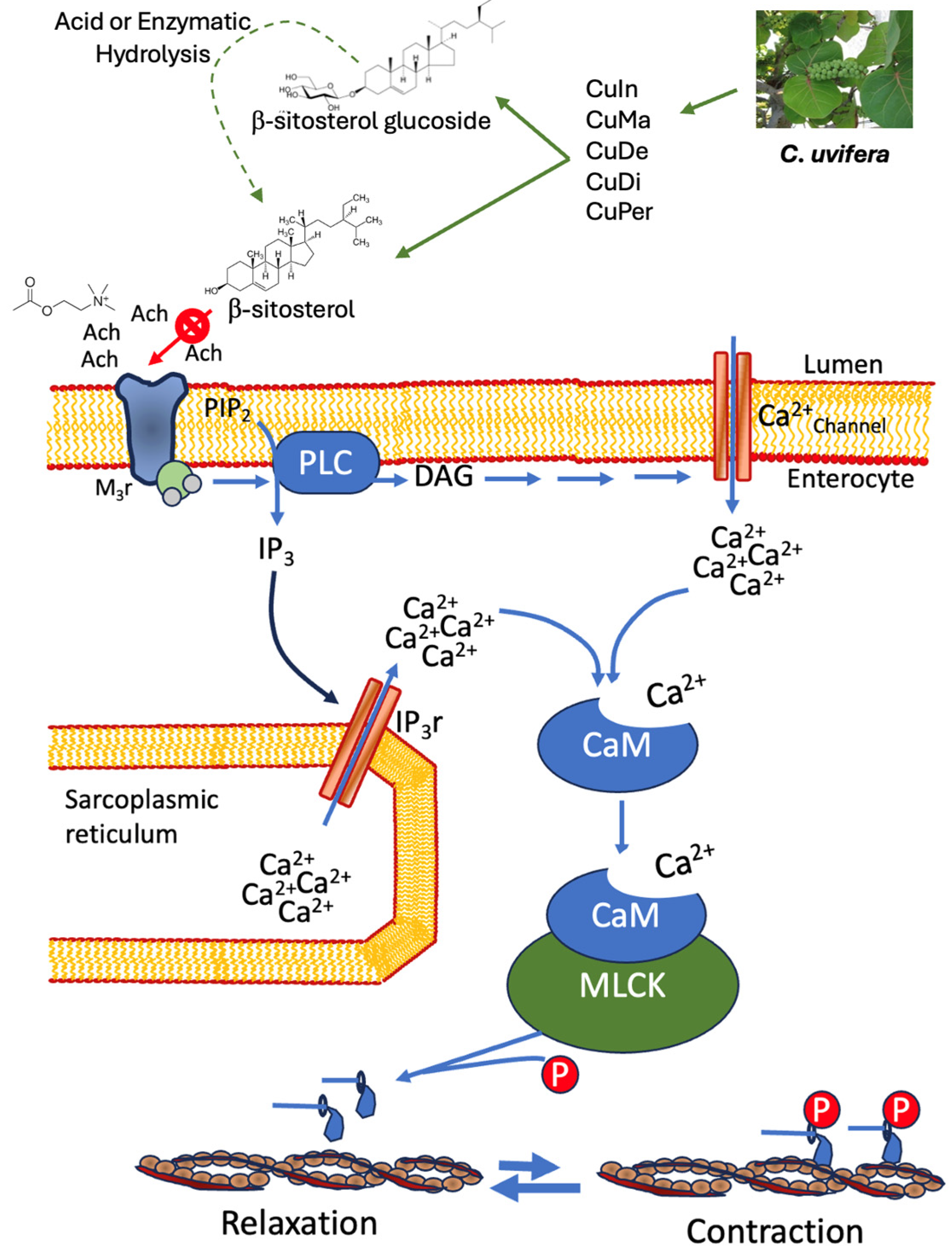
:1. Introduction
2. Materials and Methods
2.1. Plant Material
2.2. Preparation of the Extracts
2.3. Spectroscopic Analysis
2.4. Ex Vivo Evaluation
2.5. In Vivo Evaluation
2.6. In Vitro Tests
2.7. In Silico Approach
2.8. Data Analysis
3. Results
3.1. Phytochemical In Vitro Test
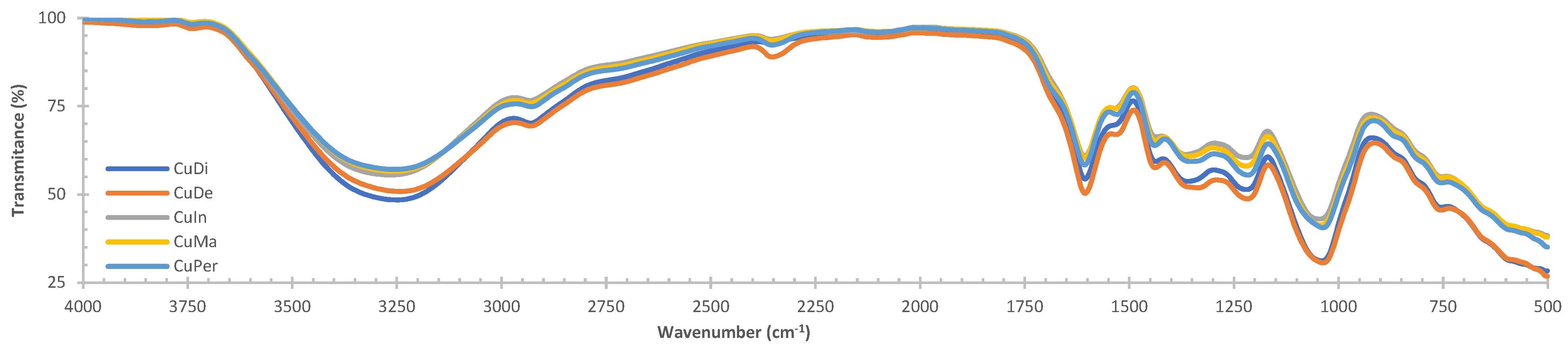
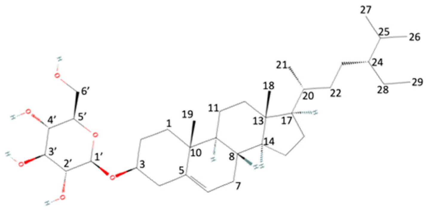
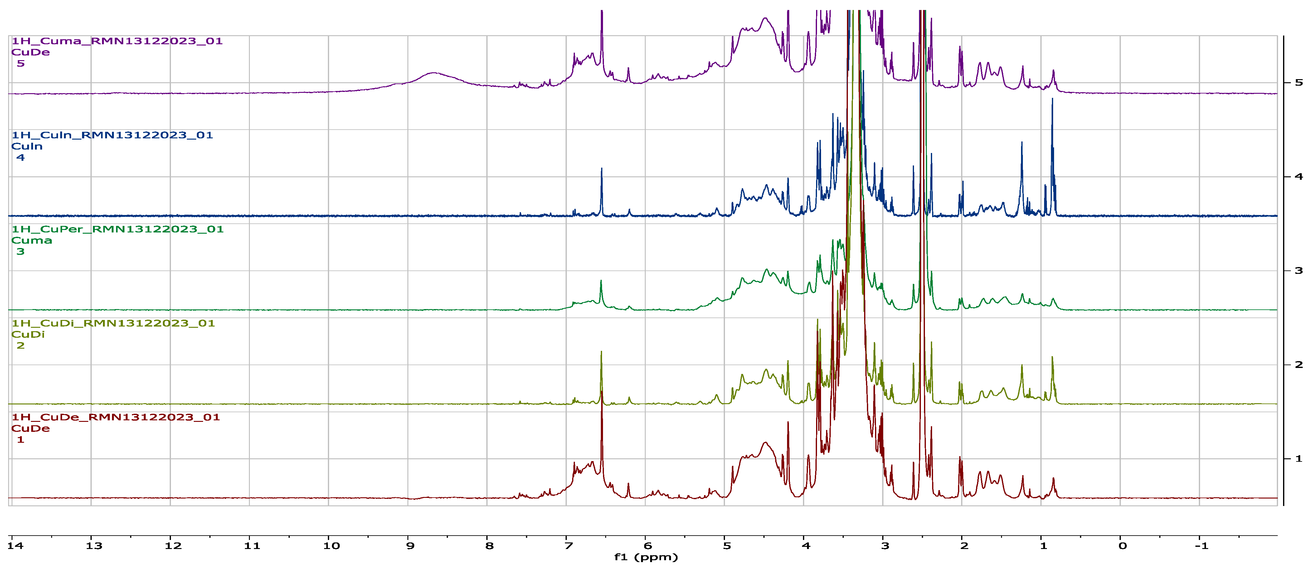
3.2. Spectroscopy Analysis
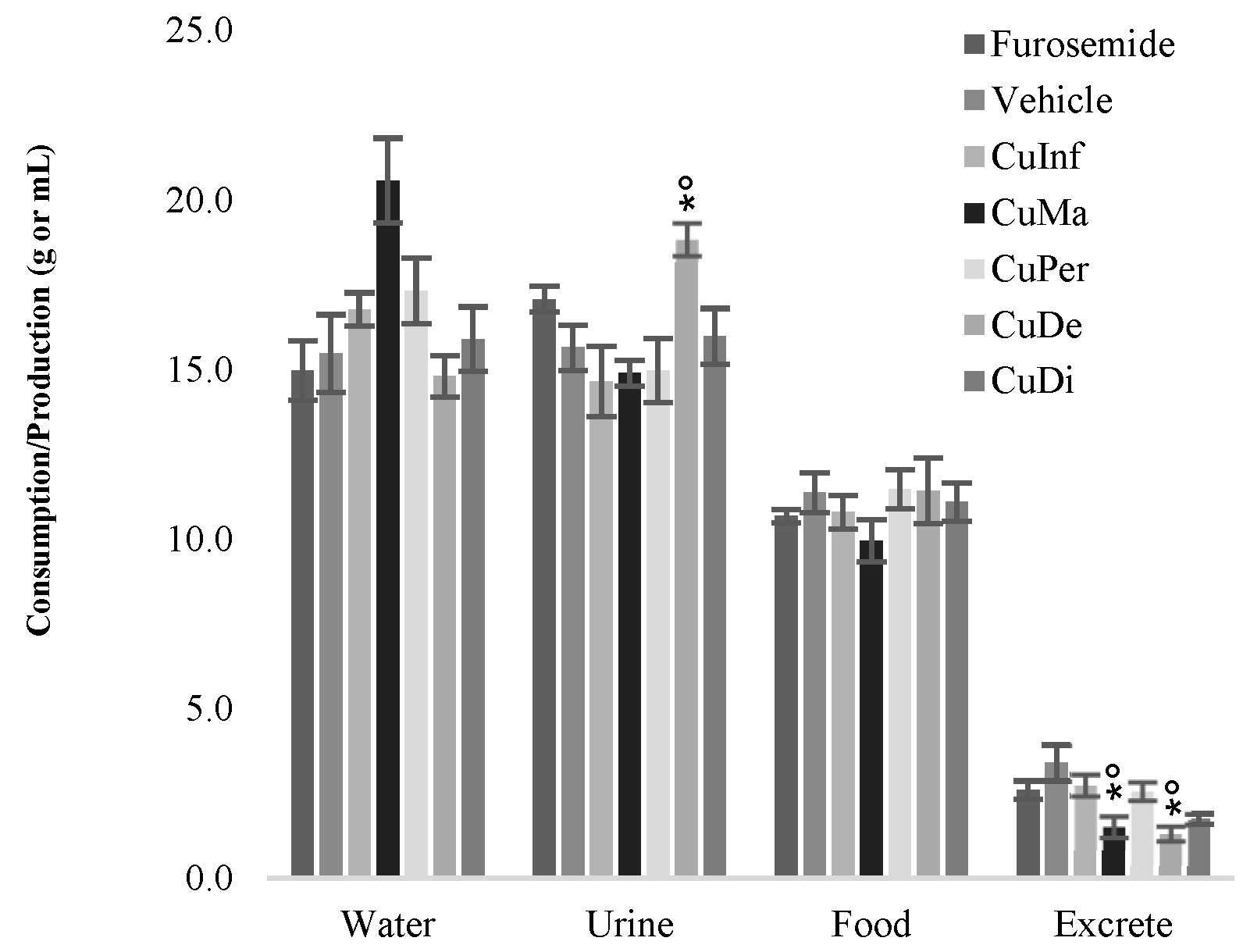
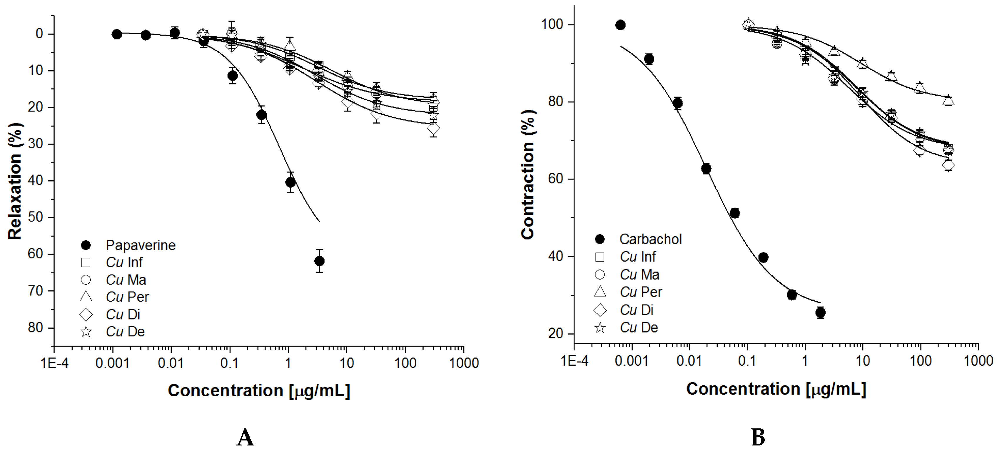
3.3. Ex Vivo and In Vivo Evaluation
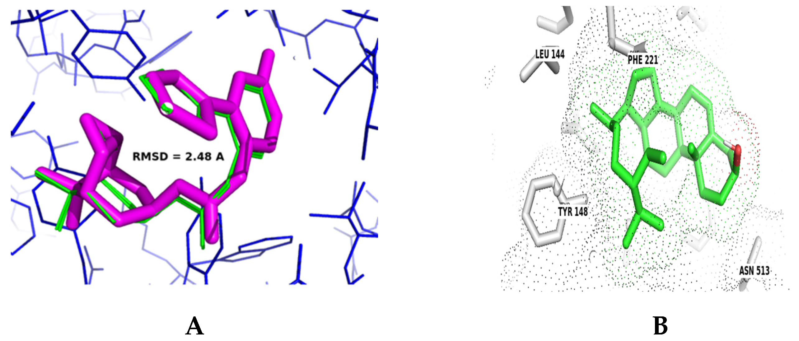
3.4. In Silico Approaches
3.5. Molecular Docking m3AChR
4. Discussion
5. Conclusions
Author Contributions
Funding
Institutional Review Board Statement
Informed Consent Statement
Data Availability Statement
Acknowledgments
Conflicts of Interest
References
- Juárez-Rosete, C.R.; Aguilar-Castillo, J.A.; Juárez-Rosete, M.E.; Bugarin-Montoya, R.; Juarez-Lopez, P.; Cruz-Crespo, E. Hierbas aromaticas y medicinales en Mexico: Tradicion e innovacion. Rev. Bio Cienc. 2013, 2, 119–129. [Google Scholar]
- La Torre-Cuadros, M.D.L.A.; Islebe, G.A. Traditional ecological knowledge and use of vegetation in southeastern Mexico: A case study from Solferino, Quintana Roo. Biodivers. Conserv. 2003, 12, 2455–2476. [Google Scholar] [CrossRef]
- Hernández, A.D.; Durán, I.S. Shortage of medicines in the health institutions in ZMG in Mexico. J. Int. Manag. Stud. 2015, 15, 27–32. [Google Scholar] [CrossRef]
- Salam, A.M.; Lyles, J.T.; Quave, C.L. Methods in the extraction and chemical analysis of medicinal plants. In Book Methods and Techniques in Ethnobiology and Ethnoecology, 2nd ed.; Albuquerque, U., de Lucena, R., Cruz da Cunha, L., Alves, R., Eds.; Springer Protocols Handbooks; Humana Press: New York, NY, USA, 2019; pp. 257–283. [Google Scholar]
- Sánchez-Aguirre, O.A.; Asseleih, L.M.C.; Medina, A.S.; González, G.S. Aplicación de Métodos Quimiométricos en la Investigación de Productos Naturales. In Miscelánea Científica de México: Biología y Química I; Solano-Sosa, C.E., Sánchez-Morales, M.A., Vázquez-García, G.V., Martínez-García, A., Ramos Guerrero, E.L., Eds.; Temacilli Editorial: Mexico City, Mexico, 2020; pp. 211–220. [Google Scholar]
- Hibbert, D.B. IUPAC project: A glossary of concepts and terms in chemometrics. Anal. Chim. Acta 2009, 642, 3–5. [Google Scholar] [CrossRef] [PubMed]
- Medina-Franco, J.L. Computational approaches for the discovery and development of pharmacologically active natural products. Biomolecules 2021, 11, 630. [Google Scholar] [CrossRef]
- Mukherjee, D.; Royce, S.G.; Sarkar, S.; Thorley, A.; Schwander, S.; Ryan, M.P.; Porter, A.P.; Chung, K.F.; Tetley, T.D.; Zhang, J.; et al. Modeling in vitro cellular responses to silver nanoparticles. J. Toxicol. 2014, 2014, 852890. [Google Scholar] [CrossRef]
- Coe, F.G. Ethnomedicine of the Rama of southeastern Nicaragua. J. Ethnobiol. 2008, 28, 1–38. [Google Scholar] [CrossRef]
- Vázquez-Yanes, C.A.I.; Batis-Muñoz, A.I.; Alcocer-Silva, M.I.; Sanchez, D. Potentially Valuable Trees and Shrubs for Ecological Restoration and Reforestation; Technical Report J084 Project; CONABIO-Instituto de Ecología, UNAM: México City, Mexico, 1990. [Google Scholar]
- Abdel-Hakim, F.; Gad, H.A.; Radwan, R.A.; Ayoub, N.; El-Shazly, M. Biological and phytochemical review on the genus Coccoloba (Polygonaceae). Arch. Pharm. Sci. Ain Shams Univ. 2019, 3, 180–194. [Google Scholar] [CrossRef]
- Gámez-Farías, Y.V. Evaluación Fitoquímica y Actividad Antibacteriana de Extractos de Coccoloba uvifera Jacq. (Polygonaceae) de la Localidad de El Peñón. Bachelor’s Degree, Universidad de Oriente, Cumaná, Venezuela, 2012. [Google Scholar]
- Povi, L.; Batomayena, B.; Hodé, T.A.; Kwashie, E.G.; Kodjo, A.; Messanvi, G. Phytochemical screening, antioxidant, and hypoglycemic activity of Coccoloba uvifera leaves and Waltheria indica roots extracts. Int. J. Pharm. Pharm. Sci. 2015, 7, 279–283. [Google Scholar]
- Segura-Campos, M.R.; Ruiz-Ruiz, J.; Chel-Guerrero, L.; Betancourt-Ancona, D. Coccoloba uvifera (L.) (Polygonaceae) fruit: Phytochemical screening and potential antioxidant activity. J. Chem. 2015, 2015, 534954. [Google Scholar] [CrossRef]
- Bailey, C.; Christian, K.R.; Pradhan, S.; Nair, M.G.; Christian, O.E. Anti-inflammatory, and antioxidant activities of Coccoloba uvifera (Seagrapes). Curr. Top. Phytochem. 2011, 10, 55–60. [Google Scholar]
- Piloto-Alfonso, A.M.; García-Simón, G.; Valdivieso-García, G.A. Estudio farmaco-toxicológico del extracto fluido de uva Caleta al 50%. Arch. Toxicol. 2001, 1, 126–131. [Google Scholar]
- Ramos-Hernández, J.A.; Calderón-Santoyo, M.; Navarro-Ocaña, A.; Barros-Castillo, J.C.; Ragazzo-Sanchez, J.A. Use of emerging technologies in the extraction of lupeol, α-amyrin and β-amyrin from sea grape (Coccoloba uvifera L.). J. Food Sci. Technol. 2018, 55, 2377–2383. [Google Scholar] [CrossRef]
- Malathi, S.; Masilamani, P.; Balasubramanian, V.; Rao, R.B.; Brindha, P. Constituents of Coccoloba uvifera leaves. Fitoterapia 1995, 66, 277. [Google Scholar]
- NOM-062-ZOO-1999; Especificaciones Técnicas para la Producción, Cuidado y uso de los Animales de Laboratorio. Secretaría de Agricultura, Ganadería, Desarrollo Rural, Pesca y Alimentación (SAGARPA): Ciudad de México, Distrito Federal, Mexico, 2001.
- Estrada-Soto, S.E.; González-Maldonado, D.; Castillo-España, P.; Aguirre-Crespo, F.J.; Sanchez-Salgado, J.C. Spasmolytic effect of Mentha pulegium L. involves ionic flux regulation in rat ileum strips. J. Smooth Muscle Res. 2010, 46, 107–117. [Google Scholar] [CrossRef]
- Aguirre-Crespo, F.; Vergara-Galicia, J.; Villalobos-Molina, R.; López-Guerrero, J.J.; Navarrete-Vazquez, J.G.; Estrada-Soto, S.E. Ursolic acid mediates the vasorelaxant activity of Lepechinia caulescens via NO release in isolated rat thoracic aorta. Life Sci. 2006, 79, 1062–1068. [Google Scholar] [CrossRef] [PubMed]
- Rajalakshmi, K.; Banu, N. Extraction, and estimation of chlorophyll from medicinal plants. Int. J. Sci. Res. 2015, 4, 209–212. [Google Scholar]
- Burke, R.W.; Diamondstone, B.I.; Velapoldi, R.A.; Menis, O. Mechanisms of the Liebermann-Burchard and Zak Color Reactions for Cholesterol. Clin. Chem. 1974, 20, 794–801. [Google Scholar] [CrossRef] [PubMed]
- Puangpronpitag, D.; Areejitranusorn, P.; Boonsiri, P.; Suttajit, M.; Yongvanit, P. Antioxidant activities of polyphenolic compounds isolated from Antidesma thwaitesianum Müll. Arg. seeds and marcs. J. Food Sci. 2008, 73, C648–C653. [Google Scholar]
- Bernard, D.; Isaac Kwabena, A.; Daniel Osei, O.; Achel Daniel, G.; Achoribo Elom, S.; Sandra, A. The effect of different drying methods on the phytochemicals and radical scavenging activity of Ceylon Cinnamon (Cinnamomum zeylanicum) plant parts. Eur. J. Med. Plants 2014, 4, 1324–1335. [Google Scholar]
- Galvez, M.A.C. Evaluation of DPPH free radical scavenging activity and phytochemical screening of selected folkloric medicinal plants in Tinoc, Ifugao, Cordillera Administrative Region, Philippines. Int. J. Sci. Res. Publ. 2015, 5, 440–445. [Google Scholar]
- Mabkhot, Y.N.; Alatibi, F.; El-Sayed, N.N.E.; Al-Showiman, S.; Kheder, N.A.; Wadood, A.; Rauf, A.; Bawazeer, S.; Hadda, T.B. Antimicrobial activity of some novel armed thiophene derivatives and petra/osiris/molinspiration (POM) analyses. Molecules 2016, 21, 222. [Google Scholar] [CrossRef] [PubMed]
- Hakkou, Z.; Maciuk, A.; Leblais, V.; Bouanini, N.E.; Mekhfi, H.; Bnouham, M.; Aziz, M.; Ziyyat, A.; Rauf, A.; Hadda, T.B.; et al. Antihypertensive and vasodilator effects of methanolic extract of Inula viscosa: Biological evaluation and POM analysis of cynarin, chlorogenic acid as potential hypertensive. Biomed. Pharmacother. 2017, 93, 62–69. [Google Scholar] [CrossRef]
- Cob-Calan, N.N.; Chi-Uluac, L.A.; Ortiz-Chi, F.; Cerqueda-García, D.; Navarrete-Vázquez, J.G.; Ruiz-Sánchez, E.; Hernández-Núñez, E. Molecular Docking and dynamics simulation of protein β-Tubulin and antifungal cyclic lipopeptides. Molecules 2019, 24, 3387. [Google Scholar] [CrossRef] [PubMed]
- Amina, M.; Amna, T.; Al-Musayeib, N.; Zabin, S.A.; Hassan, M.S.; Khil, M.S. Encapsulation of β-Sitosterol in polyurethane by sol-gel electrospinning. Appl. Biochem. Biotechnol. 2017, 182, 624–634. [Google Scholar] [CrossRef] [PubMed]
- López-Salazar, H.; Camacho-Díaz, B.H.; Ávila-Reyes, S.V.; Pérez-García, M.D.; González-Cortazar, M.; Arenas-Ocampo, M.L.; Jiménez-Aparicio, A.R. identification and quantification of β-Sitosterol β-d-glucoside of an ethanolic extract obtained by microwave-assisted extraction from Agave angustifolia Haw. Molecules 2019, 24, 3926. [Google Scholar] [CrossRef] [PubMed]
- El-shiekh, R.A.; Al-Mahdy, D.A.; Hifnawy, M.S.; Bagrel, D.; Abdelsattar, E.A. Chemical and Biological Investigation of Ochrosia elliptica in Egypt. Rec. Nat. Prod. 2017, 11, 552–557. [Google Scholar] [CrossRef]
- Veber, D.F.; Johnson, S.R.; Cheng, H.Y.; Smith, B.R.; Ward, K.W.; Kopple, K.D. Molecular properties that influence the oral bioavailability of drug candidates. J. Med. Chem. 2002, 45, 2615–2623. [Google Scholar] [CrossRef] [PubMed]
- Igarashi-Hisayoshi, Y.; Ihara, E.; Bai, X.; Higashi, C.; Ikeda, H.; Tanaka, Y.; Hirano, M.; Ogino, H.; Chinen, T.; Taguchi, Y.; et al. Determination of Region-Specific Roles of the M3 Muscarinic Acetylcholine Receptor in Gastrointestinal Motility. Dig. Dis. Sci. 2023, 68, 439–450. [Google Scholar] [CrossRef]
- Sampaio, B.L.; Edrada-Ebel, R.; Da Costa, F.B. Effect of the environment on the secondary metabolic profile of Tithonia diversifolia: A model for environmental metabolomics of plants. Sci. Rep. 2016, 6, 29265. [Google Scholar] [CrossRef]
- Kaufmann, B.; Christen, P. Recent extraction techniques for natural products: Microwave-assisted extraction and pressurized solvent extraction. Phytochem. Anal. 2002, 13, 105–113. [Google Scholar] [CrossRef] [PubMed]
- Chaudhary, A.; Nagaich, U.; Gulati, N.; Sharma, V.K.; Khosa, R.L.; Partapur, M.U. Enhancement of solubilization and bioavailability of poorly soluble drugs by physical and chemical modifications: A recent review. J. Adv. Pharm. Educ. Res. 2012, 2, 32–67. [Google Scholar]
- Garzón, G.A. Anthocyanins as natural colorants and bioactive compounds: A review. Acta Biolo. Colomb. 2008, 13, 27–36. [Google Scholar]
- Rajbhar, K.; Dawda, H.; Usha, M. Polyphenols: Methods of extraction. Sci. Revs. Chem. Commun. 2015, 5, 1–6. [Google Scholar]
- Maltese, F.; Van Der Koov, F.; Verpoorte, R. Solvent-derived artifacts in natural products chemistry. Nat. Prod. Commun. 2009, 4, 447–454. [Google Scholar] [PubMed]
- Wong, S.K.; Sytnyk, W.; Wan, J.K.S. Electron spin resonance study of the self-disproportionation of some semiquinone radicals in solution. Can. J. Chem. 1972, 50, 3052–3057. [Google Scholar] [CrossRef]
- Aguirre-Crespo, F.J.; Salgado, J.C.S. ¿Qué sabe Ud. acerca de... la curva dosis-respuesta? Rev. Mex. Cienc. Farm. 2010, 41, 57–59. [Google Scholar]
- Barbosa da Rocha, M.; Menezes-Souza, F.V.; Dos Santos, E.C.; Pizza, C.; Goulart-Santana, A.E.; Moretti-Marcal, R. Antispasmodic effect of 4’-methylepigallocatechin on guinea pig ileum. Fitoterapia 2012, 83, 1286–1290. [Google Scholar] [CrossRef] [PubMed]
- Cardoso-Lima, T.; Mendoca-Mota, M.; Barbosa-Filho, J.M.; Dos Santos, M.R.V.; De Sousa, D.P. Structural relationships and vasorelaxant activity of monoterpenes. DARU J. Pharm. Sci. 2012, 20, 23. [Google Scholar] [CrossRef]
- Estrada-Soto, S.E.; Lopez-Guerrero, J.J.; Villalobos-Molina, R.; Mata, R. Endothelium-independent relaxation of aorta rings by two stilbenoids from the orchids Scaphyglottis livida . Fitoterapia 2006, 77, 236–239. [Google Scholar] [CrossRef]
- Liu, Y.; Chu, S.; Yang, S.; Peng, Y.; Ren, S.; Wen, B.; Chen, N. Physcion and physcion 8-O-β-glucopyranoside: A review of their pharmacology, toxicities and pharmacokinetics. Chem. Biol. Interact. 2019, 310, 108722. [Google Scholar]
- Diaz-Muñoz, G.; Miranda, I.L.; Sartori, S.K.; de Rezende, D.C.; Diaz, M.A.N. Anthraquinones: An Overview. Stud. Nat. Prod. Chem. 2018, 58, 313–338. [Google Scholar]
- Ruoff, H.J.; Fladung, B.; Demol, P.; Weihrauch, T.R. Gastrointestinal receptors, and drugs in motility disorders. Digestion 1991, 48, 1–17. [Google Scholar] [CrossRef]
- Lyford, G.L.; Farrugia, G. Ion channels in gastrointestinal smooth muscle and interstitial cells of Cajal. Curr. Opin. Pharmacol. 2003, 3, 583–587. [Google Scholar] [CrossRef] [PubMed]
- Ma, T.; Verkman, A.S. Aquaporin water channels in gastrointestinal physiology. J. Physiol. 1999, 517, 317–326. [Google Scholar] [CrossRef]
- Daniel, H.; Zietek, T. Taste and move glucose and peptide transporters in the gastrointestinal tract. Exp. Physiol. 2015, 100, 1441–1450. [Google Scholar] [CrossRef] [PubMed]
- Steffansen, B.; Nielsen, C.U.; Brodin, B.; Eriksson, A.H.; Andersen, R.; Frokjaer, S. Intestinal solute carriers: An overview of trends and strategies for improving oral drug absorption. Eur. J. Pharm. Sci. 2004, 21, 3–16. [Google Scholar] [CrossRef]
- Degirolamo, C.; Sabbà, C.; Moschetta, A. Intestinal nuclear receptors in HDL cholesterol metabolism. J. Lipid. Res. 2015, 56, 1262–1270. [Google Scholar] [CrossRef]
- Flores-Soto, M.E.; Segura-Torres, J.E. Structure, and function of the acetylcholine of muscarinical and nicotinical type. Rev. Mex. Neuroci. 2005, 6, 315–326. [Google Scholar]
- Caulfield, M.P. Muscarinic receptors—Characterization, coupling and function. Pharmacol. Ther. 1993, 58, 319–379. [Google Scholar]
- Rodríguez-Rodríguez, R.; Herrera, M.D.; Perona, J.S.; Ruiz-Gutiérrez, V. Potential vasorelaxant effects of oleanolic acid and erythrodiol, two triterpenoids contained in ‘orujo’olive oil, on rat aorta. Br. J. Nutr. 2004, 92, 635–642. [Google Scholar] [CrossRef] [PubMed]
- Khan, N.M.; Hossain, M.S. Scopoletin and β-sitosterol glucoside from roots of Ipomoea digitata . J. Pharmacogn. Phytochem. 2015, 4, 5–7. [Google Scholar]
- Maldonado, E.; Amador, S.; Juárez-Jaimes, V. Secondary metabolites from Asclepias otarioides . J. Mex. Chem. Soc. 2015, 59, 50–52. [Google Scholar] [CrossRef]
- Zhang, Y.; Xie, J.; Zhang, Y.; Zhang, M. Degradation kinetics of jujuboside A by rat intestinal flora and identification of the metabolites by HPLC MS/MS. Int. J. Food Prop. 2014, 17, 1841–1849. [Google Scholar] [CrossRef]
- Tenfen, A.; Mariano, L.N.B.; Boeing, T.; Cechinel-Zanchett, C.C.; Mota da Silva, L.; de Andrade, S.F.; de Souza, P.; Cechinel-Filho, V. Effects of myricetin-3-O-α-rhamnoside (myricitrin) treatment on urinary parameters of Wistar rats. J. Pharm. Pharmacol. 2019, 71, 1832–1838. [Google Scholar] [CrossRef] [PubMed]
- Bracci, A.; Amat, A.G.; Maione, F.; Cicala, C.; Mascolo, N.; De Feo, V. Diuretic activity of Lophophytum leandri . Nat. Prod. Cammun. 2012, 7, 1934578X1200700112. [Google Scholar] [CrossRef]
- Song, X.; Tan, L.; Wang, M.; Ren, C.; Guo, C.; Yang, B.; Ren, Y.; Cao, Z.; Li, Y.; Pei, J. Myricetin: A review of the most recent research. Biomed. Pharmacother. 2021, 134, 111017. [Google Scholar] [CrossRef]
- Trezza, A.; Spiga, O.; Mugnai, P.; Saponara, S.; Sgaragli, G.; Fusi, F. Functional, electrophysiology, and molecular dynamics analysis of quercetin-induced contraction of rat vascular musculature. Eur. J. Pharmacol. 2022, 918, 174778. [Google Scholar] [CrossRef]


| Method | Time Extraction (min) | Yield (%) | Chlorophyll (mg/g) | Simple Phenols Eq. GA (mg/g) | Flavonoids Eq. Q (mg/g) | DPPH (%) |
|---|---|---|---|---|---|---|
| Cu De | 15 | 38.4 ± 1.7 * | 0.41 ± 0.02 * | 85.26 ± 2.08 * | 0.37 ± 0.008 * | 27.6 ± 1.9 |
| Cu Di | 15 | 6.7 ± 1.1 * | 0.28 ± 0.04 | 21.64 ± 0.52 * | 0.13 ± 0.006 * | 19.3 ± 0.9 |
| Cu Inf | 35 | 9.8 ± 0.9 * | 0.26 ± 0.02 | 46.75 ± 2.15 * | 0.22 ± 0.003 * | 27.1 ± 3.2 |
| Cu Ma | 1440 | 31.2 ± 2 | 0.31 ± 0.03 | 14.92 ± 1.49 | 0.09 ± 0.0.001 | 43.6 ± 1.3 |
| Cu Per | 25 | 5.1 ± 0.9 * | 0.04 ± 0.01 * | 29.36 ± 1.15 * | 0.04 ± 0.002 * | 64.3 ± 1.1 |
| C. sinensis | 1440 | N/A | N/A | N/A | N/A | 53.3 ± 0.2 |
| Quercetin | N/A | N/A | N/A | N/A | N/A | 91.6 ± 0.1 |
| Method | Ex Vivo (%) | Dose (mg/kg) | In Vivo (%) | ||
|---|---|---|---|---|---|
| Spasmolytic | Vasorelaxant | Diuretic | Antiperistaltic | ||
| CuDe | 25.6 ± 3.6 | 36.3 ± 1.3 | 246.0 ± 10.0 | 21.2 ± 3.1 ○ | −60.1 ± 6.5 ○○ |
| CuDi | 22.1 ± 5.3 | 32.6 ± 1.1 | 43.2 ± 2.4 | 2.1 ± 5.3 | −48.5 ± 4.6 |
| CiInf | 19.6 ± 3.6 | 32.1 ± 1.1 | 71.4 ± 4. 7 | −6.4 ± 4.3 | −19.1 ± 15.7 |
| CuMa | 18.6 ± 5.7 | 32.4 ± 0.8 | 278.2 ± 17.6 | −4.8 ± 2.4 | −55.2 ± 9.3 ○○ |
| CuPer | 17.8 ± 4.3 | 19.8 ± 1.1 | 37.5 ± 2.2 | −4.3 ± 6.1 | −24.3 ± 7.9 |
| Papaverine | 61.8 ± 3.1 * | N/A | N/A | N/A | N/A |
| Carbachol | N/A | 74.4 ± 1.4 ** | N/A | N/A | N/A |
| Vehicle | N/A | N/A | N/A | 0 ± 6.1 | 0 ± 8.8 |
| Furosemide | N/A | N/A | 0.57 | 9.6 ± 2.2 | −23.5 ± 7.9 |
| Metabolite | MW | cLogP | TPSA | nOH | nOHNH | V | Vol |
|---|---|---|---|---|---|---|---|
| Myricetin 3-O-rhamnoside | 466.39 | 0.39 | 210.5 | 12 | 8 | 2 | 378.2 |
| Myricetin 3-O-glucoside | 482.39 | −0.62 | 230.7 | 13 | 8 | 2 | 386.4 |
| Quercetin 3-O-rhamnoside | 450.42 | 0.68 | 190.3 | 11 | 7 | 2 | 370.2 |
| Quercetin 3-O-arabinoside | 436.37 | 0.1 | 190.3 | 11 | 7 | 2 | 353.6 |
| β-sitosterol | 414.72 | 8.62 | 20.2 | 1 | 1 | 1 | 456.5 |
| Lupeol | 440.76 | 8.61 | 20.2 | 1 | 1 | 1 | 478.4 |
| α-amyrin | 426.73 | 8.08 | 20.2 | 1 | 1 | 1 | 461.1 |
| β-amyrin | 426.73 | 8.02 | 20.2 | 1 | 1 | 1 | 460.7 |
| Royleanone | 316.44 | 4.68 | 54.4 | 3 | 1 | 0 | 314.1 |
| Gallic acid | 170.12 | 0.59 | 97.9 | 5 | 4 | 0 | 135.1 |
| Emodin | 270.24 | 3.01 | 94.8 | 5 | 3 | 0 | 223.2 |
| Chrysophanol | 254.24 | 3.54 | 74.6 | 4 | 2 | 0 | 215.2 |
| Physcion | 284.27 | 3.54 | 83.8 | 5 | 2 | 0 | 240.7 |
| Rhein | 284.22 | 3 | 111.9 | 6 | 3 | 0 | 225.6 |
| Metabolite | GPCR | ICM | KI | NRL | PI | EI | M | T | I | R | S | DL | DS |
|---|---|---|---|---|---|---|---|---|---|---|---|---|---|
| Myricetin 3-O-rhamnoside | −0.11 | −0.1 | −0.2 | −0.12 | −0.1 | 0.41 | 0 | 0 | 0 | 0 | −2.40 | 2.64 | 0.75 |
| Myricetin 3-O-glucoside | −0.05 | −0.1 | −0.1 | −0.08 | −0.1 | 0.46 | 0 | 0 | 0 | 0 | −1.90 | −2.87 | 0.40 |
| Quercetin 3-O-rhamnoside | −0.1 | −0.1 | −0.2 | −0.09 | −0.1 | 0.4 | 0 | 0 | 0 | 0 | −2.70 | 3.31 | 0.77 |
| Quercetin 3-O-arabinoside | −0.07 | −0.1 | −0.2 | −0.2 | −0.1 | 0.44 | 0 | 0 | 0 | 0 | −2.30 | −2.37 | 0.44 |
| β-sitosterol | 0.14 | 0.04 | −0.5 | 0.73 | 0.07 | 0.51 | 0 | 0 | 0 | 0 | −6.70 | −4.48 | 0.13 |
| Lupeol | 0.22 | 0.16 | −0.3 | 0.72 | 0.1 | 0.48 | 0 | 0 | 0 | 0 | −6.80 | −2.20 | 0.13 |
| α-amyrin | 0.22 | −0 | −0.4 | 0.79 | 0.19 | 0.6 | 0 | 0 | 0 | 0 | −6.60 | −4.37 | 0.14 |
| β-amyrin | 0.22 | −0.1 | −0.3 | 0.67 | 0.11 | 0.56 | 0 | 0 | 0 | 0 | −6.70 | −2.49 | 0.15 |
| Royleanone | 0.2 | 0.08 | −0.1 | 0.71 | −0.1 | 0.51 | 0 | 0 | 1 | 0 | −3.80 | −3.41 | 0.23 |
| Gallic acid | −0.77 | −0.3 | −0.9 | −0.52 | −0.9 | −0.2 | 1 | 0 | 0 | 1 | −0.70 | 0.12 | 0.27 |
| Emodin | −0.14 | −0.1 | 0.07 | 0.17 | −0.2 | 0.21 | 1 | 1 | 1 | 1 | −4.20 | −2.06 | 0.06 |
| Chrysophanol | −0.23 | −0.2 | −0.1 | 0.02 | −0.3 | 0.16 | 1 | 0 | 1 | 0 | −4.50 | −1.67 | 0.16 |
| Physcion | −0.17 | −0.2 | 0.04 | 0.11 | −0.2 | 0.14 | 1 | 0 | 1 | 0 | −4.50 | −1.92 | 0.20 |
| Rhein | −0.08 | −0.1 | 0.01 | 0.29 | −0.1 | 0.28 | 0 | 0 | 1 | 0 | −4.20 | 0.18 | 0.37 |
| Compound | H-Bonds (#) | Residue Receptor | Bond Length (Å) | Docking Score (kcal/mol) | Ki (μM) a |
|---|---|---|---|---|---|
| Scopolamine | 5 | A:SER 151: OG | 2.62 | −10.18 | 0.03 |
| A:ASN 507: ND2 | 2.85 | ||||
| A:9EC 1203: N03 | 3.10 | ||||
| A:9EC 1203: C24 | 3.05 | ||||
| A:9EC 1203: C25 | 3.66 | ||||
| β-sitosterol | 0 | --- | --- | −9.08 | 0.21 |
| Lupeol | 0 | --- | --- | −5.70 | 67.53 |
| α-amyrin | 0 | --- | --- | −5.60 | 76.17 |
| β-amyrin | 1 | A:ASN526:ND2 | 3.27 | −5.96 | 51.18 |
| Royleanone | 0 | --- | --- | −8.59 | 0.48 |
Disclaimer/Publisher’s Note: The statements, opinions and data contained in all publications are solely those of the individual author(s) and contributor(s) and not of MDPI and/or the editor(s). MDPI and/or the editor(s) disclaim responsibility for any injury to people or property resulting from any ideas, methods, instructions or products referred to in the content. |
© 2024 by the authors. Licensee MDPI, Basel, Switzerland. This article is an open access article distributed under the terms and conditions of the Creative Commons Attribution (CC BY) license (https://creativecommons.org/licenses/by/4.0/).
Share and Cite
Aguirre-Crespo, F.J.; Aragón-Gastélum, J.L.; Gutiérrez-Alcántara, E.J.; Zamora-Crescencio, P.; Gómez-Galicia, D.L.; Alatriste-Kurzel, D.R.; Alvarez, G.; Hernández-Núñez, E. β-Sitosterol Mediates Gastrointestinal Smooth Muscle Relaxation Induced by Coccoloba uvifera via Muscarinic Acetylcholine Receptor Subtype 3. Sci. Pharm. 2024, 92, 19. https://doi.org/10.3390/scipharm92020019
Aguirre-Crespo FJ, Aragón-Gastélum JL, Gutiérrez-Alcántara EJ, Zamora-Crescencio P, Gómez-Galicia DL, Alatriste-Kurzel DR, Alvarez G, Hernández-Núñez E. β-Sitosterol Mediates Gastrointestinal Smooth Muscle Relaxation Induced by Coccoloba uvifera via Muscarinic Acetylcholine Receptor Subtype 3. Scientia Pharmaceutica. 2024; 92(2):19. https://doi.org/10.3390/scipharm92020019
Chicago/Turabian StyleAguirre-Crespo, Francisco J., José L. Aragón-Gastélum, Eduardo J. Gutiérrez-Alcántara, Pedro Zamora-Crescencio, Diana L. Gómez-Galicia, Diego R. Alatriste-Kurzel, Guzman Alvarez, and Emanuel Hernández-Núñez. 2024. "β-Sitosterol Mediates Gastrointestinal Smooth Muscle Relaxation Induced by Coccoloba uvifera via Muscarinic Acetylcholine Receptor Subtype 3" Scientia Pharmaceutica 92, no. 2: 19. https://doi.org/10.3390/scipharm92020019
APA StyleAguirre-Crespo, F. J., Aragón-Gastélum, J. L., Gutiérrez-Alcántara, E. J., Zamora-Crescencio, P., Gómez-Galicia, D. L., Alatriste-Kurzel, D. R., Alvarez, G., & Hernández-Núñez, E. (2024). β-Sitosterol Mediates Gastrointestinal Smooth Muscle Relaxation Induced by Coccoloba uvifera via Muscarinic Acetylcholine Receptor Subtype 3. Scientia Pharmaceutica, 92(2), 19. https://doi.org/10.3390/scipharm92020019