Article

Antibacterial and Wound Healing Activity In Vitro of Individual and Combined Extracts of Tagetes nelsonii Greenm, Agave americana and Aloe vera

by
Karen Alejandra Olán-Jiménez
1,
Rosa Isela Cruz-Rodríguez
1,*,
Beatriz del Carmen Couder-García
2,*,
Nadia Jacobo-Herrera
3,
Nancy Ruiz-Lau
1,
Maritza del Carmen Hernández-Cruz
4 and
Víctor Manuel Ruíz-Valdiviezo
1
1
Departamento de Ingeniería Química y Bioquímica, Tecnológico Nacional de México/IT Tuxtla Gutiérrez, Carretera Panamericana Km 1080, Tuxtla Gutiérrez 29050, Mexico
2
CONAHCYT—Centro de Investigación y Asistencia en Tecnología y Diseño del Estado de Jalisco, Subsede Sureste, Parque Científico Tecnológico de Yucatán, Tablaje Catastral 31264, Mérida 97302, Mexico
3
Unidad de Bioquímica, Instituto Nacional de Ciencias Médicas y Nutrición Salvador Zubirán, Av. Vasco de Quiroga 15, Col. Belisario Domínguez Sección XVI, Tlalplan, Ciudad de México 14080, Mexico
4
Escuela de Ciencias Químicas, Universidad Autónoma de Chiapas (UNACH), Carretera Panamericana Ocozocoautla-Cintalapa Km 2.5, Ocozocoautla de Espinosa 29140, Mexico
*
Authors to whom correspondence should be addressed.
Sci. Pharm. 2024, 92(3), 41; https://doi.org/10.3390/scipharm92030041
Submission received: 5 June 2024 / Revised: 27 July 2024 / Accepted: 29 July 2024 / Published: 31 July 2024

Abstract

:
Currently, there are various physical and mechanical agents that can cause skin wounds, which are still traditionally treated with plant extracts. It has been reported that the genus Tagetes has a wide range of biological properties, including antibacterial and wound healing activity. Likewise, Agave americana extract and Aloe vera gel have shown potential in the treatment of burn wounds and other skin conditions both in vitro and in vivo. In this study, the antibacterial and wound healing activities of each of these plants were investigated, as well as the possibility of enhancing these activities by combining them. First, the secondary metabolites of the extracts were quantified, the antibacterial activity was evaluated using the Kirby-Bauer method, and their cytotoxicity was measured in 3T3 and HaCaT cells using the sulforhodamine B assay. The results revealed that Tagetes nelsonii extract had a higher amount of secondary metabolites, which is why it exhibited antibacterial activity. Finally, the scratch assay showed that the individual extracts of T. nelsonii and A. americana demonstrated greater cell migration and proliferation starting from 12 h, as well as when using the combination of A. americana extract and A. vera gel, which almost completely closed the wound compared to the control.

Graphical Abstract

1. Introduction

The human skin is susceptible to injuries or wounds caused by physical, mechanical, and chemical agents, which disrupt its integrity and induce systemic changes in health [1,2]. One of its characteristics is the ability to self-regenerate, which requires the involvement of specialized cells capable of participating in the healing process [3,4]. Indeed, although wound healing is a natural process, the presence of aerobic and anaerobic micro-organisms can lead to severe wound infections that require immediate attention [5,6,7]. In recent years, plants have been utilized in the pharmaceutical, cosmetic, and medicine industries due to their bioactive compounds with therapeutic properties. Thanks to this, they are a promising option for wound treatment and healing [8,9,10].
Tagetes nelsonii Greenm, commonly known as “chik chawa”, is a plant belonging to the Asteraceae family, native to the state of Chiapas. It has been empirically used by various ethnic groups to treat colds, respiratory problems, and stomach disorders [11,12]. Although there is limited research on the therapeutic properties of these species, the Tagetes genus is known for its anti-inflammatory, insecticidal, antioxidant, antihypertensive, an-tileukemic, antimicrobial, and wound-healing activities [13,14,15,16]. In particular, for T. nelsonii, research on its therapeutic activities is limited, and its bactericidal activity has been reported against both Gram-positive and Gram-negative microorganisms [17,18]. Additionally, it has been noted that T. nelsonii contains volatile compounds such as (E)-Tagetone, (Z)-Tagetone, β-ocimene, α-copaene, β-caryophyllene, and eucalyptol, which exhibit anti-inflammatory and antioxidant activities, among others [19,20].
On the other hand, Agave americana, from the Agavaceae family, is native to Mexico, Arizona, and Texas, and it is traditionally used as an antiseptic, diuretic, and laxative due to its phenolic acids, flavonoid glycosides, homoisoflavonoids, and saponins [21,22,23]. It has been reported to have wound-healing and antibacterial properties that contribute to the healing of wounds [24,25,26,27,28]. Similarly, Aloe vera is another plant with remarkable wound-healing properties widely used for the treatment of wounds. The parenchymatous gel from its leaves is extensively studied due to the variety of its bioactive compounds such as polysaccharides, tannins, sterols, fatty, amino and organic acids, enzymes, saponins, vitamins, and minerals [29,30]. It has been shown that the fresh gel of A. vera has the ability to promote tissue repair in wounds due to its high water content, which helps keep the wound moist, enhances the migration of epithelial cells, and stimulates hyaluronic acid synthesis [31,32]. Additionally, it has demonstrated antibacterial activity against various pathogenic microorganisms [33,34].
Medicinal plants may exhibit individual biological activities; however, it is possible that their therapeutic properties can be enhanced in a synergistic context by combining them. Previous findings have demonstrated that a combination of plant extracts significantly enhances wound healing through cell proliferation and migration compared to the use of individual extracts [35,36,37]. In Mexico, there have been few studies on the combined use of plant extracts for wound healing, so the aim of this study was to analyze the in vitro antibacterial activity and wound-healing activity of individual and combined extracts of T. nelsonii, A. americana, and A. vera gel, plants that are traditionally used by the Mexican population.

2. Materials and Methods

2.1. Plant Material

The plants were collected between July and August 2021 from different locations in Chiapas: Tagetes nelsonii Greenm in the city of San Cristobal de las Casas (16°43′22.7″ N 92°37′05.6″ W), Aloe vera (L.) Burm. f. in the municipality of Ocozocuautla de Espinoza (16°37′37.6″ N 93°20′19.7″ O), and Agave americana (L.) in the municipality of Comitan de Dominguez (16°23′58.2″ N 92°12′46.6″ W). The plants were identified with the assistance of a professional botanist from the CHIP herbarium (voucher number: 56564, 55137, and 55136), which belongs to the Dr. Faustino Miranda Botanical Garden.

2.2. Preparation of Plant Extracts

The leaves of Tagetes nelsonii and Agave americana were washed and dried in an oven at 40 °C for 3 and 20 days, respectively. Subsequently, they were ground, and mixtures with methanol (1:10) were prepared, which were macerated for 48 h. After that, they were subjected to sonication (Cole-Parmer 08855-00, Vernon Hills, IL, USA) at 20 °C for 2 h [38]. Each extract was then filtered, centrifuged (HERMLE Labortechnik, Wehingen, Germany) at 3500 rpm for 15 min, and concentrated under reduced pressure using a rotary evaporator (BUCHI-R210, Labortechnik AG, Flawil, Switzerland) at 40 °C. The obtained extract was collected with sterile water in 50 mL conical tubes and frozen for lyophilization (Labconco, Kansas City, MO, USA). The lyophilized extract was stored in 50 mL conical tubes covered with aluminum foil and frozen until further use [26,39,40,41,42]. For the extraction of Aloe vera gel, fresh healthy leaves were collected and washed, and then cut crosswise to remove the inner parenchymatous gel. Subsequently, the gel was homogenized in an electric blender (TAURUS, Oliana, Spain), centrifuged at 3500 rpm for 15 min. The gel was lyophilized and stored at 4 °C [29].

2.3. Phytochemical Analysis

The presence of sterols, terpenes, carbohydrates, and anthraquinones in the extracts was determined by previously reported protocols [43,44,45,46]. The qualitative analysis of flavonoids, tannins, saponins, and coumarins was performed using thin-layer chromatography (TLC) with silica gel 60 F254 plates, 10 × 10 cm (0.25 mm thick) (Merck®, Mexico City, Mexico), as a stationary phase. Then, 10 µL of each extract was applied to 1 cm from the lower limit. The methanolic extract sample was eluted in two movil phases: one with hexane-ethyl acetate-acetic acid (31:14:5) and one with methanol-chloroform-ammonium hydroxide (8.4:1.4:0.1). Chromatography plates were developed and revealed according to Wagner and Bladt [47].
The quantification of secondary metabolites was performed using UV-Visible spectrophotometry (HACH DR 5000, Loveland, CO, USA) through the following colorimetric methods: total phenolics were estimated using the Folin–Ciocalteu assay described by Singleton et al. [48]. This involved adding distilled water (1.5 mL), Folin–Ciocalteu reagent (Sigma-Aldrich, Merck KGaA, Darmstadt, Germany) (100 µL), and 20% sodium carbonate (Sigma-Aldrich, Merck KGaA, Darmstadt, Germany) (300 µL) to the sample. Gallic acid (1 mg mL−1) was used as standard curve at a wavelength of 760 nm (Sigma-Aldrich, Merck KGaA, Darmstadt, Germany). The results were expressed as milliequivalents of gallic acid per gram of dry extract.
The content of flavonoids was determined using the 2-aminoethylphenylborate method described by Ying and Wang [49]. It was added to the sample methanol (1 mL) and solution of 1% 2-aminoethyldiphenylborate (Sigma-Aldrich, Merck KGaA, Darmstadt, Germany) in methanol (50 µL). Rutin in methanol (0.1 mg mL−1) was used as standard curve at a wavelength of 404 nm (Sigma-Aldrich, Merck KGaA, Darmstadt, Germany). The results were expressed as milliequivalents of rutin per gram of dry extract.
The analysis of saponins was conducted according to the method described by Sim et al. [50] using a diosgenin (Sigma-Aldrich, Merck KGaA, Darmstadt, Germany) standard curve in chloroform at a wavelength of 544 nm (J.T.Baker, Philadelphia, PA, USA) (1 mg mL−1). It was added to the sample or dilution of 5% vanillin (Sigma-Aldrich, Merck KGaA, Darmstadt, Germany) in methanol (300 µL) and 72% sulfuric acid (J.T.Baker, Philadelphia, PA, USA) in water (1.20 µL). The results were expressed as milliequivalents of diosgenin per gram of dry extract.
The quantitative estimation of tannins was carried out using the method described by Broadhurst and Jones [51] with modifications. This involved 8% vainillin in methanol (1 mL) and a solution of 72% sulfuric acid in methanol (1 mL) to the sample solution. Catechin in methanol (0.1 mg mL−1) (Sigma-Aldrich, Merck KGaA, Darmstadt, Germany) was used as standard curve at a wavelength of 500 nm. The results were expressed as milliequivalents of catechin per gram of dry extract.
The content of coumarins was determined using the method described by Akyar [52] with modifications. This involved adding 0.6 mL of 80% methanol, 0.9 mL of 5% lead acetate in methanol (J.T.Baker, Philadelphia, PA, USA), and 1.5 mL of HCl (J.T.Baker, Philadelphia, PA, USA) to the sample. Umbelliferone in methanol (0.1 mg mL−1) (Sigma-Aldrich, Merck KGaA, Darmstadt, Germany) was used as standard curve at a wavelength of 320 nm. The results are expressed as milliequivalents of umbelliferone per gram of dry extract.

2.4. Identification of Components in Extracts by Gas Chromatography Coupled to Mass Spectrophotometry

The composition of volatile compounds present in the different extracts were identified by coupled GC-MS (mass spectrometry) using an Agilent Technologies 7890 (Wilmington, NC, USA), fitted with gas chromatograph (MSD VL 5975 C), and the 8270D method [53]. The identification of the compounds was carried out by comparing the mass spectra obtained with those of the NIST 2.0 library.

2.5. Bacterial Strains

Microbial cultures of Escherichia coli (ATCC 25922), Staphylococcus aureus (ATCC 25923), and Pseudomonas aeruginosa (ATCC 27853) were used, which were maintained on trypticasein soy agar (MCD LAB, Mexico City, Mexico), incubated at 37 °C.

2.6. Determination of Antibacterial Activity Using the Disc Diffusion Method

The agar diffusion method with impregnated discs was employed based on the Kirby-Bauer method. For the preparation of the inoculum, two well-defined colonies were taken with a bacteriological loop and inoculated in 3 mL of distilled water, followed by incubation at 37 °C. The density was adjusted by comparing it with the standard turbidity of tube number 5 on the McFarland scale at 620 nm using spectrophotometry until a density of 1 × 10⁸ CFU mL−1 was obtained [54,55]. All dry extracts were dissolved in dimethyl sulfoxide (DMSO) (MEYER, Mexico City, Mexico) at concentrations of 100, 400, and 800 mg mL−1. Petri dishes with Mueller–Hinton medium (DIBICO, Mexico City, Mexico) were prepared, and 20 µL of the extracts were administered on each sterile Whatman No. 1 filter paper disc, which had a diameter of 6 mm. DMSO was used as the negative control and 10 µL of ciprofloxacin (5 mg mL−1) as the positive control [26,55]. The Petri dishes were incubated at 37 °C for 24 h. The same procedure was followed for the antibacterial activity of the combined extracts. The extracts were added to the filter paper discs in the same quantities. The experiments were performed in triplicate. The inhibition zones were measured in millimeters. The results were reported as sensitive (S) (≥18 mm), intermediate (I) (13–17 mm), or resistant (R) (≤12 mm) [26,55,56,57].

2.7. Determination of Minimum Inhibitory Concentration (MIC) and Minimum Bactericidal Concentration (MBC)

The broth microdilution method in tryptic soy casein broth (TS) (MCD LAB, Mexico City, Mexico) was performed according to the protocols established by the Clinical and Laboratory Standards Institute (CLSI) using 96-well microplates [58,59]. Each test was performed in triplicate for the three microorganisms and the three extracts at concentrations of 12.5, 25, 50, and 75 mg mL−1. A positive control (antibiotic) and a negative control (DMSO) were also included. The microplates were incubated at 37 °C for 24 h. The absence of color change in the dilution indicated the MIC [60,61]. The determination of MBC was performed using the plate casting and drop plate methods. The concentration that did not produce any bacterial colony was considered as the MBC [38].

2.8. Cell Maintenance

The mouse embryonic fibroblast cell line (3T3) ATCC CRL-1658 was donated by PhD Leticia Rocha Zavaleta from Institute of Biomedical Research, UNAM [62], and the human keratinocyte cell line (HaCaT) was donated by PhD Nadia Jacobo Herrera from National Institute of Medical Sciences and Nutrition Salvador Zubiran [63]. In addition, 3T3 and HaCaT cells were cultured in Dulbecco’s Modified Eagle Medium (DMEM-F12) (Gibco, Thermo Fisher Scientific, Wilmington, MA, USA) supplemented with 10% fetal bovine serum (FBS) (Biowest LLC, Riverside, MO, USA) and 1% antibiotic-antimycotic (Caisson labs, Smithfield, UT, USA). They were then incubated (Steri-Cycle i160, Thermoscientific, Waltham, MA, USA) at 37 °C with 5% CO2 for 24 h to allow 85% cell confluence. The cells were washed with 1 mL of phosphate-buffered saline (PBS) and treated with 800 µL of 0.002% trypsin-EDTA (Sigma-Aldrich, Merck KGaA, Darmstadt, Germany), followed by incubation under the same conditions for 5 min. The cell pellets were collected and centrifuged (Labofuge 400, Thermoscientific, Wilmington, MA, USA) at 1500 rpm for 5 min. The supernatant was discarded, and the cell pellet was resuspended in fresh culture medium. The cell concentration was determined by counting in a Neubauer chamber using an inverted microscope (ECLIPSE TS100, NIKON, Tokyo, Japan) [33].

2.9. In Vitro Cytotoxicity Assay

The cytotoxicity of the crude extracts was evaluated using the sulforhodamine B (SRB) assay. First, 3T3 and HaCaT cells were seeded in 96-well plates at a concentration of 1 × 10⁴ cells per well and incubated for 24 h to allow monolayer formation. Subsequently, the cells were treated with different concentrations of the extracts dissolved in DMEM-F12 medium with 2% FBS. The concentrations of the methanolic extract (dissolved in DMSO) used were as follows: T. nelsonii: 20, 50, 90, 120, 150, 250, 350, 450, 550, 650, 750 and 1000 µg mL−1; A. americana: 1, 3, 7, 12.5, 25, 30, 40, 50, 70, 90, and 100 µg mL−1; A. vera gel: 15, 30, 45, 60, 75, 90, and 100% v/v. Each microplate was photographed using an inverted microscope (TMS, NIKON, Tokyo, Japan) at 24 and 48 h. The cells were then fixed with 100 µL of 10% trichloroacetic acid (TCA) (J.T.Baker, Philadelphia, PA, USA) for 1 h at 4 °C, washed four times with tap water, and inverted to air dry for 24 h. Subsequently, the microplates were stained with 100 µL of 0.057% (w/v) SRB (Sigma-Aldrich, Merck KGaA, Darmstadt, Germany), incubated for 30 min at room temperature, and washed four times with 1% acetic acid to remove unbound dye. The plates were inverted and air dried for 24 h. Then, 200 µL of 10 mM Tris base solution was added to solubilize the bound dye, mixed on a rotary shaker (BIO-RAD Laboratories, S.A., Alcobendas, Spain) for 10 min, and the absorbance was measured at a wavelength of 510 nm using a plate reader (Agilent Epoch-Biotek, Santa Clara, CA, USA). The percentage of cell viability was calculated using Formula (1) [64].
C e l l   v i a b i l i t y   % = O D   t r e a t e d   c e l l s O D   c o n t r o l   c e l l s × 100                                                                          
Each assay was performed for the three extracts in triplicate, using the extract-free medium with the solvent as the negative control [64]. For the calculation of the LC50 values, the Hill function analysis of the dose-response curves was obtained through the application of the GraphPad Prism 8 software (GraphPad Software, San Diego, CA, USA).

2.10. In Vitro Scratch Assay

3T3 and HaCaT cells were cultured in 6-well plates (5 × 10⁵ cells per well) with DMEM culture medium supplemented with 2% FBS and incubated at 37 °C, 5% CO2 for 24 h until reaching 90% cell confluence. After 24 h, a horizontal line was drawn at the bottom of each well, and two vertical scratches were made in each well using the tip of a 200 µL micropipette to simulate the wound. The cultures were washed with PBS to remove cellular debris. Then, 3T3 and HaCaT cells were treated with dilutions of 100 and 74 µg mL−1 of T. nelsonii extract, 20.53 and 15.5 µg mL−1 of A. americana extract, and 36.3 and 26.69% of A. vera gel. The combinations of the extracts were made according to the total volume of each well as follows:
Double combinations:
  • 1.5 mL of T. nelsonii extract + 1.5 mL of A. americana extract
  • 1.5 mL of T. nelsonii extract + 1.5 mL of A. vera gel
  • 1.5 mL of A. americana extract + 1.5 mL of A. vera gel
Triple combination:
  • 1 mL of T. nelsonii extract + 1 mL of A. americana extract + 1 mL of A. vera gel
Cells without treatment were used as the negative control. Immediately after scratching, photographs were taken with a phase contrast microscope (ECLIPSE TS100, NIKON, Japan) using a 4 X objective to observe cell migration in the wound at 0, 6, 12, 24, and 48 h [64,65,66]. The open area of the wound was determined by analyzing the images using ImageJ software (version 1.50, Bethesda, MD, USA) [67,68]. The percentage of wound closure was calculated using Formula (2).
W o u n d   c l o s u r e   % = T i T f T i × 100
where Ti = wound area at 0 h relative to the total analyzed area; Tf = wound area at 6, 12, 24, and 48 h of treatment relative to the total analyzed area.

2.11. Statistical Analysis

All experiments were performed in triplicate, and data were analyzed using GraphPad Prism™ software (version 5.01, La Jolla, CA, USA). Statistical analysis was conducted using one-way analysis of variance (ANOVA) with Tukey’s multiple comparison test. Data are presented as mean ± standard deviation (SD), with a significance level of p < 0.05.

3. Results

3.1. Phytochemical Analysis

The characterization of the bioactive compounds in the extracts of T. nelsonii, A. americana, and Aloe vera gel was performed using colorimetric methods. Table 1 displays the qualitative colorimetric tests of the methanolic extracts, wherein the abundant presence of flavonoids, sterols, and terpenes can be observed in the T. nelsonii extract, and it is important to mention that these determinations are the first reported for the T. nelsonii species. The A. vera gel mainly showed the presence of carbohydrates and saponins, while A. americana exhibited a moderate content of saponins and tannins.
The highest content of phenols (2.220 ± 0.014 mg Eq gallic acid g−1) and flavonoids (1.313 ± 0.006 mg Eq rutin g−1) was determined in the T. nelsonii extract (Table 2). As for the A. vera gel and A. americana extract, these stood out for their saponins and tannins content. However, they showed a lower concentration of metabolites compared to T. nelsonii.

3.2. Antimicrobial Activity, Minimum Inhibitory Concentration (MIC), and Minimum Bactericidal Concentration (MBC) of the Plant Extracts

The inhibition zone values obtained with concentrations of 100, 400, and 800 mg mL−1 of the extracts are showed in Table 3. The T. nelsonii extract did not have a statistically significant difference concerning the extract concentration. However, the strains of S. aureus and P. aeruginosa were found to be sensitive to the 800 mg mL−1 concentration, with inhibition zones of 19.93 ± 0.11 mm and 18.00 ± 2.00 mm, respectively, while E. coli showed resistance. On the other hand, the A. americana extract and A. vera gel did not exhibit antibacterial activity against the tested microorganisms. On the contrary, the activity of the T. nelsonii extract was not affected when combined with the A. americana extract and A. vera gel against the strains S. aureus and P. aeruginosa.
The minimum inhibitory concentration (MIC) with the T. nelsonii extract for all studied microorganisms was found to be 25 mg mL−1, with a bactericidal concentration (MBC) of 50 mg mL−1 (Table 4). The casting plate and drop plate methods showed that the inhibition persisted for 72 h after inoculation. In contrast, for A. americana extract and A. vera gel, turbidity was observed at all tested concentrations for the three microorganisms, making it impossible to determine the MIC and MBC. This was consistent with the antimicrobial activity results, where T. nelsonii was the only plant that demonstrated inhibition against P. aeruginosa and S. aureus.

3.3. Identification of Components by Gas Chromatography Coupled to Mass Spectrophotometry (GC-MS)

GC-MS analysis allowed the detection and identification of 15 compounds in total (Table 5). In T. nelsonii extract, the most abundant compounds identified included limonene, eugenol, phenol, 2,4-bis-(1,1-dimethylethyl), miristic acid, palmitic acid, 7-Hydroxy-2H-1-benzopyran-2-one, phytol, 11,14,17-Eicosatrienoic acid, docosane, and squalene.
For the A. americana extract, the most abundant compounds identified were phenol, 2,4-bis-(1,1-dimethylethyl), palmitic acid, heneicosane, 8,11-linoleic acid, caprilic acid, linolenic acid, phytol, and eicosane.

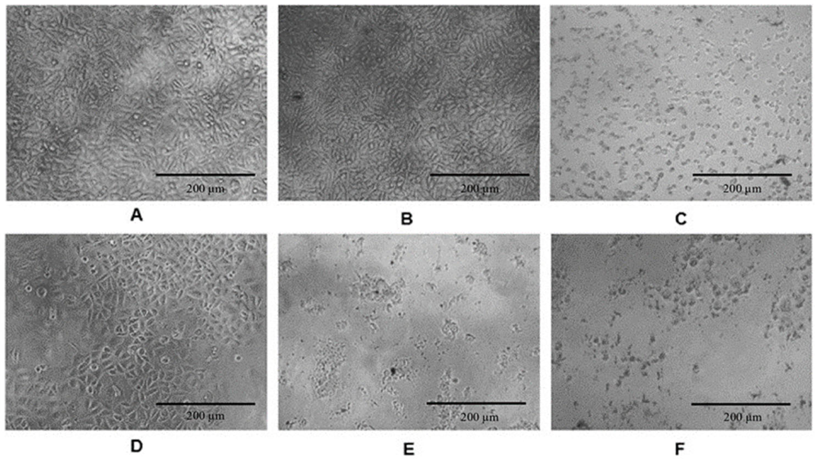
3.4. In Vitro Cytotoxicity of Plant Extracts

Cytotoxicity in cell culture is typically expressed as the LC50, which means the concentration of a substance that is lethal to 50% of the cells. The values obtained of cytotoxic effect indicated that T. nelsonii extract does not cause cell damage in lines 3T3 and HaCaT at concentrations of 20–350 µg mL−1 for 48 h (Table 6). At higher concentrations (1 mg mL−1), the extract caused a change in typical morphology and a reduction in size (Figure 1).
Likewise, A. americana extract and A. vera gel did not exhibit cytotoxicity at ranges of 1–40 µg mL−1 and 15–60% in 3T3 cells cultures, respectively, and 1–31 µg mL−1 and 15–50% in HaCaT cells cultures, respectively. Figure 2 displays the effect on the concentration of the extract and gel on the morphology of 3T3 and HaCaT cells after 48 h of exposure. Therefore, cell viability was directly proportional to the extract concentration. Additionally, the analysis of the morphology of 3T3 and HaCaT cells revealed the appearance of characteristics similar to apoptosis at concentrations higher than 50 µg mL−1 of the A. americana extract (Figure 2A–F) and at concentrations higher than 45% v/v in the case of the A. vera gel (Figure 2G–L).

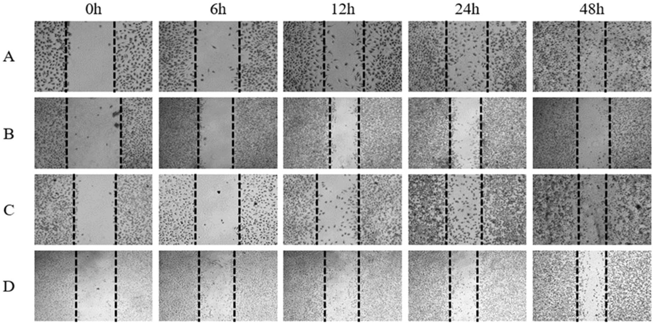
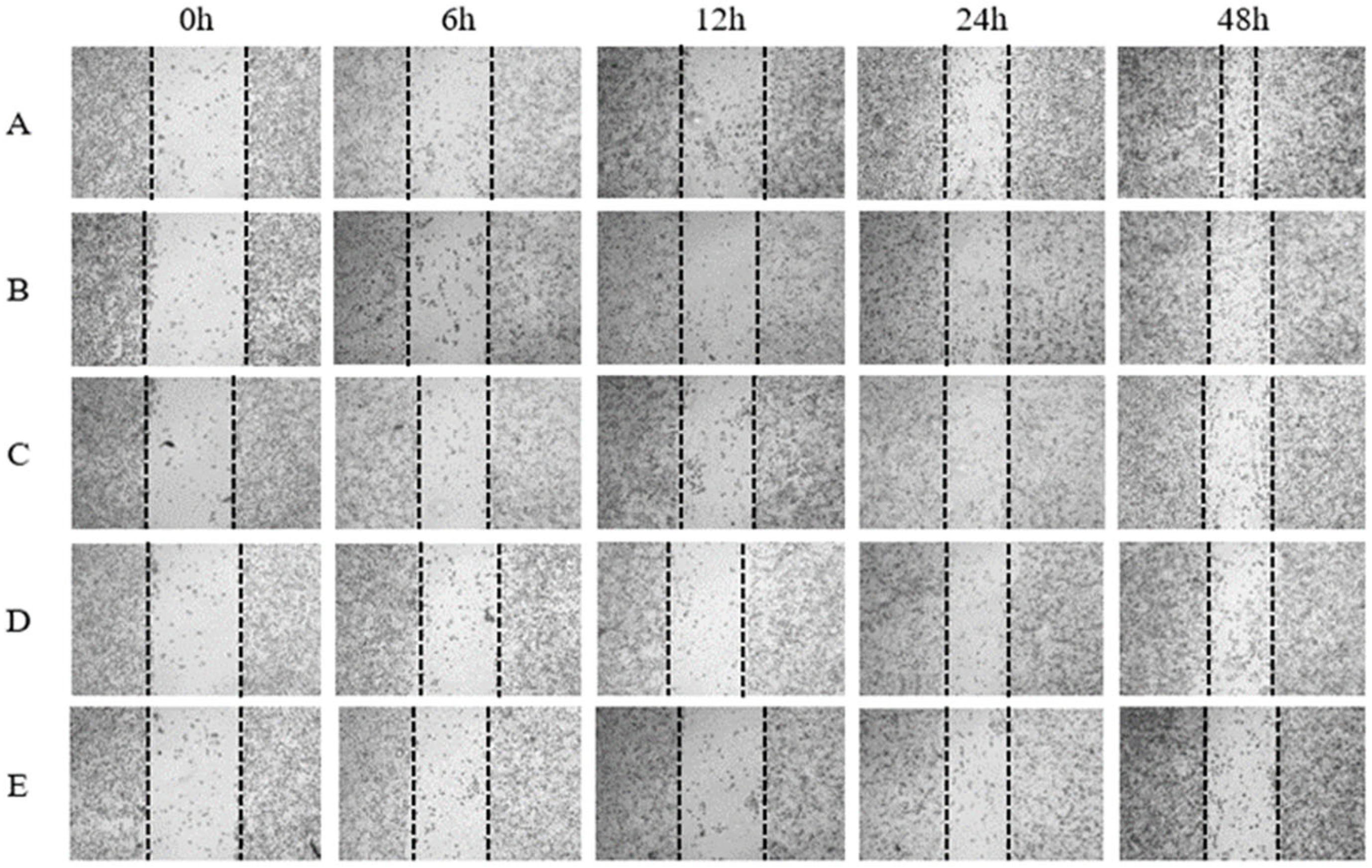
3.5. In Vitro Wound-Healing Activity with Scratch Assay of Individual Plant Extracts

Cell migration plays a crucial role in the wound-healing process; therefore, a scratch assay was performed to observe the closure of a simulated wound in response to individual and combined plant extracts, starting from non-cytotoxic concentrations. Figure 3 shows the wound closure percentages of the extracts on HaCaT and 3T3 cells from time 0 to 48 h. The statistical analysis revealed that the A. americana extract had a wound-healing effect starting from 6 h, reaching a closure of 89.74 ± 1.83% at 48 h. As seen in Figure 3A, there was no statistically significant difference between the control and the A. americana extract at 48 h, but it showed an advantage by having an effect starting from 6 h. Otherwise, for the HaCaT cell line, the statistical analysis showed that all treatments had a wound closure percentage above 70%, except for the A. vera gel at 48 h. However, it can be observed that the T. nelsonii extract showed a statistically significant difference compared to other treatments starting from 6 h (Figure 3B). Nevertheless, the A. americana and T. nelsonii extracts showed greater cell migration accelerating the reduction of the wound area compared to the A. vera gel (Figure 4 and Figure 5).

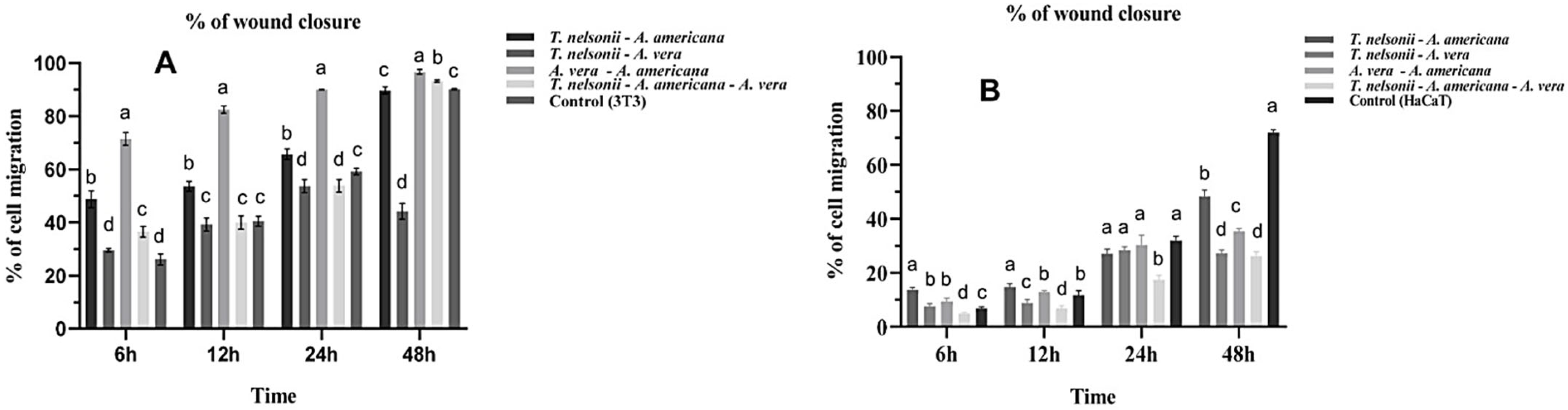
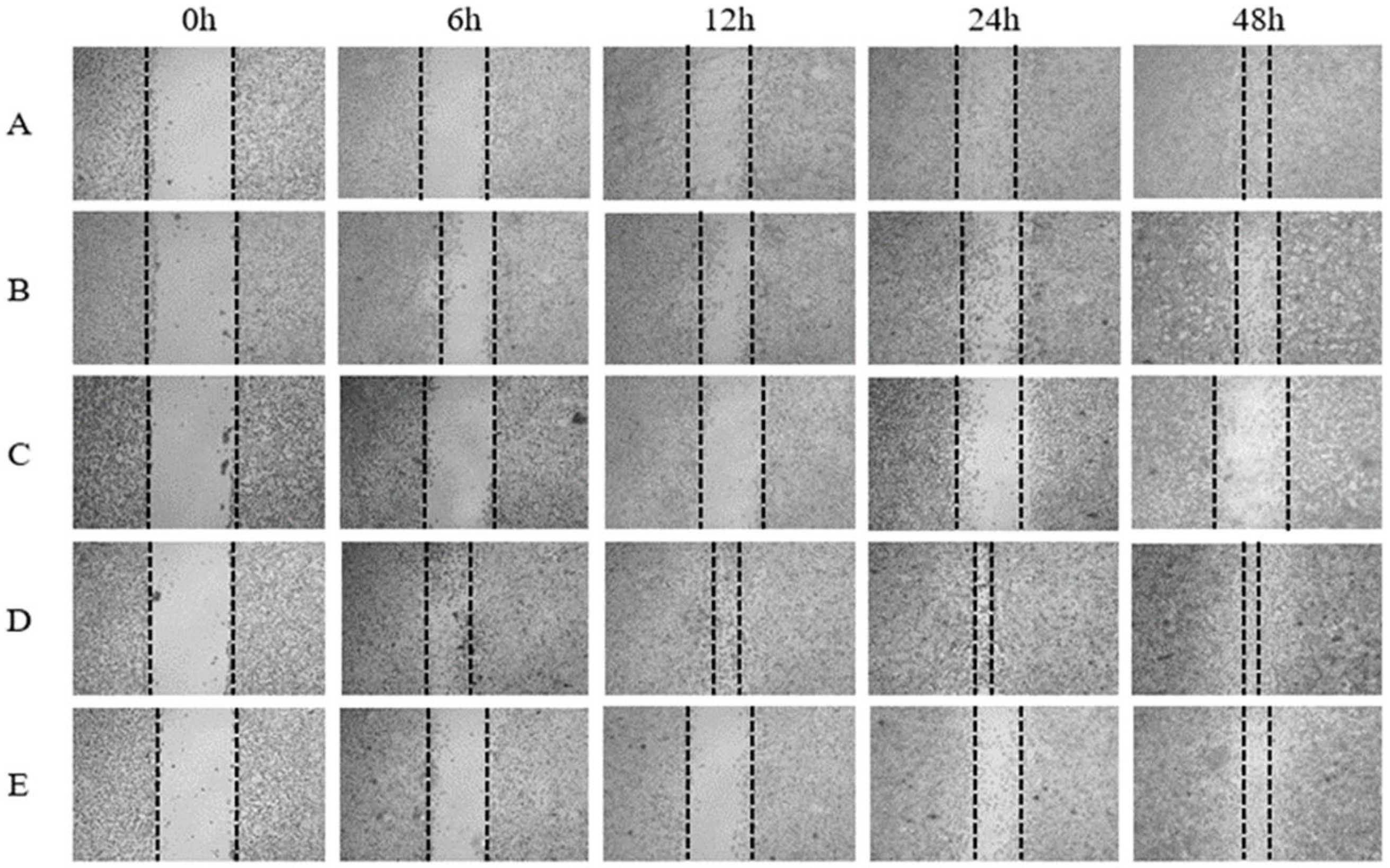
3.6. In Vitro Wound-Healing Activity with Scratch Assay of Combined Plant Extracts

The wound closure percentages of the combined extracts on 3T3 and HaCaT cells culture are showed in Figure 6; this parameter was analyzed from time 0 to 48 h. The statistical analysis revealed that the best treatment in the 3T3 cell line was the combination of A. americana extract and A. vera gel, with a closure of 71.46 ± 2.37% and 96.66 ± 0.86% at 6 and 48 h, respectively, at concentrations of 20.53 µg mL−1 and 36.3%. These was demonstrated with the micrographs, where we observed an increased cell proliferation and migration that consequently leads to a significant reduction in the wound area in the scratch assay (Figure 7). Similarly, the combination of three plants showed a wound closure of 93.33 ± 0.46% at 48 h (Figure 6A) in 3T3 cell line, also showing a statistically significant difference compared to the control; however, both were above 90%. The other combinations demonstrated wound closure above 80% at 48 h (Figure 6). In the HaCaT cell line, it was observed a decrease cell migration and proliferation when using the combination of all extracts at 48 h (Figure 8). Nevertheless, when mixes of two plants were tested, a significant difference in wound closure at 24 h was not found (Figure 6B).

4. Discussion

4.1. Phytochemical Analysis

The chemical profile observed in the T. nelsonii extract coincides with what has been reported for other Tagetes species such as T. minuta, T. erecta, and T. patula, highlighting the presence of saponins, tannins, and coumarins. However, the differences in their content may vary according to environmental and geographical conditions [69,70]. In the A. vera gel, abundant carbohydrates and saponins were found, confirming what has been reported by Hamman [29], who mentioned that carbohydrates like galactomannan, glucomannan, glucose, and fructose, among others, mainly constitute the A. vera gel. Sterols and terpenes have also been reported in the gel, which are likely to be bound to the structures of some steroidal saponins or be part of the β-carotene structure, as reported in the gel’s composition [71]. The phytochemical profile obtained from A. americana coincides with what has been reported for other Agave species, which also include flavonoids, alkaloids, sterols, and terpenes [69,72,73].
In this study, the contents of phenols and flavonoids in the T. nelsonii extract were higher than 53.92 mg g−1 reported in T. patula [21,73]. Sun and Shahrajabian [74] mentioned that these metabolites are the most widely found in the plant kingdom as they are essential for the functioning of plants and also precursors of other compounds such as coumarins, tannins, and anthocyanins, among others [75]. Burlec et al. [16] indicated that the phenolic compounds of the Tagetes genus, in synergy with terpenes and sterols, enhance the anti-inflammatory and antioxidant effects.
In the case of A. vera gel, various authors have stated that the parenchymatous gel contains 99.5% water, with the remainder being solids, which can explain the low concentration of metabolites compared to T. nelsonii (Table 2). The concentrations of phenols and flavonoids were lower compared to what was reported by Taukoorah and Mahomudally [76] and Tabatabaei et al., [77] who used a commercial gel. Nejatzadeh-Barandozi [78] also reported lower concentrations of these metabolites using different solvents.
The amount of phenols found in A. americana is consistent with other studies on Agave, where they reported a content ranging from 0.01 to 29 mg gallic acid per gram [69,72,79]. The high concentration of saponins in this genus is attributed to the effect of environmental factors such as the arid soil in which they grow, and it represents a defense mechanism against herbivores, pests, and insects [80].
Regarding the chromatographic analysis, it has been reported in the literature that the compounds found in this work present various biological activities. The 2,4-bis (1,1-dimethylethyl) [81] has been reported to have with antibacterial and antioxidant activity; terpenoids such as limonene [82], eugenol [83], squalene [84], and phytol [85] have antibacterial and antioxidant activity; fatty acids such as myristic acid [57], palmitic acid [86], 11,14, 17-eicosatrienoic acid [87], 8, 11- linoleic acid [86], caprilic acid [86], and linolenic acid [57] have anti-inflammatory, antimicrobial effects and wound-healing activity; and alkanes such as heneicosane [88] and eicosane [89] have antibacterial activity.

4.2. Antimicrobial Activity of Plant Extracts

The strains of S. aureus and P. aeruginosa showed inhibition in response to the T. nelsonii extract regardless of its concentration. S. aureus, a Gram-positive bacterium, possesses a single plasma membrane that is susceptible to different antimicrobial substances of both hydrophilic and lipophilic character. Several authors [90,91,92] have mentioned that the antimicrobial activity of the extracts obtained from plants is probably induced by the presence of phenolic compounds and sterols and terpenes, thanks to the presence of their hydroxyl groups. In this regard, T. nelsonii showed the highest concentration of phenols, mainly flavonoids, as well as the presence of terpenes such as limonene, eugenol, which, according to the literature, these compounds are capable of interacting with lipids and proteins in the membrane through hydrogen bonding, hydrophobic interactions, and electrostatic interactions, causing not only a change in the permeability and integrity of the plasma membrane but also inactivating microbial adhesins, enzymes, transport proteins in the cell wall, and alterations in energy metabolism [93,94]. The presence of tannins, saponins, anthraquinones, and coumarins was also observed, which, according to the literature, have different mechanisms of action that lead to bacterial death [91,92]. For instance, saponins are known to interact with hopanoids found in plasma membranes, causing changes in membrane fluidity; tannins, anthraquinones, and coumarins, possessing hydroxyl groups, can interact with the carbonyl group of amino acids and the oxygen atom in the head of membrane phospholipids, causing damage to the membrane, proteins, DNA, and essential functions of bacteria [79,80,90,91,92].
Otherwise, the resistance of Gram-negative bacteria like E. coli to different antimicrobial substances has already been reported [93]. These bacteria are characterized by having an outer and inner membrane composed of phospholipids, lipopolysaccharides, efflux pumps, and membrane protein structures such as porins. The latter serve as channels for entry into the bacterium, allowing certain antimicrobial substances like antibiotics to enter the bacterial cytoplasm. However, their effectiveness is influenced by various factors such as the size of the compound, its electric charge, and its hydrophobicity. Moreover, bacteria have the ability to generate mutations in porins, altering their structure and preventing the entry of substances [95].
The lack of antibacterial activity of the A. Americana extract and the A. vera gel on the microorganisms evaluated could be explained by their low content of secondary metabolites compared to the T. nelsonii extract. Additionally, the A. vera gel contains 99.5% water in its composition [29]. These results contradict other findings that highlight the antimicrobial activity of the ethanolic extract of the gel [26,94,96].

4.3. In Vitro Citotoxicity and Wound-Healing Activity

The non-toxicity of the Tagetes genus in the 3T3 and HaCaT cell lines is consistent with what has been reported by Burlec et al. [16], who evaluated the cytotoxicity of the ethanolic extract of T. erecta L. flowers in vitro on mouse fibroblastic cells and found that this species was not cytotoxic at concentrations ranging from 0.25 to 1.00 µg mL−1, with cell viability at 80%. On the other hand, this study observed that the minimum inhibitory concentration (25 mg mL−1) of the T. nelsonii extract was higher than the LC50 found for both cell lines, making its application as an antibacterial agent in various industries feasible; however, its use as a wound-healing agent should be at concentrations lower than the LC50.
The use of A. americana for wound healing has not been tested in vitro; there are studies demonstrating its in vivo wound-healing activity [24,25,97]. In this study, it was observed that the A. Americana extract presented compounds with healing and anti-inflammatory activity. To our knowledge, there are no reports on the use of T. nelsonii for wound healing, but rather the genus [98,99,100,101,102]; however, the presence of compounds with anti-inflammatory, antioxidant, and healing activity was observed. The presence of various bioactive compounds in plant extracts analyzed can contribute to wound closure through two mechanisms: (1) Antioxidant activity: reactive oxygen species (ROS) are produced when a wound occurs, which have the ability to cause damage to the cell membrane, alter metabolic pathways, induce lipid peroxidation, and inhibit proteins and DNA [103]. The presence of hydroxyl groups in these bioactive compounds allows them to donate electrons to stabilize free radicals in stable molecules [104]. (2) Anti-inflammatory activity, due to phenolic compounds that can interact with the amino acids tyrosine and serine at the active site of the enzyme cyclooxygenase-2 (COX-2), leading to its inhibition [105,106]. Additionally, saponins have been found to suppress the activity of platelet cyclooxygenase-1 (COX-1), reducing the production of thromboxanes (TXB2), prostaglandins (PGD2, PGE2) and 11-HETE [107]. These anti-inflammatory effects can help in reducing inflammation at the wound site and promote the healing process.
The use of A. vera gel for wound healing is not new, and previous studies have shown its potential in promoting cell proliferation both in vitro and in vivo, as well as in combination with other extracts [32,37]. Some compounds present in the gel have been reported to potentially contribute to wound healing. For example, mannose 6-phosphate and acemannan, present in the gel, have been shown in in vivo studies to promote wound healing. Mannose 6-phosphate binds to growth factor receptors on the surface of fibroblasts to increase the synthesis of hyaluronic acid, hydroxyproline, and collagen, thus promoting tissue repair [108,109]. Acemannan significantly increases cell proliferation and collagen synthesis [110,111,112]. The antraquinones found in the gel may suppress inflammatory responses by blocking inducible nitric oxide synthase (iNOS) and COX-2 mRNA expression, as well as enhance cell migration through cytokine and growth factor phosphorylation [57]. Sterols and saponins in the gel also increase hyaluronic acid synthesis [32,113]. It is worth noting that this study identified the presence of carbohydrates, phenols, antraquinones, and saponins in the gel.
Similarly, in the HaCaT cell line, it is evident that the combination of extracts does not increase cell proliferation, so it may require a continuous dosage of the extracts at specific intervals to enhance or maintain its effect. Therefore, it is recommended to conduct further research to determine the timing of each dose and extend the duration of the assay.

5. Conclusions

The results suggest that the methanolic extract from T. nelsonii has antibacterial activity against S. aureus and P. aeruginosa, likely attributed to the presence of phenolic compounds, sterols, terpenes, tannins, and saponins. Furthermore, the antibacterial activity of the T. nelsonii extract did not decrease when combined with the other plants. Likewise, the extracts from T. nelsonii, A. americana, and A. vera gel were shown not to be toxic in the 3T3 and HaCaT cell lines, indicating they are safe for use.
On the other hand, the results suggest that wound healing occurred more rapidly when the A. americana extract was used on 3T3 cells and the T. nelsonii extract on HaCaT cells. However, further studies with higher concentrations than those used in this study are needed for a more in-depth analysis of the wound healing capabilities of the extracts. Additionally, extract concentrations above the LC50 that exhibit antibacterial activity could potentially be useful in the food, pharmaceutical, cosmetic, agricultural, and other industries.
It is recommended to test other bacteria isolated from the skin to see if the extracts have a wide range of bacterial inhibition. Additionally, conducting an in vivo study to confirm the wound-healing activity of the extracts and considering possible clinical trials in humans are recommended.

Author Contributions

Conceptualization, K.A.O.-J. and R.I.C.-R.; methodology, K.A.O.-J., N.R.-L., M.d.C.H.-C. and R.I.C.-R.; software, K.A.O.-J.; validation, K.A.O.-J. and R.I.C.-R.; formal analysis, M.d.C.H.-C. and K.A.O.-J.; investigation, K.A.O.-J.; resources, R.I.C.-R., N.J.-H. and B.d.C.C.-G.; data curation, K.A.O.-J.; writing—original draft preparation, K.A.O.-J. and R.I.C.-R.; writing—review and editing, K.A.O.-J., R.I.C.-R., N.R.-L. and N.J.-H.; visualization, K.A.O.-J., R.I.C.-R., N.J.-H. and V.M.R.-V.; supervision, R.I.C.-R.; project administration, R.I.C.-R.; funding acquisition, R.I.C.-R. All authors have read and agreed to the published version of the manuscript.

Funding

This research was funded by CONAHCyT, becarie number 849433.

Institutional Review Board Statement

Not applicable.

Informed Consent Statement

Not applicable.

Data Availability Statement

The data presented in this study are available in this article.

Acknowledgments

Oscar Farrera Sarmiento, Head of the Department of the General Curatorship of Flora and the Office Chief of the Herbarium Chip at SEMAHN Biologist Manuel de Jesús Gutiérrez Morales for the identification of the specimens.

Conflicts of Interest

The authors declare no conflict of interest.

References

  1. Chen, K.; Wang, F.; Liu, S.; Wu, X.; Xu, L.; Zhang, D. In situ reduction of silver nanoparticles by sodium alginate to obtain silver-loaded composite wound dressing with enhanced mechanical and antimicrobial property. Int. J. Biol. Macromol. 2020, 148, 501–509. [Google Scholar] [CrossRef] [PubMed]
  2. Xu, Z.; Han, S.; Gu, Z.; Wu, J. Advances and Impact of Antioxidant Hydrogel in Chronic Wound Healing. Adv. Healthc. Mater. 2020, 9, 1901502. [Google Scholar] [CrossRef] [PubMed]
  3. Singh, S.; Young, A.; McNaught, C. The physiology of wound healing. Surgery 2017, 35, 473–477. [Google Scholar] [CrossRef]
  4. Lawton, S. Skin 1: The structure and functions of the skin. Nurs. Times 2019, 115, 30–33. [Google Scholar]
  5. Brook, I. Aerobic and Anaerobic Bacteriology of Wounds and Cutaneous Abscesses. Arch. Surg. 1990, 125, 1445. [Google Scholar] [CrossRef] [PubMed]
  6. Paladini, F.; Pollini, M. Antimicrobial Silver Nanoparticles for Wound Healing Application: Progress and Future Trends. Materials 2019, 16, 2540. [Google Scholar] [CrossRef] [PubMed]
  7. Yang, Y.; Liang, Y.; Chen, J.; Duan, X.; Guo, B. Mussel-inspired adhesive antioxidant antibacterial hemostatic composite hydrogel wound dressing via photo-polymerization for infected skin wound healing. Bioact. Mater. 2022, 8, 341–354. [Google Scholar] [CrossRef]
  8. Shedoeva, S.; Leavesley, D.; Upton, Z.; Fan, C. Wound Healing and the Use of Medicinal Plants. J. Evid. Based Complement. Altern. Med. 2019, 1, 1–30. [Google Scholar] [CrossRef]
  9. Vitale, S.; Colanero, S.; Placidi, M.; Di Emidio, G.; Tatone, C.; Amicarelli, F.; D’Alessandro, A.M. Phytochemistry and Biological Activity of Medicinal Plants in Wound Healing: An Overview of Current Research. Molecules 2022, 27, 3566. [Google Scholar] [CrossRef] [PubMed]
  10. Albahri, G.; Badran, A.; Hijazi, A.; Daou, A.; Baydoun, E.; Nasser, M.; Merah, O. The Therapeutic Wound Healing Bioactivities of Various Medicinal Plants. Life 2023, 13, 317. [Google Scholar] [CrossRef]
  11. Carod-Artal, F.J.; Vázquez-Cabrera, C. An Anthropological Study About Headache and Migraine in Native Cultures From Central and South America. J. Headache Pain 2007, 47, 834–841. [Google Scholar] [CrossRef] [PubMed]
  12. De la Cruz-Jimenez, L.; Guzman-Lucio, M.; Viveros-Valdez, E. Traditional Medicinal Plants Used for the Treatment of Gastrointestinal Diseases in Chiapas, México. World Appl. Sci. J. 2014, 31, 508–515. [Google Scholar] [CrossRef]
  13. Céspedes, C.L.; Avila, J.G.; Martínez, A.; Serrato, B.; Calderón-Mugica, J.C.; Salgado-Garciglia, R. Antifungal and antibacterial activities of Mexican tarragon (Tagetes lucida). J. Agric. Food Chem. 2006, 54, 3521–3527. [Google Scholar] [CrossRef]
  14. Singh, P.; Krishna, A.; Kumar, V.; Krishna, S.; Singh, K.; Gupta, M.; Singh, S. Chemistry and biology of industrial crop Tagetes Species: A review. J. Essent. Oil Res. 2015, 28, 1–14. [Google Scholar] [CrossRef]
  15. Walia, S.; Mukhia, S.; Bhatt, V.; Kumar, R.; Kumar, R. Variability in chemical composition and antimicrobial activity of Tagetes minuta L. essential oil collected from different locations of Himalaya. Ind. Crops Prod. 2020, 150, 112449. [Google Scholar] [CrossRef]
  16. Burlec, A.F.; Pecio, L.; Kozachok, S.; Mircea, C.; Corciovă, A.; Vereștiuc, L.; Cioancă, O.; Oleszek, W.; Hăncianu, M. Phytochemical Profile, Antioxidant Activity, and Cytotoxicity Assessment of Tagetes erecta Flowers. Molecules 2021, 26, 1201. [Google Scholar] [CrossRef] [PubMed]
  17. De La Cruz-Jiménez, L.; Hernández-Torres, M.A.; Monroy-García, I.N.; Rivas-Morales, C.; Verde-Star, M.J.; Gonzalez-Villasana, V.; Viveros-Valdez, E. Biological Activities of Seven Medicinal Plants Used in Chiapas, Mexico. Plants 2022, 11, 1790. [Google Scholar] [CrossRef] [PubMed]
  18. Espinoza, R.M.; Palomeque, R.M.A.; Salazar, S.I.; Domínguez, A.S.; Canseco, A.L.M. Análisis preliminar de la actividad antimicrobiana de la planta medicinal Chik chawa (Tagetes nelsonii Greenm.). Rev. Cuba. Plantas Med. 2009, 14, 4. [Google Scholar]
  19. Abdoul-Latif, F.M.; Elmi, A.; Merito, A.; Nour, M.; Risler, A.; Ainane, A.; Bignon, J.; Ainane, T. Essential Oils of Tagetes minuta and Lavandula coronopifolia from Djibouti: Chemical Composition, Antibacterial Activity and Cytotoxic Activity against Various Human Cancer Cell Lines. Int. J. Plant Biol. 2022, 13, 315–329. [Google Scholar] [CrossRef]
  20. Alvarado-Sansininea, J.J.; Sánchez-Sánchez, L.; López-Muñoz, H.; Escobar, M.L.; Flores-Guzmán, F.; Tavera-Hernández, R.; Jiménez-Estrada, M. Quercetagetin and patuletin: Antiproliferative, necrotic and apoptotic activity in tumor cell lines. Molecules 2018, 23, 2579. [Google Scholar] [CrossRef]
  21. Bermúdez-Bazán, M.; Castillo-Herrera, G.A.; Urias-Silvas, J.E.; Escobedo-Reyes, A.; Estarrón-Espinosa, M. Hunting Bioactive Molecules from the Agave Genus: An Update on Extraction and Biological Potential. Molecules 2021, 26, 6789. [Google Scholar] [CrossRef]
  22. López-Salazar, H.; Camacho-Díaz, B.H.; Ávila-Reyes, S.V.; Pérez-García, M.D.; González- Cortazar, M.; Arenas Ocampo, M.L.; Jiménez-Aparicio, A.R. Identification and Quantification of β-Sitosterol β-d-Glucoside of an Ethanolic Extract Obtained by Microwave-Assisted Extraction from Agave angustifolia Haw. Molecules 2019, 24, 3926. [Google Scholar] [CrossRef] [PubMed]
  23. Misra, A.K.; Varma, S.K. Effect of an Extract of Agave americana on Wound Healing Model in Experimental Animals. J. Basic Clin. Physiol. Pharmacol. 2017, 8, 45–48. [Google Scholar]
  24. Misra, A.K.; Varma, S.K.; Kumar, R. Anti-inflammatory Effect of an Extract of Agave americana on Experimental Animals. Pharmacogn. Res. 2018, 10, 104–108. [Google Scholar] [CrossRef]
  25. Elferjane, M.R.; Jovanović, A.A.; Milutinović, V.; Čutović, N.; Jovanović Krivokuća, M.; Marinković, A. From Aloe vera Leaf Waste to the Extracts with Biological Potential: Optimization of the Extractions, Physicochemical Characterization, and Biological Activities. Plants 2023, 12, 2744. [Google Scholar] [CrossRef]
  26. Pandey, B.R.; Shrestha, A.; Sharma, N.; Shrestha, B.G. Evaluation of Phytochemical, Antimicrobial, Antioxidant Activity and Cytotoxic Potentials of Agave americana. Nepal J. Biotechnol. 2019, 7, 30–38. [Google Scholar] [CrossRef]
  27. Hulle, A.; Kadole, P.; Katkar, P. Agave americana Leaf Fibers. Fibers 2015, 3, 64–75. [Google Scholar] [CrossRef]
  28. Shegute, T.; Wasihun, Y. Antibacterial Activity and Phytochemical Components of Leaf Extracts of Agave americana. J. Exp. Pharmacol. 2020, 12, 447–454. [Google Scholar] [CrossRef] [PubMed]
  29. Hamman, J.H. Composition and applications of Aloe vera leaf gel. Molecules 2008, 13, 1599–1616. [Google Scholar] [CrossRef] [PubMed]
  30. Liu, C.; Cui, Y.; Pi, F.; Cheng, Y.; Guo, Y.; Qian, H. Extraction, Purification, Structural Characteristics, Biological Activities and Pharmacological Applications of Acemannan, a Polysaccharide from Aloe vera: A Review. Molecules 2019, 24, 1554. [Google Scholar] [CrossRef]
  31. Reynolds, T.; Dweck, A.C. Aloe vera leaf gel: A review update. J. Ethnopharmacol. 1999, 68, 3–37. [Google Scholar] [CrossRef] [PubMed]
  32. Tanaka, M.; Misawa, E.; Yamauchi, K.; Abe, F.; Ishizaki, C. Effects of plant sterols derived from Aloe vera gel on human dermal fibroblasts in vitro and on skin condition in Japanese women. Clin. Cosmet. Investig. Dermatol. 2015, 8, 95–104. [Google Scholar] [CrossRef] [PubMed]
  33. Fox, L.T.; Mazumder, A.; Dwivedi, A.; Gerber, M.; du Plessis, J.; Hamman, J.H. In vitro wound healing and cytotoxic activity of the gel and whole-leaf materials from selected aloe species. J. Ethnopharmacol. 2017, 200, 1–7. [Google Scholar] [CrossRef] [PubMed]
  34. Bajpai, S. Chapter 8—Biological Importance of Aloe vera and its Active Constituents. In Synthesis of Medical Agents from Plants; Tewari, A., Tiwari, S., Eds.; Elsevier: Amsterdam, The Netherlands, 2018; pp. 177–203. [Google Scholar] [CrossRef]
  35. Kumar, D.S.; Choudhury, P.K.; Srivastava, R.; Sharma, M. Antimicrobial, anti-inflammatory and wound healing activity of polyherbal formulation. Biomed. Pharmacother. 2019, 111, 555–567. [Google Scholar] [CrossRef]
  36. Niknam, S.; Tofighi, Z.; Faramarzi, M.A.; Abdollahifar, M.A.; Sajadi, E.; Dinarvand, R.; Toliyat, T. Polyherbal combination for wound healing: Matricaria chamomilla L. and Punica granatum L. DARU J. Pharm. Sci. 2021, 29, 133–145. [Google Scholar] [CrossRef] [PubMed]
  37. Hattingh, A.; Laux, J.P.; Willers, C.; Hamman, J.; Steyn, D.; Hamman, H. In vitro wound healing effects of combinations of Aloe vera gel with different extracts of Bulbine frutescens. S. Afr. J. Bot. 2023, 158, 254–264. [Google Scholar] [CrossRef]
  38. Kuete, V.; Nguemeving, J.R.; Beng, V.P.; Blaise, A.A.G.; Etoa, F.X.; Meyer, M.; Bodo, B.; Nkengfack, A.E. Antimicrobial activity of the methanolic extracts and compounds from Vismia laurentii De Wild (Guttiferae). J. Ethnopharmacol. 2007, 109, 372–379. [Google Scholar] [CrossRef]
  39. Tereschuk, M.R.; Riera, M.V.; Castro, G.R.; Abdala, L.R. Antimicrobial activity of flavonoids from leaves of Tagetes minuta. J. Ethnopharmacol. 1997, 56, 227–232. [Google Scholar] [CrossRef] [PubMed]
  40. Chin, C.J. Development and formulation of Moringa oleifera standardised leaf extract film dressing for wound healing application. J. Ethnopharmacol. 2018, 212, 188–199. [Google Scholar] [CrossRef]
  41. Torres-Aguirre, G.A.; Muñoz-Bernal, O.A.; Álvarez-Parrilla, E.; Núñez-Gastélum, J.A.; Wall-Medrano, A.; Sáyago-Ayerdi, S.G.; de la Rosa, L.A. Optimización de la extracción e identificación de compuestos polifenólicos en anís (Pimpinella anisum), clavo (Syzygium aromaticum) y cilantro (Coriandrum sativum) mediante HPLC acoplado a espectrometría de masas. Rev. Espec. Cienc. Quim. Biol. 2018, 21, 103–115. [Google Scholar] [CrossRef]
  42. Tessema, Z.; Molla, Y. Evaluation of the wound healing activity of the crude extract of root bark of Brucea antidysentrica, the leaves of Dodonaea angustifolia and Rhamnus prinoides in mice. Heliyon 2021, 7, e05901. [Google Scholar] [CrossRef]
  43. Tadhani, M.; Subhash, R. Preliminary Studies on Stevia rebaudiana Leaves: Proximal Composition, Mineral Analysis and Phytochemical Screening. J. Med. Sci. 2006, 6, 321–326. [Google Scholar] [CrossRef]
  44. Yadav, R.N.S.; Agarwala, M. Phytochemical Analysis of Some Medicinal Plants. J. Phytol. 2011, 3, 10–14. [Google Scholar]
  45. Anburaj, G.; Marimuthu, M.; Manikandan, R.; Rajasudha, V. Phytochemical screening and GC-MS analysis of ethanolic extract of Tecoma stans (Family: Bignoniaceae) Yellow Bell Flowers. J. Pharmacogn. Phytochem. 2016, 5, 172–175. [Google Scholar]
  46. Suganandam, K.; Jeevalatha, A.; Kandeepan, C.; Kavitha, N.; Senthilkumar, N.; Sutha, S.; Seyed, M.A.; Gandhi, S.; Ramya, S.; Lydial, G.; et al. Profile of Phytochemicals and GCMS Analysis of Bioactive Compounds in Natural Dried-Seed Removed Ripened Pods Methanolic Extracts of Moringa oleifera. J. Drug Deliv. Ther. 2022, 12 (Suppl. S5), 133–141. [Google Scholar] [CrossRef]
  47. Wagner, H.; Bladt, S. Plant Drug Analysis. A Thin Layer Chromatography Atlas, 2nd ed.; Springer Science & Business Media: Berlin/Heidelberg, Germany, 1996. [Google Scholar] [CrossRef]
  48. Singleton, V.L.; Orthofer, R.; Lamuela-Raventos, R.M. Analysis of total phenols and other oxidation substrates and antioxidants by means of folin-ciocalteu reagent. Meth. Enzymol. 1999, 299, 152–178. [Google Scholar] [CrossRef]
  49. Ying, C.; Wan, D. Quantitative determination of total and individual flavonoids in stems and leaves of Buddleja davidii and Buddleja albiflora. Pharmacogn. Mag. 2012, 8, 273–279. [Google Scholar] [CrossRef] [PubMed]
  50. Sim, E.W.; Lai, S.Y.; Chang, Y.P. Antioxidant capacity, nutritional and phytochemical content of peanut (Arachis hypogaea L.) shells and roots. Afr. J. Biotechnol. 2012, 11, 11547–11551. [Google Scholar] [CrossRef]
  51. Broadhurst, R.B.; Jones, W.T. Analysis of condensed tannins using acidified vanillin. J. Sci. Food Agric. 1978, 29, 788–794. [Google Scholar] [CrossRef]
  52. Akyar, I. (Ed.) Latest Research into Quality Control; InTech: Rijeka, Croatia, 2012. [Google Scholar] [CrossRef]
  53. EPA. Method 8270D Semivolatile Organic Compounds by Gas Chromatography/Mass Spectrometry (GC/MS). 2014. Available online: https://archive.epa.gov/epa/sites/production/files/2015-12/documents/8270d.pdf (accessed on 16 March 2022).
  54. Xin, Z.; Yang, W.; Duan, Y.; Wang, W.; Niu, L.; Sun, D.; Zhang, Y. Bioactive components and antibacterial activities of hydrolate extracts by optimization conditions from Paeonia ostii T. Hong and J. X. Zhang. Ind. Crops Prod. 2022, 188, 115737. [Google Scholar] [CrossRef]
  55. Vaghasiya, Y.; Chanda, S.V. Screening of methanol and acetone extracts of fourteen indian medicinal plants for antimicrobial activity. Turk. J. Biol. 2007, 31, 243–248. [Google Scholar]
  56. NCCLS. Performance Standards for Antimicrobial Disk Susceptibility Test, 11th ed.; M02-A11; NCCLS: Wayne, PA, USA, 2012; 76p. [Google Scholar]
  57. Kim, Y.G.; Lee, J.H.; Park, S.; Kim, S.; Lee, J. Inhibition of polymicrobial biofilm formation by saw palmetto oil, lauric acid and myristic acid. Microb. Biotechnol. 2022, 15, 590–602. [Google Scholar] [CrossRef]
  58. Clinical and Laboratory Standards Institute (CLSI). Methods for Dilution Microbial Susceptibility Tests for Bacteria That Grow Aerobically; Approved Standard—Ninth Edition, CLSI Document M07-A9; Clinical and Laboratory Standards Institute: Wayne, PA, USA, 2006. [Google Scholar]
  59. Canche-Escamilla, G.; Colli-Acevedo, P.; Borges-Argaez, R.; Quintana-Owen, P.; May-Crespo, J.F.; Cáceres-Farfan, M.; Yam Puc, J.A.; Sansores-Peraza, P.; Vera-Ku, B.M. Extraction of phenolic components from an Aloe vera (Aloe barbadensis Miller) crop and their potential as antimicrobials and textile dyes. Sustain. Chem. Pharm. 2019, 14, 100168. [Google Scholar] [CrossRef]
  60. Malbrán, G. Método de determinación de sensibilidad antimicrobiana por dilución. In MIC Testing; Clinical and Laboratory Standards Institute: Wayne, PA, USA, 2012; Volume 32. [Google Scholar]
  61. Chanda, S.V.; Menpara, D.; Desai, D.; Rathod, T. Evaluation of Nutraceutical Bottle Gourd (Lagenaria siceraria) as a Potential Source of Natural Antimicrobial Agent. Am. J. Phtyomed. Clin. Ther. 2014, 2, 375–389. [Google Scholar]
  62. Lopez, T.V.; Lappin, T.R.; Maxwell, P.; Shi, Z.; Lopez-Marure, R.; Aguilar, C.; Rocha-Zavaleta, L. Autocrine/paracrine erythropoietin signalling promotes JAK/STAT-dependent proliferation of human cervical cancer cells. Int. J. Cancer 2011, 129, 2566–2576. [Google Scholar] [CrossRef] [PubMed]
  63. Fragoso-Ontiveros, V.; Alvarez-García, R.M.; Contreras-Paredes, A.; Vaca-Paniagua, F.; Alonso-Herrera, L.; López-Camarillo, C.; Jacobo-Herrera, N.; Lizano-Soberón, M.; Pérez-Plasencia, C. Gene expression profiles induced by E6 from non-European HPV18 variants reveals a differential activation on cellular processes driving to carcinogenesis. Virology 2012, 432, 81–90. [Google Scholar] [CrossRef]
  64. Basu, P.N.; Narendrakumar, U.; Arunachalam, R.; Devi, S.; Manjubala, I. Characterization and evaluation of carboxymethyl cellulose-based films for healing of full-thickness wounds in normal and diabetic rats. ACS Omega 2018, 3, 12622–12632. [Google Scholar] [CrossRef]
  65. Xie, H.C.; Chen, X.; Shen, X.; He, Y.; Chen, W.; Luo, Q.; Ge, W.; Yuan, W.; Tang, X.; Hou, D.; et al. Preparation of chitosan-collagen-alginate composite dressing and its promoting effects on wound healing. Int. J. Biol. Macromol. 2017, 107 Part A, 93–104. [Google Scholar] [CrossRef]
  66. Kisanga, E.P.; Tang, Z.; Guller, S.; Whirledge, S. In vitro Assays to Evaluate the Migration, Invasion, and Proliferation of Immortalized Human First-trimester Trophoblast Cell Lines. J. Vis. Exp. 2019, 145, 58942. [Google Scholar] [CrossRef]
  67. Nicolaus, C.J.; Junghanns, S.; Hartmann, A.; Murillo, R.; Ganzera, M.; Merfort, I. In vitro studies to evaluate the wound healing properties of Calendula officinalis extracts. J. Ethnopharmacol. 2017, 196, 94–103. [Google Scholar] [CrossRef] [PubMed]
  68. Hernández-Pasteur, G.; Silva-Bermúdez, P.S.; Reyes-Chilpa, R.; Vibrans, H.; Soto-Hernández, M. In vitro evaluation of the healing and antimicrobial activity of extracts of Buddleja cordata kunth and Vismia baccifera (L.) triana and planch. Rev. Fitotec. 2019, 42, 93–99. [Google Scholar]
  69. López-Romero, J.C.; Ayala-Zavala, J.F.; Peña-Ramos, E.A.; Hernández, J.; González-Ríos, H. Antioxidant and antimicrobial activity of Agave angustifolia extract on overall quality and shelf life of pork patties stored under refrigeration. J. Food Sci. Technol. 2018, 55, 4413–4423. [Google Scholar] [CrossRef] [PubMed]
  70. Soto-Rueda, E.M.; Rodríguez-Ruiz, Y.L.; Loango-Chamorro, N.; Landázuri, P. Extracts of Tagetes patula L. (Asteraceae): A bactericidal potential against Moko. Rev. Mex. Cienc. Agric. 2018, 9, 949–959. [Google Scholar] [CrossRef]
  71. Ammar, A.T.; Arif, A.M. Phytochemical screening and in vitro antibacterial and anticancer activities of the aqueous extract of Cucumis sativus. Saudi J. Biol. Sci. 2019, 26, 600–604. [Google Scholar] [CrossRef]
  72. Ahumada-Santos, Y.P.; Montes-Avila, J.; Uribe-Beltrán, M.J.; Díaz-Camacho, S.P.; López-Angulo, G.; Vega-Aviña, R.; López-Valenzuela, J.A.; Heredia, J.B.; Delgado-Vargas, F. Chemical characterization, antioxidant and antibacterial activities of six Agave species from Sinaloa, Mexico. Ind. Crops Prod. 2013, 49, 143–149. [Google Scholar] [CrossRef]
  73. Singh, P.; Tanwar, N.; Saha, T.; Gupta, A.; Verma, S. Phytochemical Screening and Analysis of Carica papaya, Agave americana and Piper nigrum. Int. J. Curr. Microbiol. Appl. Sci. 2018, 7, 1786–1794. [Google Scholar] [CrossRef]
  74. Sun, W.; Shahrajabian, M.H. Therapeutic Potential of Phenolic Compounds in Medicinal Plants—Natural Health Products for Human Health. Molecules 2023, 28, 1845. [Google Scholar] [CrossRef] [PubMed]
  75. Swallah, M.S.; Sun, H.; Affoh, R.; Fu, H.; Yu, H. Antioxidant Potential Overviews of Secondary Metabolites (Polyphenols) in Fruits. Int. J. Food Sci. 2020, 2020, 9081686. [Google Scholar] [CrossRef] [PubMed]
  76. Taukoorah, U.; Mahomoodally, M.F. Crude Aloe vera gel shows antioxidant propensities and inhibits pancreatic lipase and glucose movement in vitro. Adv. Pharmacol. Sci. 2016, 1, 3720850. [Google Scholar] [CrossRef]
  77. Tabatabaei, S.R.F.; Ghaderi, S.; Bahrami-Tapehebur, M.; Farbood, Y.; Rashno, M. Aloe vera gel improves behavioral deficits and oxidative status in streptozotocin-induced diabetic rats. Biomed. Pharmacother. 2017, 96, 279–290. [Google Scholar] [CrossRef]
  78. Nejatzadeh-Barandozi, F. Antibacterial activities and antioxidant capacity of Aloe vera. Org. Med. Chem. Lett. 2013, 3, 5. [Google Scholar] [CrossRef] [PubMed]
  79. Soto-Castro, D.; Pérez-Herrera, A.; García-Sánchez, E.; Santiago-García, P.A. Identification and Quantification of Bioactive Compounds in Agave potatorum Zucc. Leaves at Different Stages of Development and a Preliminary Biological Assay. Waste Biomass Valorization 2021, 12, 4537–4547. [Google Scholar] [CrossRef]
  80. Sidana, J.S.; Singh, B.; Sharma, O.P. Saponins of Agave: Chemistry and bioactivity. Phytochemistry 2016, 130, 22–46. [Google Scholar] [CrossRef] [PubMed]
  81. Devi, T.S.; Vijay, K.; Vidhyavathi, R.M.; Kumar, P.; Govarthanan, M.; Kavitha, T. Antifungal activity and molecular docking of phenol, 2,4-bis(1,1-dimethylethyl) produced by plant growth-promoting actinobacterium Kutzneria sp. strain TSII from mangrove sediments. Arch. Microbiol. 2021, 203, 4051–4064. [Google Scholar] [CrossRef]
  82. Bayer, G.; Shayganpour, A.; Bayer, I.S. Antioxidant activity of limonene modified cellulose pulp fiber-polylactic acid (PLA) composites. Cellulose 2023, 30, 1599–1622. [Google Scholar] [CrossRef]
  83. Marchese, A.; Barbieri, R.; Coppo, E.; Orhan, I.E.; Daglia, M.; Nabavi, S.F.; Izadi, M.; Abdollahi, M.; Nabavi, S.M.; Ajami, M. Antimicrobial activity of eugenol and essential oils containing eugenol: A mechanistic viewpoint. Crit. Rev. Microbiol. 2017, 43, 668–689. [Google Scholar] [CrossRef] [PubMed]
  84. Yarkent, Ç.; Oncel, S.S. Recent Progress in Microalgal Squalene Production and Its Cosmetic Application. Biotechnol. Bioprocess. Eng. 2022, 27, 295–305. [Google Scholar] [CrossRef] [PubMed]
  85. Islam, M.T.; Ali, E.S.; Uddin, S.J.; Shaw, S.; Islam, M.A.; Ahmed, M.I.; Chandra, S.M.; Karmakar, U.K.; Yarla, N.S.; Khan, I.N.; et al. Phytol: A review of biomedical activities. Food Chem. Toxicol. 2018, 121, 82–94. [Google Scholar] [CrossRef]
  86. Weimann, E.; Silva, M.B.B.; Murata, G.M.; Bortolon, J.R.; Dermargos, A.; Curi, R.; Hatanaka, E. Topical anti-inflammatory activity of palmitoleic acid improves wound healing. PLoS ONE 2018, 13, e0205338. [Google Scholar] [CrossRef]
  87. Jin, X.J.; Kim, E.J.; Oh, I.K.; Kim, Y.K.; Park, C.H.; Chung, J.H. Prevention of UV-induced skin damages by 11,14,17-eicosatrienoic acid in hairless mice in vivo. J. Korean Med. Sci. 2010, 25, 930–937. [Google Scholar] [CrossRef]
  88. Vanitha, V.; Vijayakumar, S.; Nilavukkarasi, M.; Punitha, V.N.; Vidhya, E.; Praseetha, P.K. Heneicosane—A novel microbicidal bioactive alkane identified from Plumbago zeylanica L. Ind. Crops Prod. 2020, 154, 112748. [Google Scholar] [CrossRef]
  89. Umaru, I.J.; Badruddin, F.A.; Umaru, H.A. Phytochemical Screening of Essential Oils and Antibacterial Activity and Antioxidant Properties of Barringtonia asiatica (L.) Leaf Extract. Biochem. Res. Int. 2019, 2019, 7143989. [Google Scholar] [CrossRef] [PubMed]
  90. Leyva-Jimenez, F.J.; Sanchez, J.J.; Borras-Linares, I.; Cadiz-Gurrea, M.L.; Mahmoodi-Khaledi, E. Potential antimicrobial activity of honey phenolic compounds against Gram positive and Gram negative bacteria. LWT 2019, 101, 236–245. [Google Scholar] [CrossRef]
  91. Orczyk, M.; Wojciechowski, K.; Brezesinski, G. The influence of steroidal and triterpenoid saponins on monolayer models of the outer leaflets of human erythrocytes, E. coli and S. cerevisiae cell membranes. J. Colloid. Interface Sci. 2020, 563, 207–217. [Google Scholar] [CrossRef] [PubMed]
  92. Villa-Silva, P.Y.; Iliná, A.; Ascacio-Valdés, J.A.; Esparza-González, S.C.; Cobos-Puc, L.E.; Rodríguez-Herrera, R.; Silva-Belmares, S.Y. Phenolic compounds of Tagetes lucida Cav. with antibacterial effect due to membrane damage. Bol. Latinoam. Caribe Plantas Med. Aromat. 2020, 19, 580–590. [Google Scholar] [CrossRef]
  93. Lepe, J.A.; Martínez-Martínez, L. Mecanismos de resistencia en bacterias gramnegativas. Med. Intensiv. 2022, 46, 392–402. [Google Scholar] [CrossRef]
  94. Adzitey, F.; Agbolosu, A.A.; Udoka, U.J. Antibacterial effect of Aloe vera gel extract on Escherichia coli and Salmonella enterica isolated from the gastrointestinal tract of Guinea Fowls. World’s Vet. J. 2019, 9, 166–173. [Google Scholar] [CrossRef]
  95. Breijyeh, Z.; Jubeh, B.; Karaman, R. Resistance of Gram-Negative Bacteria to Current Antibacterial Agents and Approaches to Resolve It. Molecules 2020, 25, 1340. [Google Scholar] [CrossRef] [PubMed]
  96. Arbab, S.; Buriro, R.; Bhugio, S.U.; Shah, A.H.; Soomro, J.; Kalwar, Q.; Fazilani, S.A.; Vistro, W.A. Antimicrobial Properties of Aloe vera Gel Extracts against Bacterial Isolates from Wound of Donkey. Pak. J. Zool. 2020, 52, 2333–2339. [Google Scholar] [CrossRef]
  97. Solanke, S.B.; Bhujbal, S.S.; Tawar, M.G. Development and Evaluation of Surgical Sutures from Agave americana Linn. Var. Americana. Am. J. PharmTech Res. 2018, 8, 114–125. [Google Scholar] [CrossRef]
  98. Chatterjee, S.P. Comparative efficacy of Tagetes erecta and Centella asiatica extracts on wound healing in albino rats. Chin. Med. 2011, 2, 138–142. [Google Scholar] [CrossRef]
  99. Singh, N.; Mrinal; Thakur, R. Review on Pharmacological aspects of Tagetes erecta Linn. PharmaTutor 2019, 7, 16–24. [Google Scholar]
  100. Selvam, S.I.; Joicesky, S.M.B.; Dashli, A.A.; Vinothini, A.; Premkumar, K. Assessment of anti-bacterial, anti-inflammation and wound healing activity in Wistar albino rats using green silver nanoparticles synthesized from Tagetes erecta leaves. J. Appl. Nat. Sci. 2021, 13, 343–351. [Google Scholar] [CrossRef]
  101. Sultana, A.; Hasan, M.; Rahman, M.; Alam, M.M. Healing potentials of Marigold flower (Tagetes erecta) on full thickness dermal wound in caprine model. Eur. Respir. J. 2021, 7, 332–339. [Google Scholar] [CrossRef]
  102. Manisha, U.K.; Ghosh, T.; Apoorva, V.P.; Divya, B.; Swathy, S.P.; Paul, A.; Basavraj, B.V. Isolation, phytochemical elucidation, and wound healing potential of chitosan-based film loaded with Tagetes erecta. In Materials Today: Proceedings; Elsevier: Amsterdam, The Netherlands, 2022. [Google Scholar] [CrossRef]
  103. Tan, B.L.; Norhaizan, M.E.; Liew, W.P.P.; Sulaiman, R.H. Antioxidant and Oxidative Stress: A Mutual Interplay in Age-Related Diseases. Front. Pharmacol. 2018, 9, 1162. [Google Scholar] [CrossRef] [PubMed]
  104. Olszowy, M. What is responsible for antioxidant properties of polyphenolic compounds from plants? Plant Physiol. Biochem. 2019, 144, 135–143. [Google Scholar] [CrossRef] [PubMed]
  105. Yang, M.H.; Yoon, K.D.; Chin, Y.W.; Park, J.H.; Kim, J. Phenolic compounds with radical scavenging and cyclooxygenase-2 (COX-2) inhibitory activities from Dioscorea opposita. Bioorg. Med. Chem. 2009, 17, 2689–2694. [Google Scholar] [CrossRef] [PubMed]
  106. Ribeiro, D.; Freitas, M.; Tomé, S.M.; Silva, A.M.; Laufer, S.; Lima, J.L.; Fernandes, E. Flavonoids inhibit COX-1 and COX-2 enzymes and cytokine/chemokine production in human whole blood. Inflammation 2015, 38, 858–870. [Google Scholar] [CrossRef]
  107. Chummun, I.; Bekah, D.; Goonoo, N.; Bhaw-Luximon, A. Assessing the mechanisms of action of natural molecules/extracts for phase-directed wound healing in hydrogel scaffolds. RSC Med. Chem. 2021, 12, 1476–1490. [Google Scholar] [CrossRef]
  108. Davis, R.H.; Donato, J.J.; Hartman, G.M.; Haas, R.C. Anti-inflammatory and wound healing activity of a growth substance in Aloe vera. J. Am. Podiatr. Med. Assoc. 1994, 84, 77–81. [Google Scholar] [CrossRef] [PubMed]
  109. Radha, M.H.; Laxmipriya, N.P. Evaluation of biological properties and clinical effectiveness of Aloe vera: A systematic review. J. Tradit. Complement. Med. 2014, 5, 21–26. [Google Scholar] [CrossRef] [PubMed]
  110. Sierra-García, G.D.; Castro-Ríos, R.; González-Horta, A.; Lara-Arias, J.; Chávez-Montes, A. Acemannan, an Extracted Polysaccharide from Aloe vera: A Literature Review. Nat. Prod. Commun. 2014, 9, 1217–1221. [Google Scholar] [PubMed]
  111. Silva, S.S.; Fernandes, E.M.; Pina, S.; Silva-Correia, J.; Vieira, S.; Oliveira, J.M.; Reis, R.L. Polymers of Biological Origin. Compr. Biomater. II 2017, 2, 228–252. [Google Scholar] [CrossRef]
  112. Fouché, M.; Willers, C.; Hamman, S.; Malherbe, C.; Steenekamp, J. Wound Healing Effects of Aloe muth-muth: In vitro Investigations Using Immortalized Human Keratinocytes (HaCaT). Biology 2020, 9, 350. [Google Scholar] [CrossRef]
  113. Mukherjee, P.K.; Nema, N.K.; Maity, N.; Mukherjee, K.; Harwansh, R.K. Phytochemical and therapeutic profile of Aloe vera. J. Nat. Remedies 2014, 14, 1–26. [Google Scholar] [CrossRef]
Figure 1. Morphological changes in 3T3 and HaCaT cells cultures exposed to different concentrations of T. nelsonii extract for 48 h. Images were taken using an inverted phase contrast microscope (NIKON TMS) at a X10× magnification. (A) 3T3 control cells; (B) 3T3 cells with 300 µg mL−1 without changes; (C) 3T3 cells with 1000 µg mL−1; cell detachment; (D) HaCaT cells with 100 µg mL−1; (E) HaCaT cells with 250 µg mL−1 cytoplasmic elongation; (F) HaCaT cells with 550 µg mL−1 formation of apoptotic bodies.
Scipharm 92 00041 g001
Figure 2. Morphological changes in 3T3 and HaCaT cells exposed for 48 h to different concentrations of A. americana extract (AF) and A. vera gel (GL). Images were taken using an inverted phase contrast microscope (NIKON TMS) at a X10× magnification. (A) 3T3 control; (B) 3T3 cells with 30 µg mL−1 formation of plasma membrane extensions; (C) 3T3 cells with 50 µg mL−1; the cells exhibit intracellular granules; (D) HaCaT control cells; (E) HaCaT cells with 7 µg mL−1; formation of plasma membrane extensions; (F) HaCaT cells with 50 µg mL−1; formation of apoptotic bodies; (G) 3T3 cells with 15% without changes; (H) 3T3 cells with 45% The cells begin to undergo morphological changes; (I) 3T3 cells with 75% cellular shrinkage; (J) HaCaT cells with 15% cellular condensation; (K) HaCaT cells with 30% cellular size reduction; (L) HaCaT cells with 60% cells becoming rounder and darker.
Scipharm 92 00041 g002
Figure 3. Effect of the extracts on wound closure expressed as percentage of cell migration. (A) on 3T3 cell culture with T. nelsonii extract (100 µg mL−1), A. americana extract (20.53 µg mL−1), A. vera gel (36.93% v/v), and control (untreated cells). (B) on HaCaT cell culture with T. nelsonii extract (74 µg mL−1), A. americana extract (15.5 µg mL−1), A. vera gel (26.69% v/v), and control (untreated cells). Different letters indicate statistically significant differences among the extracts with respect to each time according to the Tukey’s multiple range test (p < 0.05).
Scipharm 92 00041 g003
Figure 4. Effect of extracts on fibroblast (3T3) migration with the scratch assay from 0 h to 48 h of incubation: (A) untreated cells as control; (B) cells treated with T. nelsonii extract. No significant reduction in the wound area was observed in the evaluated time periods; (C) cells treated with A. americana extract. Cellular migration began at 12 h, and a reduction in the wound area was observed at 48 h; (D) cells treated with A. vera gel. Cellular migration began at 6 h, and a reduction in the wound area was observed at 48 h. Dotted lines delineate the wound edges. Fibroblast migration was photographed using an inverted microscope at a X10× magnification.
Scipharm 92 00041 g004
Figure 5. Effect of extracts on keratinocyte (HaCaT) migration with the scratch assay from 0 h to 48 h of incubation: (A) untreated cells as control; (B) cells treated with T. nelsonii extract. Cellular proliferation and migration started at 6 and 12 h, respectively; (C) cells treated with A. americana extract. The wound closure area at 48 h was the same for rows A and B; (D) cells treated with A. vera gel. Cellular migration was observed to begin at 12 h. Dotted lines delineate the wound edges. Keratinocyte migration was photographed using an inverted microscope at a X10× magnification.
Scipharm 92 00041 g005
Figure 6. Effect of combined extracts on wound closure. (A) Percentage of wound closure relative to 3T3 cell migration with the combined extracts. (B) Percentage of wound closure relative to HaCaT cell migration with the combined extracts. Lowercase letters indicate statistically significant differences among the extracts with respect to each time according to Tukey’s multiple range test (p < 0.05).
Scipharm 92 00041 g006
Figure 7. Wound-healing activity of combined vegetable extracts on fibroblast (3T3) migration with the scratch assay from 0 h to 48 h of incubation: (A) untreated cells as control; (B) cells treated with combined T. nelsonii-A. americana extract; (C) cells treated with combined T. nelsonii-A. vera extract. Cellular migration and proliferation were not observed; (D) cells treated with combined A. americana-A. vera gel extract. There was a reduction in the wound area starting from 6 h; (E) cells treated with combined extracts of all three plants. Cellular migration was observed starting at 24 h. Dotted lines delineate the wound edges. Fibroblast migration was photographed using an inverted microscope at a X10 magnification.
Scipharm 92 00041 g007
Figure 8. Wound-healing activity of combined vegetable extracts on keratinocyte (HaCaT) migration with the scratch assay from 0 h to 48 h of incubation: (A) untreated cells as control; (B) cells treated with combined T. nelsonii-A. americana extract. There was a reduction in the wound area at 24 h, but it was not greater than the control; (C) cells treated with combined T. nelsonii-A. vera extract; (D) cells treated with combined A. americana-A. vera gel extract. Cellular migration was observed at 24 h; (E) cells treated with combined extracts of all three plants. No significant reduction in the wound area was observed in the evaluated time periods. Dotted lines delineate the wound edges. Keratinocyte migration was photographed using an inverted microscope at a X10 magnification.
Scipharm 92 00041 g008
Table 1. Qualitative screening of phytochemical components in the methanolic extracts from Tagetes nelsonii, Agave americana, and Aloe vera gel.
Secondary MetabolitesT. nelsoniiA. americanaA. vera
Sterols+++++
Terpenes+++++
Anthraquinones+++
Carbohydrates++++
Saponins+++++
Flavonoids+++++
Tanins++++
Coumarins+++
The presence of phytochemicals is shown as abundant (+++), moderate (++), low (+), and absence (−) [34,35].
Table 2. Phytochemical quantification in the methanolic extracts from Tagetes nelsonii, Agave americana, and Aloe vera gel.
Secondary MetabolitesT. nelsoniiA. americanaA. vera
Total phenols2.220 ± 0.0140.102 ± 0.0050.138 ± 0.007
Flavonoids1.313 ± 0.0060.026 ± 0.0020.019 ± 0.001
Saponins0.406 ± 0.0181.009 ± 0.0020.188 ± 0.014
Tannins0.871 ± 0.0110.518 ± 0.0030.014 ± 0.002
Coumarins0.103 ± 0.0070.030 ± 0.0010.020 ± 0.001
Values expressed in milligrams (equivalent of gallic acid, rutin, diosgenin, catechin, and umbeliferone as appropriate) per gram of dry extract (mg g−1).
Table 3. Antimicrobial activity of the methanolic extracts from Tagetes nelsonii, Agave americana, and Aloe vera gel.
Concentration100 mg mL−1400 mg mL−1800 mg mL−1
Inhibition Zone (mm)
MicroorganismP.aE.cS.aP.aE.cS.aP.aE.cS.a
T. nelsonii17.00 Aa10.67 Aa17.33 Aa18.33 Aa13.67 Aa18.66 Aa18.00 Aa10.67 Aa19.93 Aa
A. veraWAWAWAWAWAWAWAWAWA
A. americana6.83 B6.33 BWA6.83 B6.33 BWA6.83 B6.33 BWA
Mean values of zone of inhibition in millimeters. E.c., Escherichia coli; P.a., Pseudomonas aeruginosa; S.a., Staphylococcus aureus; WA., without activity. Values with the same letter are not significantly different according to the Tukey’s multiple range test (p < 0.05). Capital letters indicate difference between plants and microorganism. Lowercase letters indicate difference with respect to concentration. Ciprofloxacin as positive control had inhibition zones of 28 (P.a), 25.33 (E.c), and 29.67 mm (S.a).
Table 4. Minimum inhibitory concentration (MIC) and minimum bactericidal concentration (MBC) of methanolic extract from T. nelsonii.
MicroorganismT. nelsoniiCiprofloxacin
MIC mg mL−1MBC mg mL−1MIC mg mL−1MBC mg mL−1
Escherichia coli25500.000160.00032
Pseudomonas aeruginosa25500.000320.00064
Staphylococcus aureus25500.000120.00025
Table 5. Metabolites of T. nelsonii and A. americana leaves identified by CG-MS.
ExtractR.T. (min)NameClass
T. nelsonii5.839LimoneneTerpenoids
10.859EugenolTerpenoids
12.542Phenol, 2,4-bis-(1,1-dimethylethyl)Phenols
15.578Myristic acidFatty acid
17.281Palmitic acidFatty acid
17.7097-Hydroxy-2H-1-benzopyran-2-oneCoumarin
19.124PhytolTerpenoids
19.62011,14,17-Eicosatrienoic acidFatty acid
22.523DocosaneAlkane
24.923SqualeneTerpenoids
A americana12.540Phenol, 2,4-bis-(1,1-dimethylethyl)Phenols
17.286Palmitic acidFatty acid
18.868HeneicosaneAlkane
19.0028,11-Linoleic acidFatty acid
19.043Caprilic acidFatty acid
19.081Linolenic acidFatty acid
19.124PhytolTerpenoids
22.426EicosaneAlkane
Table 6. Lethality concentration 50 (LC50) of the methanolic extracts from Tagetes nelsonii, Agave americana, and Aloe vera gel against 3T3 and HaCaT cells cultures.
Extract3T3HaCaT
LC50 µg mL−1LC50 µg mL−1
T. nelsonii346.19 ± 2.47148.40 ± 1.60
A. americana40.22 ± 2.4331.19 ± 1.99
A. vera64.71 ± 2.9953.38 ± 4.17
Disclaimer/Publisher’s Note: The statements, opinions and data contained in all publications are solely those of the individual author(s) and contributor(s) and not of MDPI and/or the editor(s). MDPI and/or the editor(s) disclaim responsibility for any injury to people or property resulting from any ideas, methods, instructions or products referred to in the content.

