Article

A Network Pharmacology and Molecular Docking Technology to Identify and Explore Mechanism of Bioactive Components of Fucus vesiculosus against Gut Disorders

Department of Food and Biotechnology, South Ural State University, Chelyabinsk 454080, Russia
*
Author to whom correspondence should be addressed.
Sci. Pharm. 2024, 92(3), 49; https://doi.org/10.3390/scipharm92030049
Submission received: 13 May 2024 / Revised: 24 July 2024 / Accepted: 25 July 2024 / Published: 30 August 2024

Abstract

:
The gut is an intricate and diverse organ system for investigating visceral pattern generation. The gut made an early evolutionary breakthrough. There is evidence that the molecular mechanisms governing the development of gastrointestinal patterns are species-specific. Some have suggested using marine drugs to treat gastrointestinal disorders. The study, which used an integrated network pharmacology approach, aimed to find out how well fucoidan worked and what Fucus vesiculosus might be doing to help prevent digestive problems. Researchers gathered information on how potential bioactive components of brown seaweed and their associated targets interact with the disease gene targets of gastrointestinal disorders. We studied the signaling pathways linked to F. vesiculosus in the context of gut health treatments using a KEGG (Kyoto Encyclopedia of Genes and Genomes pathway) and GO (Gene ontology) enrichment analysis. We built and studied the protein–protein interaction (PPI) and compound-target networks using Cytoscape. PPI network analysis yielded a total of 44 important targets, including BCL2, ESR1, and STAT3. The KEGG enrichment analysis revealed a strong correlation between the signaling pathways used in the treatment and the prevention of gastrointestinal disorders. We docked significant genes with TNF, STAT3, BCL2, and ALB. Fucoxanthin and fucoidan possess beneficial properties for improving gut health by treating gastric-intestinal issues. This study showed that by controlling several targets and many pathways with multiple components, F. vesiculosus exhibited preventive effects against gastrointestinal disorders.

1. Introduction

All living things, including the human gut, require food consumption and digestion to obtain the necessary nutrients for maintaining physical strength. A variety of metabolic processes, including growth, development, and reproduction, can use the energy produced by nutrient absorption [1,2,3]. Thus, maintaining both host protection against invasive infections and host health depends heavily on a well-balanced diet. In this context, immune system-boosting dietary additives can help the host in other ways. Increasing healthcare concerns have led to a decrease in the use of synthetic chemicals, including antibiotics. As a result, scientists have been searching for naturally occurring compounds that may improve human physiology with no negative effects. However, bioactive substances derived from brown seaweed, such as microalgae and macroalgae, are good contenders because they have various advantageous properties, such as anti-inflammatory, anti-cancer, and antioxidant properties [4,5,6]. The gut bacteria, intestinal mucosa, and intestinal immune system make up the incredibly complex environment known as intestinal microecology. The microbiology of the intestines is critical to controlling the human body’s metabolism [7]. Creating a selective barrier to stop toxic bacteria and other substances from entering the bloodstream is one of the main functions of intestinal microbiology.
Fucoidan, which is a polysaccharide with a fucose moiety and a sulfate group, is isolated from various species of brown seaweed, including Undaria pinnatifida, F. vesiculosus, Laminaria japonica, and Cladosiphon okamuranus [8]. Fucoidans are known to be present in seaweed cell walls to maintain the cell membrane’s stability and protect the structure from dehydration [9]. Fucoidans have pharmacological and immunological functions, such as lowering blood sugar, reducing inflammation, lowering cholesterol, stopping blood clots, protecting cells from damage, fighting viruses, stopping metastasis, and fighting cancer [10]. These functions are linked to the way they are structured. Despite the widespread use of fucoidan extracts in various healthcare products, like food supplements [11] and cosmetics [12], studies have demonstrated that alterations in their structural composition influence their bioactivities. Studies have indicated a close relationship between the structural formula, sugar composition, sulfate concentration, and sulfate group placement of fucoidan extracts and their functional properties [13]. Therefore, to fully comprehend the functional aspects of fucoidan, additional understanding regarding its structural and chemical properties is necessary.
The gut, which absorbs and digests nutrition, is the largest and most active portion of the immune system. Furthermore, it is critical for immunity to preserve the intestine’s homeostasis through a variety of communications between microorganisms and immune cells. The immune-responsible cells actively track, identify, and distinguish between dietary antigens and exogenous antigens, such as infections [14]. Thus, using immunostimulants (also known as immune modulators) to modify the gut immune system has been the subject of numerous investigations. Stimulants are generally known to induce intestine immunity by activating the signaling pathways of pattern recognition receptors in epithelial cells and antigen-presenting cells, leading to the production of certain cytokines or chemokines [15,16]. Because fucoidan is important for the immune system as a functional supplement, an effective immunostimulant may make the intestines more resistant to illness. Researchers recently looked at zebrafish and found that fucoidan from C. okamuranus changed the gut microbiota and the number of macrophages and neutrophils in the gut [17]. Moreover, feeding fucoidan to adult zebrafish dramatically reduced pro-inflammatory gene (IL-1b) expression, suggesting that fucoidan may have immunomodulatory properties.
Network pharmacology (NP), which combines a number of disciplines and methodologies, has emerged as computer technology advances [18]. Since it aligns with the holistic approach of utilizing multiple components, their targets, and reaching pathways, NP has effectively unraveled the therapeutic processes of numerous bioactive phytomedicines [19,20]. For this study, NP was used to find the disease network components, pathways, and targets so that F. vesiculosus could be looked at for its possible role in preventing gastrointestinal disorders. Initially, we generated a collection of potential chemicals and intersecting genes related to gastrointestinal disorders. Next, we constructed the components-targets-pathways network and the PPI. We also conducted analyses using gene ontology (GO) and KEGG enrichment. Lastly, the molecular docking experiments provided additional validation for the possible bioactive ingredients and main targets.

2. Materials and Methods

We used different software for the following analysis, such as Autodoc vina 4.2 (developed by Forli Lab. (La Jolla, CA, USA) at The Scripps Research Institute), Cytoscape 3.10.2 (created at the Institute of Systems Biology in Seattle, WA, USA), String Version 12 (UK), STITCH V 5 (Vancouver, BC, Canada), SwissTargetPredication ChEMBL23 and Swiss ADME (http://www.swissadme.ch/ (accessed on 24 July 2024)) (Molecular Modeling Group of the SIB), OMIM V2 (World Wide Web by NCBI, the National Center for Biotechnology Information, Bethesda, MD, USA), GeneCard V5.21 (Department of Molecular Genetics at the Weizmann Institute of Science in Israel), Pymol V3 (Schrödinger Inc., New York, NY, USA), and Protox V3.

2.1. Active Components and Corresponding Target Selection

We gathered the literature on the bioactive components of F. vesiculosus from PubMed, IMPPAT, and the KNapSAcK database. The latter is a platform that includes pharmacokinetic features, associated protein targets, and several chemicals from herbal medicine. We utilized SMILES (Canonical Simplified Molecular Input Line Entry System) to extract the potent bioactive phytoconstituents of F. vesiculosus from PubChem. The metrics of OB (oral bioavailability; 30%) and DL (drug-likeness; 0.18) were filtered out as they are critical components of the drug’s Absorption, Distribution, Metabolism, and Elimination (ADME) profile [21]. The preliminary phases of drug development frequently employ DL, a qualitative characteristic of compounds. The term “oral drug absorption” (OB) describes how much and how quickly an oral medication enters the body’s systemic circulation. Molsoft and SwissADME calculate OB and DL to eliminate substances unlikely to be pharmaceuticals. Protox-2 was used to examine the toxicity of the chosen active chemicals to determine their active and inactive characteristics. Protein targets that interacted with those putative active chemicals were then predicted using the STITCH and Swiss Target Prediction databases. We fixed the target’s probability at >0.5.

2.2. Procurement of Candidate Gene and Disease-Associated Targets Genes

We searched for known targets linked to the etiology of gastro-intestinal disorders using keywords like “gastro-intestinal” or “gastric intestinal” from two significant databases, namely, OMIM and GeneCards [22]. We kept only the carefully chosen targets linked to the illness, eliminating repeat targets to reduce the number of false positives. Next, F. vesiculosus disease-related protein targets and bioactive compounds were imported into Uniprot to obtain the corresponding gene name, symbol, ID, and their respective functions. Acquired genes were then entered into the Venn diagram drawing tool to find the intersected gene as a potential gene.

2.3. Protein-Protein Interaction (PPI) Construction Network

We built a PPI network with a maximum confidence of 0.9 by importing the candidate genes into the STRING database (Search Tool for the Retrieval of Interacting Genes) [23]. Cytoscape (v3.9.1) provided a picture of the PPI network. The important genes were then determined by calculating two topological features: betweenness and degree. The degree of a node indicated how many edges were present. The shortest pathways between two nodes were denoted by betweenness [24].

2.4. GO Enrichment and KEGG Pathway Analysis

The GO and KEGG enrichments were carried out by the David and Metascape databases. Functional annotation of key genes by GO is described by three primary terms: biological processes (BPs), cellular components (CCs), and molecular functions (MFs). Key genes reveal a potential biological pathway through KEGG enrichment analysis. Furthermore, the bioinformatics platform was utilized to do the KEGG enrichment analysis and GO bubble chart.

2.5. Drug-Target Pathways Network Construction

The PPI-constructed network from the STRING database was then studied with Cytoscape to analyze the constructed compound-protein interaction (CPI) network to characterize the therapeutic action of F. vesiculosus for gastrointestinal disorders. The nodes in the constructed network represent the drug components, target or disease-related genes, and pathways with different shapes and colors. An “edge” refers to an association between the nodes.

2.6. Molecular Docking Studies

The discovery and selection of a suitable drug target or receptor is the first stage in the in silico drug design process. We used Autodock 4.2 for docking to investigate the type of interaction, binding mechanisms, and specific gut-intestinal receptors with F. vesiculosus secondary metabolites. The RCSB protein data library provided the crystal structures of the target enzymes, including TNF (6RMJ), STAT3 (6NJS), ALB (1AO6), BCL2 (6QGG), and CASP3 (1NME). We downloaded the following ligands as SDF files and 3D images from PubChem: fucoidan, sodium alginate, fucoxanthin, fucosterol, tetrafucol A, and zeaxanthin. The receptors were introduced to PyMOL 3.0 (https://pymol.org/2/ (accessed on 28 March 2024)) and AutoDock 1.5.7 (http://autodock.scripps.edu/ (accessed on 27 March 2024)) [25] for the removal of water molecules and heteroatoms and the addition of charges and hydrogen atoms. Subsequently, AutoDock (AD) 4.2 tools predicted the conformations of ligand-receptor binding. We incorporated Gasteiger charges into the ligand molecule and set all bonds to be rotatable. The method of applying the Lamarckian Genetic Algorithm (LGA) was employed for protein-ligand docking. Using AD 4.2, the optimized ligands docked with gastrointestinal receptors. We examined the docking results using the molecular graphics visualization tool PyMOL. For coordinate files, the PDBQT file, which has partial charges in the atom, was used. The AD facilitated the generation of PDBQT files from traditional PDB files [25]. We utilized PyMOL to evaluate the target proteins and active compound affinities as mentioned by Shivanika C et al., 2022 [26]. Higher scores meant that the target protein was actively interacting with the parts. The threshold was set by the protein’s PyMOL and its corresponding prototype ligand.

3. Results and Discussion

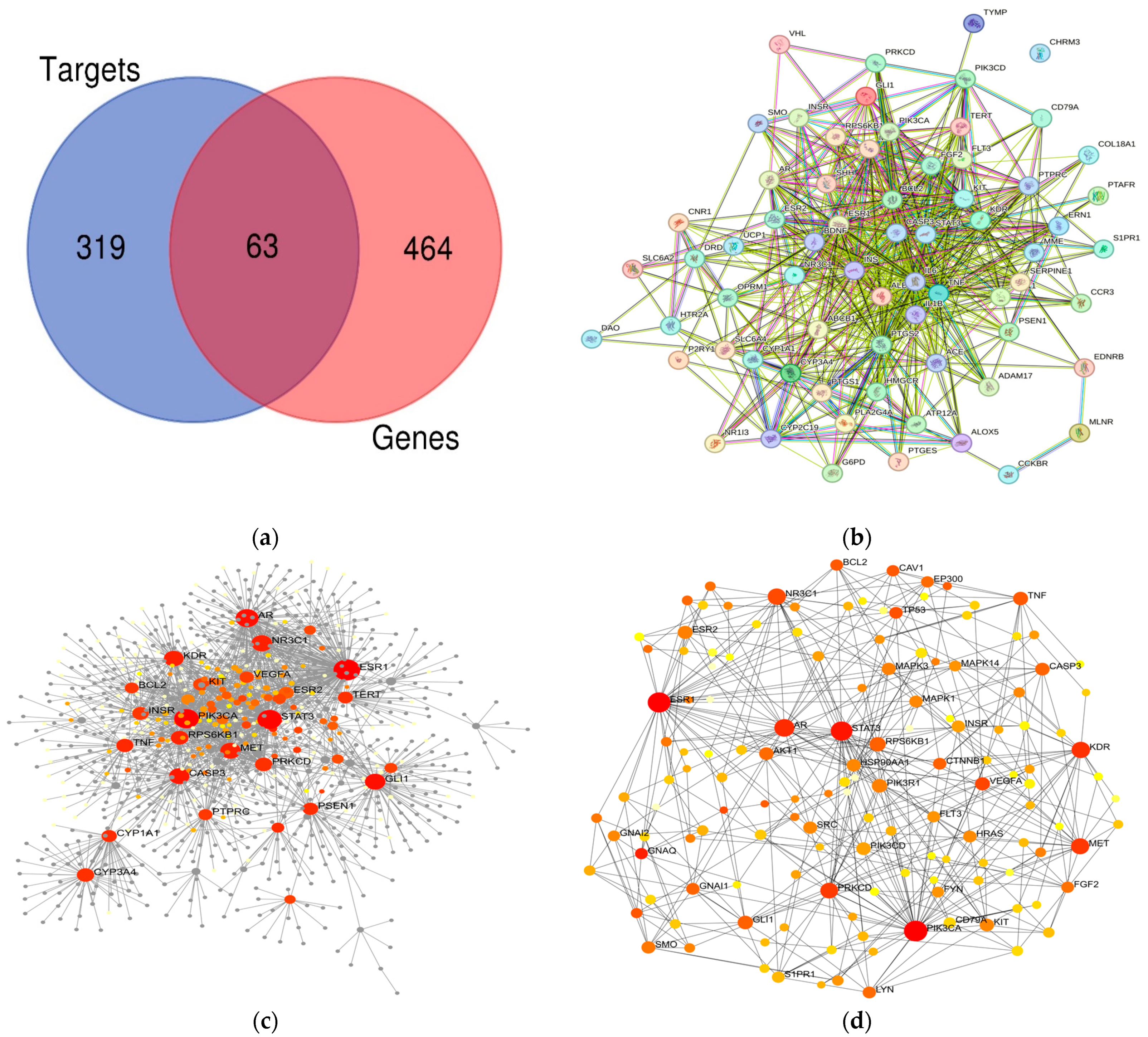
3.1. F. vesiculosus Bioactive Components and Target Gene Related to Disorder Identification

From the IMPPAT and KNapSAcK databases, were chosen following searching, filtering, and duplication removal (Table 1).
The current study also included the marine component fucodan, which boasts a high yield and a variety of pharmacological activities despite its DL value of −1.63. Though the DL and OB for fucosterol were higher than the set limits, other phytocompounds were of pharmacological interest and were therefore considered for the study. We carry out the difficult phase of drug discovery, known as ADMET analysis, using the SwissADE tool to assess various pharmacokinetic parameters that forecast ADE and the toxicity (T) of the most highly significant novel therapeutic phytocomponents. The ADMET sketch showed that none of the top-ranked fractions have any negative effects, suggesting that the potent components have pharmacokinetic properties (Table 2). The ADMET properties of potent medications for a variety of paradigms, such as GI absorption, P-glycoprotein substrates, BBB penetration, and various cytochrome inhibitors, such as CYP1A2, CYP2C19, CYP2C9, CYP2D6, and CYP3A4, have produced encouraging outcomes that significantly substantiate the potency of these fractions. Log Kp values, which penetrate the stratum corneum through intercellular, appendageal, or transcellular channels, determine the size and chemo-physical characteristics of the skin. Unknown processes contribute to gastric and intestinal disorders. We found high GI absorption of fucoidan, suggesting its potential to induce intestinal immunity by stimulating pattern recognition receptor signaling pathways that regulate epithelial cell proliferation, leading to the production of certain cytokines or chemokines. Fucoidan, an immunostimulant candidate, may improve host intestinal immunity, considering its immunological role as a functional supplement. While fucoidan functions as an anti-cancer drug, prolonged use has demonstrated its carcinogenic effects. Fucoidan inhibits metastasis by preventing the adhesion of tumor cells to the extracellular matrix, whereas other phytocompounds are free from any type of toxicity. All the phytocompounds demonstrated inactivity against various cytochrome inhibitors, suggesting that other drugs did not hinder their absorption in the body. Scholars have proposed several theories to elucidate the underlying process. Therefore, researchers explored the molecular mechanism of bioactive components using network pharmacology for the treatment of gastrointestinal disorders.
Meanwhile, we gathered a total of 382 target genes that interacted with those expected components; we retrieved 539 of these from the STP database and 26 from the STITCH after removing duplication. Furthermore, we gathered 527 disease genes related to the gastric-intestinal tract, which includes 358 found in GeneCards and 393 targets in OMIM, after the removal of duplicate genes. Hence, through the Venn diagram, we found 319 unique compound targets and 464 disease genes. We considered the revealed targets and compounds to be important factors in analyzing the gut disorder’s treatment outcomes. Finally, 63 intersection genes or overlapping genes of targets identified as co-expressed targets, also known as candidate genes, would help with more research into how F. vesiculosus treats gastrointestinal disorders (Figure 1a). Identifying the exact cellular and molecular locations of the genes is required to identify a protein at the transcriptional level. Most of the gastro-intestinal target genes were expressed in smooth muscle (expressed value above 6), while others were expressed in HepG2 (around 2.8) and the small intestine (around 2.6) (Figure S1). At the cellular level, the Descartes fetal cerebellum microglia expressed the majority of the genes (above five), while the muraro pancreas mesenchymal stroma cell and the aizarani liver C23 kupffer cells 3 (near to 4) also showed high expression (above four). We thoroughly distributed the genes in the other gastric and intestine cells (Figure S2). Lastly, our analysis revealed that various diseases and carcinomas expressed the majority of these genes (Figure S3).

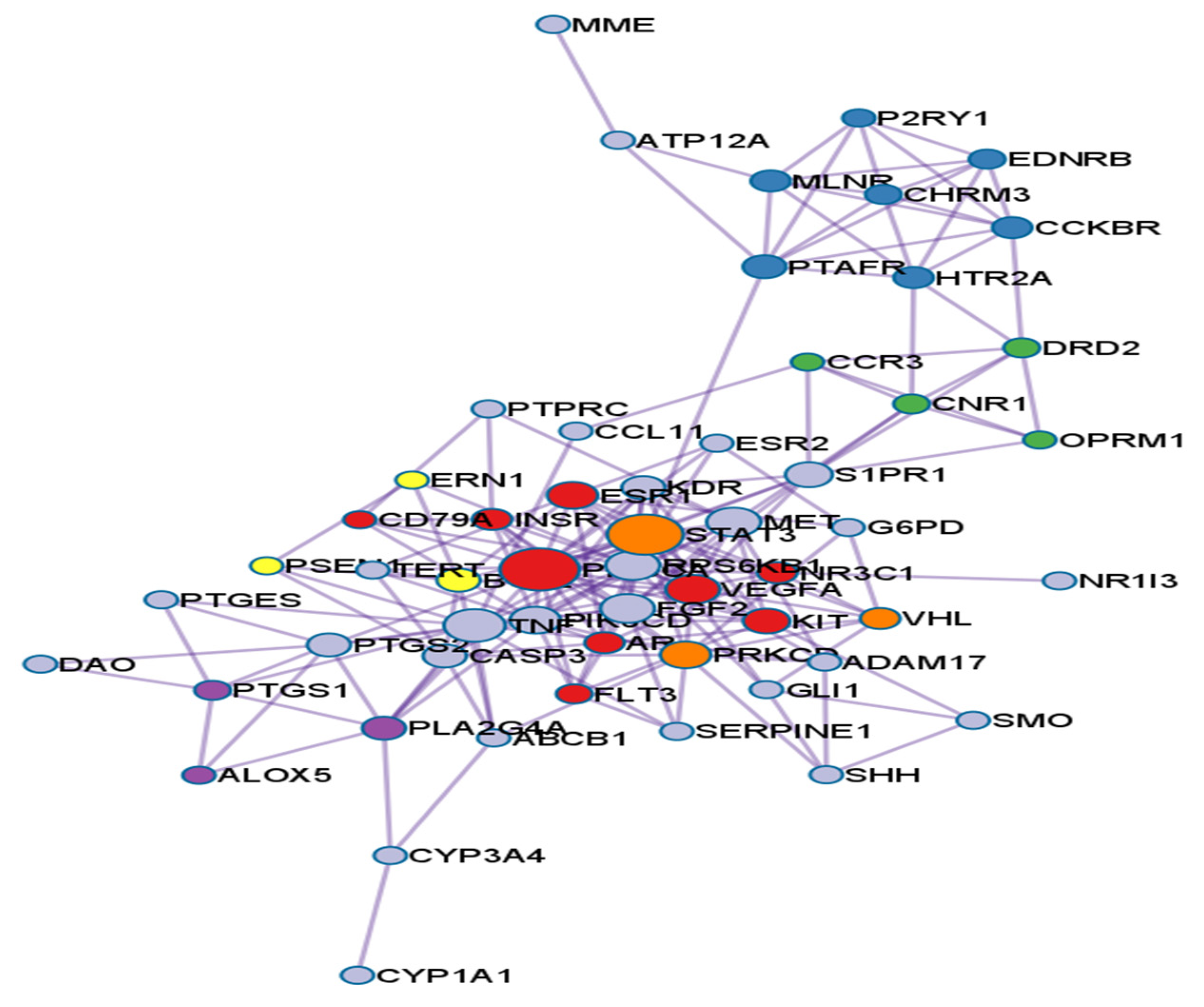
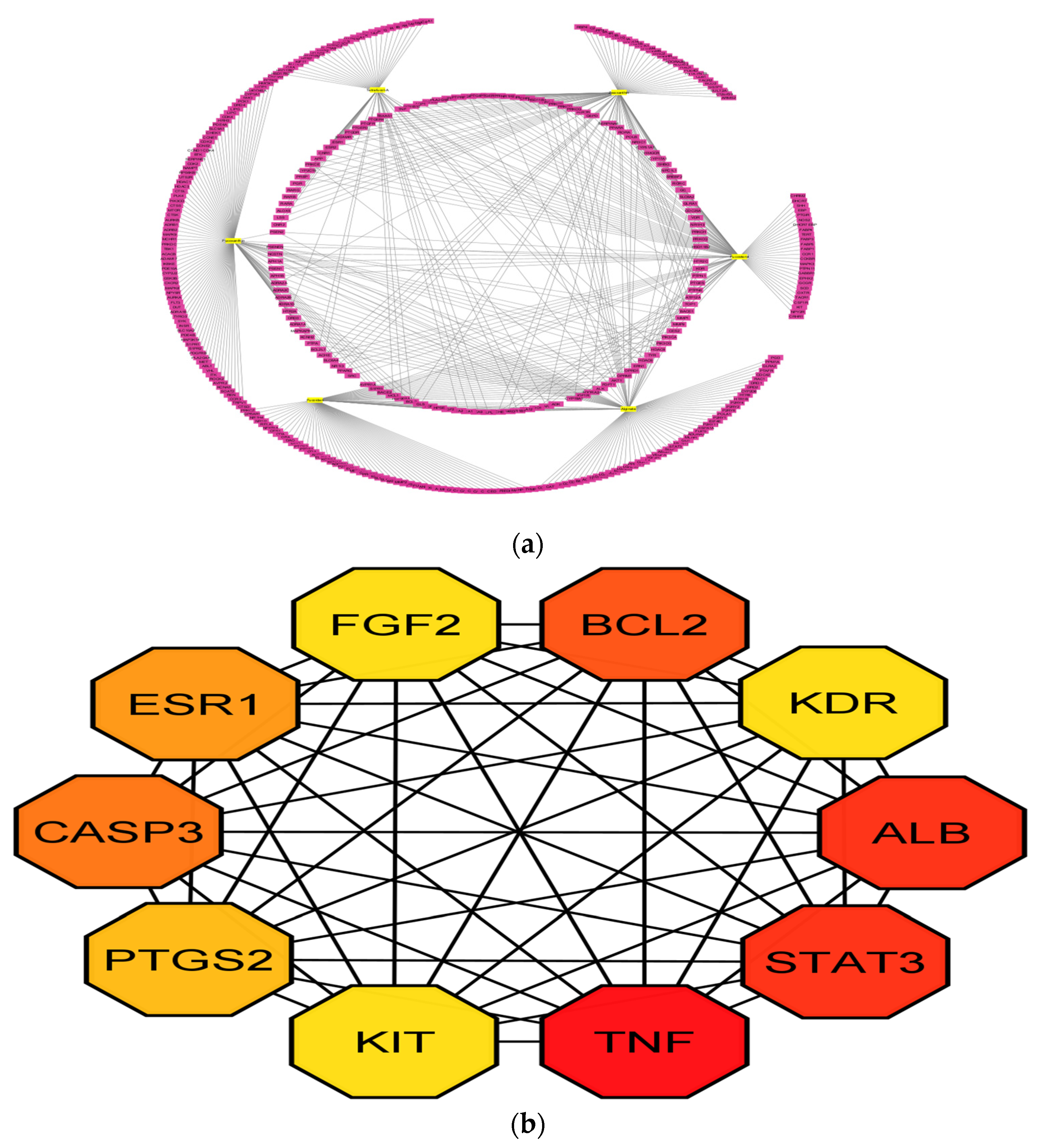
3.2. PPI Network Analysis

We selected the 63 potential genes using STITCH, resulting in a first-constructed PPI (Figure 1b) network with 1049 nodes, 1717 edges, and 44 isolated target seed genes (Figure 1c). Furthermore, we built a minimal network of protein-protein interaction clusters (Figure 1d) with 141 nodes and 435 edges. The network analysis identified only genes with greater degree and betweenness values as the primary targets of F. vesiculosus for gastrointestinal disorders. We ultimately gathered 44 important targets for pathway enrichment analysis, with ESR1, PIK3CA, and STAT3 ranking highest (Table 3). The highest degree by ESR1, found to be 168, indicated the connection of edges with the node, i.e., nodes are the main proteins connected to sub-proteins in the network. Further high betweenness of the protein gives the power to have more control over the network, and hence it will help to pass more information through that node, as easily shown in Figure 1d.

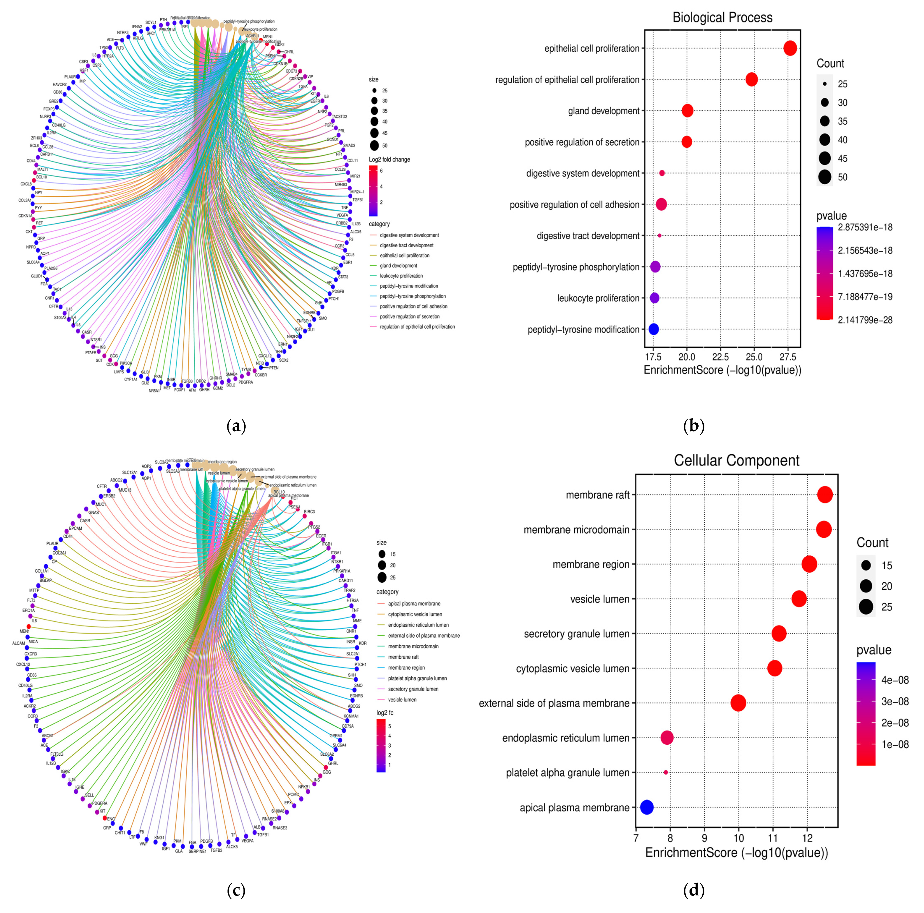
3.3. Functional Annotation and GO Analysis

The pharmacological role of each target gene of F. vesiculosus for gastrointestinal disorders was examined using functional annotation and enrichment analysis. We found these genes enriched in 2737 biological process (BP) terms, 200 molecular functions (MF) terms, and 95 cellular components (CC) terms for gastric and gastrointestinal disorders. The BPs were obtained using the SR plot with 63 interaction genes, as observed in Figure 2a. The BPs significantly enriched the top three terms, gland development (GO: 0048732), epithelial cell proliferation (GO: 0050673), and regulation of epithelial cell proliferation (GO: 0050678), with a p-value of less than 0.01. The primary BPs include peptidyl-tyrosine phosphorylation and modification, leukocyte proliferation, gland development, positive regulation of secretion, digestive system development, positive regulation of cell adhesion, and digestive tract development (Figure 2a). The size of the dot in the bubble chart indicates the number of target genes in the related function pathway, and the enrichment mentions the ratio of the numerous target genes to all annotated functional genes in the pharmacological pathway (Figure 2b). The highest enrichment was found in the perinuclear region of the membrane region (GO: 0098589), the membrane raft (GO: 0045121), and the membrane microdomain (GO: 0098857) of the CCs. The primary components of the CCs were the apical plasma membrane, the platelet alpha granule lumen, the endoplasmic reticulum lumen, the secretory granule lumen, the external side of the plasma membrane, the membrane area, the membrane raft, the cytoplasmic vesicle lumen, the membrane microdomain, and the vesicle lumen (Figure 2c,d). The MFs are involved in signaling receptor activator activity (GO: 0030546), receptor-ligand activity (GO: 0048018), and hormone activity (GO: 0005179). GO: 0005179. The MFs were involved in signaling receptor activator activity, cytokine activity, hormone activity, G protein-coupled receptor binding, protease binding, neuropeptide hormone activity, growth factor binding, and finally growth factor receptor binding, as could be seen in Figure 2e,f. Figure 3 displays the enrichment of BP among the top targets. According to GO functional analysis, targets were associated with gastrointestinal treatment, epithelial cell proliferation, gland development, positive secretion regulation, digestive tract development, and so on, as shown in Table 4.

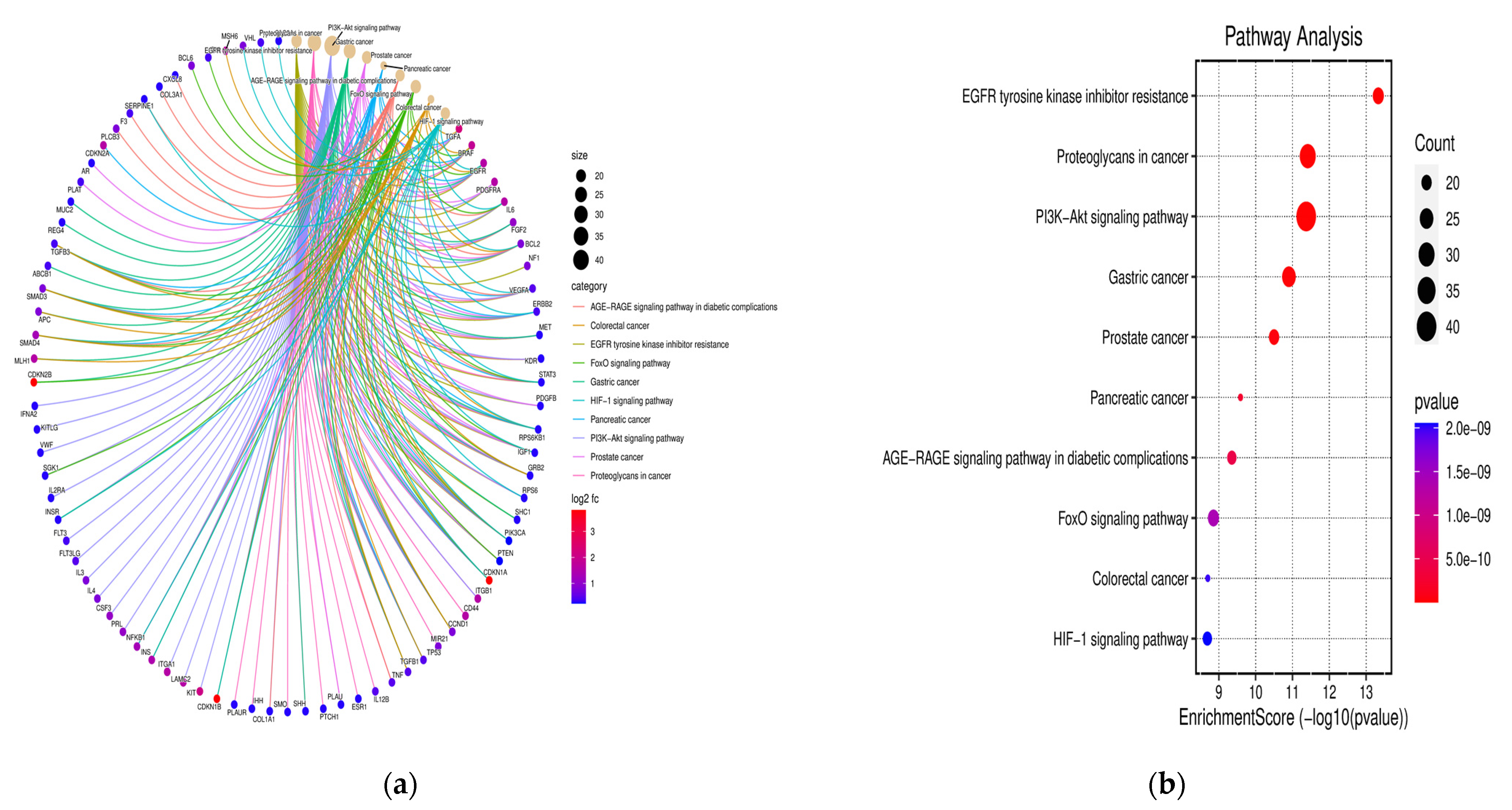
3.4. KEGG Pathway Examination

The KEGG analysis highlights the therapeutic potential of Fucus vesiculosus in treating gastrointestinal disorders through its action on various signaling pathways. The study identified ten significant pathways, with p-values less than 0.01, based on 63 key targets for further investigation. The top three pathways identified were the PI3K-Akt signaling pathway (hsa04151), proteoglycans in cancer (hsa05205), and EGFR tyrosine kinase inhibitor resistance (hsa01521). The PI3K-Akt signaling pathway plays a crucial role in regulating cell growth, proliferation, and survival. In the context of gastrointestinal disorders, modulation of this pathway by F. vesiculosus can help manage inflammation and promote mucosal healing. This pathway’s involvement in cellular processes underscores its potential for therapeutic intervention in gastrointestinal diseases, where epithelial integrity and cellular homeostasis are compromised.
Proteoglycans in cancer, another significant pathway, are essential for maintaining the structural integrity of tissues and regulating cellular functions. The involvement of this pathway suggests that F. vesiculosus may exert its therapeutic effects by influencing the extracellular matrix components, which are crucial in both cancer progression and tissue repair mechanisms in gastrointestinal disorders. The EGFR tyrosine kinase inhibitor resistance pathway highlights the role of F. vesiculosus in potentially overcoming drug resistance in gastrointestinal cancers. EGFR signaling is critical for cell proliferation and survival, and its dysregulation is common in various cancers. By targeting this pathway, F. vesiculosus may enhance the efficacy of existing treatments and prevent resistance development, thus offering a complementary approach to cancer therapy.
Additionally, the KEGG pathway analysis indicated that the selected targets of F. vesiculosus were significantly enriched in various other pathways, including gastric cancer, prostate cancer, the AGE-RAGE signaling pathway in diabetic complications, pancreatic cancer, colorectal cancer, FoxO signaling, and the HIF-1 signaling pathway. These pathways are involved in a wide range of biological processes, from metabolic regulation and stress response to tumor growth and hypoxia adaptation. The KEGG analysis provides valuable insights into the multiple mechanisms through which F. vesiculosus can exert its therapeutic effects on gastrointestinal disorders. By modulating key signaling pathways, this marine algae shows promise in managing inflammation, enhancing tissue repair, and potentially improving cancer treatment outcomes. Further research is needed to fully understand the molecular interactions and clinical implications of these findings.
Among these, EGFR tyrosine kinase inhibitor resistance was shown to be a group of significant critical pathways with the lowest p value and the highest target enrichment. KEGG pathway analysis revealed that TNF, STAT3, PIK3G, KDR, IL3, IL4, SERPINE1, VWF, SMO, ESR1, MET, and many more were significantly enriched genes involved in various pathways, like the diabetic complication AGE-RAGE signaling pathway, gastric cancer, pancreatic cancer, and the signaling pathway of HIF-1, as shown in Figure 4a,b.

3.5. Compound-Protein/Target Interaction (CPI) Network

Figure 5 displays the component-protein network, which consisted of six compounds and 63 targets. Among these targets, the gastrointestinal target CHRM3 did not interact with other targets that consisted of 63 nodes and 421 edges (Figure 1b). Several F. vesiculosus components act upon at least one target gene, according to the network analysis. Moreover, two or more active components control many selected gene targets, and each pathway associated with gastrointestinal disorders may include at least ten genes. We then developed the network of targets with overlapped PPI genes using Cytoscape, as shown in Figure 5a. The top 10 genes were selected from Cytoscape cytohubba (Figure 5b). We observed a high degree of interaction between TNF (46), STAT3 (39), ALB (39), BCL2 (36), CASP3 (34), ESR1 (33), PTGS2 (29), KDR (25), FGF2 (25), and KIT (25), indicating that these genes are hub targets. The PPI identified the target gene network with a moderate confidence score (0.50). Furthermore, the developed PPI reveals a partial physiological relationship between the proteins and all gastro-intestinal diseases, along with a significantly higher number of interactions than initially expected. The compounds Fucoxanthin, Fucoidan, Fucosterol, and Fucosterol were the ones that interacted with targeted genes the most strongly in the CPI analysis. These were followed by Tetrafucol A, Sodium Alginate, and Zeaxanthin.
The results showed that genes such as TNF, STAT3, ALB, BCL2, CASP3, ESR1, PTGS2, KDR, FGF2, and FIT are the most prominent functional genes that interact with marine secondary metabolites of various origins. These include Fucoidan and Sodium Alginate (polysaccharide), Fucoxanthin (xanthophyl carotenoid), Fucosterol (sterol derivative), Tetrafucol A (phlorotannin), and Zeaxanthin (carotenoid). These genes are involved in positive regulation of secretion, digestive system development, peptidyl-tyrosine phosphorylation, and activation of proteins.

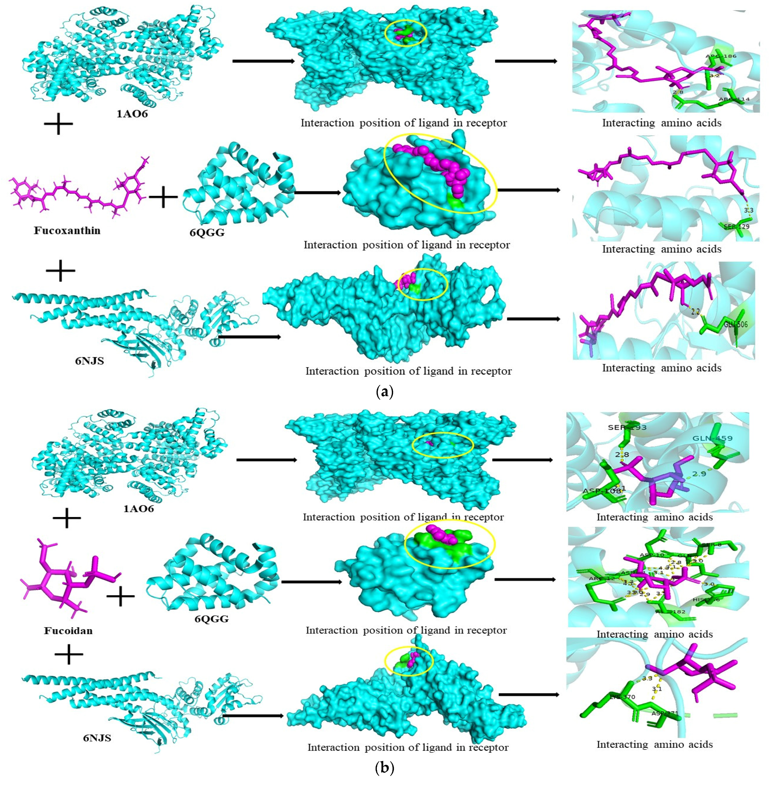
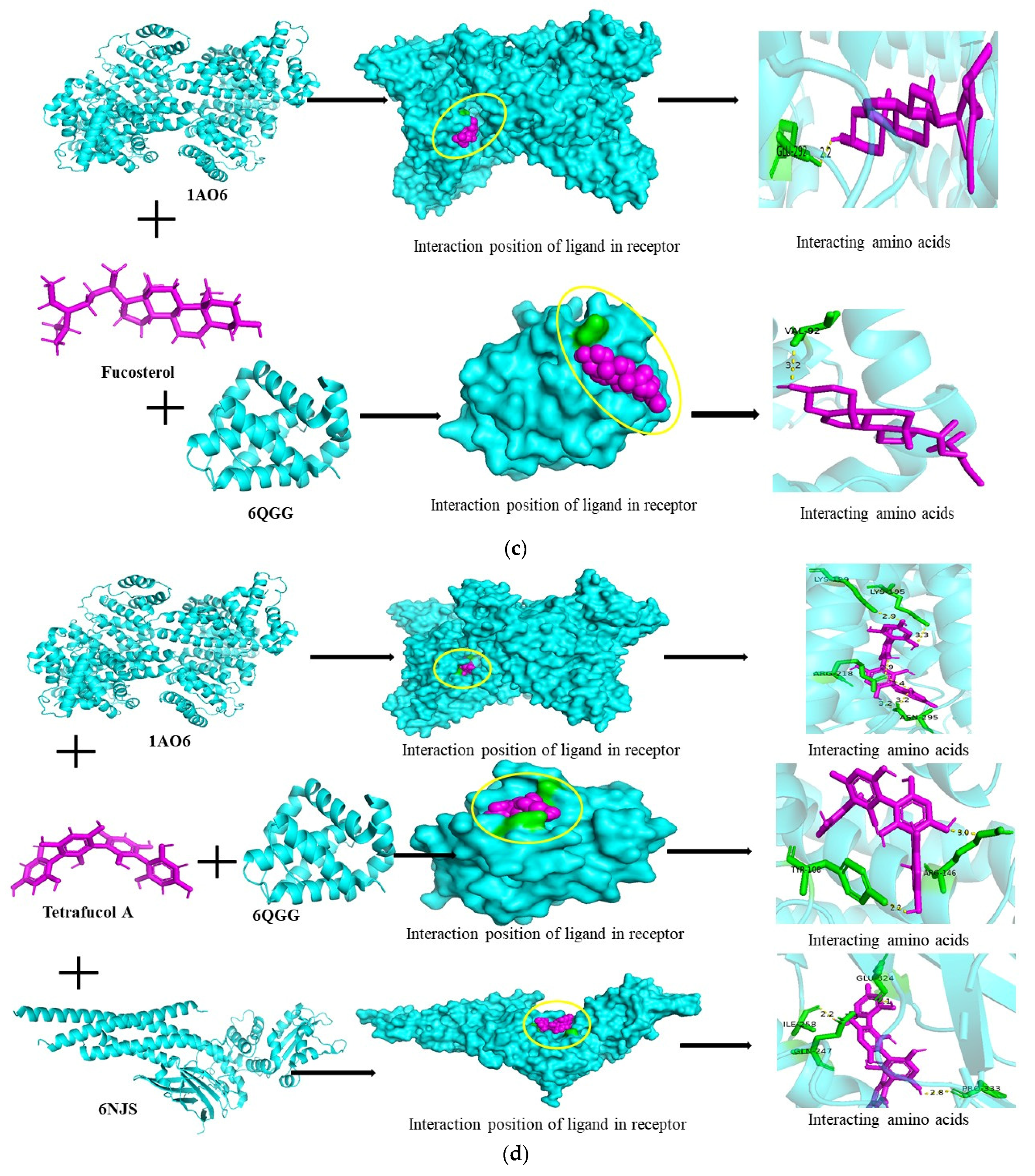
3.6. Molecular Docking Studies

Recently, research topics like gut disorders have been a major challenge for scientists. To address challenges like the lack of efficacy and the development of resistance to single-targeted drugs, drug discovery frequently necessitates a system-level pharmacology approach. Network pharmacology techniques are created and used more frequently to identify new therapeutic possibilities and repurpose current medications. To assess the pharmacological significance of the primary target discovered using microbiome analysis, we conducted a network pharmacology inquiry. Using data-driven analysis, we investigated the interaction of gastrointestinal disorders with gut health. Malnutrition, hypertension or hypotension, microinflammation, immune system dysbiosis, and numerous oxidative stresses are frequently associated with gut health and can all be treated with phytocompounds of F. vesiculosus.
We used molecular docking to assess the binding similarity between the six important targets inspected from the PPI construction and the top five disease gene targets selected from the disease gene-target constructed network. All the key genes showed strong binding affinities with the key targets, as confirmed by the data. Every docking module’s minimal binding energy was noted in Table 5, and Figure 6 displays the docking modules whose minimum binding energies were up to −8.3 kcal/mol.
The interactions between TNF, STAT3, KDR, FGF2, and KIT on the surface of gastrointestinal receptors play a crucial role in initiating adhesion and tethering processes to the exposed sub-endothelium, promoting cell proliferation and inflammatory responses. These molecules are integral to the signaling pathways that regulate various physiological processes, including inflammation and acid secretion in the stomach. The flow of bodily fluids significantly influences these interactions, triggering signaling cascades that regulate inflammation, enhance stomach acid secretion, and fortify focal adhesion.
TNF (Tumor Necrosis Factor) is a key inflammatory mediator that can activate multiple signaling pathways, including NF-κB and MAPK, leading to the expression of pro-inflammatory genes and the secretion of stomach acid. STAT3 (Signal Transducer and Activator of Transcription 3) is involved in cell growth and survival, and its activation is crucial for the inflammatory response and cellular proliferation. KDR (Kinase Insert Domain Receptor) and FGF2 (Fibroblast Growth Factor 2) play essential roles in angiogenesis and tissue regeneration, contributing to the healing and proliferation of the gastrointestinal epithelium. KIT, a receptor tyrosine kinase, is involved in cell differentiation and proliferation, particularly in the gastrointestinal tract.
The interactions of these agonists with their respective receptors, such as ALB (Albumin), STAT3, and BCL2 (B-cell lymphoma 2), initiate inside-out signaling mechanisms. This signaling enhances the affinity and activity of BCL2 and STAT3 through conformational changes, leading to increased cell survival and proliferation. BCL2 is particularly important in regulating apoptosis, and its upregulation can prevent cell death, thereby supporting tissue regeneration and maintaining gastrointestinal integrity.
In contrast, the coordinated actions of TNF, STAT3, KDR, FGF2, and KIT, along with their interactions with receptors like ALB, STAT3, and BCL2, are crucial for managing inflammation, promoting stomach acid secretion, and strengthening cellular adhesion in the gastrointestinal tract. These processes are essential for maintaining gastrointestinal health and responding to injury or infection. Understanding these molecular interactions offers insights into potential therapeutic targets for gastrointestinal disorders characterized by inflammation and impaired cell adhesion and proliferation.
According to Chen et al. [27], inflammation is a complex protective pharmacological reaction that the body uses to combat irritants, pathogens, and damaged cells. Over time, the inflammatory response may result in internal scarring, tissue death, and damage to DNA [28]. Leukotrienes and prostaglandins, as lipid mediators and cytokines, control the inflammatory responses [29]. Fucoxanthin promoted G2/M phase apoptosis in the MGC-803 cell line and cell-cycle arrest in gastric cancer by downregulating STAT3, Bcl-2, and cyclin B1 [30], as well as further upregulating CASP-3 [31]. Zhu et al. [32] discovered that fucoxanthin cut CASP-3, lowered Bcl-2, and ultimately lowered the viability of SGC-7901 cells in a different study. These results demonstrate the importance of fucoxanthin as an anti-gastric cancer treatment. Researchers have also demonstrated the impact of fucoxanthin on colon cancer, causing apoptosis, DNA fragmentation, G0/G1 arrest, and downregulating Bcl-2 and upregulating p21 (Table 4; Figure 6a) [33].
It downregulated Bcl-2, raised Bax, and triggered apoptosis in prostate cancer through G1 cell cycle arrest [34]. It was shown that fucoxanthin could lower the levels of pro-inflammatory mediators like PGE2, NO, TNF-α, IL-1β, and IL-6 by stopping NF-κB activation and MAPK phosphorylation. With molecular docking, it was clear that ALB (−8.3 kcal/mol) interacting with ARG-186/114 and Bcl-2 (−8.10 kcal/mol) interacting with SER-129 had a good bond, as seen in Figure 6a. When interacting with GLU-506, fucoxanthin also showed a favorable interaction with STAT3, with a binding energy of −6.8 kcal/mol [35,36].
Constipation, one of the most prevalent chronic gastrointestinal diseases, has greatly impacted the physical and mental health of patients [16]. The membrane protein of eggshells made of chitosan and fucoidan nanoparticles lowered the levels of NO, IL-6, and TNF-α in Caco-2 cell lines [37]. Fluorescein isothiocyanate-dextran increased the permeability of paracellular structures, highlighting the importance of boosting fucoidan bioavailability to maximize its impact on immunity. Fucoidan has been shown in other mouse studies to hinder cryptosporidiosis by decreasing the Cryptosporidium parvumoocysts adherence in the epithelial cells of the intestine, promoting proliferation of T cells (CD4+ and CD8 T+), and increasing the levels of IL-6, TNF-α, IL-12, CD40, CD86, and CD80 in the spleen.
IL-6 is involved in the pathogenesis of various gut disorders, including inflammatory bowel disease (IBD). It contributes to inflammation by promoting the differentiation of T cells and the production of other inflammatory cytokines. Targeting IL-6 with specific inhibitors, such as tocilizumab, can reduce inflammation and improve symptoms in IBD patients. TNF-α is a central mediator of inflammation in gut disorders like Crohn’s disease and ulcerative colitis. It promotes the release of other pro-inflammatory cytokines and the recruitment of immune cells to the gut. Anti-TNF therapies, such as infliximab and adalimumab, have been effective in reducing inflammation and maintaining remission in IBD.
IL-12 is involved in the differentiation of naive T cells into Th1 cells, which produce interferon-gamma and perpetuate inflammation. Elevated IL-12 levels are associated with severe gut inflammation. Therapies targeting the IL-12/IL-23 pathway, like ustekinumab, have shown efficacy in treating Crohn’s disease by reducing inflammation. CD40, a co-stimulatory protein found on antigen-presenting cells, plays a role in T cell activation and inflammation. In gut disorders, CD40 signaling contributes to chronic inflammation. Blocking CD40-CD40L interactions may reduce inflammation and is a potential therapeutic strategy. CD86 and CD80 are co-stimulatory molecules expressed on antigen-presenting cells. They interact with CD28 and CTLA-4 on T cells to regulate immune responses. In gut disorders, these molecules are upregulated, leading to increased T cell activation and inflammation. Therapeutics like abatacept, which modulates CD80/CD86 interactions, can potentially reduce T cell-mediated inflammation in IBD. Collectively, targeting these molecules offers therapeutic strategies for managing gut disorders by modulating immune responses and reducing inflammation.
These findings suggest that fucoidan might accelerate Th1 immune function. Dietary fucoidan has immunomodulatory and anti-inflammatory properties that can also improve the functioning of the gut immune system. Moreover, fucoidan helps treat conditions linked to inflammation, such as inflammatory bowel disorders. According to a recent study, fucoidan may have a protective effect on colon health since it was shown to reduce TNF-α, IL-10, IL-6, and IL-1 levels in the colon cells of mice when given after antibiotic therapy [38].
According to Apostolova et al. [39], the most plausible mechanistic approach to date is the downregulation of NF-B and MAPK signaling pathways in immunology and structural cell effects, such as epithelial cells. This leads to a drop in pro-inflammatory and an increase in anti-inflammatory cytokines (Figure 6b). However, the most prevalent intestinal condition is IBD, consisting of Crohn’s disease (CD) and ulcerative colitis (UC). Multiple inflammation reactions in the intestine that have no apparent reason are the hallmarks of IBD, which is becoming more prominent worldwide. Colorectal cancer, the third most common cancer globally, is another intestinal disease that significantly jeopardizes human health. Current research suggests that fucoidan could potentially serve as an anticancer drug in the future. Though fucoidan’s molecular size affects the absorption by intestine epithelial cells, through the Ras/Raf/ERK pathway, fucoidan stops the insulin-like growth factor (IRF)-I receptor from working. This controls cell survival and the biological cycle [40]. Docking revealed that fucoidan exhibited strong binding with ALB (−5.8 kcal/mL), Bcl-2 (−5.5 kcal/mol), and STAT3 (−4.9 kcal/mol). We also conducted a binding study of fucoidan with TNF and CASP-3 but found no interaction (Figure 6b).
Fucosterol, a bioactive compound found in marine algae, has demonstrated significant anti-inflammatory effects by inhibiting the expression of inducible nitric oxide synthase (iNOS), interleukin-6 (IL-6), and tumor necrosis factor-alpha (TNF-α). It also suppresses the NF-κB and p38 MAPK pathways, key regulators of inflammation. As an anti-atopic agent, fucosterol inhibits IL-4 and TNF-α while increasing the secretion of interferon-gamma (IFN-γ), highlighting its role in modulating immune responses.
Additionally, fucosterol exhibits strong antiproliferative effects by regulating signaling proteins, including MAPK, NF-κB, and the caspase (CASP) family. This regulation is crucial in controlling cell growth and apoptosis. Previous studies have shown that fucosterol downregulates apoptotic pathways mediated by MAPK, STAT, and NF-κB, leading to the induction of apoptosis in cancer cells. By modulating these pathways, fucosterol can inhibit cancer cell proliferation and promote cell death.
Moreover, fucosterol’s ability to inhibit inflammatory cytokines and signaling pathways, along with its anti-proliferative and pro-apoptotic effects, makes it a promising candidate for the development of new therapeutic strategies for inflammatory diseases and cancer. Further research is warranted to fully understand its mechanisms and potential clinical applications [41,42].
Another effect was observed in glial cells, where excessive activation of cells has shown pro-inflammatory action that results in neurodegeneration due to synaptic dysfunction, loss of synapses, and neuronal death. Pro-inflammatory mediators (iNOS, IL-1, IL-6, TNF-α, IL-2, COX2, ROS/RNS, MMPs) and chemokines were overproduced as a result of activated microglia [43]. Numerous intracellular pathways, such as NF-κB, MAPKs, STAT, and PI3K, regulate signal transduction molecules, enhancing factors, and enzymes. The PI3K/Akt cascade is activated by growth factors, LPS, and cytokines. Docking studies of fucosterol revealed strong binding to Bcl-2 (−6.8 kcal/mol) interacting with VAL-92, STAT3 (−6.2 kcal/mol), and ALB (−5.7 kcal/mol). These interactions suggest fucosterol’s potential in modulating these pathways, highlighting its therapeutic promise in regulating cellular processes and mitigating inflammation [44,45,46].
Tetrafucol A and sodium alginate have the potential to protect mucous membranes and hemostatis. Moreover, feces eliminate it, and its absorption is minimal; reports of adverse effects are rare. Tetrafucol A and sodium alginate have thus been employed extensively in the treatment of bleeding [47], nonerosive reflux disease, and stomach ulcers. In animal models, sodium alginate also prevents the gastrointestinal mucositis caused by methotrexate [48].
Sodium alginate and Tetrafucol A have been shown to have a mucoprotective effect by coating the GI tract’s surface and preventing bacterial translocation. Tetrafucol A, on the other hand, has demonstrated its ability to positively regulate peptidyl-tyrosine phosphorylation, inflammatory responses, and epithelial cell proliferation after interacting with STAT3.
Further interaction with Bcl-2 regulated lymphocytes, leukocytes, and activated T cells, leading to digestive tract development, as shown in Figure 4a and Table 4 [49].
In the investigation, they increased the expressions of beclin-1 and LC3, and then they cleaved CASP-3, downregulated Bcl-2 expression, and reduced the viability of the cells in SGC-7901 cells, as mentioned in Table 4 and Table 5. These medications are essential to the GI tract’s epithelium surface’s protection and functionality. The docking studies of Tetrafucol A and sodium alginate showed good binding with ALB (−8.3 and −6.0 kcal/mol, respectively), respectively, followed by STAT3 (−7.7 kcal/mL and −5.5 kcal/mol). Tetrafucol A also showed good binding with Bcl-2 (−7.5 kcal/mol) (Figure 6d,e). The strong local immune response draws in macrophages and lymphocytes and releases proinflammatory cytokines and inflammatory mediators thereafter [50]. According to Ying et al. [51], zeaxanthin prevents mesangial cell death caused by hyperglycemia by raising serine/threonine protein kinase levels that are phosphorylated, decreasing Bcl-2-associated X protein levels, and cleaving CASP-3 levels. Increased fluid and fiber intake is advised for those with Crohn’s and Colitis, but this is ineffective for IBD patients [52]. This is because of a decrease in colonic content volume, an increase in colonic concentration of toxic compounds, and mucosal exposition [53].
Zeaxanthin, a dietary carotenoid known for its antioxidant properties, has been shown to induce apoptosis in cancer cells through multiple pathways. One of the key mechanisms by which zeaxanthin promotes apoptosis is by altering the mitochondrial membrane potential. This disruption leads to the release of cytochrome C into the cytosol, a crucial step in the intrinsic apoptotic pathway. The increased cytochrome C expression facilitates the activation of Bax, a pro-apoptotic protein, and the cleavage of caspase-3 (cleaved-CASP-3) and poly (ADP-ribose) polymerase (cleaved-PARP). These cleaved forms are active in executing apoptosis, resulting in cell death. Simultaneously, zeaxanthin reduces the levels of anti-apoptotic proteins such as Bcl-2, pro-caspase-3 (pro-CASP-3), and pro-PARP, further tipping the balance towards cell death. The reduction in Bcl-2, a key regulator that prevents apoptosis, enhances the pro-apoptotic effects of zeaxanthin.
Moreover, zeaxanthin administration increases the levels of reactive oxygen species (ROS), phosphorylated p38 (p-p38), phosphorylated c-Jun N-terminal kinase (p-JNK), and I-κB, indicating activation of stress-related pathways. The elevated ROS levels contribute to oxidative stress, which can damage cellular components and trigger apoptosis. The phosphorylation of p38 and JNK, components of the mitogen-activated protein kinase (MAPK) pathway, further promotes apoptotic signaling. I-κB, an inhibitor of NF-κB, when increased, prevents the translocation of NF-κB to the nucleus, thereby inhibiting its anti-apoptotic signaling.
Conversely, zeaxanthin decreases the expression of phosphorylated extracellular signal-regulated kinase (p-ERK), signal transducer and activator of transcription 3 (STAT3), phosphorylated Akt (p-AKT), and NF-κB. The downregulation of these proteins, particularly p-ERK and p-AKT, which are often associated with cell survival and proliferation, shifts the cell towards apoptosis. STAT3 and NF-κB are transcription factors that promote cell survival and proliferation; their inhibition by zeaxanthin further enhances apoptotic processes.
Interestingly, the induction of apoptosis by zeaxanthin is attenuated by MAPK inhibitors, indicating that the MAPK pathway plays a critical role in zeaxanthin-mediated apoptosis. This suggests that zeaxanthin-induced apoptosis is at least partly dependent on the activation of the MAPK pathway.
In summary, zeaxanthin induces apoptosis in cancer cells through a multifaceted mechanism involving the disruption of mitochondrial function, modulation of apoptotic and anti-apoptotic proteins, and regulation of key signaling pathways such as MAPK, STAT3, and NF-κB. By influencing these pathways, zeaxanthin effectively promotes cell death and has potential therapeutic applications in cancer treatment. Further research is warranted to fully elucidate its mechanisms and optimize its use in clinical settings [54].

4. Conclusions

When it comes to treating diseases, marine medications are multi-ingredient and multi-target, and it might be challenging to pinpoint their pharmacodynamic components and molecular mechanisms. We found that six selected metabolites, along with three gene targets, were related to gastrointestinal disorders and gut health diseases. Even though fucoidan is one of the novel compounds in recent times that has shown multiple pharmacological actions. However, with our findings, it was found that fucoxanthin, tetrafucol A, and zeaxanthin could be useful in the treatment of gut health disorders and gastrointestinal diseases. Moreover, Fucoxanthin and tetrafucol A showed good interaction with Alb and Bcl2, possessing beneficial properties for improving gut health by treating gastric-intestinal issues. This study showed that by controlling several targets and many pathways with multiple components, F. vesiculosus can exhibit preventive effects against gastrointestinal disorders. With the advancement of network technology and bioinformatics, researchers have developed computer-aided identification strategies for drug-target interactions, such as NP and molecular docking, to study herbs with complex action mechanisms. They increase the likelihood of identifying the best possible drug candidates and significantly cut down on the time and expense of the initial experimental identification of drug-target interactions.

Supplementary Materials

The following supporting information can be downloaded at: https://www.mdpi.com/article/10.3390/scipharm92030049/s1.

Author Contributions

V.A. was responsible for conceptualization, methodology, validation, formal analysis, investigation, resources, data curation, writing—original draft preparation, and writing—review; U.B. and I.P. were responsible for editing and formatting and funding resources; and A.K. was responsible for formal analysis and validation. All authors have read and agreed to the published version of the manuscript.

Funding

The authors acknowledge the RSF grant 22-76-10049 support for the experimental and characterization of material for this manuscript.

Institutional Review Board Statement

Not applicable.

Informed Consent Statement

Not applicable.

Data Availability Statement

The datasets used and/or analyzed during the current study are available from the corresponding author upon reasonable request.

Acknowledgments

The authors acknowledge the Department of Food and Biotechnology, South Ural State University, Chelyabinsk, Russian Federation, and RSF grant 22-76-10049 for financial support.

Conflicts of Interest

The authors declare no conflicts of interest. The funders had no role in the design of the study; in the collection, analyses, or interpretation of data; in the writing of the manuscript; or in the decision to publish the results.

References

  1. Beitz, D.C. Physiological and metabolic systems important to animal growth: An overview. J. Anim. Sci. 1985, 61, 1–20. [Google Scholar] [CrossRef]
  2. Owens, F.N.; Dubeski, P.; Hanson, C.F. Factors that alter the growth and development of ruminants. J. Anim. Sci. 1993, 71, 3138–3150. [Google Scholar] [CrossRef] [PubMed]
  3. Martin, G.B.; Rodger, J.; Blache, D. Nutritional and environmental effects on reproduction in small ruminants. Reprod. Fertil. Dev. 2004, 16, 491–501. [Google Scholar] [CrossRef] [PubMed]
  4. Holdt, S.L.; Kraan, S. Bioactive compounds in seaweed: Functional food applications and legislation. J. Appl. Phycol. 2011, 23, 543–597. [Google Scholar] [CrossRef]
  5. Simpi, C.C.; Nagathan, C.V.; Karajgi, S.R.; Kalyane, N.V. Evaluation of marine brown algae Sargassum ilicifolium extract for analgesic and anti-inflammatory activity. Pharmacogn. Res. 2013, 5, 146–149. [Google Scholar] [CrossRef]
  6. Subramaniam, S.; Selvaduray, K.R.; Radhakrishnan, A.K. Bioactive compounds: Natural defense against cancer? Biomolecules 2019, 9, 758. [Google Scholar] [CrossRef]
  7. Lin, S.; Wang, Z.; Lam, K.L.; Zeng, S.; Tan, B.K.; Hu, J. Role of intestinal microecology in the regulation of energy metabolism by dietary polyphenols and their metabolites. Food Nutr. Res. 2019, 63, 1–12. [Google Scholar] [CrossRef]
  8. Fitton, J.H. Therapies from fucoidan; Multifunctional marine polymers. Mar. Drugs 2011, 9, 1731–1760. [Google Scholar] [CrossRef]
  9. Vo, T.S.; Kim, S.K. Fucoidans as a natural bioactive ingredient for functional foods. J. Funct. Foods 2013, 5, 16–27. [Google Scholar] [CrossRef]
  10. Li, B.; Lu, F.; Wei, X.J.; Zhao, R.X. Fucoidan: Structure and bioactivity. Molecules 2008, 13, 1671–1695. [Google Scholar] [CrossRef]
  11. Ikeguchi, M.; Saito, H.; Miki, Y.; Kimura, T. Effect of fucoidan dietary supplement on the chemotherapy treatment of patients with unresectable advanced gastric cancer. J. Cancer Ther. 2015, 6, 1020–1026. [Google Scholar] [CrossRef]
  12. Fitton, J.H.; Dell’Acqua, G.; Gardiner, V.A.; Karpiniec, S.S.; Stringer, D.N.; Davis, E. Topical benefits of two fucoidan-rich extracts from marine macroalgae. Cosmetics 2015, 2, 66–81. [Google Scholar] [CrossRef]
  13. Palanisamy, S.; Vinosha, M.; Marudhupandi, T.; Rajasekar, P.; Prabhu, N.M. Isolation of fucoidan from Sargassum polycystum brown algae: Structural characterization, in vitro antioxidant and anticancer activity. Int. J. Biol. Macromol. 2017, 102, 405–412. [Google Scholar] [CrossRef] [PubMed]
  14. Flannigan, K.L.; Geem, D.; Harusato, A.; Denning, T.L. Intestinal antigen-presenting cells Key regulators of immune homeostasis and inflammation. Am. J. Pathol. 2015, 185, 1809–1819. [Google Scholar] [CrossRef]
  15. Lebedev, K.A.; Poniakina, I.D. Immunophysiology of epithelial cells and pattern-recognition receptors. Fiziol. Cheloveka 2006, 32, 114–126. [Google Scholar] [CrossRef]
  16. Fukata, M.; Arditi, M. The role of pattern recognition receptors in intestinal inflammation. Mucosal Immunol. 2013, 6, 451–463. [Google Scholar] [CrossRef]
  17. Ikeda-Ohtsubo, W.; Lopez Nadal, A.; Zaccaria, E.; Iha, M.; Kitazawa, H.; Kleerebezem, M.; Brugman, S. Intestinal microbiota and immune modulation in zebrafish by fucoidan from Okinawa mozuku (Cladosiphon okamuranus). Front. Nutr. 2020, 7, 67. [Google Scholar] [CrossRef]
  18. Liu, S.; Wu, J.; Zhu, Y.; Zhou, W.; Liu, X.; Fu, C.; Ding, Z.; Xu, L.; Zhang, Y.; Meng, Z.; et al. Network pharmacology-based approach to investigate the mechanisms of Shenqi Fuzheng injection in the treatment of breast cancer. Eur. J. Integr. Med. 2020, 34, 101064. [Google Scholar] [CrossRef]
  19. Song, X.; Zhang, Y.; Dai, E.; Wang, L.; Du, H. Prediction of triptolide targets in rheumatoid arthritis using network pharmacology and molecular docking. Int. Immunopharmacol. 2020, 80, 106179. [Google Scholar] [CrossRef]
  20. Kim, S.K.; Lee, S.; Lee, M.K.; Lee, S. A systems pharmacology approach to investigate the mechanism of Oryeong-sanformula for the treatment of hypertension. J. Ethnopharmacol. 2019, 244, 112–129. [Google Scholar] [CrossRef]
  21. Cui, Q.; Zhang, Y.L.; Ma, Y.H.; Yu, H.Y.; Zhao, X.Z.; Zhang, L.H.; Ge, S.Q.; Zhang, G.W.; Qin, X.D. A network pharmacology approach to investigate the mechanism of Shuxuening injection in the treatment of ischemic stroke. J. Ethnopharmacol. 2020, 257, 112891. [Google Scholar] [CrossRef] [PubMed]
  22. Gfeller, D.; Grosdidier, A.; Wirth, M.; Daina, A.; Michielin, O.; Zoete, V. SwissTargetPrediction: A web server for target prediction of bioactive small molecules. Nucleic Acids Res. 2014, 42, W32–W38. [Google Scholar] [CrossRef] [PubMed]
  23. Szklarczyk, D.; Gable, A.L.; Lyon, D.; Junge, A.; Wyder, S.; Huerta-Cepas, J.; Simonovic, M.; Doncheva, N.T.; Morris, J.H.; Bork, P.; et al. STRING v11: Protein-protein association networks with increased coverage, supporting functional discovery in genome-wide experimental datasets. Nucleic Acids Res. 2019, 47, D607–D613. [Google Scholar] [CrossRef] [PubMed]
  24. Kohl, M.; Wiese, S.; Warscheid, B. Cytoscape: Software for visualization and analysis of biological networks (M)//data mining in proteomics. Methods Mol. Biol. 2011, 96, 291–303. [Google Scholar]
  25. Morris, G.M.; Huey, R.; Lindstrom, W.; Sanner, M.F.; Belew, R.K.; Goodsell, D.S.; Olson, A.J. AutoDock 4 and AutoDock Tools 4: Automated docking with selective receptor flexibility. J. Comput. Chem. 2009, 30, 2785–2791. [Google Scholar] [CrossRef]
  26. Shivanika, C.; Kumar, S.D.; Ragunathan, V.; Tiwari, P.; Sumitha, A.; Devi, P.B. Molecular docking, validation, dynamics simulations, and pharmacokinetic prediction of natural compounds against the SARS-CoV-2 main-protease. J. Biomol. Struct. Dyn. 2022, 40, 585–611. [Google Scholar] [CrossRef]
  27. Chen, L.; Deng, H.; Cui, H.; Fang, J.; Zuo, Z.; Deng, J.; Li, Y.; Wang, X.; Zhao, L. Inflammatory Responses and Inflammation-Associated Diseases in Organs. Oncotarget 2018, 9, 7204–7218. [Google Scholar] [CrossRef]
  28. Jounai, N.; Kobiyama, K.; Takeshita, F.; Ishii, K.J. Recognition of Damage-Associated Molecular Patterns Related to Nucleic Acids during Inflammation and Vaccination. Front. Cell. Infect. Microbiol. 2012, 2, 168. [Google Scholar] [CrossRef]
  29. Ross, J. The Biology of the Macrophage; ScienceOpen, Inc.: Burlington, MA, USA, 2002. [Google Scholar]
  30. Yu, R.X.; Hu, X.M.; Xu, S.Q.; Jiang, Z.J.; Yang, W. Effects of Fucoxanthin on Proliferation and Apoptosis in Human Gastric Adenocarcinoma MGC-803 Cells via JAK/STAT Signal Pathway. Eur. J. Pharmacol. 2011, 657, 10–19. [Google Scholar] [CrossRef]
  31. Yu, R.X.; Yu, R.T.; Liu, Z. Inhibition of Two Gastric Cancer Cell Lines Induced by Fucoxanthin Involves Downregulation of Mcl-1 and STAT3. Hum. Cell 2018, 31, 50–63. [Google Scholar] [CrossRef]
  32. Zhu, Y.; Cheng, J.; Min, Z.; Yin, T.; Zhang, R.; Zhang, W.; Hu, L.; Cui, Z.; Gao, C.; Xu, S.; et al. Effects of Fucoxanthin on Autophagy and Apoptosis in SGC-7901cells and the Mechanism. J. Cell. Biochem. 2018, 119, 7274–7284. [Google Scholar] [CrossRef] [PubMed]
  33. Das, S.K.; Hashimoto, T.; Shimizu, K.; Yoshida, T.; Sakai, T.; Sowa, Y.; Komoto, A.; Kanazawa, K. Fucoxanthin Induces Cell Cycle Arrest at G0/G1 Phase in Human Colon Carcinoma Cells through Up-Regulation of p21WAF1/Cip1. Biochim. Biophys. Acta 2005, 1726, 328–335. [Google Scholar] [CrossRef] [PubMed]
  34. Kotake-Nara, E.; Asai, A.; Nagao, A. Neoxanthin and Fucoxanthin Induce Apoptosis in PC-3 Human Prostate Cancer Cells. Cancer Lett. 2005, 220, 75–84. [Google Scholar] [CrossRef] [PubMed]
  35. Bteich, M. An overview of albumin and alpha-1-acid glycoprotein main characteristics: Highlighting the roles of amino acids in binding kinetics and molecular interactions. Heliyon 2019, 5, e02879. [Google Scholar] [CrossRef] [PubMed]
  36. Murray, J.B.; Davidson, J.; Chen, I.; Davis, B.; Dokurno, P.; Graham, C.J.; Harris, R.; Jordan, A.; Matassova, N.; Pedder, C.; et al. Establishing Drug Discovery and Identification of Hit Series for the Anti-apoptotic Proteins, Bcl-2 and Mcl-1. ACS Omega 2019, 4, 8892–8906. [Google Scholar] [CrossRef]
  37. Lee, M.C.; Huang, Y.C. Soluble eggshell membrane protein-loaded chitosan/fucoidan nanoparticles for treatment of defective intestinal epithelial cells. Int. J. Biol. Macromol. 2019, 131, 949–958. [Google Scholar] [CrossRef]
  38. Wang, L.; Ai, C.; Wen, C.; Qin, Y.; Liu, Z.; Wang, L.; Gong, Y.; Su, C.; Wang, Z.; Song, S. Fucoidan isolated from Ascophyllum nodosum alleviates gut microbiota dysbiosis and colonic inflammation in antibiotic-treated mice. Food Funct. 2020, 11, 5595–5606. [Google Scholar] [CrossRef]
  39. Apostolova, E.; Lukova, P.; Baldzhieva, A.; Katsarov, P.; Nikolova, M.; Iliev, I.; Peychev, L.; Trica, B.; Oancea, F.; Delattre, C.; et al. Immunomodulatory and Anti-Inflammatory Effects of Fucoidan: A Review. Polymers 2020, 12, 2338. [Google Scholar] [CrossRef]
  40. Kim, I.H.; Nam, T.J. Fucoidan downregulates insulin-like growth factor-I receptor levels in HT-29 human colon cancer cells. Oncol. Rep. 2018, 39, 1516–1522. [Google Scholar] [CrossRef]
  41. Bandaranayake, W.M. The nature and role of pigments of marine invertebrates. Nat. Prod Rep. 2006, 23, 223–255. [Google Scholar] [CrossRef]
  42. Mondal, A.; Bose, S.; Banerjee, S.; Patra, J.K.; Malik, J.; Mandal, S.K.; Kilpatrick, K.L.; Das, G.; Kerry, R.G.; Fimognari, C.; et al. Marine cyanobacteria and microalgae metabolites—A rich source of potential anticancer drugs. Mar. Drugs 2020, 18, 476. [Google Scholar] [CrossRef] [PubMed]
  43. Zeng, W.X.; Han, Y.L.; Zhu, G.F.; Huang, L.Q.; Deng, Y.Y.; Wang, Q.S.; Jiang, W.Q.; Wen, M.Y.; Han, Q.P.; Xie, D.; et al. Hypertonic saline attenuates expression of Notch signaling and proinflammatory mediators in activated microglia in experimentally induced cerebral ischemia and hypoxic BV-2 microglia. BMC Neurosci. 2017, 18, 32. [Google Scholar] [CrossRef] [PubMed]
  44. Shabab, T.; Khanabdali, R.; Moghadamtousi, S.Z.; Kadir, H.A.; Mohan, G. Neuroinflammation pathways: A general review. Int. J. Neurosci. 2017, 127, 624–633. [Google Scholar] [CrossRef] [PubMed]
  45. Teismann, P.; Sathe, K.; Bierhaus, A.; Leng, L.; Martin, H.L.; Bucala, R.; Weigle, B.; Nawroth, P.P.; Schulz, J.B. Receptor for advanced glycation endproducts (RAGE) deficiency protects against MPTP toxicity. Neurobiol. Aging 2012, 33, 2478–2490. [Google Scholar] [CrossRef] [PubMed]
  46. Bai, L.; Zhou, H.; Xu, R.; Zhao, Y.; Chinnaswamy, K.; McEachern, D.; Chen, J.; Yang, C.Y.; Liu, Z.; Wang, M.; et al. A Potent and Selective Small-Molecule Degrader of STAT3 Achieves Complete Tumor Regression In Vivo. Cancer Cell 2019, 36, 498–511.e17. [Google Scholar] [CrossRef]
  47. Diago, K.; Yamada, C.; Yamaji, M.; Nakagiri, N.; Okada, M.; Komiya, H.; Horiuchi, Y.; Miyazato, T. Pharmacological studies of sodium alginate. V. Effect of sodium alginate on platelet aggregation. Yakugaku Zasshi 1985, 105, 171–182. [Google Scholar] [CrossRef]
  48. Yamamoto, A.; Itoh, T.; Nasu, R.; Kajiwara, E.; Nishida, R. Sodium alginate inhibits methotrexate-induced gastrointestinal mucositis in rats. Biol. Pharm. Bull. 2013, 36, 1528–1534. [Google Scholar] [CrossRef]
  49. Yamamoto, A.; Itoh, T.; Nasu, R.; Nishida, R. Sodium alginate ameliorates indomethacin-induced gastrointestinal mucosal injury via inhibiting translocation in rats. World J. Gastroenterol. 2014, 20, 2641–2652. [Google Scholar] [CrossRef]
  50. Guan, Q.; Zhang, J. Recent advances: The imbalance of cytokines in the pathogenesis of inflammatory bowel disease. Mediat. Inflamm. 2017, 2017, 4810258. [Google Scholar] [CrossRef]
  51. Ying, C.; Chen, L.; Wang, S.; Mao, Y.; Ling, H.; Li, W.; Zhou, X. Zeaxanthin ameliorates high glucose-induced mesangial cell apoptosis through inhibiting oxidative stress via activating AKT signalling-pathway. Biomed. Pharmacother. 2017, 90, 796–805. [Google Scholar] [CrossRef]
  52. Reif, S.; Klein, I.; Lubin, F.; Farbstein, M.; Hallak, A.; Gilat, T. Pre-illness dietary factors in inflammatory bowel disease. Gut 1997, 40, 754–760. [Google Scholar] [CrossRef] [PubMed]
  53. Lubin, F.; Rozen, P.; Arieli, B.; Farbstein, M.; Knaani, Y.; Bat, L. Nutritional and lifestyle habits and water-fiber interaction in colorectal adenoma etiology. Cancer Epidemiol. Biomark. Prev. 1997, 6, 79–85. [Google Scholar]
  54. Głąbska, D.; Guzek, D.; Zakrzewska, P.; Lech, G. Intake of Lutein and Zeaxanthin as a Possible Factor Influencing Gastrointestinal Symptoms in Caucasian Individuals with Ulcerative Colitis in Remission Phase. J. Clin. Med. 2019, 8, 77. [Google Scholar] [CrossRef] [PubMed]
Figure 1. (a) The Venn diagram showing 63 interaction genes; (b) PPI-constructed network; (c) PPI cluster (1049 nodes and 1717 edges); and (d) PPI cluster (141 nodes and 435 edges).
Scipharm 92 00049 g001
Figure 2. (a): Enrichment analysis (EA) of BPs; (b): bubble chart of BPs; (c): EA with CCs of 63 key targets; (d): bubble chart of CCs; (e): EA with MFs; (f): bubble chart of MFs.
Scipharm 92 00049 g002aScipharm 92 00049 g002b
Figure 3. Enriched BPs top-ranked target gene.
Scipharm 92 00049 g003
Figure 4. (a): Enrichment analysis on KEGG; (b): bubble chart of KEGG pathway.
Scipharm 92 00049 g004
Figure 5. (a): Compound–Target interaction; (b): top 10 genes selected based on degree.
Scipharm 92 00049 g005
Figure 6. (a): Docking of Fucoxanthin with ALB, BCL2, and STAT3 showing interacting residue. (b): Docking of Fucoidan with ALB, BCL2, and STAT3 shows interacting residues. (c): Docking of Fucosterol with ALB and BCL2 shows interacting residues. (d): Docking of Tetrafucol A with ALB, BCL2, and STAT3 shows interacting residues. (e): Docking of sodium alginate with ALB, BCL2, and STAT3 shows interacting residues. (f): Docking of zeaxanthin with ALB and BCL2 shows interacting residues.
Scipharm 92 00049 g006aScipharm 92 00049 g006bScipharm 92 00049 g006c
Table 1. The active compounds and their properties and structures.
Compound_IDActive CompoundDLOB%Structure
129532628Fucoidan−1.630.56Scipharm 92 00049 i001
71308278Tetrafucol A−1.820.17Scipharm 92 00049 i002
5281239Fucoxanthin−0.350.17Scipharm 92 00049 i003
5281328Fucosterol0.850.55Scipharm 92 00049 i004
5280899Zeaxanthin−0.180.17Scipharm 92 00049 i005
5102882Alginate−0.080.11Scipharm 92 00049 i006
Table 2. ADMET profiling of compounds.
Standard ParametersFucoxanthinFucoidanFucosterolTetrafucol ASodium AlginateZeaxanthin
GI absorptionLowHighLowLowLowLow
BBBNoNoYesYesNoNo
P-gp substrateYesNoNoNoYesYes
CYP1A2 inhibitorNoNoNoNoNoNo
CYP2C19 inhibitorNoNoNoNoNoNo
CYP2C9 inhibitorNoNoNoYesNoNo
CYP2D6 inhibitorNoNoNoNoNoNo
CYP3A4 inhibitorNoNoNoNoNoNo
Log Kp (Skin permeation; cm/s)−4.60−8.56−2.53−7.54−11.71−2.02
Toxicity
HepatoxicityNoNoNoNoNoNo
CarcinogenicityYesYesNoNoNoNo
ImmunotoxicityYesNoYesNoNoNo
MutangenicityNoNoNoNoNoNo
CytotoxicityNoNoNoNoNoNo
Table 3. The major targets of F. vesiculosus for gastro-intestinal treatment and the topological parameters.
Uniprot IDGene SymbolTarget NameDegreeBetweenness
2099ESR1Estrogen Receptor 1168166,699.5
5290PIK3CAPhosphatidylinositol-4,5-Bisphosphate 3-Kinase Catalytic Subunit Alpha131117,015
6774STAT3Signal transducer and activator of transcription 312893,667.13
367ARAndrogen receptor10768,610.89
2908NR3C1Nuclear receptor subfamily 3 group C member 18041,933.35
2735GLI1Glioma-associated oncogene homologue 17764,882.65
836CASP3cysteine-aspartic acid protease7652,311.44
4233METMesenchymal Epithelial Transition7642,045.77
3791KDRKinase inserts domain receptor7041,273.74
6198RPS6KB1Ribosomal protein S6 kinase B15138,342.19
5580PRKCDProtein kinase C delta5026,321.13
1576CYP3A4Cytochrome P450 3A44926,557.22
7124TNFTumor Necrosis Factor4733,392.7
3643INSRInsulin Receptor4319,377.29
3815KITKIT Proto-Oncogene, Receptor Tyrosine Kinase4014,474.71
5663PSEN1Presenilin-13827,637.65
1543CYP1A1Cytochrome P450 Family 1 Subfamily A Member 13630,323.08
2100ESR2Estrogen receptor 2355959.107
7015TERTTelomerase reverse transcriptase3425,487.66
7422VEGFAVascular endothelial growth factor A3115,861.87
5788PTPRCProtein tyrosine phosphatase receptor type C2920,399.73
596BCL2B-cell lymphoma 22819,894.33
2247FGF2Fibroblast growth factor 22512,124.24
5293PIK3CDPhosphatidylinositol-4,5-bisphosphate 3-kinase catalytic subunit delta251706.552
6469SHHSonic Hedgehog Signaling Molecule2315,708.06
1232CCR3C-C motif chemokine receptor 32327,637.78
6608SMOSmoothened, frizzled class receptor1910,874.2
7428VHLVon Hippel-Lindau tumor suppressor1912,073.5
2322FLT3FMS-like tyrosine kinase 3183634.476
213ALBAlbumin1716,632
1910EDNRBEndothelin Receptor Type B155792.32
5054SERPINE1Endothelial plasminogen activator inhibitor 11417,182.99
5295PIK3R1Phosphoinositide-3-kinase regulatory subunit 1136946.97
1901S1PR1Sphingosine-1-phosphate receptor 1132106.34
1813DRD2Dopamine receptor D2126695.78
973CD79ACluster of differentiation 79 associated protein alpha123759.8
1268CNR1Cannabinoid Receptor 1123593.6
207AKT1AKT serine/threonine kinase 11114,063.64
3156HMGCR3-hydroxy-3-methylglutaryl-CoA reductase107971.86
5595MAPK3Mitogen-activated protein kinase 3107717.44
3320HSP90AA1Heat shock protein 90 alpha family class A member 1106916.55
6868ADAM17A disintegrin and metalloprotease 17105706.29
6714SRCProto-oncogene tyrosine-protein kinase103812.01
2885GRB2Growth factor receptor bound protein 2103631.08
Table 4. Top genes ranked by degree method.
Gene NameCompoundScorePathways
TNFFucosterol46Regulation of epithelial cell proliferation, gland development, positive regulation of secretion, digestive system development, digestive tract development, peptidyl-tyrosine phosphorylation, activation of protein kinase activity, positive regulation of secretion by cell, leukocyte cell-cell adhesion, positive regulation of peptidyl-tyrosine phosphorylation, regulation of peptide secretion, protein kinase B signaling, positive regulation of MAP kinase activity, positive regulation of peptide secretion, hormone transport, regulation of response to wounding, membrane microdomain, cytokine activity, TNF signaling pathway, NF-kappa B signaling pathway, inflammatory bowel disease
STAT3Sodium Alginate/Fucoxanthin/Tetrafucol A/Zeaxanthin39Positive regulation of epithelial cell proliferation, peptidyl-tyrosine phosphorylation, positive regulation of peptidyl-tyrosine phosphorylation, T cell activation, regulation of inflammatory response, G protein-coupled receptor binding, inflammatory bowel disease
ALBFucoxanthin/Tetrafucol A/Fucosterol/Zeaxanthin/Fucoidan39Blood microparticle, vesicle lumen, secretory granule lumen, cytoplasmic vesicle lumen, endoplasmic reticulum lumen, drug binding
BCL2Fucoxanthin/Tetrafucol A/Fucosterol/Fucoidan36Gland development, digestive system development, digestive tract development, response to nutrient levels, ossification, positive regulation of lymphocyte and leukocyte activation, multi-multicellular organism process, positive regulation of cell activation, T cell activation, membrane raft, focal adhesion
CASP3Fucoidan34Signaling receptor activator activity
ESR1Tetrafucol A33Multi-multicellular organism process, epithelial tube morphogenesis, regulation of inflammatory response, DNA-binding transcription factor binding
PTGS2Fucoidan/Fucoxanthin/Sodium alginate29Response to nutrient levels, regulation of blood pressure, ossification, multi-multicellular organism process, response to lipopolysaccharide, regulation of inflammatory response, muscle cell proliferation, response to hypoxia, response to corticosteroid, plasma membrane raft, arachidonic acid metabolism, gastric acid secretion
KDRFucosterol25Positive regulation of epithelial cell proliferation, peptidyl-tyrosine phosphorylation, protein kinase B signaling, epithelial tube morphogenesis, positive regulation of ERK1 and ERK2 cascade, membrane microdomain, membrane region, protein tyrosine kinase activity, focal adhesion
FGF2Fucoidan/Fucoxanthin25Regulation of epithelial cell proliferation, activation of protein kinase activity, protein kinase B signaling, positive regulation of MAP kinase activity, positive regulation of ERK1 and ERK2 cascade, muscle cell proliferation, transcription coregulator activity
KITFucosterol/Fucoxanthin/Tetrafucol A25Epithelial cell proliferation, digestive system development, digestive tract development, peptidyl-tyrosine phosphorylation, activation of protein kinase activity, positive regulation of peptidyl-tyrosine phosphorylation, protein kinase B signaling, positive regulation of MAP kinase activity, protein tyrosine kinase activity
Table 5. The interaction energy analysis of marine components obtained from brown algae responsible for gastrointestinal treatment using the PyMOL molecular graphics system 1.7.4.2.
Ligand NameProtein NameDocking SpeedBinding Energy (Kcal/mol) Interacting Residues
Fucoidan1AO6−1,240,832,140−5.8SER-193, GLN-459, ASP-108
1NME521,076,508−0.0No binding
6NJS1,503,384,624−4.9LYS-370, ASP-371
6QGG−1,184,102,352−5.5ARG-12, ASN-11, ASP-10, ASN-182, GLN-9, SER-8, HIS-186
6RMJ1,792,153,888−0.0No binding
Alginate1AO6−1,949,256,596−6.0ARG-145, SER-193, GLN-459
1NME−1,074,133,996+0.0No binding
6NJS1,628,093,584−5.5GLN-239, GLN-141, ARG-246, CYS-259
6QGG309,544,064−4.9ARG-12, ASN-11, ASP-10, SER-8, HIS-186, ASN-182, GLN-9
6RMJ721,266,312+0.0No binding
Fucosterol1AO6−339,712,888−5.7GLN-292
1NME1,358,786,528+0.0No binding
6NJS265,472,888−6.2Hydrophobic interaction
6QGG1,571,179,760−6.8VAL-92
6RMJ709,042,400−0.0No binding
Fucoxanthin1AO626,901,568−8.3ARG-186, ARG-114
1NME−1,890,328,104−0.0No binding
6NJS1,489,833,176−6.8GLU-506
6QGG1,833,423,984−8.1SER-129
6RMJ771,959,900−0.0No binding
Tetrafucol A1AO62,005,618,124−8.3LYS-199, LYS-195, ARG-218, ASN-295
1NME−1,003,280,536−0.0No binding
6NJS1,664,256,024−7.7GLU-324, ILE-258, GLN-247, PRO-333
6QGG−429,812,260−7.5TYR-108, ARG-146
6RMJ1,619,515,552−0.0No binding
Zeaxanthin1AO6612,375,932−6.2GLU-297
1NME910,425,852−0.0No binding
6NJS−675,009,912−7.3LYS-233
6QGG−734,827,780−6.6Hydrophobic interaction
6RMJ−178,329,644+0.0No binding
Disclaimer/Publisher’s Note: The statements, opinions and data contained in all publications are solely those of the individual author(s) and contributor(s) and not of MDPI and/or the editor(s). MDPI and/or the editor(s) disclaim responsibility for any injury to people or property resulting from any ideas, methods, instructions or products referred to in the content.

