Review

Resveratrol: Extraction Techniques, Bioactivity, and Therapeutic Potential in Ocular Diseases

Department of Drug Science and Technology, University of Turin, 10125 Turin, Italy
*
Author to whom correspondence should be addressed.
Sci. Pharm. 2024, 92(4), 59; https://doi.org/10.3390/scipharm92040059
Submission received: 23 September 2024 / Revised: 6 November 2024 / Accepted: 12 November 2024 / Published: 14 November 2024

Abstract

:
Resveratrol (RV), a natural polyphenol found in various plants, exhibits a wide range of bioactive properties and mechanisms of action. Its potential therapeutic benefits in several diseases and, more specifically, in ocular diseases have garnered significant attention, with studies exploring RV properties at cellular, molecular, and physiological levels. Like many natural derivatives, RV can be obtained through various extraction methods from plant sources, with a growing interest in sustainable techniques that align with recent trends in sustainability, circular economy, and green chemistry. This review begins by describing the most efficient and sustainable extraction techniques of RV from natural sources and then delves into its numerous bioactive properties and its synergistic effects with other active substances and drugs. Furthermore, an overview of the scientific literature on RV as a therapeutic agent for ocular diseases, both in its pure form and entrapped in nanoparticulate systems, is provided.

1. Introduction

The antioxidant, anti-inflammatory, anticancer, cardioprotective, neuroprotective, and anti-aging properties of resveratrol (RV) have attracted considerable attention in the last decades, giving these multiple health-beneficial effects [1,2], and also, due to its abundance in grapes, peanuts, and berries [3], it is widely studied. As a member of the stilbenoid family, RV’s positive effects on vascular function, atherosclerosis, oxidative stress, and other injuries have been extensively substantiated through numerous investigations. The interest around RV escalated significantly following Renaud’s 1992 epidemiological review aimed at unraveling the French paradox. This paradox posited an inverse correlation between wine consumption and cardiovascular disease mortality rates [4]. Since then, the interest in the healthy potential of RV has grown exponentially.
Despite being studied for many years, RV continues to gain the interest of the scientific community. This is evident from the fact that in the past year alone, approximately 200 articles have been published exploring the benefits of this molecule. Data extracted from Scopus using the keywords “resveratrol” and “health” corroborate this trend. These studies have investigated various health aspects linked to resveratrol, including its antioxidant properties, enhancement of cardiovascular function, regulation of blood pressure, management of diabetes and insulin resistance, and potential effects on bone mineral density and metabolic conditions. Such findings underscore the persistent interest within the scientific community regarding the therapeutic potential of resveratrol and its significance in promoting health and preventing diseases.
Recent scientific studies have underscored the challenging and cutting-edge potentials of RV. For instance, one study inquired into the role of RV in fluoride neurotoxicity, while highlighting its intervention effects in this context [5]. Another important area of research has been the modulation of gut microbiota through polyphenols found in sulfur dioxide-free wine. This has led to evidence that the Nrf2 pathway has a role in promoting lipid metabolism [6]. Moreover, studies have demonstrated RV’s potential for improving cognitive function in post-stroke depression rats, combating chronic diseases through mitochondrial enhancement, and alleviating intestinal epithelial barrier dysfunction [7]. Additional investigations have explored its preventive effects on age-related heart impairment, its role in hepatoprotection, and its impact on nephroprotection in diabetic rats [8]. Furthermore, studies have investigated its potential in alleviating perinatal methylmercury-induced neurobehavioral impairments [9], underscoring its wide-ranging therapeutic applications across various health domains.
Notably, several pieces of evidence suggest its potential role in ocular disease management [10,11]. From a Scopus search limited to research articles published from 2022 to the present day, more than ten papers deal with the exploitation of RV, either alone or in combination with other compounds, for the treatment or prevention of various ocular diseases such as retinopathies, age-related macular degeneration, and dry eye disease. Indeed, it is well known that RV possesses the ability to prevent oxidative stress-induced damage, inhibit inflammation, promote mitochondrial function, and regulate key signaling pathways implicated in ocular diseases. However, despite its beneficial properties, the utilization of RV in treating ocular diseases faces limitations primarily due to its inherent instability and the physiological barriers present in the ocular environment. RV susceptibility to degradation and poor bioavailability pose significant challenges to its effective delivery to ocular tissues. Moreover, the intricate structures of the eye, including the retinal and blood–aqueous barriers, impede the penetration of therapeutic agents like RV into the desired ocular compartments. Consequently, while the therapeutic potential of RV in ocular diseases is recognized, its clinical application is hindered by these complex biological and chemical factors. In recent papers, different delivery systems, including self-nanoemulsifying drug delivery systems (SNEDDSs) [12], flexible liposomes (LPs) [13], and thermoresponsive hydrogel and nanoparticles (NPs) [14], are explored to enhance the bioavailability and efficacy of RV in ocular tissues. This search highlights the current researchers’ interest in exploring innovative ways to improve the bioavailability and efficacy of RV, particularly in ocular tissues, to address a variety of pathological conditions. However, further research and clinical studies are still needed to fully elucidate the therapeutic benefits and optimize RV delivery strategies for maintaining ocular health and disease management.
Overall, the first part of this review elucidates essential extraction methodologies from natural sources, including the innovative utilization of waste materials as a sustainable source of RV, aligning with the principles of the green approach contest. It then shifts to exploring recent advancements in exploiting the bioactive properties of RV with a focus on its synergistic effect for addressing various pathological conditions. Subsequently, the development of delivery systems aimed at improving RV therapeutic efficacy in ocular diseases is thoroughly examined and discussed.

2. Natural Sources and Extraction Methods of RV

RV exists in both cis and trans forms, with the trans structure being predominant in nature. Furthermore, the trans isomer is generally considered the more biologically active and potent form compared to the cis isomer. This is largely due to its stability and stronger affinity for biological targets associated with its therapeutic effects. However, trans-RV can convert to the cis isomer under certain conditions, including exposure to ultraviolet light, heat, and acidic environments [15]. Therefore, efficient extraction of RV from natural sources is crucial for maximizing its yield while maintaining bioactivity [3]; in today’s context, it is equally important to consider the sustainability of extraction processes. Accordingly, the main point is the employment of sustainable extraction methods that do not promote the isomerization of resveratrol into the less active cis form.
The scheme in Figure 1, as an example, illustrates the circular economy process, starting with the cultivation and harvest of grapes and peanuts. Post-harvest, waste from both sources is generated, which is then recycled and processed to extract RV. The extracted compound is subsequently used for various applications, promoting sustainability and resource efficiency by converting agricultural waste into valuable bioactive compounds.
Various extraction methods are available to obtain RV from natural sources (Table 1). Traditionally, solvent extraction (SE) has been the most widely used approach [16,17]. However, there is a growing interest in alternative methods that are more environmentally friendly and capable of selectively extracting bioactive compounds like RV from plant-based waste. Notable among these are supercritical fluid extraction (SFE) [18] and microwave-assisted extraction (MAE) [19,20]. Both these methods reduce costs, processing time, solvent usage, energy consumption, and carbon dioxide emissions compared to conventional techniques. Additionally, ultrasound-assisted extraction (UAE) provides similar benefits with a strong environmental advantage [21]. Another promising method is enzyme-assisted extraction (EAE), which employs enzymes like cellulase, pectinase, and β-glucosidase to break down plant cell walls and release intracellular compounds [22,23]. Finally, matrix solid-phase dispersion (MSPD) stands out for its high selectivity, as it interacts directly with phenolic compounds in grape pomace and grapefruit residues [24].
Efficient extraction techniques from plant waste not only promote the sustainable utilization of agricultural by-products but also yield economic and environmental benefits by reducing waste and eliminating the need for toxic reagents and solvents. The optimization of extraction parameters and the adoption of innovative green extraction technologies play a pivotal role in maximizing RV recovery while minimizing environmental impact. Further research is warranted to explore innovative extraction methods and establish scalable processes for the commercial production of RV-rich extracts.

3. Mechanisms of Action and Bioactivities of RV

3.1. Bioactive Properties and Molecular Pathways

RV exerts its effects through various mechanisms, including antioxidant activity, modulation of cellular signaling pathways, and gene expression regulation. Its ability to scavenge free radicals and inhibit oxidative stress contributes to its overall health-promoting effects. In relation to this point, RV in particular is able to decrease the production of reactive oxygen species (ROS) and nitric oxide (NO) produced in activated macrophages by cytosolic inducible NO synthase (iNOS) protein [25]. They play a key role, if accumulated, in oxidative stress; therefore, the reduction of these radicals becomes important in order to avoid the establishment of some pathological conditions, such as chronic inflammation and cancer [26]. Furthermore, RV can counteract the production of free radicals implementing the activities of some detoxifying enzymes [25].
Even though RV antioxidant activity is strongly correlated with its anti-inflammatory activity, other potential pathways for RV anti-inflammatory activity have been proposed as well. Some studies have shown, for example, that RV can have an effect on the arachidonic acid pathway. Arachidonic acid is produced by the cleavage of membrane phospholipids by phospholipase A2; then, it is metabolized by cyclooxygenases (COX), which leads to the generation of prostaglandins (PGs), such as PGD2, PGE2, PGI2, and thromboxane A2, that have an important role in the inflammation process [27]. In this pathway, RV is able to inhibit COX activity, as well as to suppress the production of PGs. The results of previous works suggest that RV is more selective for one of the two COX forms; in particular, it is a potent inhibitor of COX-1, but only a poor inhibitor of COX-2 peroxidase activity, the target of non-steroidal anti-inflammatory drugs [28]. In contrast, Murias et al. [29] demonstrated the ability of a series of methoxylated and hydroxylated RV derivatives to inhibit COX-1 and COX-2 isoenzymes in vitro.
Beyond its ability to block the activity of COX-2, inhibiting the synthesis of PGE2, it is also able to induce a decrease in COX-2 expression through the down-regulation of the coding gene without changing the transcription of COX-1; this was especially verified in PMA-treated human mammary epithelial cells. Furthermore, the literature reports that in rats, RV indirectly produces a decrease in COX-2 transcription, by inhibiting protein kinase C (PKC), ERK1, c-JUNS, and AP-1 activities; this is an additional mechanism of inactivation of COX-2 for the polyphenol [30].
Another pathway in which RV seems to operate is the NF-kB pathway. NF-kB is a ubiquitous nuclear transcription factor family that modulates a wide variety of gene expressions also involved in the inflammatory response; this family includes NF-kB1 (p50/p150), NF-kB2 (p52/p100), p65 (RelA), RelB, and c-REL. They normally exist in the cytoplasm in an inactive form [31]. The activation of these transcription factors can lead to the expression of inflammatory cytokines such as IL-1, IL-6, IL-10, and TNF [32]. It has been seen that RV can reduce NF-kB gene expression and induce less activation of NF-kB, through the blocking of IkB kinase activation.

3.2. Evidence from Clinical Studies of RV

RV daily intake is reported to potentially improve the therapeutic outcome in patients suffering from diabetes mellitus, obesity, colorectal cancer, breast cancer, multiple myeloma, metabolic syndrome, hypertension, Alzheimer’s disease, stroke, cardiovascular diseases, kidney diseases, inflammatory diseases, and rhinopharyngitis [33]. RV is a safety molecule, and it is well tolerated by experimental models with no major adverse effects. Specifically, orally administered RV at certain doses did not show any apparent side effects in rats and dogs [34]. Nevertheless, in the absence of a universally recommended dose, clinical trials have indicated that taking resveratrol in doses of up to 5 g per day is technically safe, but doses in the range of 2–5 g per day can lead to certain side effects such as light and mild diarrhea, nausea, hypersensitivity, and anal pruritus [35]. Additionally, RV at high doses may increase DNA damage and proteolysis, and alter human cytokine, blood, and liver parameters [36]. Most recent clinical studies, however, employ resveratrol doses between 400 and 1000 mg per day. For example, in a clinical trial involving patients with type 2 diabetes mellitus, a daily dose of 500 mg of resveratrol for six months demonstrated antidiabetic effects, including reduced blood glucose levels, decreased insulin resistance, and increased high-density lipoprotein cholesterol [37]. The same activity was investigated focusing on the oxidative stress in diabetic patients treated with 500 mg/day or 1000 mg/day of RV. This trial suggested that the higher dose exerts a major antioxidant effect [38]. In addition, another study demonstrated a potential neuroprotective role of a 500 mg/day RV dose in the treatment of moderate to mild Alzheimer’s disease. In this case, the mechanism may involve a reduction in the accumulation and toxicity of amyloid-beta protein in the brains of patients [39]. Finally, focusing on the most recent findings, RV supplementation of 400 mg/day could be used as an adjunct to conventional therapies for hypertensive heart disease for its ability to alleviate atrial remodeling, improve ventricular function, and reduce cardiac fibrosis [40].

3.3. Therapeutic Effects and Synergy with Other Active Molecules

Recently published research articles have highlighted the remarkable therapeutic potential of RV, demonstrating a wide range of health benefits, including its anticancer activity, protection against neurological disorders, benefits in managing heart diseases, antimicrobial action, and ability to regulate blood glucose levels in metabolic conditions such as diabetes. Recent reports have also corroborated the role of RV in respiratory conditions as well as its impact on the immunomodulatory system [41]. Additionally, increasing attention is being given to the synergistic effect of RV when combined with other bioactive compounds and drugs, as numerous studies have shown that such associations enhance therapeutic efficacy in the treatment of various diseases. In fact, combining therapies have emerged as a novel and crucial strategy in modern medicine, particularly in fields such as neurodegenerative diseases and oncology. This approach holds promise in addressing the limitations of single-agent treatments, such as adverse reactions and the development of drug resistance, while simultaneously enhancing therapeutic efficacy. In support of this, by exploiting the synergistic interactions between RV and the conventional Parkinson’s treatment L-DOPA, researchers observed significantly enhanced therapeutic outcomes. Particularly, in a study by Liu et al. [42] employing a mouse model of Parkinson’s disease induced by 1-methyl-4-phenyl-1,2,3,6-tetrahydropyridine, the administration of RV mitigated loss of dopaminergic neurons, subdued astroglial activation in the nigrostriatal pathway, and reduced α-synuclein expression in the striatum. Furthermore, RV induced anti-apoptotic effects and alleviated motor dysfunction. Additionally, the combination of RV with a lower dose of L-DOPA yielded effects equivalent to a higher dose of L-DOPA alone, suggesting a potential dose-sparing effect and a reduction in associated adverse reactions. In the context of cancer therapeutics, particularly against gastric cancer, cisplatin stands as a milestone chemotherapeutic agent. However, its clinical utility is curtailed by dose-dependent systemic toxicity. In this light, RV emerges as a promising adjunctive therapy due to its well-documented chemopreventive properties. Ren et al. [43], through a series of in vitro experiments on gastric adenocarcinoma cells, investigated the synergistic effect of RV in combination with cisplatin. Their co-administration resulted in significant inhibition of cell viability, induction of apoptosis, and G2/M phase cell cycle arrest. Mechanistically, this combination therapy triggered endoplasmic reticulum stress-mediated apoptosis via the activation of the PERK/eIF2α/ATF4/CHOP signaling pathway and caspase-12, alongside modulation of cell cycle regulators, underscoring the potential of RV as a promising avenue for improving the outcomes of conventional chemotherapy.
Similarly, a recent study [44] focused on the synergistic effects of RV and doxazosin (DOX) in inhibiting breast cancer cell proliferation. The results demonstrated that while both DOX and RV individually exerted significant cytotoxic effects on cancer cells, their combination exhibited superior efficacy, as evidenced by increased cytotoxicity and inhibition of colony formation. Furthermore, the combination treatment induced higher expression of caspase-3, indicating enhanced apoptosis and suggesting the induction of autophagy. The study underscores the potential of combining DOX and RV as a potent strategy to reduce breast cancer proliferation, highlighting the importance of synergistic drug combinations in cancer therapy.
Recent reports also underlined specific actions of RV on the skeletal muscle tissue [45]. It appears to be a potential anti-osteoarthritic agent for the widespread musculoskeletal disorder. As an example, S.A. Hussain and co-workers investigated the efficacy and safety of co-administration of RV with meloxicam in patients with knee osteoarthritis. In enrolled patients, the co-administration was found to be safer and more efficacious than meloxicam alone for treating pain and improving physical function [46].
Furthermore, RV has additional effects related to its antioxidant and anti-inflammatory properties. Notably, it has demonstrated efficacy in treating atherosclerosis [47], and there is growing evidence of its neuroprotective effects mentioned above. Studies suggest that RV may reduce neuronal damage and apoptosis, showing promise for conditions affecting the central nervous system [48]. More specifically, it has been reported that it exerts neuroprotection in amyloid in mice models with Alzheimer’s disease by improving cognitive functions [49]. Likewise, RV is also advantageous in the case of ischemia and reperfusion injury thanks to the reduction in the cerebral infarcted volume, as observed in rat brain; it is also able to induce a higher expression of anti-apoptotic elements, including kinases, like JAK2 and PI3K, and to decrease the expression of pro-apoptotic ones, such as caspase-3 and Bax [50]. Lastly, it is crucial to highlight its cardioprotective effects. Some studies have investigated the effects on the heart of both low and high doses of RV; it turned out that an intake of low doses of RV (10 mg/day) in patients with stable coronary disease leads to a significant decrease in low-density lipoprotein cholesterol (LDL-C) levels, enhances endothelial function and left ventricular diastolic function, and protects against some unfavorable changes in blood rheological properties [51]. The effects on the cardiovascular system of hamsters with a high-fat diet were also evaluated: the results reported that RV is able to prevent aortic fatty streak deposition thanks to its antioxidant and anti-inflammatory activities [52].

4. Resveratrol in Ocular Disease

4.1. Role and Mechanisms

RV has demonstrated potential therapeutic efficacy in various ocular diseases due to its anti-inflammatory, antioxidant, and antiangiogenic properties. All these characteristics make RV a promising candidate for mitigating ocular pathologies including age-related macular degeneration (AMD), diabetic retinopathy, glaucoma, retinal vascular degeneration, cataracts, uveitis, retinopathy of prematurity (ROP), corneal infection, and dry eye disease (DED) [53].
A primary benefit of RV for treating ocular pathologies is its antioxidant properties. Oxidative stress is correlated with diabetic cataracts, where high glucose levels induce oxidative damage in human lens epithelial cells (HLECs) [54] and also play a role in DED, where conjunctival cells show increased stress markers [55]. Similarly, due to the metabolic activity of retinal pigment epithelial (RPE) cells, oxidative stress is one of the most detrimental to RPE cell function. RPE cells are also protected by RV from autoimmune antibody-induced apoptosis in vitro, which is of high importance in autoimmune-related retinopathies [56]. Here, RV acts by activating eukaryotic elongation factor-2 (eEF2) kinase and suppresses vascular endothelial growth factor (VEGF) secretion. It also inhibits endothelial cell proliferation and migration by a novel SIRT1-independent pathway; this prevents pathologically aberrant angiogenesis induced by injury [57].
Besides its role as an antioxidant, RV can exert other favorable activities in the treatment of ocular disorders. For example, the topical administration of RV increases tear film production, improves tear break-up time and goblet cell production, and decreases the secretion of VEGF and inflammatory markers: IL-1 and CD4 + T cells [58]. Moreover, RV seems to exert a certain activity in glaucoma prevention by preventing increased production of intracellular reactive oxygen species (iROS) and inflammatory markers (IL1α, IL6, IL8, and ELAM-1) [59]. In addition, a study reported that topical application of trans-RV reduces the intraocular pressure (IOP) in rat eyes by modulating the adenosine receptor expression and TGF-β2 signaling, both of which play a role in regulating IOP [60].

4.2. Advanced Nanotechnologies for the Ocular Delivery of RV

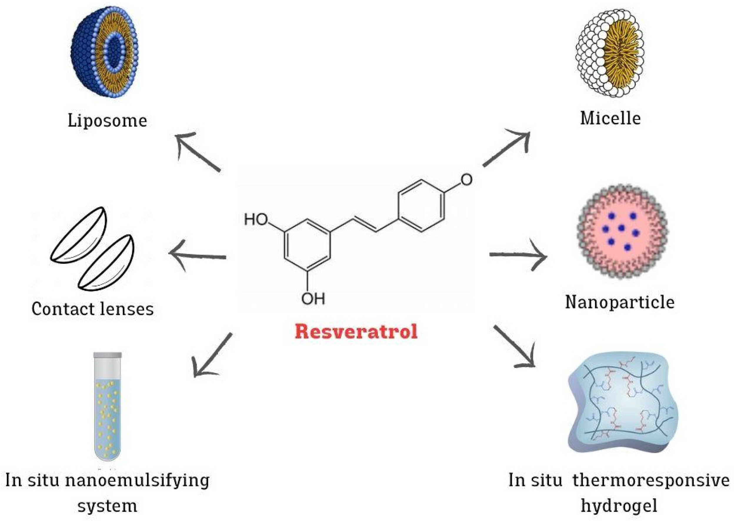
Scientific studies have explored the therapeutic potential of RV in ocular diseases. Whether administered in its pure form or entrapped within nanoparticulate delivery systems, RV exhibits promising outcomes in preclinical and clinical settings. One of its major limitations is deliverability and tissue-specific bioavailability. Being a hydrophobic agent with low water solubility, both the topical instillation into the eye and the delivery to the posterior segment of the eye are challenging; therefore, it must be used either with a nanocarrier or in combination with another vehicle that aids in its intracellular delivery. Figure 2 shows various nanotechnology-based systems designed for the effective delivery of RV to the eye. These systems include NPs, LPs, and micelles, which enhance the bioavailability, stability, and targeted delivery of RV, improving its therapeutic efficacy in treating ocular diseases.
Polymeric NPs have shown considerable promise in enhancing the delivery and therapeutic efficacy of RV in the treatment of ocular diseases. For instance, Bhatt, P, and coworkers demonstrated that incorporating RV into poly(lactic-co-glycolic acid) (PLGA) NPs significantly improved cellular uptake and increased the inhibition of VEGF expression in ARPE-19 cells tested in vitro [61].
Staying within the context of polymeric NPs and referring to the previously mentioned synergistic potential of RV when combined with other compounds, significant attention has been drawn to the work of Natesan et al. [62]. They demonstrated that loading RV and quercetin in pegylated chitosan NPs improved both the delivery and synergistic effects on reducing IOP in the treatment of glaucoma. Moreover, ex vivo corneal permeation tests showed that co-entrapment with quercetin improves RV corneal permeation compared to RV alone. Chitosan has also been proposed in the form of polymer nanogels (NGs), which are NPs composed of a cross-linked polymer network. NGs have emerged as promising and innovative drug delivery carriers in recent years. Buosi and coauthors [63] exploited such a system to achieve RV entrapment, addressing its limitations in terms of photostability. The group tested the biological application of these NGs on ARPE-19 cells, determining their biocompatibility through cytotoxicity and proinflammatory profiles, as well as cellular uptake. The results were promising, indicating biocompatibility and efficient cellular uptake, but further studies are needed to confirm these findings and to fully realize the therapeutic potential of NGs for controlled RV release in human retinal cells. The longer-term technological objective is to deliver these NGs into human retinal cells, enabling the controlled release of RV for therapeutic purposes and ultimately enhancing the efficacy and stability of RV in ocular treatments. Additionally, chitosan finds application in coating niosomes, providing a protective layer and enhancing their stability and bioavailability. More specifically, El-Haddad and coworkers [64] exploited RV-loaded chitosan-coated niosomes, chitoniosomes, for the topical treatment of induced ocular inflammation in a rabbit model. Coating with chitosan enhanced the mucoadhesive efficiency of niosomes compared to uncoated formulations. Notably, the treated group exhibited good ocular tolerance without any inflammatory response in rabbit eyes. In vivo tests of the anti-inflammatory effect of topically administered RV from chitoniosomes demonstrated a significant decrease in the expression levels of TNFα and IL-6 genes in all treated groups compared to the control group, suggesting its potential as an alternative treatment for ocular inflammation.
Recent studies have explored further strategies to enhance the therapeutic efficacy of ocular treatments consisting of combining polymeric NPs with in situ gelling polymers, aimed at prolonging corneal residence time and reducing the frequency of administration. This strategy has proven effective in addressing DED, a condition typically managed with eye drops and gels, which stand as the most common and convenient topical ophthalmic formulations. In particular, NPs modified with acetylated polyethyleneimine-poly(lactic-co-glycolic acid) (PLGA-PEI) and loaded with RV were dispersed into poloxamer 407 hydrogel with the aim of reducing the frequency of administration. This formulation exhibited sustained release of RV for 3 days, maintaining the antioxidant and anti-inflammatory effects on corneal epithelial cells [14].
An even more innovative approach was reported by Nguyen et al. [65], involving the development of NPs made of the biopolymer poly(ε-caprolactone) (PCL) that simultaneously load RV and metformin for the treatment of retinal pathologies, particularly AMD. Metformin is capable of inhibiting choroidal neovascularization, thereby providing a synergistic effect when combined with RV. The novelty of this system lies in exploiting a targeted therapeutic approach: the PCL-NPs loaded with RV and metformin were modified by linking them with a transcription activator cell-penetrating peptide to enhance retinal permeability and ensure sustained bioactive delivery. Cell-penetrating peptides represent a promising advancement in improving drug and nanocarrier uptake in cells. According to in vivo studies, a single-dose intravitreal injection of this nanoformulation increased retinal permeability for two months and inhibited abnormal vessel growth, suggesting that these nanoformulations could be effective in treating complex posterior segment diseases such as neovascular AMD.
Shifting the discussion from polymeric NPs to LPs, recent research has highlighted the potential of liposomal systems in ocular drug delivery. Recently, there has been increased attention on the damage induced by LED-related blue light on vision. In this context, Huan Gu et al. [13] proposed LPs based on trimethylated chitosan (TMC) to deliver RV, enhancing its mucoadhesive properties. These LPs reduce H2O2-induced damage to ARPE-19 cells, protecting them and restoring mitochondrial membrane potential. The study of ocular penetration demonstrated that LPs effectively delivered RV to the retina, showing a protective effect against blue light-induced retinal damage in mouse retinas.
Micelles, another effective carrier, have shown great promise for enhancing RV stability and ocular delivery. To overcome the limited solubility of RV, its rapid degradation, and low ocular permeability, single and mixed micelles of Pluronic® F127 and casein have been proposed as nanocarriers for the ophthalmic administration of RV [66]. For one month, these micelles were stable and maintained the sol-to-gel transition temperature. In addition, this formulation contributed to preserving the antioxidant properties of RV, preventing biofilm development, and enhancing RV permeation through corneal and scleral tissues.
SNEDDSs are another promising type of carrier for ocular drug delivery as they enhance the solubility, stability, and bioavailability of poorly water-soluble drugs. SNEDDSs are isotropic mixtures of oils, surfactants, and co-surfactants that spontaneously form fine oil-in-water nanoemulsions when exposed to aqueous environments, such as ocular fluids. As an example, Zingale et al. [12] developed an in situ SNEDDS for the topical ophthalmic delivery of RV and melatonin, aiming to treat diabetic retinopathy. This system demonstrated a slight mucoadhesive capacity and proved to be cytocompatible with corneal epithelial cells. However, in vivo studies are necessary to evaluate the drug diffusion mechanism towards the posterior chamber and its pharmacological activity.
Gold NPs have emerged as an interesting ocular delivery system due to their unique properties and potential for targeted drug delivery. For example, Dong et al. [67] developed RV-coated gold NPs (AuNPs) via a green synthetic method, avoiding the use of toxic reductants, for their application in treating diabetic retinopathy. The effects of these NPs were tested in streptozotocin-induced diabetic rats. The results demonstrated that the group treated with AuNPs showed a reduction in the retinal mRNA expression of VEGF-1, TNFα, adhesion molecules, and interleukins (IL-6, IL-1β). Furthermore, AuNPs were found to inhibit the signaling pathway of ERK1/2, leading to an improvement in retinal inflammation through the transrepression of NF-κB. More recently, Chen et al. [68] prepared gold NPs containing RV (RGNPs) as an anti-aging agent to delay the onset of cataracts. The spherical RGNPs demonstrated a certain ability to inhibit oxidative stress damage, including reactive oxygen species production, and glutathione consumption in lens epithelial cells. Overall, the data showed that cell senescence was reduced, and cataract formation was delayed upon treatment with RGNPs, through the activation of the Sirt1/Nrf2 signaling pathway, highlighting their potential to delay cataract development.
In addition to the aforementioned delivery systems, innovative approaches have extended to include the incorporation of ophthalmic drugs into contact lenses. These lenses serve as an ingenious platform for sustained drug delivery directly to the ocular surface. By incorporating drugs or NPs into the lens material, they offer a convenient and non-invasive method for administering therapeutics. The controlled release of drugs from these lenses ensures prolonged efficacy while minimizing systemic side effects. With the potential to revolutionize ocular drug delivery, drug-loaded contact lenses represent a promising strategy for the treatment of various eye conditions, including cataracts, diabetic retinopathy, and ocular inflammation. Recently, Vivero-Lopez and coworkers [69] developed phosphorylcholine-based contact lenses for sustained release of RV. More specifically, they explored the feasibility of using phosphorylcholine as a comonomer of 2-hydroxyethyl methacrylate-based hydrogels, allowing for the loading of hydrophobic RV. The developed RV-loaded contact lens showed excellent anti-inflammatory properties and biocompatibility in in vitro and in vivo tests and provided higher and more prolonged levels of RV in tear fluid, favoring its biodistribution in anterior and posterior eye segments compared to eye drops. Table 2 summarizes the main delivery systems proposed in the literature for the ocular administration of RV.

5. Conclusions

RV, a versatile natural polyphenol, demonstrates significant potential in the treatment of various disorders, including ocular diseases, due to its broad spectrum of bioactive properties and mechanisms of action. This review provides an overview of the principal extraction techniques used to derive RV from plant sources, emphasizing the growing importance of sustainability in these processes. It also explores the therapeutic benefits of RV at cellular, molecular, and physiological levels, not only highlighting its inherent bioactivity but also the promising synergistic effects achievable with other active substances and drugs, paving the way for enhanced therapeutic outcomes.
However, the main focus of this paper is on the role nanoparticulate systems can play in improving RV stability, bioavailability, and targeted delivery, such as polymeric nanoparticles, liposomes, and micelles. Emerging delivery methods, including drug-loaded contact lenses, represent significant advancements in sustained and controlled release, further expanding RV potential in ocular therapeutics.
Looking ahead, the inclusion of RV into advanced delivery platforms, along with robust ex vivo/in vivo studies, will be essential for addressing current limitations and optimizing its therapeutic efficacy. Continued progress in these innovative technologies holds considerable promise for RV-based treatments, potentially transforming the management of ocular diseases and substantially enhancing patient outcomes.

Author Contributions

Conceptualization, G.A. and D.C.; methodology, F.T. and E.P.; writing—original draft preparation, G.A. and F.T.; writing—review and editing, S.S., D.C. and E.P.; supervision, S.S. and M.G. All authors have read and agreed to the published version of the manuscript.

Funding

This review was funded by the European Union: PHENOCYCLES—Grant number 101131420.

Data Availability Statement

We searched PubMed, Web of Science, and Scopus for the health benefits of RV and its role in ocular diseases and retrieved relevant references from research articles and reviews. The search terms included “resveratrol” AND “health” or “bioactive properties” or “ocular diseases” or “drug delivery systems”; “resveratrol” AND “extraction technologies”; “resveratrol” AND “synergistic effect”. Search dates: February–May 2024.

Conflicts of Interest

The authors declare no conflicts of interest.

Abbreviations

AMDage-related macular degeneration
CSchitosan
COXcyclooxygenase
DEDdry eye disease
DESdeep eutectic solvent
DOXdoxazosin
EAEenzyme-assisted extraction
eEF2eukaryotic elongation factor-2
HLEChuman lens epithelial cells
ILinterleukins
iNOSinducible NO synthase
IOPintraocular pressure
iROSintracellular reactive oxygen species
LDL-Clow-density lipoprotein cholesterol
LPliposome
MAEmicrowave-assisted extraction
MPC2-methacryloyloxyethyl phosphorylcholine
MSPDmatrix solid-phase dispersion
NGnanogel
NOnitric oxide
NPnanoparticle
PCLpoly(ε-caprolactone)
PEGpoly ethylene glycol
PGprostaglandin
PKCprotein kinase C
PLGApoly(lactic-co-glycolic acid)
PLGA-PEIacetylated polyethyleneimine-poly(lactic-co-glycolic acid)
RGNPgold nanoparticles encapsulated resveratrol
ROPretinopathy of prematurity
ROSreactive oxygen species
RPEretinal pigment epithelial
RVresveratrol
SEsolvent extraction
SFEsupercritical fluid extraction
SNEDDSsself-nanoemulsifying drug delivery systems
TMCtrimethylated chitosan
UAEultrasound-assisted extraction
VEGFvascular endothelial growth factor

References

  1. Akinwumi, B.C.; Bordun, K.A.M.; Anderson, H.D. Biological Activities of Stilbenoids. Int. J. Mol. Sci. 2018, 19, 792. [Google Scholar] [CrossRef]
  2. Duta-Bratu, C.G.; Nitulescu, G.M.; Mihai, D.P.; Olaru, O.T. Resveratrol and Other Natural Oligomeric Stilbenoid Compounds and Their Therapeutic Applications. Plants 2023, 12, 2935. [Google Scholar] [CrossRef] [PubMed]
  3. Tian, B.; Liu, J. Resveratrol: A Review of Plant Sources, Synthesis, Stability, Modification and Food Application. J. Sci. Food Agric. 2020, 100, 1392–1404. [Google Scholar] [CrossRef]
  4. Renaud, S.; de Lorgeril, M. Wine, Alcohol, Platelets, and the French Paradox for Coronary Heart Disease. Lancet 1992, 339, 1523–1526. [Google Scholar] [CrossRef]
  5. Tang, H.; Hou, H.; Song, L.; Tian, Z.; Liu, W.; Xia, T.; Wang, A. The Role of MTORC1/TFEB Axis Mediated Lysosomal Biogenesis and Autophagy Impairment in Fluoride Neurotoxicity and the Intervention Effects of Resveratrol. J. Hazard. Mater. 2024, 467, 133634. [Google Scholar] [CrossRef]
  6. Ma, Y.; Yu, K.; Wang, N.; Xiao, X.; Leng, Y.; Fan, J.; Du, Y.; Wang, S. Sulfur Dioxide-Free Wine with Polyphenols Promotes Lipid Metabolism via the Nrf2 Pathway and Gut Microbiota Modulation. Food Chem. X 2024, 21, 101079. [Google Scholar] [CrossRef]
  7. Bai, Y.; Sui, R.; Zhang, L.; Bai, B.; Zhu, Y.; Jiang, H. Resveratrol Improves Cognitive Function in Post-Stroke Depression Rats by Repressing Inflammatory Reactions and Oxidative Stress via the Nrf2/HO-1 Pathway. Neuroscience 2024, 541, 50–63. [Google Scholar] [CrossRef]
  8. Golestaneh, E.; Dehkordi, A.H.; Yalameha, B.; Noorshargh, P.; Nasri, P.; Nasri, H. Comparative Study of Nephroprotective Effects of Resveratrol and Silymarin in Diabetic Rats; an Experimental Histopathologic Study. J. Nephropharmacol. 2024, 13, e10381–e10385. [Google Scholar] [CrossRef]
  9. Chen, F.; Zhang, L.; Liu, Y.; Zhang, A.; Wang, W. Resveratrol alleviates perinatal methylmercury-induced neurobehavioral impairments by modulating the gut microbiota composition and neurotransmitter disturbances. Environ. Toxicol. 2024, 39, 329–340. [Google Scholar] [CrossRef]
  10. Delmas, D.; Cornebise, C.; Courtaut, F.; Xiao, J.; Aires, V. New Highlights of Resveratrol: A Review of Properties against Ocular Diseases. Int. J. Mol. Sci. 2021, 22, 1295. [Google Scholar] [CrossRef]
  11. Bungau, S.; Abdel-Daim, M.M.; Tit, D.M.; Ghanem, E.; Sato, S.; Maruyama-Inoue, M.; Yamane, S.; Kadonosono, K. Health Benefits of Polyphenols and Carotenoids in Age-Related Eye Diseases. Oxidative Med. Cell. Longev. 2019, 2019, 9783429. [Google Scholar] [CrossRef] [PubMed]
  12. Zingale, E.; Bonaccorso, A.; D’Amico, A.G.; Lombardo, R.; D’Agata, V.; Rautio, J.; Pignatello, R. Formulating Resveratrol and Melatonin Self-Nanoemulsifying Drug Delivery Systems (SNEDDS) for Ocular Administration Using Design of Experiments. Pharmaceutics 2024, 16, 125. [Google Scholar] [CrossRef] [PubMed]
  13. Gu, H.; Chen, P.; Liu, X.; Lian, Y.; Xi, J.; Li, J.; Song, J.; Li, X. Trimethylated Chitosan-Coated Flexible Liposomes with Resveratrol for Topical Drug Delivery to Reduce Blue-Light-Induced Retinal Damage. Int. J. Biol. Macromol. 2023, 252, 126480. [Google Scholar] [CrossRef] [PubMed]
  14. De Luca, I.; Di Cristo, F.; Conte, R.; Peluso, G.; Cerruti, P.; Calarco, A. In-Situ Thermoresponsive Hydrogel Containing Resveratrol-Loaded Nanoparticles as a Localized Drug Delivery Platform for Dry Eye Disease. Antioxidants 2023, 12, 993. [Google Scholar] [CrossRef]
  15. Li, W.; Yuan, H.; Liu, Y.; Wang, B.; Xu, X.; Xu, X.; Hussain, D.; Ma, L.; Chen, D. Current analytical strategies for the determination of resveratrol in foods. Food Chem. 2024, 431, 137182. [Google Scholar] [CrossRef]
  16. Angelov, G.; Boyadzhiev, L.; Georgieva, S. Useful Bioactive Substances from Wastes: Recovery of Trans-Resveratrol from Grapevine Stems. Open Chem. Eng. J. 2016, 10, 4–9. [Google Scholar] [CrossRef]
  17. Piyaratne, P.S.; Leblanc, R.; Myracle, A.D.; Cole, B.J.W.; Fort, R.C. Extraction and Purification of (E)-Resveratrol from the Bark of Conifer Species. Processes 2022, 10, 647. [Google Scholar] [CrossRef]
  18. Jitrangsri, K.; Chaidedgumjorn, A.; Satiraphan, M. Supercritical Fluid Extraction (SFE) Optimization of Trans-Resveratrol from Peanut Kernels (Arachis hypogaea) by Experimental Design. J. Food Sci. Technol. 2020, 57, 1486–1494. [Google Scholar] [CrossRef]
  19. Akhtar, I.; Javad, S.; Yousaf, Z.; Iqbal, S.; Jabeen, K. Microwave Assisted Extraction of Phytochemicals an Efficient and Modern Approach for Botanicals and Pharmaceuticals. Pak. J. Pharm. Sci. 2019, 32, 223–230. [Google Scholar]
  20. Piñeiro, Z.; Marrufo-Curtido, A.; Vela, C.; Palma, M. Microwave-Assisted Extraction of Stilbenes from Woody Vine Material. Food Bioprod. Process. 2017, 103, 18–26. [Google Scholar] [CrossRef]
  21. Sun, H.; Lin, Q.; Wei, W.; Qin, G. Ultrasound-Assisted Extraction of Resveratrol from Grape Leaves and Its Purification on Mesoporous Carbon. Food Sci. Biotechnol. 2018, 27, 1353–1359. [Google Scholar] [CrossRef] [PubMed]
  22. Wang, C.; Liu, X.; Zhang, M.; Shao, H.; Zhang, M.; Wang, X.; Wang, Q.; Bao, Z.; Fan, X.; Li, H. Efficient Enzyme-Assisted Extraction and Conversion of Polydatin to Resveratrol from Polygonum Cuspidatum Using Thermostable Cellulase and Immobilized β-Glucosidase. Front. Microbiol. 2019, 10, 445. [Google Scholar] [CrossRef] [PubMed]
  23. Lin, J.A.; Kuo, C.H.; Chen, B.Y.; Li, Y.; Liu, Y.C.; Chen, J.H.; Shieh, C.J. A Novel Enzyme-Assisted Ultrasonic Approach for Highly Efficient Extraction of Resveratrol from Polygonum Cuspidatum. Ultrason. Sonochem 2016, 32, 258–264. [Google Scholar] [CrossRef]
  24. Gómez-Mejía, E.; Mikkelsen, L.H.; Rosales-Conrado, N.; León-González, M.E.; Madrid, Y. A Combined Approach Based on Matrix Solid-Phase Dispersion Extraction Assisted by Titanium Dioxide Nanoparticles and Liquid Chromatography to Determine Polyphenols from Grape Residues. J. Chromatogr. A 2021, 1644, 462128. [Google Scholar] [CrossRef]
  25. Meng, T.; Xiao, D.; Muhammed, A.; Deng, J.; Chen, L.; He, J. Anti-Inflammatory Action and Mechanisms of Resveratrol. Molecules 2021, 26, 229. [Google Scholar] [CrossRef] [PubMed]
  26. Reuter, S.; Gupta, S.C.; Chaturvedi, M.M.; Aggarwal, B.B. Oxidative Stress, Inflammation, and Cancer: How Are They Linked? Free Radic. Biol. Med. 2010, 49, 1603–1616. [Google Scholar] [CrossRef] [PubMed]
  27. Chandrasekharan, N.V.; Dai, H.; Roos, K.L.T.; Evanson, N.K.; Tomsik, J.; Elton, T.S.; Simmons, D.L. COX-3, a Cyclooxygenase-1 Variant Inhibited by Acetaminophen and Other Analgesic/Antipyretic Drugs: Cloning, Structure, and Expression. Proc. Natl. Acad. Sci. USA 2002, 99, 13926–13931. [Google Scholar] [CrossRef]
  28. Szewczuk, L.M.; Forti, L.; Stivala, L.A.; Penning, T.M. Resveratrol Is a Peroxidase-Mediated Inactivator of COX-1 but Not COX-2: A Mechanistic Approach to the Design of COX-1 Selective Agents. J. Biol. Chem. 2004, 279, 22727–22737. [Google Scholar] [CrossRef]
  29. Murias, M.; Handler, N.; Erker, T.; Pleban, K.; Ecker, G.; Saiko, P.; Szekeres, T.; Jäger, W. Resveratrol Analogues as Selective Cyclooxygenase-2 Inhibitors: Synthesis and Structure-Activity Relationship. Bioorg. Med. Chem. 2004, 12, 5571–5578. [Google Scholar] [CrossRef]
  30. Subbaramaiah, K.; Chung, W.J.; Michaluart, P.; Telang, N.; Tanabe, T.; Inoue, H.; Jang, M.; Pezzuto, J.M.; Dannenberg, A.J. Resveratrol Inhibits Cyclooxygenase-2 Transcription and Activity in Phorbol Ester-Treated Human Mammary Epithelial Cells. J. Biol. Chem. 1998, 273, 21875–21882. [Google Scholar] [CrossRef]
  31. Chen, F.; Castranova, V.; Shi, X. Review New Insights into the Role of Nuclear Factor-B in Cell Growth Regulation. Am. J. Pathol. 2001, 159, 387–397. [Google Scholar] [CrossRef] [PubMed]
  32. Wang, T.; Wu, F.; Jin, Z.; Zhai, Z.; Wang, Y.; Tu, B.; Yan, W.; Tang, T. Plumbagin Inhibits LPS-Induced Inflammation through the Inactivation of the Nuclear Factor-Kappa B and Mitogen Activated Protein Kinase Signaling Pathways in RAW 264.7 Cells. Food Chem. Toxicol. 2014, 64, 177–183. [Google Scholar] [CrossRef] [PubMed]
  33. Berman, A.Y.; Motechin, R.A.; Wiesenfeld, M.Y.; Holz, M.K. The therapeutic potential of resveratrol: A review of clinical trials. NPJ Precis. Oncol. 2017, 1, 35. [Google Scholar] [CrossRef] [PubMed]
  34. Johnson, W.D.; Morrissey, R.L.; Usborne, A.L.; Kapetanovic, I.; Crowell, J.A.; Muzzio, M.; McCormick, D.L. Subchronic oral toxicity and cardiovascular safety pharmacology studies of resveratrol, a naturally occurring polyphenol with cancer preventive activity. Food Chem. Toxicol. 2011, 49, 3319–3327. [Google Scholar] [CrossRef] [PubMed]
  35. Ramírez-Garza, S.L.; Laveriano-Santos, E.P.; Marhuenda-Muñoz, M.; Storniolo, C.E.; Tresserra-Rimbau, A.; Vallverdú-Queralt, A.; Lamuela-Raventós, R.M. Health Effects of Resveratrol: Results from Human Intervention Trials. Nutrients 2018, 10, 1892. [Google Scholar] [CrossRef]
  36. Shaito, A.; Posadino, A.M.; Younes, N.; Hasan, H.; Halabi, S.; Alhababi, D.; Al-Mohannadi, A.; Abdel-Rahman, W.M.; Eid, A.H.; Nasrallah, G.K.; et al. Potential Adverse Effects of Resveratrol: A Literature Review. Int. J. Mol. Sci. 2020, 21, 2084. [Google Scholar] [CrossRef]
  37. Ma, N.; Zhang, Y. Effects of resveratrol therapy on glucose metabolism, insulin resistance, inflammation, and renal function in the elderly patients with type 2 diabetes mellitus: A randomized controlled clinical trial protocol. Medicine 2022, 101, e30049. [Google Scholar] [CrossRef]
  38. García-Martínez, B.I.; Ruiz-Ramos, M.; Pedraza-Chaverri, J.; Santiago-Osorio, E.; Mendoza-Núñez, V.M. Effect of Resveratrol on Markers of Oxidative Stress and Sirtuin 1 in Elderly Adults with Type 2 Diabetes. Int. J. Mol. Sci. 2023, 24, 7422. [Google Scholar] [CrossRef]
  39. Gu, J.; Li, Z.; Chen, H.; Xu, X.; Li, Y.; Gui, Y. Neuroprotective Effect of Trans-Resveratrol in Mild to Moderate Alzheimer Disease: A Randomized, Double-Blind Trial. Neurol. Ther. 2021, 10, 905–917. [Google Scholar] [CrossRef]
  40. Gimblet, C.J.; Kruse, N.T.; Geasland, K.; Michelson, J.; Sun, M.; Mandukhail, S.R.; Wendt, L.H.; Eyck, P.T.; Pierce, G.L.; Jalal, D.I. Effect of Resveratrol on Endothelial Function in Patients with CKD and Diabetes: A Randomized Controlled Trial. Clin. J. Am. Soc. Nephrol. 2024, 19, 161–168. [Google Scholar] [CrossRef]
  41. Zhang, L.X.; Li, C.X.; Kakar, M.U.; Khan, M.S.; Wu, P.F.; Amir, R.M.; Dai, D.F.; Naveed, M.; Li, Q.Y.; Saeed, M.; et al. Resveratrol (RV): A Pharmacological Review and Call for Further Research. Biomed. Pharmacother. 2021, 143, 112164. [Google Scholar] [CrossRef] [PubMed]
  42. Liu, Q.; Zhu, D.; Jiang, P.; Tang, X.; Lang, Q.; Yu, Q.; Zhang, S.; Che, Y.; Feng, X. Resveratrol Synergizes with Low Doses of L-DOPA to Improve MPTP-Induced Parkinson Disease in Mice. Behav. Brain Res. 2019, 367, 10–18. [Google Scholar] [CrossRef] [PubMed]
  43. Ren, M.; Zhou, X.; Gu, M.; Jiao, W.; Yu, M.; Wang, Y.; Liu, S.; Yang, J.; Ji, F. Resveratrol Synergizes with Cisplatin in Antineoplastic Effects against AGS Gastric Cancer Cells by Inducing Endoplasmic Reticulum Stress-Mediated Apoptosis and G2/M Phase Arrest. Oncol. Rep. 2020, 44, 1605–1615. [Google Scholar] [CrossRef] [PubMed]
  44. Alhayali, A.S.A.; Hasan, W.A.; Salah, F.S. Autophagy Induction Using Resveratrol Enhances the Anti-Cancer Efficacy of Doxazosin in Breast Cancer Cells. Bionatura 2023, 8, 63. [Google Scholar] [CrossRef]
  45. Toniolo, L.; Fusco, P.; Formoso, L.; Mazzi, A.; Canato, M.; Reggiani, C.; Giacomello, E. Resveratrol Treatment Reduces the Appearance of Tubular Aggregates and Improves the Resistance to Fatigue in Aging Mice Skeletal Muscles. Exp. Gerontol. 2018, 111, 170–179. [Google Scholar] [CrossRef]
  46. Hussain, S.A.; Marouf, B.H.; Ali, Z.S.; Ahmmad, R.S. Efficacy and Safety of Co-Administration of Resveratrol with Meloxicam in Patients with Knee Osteoarthritis: A Pilot Interventional Study. Clin. Interv. Aging 2018, 13, 1621–1630. [Google Scholar] [CrossRef]
  47. Farrokhi, E.; Ghatreh-Samani, K.; Salehi-Vanani, N.; Mahmoodi, A. The Effect of Resveratrol on Expression of Matrix Metalloproteinase 9 and Its Tissue Inhibitors in Vascular Smooth Muscle Cells. ARYA Atheroscler. 2018, 14, 157–162. [Google Scholar] [CrossRef] [PubMed]
  48. Galiniak, S.; Aebisher, D.; Bartusik-Aebisher, D. Health Benefits of Resveratrol Administration. Acta Biochim. Pol. 2019, 66, 13–21. [Google Scholar] [CrossRef]
  49. Corpas, R.; Griñán-Ferré, C.; Rodríguez-Farré, E.; Pallàs, M.; Sanfeliu, C. Resveratrol Induces Brain Resilience Against Alzheimer Neurodegeneration Through Proteostasis Enhancement. Mol. Neurobiol. 2019, 56, 1502–1516. [Google Scholar] [CrossRef]
  50. Hou, Y.; Wang, K.; Wan, W.; Cheng, Y.; Pu, X.; Ye, X. Resveratrol Provides Neuroprotection by Regulating the JAK2/STAT3/PI3K/AKT/MTOR Pathway after Stroke in Rats. Genes Dis. 2018, 5, 245–255. [Google Scholar] [CrossRef]
  51. Magyar, K.; Halmosi, R.; Palfi, A.; Feher, G.; Czopf, L.; Fulop, A.; Battyany, I.; Sumegi, B.; Toth, K.; Szabados, E. Cardioprotection by Resveratrol: A Human Clinical Trial in Patients with Stable Coronary Artery Disease. Clin. Hemorheol. Microcirc. 2012, 50, 179–187. [Google Scholar] [CrossRef]
  52. Romain, C.; Gaillet, S.; Carillon, J.; Vidé, J.; Ramos, J.; Izard, J.C.; Cristol, J.P.; Rouanet, J.M. Vineatrol and Cardiovascular Disease: Beneficial Effects of a Vine-Shoot Phenolic Extract in a Hamster Atherosclerosis Model. J. Agric. Food Chem. 2012, 60, 11029–11036. [Google Scholar] [CrossRef]
  53. Bryl, A.; Falkowski, M.; Zorena, K.; Mrugacz, M. The Role of Resveratrol in Eye Diseases—A Review of the Literature. Nutrients 2022, 14, 2974. [Google Scholar] [CrossRef] [PubMed]
  54. Doganay, S.; Borazan, M.; Iraz, M.; Cigremis, Y. The Effect of Resveratrol in Experimental Cataract Model Formed by Sodium Selenite. Curr. Eye Res. 2006, 31, 147–153. [Google Scholar] [CrossRef]
  55. Wakamatsu, T.H.; Dogru, M.; Matsumoto, Y.; Kojima, T.; Kaido, M.; Ibrahim, O.M.A.; Sato, E.A.; Igarashi, A.; Ichihashi, Y.; Satake, Y.; et al. Evaluation of Lipid Oxidative Stress Status in Sjögren Syndrome Patients. Investig. Ophthalmol. Vis. Sci. 2013, 54, 201–210. [Google Scholar] [CrossRef] [PubMed]
  56. Anekonda, T.S.; Adamus, G. Resveratrol Prevents Antibody-Induced Apoptotic Death of Retinal Cells through Upregulation of Sirt1 and Ku70. BMC Res. Notes 2008, 1, 122. [Google Scholar] [CrossRef] [PubMed]
  57. Khan, A.A.; Dace, D.S.; Ryazanov, A.G.; Kelly, J.; Apte, R.S. Resveratrol Regulates Pathologic Angiogenesis by a Eukaryotic Elongation Factor-2 Kinase-Regulated Pathway. Am. J. Pathol. 2010, 177, 481–492. [Google Scholar] [CrossRef]
  58. Shetty, R.; Joshi, P.D.; Mahendran, K.; Jayadev, C.; Das, D. Resveratrol for Dry Eye Disease—Hope or Hype? Indian. J. Ophthalmol. 2023, 71, 1270–1275. [Google Scholar] [CrossRef]
  59. Luna, C.; Li, G.; Liton, P.B.; Qiu, J.; Epstein, D.L.; Challa, P.; Gonzalez, P. Resveratrol Prevents the Expression of Glaucoma Markers Induced by Chronic Oxidative Stress in Trabecular Meshwork Cells. Food Chem. Toxicol. 2009, 47, 198–204. [Google Scholar] [CrossRef]
  60. Razali, N.; Agarwal, R.; Agarwal, P.; Froemming, G.R.A.; Tripathy, M.; Ismail, N.M. IOP Lowering Effect of Topical Trans-Resveratrol Involves Adenosine Receptors and TGF-Β2 Signaling Pathways. Eur. J. Pharmacol. 2018, 838, 1–10. [Google Scholar] [CrossRef]
  61. Bhatt, P.; Fnu, G.; Bhatia, D.; Shahid, A.; Sutariya, V. Nanodelivery of Resveratrol-Loaded PLGA Nanoparticles for Age-Related Macular Degeneration. AAPS PharmSciTech 2020, 21, 291. [Google Scholar] [CrossRef] [PubMed]
  62. Natesan, S.; Pandian, S.; Ponnusamy, C.; Palanichamy, R.; Muthusamy, S.; Kandasamy, R. Co-Encapsulated Resveratrol and Quercetin in Chitosan and Peg Modified Chitosan Nanoparticles: For Efficient Intra Ocular Pressure Reduction. Int. J. Biol. Macromol. 2017, 104, 1837–1845. [Google Scholar] [CrossRef]
  63. Buosi, F.S.; Alaimo, A.; Di Santo, M.C.; Elías, F.; García Liñares, G.; Acebedo, S.L.; Castañeda Cataña, M.A.; Spagnuolo, C.C.; Lizarraga, L.; Martínez, K.D.; et al. Resveratrol Encapsulation in High Molecular Weight Chitosan-Based Nanogels for Applications in Ocular Treatments: Impact on Human ARPE-19 Culture Cells. Int. J. Biol. Macromol. 2020, 165, 804–821. [Google Scholar] [CrossRef]
  64. El-Haddad, M.E.; Hussien, A.A.; Saeed, H.M.; Farid, R.M. Down Regulation of Inflammatory Cytokines by the Bioactive Resveratrol-Loaded Chitoniosomes in Induced Ocular Inflammation Model. J. Drug Deliv. Sci. Technol. 2021, 66, 102787. [Google Scholar] [CrossRef]
  65. Nguyen, D.D.; Luo, L.J.; Yang, C.J.; Lai, J.Y. Highly Retina-Permeating and Long-Acting Resveratrol/Metformin Nanotherapeutics for Enhanced Treatment of Macular Degeneration. ACS Nano 2023, 17, 168–183. [Google Scholar] [CrossRef] [PubMed]
  66. Vivero-Lopez, M.; Sparacino, C.; Quelle-Regaldie, A.; Sánchez, L.; Candal, E.; Barreiro-Iglesias, A.; Huete-Toral, F.; Carracedo, G.; Otero, A.; Concheiro, A.; et al. Pluronic®/Casein Micelles for Ophthalmic Delivery of Resveratrol: In Vitro, Ex Vivo, and in Vivo Tests. Int. J. Pharm. 2022, 628, 122281. [Google Scholar] [CrossRef]
  67. Dong, Y.; Wan, G.; Yan, P.; Qian, C.; Li, F.; Peng, G. Fabrication of Resveratrol Coated Gold Nanoparticles and Investigation of Their Effect on Diabetic Retinopathy in Streptozotocin Induced Diabetic Rats. J. Photochem. Photobiol. B 2019, 195, 51–57. [Google Scholar] [CrossRef] [PubMed]
  68. Chen, Q.; Gu, P.; Liu, X.; Hu, S.; Zheng, H.; Liu, T.; Li, C. Gold Nanoparticles Encapsulated Resveratrol as an Anti-Aging Agent to Delay Cataract Development. Pharmaceuticals 2023, 16, 26. [Google Scholar] [CrossRef]
  69. Vivero-Lopez, M.; Pereira-Da-Mota, A.F.; Carracedo, G.; Huete-Toral, F.; Parga, A.; Otero, A.; Concheiro, A.; Alvarez-Lorenzo, C. Phosphorylcholine-Based Contact Lenses for Sustained Release of Resveratrol: Design, Antioxidant and Antimicrobial Performances, and in Vivo Behavior. ACS Appl. Mater. Interfaces 2022, 14, 55431–55446. [Google Scholar] [CrossRef]
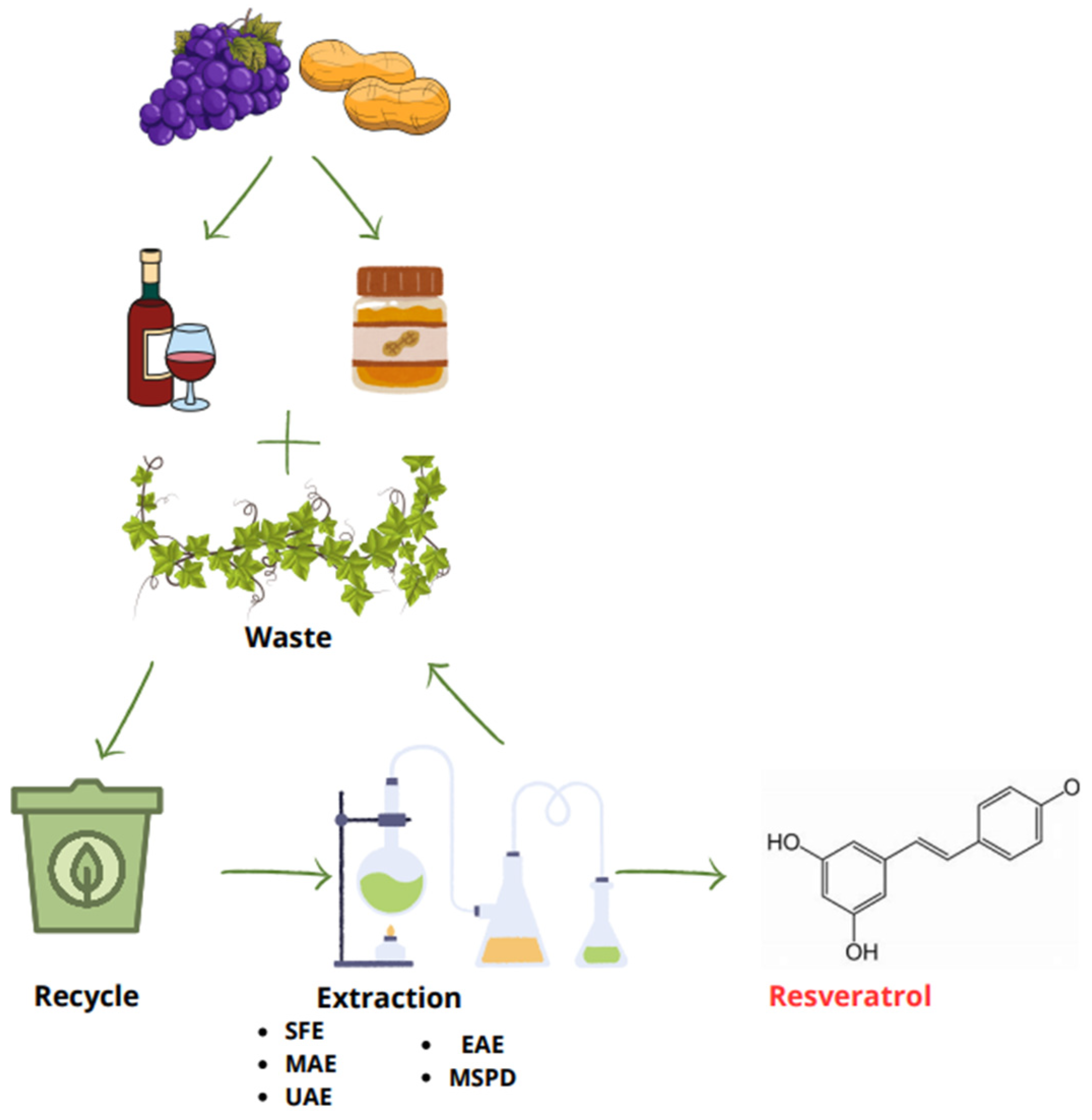
Figure 1. Circular economy scheme for grape and peanut utilization: from waste to resveratrol (RV) extraction.
Scipharm 92 00059 g001
Figure 2. Advanced nanotechnologies for the ocular delivery of RV.
Scipharm 92 00059 g002
Table 1. List of the main sustainable extraction techniques used to recover RV from plant sources.
Method of ExtractionSolventExtraction ConditionNatural SourceRef.
SFEEthanol70 °C, 48 MPa of pressure for 50 minPeanut kernels[18]
MAEEthanol/water (80%)125 °C and 750 W for 5 minGrape stem and canes[20]
UAEEthanol/water (40%)50 °C in ultrasonic bath
(40 kHz, 100 W)
Grape leaves[21]
EAECitric acid–NaOH buffer (50 mM, pH 5.0), 27.0 U cellulase; Methanol25–50 °C, with shaking at 150 rpmPolygonum cuspidatum roots[23]
MSPDTiO2 NPs and diatomaceous earthRoom temperature and pressureGrape pomace and grape fruit[24]
Table 2. List of the main drug delivery systems investigated for the ocular delivery of RV.
NanosystemLoaded
Molecules
CompositionMajor OutcomeCell Line/
Animal Model
Reference
NPsRVPoly (lactic-co-glycolic acid) (PLGA)<VEGF expressionIn vitro (ARPE-19)[61]
RV and quercetinPolyethylene glycols (PEGs) modified chitosan (CS)<IOPIn vivo (rabbit)[62]
RV and metforminBiopolymer poly(ε-caprolactone) (PCL)Antioxidant, anti-inflammatory, and antiangio-genic activitiesIn vitro (HUVECs)/
in vivo (rats)
[65]
RVGold<mRNA expression of VEGF-1, TNFα, adhesion molecules, IL-6, IL-1βIn vivo (rat)/
ex vivo
[67]
RVGold<ROS, expression levels of senescence markers (p16, p21, BAX, BCL-2 and SASP) >GSHIn vitro (HLECB3)/
in vivo (rats)
[68]
In situ thermoresponsive hydrogelRVAcetylated polyethyleneimine-modified poly lactic-co-glycolic acid- (PLGA-PEI) NPs into poloxamer 407 hydrogelAntioxidant and anti-inflammatory effectsIn vitro (HCECs)[14]
NGRVHigh weight chitosanAnti-inflammatory effectsIn vitro (ARPE-19)[63]
LPsRVTrimethylated chitosan-coated<H2O2-induced damageIn vitro (ARPE-19),
in vivo (mouse)
[13]
NiosomesRVChitosan-coated<TNFα, IL-6In vivo[64]
MicelleRVPluronic® F127 and casein>Solubilize RV, preserve antioxidant properties, <biofilm developmentIn vitro (HCECs),
ex vivo (porcine eye),
in vivo (rabbit)
[66]
SNEEDSRV and melatoninCapryol® PGMC, Tween® 80, and Transcutol® POptimization of formulations for ocular administrationIn vitro (SIRC)[12]
Contact lensesRVDaily contact lens coating with 2-methacryloyloxyethyl phosphorylcholine (MPC)<Inflammation and biofilm development, antibiofoulingIn vitro (THP-1),
in vivo (rabbit)
[69]
Disclaimer/Publisher’s Note: The statements, opinions and data contained in all publications are solely those of the individual author(s) and contributor(s) and not of MDPI and/or the editor(s). MDPI and/or the editor(s) disclaim responsibility for any injury to people or property resulting from any ideas, methods, instructions or products referred to in the content.