Communication

Can Gastric Juice Analysis with EndoFaster® Reduce the Environmental Impact of Upper Endoscopy?

1
Gastroenterology Unit, “Nuovo Regina Margherita” Hospital, 00153 Rome, Italy
2
Department of Translational Research and New Technologies in Medicine and Surgery, University of Pisa, 56126 Pisa, Italy
3
Gastroenterology and Endoscopy Unit, Department of Biomedical Sciences, IRCCS Humanitas Research Hospital, 20089 Milan, Italy
*
Author to whom correspondence should be addressed.
Healthcare 2023, 11(24), 3186; https://doi.org/10.3390/healthcare11243186
Submission received: 9 November 2023 / Revised: 12 December 2023 / Accepted: 14 December 2023 / Published: 17 December 2023
(This article belongs to the Section Environmental Factors and Global Health)

Abstract

:
Gastrointestinal (GI) endoscopy services are in third place as major contributors to CO2 emissions among healthcare facilities, especially due to their massive waste production. One of the measures suggested to reduce this environmental impact is a reduction in histological examinations performed on biopsy specimens taken during endoscopy. A reliable candidate to reduce the rate of biopsies and, consequently, the impact of CO2 emissions could be EndoFaster®, an innovative medical device that allows one to suspect or rule out both H. pylori infection and precancerous lesions on the gastric mucosa by analyzing a small amount of gastric juice aspirated during endoscopy in real time. In the present study, we investigated the ability of EndoFaster® to reduce the environmental impact of upper endoscopy, comparing the CO2 production of standard biopsy sampling as suggested in guidelines and biopsies guided by real-time EndoFaster® results during endoscopy. By estimating an overall 90% rate of biopsies according to standard guidelines and a reduction of 50% of gastric biopsies based on EndoFaster® results, we calculated a 44% overall reduction in CO2 emissions, demonstrating that by using this tool, it is possible to distinctly reduce the contribution of upper endoscopy to global warming.

1. Introduction

Human activities are responsible for climate changes, which already have evident effects on the environment and human health [1]. The critical connection between human activities and environmental temperature increments is represented by greenhouse gases (GHGs) because of their impact on energy retention [2]. Up to 85% of all GHGs are represented by carbon dioxide (CO2). In order to indicate the total amount of CO2 equivalents released into the atmosphere as a result of the activities of an individual, a product, an institution, or a service, the term ‘carbon footprint’ is used.
The mission of healthcare organizations is to protect and enhance human health and well-being. However, it is estimated that healthcare activities have a remarkable carbon footprint, accounting for 1% to 5% of human environmental impact and about 4.4% of GHG emissions worldwide [3]. Among these activities, gastrointestinal (GI) endoscopy is reported as one of the largest polluters in terms of CO2 emissions and the third largest contributor to hazardous waste production in healthcare facilities, with about 3 kg of waste produced for each digestive endoscopy bed every day [4]. Therefore, different scientific societies, including the European Society of Gastrointestinal Endoscopy (ESGE), the European Society of Gastroenterology and Endoscopy Nurses and Associates (ESGENA), and the Italian Association of Hospital Gastroenterologists (AIGO), released specific documents aimed at outlining strategies to achieve a sustainable endoscopy practice, the so-called ‘Green Endoscopy’ [1,2].
One of the components that contributes the most to the high carbon footprint of digestive endoscopy is tissue sampling, which requires histological analysis because biopsy processing needs additional energy and generates hazardous waste [1,2]. Currently, in all the appropriate upper endoscopies, the standard gastric mucosa sampling consists of two biopsies on the antral (plus one on the incisura angularis) and two on the gastric body mucosa to be put in two different jars, which allows for the correct diagnosis of H. pylori infection and the disclosure of the presence and extension of precancerous lesions in the stomach [5,6]. H. pylori is the main cause of both benign (non-ulcer dyspepsia, peptic ulcer) and malignant (cancer, lymphoma) diseases, and the presence of diffuse precancerous lesions (atrophy, metaplasia) on the gastric mucosa distinctly increases the risk of cancer development [5,6]. It is widely reported that both of these conditions may be detected through histological assessment of biopsies taken on even normally appearing mucosa at white-light endoscopic examination [5,6,7,8,9]. Nevertheless, it is important to consider that routine histological analysis is unnecessary in a consistent percentage of patients with normal-appearing mucosa that eventually tests negative for both H. pylori infection and precancerous lesions. Indeed, the frequency of infection is relentlessly decreasing in developed countries, and the prevalence of diffuse gastric precancerous lesions is quoted as low as 3–7% in Western countries [10,11].
In current European guidelines, upper endoscopy is suggested in patients with an increased risk of gastric cancer (patients >50 years old) and in those with alarm symptoms (bleeding, anemia, weight loss, persistent vomiting, dysphagia), while in dyspeptic, uninvestigated young patients, the 13C-Urea Breath Test (UBT), a less invasive alternative, should be preferred [5,6]. Nevertheless, even when upper endoscopy is appropriate, strategies for GHG emission reduction are needed, which can be summarized in the 3R principle (Reduce, Reuse, Recycle), with the reduction in waste and product generation as the top priority [1,2]. In GI endoscopy, by ensuring that only appropriate histological examinations are undertaken, it is possible to reduce the number of useless biopsy samples in low-risk patients without altering their management. Likewise, this approach is the most effective measure to lessen the impact on GHG emissions, according to the ESGE-ESGENA recommendations [1]. In line with this perspective, EndoFaster® (producer: NISO Biomed, Turin, Italy; distributor: Waldner Tecnologie Medicali, Trento, Italy) may be a valid tool to select those patients who really require biopsy sampling of the gastric mucosa, avoiding inappropriate biopsies in low-risk patients and, consequently, reducing the environmental impact. Indeed, EndoFaster® is an innovative, intuitive, and easy-to-use medical device that automatically analyzes a small (3 mL) amount of gastric juice aspirated during upper endoscopy, allowing one to suspect or rule out both H. pylori infection and diffuse precancerous lesions (atrophy with or without metaplasia) on the gastric mucosa in real time [12,13]. In detail, the machine is interposed between the endoscope and the suction system so that no adjunctive invasive procedure is required and there is no discomfort for the patient (Figure 1). The diagnosis is based on the determination of ammonium concentration and pH levels. The first is linked to the urease activity of the bacterium and provides information on the infection, while the latter allows the detection of hypochlorhydric conditions in gastric juice related to atrophy/metaplasia involving the gastric body mucosa.
Based on these considerations, we designed this study to evaluate the ability of EndoFaster® to reduce the environmental impact of upper endoscopy, comparing the CO2 production of standard biopsy sampling performed in all patients as suggested in guidelines and biopsies guided by real-time EndoFaster® results during endoscopy.

2. Materials and Methods

We estimated the CO2 production for either biopsy sampling guided by real-time EndoFaster® results during endoscopy or standard biopsy sampling performed in all patients, as suggested in guidelines [5,6,7,8,9].
In detail, for the application of EndoFaster® procedures, we first evaluated the daily CO2 production due to the energy consumed by the machine (51.04 Watts/day), taking as reference the data from an ISPRA report [14]. In addition, we considered the contribution of the following consumable materials with a relevant carbon component: (a) 3 bottles for calibration plus a liquid-draining system; (b) cardboard box for the 3 bottles; (c) washing solution tank; and (d) gastric juice suction tube. We did not include calibration liquids and reagents in this calculation, as they are highly diluted aqueous solutions that, in waste treatment plants, provide a positive contribution to the treatment process by helping to dilute solid waste without adding carbon. Furthermore, we computed the CO2 impact due to the disposal of liquid residues eliminated by the machine (5.308 L/day), transported by a 30,000 L chemical tanker traveling about 30 km between the hospital and disposal plant.
For the histological assessment, we also calculated the contribution of the following consumable materials: (a) biopsy forceps and (b) biopsy jar. In addition, we considered the entire biopsy processing in the pathology laboratory, including 11 steps (from specimen arriving in the laboratory to the pathologist’s review and report), accounting for 0.560 kg CO2/test, as accurately described elsewhere [15].
In order to calculate the CO2 production due to consumable material disposal, we used conversion factors adopted by the Institute for Sustainability Leadership of the University of Cambridge [16] and the IPCC Intergovernmental Panel on Climate Change [17]. In particular, the GHG emission factor from incineration of plastics was calculated at 2.697 kg CO2e/t of plastic waste using the formula kg CO2 = kg waste for incineration × oxidation factor of carbon in incinerator (0.98) × conversion factor of C to CO2 (3.67) × Σ(waste fraction (in %)) × dry matter content × carbon content (g/g dry weight). The dry matter content of plastic waste was equal to 1. The carbon content of plastic waste was 0.75 (Gg C/Gg dry weight waste). Moreover, the end-of-life emissions varied between different plastic types. The emissions for incineration, for instance, were higher for polystyrene (PS) and polyethylene (PE) (around 3 kg/kg plastic) and lower for polypropylene (PP) and polyurethane (PUR) (around 2.5 kg/kg plastic). For the purpose of this work, 2.7 kg CO2/kg plastic have been used for all incinerated end-of-life plastics.

3. Results

By hypothetically considering an endoscopy unit in which 2000 upper endoscopies are performed yearly, the CO2 production due to the energy consumed by the EndoFaster® machine was estimated at 260.5 g CO2/kWh, corresponding to 21 kg CO2 /year (51.04 Watt × 8 h utilization/day × 200 days of use at full work/year). The contribution of consumable materials resulted in 50 kg/year (0.025 kg/test), a value conservatively calculated without considering the portion of materials that can be recycled (Table 1). For the disposal of liquid residues produced by the machine, we computed 0.98 kg/year (assuming 2.62 kg CO2/L of diesel).
For standard biopsy sampling, with the use of one biopsy forceps and two jars with formaldehyde, the production of CO2 resulted in 1262 kg/year (0.70 kg/test) following the entire process up to the histological report (Table 1).
Thus, by estimating an overall 90% rate of biopsies, according to what is advised by current guidelines to perform a high-quality upper endoscopy, and a reduction of 50% of gastric biopsies, based on the EndoFaster® results, the yearly CO2 production would be 704 kg instead of 1262 kg, accounting for an overall 558 kg CO2 reduction (44%) (Table 2).
Since the number of endoscopic examinations performed yearly and the rate of standard biopsies taken during endoscopy may vary in different centers [18], we constructed a nomogram to calculate various scenarios of CO2 reduction by using EndoFaster® (Figure 2).

4. Discussion

A large amount of carbon is emitted by healthcare institutions. Between 2000 and 2019, there was an annual increase of 1.6% in energy usage, according to the Energy Statistics Handbook (2021). Therefore, carbon footprint represents an important area of interest for sustainable healthcare in the future [19].
Within the healthcare field, GI endoscopy is a larger contributor to the carbon footprint than other fields because it is associated with high daily caseloads, the production of high-volume non-renewable waste, the utilization of single-use devices, reprocessing or decontamination processes, and repetitive travel by patients and their families [19]. In the USA, the impact of endoscopy was estimated at 85,768 metric tonnes of CO2 emissions annually, corresponding to >9 million gallons of gasoline consumed, 94 million pounds of coal burned, and 212 million miles driven in an average non-electric car [20].
A significant contribution to the high environmental impact of digestive endoscopy comes from the routine histological analysis of biopsy specimens taken during the endoscopy procedure. This has been clearly demonstrated in the study from which we derived the CO2 amount produced during the entire biopsy processing in the pathology laboratory, which showed that the environmental impact of one cassette processed by the pathology laboratory corresponds to 0.28 kg of carbon dioxide equivalents (CO2e) per examination in the case of one biopsy jar and 0.79 kg of CO2e in the case of three jars, corresponding to a car with a passenger traveling 0.7 and 2 miles, respectively. By applying this to more than 20 million biopsies performed in the US annually, it turns out that emissions from biopsy processing are equivalent to the yearly GHG emissions from 1200 passenger cars [15]. Another study demonstrated that just placing small tissue samples obtained from polypectomy collectively in a single specimen pot would result in a reduction in carbon footprint equivalent to 396 kg CO2e (emissions from 982 miles driven by an average passenger car) [21].
The EndoFaster® tool has been proven by previous studies to enable a significant reduction in gastric biopsies, with consequent decreased costs (biopsy forceps, formalin vials, histologic preparation, histologic analysis, etc.) and important health resource savings [22]. The data from the present study found that EndoFaster® could also distinctly reduce the environmental impact of upper endoscopy. Indeed, we calculated that through the application of this technology, it is possible to obtain a reduction of 44% in the yearly CO2 production of upper endoscopy in a center hypothetically performing 2000 endoscopies a year with an estimated 90% rate of biopsies, as advised by current guidelines to perform a high-quality upper endoscopy [9]. In detail, by using the device, a reduction of 558 kg of CO2 is expected. By considering that a tree absorbs an average of 22 kg of CO2 per year, the use of EndoFaster® would correspond to planting 27 trees around an endoscopy center that performs 2000 endoscopies per year. Of note, this reduction can be obtained without losing clinically relevant information for the patients, since EndoFaster® excludes the presence of both H. pylori infection and diffuse precancerous lesions in the stomach with negative predictive values (NPVs) as high as 97–98% in a population with a low prevalence of these conditions [12,13]. This would mean that only 2–3 of every 100 patients classified as negative by the test would eventually be infected or have a precancerous lesion, even lower when considering that some false-negative results for one finding (i.e., pH results) could be recovered by a positive result at the other finding (i.e., ammonium concentrations).
The reduced environmental impact achievable through the use of EndoFaster® compared to standard biopsy sampling performed in all patients further supports the possibility of using this technology to minimize the use of histology in appropriate clinical pathways. Indeed, in the majority of patients with normal-appearing gastric mucosa who eventually test negative for both H. pylori infection and precancerous lesions, routine histological analysis is substantially useless [10]. Thus, a strategy that allows for the avoidance of inappropriate and invasive biopsies, identifying patients who really need biopsy sampling and those who do not, also reduces CO2 emissions, proving advantageous.
To the best of our knowledge, the present study is the first to demonstrate how the possibility of avoiding foreseeable negative gastric biopsies in a definite portion of patients through the application of EndoFaster® could distinctly reduce the environmental impact of upper endoscopy.

5. Conclusions

Healthcare processes are among the activities producing GHGs, so more efforts within the healthcare community are needed to promote environmental sustainability by reducing healthcare-associated emissions and implementing energy-efficient practices. Undeniably, GI endoscopic procedures are among the highest generators of waste, mainly due to the employment of several single-use devices. One of the activities that implies a major consumption of materials, considering the high rate of endoscopic examinations performed yearly, is the histological assessment of the mucosa. Furthermore, histological analysis is frequently negative in several patients, which implies a useless consumption of healthcare resources and avoidable CO2 emissions. Thus, technologies that minimize the use of histology should be implemented within appropriate clinical pathways from the perspective of a Green Endoscopy.

Author Contributions

Conceptualization, A.Z. and B.C.; methodology, A.Z. and C.H.; formal analysis, A.Z. and C.H.; investigation, A.Z.; resources, A.Z.; data curation, A.Z. and C.H.; writing—original draft preparation, A.Z., F.C., and E.E.; writing—review and editing, A.Z. and B.C. All authors have read and agreed to the published version of the manuscript.

Funding

This research received no external funding.

Institutional Review Board Statement

Not applicable.

Informed Consent Statement

Not applicable.

Data Availability Statement

Data are contained within the article.

Conflicts of Interest

The authors declare no conflict of interest.

References

  1. De Santiago, E.R.; Dinis-Ribeiro, M. Reducing the environmental footprint of gastrointestinal endoscopy: European Society of Gastrointestinal Endoscopy (ESGE) and European Society of Gastroenterology and Endoscopy Nurses and Associates (ESGENA) Position Statement. Endoscopy 2022, 54, 797–826. [Google Scholar] [CrossRef] [PubMed]
  2. Bortoluzzi, F.; Sorge, A. Sustainability in gastroenterology and digestive endoscopy: Position Paper from the Italian Association of Hospital Gastroenterologists and Digestive Endoscopists (AIGO). Dig. Liver Dis. 2022, 54, 1623–1629. [Google Scholar] [CrossRef] [PubMed]
  3. Lenzen, M.; Malik, A. The environmental footprint of health care: A global assessment. Lancet Planet. Health 2020, 4, 271–279. [Google Scholar] [CrossRef] [PubMed]
  4. Vaccari, M.; Tudor, T. Costs associated with the management of waste from healthcare facilities: An analysis at national and site level. Waste Manag. Res. 2018, 36, 39–47. [Google Scholar] [CrossRef] [PubMed]
  5. Malfertheiner, P.; Megraud, F. Management of Helicobacter pylori infection: The Maastricht VI/Florence consensus report. Gut 2022, 71, 1724–1762. [Google Scholar] [CrossRef] [PubMed]
  6. Pimentel-Nunes, P.; Libanio, D. Management of epithelial precancerous conditions and lesions in the stomach (MAPS II): European Society of Gastrointestinal Endoscopy (ESGE), European Helicobacter and Microbiota Study Group (EHMSG), European Society of Pathology (ESP), and Sociedade Portuguesa de Endoscopia Digestiva (SPED) guideline update 2019. Endoscopy 2019, 51, 365–388. [Google Scholar] [CrossRef] [PubMed]
  7. Pouw, R.E.; Barret, M. Endoscopic tissue sampling—Part 1: Upper gastrointestinal and hepatopancreatobiliary tracts. European Society of Gastrointestinal Endoscopy (ESGE) guideline. Endoscopy 2021, 53, 1174–1188. [Google Scholar] [CrossRef] [PubMed]
  8. Banks, M.; Graham, D. British Society of Gastroenterology guidelines on the diagnosis and management of patients at risk of gastric adenocarcinoma. Gut 2019, 68, 1545–1575. [Google Scholar] [CrossRef] [PubMed]
  9. De Francesco, V. Alicante. Quality performance measures in upper gastrointestinal endoscopy for lesion detection: Italian AIGO-SIED-SIGE joint position statement. Dig. Liver Dis. 2022, 54, 1479–1485. [Google Scholar] [CrossRef] [PubMed]
  10. Lahner, E.; Zullo, A. Detection of gastric precancerous conditions in daily clinical practice: A nationwide survey. Helicobacter 2014, 19, 417–424. [Google Scholar] [CrossRef] [PubMed]
  11. O’Connor, H.J. Forty years of Helicobacter pylori infection and changes in findings at esophagogastroduodenoscopy. Helicobacter 2023, 28, e13026. [Google Scholar] [CrossRef] [PubMed]
  12. Vasapolli, R.; Ailloud, F. Intraprocedural gastric juice analysis as compared to rapid urease test for real-time detection of Helicobacter pylori. World J. Gastroenterol. 2023, 29, 1638–1647. [Google Scholar] [CrossRef] [PubMed]
  13. Esposito, G.; Libanio, D. Real-time assessment of H. pylori during the endoscopic assessment of individuals with gastric intestinal metaplasia: A possible way to reduce the burden of care. Eur. J. Gastroenterol. Hepatol. 2023, 35, 1154–1158. [Google Scholar] [CrossRef] [PubMed]
  14. Caputo, A. Indicatori di Efficienza e Decarbonizzazione del Sistema Energetico Nazionale e del Settore Elettrico; Rapporti 343/2021; ISPRA: Rome, Italy, 2021; ISBN 978-88-448-1049-8. [Google Scholar]
  15. Gordon, I.O.; Sherman, J.D. Life Cycle Greenhouse Gas Emissions of Gastrointestinal Biopsies in a Surgical Pathology Laboratory. Am. J. Clin. Pathol. 2021, 156, 540–549. [Google Scholar] [CrossRef] [PubMed]
  16. Material Economics. Industrial Transformation 2050: Pathways to Net-Zero Emissions from EU Heavy Industry; University of Cambridge Institute for Sustainability Leadership (CISL): Cambridge, UK, 2019. [Google Scholar]
  17. Pipatti, R.; Sharma, C. Waste generation, composition and management data. In 2006 IPCC Guidelines for National Greenhouse Gas Inventories; Eggleston, S., Buendia, L., Eds.; Institute for Global Environmental Strategies: Hayama, Japan, 2006; Volume 5. [Google Scholar]
  18. Zullo, A.; Brighi, S. Current practice for upper gastrointestinal endoscopy: A multicentre study in Lazio, Italy. J. Gastrointestin Liver Dis. 2023, 32, 261–262. [Google Scholar] [CrossRef] [PubMed]
  19. Park, S.B.; Cha, J.M. Gastrointestinal endoscopy’s carbon footprint. Clin. Endosc. 2023, 56, 263–267. [Google Scholar] [CrossRef] [PubMed]
  20. Sebastian, S.; Dhar, A. Green endoscopy: British Society of Gastroenterology (BSG), Joint Accreditation Group (JAG) and Centre for Sustainable Health (CSH) joint consensus on practical measures for environmental sustainability in endoscopy. Gut 2023, 72, 12–26. [Google Scholar] [CrossRef] [PubMed]
  21. Yong, K.K.; He, Y. Rationalising the use of specimen pots following colorectal polypectomy: A small step towards greener endoscopy. Frontline Gastroenterol. 2023, 14, 295–299. [Google Scholar] [CrossRef] [PubMed]
  22. Costamagna, G.; Zullo, A. Real-time diagnosis of H. pylori infection during endoscopy: Accuracy of an innovative tool (EndoFaster). United Eur. Gastroenterol. J. 2015, 4, 339–342. [Google Scholar] [CrossRef] [PubMed]
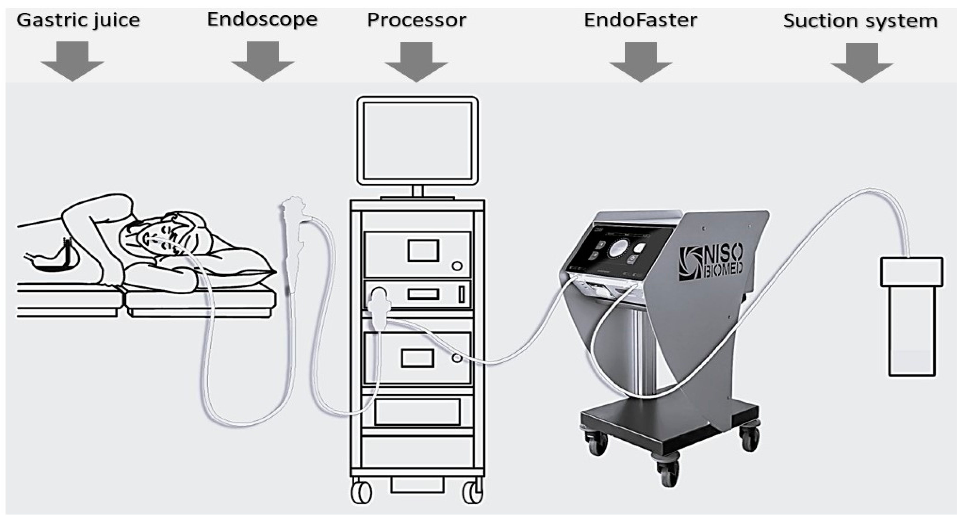
Figure 1. Gastric juice is aspirated during gastroscopy, passes through EndoFaster®, where it is analyzed in real time for both pH and ammonium concentration, and then it is discarded into the suction system.
Healthcare 11 03186 g001
Figure 2. Reduction in yearly CO2 production (kg) by using EndoFaster® according to the number of endoscopies and rate of standard biopsies performed. By considering values >1500, 1500–500, and <500 (arbitrarily chosen), the reduction could be considered high, intermediate, and low, respectively.
Healthcare 11 03186 g002
Table 1. CO2 production for consumable materials of EndoFaster® and standard biopsy sampling.
MaterialTypeQuantity
(g)
Waster
Disposal
Quantity/
Test
kg CO2/
kg Waste
kg CO2/
Test
Bottle 1 for calibration + liquid-draining systemPolyethylene (PE)78.6Plastic recycling1.313.00.004
Bottle 2 for calibration + liquid-draining systemPE51.6Plastic recycling0.863.00.003
Bottle 3 for calibration + liquid-draining systemPE51.6Plastic recycling0.863.00.003
Cardboard box for the 3 bottlesCarton137Cardboard
recycling
2.280.950.002
Washing solution tankHigh-density polyethylene
(HDPE)
146Plastic recycling2.433.00.007
Gastric juice suction tubePlastic (mixed)25Infected waste2.083.00.006
Biopsy forcepsPlastic (mixed)20Infected waste13.00.060
Biopsy jarPlastic-Infected waste23.00.081
Table 2. Estimation of yearly CO2 reduction by performing biopsies based on EndoFaster® results.
Total procedures/year2000
Standard biopsy sampling90%
Procedures with biopsy sampling1800
Reduction in biopsies through the use of EndoFaster®50%
Biopsy sampling avoided900
kg CO2 produced by EndoFaster® materials50
kg CO2 produced by EndoFaster® due to energy consumption and liquid residue disposal22
kg CO2 total normally produced (without biopsy reduction) 1262
kg CO2 total saved yearly through the use of EndoFaster®630
kg CO2 reduction558
Percentage of CO2 reduction 44%
Disclaimer/Publisher’s Note: The statements, opinions and data contained in all publications are solely those of the individual author(s) and contributor(s) and not of MDPI and/or the editor(s). MDPI and/or the editor(s) disclaim responsibility for any injury to people or property resulting from any ideas, methods, instructions or products referred to in the content.