


Validity and Reliability of a Non-Radiographic Postural Analysis Device Based on an RGB-Depth Camera Comparing EOS 3D Imaging: A Prospective Observational Study

Abstract
1. Introduction
- In people with somatic dysfunction, is PAViR reliable when shooting repeatedly?
- When applied as diagnostic imaging, is PAViR valid compared to the parameters of EOS?
2. Materials and Methods
2.1. Participants
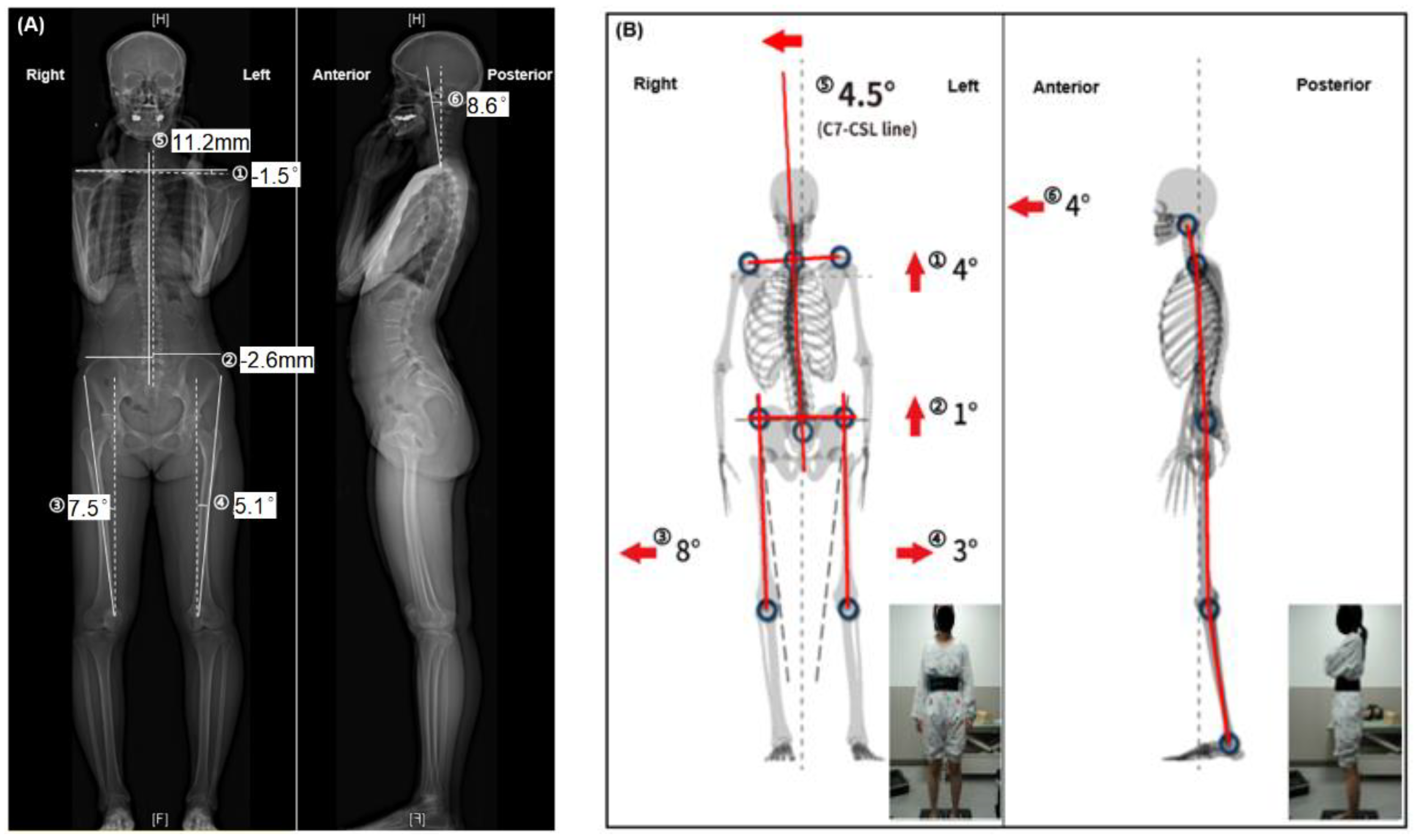
2.2. EOS
2.3. PAViR
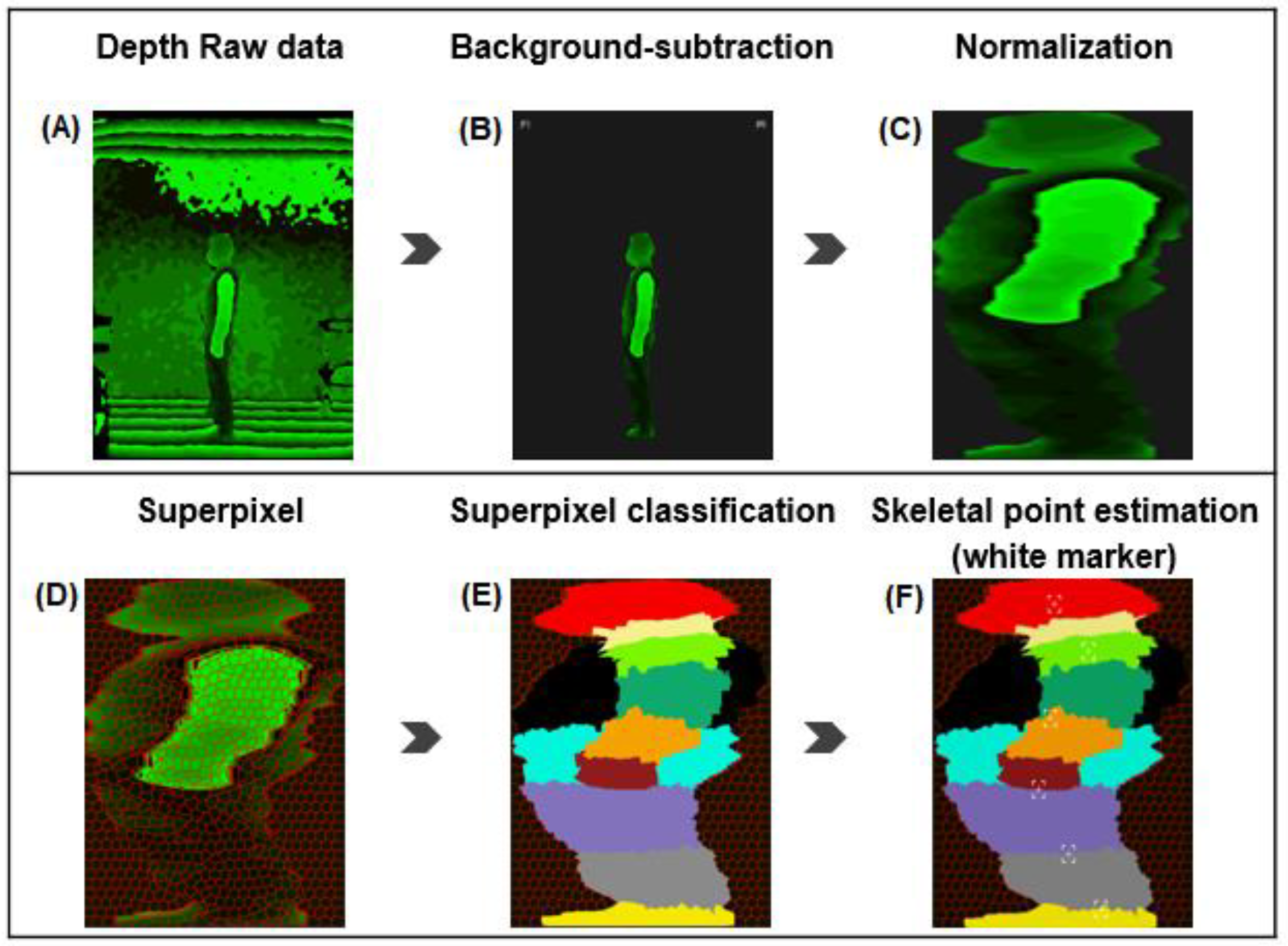
2.3.1. RGB-D Camera
2.3.2. Support Vector Machine
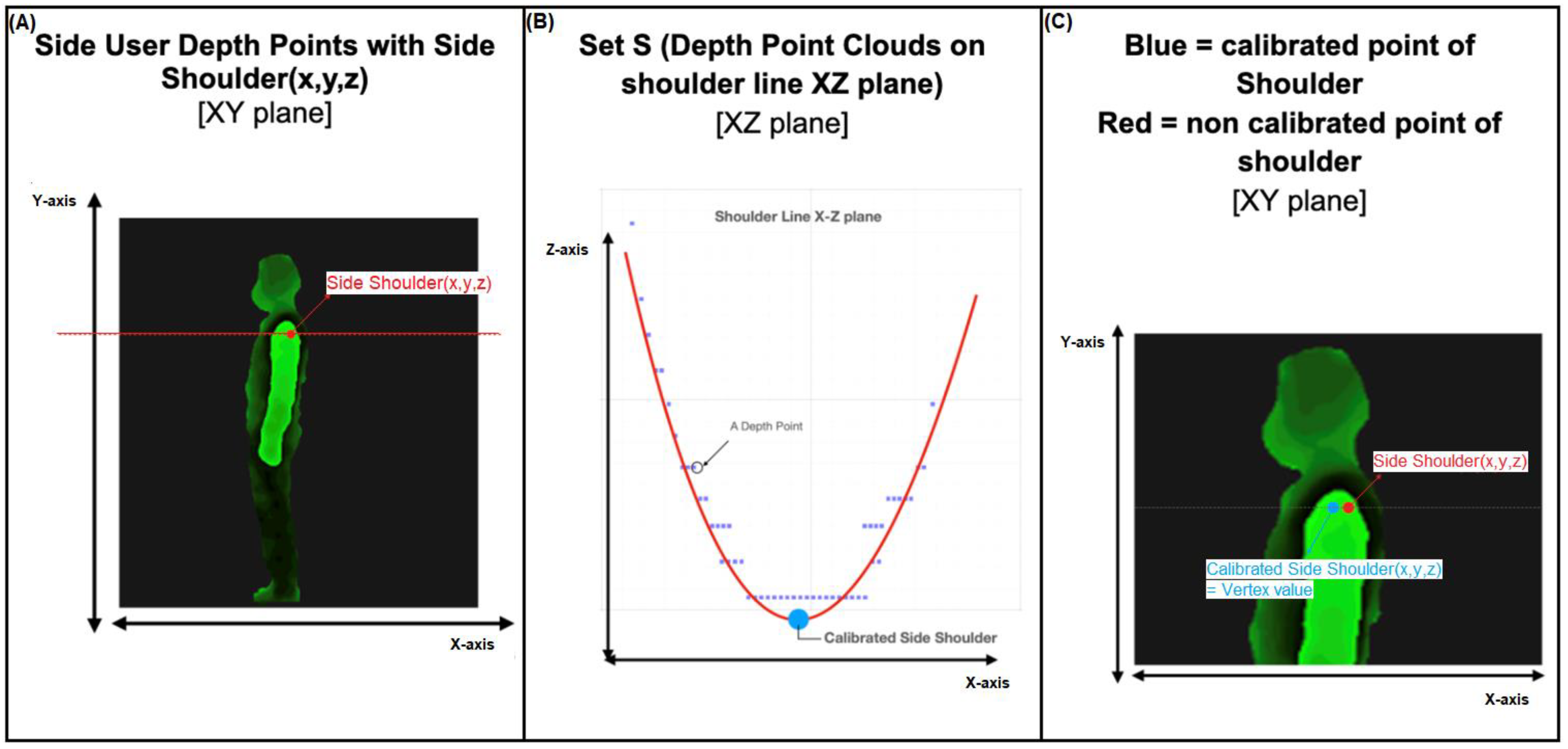
2.3.3. Geometric Method
2.4. Outcome Measures
2.5. Data Analysis
3. Results
3.1. Outcomes of Measuring with EOS and PAViR
3.2. Intra-Rater Reliability of PAViR
3.3. Validation of PAViR Compared to Parameters of EOS
4. Discussion
Study Limitations
5. Conclusions
Author Contributions
Funding
Institutional Review Board Statement
Informed Consent Statement
Data Availability Statement
Acknowledgments
Conflicts of Interest
References
- Raine, S.; Twomey, L. Attributes and qualities of human posture and their relationship to dysfunction or musculoskeletal pain. Crit. Rev. Phys. Rehabil. Med. 1994, 6, 409. [Google Scholar]
- Kamper, D.G.; Fischer, H.C.; Cruz, E.G. Impact of finger posture on mapping from muscle activation to joint torque. Clin. Biomech. (Bristol Avon.) 2006, 21, 361–369. [Google Scholar] [CrossRef]
- Magee, D.J. Orthopedic Physical Assessment (Musculoskeletal Rehabilitation); Saunders: Toronto, Canada, 2014. [Google Scholar]
- Pope, P.M. Severe and Complex Neurological Disability: Management of the Physical Condition; Butterworth-Heinemann: Oxford, UK, 2007. [Google Scholar]
- Läubli, T.; Karpilow, C. Global Occupational Health; Oxford university press: Oxford, UK, 2011; pp. 277–299. [Google Scholar]
- Takasaki, H.; May, S. Mechanical diagnosis and therapy has similar effects on pain and disability as ‘wait and see’ and other approaches in people with neck pain: A systematic review. J. Physiother. 2014, 60, 78–84. [Google Scholar] [CrossRef] [PubMed]
- Rey-Matias, R.R. 16—Manipulation, Traction, and Massage. In Braddom’s Rehabilitation Care: A Clinical Handbook; Cifu, D.X., Lew, H.L., Eds.; Elsevier: Amsterdam, The Netherlands, 2018; pp. 111–118.e8. [Google Scholar]
- Chaitow, L. Chapter 6—Osteopathic Assessment and Treatment of Thoracic and Respiratory Dysfunction. In Multidisciplinary Approaches to Breathing Pattern Disorders; Chaitow, L., Bradley, D., Gilbert, C., Ley, R., Eds.; Churchill Livingstone: Edinburgh, Scotland, 2002; pp. 131–172. [Google Scholar]
- Williams, N. Managing back pain in general practice is osteopathy the new paradigm? Br. J. Gen. Pract. 1997, 47, 653–655. [Google Scholar]
- Muller, R.; Linz, W.; Buchmann, J. Manual medicine--a powerful “hands on” facility to treat somatic and segmental dysfunction with musculosceletal pain, increased muscule tension, restrictions of fascia and posture asymmetries. MMW Fortschr. Med. 2011, 153, 27–30. [Google Scholar]
- Fortin, C.; Ehrmann Feldman, D.; Cheriet, F.; Labelle, H. Clinical methods for quantifying body segment posture: A literature review. Disabil. Rehabil. 2011, 33, 367–383. [Google Scholar] [CrossRef] [PubMed]
- Lim, Y.Z.; Chou, L.; Au, R.T.M.; Seneviwickrama, K.L.M.D.; Cicuttini, F.M.; Briggs, A.M.; Sullivan, K.; Urquhart, D.M.; Wluka, A.E. People with low back pain want clear, consistent and personalised information on prognosis, treatment options and self-management strategies: A systematic review. J. Physiother. 2019, 65, 124–135. [Google Scholar] [CrossRef]
- Legaye, J. Follow-up of the sagittal spine by optical technique. Ann Phys Rehabil Med 2012, 55, 76–92. [Google Scholar] [CrossRef]
- Cohen, L.; Kobayashi, S.; Simic, M.; Dennis, S.; Refshauge, K.; Pappas, E. Non-radiographic methods of measuring global sagittal balance: A systematic review. Scoliosis Spinal Disord. 2017, 12, 30. [Google Scholar] [CrossRef]
- Furlanetto, T.S.; Sedrez, J.A.; Candotti, C.T.; Loss, J.F. Photogrammetry as a tool for the postural evaluation of the spine: A systematic review. World J. Orthop. 2016, 7, 136–148. [Google Scholar] [CrossRef]
- Dubousset, J.; Charpak, G.; Dorion, I.; Skalli, W.; Lavaste, F.; Deguise, J.; Kalifa, G.; Ferey, S. A new 2D and 3D imaging approach to musculoskeletal physiology and pathology with low-dose radiation and the standing position: The EOS system. Bull. De L’academie Natl. De Med. 2005, 189, 287–297; discussion 297. [Google Scholar]
- Somoskeöy, S.; Tunyogi-Csapó, M.; Bogyó, C.; Illés, T. Accuracy and reliability of coronal and sagittal spinal curvature data based on patient-specific three-dimensional models created by the EOS 2D/3D imaging system. Spine J. 2012, 12, 1052–1059. [Google Scholar] [CrossRef] [PubMed]
- Deschenes, S.; Charron, G.; Beaudoin, G.; Labelle, H.; Dubois, J.; Miron, M.C.; Parent, S. Diagnostic imaging of spinal deformities: Reducing patients radiation dose with a new slot-scanning X-ray imager. Spine (Phila Pa 1976) 2010, 35, 989–994. [Google Scholar] [CrossRef] [PubMed]
- Hui, S.C.N.; Pialasse, J.-P.; Wong, J.Y.H.; Lam, T.-p.; Ng, B.K.W.; Cheng, J.C.Y.; Chu, W.C.W. Radiation dose of digital radiography (DR) versus micro-dose x-ray (EOS) on patients with adolescent idiopathic scoliosis: 2016 SOSORT- IRSSD “John Sevastic Award” Winner in Imaging Research. Scoliosis Spinal Disord. 2016, 11, 46. [Google Scholar] [CrossRef]
- Wall, B.F. Ionising Radiation Exposure of the Population of the United States: NCRP Report no. 160; Oxford University Press: Oxford, UK, 2009. [Google Scholar]
- Metaxas, V.I.; Messaris, G.A.; Lekatou, A.N.; Petsas, T.G.; Panayiotakis, G.S. Patient Doses in Common Diagnostic X-ray Examinations. Radiat. Prot. Dosim. 2019, 184, 12–27. [Google Scholar] [CrossRef]
- Kim, H.; Lee, S.; Lee, D.; Choi, S.; Ju, J.; Myung, H. Real-time human pose estimation and gesture recognition from depth images using superpixels and SVM classifier. Sensors 2015, 15, 12410–12427. [Google Scholar] [CrossRef]
- Straka, M.; Hauswiesner, S.; Rüther, M.; Bischof, H. Skeletal Graph Based Human Pose Estimation in Real-Time. In Proceedings of the British Machine Vision Conference, Dundee, UK, 29 August–2 September 2011; pp. 1–12. [Google Scholar]
- Achilles, F.; Ichim, A.-E.; Coskun, H.; Tombari, F.; Noachtar, S.; Navab, N. Patient MoCap: Human Pose Estimation under Blanket Occlusion for Hospital Monitoring Applications; Springer International Publishing: Cham, Switzerland, 2016; pp. 491–499. [Google Scholar]
- Faro, F.D.; Marks, M.C.; Pawelek, J.; Newton, P.O. Evaluation of a functional position for lateral radiograph acquisition in adolescent idiopathic scoliosis. Spine (Phila Pa 1976) 2004, 29, 2284–2289. [Google Scholar] [CrossRef]
- Garg, B.; Mehta, N.; Bansal, T.; Malhotra, R. EOS(R) imaging: Concept and current applications in spinal disorders. J. Clin. Orthop. Trauma 2020, 11, 786–793. [Google Scholar] [CrossRef]
- Pumberger, M.; Schmidt, H.; Putzier, M. Spinal Deformity Surgery: A Critical Review of Alignment and Balance. Asian Spine J. 2018, 12, 775–783. [Google Scholar] [CrossRef]
- Vergari, C.; Skalli, W.; Clavel, L.; Demuynck, M.; Valentin, R.; Sandoz, B.; Similowski, T.; Attali, V. Functional analysis of the human rib cage over the vital capacity range in standing position using biplanar X-ray imaging. Comput. Biol. Med. 2022, 144, 105343. [Google Scholar] [CrossRef]
- Vinod Gutta, E.D.L. Natalie Baddour, Pascal Fallavollita, A Comparison of Depth Sensors for 3D Object Surface Reconstruction. In The Canadian Medical and Biological Engineering Society; CMBES/SCGB: Ottawa, ON, Canada, 2019; Volume 42. [Google Scholar]
- Jang, C.W.; Park, J.; Cho, H.E.; Park, J.H. Appraisal of the new posture analyzing and virtual reconstruction device (PAViR) for assessing sagittal posture parameters: A prospective observational study. Int. J. Env. Res. Public Health 2022, 19, 11109. [Google Scholar] [CrossRef] [PubMed]
- Camplani, M.; Salgado, L. Background foreground segmentation with RGB-D Kinect data: An efficient combination of classifiers. J. Vis. Commun. Image Represent. 2014, 25, 122–136. [Google Scholar] [CrossRef]
- Achanta, R.; Shaji, A.; Smith, K.; Lucchi, A.; Fua, P.; Susstrunk, S. SLIC superpixels compared to state-of-the-art superpixel methods. IEEE Trans. Pattern Anal. Mach. Intell. 2012, 34, 2274–2282. [Google Scholar] [CrossRef] [PubMed]
- Fleiss, J.L.; Levin, B.; Paik, M.C. Statistical Methods for Rates and Proportions; John Wiley & Sons: Hoboken, NJ, USA, 2013. [Google Scholar]
- Campos, S.; Zhang, L.; Sinclair, E.; Tsao, M.; Barnes, E.A.; Danjoux, C.; Sahgal, A.; Goh, P.; Culleton, S.; Mitera, G. The palliative performance scale: Examining its inter-rater reliability in an outpatient palliative radiation oncology clinic. Support. Care Cancer 2009, 17, 685–690. [Google Scholar] [CrossRef] [PubMed]
- Kasten, K.M.; Lewis, D.D. High-Velocity, Low-Amplitude Management of Posterior Rib Somatic Dysfunction. J. Am. Osteopath. Assoc. 2020, 120, e1–e2. [Google Scholar] [CrossRef] [PubMed]
- Rousseau, M.-A.; Brusson, A.; Lazennec, J.-Y. Assessment of the axial rotation of the pelvis with the EOS® imaging system: Intra-and inter-observer reproducibility and accuracy study. Eur. J. Orthop. Surg. Traumatol. 2014, 24, 891–895. [Google Scholar] [CrossRef] [PubMed]
- Cicchetti, D.V. Guidelines, criteria, and rules of thumb for evaluating normed and standardized assessment instruments in psychology. Psychol. Assess. 1994, 6, 284. [Google Scholar] [CrossRef]
- Humbert, L.; De Guise, J.A.; Aubert, B.; Godbout, B.; Skalli, W. 3D reconstruction of the spine from biplanar X-rays using parametric models based on transversal and longitudinal inferences. Med. Eng. Phys. 2009, 31, 681–687. [Google Scholar] [CrossRef] [PubMed]
- Kösling, S.; Dietrich, K.; Steinecke, R.; Klöppel, R.; Schulz, H.-G. Diagnostic value of 3 D CT surface reconstruction in spinal fractures. Eur. Radiol. 1997, 7, 61–64. [Google Scholar] [CrossRef]
- Hocquelet, A.; Cornelis, F.; Jirot, A.; Castaings, L.; de Sèze, M.; Hauger, O. Patient-specific 3D models created by 3D imaging system or bi-planar imaging coupled with Moiré–Fringe projections: A comparative study of accuracy and reliability on spinal curvatures and vertebral rotation data. Eur. Spine J. 2016, 25, 3154–3161. [Google Scholar] [CrossRef]
- Daruwalla, J.; Balasubramaniam, P. Moiré topography in scoliosis. Its accuracy in detecting the site and size of the curve. J. Bone Joint Surg. Br. Vol. 1985, 67, 211–213. [Google Scholar]
- Berryman, F.; Pynsent, P.; Fairbank, J.; Disney, S. A new system for measuring three-dimensional back shape in scoliosis. Eur. Spine J. 2008, 17, 663–672. [Google Scholar] [CrossRef] [PubMed]


| Variables (n = 100) | Values | Range |
|---|---|---|
| Gender, n male/female | 44/56 | |
| Age (y), (mean ± SD) | 47.2 ± 16.5 | 19~81 |
| Weight (kg), (mean ± SD) | 63.3 ± 13.1 | 37.4~89.3 |
| Body height (cm), (mean ± SD) | 163.4 ± 19.3 | 143.0~183.0 |
| Body mass index (kg/m2), (mean ± SD) | 23.1 ± 3.5 | 15.6~30.8 |
| EOS | PAViR | ||||
|---|---|---|---|---|---|
| Parameters | Mean ± SD | Range | Mean ± SD | Range | |
| Coronal view | Asymmetric clavicle height (°) | 0.1 ± 2.8 | −8.0 a~16.0 | −1.0 ± 1.6 | −5.0~3.8 |
| Pelvic oblique (mm, °) b | −0.3 ± 5.0 | −12.0~14.0 | 0.7 ± 1.6 * | −2.6~6.4 | |
| Right Q angle (°) | 6.1± 1.7 | −1.8~10.4 | 0.9 ± 7.9 * | −8.0~14.0 | |
| Left Q angle (°) | 5.6 ± 1.6 | 0.9~9.8 | −3.1 ± 4.0 * | −7.9~13.5 | |
| C7-CSL (mm, °) b | −3.0 ± 13.3 | −59.0~36.0 | −1.3 ± 2.2 * | −8.1~4.3 | |
| Sagittal view | Forward head posture (°) | 7.0 ± 6.9 | −5.1~29.4 | 7.9 ± 6.3 | −5.0~29.0 |
| Parameters | Coefficient Value | p-Value |
|---|---|---|
| Asymmetric clavicle height | 0.69 | 0.005 |
| Pelvic oblique | 0.72 | 0.002 |
| Right Q angle of knee | 0.72 | 0.001 |
| Left Q angle of knee | 0.79 | 0.001 |
| C7-CSL | 0.84 | 0.002 |
| Forward head posture | 0.76 | 0.001 |
| Parameters | Correlation Coefficient | p-Value |
|---|---|---|
| Asymmetric clavicle height | 0.37 | <0.002 |
| Pelvic oblique | 0.32 | <0.002 |
| Right Q angle of knee | −0.47 | 0.14 |
| Left Q angle of knee | −0.15 | 0.15 |
| C7-CSL | 0.42 | <0.001 |
| Forward head posture | 0.39 | <0.002 |
Disclaimer/Publisher’s Note: The statements, opinions and data contained in all publications are solely those of the individual author(s) and contributor(s) and not of MDPI and/or the editor(s). MDPI and/or the editor(s) disclaim responsibility for any injury to people or property resulting from any ideas, methods, instructions or products referred to in the content. |
© 2023 by the authors. Licensee MDPI, Basel, Switzerland. This article is an open access article distributed under the terms and conditions of the Creative Commons Attribution (CC BY) license (https://creativecommons.org/licenses/by/4.0/).
Share and Cite
Lee, H.J.; Cho, H.E.; Kim, M.; Chung, S.Y.; Park, J.H. Validity and Reliability of a Non-Radiographic Postural Analysis Device Based on an RGB-Depth Camera Comparing EOS 3D Imaging: A Prospective Observational Study. Healthcare 2023, 11, 686. https://doi.org/10.3390/healthcare11050686
Lee HJ, Cho HE, Kim M, Chung SY, Park JH. Validity and Reliability of a Non-Radiographic Postural Analysis Device Based on an RGB-Depth Camera Comparing EOS 3D Imaging: A Prospective Observational Study. Healthcare. 2023; 11(5):686. https://doi.org/10.3390/healthcare11050686
Chicago/Turabian StyleLee, Hyo Jeong, Han Eol Cho, Myungsang Kim, Seok Young Chung, and Jung Hyun Park. 2023. "Validity and Reliability of a Non-Radiographic Postural Analysis Device Based on an RGB-Depth Camera Comparing EOS 3D Imaging: A Prospective Observational Study" Healthcare 11, no. 5: 686. https://doi.org/10.3390/healthcare11050686
APA StyleLee, H. J., Cho, H. E., Kim, M., Chung, S. Y., & Park, J. H. (2023). Validity and Reliability of a Non-Radiographic Postural Analysis Device Based on an RGB-Depth Camera Comparing EOS 3D Imaging: A Prospective Observational Study. Healthcare, 11(5), 686. https://doi.org/10.3390/healthcare11050686