Article

Antimicrobial Resistance in Sepsis Cases Due to Escherichia coli and Klebsiella pneumoniae: Pre-Pandemic Insights from a Single Center in Southwestern Romania

by
Lucian-Ion Giubelan
1,2,
Alexandru Ionuț Neacșu
1,*,
Alexandra Daniela Rotaru-Zavaleanu
3 and
Eugen Osiac
3,4
1
Department of Infectious Diseases, University of Medicine and Pharmacy of Craiova, 200349 Craiova, Romania
2
Infectious Diseases and Pulmonology ‘Victor Babes’ Hospital, 200515 Craiova, Romania
3
Experimental Research Center for Normal and Pathological Aging, Department of Functional Sciences, University of Medicine and Pharmacy of Craiova, 200349 Craiova, Romania
4
Department of Biophysics, University of Medicine and Pharmacy of Craiova, 200349 Craiova, Romania
*
Author to whom correspondence should be addressed.
Healthcare 2024, 12(17), 1713; https://doi.org/10.3390/healthcare12171713
Submission received: 11 July 2024 / Revised: 26 August 2024 / Accepted: 26 August 2024 / Published: 27 August 2024

Abstract

:
Sepsis is an uncontrolled reaction of the body to an infection, and if not effectively treated, it can progress to septic shock, multiple organ failure, and ultimately, death. Objective: To determine the resistance profile of Escherichia coli (E. coli) and Klebsiella pneumoniae (K. pneumoniae) strains isolated in sepsis cases diagnosed at the Infectious Diseases Clinic in Craiova, Romania. Methods: The bacteria responsible for sepsis cases were identified using the Vitek 2 Systems version 06.01, which was then employed to assess their antimicrobial susceptibility (Global CLSI and Phenotypic 2017). Results: We have identified 989 patients diagnosed with bacterial sepsis. Among these, 953 cases were caused by Gram-negative rods, with 415 attributed to E. coli and 278 to K. pneumoniae. High levels of resistance to ampicillin were recorded for E. coli strains isolated in sepsis cases (64.6%); adding sulbactam lowers the level of resistance to 41.8%. Resistance to 3rd generation cephalosporins varied between 7.47 and 14.6% and another 3.41 to 11.1% are dose-dependent susceptibility strains. Resistance to carbapenems (i.e., ertapenem, meropenem) is low—2.18–2.42%. More than 95% of the tested K. pneumoniae strains were resistant to ampicillin and adding sulbactam as a β-lactamase inhibitor only halves that level. Resistance to 3rd generation cephalosporins varied between 20.7% and 22.5%; resistance levels for K. pneumoniae were notably higher than those for E. coli. Over 95% of K. pneumoniae strains showed resistance to ampicillin, and resistance to 3rd generation cephalosporins varied between 20.7% and 22.5%. Additionally, K. pneumoniae exhibited higher resistance to carbapenems (13.7–19.5%) compared to E. coli (2.18–2.42%). Conclusions: Antimicrobial resistance levels are generally lower than continental and national data, except for ampicillin and carbapenems (meropenem and ertapenem). K. pneumoniae strains are significantly more resistant than E. coli strains.

1. Introduction

Sepsis, a life-threatening condition caused by the body’s extreme response to infection, remains a significant cause of morbidity and mortality worldwide [1,2,3]. Data from 2017 estimated that approximately 49 million cases of sepsis and 11 million deaths occurred worldwide [4]. According to the Centers for Disease Control and Prevention (CDC), 33% of patients who died in the hospital also developed sepsis [5]. In recent years, the incidence of sepsis appears to be decreasing; however, the mortality rate remains high, with 24% occurring after 30 days and 32% after 90 days [6]. Early recognition of the syndrome is crucial, since it was estimated that 87% of the patients had sepsis or the causative infections before hospitalization [5]. While sepsis can be caused by viruses, fungi, or parasites, bacteria, particularly Gram-positive cocci and Gram-negative rods, represent the most significant etiology of the clinical syndrome [7]. Sepsis treatment is complicated by bacterial resistance to antimicrobials, and the degree of resistance (multidrug-resistant, extensively drug-resistant, and pandrug-resistant) is well-defined [8,9,10]. In 2017, the World Health Organization (WHO) published a list of bacteria for which the development of new antimicrobials is urgently needed [11]; five of these bacteria are Gram-negative rods (the first three of them listed as priority 1—critical), and their resistance to third-generation cephalosporins, carbapenems, and fluoroquinolones is on the focus, while another matter of concern is resistance to polymyxin [12,13]. The rapid rise in antimicrobial resistance has emerged as a formidable challenge in treating bacterial infections, particularly in sepsis cases [14].
Antimicrobial resistance in E. coli and K. pneumoniae has been a focal point of numerous studies due to its significant impact on public health [15]. Recent research by Ventola emphasizes that antibiotic resistance is a critical challenge in healthcare, necessitating urgent action to develop new treatment strategies and stewardship programs [16]. Additionally, a study by Cassini et al. quantified the burden of antimicrobial resistance in Europe, demonstrating that resistant infections are associated with substantial morbidity and mortality [17].
This study aims to provide a detailed demographic and resistance profile analysis of E. coli and K. pneumoniae sepsis cases at the ‘Victor Babeș’ Hospital in Craiova. By examining the distribution of these infections across different specimen types and their resistance patterns, we seek to contribute valuable insights that can better inform clinical practice and public health strategies.
This study focuses on the pre-pandemic period, aiming to provide baseline data on antimicrobial resistance in sepsis cases caused by E. coli and K. pneumoniae. Understanding the resistance patterns in this period can help in comparing and contrasting with post-pandemic data, which may be influenced by changes in healthcare practices, antibiotic usage, and infection control measures brought on by the COVID-19 pandemic.

2. Materials and Methods

2.1. Study Design and Setting

A descriptive, retrospective study was conducted at the Infectious Diseases Clinic of ‘Victor Babeș’ Hospital in Craiova, Romania, from 1 January 2017 to 31 December 2019. This period was chosen to represent the pre-pandemic era of the Severe Acute Respiratory Syndrome Coronavirus 2 (SARS-CoV-2).

2.2. Data Collection

The data retrieved from the patients’ files (demographic, date of sampling, specimen sampled, identified germs, and susceptibility to antimicrobials) were used to create a Microsoft Excel database.

2.3. Inclusion Criteria

Patients diagnosed with sepsis (SOFA score ≥ 2) based on the Third International Consensus Definitions for Sepsis and Septic Shock (Sepsis-3) were included [1]. The bacteria involved in sepsis cases were isolated from regular wards.

2.4. Microbiological Identification

Bacterial identification and antimicrobial susceptibility testing were performed using the Vitek 2 Systems version 06.01, adhering to Global CLSI and Phenotypic 2017 standards [18,19,20].

2.5. Data Analysis

The Multiple Antibiotic Resistance Index (MAR index) was calculated for each strain, with values ranging from 0 to 1 [21]. Statistical analysis was conducted using the two-tailed Chi-squared test with Yates correction and the unpaired t-Student test, with significance set at p < 0.05 [22]. Microsoft Excel 365, version 2407 (Build 17830.20166, Click-to-Run) was used to perform the calculations.

2.6. Ethical Considerations

Informed consent was obtained from all subjects, and the study was approved by the Ethics Committee of the University of Medicine and Pharmacy from Craiova, Romania (nr. 251/06.11.2023). Our hospital adhered to General Data Protection Regulation (GDPR) rules, and all patients’ names were coded to ensure confidentiality.

3. Results

Between 2017 and 2019, we identified 989 patients diagnosed with bacterial sepsis (Gram-positive cocci and Gram-negative bacilli). Out of this number, there were 953 cases of sepsis due to Gram-negative rods, with 415 (43%) cases attributed to E. coli, 278 (26%) to K. pneumoniae, and 260 other species (Proteus spp., Pseudomonas aeruginosa, Acinetobacter baumannii). This study focused on Escherichia coli and Klebsiella pneumoniae as these pathogens accounted for 72.7% of the total Gram-negative sepsis cases. The remaining 260 cases, though identified as sepsis contributors, involved diverse bacterial species with smaller individual sample sizes. Due to the low occurrence of these species and to maintain statistical significance, they were excluded from the analysis.
Table 1 presents the demographic characteristics of patients with sepsis caused by E. coli and K. pneumoniae. Noteworthy findings include a significantly higher proportion of females in the E. coli group, while males were more predominant in the K. pneumoniae group. Additionally, the average age of patients in the K. pneumoniae group was higher than in the E. coli group. The average age of E. coli cases was 53.03 ± 19.31 years (ranging from 18 to 94 years), while in K. pneumoniae cases, it was 56.61 ± 15.16 years (ranging from 16 to 88 years), with a significant difference (p = 0.0117). The majority of patients in E. coli cases were females, accounting for 282 (67.95%), while in K. pneumoniae cases, males were predominant, comprising 155 (55.76%) of the cases, with a highly significant difference (p < 0.0001). Most patients resided in rural settings in both groups, with 217 (52.29%) in E. coli cases and 144 (51.8%) in K. pneumoniae cases. Subjects were recruited from the following counties: Dolj (555, 80.09%), Olt (74, 10.66%), Gorj (40, 5.77%), Vȃlcea (13, 1.88%), Mehedinți (8, 1.15%), Argeș (2, 0.29%), and Caraș-Severin (1, 0.14%), covering the southwestern part of Romania. Demographic data of E. coli and K. pneumoniae are shown in Table 1.
The demographic data in Table 1 present the burden of E. coli and K. pneumoniae infections in the ‘Victor Babeș’ Hospital. This study observed differences in infection rates between counties. Dolj county had the highest number of sepsis cases (555, 80.09%), while Mehedinți and other smaller counties had significantly fewer cases.
For all patients, the median SOFA score was 2 (ranging from 2 to 3), with no statistically significant differences observed between E. coli and K. pneumoniae cases. All microbiological samples were collected prior to the introduction of antimicrobials.
Table 2 presents data on the sources of sepsis caused by E. coli and K. pneumoniae, detailing the number and percentage of cases from various specimen types.
The majority of E. coli cases were detected through urine samples (323, 77.83%), whereas K. pneumoniae cases predominantly originated from sputum samples (175, 62.95%).
The significant differences in the distribution of cases from sputum and urine between the two pathogens suggest varying primary infection sites and subsequent pathways to sepsis. These findings underscore the importance of targeted diagnostic and treatment strategies based on the underlying source of infection.
Figure 1 presents a heatmap of the antimicrobial susceptibility patterns of E. coli isolates. The heatmap indicates the percentage of isolates susceptible, intermediate, and resistant to various antibiotics.
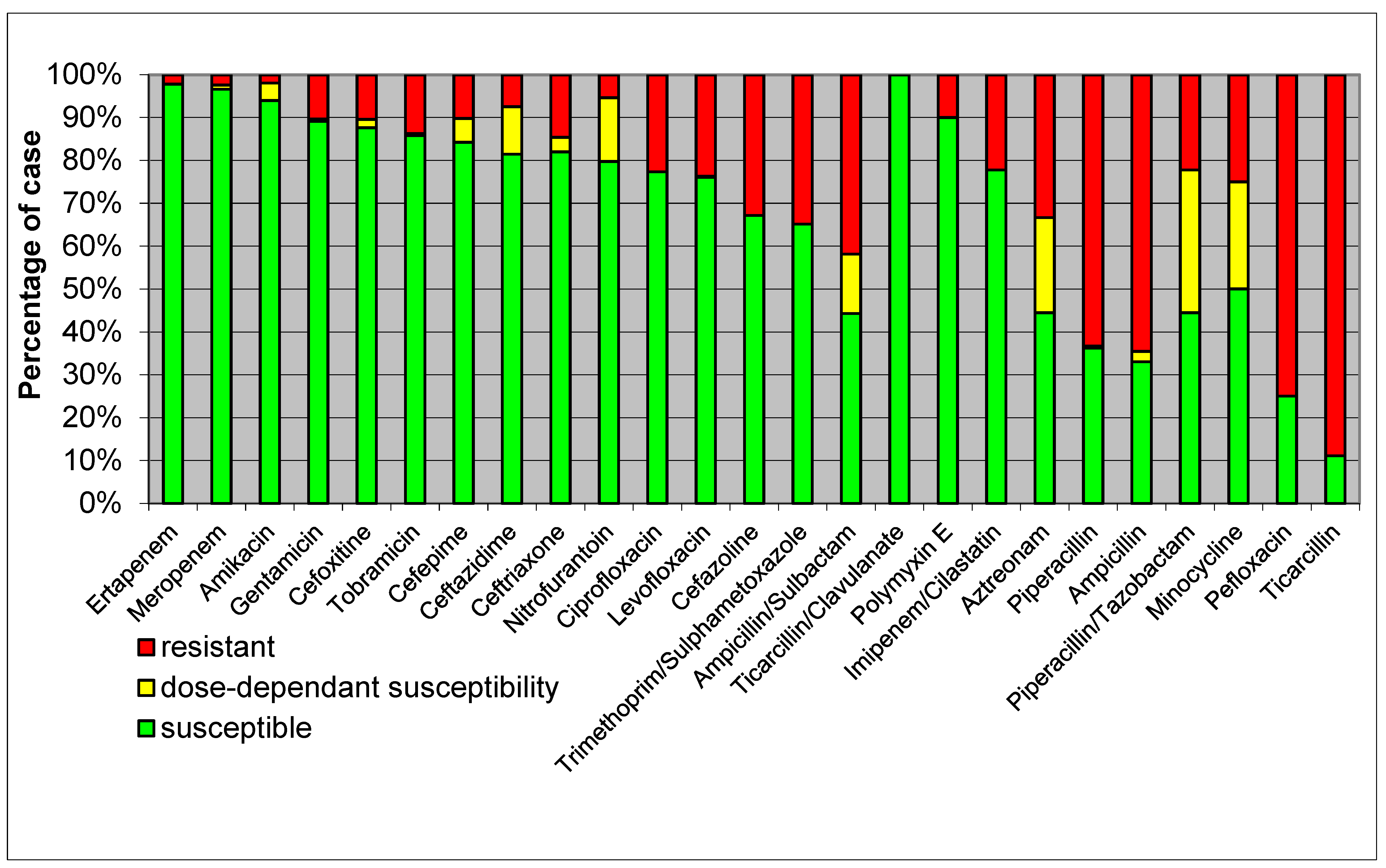
  • High Resistance: E. coli isolates showed high resistance to ampicillin, reflecting common resistance patterns seen globally.
  • Moderate Resistance: there was moderate resistance to cephalosporins such as ceftriaxone and ceftazidime, highlighting the presence of extended-spectrum β-lactamase (ESBL)-producing strains.
  • Low Resistance: the lowest resistance rates were observed for carbapenems like meropenem and imipenem, indicating their continued effectiveness, though vigilance is required due to emerging resistance.
Elevated levels of resistance to ampicillin were noted among E. coli strains isolated from sepsis cases (64.6%). The inclusion of sulbactam decreased the resistance level to 41.8%; however, it remains insufficient for empirical treatment in these instances. The antimicrobial susceptibility of E. coli strains isolated in sepsis cases is depicted in Figure 1.
Figure 2 displays a heatmap of the antimicrobial susceptibility patterns of K. pneumoniae isolates, similar to Figure 1, showcasing their response to different antibiotics.
  • High Resistance: K. pneumoniae exhibited high resistance to β-lactams, including ampicillin and cefazoline, suggesting widespread β-lactamase production.
  • Moderate Resistance: there was moderate resistance to quinolones such as ciprofloxacin, indicating a significant but lesser extent of resistance compared to β-lactams.
  • Low Resistance: susceptibility to carbapenems like meropenem remained relatively high, although the presence of carbapenem-resistant strains is a growing concern.
Over 95% of the tested K. pneumoniae strains exhibited resistance to ampicillin, and the addition of sulbactam as a β-lactamase inhibitor only reduced this level by half. Nevertheless, dose-dependent susceptibility increased from 2.67% to 17.3%. Antimicrobial susceptibility of K. pneumoniae isolated in sepsis cases is shown in Figure 2.
The average Multiple Antibiotic Resistance (MAR) index of E. coli isolated in sepsis cases was 0.21 ± 0.21. The average MAR index of K. pneumoniae isolated in sepsis cases was 0.35 ± 0.25 (p < 0.0001 as compared with E. coli cases); Figure 3 illustrates the relationship between the number of E. coli and K. pneumoniae strains and specific MAR index values.
There were no statistically significant differences related to patients’ gender or settings. As for age groups, the data are presented in Figure 4.

4. Discussion

Sepsis is one of the most critical emergency issues in practicing medicine. Among the top 10 leading global causes of sepsis are lower respiratory tract infections, diarrheal diseases, HIV infection, malaria, and tuberculosis [4]. The highest incidence of sepsis cases is found in Africa, South and Central Americas, and South and Southeast Asia (ranging from 340 cases per 100,000 inhabitants to up to 10 times this number). In Europe (excluding the European part of Russia, Ukraine, and Turkey), the incidence varies between 120 and 270 cases per 100,000 inhabitants [4]. However, estimating sepsis cases and incidence based on the International Statistical Classification of Diseases and Related Health Problems, 10th version (ICD 10) might not be quite accurate. A recent paper evaluating cases from Scania, Sweden suggests that the real incidence is much higher, with about 747 cases per 100,000 habitants [23]. The resistance patterns observed in E. coli and K. pneumoniae underscore the ongoing challenge of managing infections caused by these pathogens, especially in vulnerable patient populations [24].
Among sepsis cases with identified causes, bacterial etiologies are predominant, with Gram-negative rods (mainly E. coli) responsible for 62.2% of cases, while Gram-positive bacteria (primarily Staphylococcus aureus) account for 46.8%. Polymicrobial infections are also observed [25]. Our study has found a striking discrepancy between Gram-negative and Gram-positive bacteria causing sepsis, the former being almost 25 times more isolated than the latter. The observed difference could potentially be attributed to our clinic’s patient profile, as cases related to pulmonology (including tuberculosis) are managed in a nearby facility. Tuberculosis (TB) is a major cause of pulmonary infections worldwide. In regions with a high incidence of TB, the disease can substantially impact the overall burden of pulmonary infections. A study highlighted that TB-related complications such as acute respiratory distress syndrome (ARDS), hospital-acquired pneumonia, and sepsis are common in critically ill patients, contributing to high mortality rates in these patients. The high incidence of TB in our region and its management in a dedicated facility could explain the lower numbers of pulmonary infections reported in our study, as our data primarily reflect non-tuberculosis pulmonary infections [26,27]. In our clinic, tuberculosis cases are managed separately in a dedicated facility, which might influence the lower numbers of pulmonary infections reported in this manuscript. The separation ensures that tuberculosis patients receive specialized care and reduces the risk of transmission to other patients. Consequently, the numbers reported in our study primarily reflect non-tuberculosis pulmonary infections, providing a more accurate picture of sepsis cases caused by other bacterial pathogens. This discrepancy aligns with general expectations where Gram-negative bacteria are more frequently isolated in sepsis cases compared to Gram-positive bacteria. For instance, a study reported that Gram-negative bacteria accounted for approximately 62% of sepsis cases, while Gram-positive bacteria accounted for about 47% [28,29]. The current study also confirms that among all cases of Gram-negative bacterial sepsis, E. coli is the most frequently isolated pathogen, with K. pneumoniae isolates being identified almost 1.5 times less frequently.
In relation to gender, sepsis appears to be more common in males [4]. However, the current study reports approximately 20% more female cases than male cases diagnosed with sepsis. Nevertheless, when considering specific etiologies, E. coli and urosepsis are more prevalent in females, while K. pneumoniae and respiratory causes of sepsis are more common in males.
High levels of resistance to ampicillin were observed in E. coli strains isolated from sepsis cases (64.6%) [30]. The addition of sulbactam reduced the level of resistance to 41.8%, but it remains suboptimal for empirical treatment in such cases. This level of resistance aligns with European data, where the average resistance to ampicillin alone is 57.4%, and it is consistent with the national antimicrobial resistance study CARMIN-ROM 2017, which reported a resistance rate of 68.7% [31,32]. Resistance to 3rd generation cephalosporins varied between 7.47% and 14.6%, with an additional 3.41% to 11.1% being dose-dependent susceptibility strains. This is slightly lower than the European results, which show a resistance rate of 15.1%, and national data, which are around 20% [31,32]. Except for imipenem/cilastatin, to which only a few E. coli strains have been tested, resistance to carbapenems (ertapenem, meropenem) is low, ranging from 2.18% to 2.42%. However, European and national data show even lower percentages (0.1% European data vs. 0.39% national data) [31,32]. Antimicrobial policies implemented in our clinic restricted the use of carbapenems and future studies will assess the effect of these rules. The resistance levels for ciprofloxacin and levofloxacin are 22.7% and 23.7%, respectively, slightly lower than continental and national data [31,32]. We have also recorded a low level of resistance to aminoglycosides (1.93 up to 13.7%), the lowest being recorded for amikacin. Polymyxin E is considered the last resort in the fight against Gram-negative rods [33]; we have tested only ten E. coli strains, but, worrisome, one of them proved resistant to this antimicrobial. Evolution in time of the resistance levels and the effects of local antimicrobial policies will be assessed by future studies.
More than 95% of the tested K. pneumoniae strains were resistant to ampicillin, and adding sulbactam as a β-lactamase inhibitor only halved that level; however, the dose-dependent susceptibility increased from 2.67% to 17.3%. Continental and national data basically skipped this testing due to extremely high levels of resistance. Resistance to 3rd generation cephalosporins varied between 20.7% and 22.5% (depending on the tested antimicrobial), and another 16.8–21.4% of the strains showed dose-dependent susceptibility. This level of resistance is lower than the 31.2–31.7% European data [31]. The resistance levels for meropenem and ertapenem were 13.7% and 19.5%, respectively, which is 2 to 3 times higher than the continental average, raising serious concerns. Of the slightly over 200 tested K. pneumoniae strains, the resistance to levofloxacin and ciprofloxacin was 24.5–26.2%, which is lower than EU data and significantly 2.5 times lower than national data [31,32]. Also, aminoglycosides’ level of resistance is comparable with that recorded in Europe (19.8% vs. 22–24%) [31]. Another concerning finding is that we identified two Klebsiella pneumoniae strains resistant to polymyxin E, although only 20 isolates were tested for this antimicrobial. It is to be mentioned that our study found out that, for every specific age group, K. pneumoniae strains were 1.5 up to 2 times more resistant to antimicrobials as compared with E. coli isolates. Understanding the mechanisms underlying antibiotic resistance, such as β-lactamase production, is crucial for developing effective treatment strategies [34].
According to Krumperman PH [21], bacterial strains with an MAR index below 0.2 are acquired from sources with no or little exposure to antimicrobials (i.e., community). MAR indices above 0.2 suggest hospital-related sources. From this perspective, our E. coli and K. pneumoniae isolates originated almost equally from community and hospital sources. The average MAR index demonstrated that K. pneumoniae is significantly more resistant to antimicrobials compared to E. coli. An interesting aspect is the variation in the average MAR index across different age groups. Although the number of isolates is limited for each category (E. coli and K. pneumoniae), it is noteworthy that higher-than-average levels of resistance were observed in very young and very old individuals, while those in middle age exhibited MAR index levels lower than the average. Generally, we would anticipate a consistent increase in the MAR index as an individual’s age increases. This aligns with findings from Naqvi and Drlica, which highlight how fluoroquinolone resistance can complicate treatment strategies and influence resistance trends over time [35]. We will continue to explore this aspect, but the authors’ opinion is that, for our patients, these data should be viewed as a temporal trend and may indicate an increase in the average level of antimicrobial resistance among younger generations compared to middle-aged generations.
The findings from this study reveal distinct patterns in the sources of sepsis caused by E. coli and K. pneumoniae, as well as their respective resistance profiles. The predominance of E. coli in urinary tract infections leading to sepsis is consistent with previous studies [36]. This pathogen’s high incidence in urine samples (77.83%) underscores the need for effective urinary tract infection management to prevent the progression to sepsis.
K. pneumoniae, on the other hand, is primarily associated with respiratory tract infections, as indicated by the high proportion of sputum samples (62.95%). This aligns with research by Podschun and Ullmann [37], which highlights the pathogen’s role in pneumonia, particularly in hospital settings. The statistical significance of the differences in sputum and urine sources between the two pathogens (p < 0.0001) further supports the need for tailored diagnostic and therapeutic approaches.
The present research is a descriptive and retrospective, single-center analysis, with a relatively small number of cases. Most of our subjects were only from Dolj County. All types of samples from different areas of the clinic have been counted.

4.1. Study Limitations

While this study does have some limitations, it still provides important insights into antimicrobial resistance. It is true that being a single-center, retrospective study means the findings might not apply everywhere, as the data came from the Infectious Diseases Clinic of ‘Victor Babeș’ Hospital in Craiova, Romania, and might not reflect broader trends. This study also only covers the pre-pandemic years (2017–2019), so any changes in resistance patterns due to the COVID-19 pandemic are not captured here. Additionally, the sample size, while sufficient for the main analysis, might not be large enough to pick up on smaller differences in resistance patterns or demographic factors. Despite these limitations, this study still offers valuable information that can help guide future research and deepen our understanding of antimicrobial resistance in this specific context.

4.2. Implications for Future Research and Practice

The pre-pandemic data serve as a crucial baseline for understanding the impact of the COVID-19 pandemic on antimicrobial resistance [38]. During the pandemic, changes in antibiotic prescribing practices, increased use of broad-spectrum antibiotics, and shifts in hospital infection control protocols may have influenced resistance patterns. By comparing these findings with post-pandemic data, we can assess the impact of the pandemic on antimicrobial resistance and develop more effective strategies for managing sepsis in the future.
This study highlights the importance of continuous monitoring of antimicrobial resistance and the need for targeted interventions to reduce the burden of resistant infections. The demographic data presented underscore the significant burden of E. coli and K. pneumoniae infections, particularly in rural settings and among older populations. Tailoring infection control and prevention strategies to these demographics can help mitigate the impact of these infections.
The authors plan to use a similar approach for post-pandemic analysis, emphasizing the comparison of resistance patterns before and after the COVID-19 pandemic. This will involve collecting data from the same clinical settings and using consistent methodologies for identifying bacterial strains and assessing antimicrobial susceptibility. The analysis will account for changes in healthcare practices, antibiotic usage, and infection control measures due to the pandemic. By maintaining methodological consistency, the authors aim to provide a clear comparison of how the pandemic has influenced antimicrobial resistance in sepsis cases in future articles.

5. Conclusions

Our data reveal that most sepsis cases with an identified cause diagnosed at the ‘Victor Babeș’ Hospital in Craiova, Romania were attributed to Gram-negative rods. E. coli and K. pneumoniae together accounted for over 70% of these cases. Urinary sources are the predominant cause of sepsis in E. coli cases, while respiratory sources are more prevalent in K. pneumoniae cases. Antimicrobial resistance levels are generally lower than continental and national data, except for ampicillin and carbapenems (meropenem and ertapenem). K. pneumoniae strains exhibit significantly higher resistance compared to E. coli stains. There might be an increase in the average level of antimicrobial resistance in the younger generation compared to the middle-aged generation. This study will serve as a foundation for our further investigations into the etiology of sepsis and antimicrobial resistance in the post-pandemic period.
This study provides valuable insights into the demographic characteristics, sources, and antimicrobial resistance profiles of E. coli and K. pneumoniae sepsis cases at the ‘Victor Babeș’ Hospital in Craiova. The distinct patterns observed underscore the importance of targeted diagnostic and therapeutic strategies. Addressing the high resistance rates requires coordinated efforts across clinical practice and public health domains. Future research should focus on longitudinal studies to monitor resistance trends and evaluate the impact of intervention strategies.
The high resistance rates observed necessitate urgent action to revise treatment guidelines and ensure the judicious use of antibiotics. Clinicians should consider local resistance patterns when selecting empirical therapies for sepsis. A 2023 review emphasized the importance of timely management of sepsis and septic shock. Key recommendations include the prompt administration of empirical antimicrobial treatment, appropriate fluid replacement, and the use of vasoactive agents like norepinephrine to maintain blood pressure. These measures are crucial for reducing mortality rates associated with sepsis [39]. Moreover, enhancing diagnostic capabilities to rapidly identify resistant strains can improve treatment outcomes and reduce the spread of resistant antibiotic infections. Future research should prioritize the development of novel antimicrobial agents and the implementation of robust surveillance systems to monitor resistance trends [40,41].

Author Contributions

Conceptualization, L.-I.G., A.I.N. and E.O.; methodology, L.-I.G. and E.O.; software, A.I.N.; validation, L.-I.G., A.I.N., E.O. and A.D.R.-Z.; formal analysis, L.-I.G. and A.D.R.-Z.; investigation, L.-I.G. and A.I.N.; resources, L.-I.G.; data curation, A.I.N. and L.-I.G.; writing—original draft preparation, L.-I.G. and A.I.N.; writing—review and editing, L.-I.G. and A.I.N.; visualization, L.-I.G. and A.I.N.; supervision, L.-I.G. and E.O.; project administration, L.-I.G., A.I.N. and E.O. All authors have read and agreed to the published version of the manuscript.

Funding

This research received no external funding.

Institutional Review Board Statement

This study was approved by the Ethics Committee of the University of Medicine and Pharmacy from Craiova, Romania (nr. 251/06.11.2023, approved on 6 November 2023).

Informed Consent Statement

Informed consent was obtained from all subjects involved in the study.

Data Availability Statement

The data presented in this study are available upon request from the corresponding authors.

Conflicts of Interest

The authors declare no conflicts of interest.

References

  1. Singer, M.; Deutschman, C.S.; Seymour, C.W.; Shankar-Hari, M.; Annane, D.; Bauer, M.; Bellomo, R.; Bernard, G.R.; Chiche, J.D.; Coopersmith, C.M.; et al. The Third International Consensus Definitions for Sepsis and Septic Shock (Sepsis-3). JAMA 2016, 315, 801–810. [Google Scholar] [CrossRef] [PubMed]
  2. Fleischmann, C.; Scherag, A.; Adhikari, N.K.; Hartog, C.S.; Tsaganos, T.; Schlattmann, P.; Angus, D.C.; Reinhart, K. Assessment of Global Incidence and Mortality of Hospital-treated Sepsis. Current Estimates and Limitations. Am. J. Respir. Crit. Care Med. 2016, 193, 259–272. [Google Scholar] [CrossRef]
  3. Cohen, J.; Vincent, J.L.; Adhikari, N.K.; Machado, F.R.; Angus, D.C.; Calandra, T.; Jaton, K.; Giulieri, S.; Delaloye, J.; Opal, S.; et al. Sepsis: A roadmap for future research. Lancet Infect. Dis. 2015, 15, 581–614. [Google Scholar] [CrossRef]
  4. Rudd, K.E.; Johnson, S.C.; Agesa, K.M.; Shackelford, K.A.; Tsoi, D.; Kievlan, D.R.; Colombara, D.V.; Ikuta, K.S.; Kissoon, N.; Finfer, S.; et al. Global, regional, and national sepsis incidence and mortality, 1990–2017: Analysis for the Global Burden of Disease Study. Lancet 2020, 395, 200–211. [Google Scholar] [CrossRef]
  5. Center for Disease Control and Prevention (CDC). What Is Sepsis? Available online: https://www.cdc.gov/sepsis/about/?CDC_AAref_Val=https://www.cdc.gov/sepsis/what-is-sepsis.html (accessed on 15 October 2023).
  6. Bauer, M.; Gerlach, H.; Vogelmann, T.; Preissing, F.; Stiefel, J.; Adam, D. Mortality in sepsis and septic shock in Europe, North America and Australia between 2009 and 2019-results from a systematic review and meta-analysis. Crit. Care 2020, 24, 239. [Google Scholar] [CrossRef]
  7. Evans, L.; Rhodes, A.; Alhazzani, W.; Antonelli, M.; Coopersmith, C.M.; French, C.; Machado, F.R.; Mcintyre, L.; Ostermann, M.; Prescott, H.C.; et al. Surviving sepsis campaign: International guidelines for management of sepsis and septic shock 2021. Intensive Care Med. 2021, 47, 1181–1247. [Google Scholar] [CrossRef]
  8. Magiorakos, A.P.; Srinivasan, A.; Carey, R.B.; Carmeli, Y.; Falagas, M.E.; Giske, C.G.; Harbarth, S.; Hindler, J.F.; Kahlmeter, G.; Olsson-Liljequist, B.; et al. Multidrug-resistant, extensively drug-resistant and pandrug-resistant bacteria: An international expert proposal for interim standard definitions for acquired resistance. Clin. Microbiol. Infect. 2012, 18, 268–281. [Google Scholar] [CrossRef] [PubMed]
  9. Livermore, D.M. Has the era of untreatable infections arrived? J. Antimicrob. Chemother. 2009, 64, i29–i36. [Google Scholar] [CrossRef] [PubMed]
  10. Laxminarayan, R.; Duse, A.; Wattal, C.; Zaidi, A.K.; Wertheim, H.F.; Sumpradit, N.; Vlieghe, E.; Hara, G.L.; Gould, I.M.; Goossens, H.; et al. Antibiotic resistance—The need for global solutions. Lancet Infect. Dis. 2013, 13, 1057–1098. [Google Scholar] [CrossRef]
  11. WHO Global Priority List of Antibiotic-Resistant Bacteria to Guide Research, Discovery, and Development of New Antibiotics. 2017. Available online: https://remed.org/wp-content/uploads/2017/03/lobal-priority-list-of-antibiotic-resistant-bacteria-2017.pdf (accessed on 17 October 2023).
  12. Bassetti, M.; Garau, J. Current and future perspectives in the treatment of multidrug-resistant Gram-negative infections. J. Antimicrob. Chemother. 2021, 76, iv23–iv37. [Google Scholar] [CrossRef]
  13. Breijyeh, Z.; Jubeh, B.; Karaman, R. Resistance of Gram-Negative Bacteria to Current Antibacterial Agents and Approaches to Resolve It. Molecules 2020, 25, 1340. [Google Scholar] [CrossRef] [PubMed]
  14. Davies, J.; Davies, D. Origins and Evolution of Antibiotic Resistance. Microbiol. Mol. Biol. Rev. 2010, 74, 417–433. [Google Scholar] [CrossRef] [PubMed]
  15. Russo, T.A.; Johnson, J.R. Medical and economic impact of extraintestinal infections due to Escherichia coli: Focus on an increasingly important endemic problem. Microbes Infect. 2003, 5, 449–456. [Google Scholar] [CrossRef] [PubMed]
  16. Ventola, C.L. The antibiotic resistance crisis: Part 1: Causes and threats. Pharm. Ther. 2015, 40, 277–283. [Google Scholar]
  17. Cassini, A.; Högberg, L.D.; Plachouras, D.; Quattrocchi, A.; Hoxha, A.; Simonsen, G.S.; Colomb-Cotinat, M.; Kretzschmar, M.E.; Devleesschauwer, B.; Cecchini, M.; et al. Burden of AMR Collaborative Group. Attributable deaths and disability-adjusted life-years caused by infections with antibiotic-resistant bacteria in the EU and the European Economic Area in 2015: A population-level modelling analysis. Lancet Infect. Dis. 2019, 19, 56–66. [Google Scholar] [CrossRef]
  18. Clinical and Laboratory Standards Institute, Performance Standards for Antimicrobial Susceptibility Testing 2017. Available online: https://webstore.ansi.org/standards/clsi/clsim100s27 (accessed on 22 October 2023).
  19. Cockerill, F.R.; Wikler, M.A.; Alder, J.; Dudley, M.N.; Eliopoulos, G.M.; Ferraro, M.J.; Hardy, D.J.; Hecht, D.W.; Hindler, J.A.; Patel, J.B.; et al. Performance Standards for Antimicrobial Disk Susceptibility Tests, 13th ed.; Clinical and L. S. Institute: Wayne, PA, USA, 2018. [Google Scholar]
  20. Weinstein, M.P.; Patel, J.B.; Burnham, C.-A.; Campeau, S.; Conville, P.S.; Doern, C.; Eliopoulos, G.M.; Galas, M.F.; Humphries, R.M.; Jenkins, S.G.; et al. Methods for Dilution Antimicrobial Susceptibility Tests for Bacteria That Grow Aerobically, 11th ed.; Clinical and L. S. Institute: Wayne, PA, USA, 2018. [Google Scholar]
  21. Krumperman, P.H. Multiple antibiotic resistance indexing of Escherichia coli to identify high-risk sources of fecal contamination of foods. Appl. Environ. Microbiol. 1983, 46, 165–170. [Google Scholar] [CrossRef]
  22. Altman, D.G. Practical Statistics for Medical Research; Chapman and Hall/CRC: Boca Raton, FL, USA, 1990. [Google Scholar] [CrossRef]
  23. Mellhammar, L.; Wollter, E.; Dahlberg, J.; Donovan, B.; Olséen, C.J.; Wiking, P.O.; Rose, N.; Schwarzkopf, D.; Friedrich, M.; Fleischmann-Struzek, C.; et al. Estimating Sepsis Incidence Using Administrative Data and Clinical Medical Record Review. JAMA Netw. Open 2023, 6, e2331168. [Google Scholar] [CrossRef]
  24. Trecarichi, E.M.; Giuliano, G.; Cattaneo, C.; Ballanti, S.; Criscuolo, M.; Candoni, A.; Marchesi, F.; Laurino, M.; Dargenio, M.; Fanci, R.; et al. Bloodstream infections caused by Escherichia coli in onco-haematological patients: Risk factors and mortality in an Italian prospective survey. PLoS ONE 2019, 14, e0224465. [Google Scholar] [CrossRef]
  25. Dolin, H.H.; Papadimos, T.J.; Chen, X.; Pan, Z.K. Characterization of Pathogenic Sepsis Etiologies and Patient Profiles: A Novel Approach to Triage and Treatment. Microbiol. Insights 2019, 12, 1178636118825081. [Google Scholar] [CrossRef]
  26. Yan, H.; Guo, L.; Pang, Y.; Liu, F.; Liu, T.; Gao, M. Clinical characteristics and predictive model of pulmonary tuberculosis patients with pulmonary fungal coinfection. BMC Pulm. Med. 2023, 23, 56. [Google Scholar] [CrossRef]
  27. Galvin, J.; Tiberi, S.; Akkerman, O.; Kerstjens, H.A.M.; Kunst, H.; Kurhasani, X.; Ambrosino, N.; Migliori, G.B. Pulmonary tuberculosis in intensive care setting, with a focus on the use of severity scores, a multinational collaborative systematic review. Pulmonology 2022, 28, 297–309. [Google Scholar] [CrossRef] [PubMed]
  28. Niederman, M.S.; Baron, R.M.; Bouadma, L.; Calandra, T.; Daneman, N.; DeWaele, J.; Kollef, M.H.; Lipman, J.; Nair, G.B. Initial antimicrobial management of sepsis. Crit. Care 2021, 25, 307. [Google Scholar] [CrossRef] [PubMed]
  29. Gauer, R.; Forbes, D.; Boyer, N. Sepsis: Diagnosis and Management. Am. Fam. Physician 2020, 101, 409–418. [Google Scholar] [PubMed]
  30. de Kraker, M.E.A.; Wolkewitz, M.; Davey, P.G.; Koller, W.; Berger, J.; Nagler, J.; Icket, C.; Kalenic, S.; Horvatic, J.; Seifert, H.; et al. Burden of antimicrobial resistance in European hospitals: Excess mortality and length of hospital stay associated with bloodstream infections due to Escherichia coli resistant to third-generation cephalosporins. J. Antimicrob. Chemother. 2011, 66, 398–407. [Google Scholar] [CrossRef]
  31. ECDC Surveillance of Antimicrobial Resistance in Europe 2018. Available online: https://www.ecdc.europa.eu/sites/default/files/documents/surveillance-antimicrobial-resistance-Europe-2018.pdf (accessed on 15 October 2023).
  32. Popescu, G.A.; Şerban, R.; Niculcea, A. 2019 CARMIN-ROM 2017 Consumul de Antibiotice, Rezistenţa Microbiană şi Infecţiile Asociate Asistenţei Medicale (Nosocomiale) în România—2017. Available online: www.cnscbt.ro/index.php/analiza-date-supraveghere/infectii-nosocomiale-1/1309-consumul-de-antibiotice-rezistenta-microbiana-si-infectii-asociate-asistentei-medicale-nosocomiale-in-romania-2017/file (accessed on 22 October 2023).
  33. Mae, E.-S.A.; Zhong, L.L.; Shen, C.; Yang, Y.; Tian, G.B. Colistin and its role in the Era of antibiotic resistance: An extended review (2000–2019). Emerg. Microbes Infect. 2020, 9, 868–885. [Google Scholar]
  34. Munita, J.M.; Arias, C.A. Mechanisms of Antibiotic Resistance. Microbiol. Spectr. 2016, 4, 464–473. [Google Scholar] [CrossRef]
  35. Naqvi, S.A.R.; Drlica, K. Fluoroquinolones as imaging agents for bacterial infection. Dalton Trans. 2017, 46, 14452–14460. [Google Scholar] [CrossRef]
  36. Pitout, J.D. Extraintestinal Pathogenic Escherichia coli: A Combination of Virulence with Antibiotic Resistance. Front. Microbiol. 2012, 3, 9. [Google Scholar] [CrossRef]
  37. Podschun, R.; Ullmann, U. Klebsiella spp. as nosocomial pathogens: Epidemiology, taxonomy, typing methods, and pathogenicity factors. Clin. Microbiol. Rev. 1998, 11, 589–603. [Google Scholar] [CrossRef]
  38. Buehrle, D.J.; Decker, B.K.; Wagener, M.M.; Adalja, A.; Singh, N.; McEllistrem, M.C.; Nguyen, M.H.; Clancy, C.J. Antibiotic Consumption and Stewardship at a Hospital outside of an Early Coronavirus Disease 2019 Epicenter. Antimicrob. Agents Chemother. 2020, 64, e01011-20. [Google Scholar] [CrossRef]
  39. Guarino, M.; Perna, B.; Cesaro, A.E.; Maritati, M.; Spampinato, M.D.; Contini, C.; De Giorgio, R. Update on Sepsis and Septic Shock in Adult Patients: Management in the Emergency Department. J. Clin. Med. 2023, 12, 3188. [Google Scholar] [CrossRef] [PubMed]
  40. Holmes, A.H.; Moore, L.S.; Sundsfjord, A.; Steinbakk, M.; Regmi, S.; Karkey, A.; Guerin, P.J.; Piddock, L.J. Understanding the mechanisms and drivers of antimicrobial resistance. Lancet 2016, 387, 176–187. [Google Scholar] [CrossRef] [PubMed]
  41. Bartlett, J.G.; Gilbert, D.N.; Spellberg, B. Seven Ways to Preserve the Miracle of Antibiotics. Clin. Infect. Dis. 2013, 56, 1445–1450. [Google Scholar] [CrossRef] [PubMed]
Figure 1. Antimicrobial susceptibility of E. coli isolated in sepsis cases. Legend: green = susceptible; yellow = dose-dependent susceptible; red = resistant.
Healthcare 12 01713 g001
Figure 2. Antimicrobial susceptibility of K. pneumoniae isolated in sepsis cases. Legend: green = susceptible; yellow = dose-dependent susceptible; red = resistant.
Healthcare 12 01713 g002
Figure 3. The average Multiple Antibiotic Resistance (MAR) index of E. coli and K. pneumoniae isolated in sepsis cases. Legend: red = E. coli, blue = K. pneumoniae. Note: Px = patient.
Healthcare 12 01713 g003
Figure 4. Average MAR index for E. coli and K. pneumoniae in specific age groups and trendline. Legend: red = E. coli, blue = K. pneumoniae.
Healthcare 12 01713 g004
Table 1. Demographic data of E. coli and K. pneumoniae identified in the ‘Victor Babeș’ Hospital from Craiova.
CharacteristicE. coli (n = 415)K. pneumoniae (n = 278)p-Value
Environment
  -
Urban
198 (47.71%)134 (48.20%)
  -
Rural
217 (52.29%)144 (51.80%)
Age (years)
  -
Mean ± SD
53.03 ± 19.3156.51 ± 15.160.0117
  -
Median (range)
56 (18–94)59 (16–88)
Sex
  -
Male
133 (32.05%)155 (55.76%)
  -
Female
282 (67.95%)123 (44.24%)
  -
Total
415278p < 0.001
Table 2. The sources of sepsis due to E. coli and K. pneumoniae identified in the ‘Victor Babeș’ Hospital from Craiova. N/A: not applicable due to insufficient sample size for statistical analysis.
SpecimenE. coli Cases%K. pneumoniae Cases%p
Sputum5713.7317562.95<0.0001
Bronchial aspirate00.0041.44N/A
Pus from wound10.2462.16N/A
Blood296.9993.240.0128
Urine32377.837928.42<0.0001
Feces20.4800.00N/A
Vaginal secretion20.4820.72N/A
Pericardial fluid10.2400.00N/A
Cerebrospinal fluid00.0010.36N/A
Peritoneal fluid00.0010.36N/A
Bile00.0010.36N/A
Total415 278
Disclaimer/Publisher’s Note: The statements, opinions and data contained in all publications are solely those of the individual author(s) and contributor(s) and not of MDPI and/or the editor(s). MDPI and/or the editor(s) disclaim responsibility for any injury to people or property resulting from any ideas, methods, instructions or products referred to in the content.

