Article

Screening of Chemical Composition, Antimicrobial and Antioxidant Activities of Essential Oil and Volatile Fraction from Olive Mill Wastewater

1
Laboratory of Natural Products Chemistry and Biomolecules, Department of Chemistry, University of Blida 1, Blida 9000, Algeria
2
Valorization of Natural Resources, Bioactive Molecules and Biological Analysis Unit, Department of Chemistry, University of Mentouri Constantine 1, Constantine 25017, Algeria
3
Department of Analytical and Food Chemistry, Faculty of Pharmacy, Wadi International University, Homs, Syria
4
Department of Analytical and Food Chemistry, Faculty of Pharmacy, Tartous University, Tartus C5335, Syria
*
Author to whom correspondence should be addressed.
Chemosensors 2022, 10(11), 491; https://doi.org/10.3390/chemosensors10110491
Submission received: 6 October 2022 / Revised: 7 November 2022 / Accepted: 17 November 2022 / Published: 19 November 2022
(This article belongs to the Special Issue GC, MS and GC-MS Analytical Methods: Opportunities and Challenges)

Abstract

:
Oil mill wastewater is the main by-product of the olive oil industry resulting mainly from the treatment and pressing of olives in mills. It is a rich source of nutrients and phytochemicals with a wide spectrum of biological properties. The present study focuses on the chemical analysis and evaluation of the antimicrobial and antioxidant activity of the essential oil (EO) and the volatile fraction (VF) obtained, respectively, by hydrodistillation. Chemical analysis by gas chromatography coupled to mass spectrometry (GC/MS) and a flame ionisation detector (GC/FID) revealed the predominance of phenolic compounds (25.71%, 60.36%) and fatty acids (62.37%, 38.25%) for the VF and EO, respectively. It was also shown that the main compounds were oleic acid (24.9%) for the VF and 4-ethylphenol (28.5%) for the EO. The results of the antimicrobial activity in terms of MIC values against twelve microorganisms showed that, overall, the VF was more active than the EO. The antioxidant activity of the VF and EO was evaluated using the DPPH assay and expressed as half-maximal inhibitory concentration (IC50), where the EO (218 μg/mL) showed better antioxidant activity than the VF (244 μg/mL). The results also revealed that the antimicrobial activity and antioxidant activity values for both oils were significantly lower than the standards used.

1. Introduction

The Mediterranean region constitutes the best environment for the growth and the development of olive trees and the largest producer of olive oil. More than 98% of the world’s olive oil is produced there, with Spain, Italy, Greece, Turkey and Portugal being the main producing countries [1]. The International Olive Council (IOC) reported that for the 2020–2021 harvest periods, world olive oil production was estimated at 3,034,000 tons. In addition, Algeria is the ninth-largest olive oil producer in the world, with about 35 million trees whose production of olives and olive oil is estimated at about one million and 80,000 tons per year, respectively [2,3]. Nowadays, numerous olive-oil-extraction processes are used, including the traditional extraction, press system, two-phase and three-phase extraction. The two-phase centrifugation process generates effluents in smaller quantities as compared to the former process and the three-phase centrifugation system [4]. This later produces from 10 to more than 30 million cubic meters annually of olive mill wastewater (OMW) [5], which includes 83 to 92% water coming from the olives themselves and the water used in the olive-oil-extraction process [6]. It is characterised by a dark colour (red to brown), an acidic pH (between 3.0 and 6.0), high conductivity, and a solid content ranging from 4.1 to 16.4% [7]. It is well recognised that many factors (origin and location, type of species and their cultivation, maturity of olives, storage time, climatic conditions and extraction procedure) have a direct impact on the characteristics and chemical composition of olive oil and OMW [8]. The chemical analysis of the OMW has revealed the presence of a high content of organic matter (2–8 g/100 g) [9] including nitrogen compounds, sugars, organic acids, and phenolic compounds, which leads to an increase in organic load, high chemical oxygen demand (COD) (between 40 and 220 g/L) and biochemical oxygen demand (BOD) (between 35 and 170 g/L) [10]. In addition, pectins, mucilages, lignins and tannins (1.0–1.5%), lipids (0.03–1.1%) and inorganic substances (0.4–2.5%) have also been described in OMW [11]. It should be noted that during the extraction of olive oil, more than 99% of the important phenolic compounds present in the olive fruit are completely transported in the wastewater of the oil mill. Currently, more than 50 and 40 phenolic compounds have been isolated and characterised in OMW and olive oil, respectively [11]. These phenolic compounds constitute the major chemical class with concentrations ranging from 0.5 to 24 g/L [12], where flavonoids are the main components of this effluent, representing 45–65% of the total phenolic content [13]. However, they are phytotoxic [14] and cause environmental problems such as water pollution for aquatic organisms and soil contamination for microorganisms [15,16].
Phenolic compounds are characterised by their high bioactivity and great therapeutic potential, including antioxidant activity, antimicrobial activity [17,18], as well as anti-inflammatory [18,19], antibiotic/antiviral [20], antiproliferative and anti-atherogenic activities [21]. Recent studies performed on pharmacological activities of OMW revealed interesting antioxidant [17,22], antimicrobial [17,23], anticoagulant [24], anti-haemolytic and anti-inflammatory properties [25].
The smell of olive oil is associated with volatile components such as aldehydes, alcohols, ketones, ethers and esters. More than 120 volatile compounds have been identified and different oils can be classified based on their organoleptic properties [26]. This may be related to characteristic compounds in the volatile fraction of olive oil that are responsible for specific aromas [27]. These compounds are biogenerated through the lipoxygenase pathway (LOX) from polyunsaturated fatty acids when the cells of olive fruits are disrupted by crushing and oil extraction [28].
As a follow-up to our review [29], the present study focuses on the chemical analysis of the EO and the VF obtained by hydrodistillation from the crude OMW and the crude extract, prepared by liquid–liquid extraction of OMW, respectively. Both oils were first assessed for their antimicrobial activity against twelve microorganisms and then for their antioxidant activity by the DPPH assay. Based on the literature review [30] and to the extent of our knowledge, this is the first time that a study of the chemical analysis of OMW’s VF and EO and their biological activities has been carried out.

2. Materials and Methods

2.1. Sampling and Origin of Olive Mill Wastewater

Crude OMW was obtained from an olive oil mill located in Larbaa (Lat. 36°33′54.20″ N—Long. 3° 9′5.63″ E)—Blida (Algeria) in November, during the 2019/2020 olive season. The recovered OMW was the by-product of a three-phase olive-oil-extraction process. The samples were subjected to filtration followed by centrifugation to remove suspended solids. Finally, storage was carried out at room temperature in a dry and ventilated area.

2.2. Chemicals

1,1-Diphenyl-2-picrylhydrazyl (DPPH), butylated hydroxytoluene (BHT), butylated hydroxyanisole (BHA), α-tocopherol, ethyl acetate, hexane, methanol, hydrochloric acid and sodium sulphate anhydrous (99%) were purchased from Prochima-Sigma (Algeria). All reagents and solvents used were pure and of analytical grade.

2.3. Preparation of Crude Extract

First, 300 g of crude OMW (pH = 5) were centrifuged for 10 min at 4500 rpm and 5 °C, then the pH was adjusted to 2 using HCl (0.5 N). The obtained solution was treated with 100 mL of hexane twice to remove the lipid fraction. The aqueous phase was collected for further liquid–liquid extraction using 100 mL of ethyl acetate. The organic phase obtained after five repeated extractions was dried over anhydrous sodium sulphate and the solvent was evaporated to give 1.5 g of a crude extract, which corresponds to 0.5% (w/w) yield.

2.4. Extraction of Volatile Fraction

The VF of the OMW was prepared using a modified Dean-Stark apparatus. First, 300 mg of crude extract were steam-distilled with 500 mL of distilled water for 1 h.
The VF was separated from the water by liquid–liquid extraction with ethyl acetate. The organic phase was dried over anhydrous sodium sulphate and the solvent was removed using a rotary evaporator resulting in 20 mg of VF, corresponding to a yield of 6.6% (w/w).

2.5. Extraction of Essential Oil

First, 400 g of the centrifuged OMW were mixed with 1 L of distilled water. The hydrodistillation process was carried out for 3 h. The separation of the EO from the aqueous phase was carried out in a similar way to that of the VF. The extraction yield of the EO was 0.0075% (w/w). The obtained EO was weighed and stored in brown vials at 4 °C. It should be noted that the extraction time for the preparation of the VF and EO by hydrodistillation was chosen on the basis of preliminary experiments and in reference to our previous work [31].

2.6. Chromatographic Analyses of VF and EO

The coupled gas chromatography/mass spectrometry (GC/MS) and gas chromatography/flame ionisation detection (GC/FID) analyses were performed for qualitative and quantitative purposes, respectively. The analysis was performed using a GC-QP2010 system (Shimadzu, Kyoto, Japan) coupled to an FID detector. The analysis system was equipped with an auto-sampler injector and a separation capillary column HP-5 fused silica column (30 m × 0.25 mm i.d., df 0.25 μm). The oven temperature was held at 60 °C for 5 min, and then increased to 250 °C at a rate of 3 °C·min−1. The injector temperature was set at 250 °C. Helium was used as a carrier gas at a constant rate of 28.8 mL/min and a linear velocity of 33.3 cm/s corresponding to an inlet pressure of 43.6 kPa. The interface and ion source temperatures were 250 °C and 200 °C, respectively. The acquisition was made in full scan mode in the mass range of 40–500 m/z, with a scanning rate interval of 0.2 s. Samples were dissolved in ethyl acetate (10% w/w) and then, 1 μL was injected with a split ratio 1:90. The mass range scanned was 35–550 m/z. The delay time was set at 5 min. The GC/FID analysis was also performed under the same conditions. To identify the chemical composition, the following databases were used: W11N17 (DB1) (Wiley11-Nist17, Wiley, Hoboken, NJ, USA; and FFNSC 3.0 (DB2) (Shimadzu, Kyoto, Japan). The identification was performed by applying two filters, namely spectral similarity match over 85% and linear retention index (LRI) match calculated using a C7 and C40 saturated n-alkane homologue series (1000 g/mL, 49452-U) supplied by Merck KGaA (Darmstadt, Germany) with a filter window of ±10 LRI units. Further identification was achieved based on mass spectra reported by specialized [32].

2.7. ATR-FTIR Spectroscopy Analyses of VF and EO

An FTIR spectrometer, model Nicolet iS10, was used for ATR dry film measurements. It was equipped with a deuterated triglycine sulphate (DTGS) detector, 0.5 µL liquid samples (EO or VF) were dried over the diamond ATR crystal of mono-reflection Dura Sample IR II accessory for liquids, from Smiths Detection Inc. (Andover, MA, USA). The OMNIC 9.8 software, from Nicolet iS10, was used for instrument control and data treatment. The absorbance was recorded over the range 4000–525 cm−1 at a resolution of 4 cm−1 with an average of 32 scans per spectrum.

2.8. Antimicrobial Assay of VF and EO

2.8.1. Test Microorganisms

Ten bacterial strains, including the Gram-positive bacteria Staphylococcus aureus (ATCC 44300), Bacillus cereus (ATCC 14975), Listeria monocytogenes (ATCC 13932), Bacillus subtilis (ATCC 6633), Micrococcus luteus (ATCC 14110), and the Gram-negative Escherichia coli (ATCC 25922), Pseudomonas aeruginosa (ATCC 9027), Agrobacterium tumefaciens (ATCC 23308), Enterococcus faecium (ATCC 51559) and Enterobacter cloacae (ATCC 13047) were used to assess the anti-bacterial properties and the anti-fungal activity against Candida albicans (ATCC 10231) and Saccharomyces cerevisiae (ATCC 9763) of the VF and EO. These strains have been provided by Pasteur Institute of Algiers (Algeria).

2.8.2. Determination of the Minimum Inhibitory Concentration of VF and EO

The minimum inhibitory concentration (MIC) values were determined according to the CLSI guidelines. Inoculums were prepared by transferring one colony of each strain from the agar plate into flasks containing the nutrient broth. They were then incubated for 18 h at 37 °C. The microbial suspensions were adjusted to 1 × 10⁸ CFU/mL for bacteria and 1 × 10⁵ CFU/mL for C. albicans (ATCC 10231) and S. cerevisiae (ATCC 9763) using a UV–visible spectrometer at a wavelength of 625 nm. The 96-well plates were prepared by adding 100 μL of the VF and EO into the first well. Then, 50 μL of nutrient broth were added to each well, from the second to the twelfth well. A series of two-fold dilutions of the EO was performed in a concentration range from 0.0488 to 50 mg/mL by successively transferring 50 μL from the first to the eleventh well. The last well containing 50 μL of nutrient broth without the VF and EO was used as a negative control, and then 50 μL of the suspensions were added to the wells of each strip leading to a final volume of 100 μL. The plates were incubated at 37 °C for 18 to 24 h for bacteria and 36 to 48 h for the antifungal test. Kanamycin and chloramphenicol were used as references; their highest concentration was 10 mg/mL. All tests were performed in triplicate under sterile conditions.

2.9. Antioxidant Activity of VF and EO

The DPPH free-radical-scavenging test was performed to determine antioxidant activity [33]. A 0.4 mL volume of the methanolic solution of the VF and EO at different concentrations (from 7.81 to 4000 µg/mL) was mixed with 1.6 mL of DPPH methanolic solution (0.1 mM). The mixture was incubated at 37 °C and left in the dark for 30 min, and then the absorbance was measured at 517 nm, using a UV–visible reader. Butylhydroxytoluene (BHT), butylhydroxyanisole (BHA) and α-tocopherol were used as antioxidant standards. Results were expressed as IC50. The scavenging activity was calculated using Equation (1):
% DPPH   scavenging = A control     A sample   A control ×   100
where Acontrol is the absorbance of the negative control and Asample is the absorbance of the sample oils at 517 nm.

3. Results and Discussions

3.1. ATR-FTIR Analyses of VF and EO

The analysis of the infrared spectrum of the EO and VF (Figure 1) revealed the presence of strong absorption bands at 1463 cm−1, 2923 cm−1, 2853 cm−1, most likely corresponding to methyne, methylene and methyl groups. The IR spectrum also showed a strong absorption band between 1600 and 1800 cm−1 centred around 1700 cm−1. This broad band could regroup the elongation movements of the C=C at 1600 cm−1, indicating the presence of aromatic compounds and those of the carbonyl group C=O at 1720–1740 cm−1, in relation to the presence of compounds bearing a carbonyl function. The presence of aromatic compounds is most likely associated with the presence of phenolic compounds. This hypothesis is most plausibly confirmed by the presence of OH groups of phenolic compounds, but also of alcohols, at 3358 cm−1 [34]. The correlation between the VF and EO spectra was estimated at 71.69%.

3.2. GC/MS and GC/FID Analyses of VF and EO

First, it should be noted that VF and EO displayed different chromatographic profiles. Forty-one and thirty-nine compounds accounting for 90.17 and 99.26% of the total compositions were identified in the VF and EO, respectively (Table 1). As described above, GC/MS allowed the identification of the different compounds on the basis of the MS database. The GC/FID analysis has a double objective including the calculation of retention indices and the determination of the percentage content of different compounds.
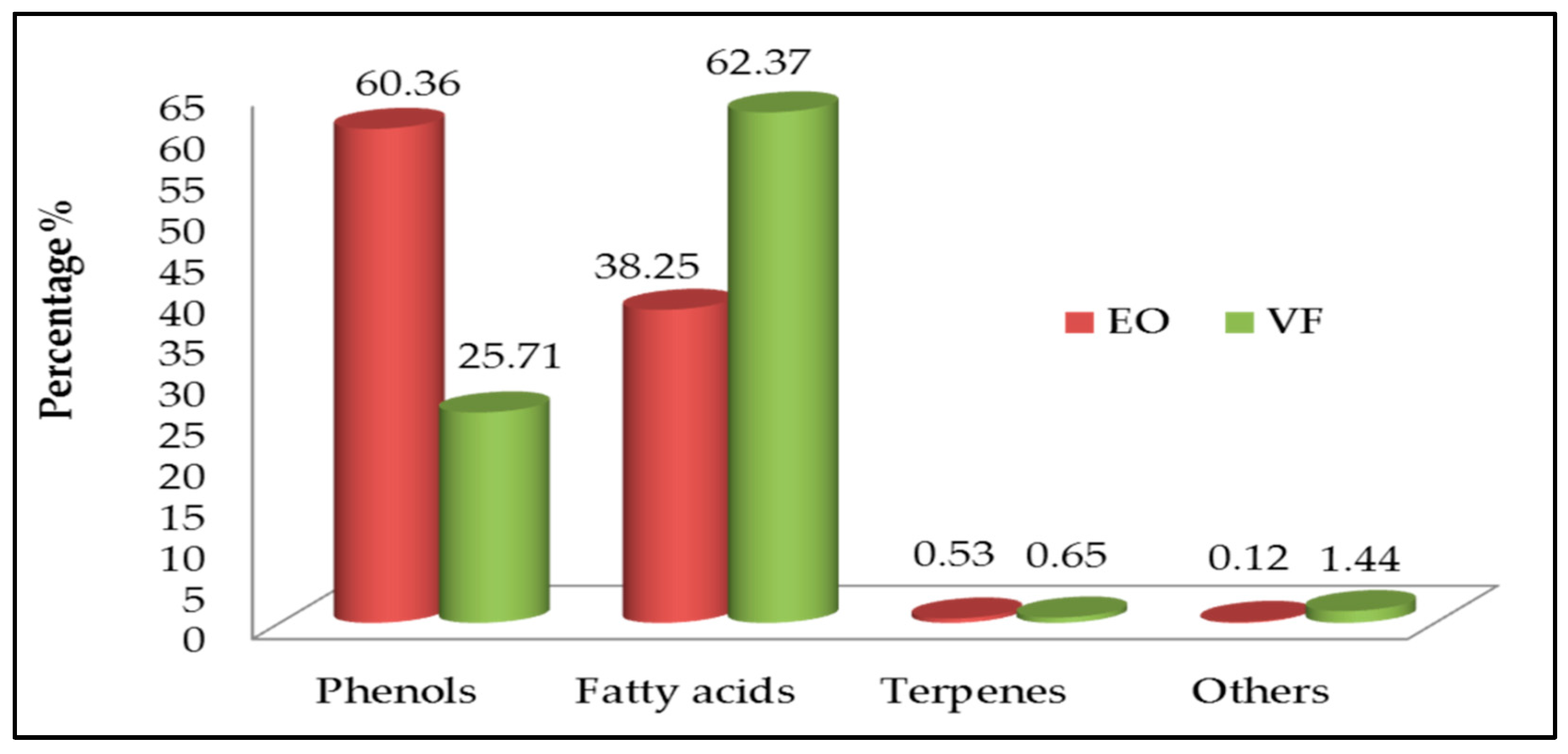
The chemical composition of the VF and EO can be divided into four chemical classes (Table 1 and Figure 2): terpenes (VF—0.65%; EO—0.53%), phenolic compounds (VF—25.71%; EO—60.36%), fatty acids (VF—62.37%; EO—38.25%) and others (variously functionalised compounds; VF—1.44%; EO—0.12%). Previous studies have already reported the presence of fatty acids [35], phenols [36], and terpenes [37,38] as the main chemical composition of olive oil and OMW.
A closer look at Figure 2 shows that the sum of the percentages of phenolic compounds and fatty acids in the VF and EO are 88.08% and 98.61%, respectively. Thus, it can be seen that almost the entire chemical composition of both oils is phenolic compounds and/or fatty acids at approximately similar percentages. The findings are most probably related to a bioconversion/degradation process of phenols into fatty acids and vice versa. As mentioned above and according to Figure 2, phenolics and fatty acids are the predominant chemical classes in the VF and EO, respectively. This result is rather surprising given that the two oils, obtained from OMW, should, in all likelihood, have a relatively similar chemical composition, especially with regard to the chemical nature of the main class. It seems, therefore, that the hypothesis of the intervention of a degradation process is more than likely. Thus, as the EO was directly prepared from the OMW while the VF was obtained following two steps including the preparation of the extract and then its hydrodistillation, it is plausible that the phenols, which are very sensitive to heat, within the VF underwent degradation reactions into fatty acids. It is possible that the degradation process followed the same chemical mechanism as the bioconversion of phenols to fatty acids.
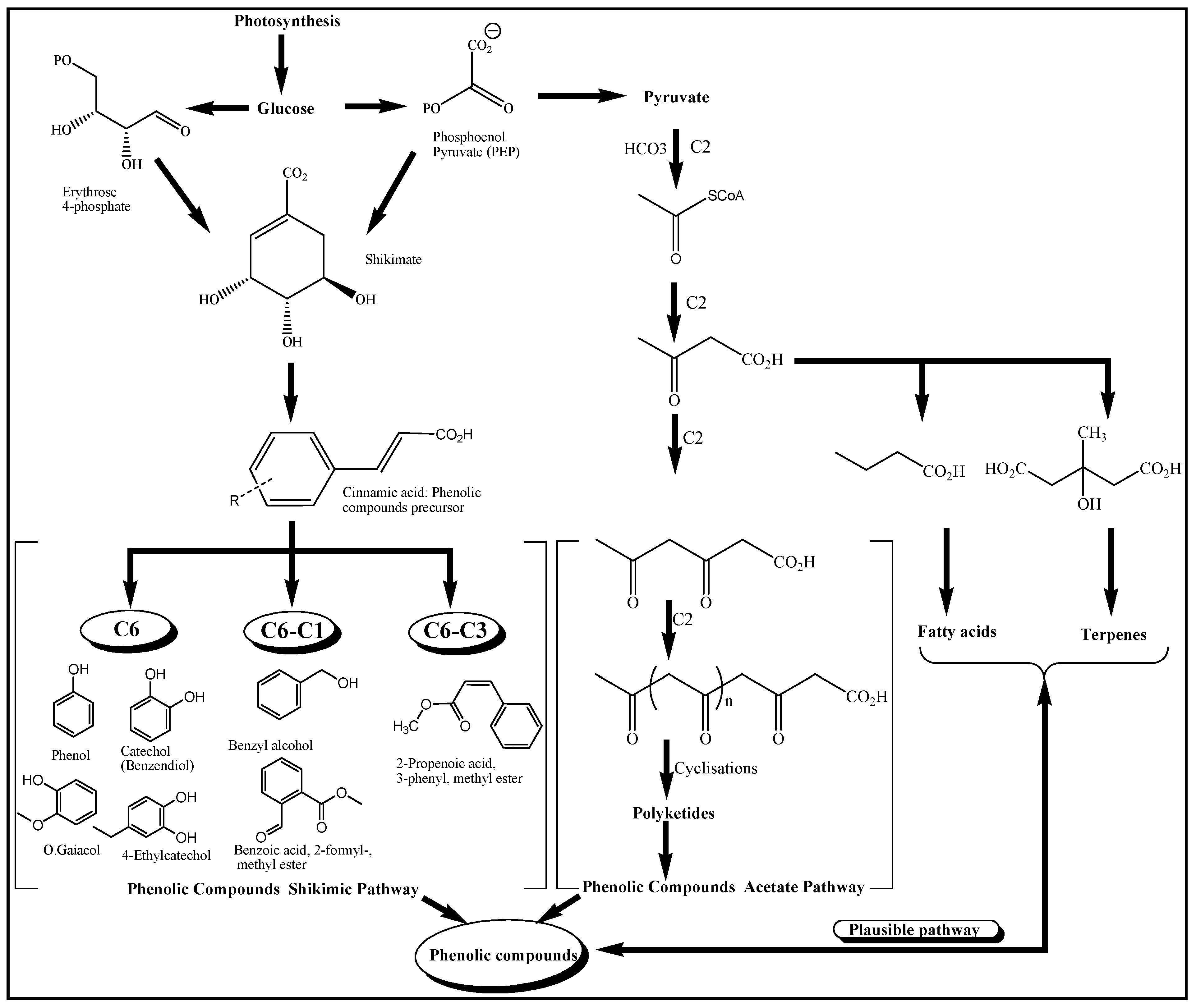
Thus, the simultaneous coexistence of the four main classes in the chemical composition of the VF and EO could most likely be related to biosynthetic considerations, as illustrated in Figure 3 [39], which describes the biosynthetic pathways of the different chemical classes and some plausible pathways suggested based on the chemical composition of the VF and EO. The results also revealed that the main chemical class in the EO was made up of phenolic compounds (60.36%), of which 4-ethylphenol (28.5%) was the predominant one. On the other hand, and unexpectedly, the volatile fraction was characterised by the presence of fatty acids as the predominant class (62.37%), with oleic acid being the main compound at 24.9%. The low content of terpenes in the VF and EO is probably related either to their loss during the preparation process of the OMW extract by liquid extraction, or to their degradation under the effect of heat, although this last hypothesis remains less probable. In the present study, we noticed the presence of cis-limonene oxide, cis-pyran linalool oxide, isomenthol and camphor as monoterpenes and farnesene as a sesquiterpene.
As illustrated in Figure 3, shikimic acid is the biosynthetic precursor of the majority of phenolic compounds, whereas acetyl Coenzyme A is the precursor of fatty acids, which is also the precursor of terpenes via mevalonic acid and of phenolic compounds via malonyl Coenzyme A. It is quite permissible that the contents of the four chemical classes are most likely inter-related.
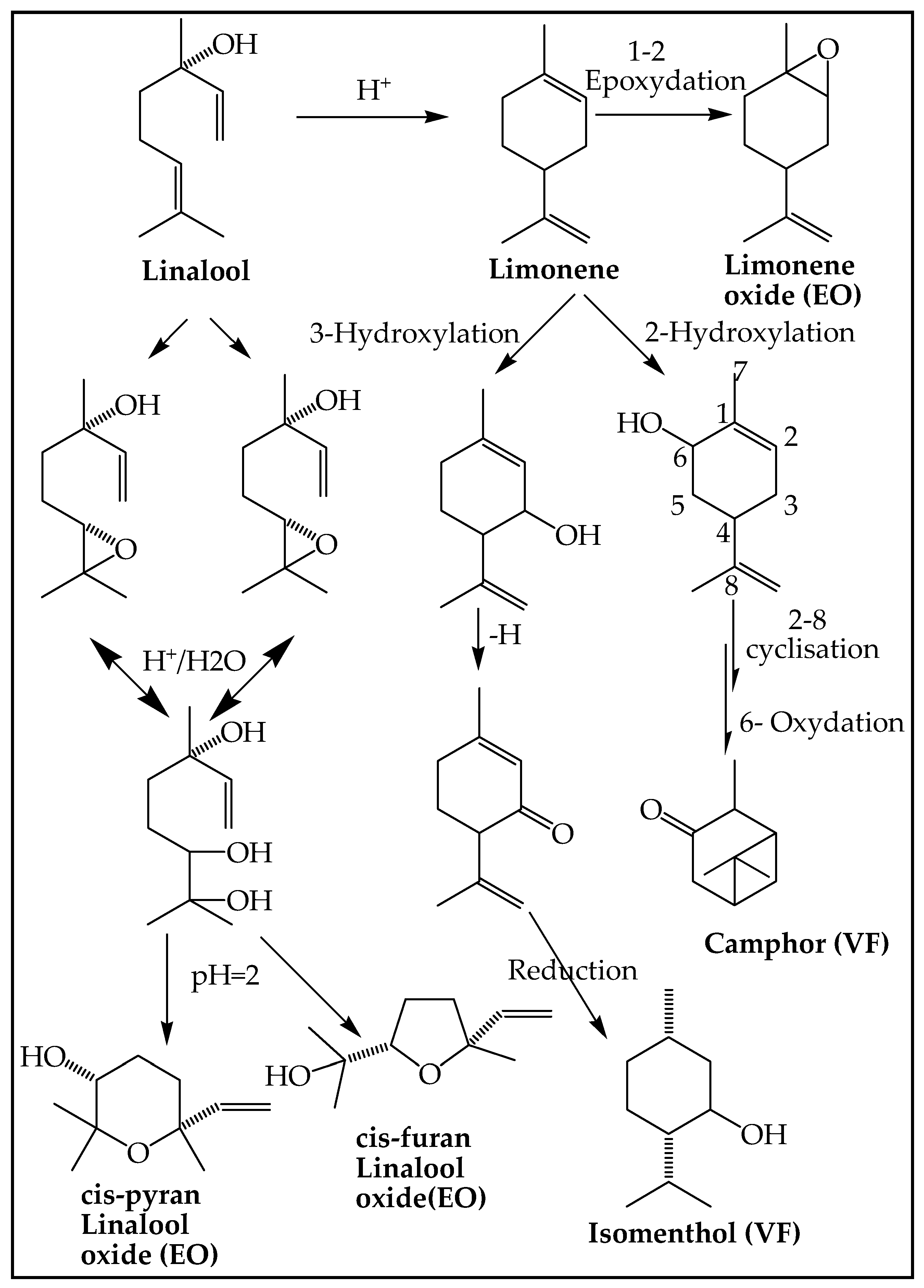
Thus, it is worth noting the presence of linalool and limonene in the volatile fraction of olive oil [40,41], while, in the present study, they were described in the EO as limonene oxide and linalool oxide (furan and pyran). This finding is most likely related to their chemical transformation under the effect of steam or their bioconversion by specific microorganisms present in the medium. In this context, the biotransformation of linalool to limonene oxide via limonene, as well as to furanoid and pyranoid linalool oxide was reported [42] using different strains of A. niger, a fungus found and isolated in OMW [43]. In the same order, plausible pathways (PPs) have been proposed for obtaining camphor and isomenthol from limonene. Additionally, the biosynthesis of neomenthol (isomer of isomenthol) has been described from limonene in Mentha arvensis L. essential oils [44]. The previous several reactions are illustrated by Figure 4, which clearly explains the presence of limonene oxide pyran and furan linalool oxide in the EO.
Furthermore, the presence of farnesene, even in trace amounts, in the EO, and not in the VF, is linked to the relatively long hydrodistillation time (3 h) which favours its entrainment since it is a compound with a high molecular weight and low vapor pressure. The presence of farnesene and limonene has previously been proven in olive oil treated by hydrodistillation and solid-phase microextraction (SPME) [38]. It should be pointed out that monoterpenes, sesquiterpenes and benzenoids have been described as being more abundant in the volatile fraction of virgin olive oil obtained by hydrodistillation than by SPME [45], and vice versa for the abundance of their fatty acids.
A further examination of the chemical composition (Table 1) revealed the presence of oleic acid and its isomer elaidic acid (identified as an ethyl ester), tyrosol and its derivative tyrosol acetate (considered a secoiridoid); both compounds have previously been reported in virgin olive oil [46].

3.3. Antimicrobial Activity

According to Table 2, the VF exhibited strong antimicrobial activity compared to the EO. Thus, both the VF and EO inhibited the growth of all the tested microorganisms at different concentrations. The MIC values were in the range of 195.30–1562.5 µg/mL and 390.62–3125 µg/mL for the VF and EO, respectively. Based on the above data, it is evident that the VF exhibited a stronger inhibitory effect with respect to B. subtilis, M. luteus, E. coli, A. tumefaciens, E. feacium, C. albicans and S. cerevisiae. Furthermore, both oils showed the same activity against B. cereus, S. aureus, L. monocytogenes and P. aeruginosa. However, the EO was ahead of the VF only once against E. cloacae. Kanamycin and chloramphenicol showed the lowest MIC values, i.e., a very strong inhibitory effect against all the microorganisms tested in comparison with the VF and EO. It must be noted that B. subtilis, B. cereus, L. monocytogenes and P. aeruginosa were the most resistant microorganisms. Conversely, E. coli and S. cerevisiae were the most sensitive ones.
The high antibacterial activity of the VF could be related to its chemical composition, which is particularly rich in fatty acids, especially unsaturated ones. The latter are also famous for their high biological activity and are involved in various biosynthetic processes. In this regard, long-chain unsaturated fatty acids are endowed with antibacterial activity [47]. In addition, it has been reported that fatty acids inhibit bacterial growth by disrupting bacterial membranes or by inhibiting fatty-acid synthesis [48]. According to the GC/MS analysis of the VF, the fatty acid fraction consisted of oleic acid and its isomer elaidic acid, linoleic acid, their ethyl ester and stearic acid ethyl ester, which are known for their antibacterial activity against several microorganisms such as E. coli, P. aeruginosa, B. subtilis and S. aureus, among others [49].
Furthermore, synergism between these fatty acids could take place and boost the antimicrobial activity of the VF. In this regard, it was found that oleic and linoleic acid exhibited a synergistic antimicrobial effect when they were mixed [50]. In addition, the presence of tyrosol in the VF (2%) composition at a higher amount as compared to the EO (0.3%), and tyrosol acetate (0.2%), which was only found in the VF could also be reasons for its high activity. In this scenario, it has been reported that these two components are endowed with good antimicrobial activities [51].

3.4. Antioxidant Capacity

The capacity of scavenging the DPPH free radicals by the VF, EO and standards was obtained graphically using a linear curve by plotting radical-scavenging percentages with respect to the sample concentrations. The antioxidant activity of both the oils and standards was expressed in terms of IC50 (µg/mL). The results in Table 3 show that the antioxidant activity of the VF and EO was significantly lower than all the controls used. The recorded IC50 for the EO was relatively better than for VF. It has previously been reported that OMW alkyl aromatic alcohols show significant antioxidant properties and that the best result was associated with the presence of polyphenols [51,52]. This result is mainly due to the high phenolic content in the EO as they represent the main chemical class. The difference in antioxidant activity was most probably related to the presence of gaiacol, phenol and 5-tertbutylpyrogallol, which were only present in the EO. In addition, benzyl alcohol (4.9%), benzene ethanol (7%) and 4-ethylphenol (28.5%) were comparatively much more present in the EO than in the VF.

4. Conclusions

In the present work, the chemical analysis of the VF and the EO of OMW revealed the presence of four chemical classes including phenolics, fatty acids, terpenes and a group of variously functionalised compounds. The antimicrobial test revealed that the VF showed a strong inhibitory effect compared to the EO against most of the microorganisms tested, including B. subtilis, M. luteus, E. coli, A. tumefaciens, E. feacium, and especially the yeasts, C. albicans and S. cerevisiae. This high antimicrobial activity is most likely related to its richness in fatty acids, especially unsaturated fatty acids, which are known to have strong antimicrobial properties. The antioxidant capacity of the two oils by the DPPH method, through the free-radical-scavenging capacity, showed that the EO had a relatively higher antioxidant activity than the VF in terms of IC50.
In general, the in vitro assays carried out showed that the essential oil and the volatile fraction of OMW provide an important source of natural antimicrobial and antioxidant agents that could be used in the food, cosmetic or pharmaceutical industries, while reducing their hazardous impact on the environment.

Author Contributions

Collecting the OMW sample, running the laboratory work, analysis of all the data and drafted the paper, C.A.S.S.; reading of antimicrobial activity results, N.R. and M.R.Z.; interpretation of antioxidant activity results, O.T.; critical reading of the manuscript, S.S., W.Z. and S.A.; designed the study, supervised the laboratory work and contributed to critical reading of all the manuscript, M.E.H. All authors have read and agreed to the published version of the manuscript.

Funding

We gratefully acknowledge the Algerian DGRSDT (http://www.dgrsdt.dz/en, accessed on 15 March 2020) for the financial support (008/15 March 2020).

Acknowledgments

We would like to express our gratitude to the Pasteur Institute of Algiers for its material contribution.

Conflicts of Interest

The authors declare no competing interests.

References

  1. Shabir, S.; Ilyas, N.; Saeed, M.; Bibi, F.; Sayyed, R.; Almalki, W.H. Treatment technologies for olive mill wastewater with impacts on plants. Environ. Res. 2022, 216, 114399. [Google Scholar] [CrossRef] [PubMed]
  2. Rocha, C.; Soria, M.A.; Madeira, L.M. Olive Mill Wastewater Valorization through Steam Reforming Using Multifunctional Reactors: Challenges of the Process Intensification. Energies 2022, 15, 920. [Google Scholar] [CrossRef]
  3. Mekersi, N.; Kadi, K.; Casini, S.; Addad, D.; Bazri, K.E.; Marref, S.E.; Lekmine, S.; Amari, A. Effects of single and combined olive mill wastewater and olive mill pomace on the growth, reproduction, and survival of two earthworm species (Aporrectodea trapezoides, Eisenia fetida). Appl. Soil Ecol. 2021, 168, 104123. [Google Scholar] [CrossRef]
  4. Halimi, C.W.; Laamari, M.; Goldarazena, A. A Preliminary Survey of Olive Grove in Biskra (Southeast Algeria) Reveals a High Diversity of Thrips and New Records. Insects 2022, 13, 397. [Google Scholar] [CrossRef]
  5. Ochando-Pulido, J.M.; Vellido-Pérez, J.A.; González-Hernández, R.; Martínez-Férez, A. Optimization and modeling of two-phase olive-oil washing wastewater integral treatment and phenolic compounds recovery by novel weak-base ion exchange resins. Sep. Purif. Technol. 2020, 249, 117084. [Google Scholar] [CrossRef]
  6. Azzam, M.O.; Al-Gharabli, S.I.; Alrawash, F.F. Air gap membrane distillation applied to olive mill wastewater. J. Environ. Chem. Eng. 2022, 10, 108465. [Google Scholar] [CrossRef]
  7. Hattab, A.; Bagané, M.; Ben Amor, H. Combined Treatment by Coagulation-Flocculation and Oxidation of Olive Mill Wastewater. J. Mater. Environ. Sci. 2020, 11, 522–530. [Google Scholar]
  8. De Leonardis, A.; Macciola, V.; Iorizzo, M.; Lombardi, S.J.; Lopez, F.; Marconi, E. Effective assay for olive vinegar production from olive oil mill wastewaters. Food Chem. 2018, 240, 437–440. [Google Scholar] [CrossRef] [PubMed]
  9. Miklavčič Višnjevec, A.; Baker, P.; Charlton, A.; Preskett, D.; Peeters, K.; Tavzes, Č.; Kramberger, K.; Schwarzkopf, M. Developing an olive biorefinery in slovenia: Analysis of phenolic compounds found in olive mill pomace and wastewater. Molecules 2020, 26, 7. [Google Scholar] [CrossRef]
  10. Solomakou, N.; Goula, A.M. Treatment of olive mill wastewater by adsorption of phenolic compounds. Rev. Environ. Sci. Biotechnol. 2021, 20, 839–863. [Google Scholar] [CrossRef]
  11. Rahmanian, N.; Jafari, S.M.; Galanakis, C.M. Recovery and removal of phenolic compounds from olive mill wastewater. J. Am. Oil Chem. Soc. 2014, 91, 1–18. [Google Scholar] [CrossRef]
  12. Gueboudji, Z.; Addad, D.; Kadi, K.; Nagaz, K.; Secrafi, M.; Yahya, L.B.; Lachehib, B.; Abdelmalek, A. Biological activities and phenolic compounds of olive oil mill wastewater from Abani, endemic Algerian variety. Sci. Rep. 2022, 12, 6042. [Google Scholar] [CrossRef] [PubMed]
  13. Lončarić, M.; Habuda-Stanić, M.; Molnar, M. Various techniques for phenolic removal from olive mill wastewater. Croat. J. Food Sci. Technol. 2021, 13, 268–276. [Google Scholar] [CrossRef]
  14. Bouknana, D.; Jodeh, S.; Sbaa, M.; Hammouti, B.; Arabi, M.; Darmous, A.; Slamini, M.; Haboubi, K. A phytotoxic impact of phenolic compounds in olive oil mill wastewater on fenugreek “Trigonella foenum-graecum”. Environ. Monit. Assess 2019, 191, 1–20. [Google Scholar] [CrossRef] [PubMed]
  15. Babić, S.; Malev, O.; Pflieger, M.; Lebedev, A.T.; Mazur, D.M.; Kužić, A.; Čož-Rakovac, R.; Trebše, P. Toxicity evaluation of olive oil mill wastewater and its polar fraction using multiple whole-organism bioassays. Sci. Total Environ. 2019, 686, 903–914. [Google Scholar] [CrossRef] [PubMed]
  16. Morón, M.; Pozo-Morales, L.; Benito Mora, C.; Garvi, D.; Lebrato, J. OMW spillage control tool based on tracking p-Coumaric acid degradation by HPLC. Environ. Technol. 2019, 40, 2157–2172. [Google Scholar] [CrossRef] [PubMed]
  17. El Yamani, M.; Boussakouran, A.; Benali, T.; Rharrabti, Y. Antibacterial and antioxidant potentials of phenolic extracts from olive mill wastewater and their use to enhance the stability of olive oil. Riv. Ital. Delle Sostanze Grasse 2020, 97, 31–42. [Google Scholar]
  18. Laganà, P.; Anastasi, G.; Marano, F.; Piccione, S.; Singla, R.K.; Dubey, A.K.; Delia, S.; Coniglio, M.A.; Facciolà, A.; Di Pietro, A. Phenolic substances in foods: Health effects as anti-inflammatory and antimicrobial agents. J. AOAC Int. 2019, 102, 1378–1387. [Google Scholar] [CrossRef] [PubMed]
  19. Xing, L.; Zhang, H.; Qi, R.; Tsao, R.; Mine, Y. Recent advances in the understanding of the health benefits and molecular mechanisms associated with green tea polyphenols. J. Agric. Food Chem. 2019, 67, 1029–1043. [Google Scholar] [CrossRef]
  20. Kandeil, A.; Mostafa, A.; Kutkat, O.; Moatasim, Y.; Al-Karmalawy, A.A.; Rashad, A.A.; Kayed, A.E.; Kayed, A.E.; El-Shesheny, R.; Kayali, G. Bioactive polyphenolic compounds showing strong antiviral activities against severe acute respiratory syndrome coronavirus 2. Pathogens 2021, 10, 758. [Google Scholar] [CrossRef] [PubMed]
  21. Bonetti, A.; Venturini, S.; Ena, A.; Faraloni, C. Innovative method for recovery and valorization of hydroxytyrosol from olive mill wastewaters. Water Sci. Technol. 2016, 74, 73–86. [Google Scholar] [CrossRef] [PubMed] [Green Version]
  22. Kreatsouli, K.; Fousteri, Z.; Zampakas, K.; Kerasioti, E.; Veskoukis, A.S.; Mantas, C.; Gkoutsidis, P.; Ladas, D.; Petrotos, K.; Kouretas, D. A polyphenolic extract from olive mill wastewaters encapsulated in whey protein and maltodextrin exerts antioxidant activity in endothelial cells. Antioxidants 2019, 8, 280. [Google Scholar] [CrossRef] [PubMed]
  23. Russo, E.; Spallarossa, A.; Comite, A.; Pagliero, M.; Guida, P.; Belotti, V.; Caviglia, D.; Schito, A.M. Valorization and Potential Antimicrobial Use of Olive Mill Wastewater (OMW) from Italian Olive Oil Production. Antioxidants 2022, 11, 903. [Google Scholar] [CrossRef] [PubMed]
  24. Kadi, K.; Mrah, R.; Hamli, S.; Lekmine, S.; Dib, D.; Addad, D.; Boukeria, S.; Gueboudji, Z.; Hafsaoui, I. Evaluation of the anticoagulant activity of margins from olives extraction in the Khenchela region. J. Fund. Appl. Sci. 2020, 12, 634–649. [Google Scholar]
  25. Silvan, J.M.; Pinto-Bustillos, M.A.; Vásquez-Ponce, P.; Prodanov, M.; Martinez-Rodriguez, A. Olive mill wastewater as a potential source of antibacterial and anti-inflammatory compounds against the food-borne pathogen Campylobacter. Innov. Food Sci. Emerg. Technol. 2019, 51, 177–185. [Google Scholar] [CrossRef] [Green Version]
  26. Ben Mansour, A.; Chtourou, F.; Khbou, W.; Flamini, G.; Bouaziz, M. Phenolic and volatile compounds of Neb Jmel olive oil cultivar according to their geographical origin using chemometrics. Eur. Food Res. Technol. 2017, 243, 403–418. [Google Scholar] [CrossRef]
  27. Olmo-Cunillera, A.; Casadei, E.; Valli, E.; Lozano-Castellón, J.; Miliarakis, E.; Domínguez-López, I.; Ninot, A.; Romero-Aroca, A.; Lamuela-Raventós, R.M.; Pérez, M. Aromatic, Sensory, and Fatty Acid Profiles of Arbequina Extra Virgin Olive Oils Produced Using Different Malaxation Conditions. Foods 2022, 11, 3446. [Google Scholar] [CrossRef]
  28. Vidović, N.; Pasković, I.; Marcelić, Š.; Lukić, I.; Brkić Bubola, K.; Klisović, D.; Novoselić, A.; Palčić, I.; Polić Pasković, M.; Herak Ćustić, M. Effect of Combined Sulfur and Nitrogen Foliar Supply on Olive Oil Volatile Compounds and Sensory Attributes. Hortic 2022, 8, 912. [Google Scholar] [CrossRef]
  29. Zahi, M.R.; Zam, W.; El Hattab, M. State of knowledge on chemical, biological and nutritional properties of olive mill wastewater. Food Chem. 2022, 381, 132238. [Google Scholar] [CrossRef]
  30. Ladhari, A.; Zarrelli, A.; Ghannem, M.; Ben Mimoun, M. Olive wastes as a high-potential by-product: Variability of their phenolic profiles, antioxidant and phytotoxic properties. Waste Biomass Valorization 2021, 12, 3657–3669. [Google Scholar] [CrossRef]
  31. Riad, N.; Zahi, M.R.; Trovato, E.; Bouzidi, N.; Daghbouche, Y.; Utczás, M.; Mondello, L.; El Hattab, M. Chemical screening and antibacterial activity of essential oil and volatile fraction of Dictyopteris polypodioides. Microchem. J. 2020, 152, 104415. [Google Scholar] [CrossRef]
  32. Adams, R.P. Identification of Essential Oil Components by Gas Chromatography/Mass Spectrometry, 4th ed.; Allured Publishing: Carol Stream, IL, USA, 2007. [Google Scholar]
  33. Brand-Williams, W.; Cuvelier, M.-E.; Berset, C. Use of a free radical method to evaluate antioxidant activity. LWT Food Sci. Technol. 1995, 28, 25–30. [Google Scholar] [CrossRef]
  34. Hafidi, M.; Amir, S.; Revel, J.C. Structural characterization of olive mill waster-water after aerobic digestion using elemental analysis, FTIR and 13C NMR. Process Biochem. 2005, 40, 2615–2622. [Google Scholar] [CrossRef]
  35. Jimenez-Lopez, C.; Carpena, M.; Lourenço-Lopes, C.; Gallardo-Gomez, M.; Lorenzo, J.M.; Barba, F.J.; Prieto, M.A.; Simal-Gandara, J. Bioactive compounds and quality of extra virgin olive oil. Foods 2020, 9, 1014. [Google Scholar] [CrossRef]
  36. Zakia, G.; Kenza, K.; Kamel, N. Extraction and quantification of polyphenols of olive oil mill wastewater from the cold extraction of olive oil in the region of Khenchela-Algeria. Genet. Biodivers. J. 2021, 5, 116–122. [Google Scholar] [CrossRef]
  37. Suárez Montenegro, Z.J.; Álvarez-Rivera, G.; Sánchez-Martínez, J.D.; Gallego, R.; Valdés, A.; Bueno, M.; Cifuentes, A.; Ibáñez, E. Neuroprotective effect of terpenoids recovered from olive oil by-products. Foods 2021, 10, 1507. [Google Scholar] [CrossRef]
  38. Vichi, S.; Guadayol, J.M.; Caixach, J.; López-Tamames, E.; Buxaderas, S. Monoterpene and sesquiterpene hydrocarbons of virgin olive oil by headspace solid-phase microextraction coupled to gas chromatography/mass spectrometry. J. Chromatogr. A 2006, 1125, 117–123. [Google Scholar] [CrossRef] [PubMed]
  39. Yang, Z.; Baldermann, S.; Watanabe, N. Recent studies of the volatile compounds in tea. Food Res. Int. 2013, 53, 585–599. [Google Scholar] [CrossRef]
  40. Genovese, A.; Caporaso, N.; Leone, T.; Paduano, A.; Mena, C.; Perez-Jimenez, M.A.; Sacchi, R. Use of odorant series for extra virgin olive oil aroma characterisation. J. Sci. Food Agric. 2019, 99, 1215–1224. [Google Scholar] [CrossRef]
  41. Kesen, S.; Kelebek, H.; Selli, S. Characterization of the volatile, phenolic and antioxidant properties of monovarietal olive oil obtained from cv. Halhali. J. Am. Oil Chem. Soc. 2013, 90, 1685–1696. [Google Scholar] [CrossRef]
  42. Demyttenaere, J.C. Biotransformation of terpenoids by microorganisms. Stud. Nat. Prod. Chem. 2001, 25, 125–178. [Google Scholar] [CrossRef]
  43. Aissam, H.; Errachidi, F.; Penninckx, M.; Merzouki, M.; Benlemlih, M. Production of tannase by Aspergillus niger HA37 growing on tannic acid and olive mill waste waters. World J. Microbiol. Biotechnol. 2005, 21, 609–614. [Google Scholar] [CrossRef]
  44. Bose, S.K.; Yadav, R.K.; Mishra, S.; Sangwan, R.S.; Singh, A.; Mishra, B.; Srivastava, A.; Sangwan, N.S. Effect of gibberellic acid and calliterpenone on plant growth attributes, trichomes, essential oil biosynthesis and pathway gene expression in differential manner in Mentha arvensis L. Plant Physiol. Biochem. 2013, 66, 150–158. [Google Scholar] [CrossRef] [PubMed]
  45. Flamini, G.; Cioni, P.L.; Morelli, I. Volatiles from leaves, fruits, and virgin oil from Olea europaea Cv. Olivastra Seggianese from Italy. J. Agric. Food Chem. 2003, 51, 1382–1386. [Google Scholar] [CrossRef] [PubMed]
  46. Termopoli, V.; Piergiovanni, M.; Cappiello, A.; Palma, P.; Famiglini, G. Tyrosol and Hydroxytyrosol Determination in Extra Virgin Olive Oil with Direct Liquid Electron Ionization–Tandem Mass Spectrometry. Separations 2021, 8, 173. [Google Scholar] [CrossRef]
  47. Zheng, C.J.; Yoo, J.-S.; Lee, T.-G.; Cho, H.-Y.; Kim, Y.-H.; Kim, W.-G. Fatty acid synthesis is a target for antibacterial activity of unsaturated fatty acids. FEBS Lett. 2005, 579, 5157–5162. [Google Scholar] [CrossRef] [Green Version]
  48. Thormar, H. Lipids and Essential Oils as Antimicrobial Agents; John Wiley & Sons, Ltd.: London, UK, 2010. [Google Scholar]
  49. McGaw, L.; Jäger, A.; Van Staden, J. Antibacterial effects of fatty acids and related compounds from plants. S. Afr. J. Bot. 2002, 68, 417–423. [Google Scholar] [CrossRef] [Green Version]
  50. Skalicka-Woźniak, K.; Los, R.; Głowniak, K.; Malm, A. Antimicrobial activity of fatty acids from fruits of Peucedanum cervaria and P. alsaticum. Chem. Biodivers. 2010, 7, 2748–2754. [Google Scholar] [CrossRef]
  51. Leouifoudi, I.; Harnafi, H.; Zyad, A. Olive mill waste extracts: Polyphenols content, antioxidant, and antimicrobial activities. Adv. Pharmacol. Sci. 2015, 2015, 714138. [Google Scholar] [CrossRef]
  52. Azaizeh, H.; Halahlih, F.; Najami, N.; Brunner, D.; Faulstich, M.; Tafesh, A. Antioxidant activity of phenolic fractions in olive mill wastewater. Food Chem. 2012, 134, 2226–2234. [Google Scholar] [CrossRef]
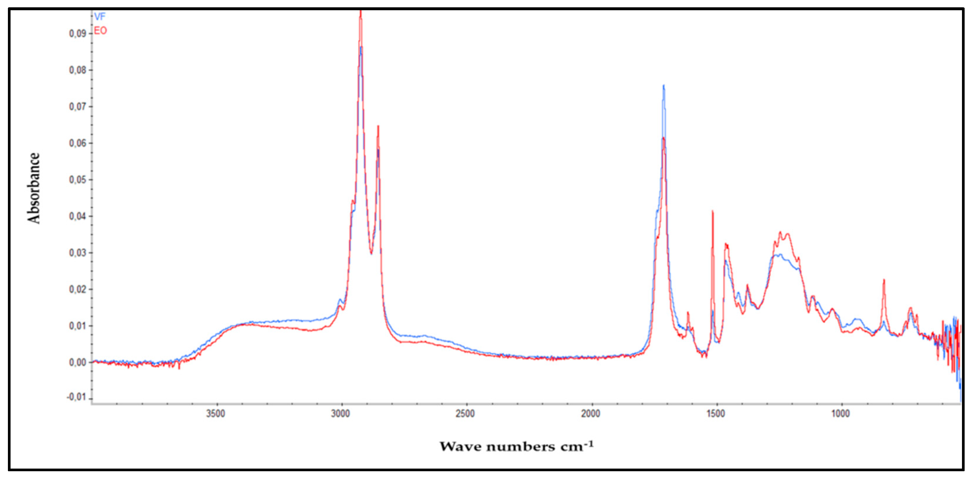
Figure 1. ATR—FTIR spectra of the VF (Blue) and EO (Red) extracted from OMW.
Chemosensors 10 00491 g001
Figure 2. Percentage composition of the four chemical classes in VF and EO.
Chemosensors 10 00491 g002
Figure 3. Interconnection of the biosynthetic pathways of the different chemical classes present in VF and EO.
Chemosensors 10 00491 g003
Figure 4. Bioconversion pathways of limonene and linalool using Aspergillus niger and proposed pathways (PPs) leading towards camphor and isomenthol.
Chemosensors 10 00491 g004
Table 1. Chemical composition of VF and EO.
CompoundDatabaseChem. ClassLRIlitLRIcalVF (%)EO (%)
1Ethanol, 2,2′-oxybis-DB1, DB2O9679620.5t
2Hexanoic AcidDB1, DB2FA9849800.230.3
3PhenolDB1, DB2PC9869820.40.5
4Benzyl alcoholDB1, DB2PC1032103324.9
5cis-furan Linalool oxideDB1, DB2T10701072_0.12
6p-CresolDB1, DB2PC107710770.32_
7o. GuaiacolDB1, DB2PC108810880.110.5
8NonanalDB1, DB2O110411040.17_
9Benzene ethanolDB1, DB2PC111611123.617
10Cyclohexanecarboxylic acidDB1, DB2O11271120_0.12
11cis-Limonene oxideDB1, DB2T11341134_t
12CamphorDB1, DB2T11431141t_
134-EthylphenolDB1, DB2PC1170117011.728.5
14cis-pyran Linalool oxideDB1, DB2T11731174_0.41
15Octanoic acidDB1, DB2FA117811750.8_
16IsomentholDB1, DB2T118211820.65_
17CatecholDB1, DB2PC119712051.10.2
184-vinylphenolDB1, DB2PC122612210.30.7
19cis-p-PropenylanisoleDB1, DB2PC12691265_0.3
20Nonanoic acidDB1, DB2FA127212720.8t
214-EthylguaiacolDB1, DB2PC128212791.313.8
22Cinnamic acid, methyl esterDB1, DB2PC135013520.201.4
23Neric acidDB1, DB2FA13471358_0.2
24o-Toluic acid, methyl esterDB1, DB2FA136213650.20.4
25EugenolDB1, DB2PC13611368_11
26Capric acidDB1, DB2FA13801370_0.17
274-EthylcatecholDB1, DB2PC139213882.110.8
28TyrosolDB1, DB2PC1427142720.36
299-Oxononanoic acidDB1, DB2FA148314800.260.12
30α-FarneseneDB1, DB2T15081509_t
312,4-di-tert-ButylphenolDB1, DB2PC151915140.15_
325-tert-ButylpyrogallolDB1, DB2PC152615270.20.7
33Tyrosol, acetateDB1, DB2PC156715660.20_
34Tetradecanoic acidDB1, DB2FA176817610.10.16
35Palmitoleic acid, methyl esterDB1, DB2FA18901905_0.2
36Palmitic acid, ethyl esterDB1, DB2FA192619270.141.5
37Palmitoleic acidDB1, DB2FA194419430.551.7
38Palmitic acidDB1, DB2FA196919673.32.1
39Ethyl (E)-9-palmitoleateDB1, DB2FA197819741.041.1
40Palmitic acid, ethyl esterDB1, DB2FA199019954.47.4
41cis-Linoleic acid, methyl esterDB1, DB2FA209620950.70.7
42Elaidic acid, methyl esterDB1, DB2FA211021012.012
43Oleic AcidDB1, DB2FA2146214524.96
44Linoleic acid, ethyl esterDB1, DB2FA216421645.73
45Elaidic acid, ethyl esterDB1, DB2FA2174217016.4211
46Stearic acid, ethyl esterDB1, DB2FA219921950.30.2
47Isopentyl palmitateDB1, DB2FA224622460.21_
48Tributyl acetylcitrateDB1, DB2O225422690.4_
49TetracosneDB1, DB2O240024000.2_
502-Octyldodecyl propionateDB1, DB2O241124110.17_
51Oleic acid, pentyl esterDB1, DB2FA242124210.31_
T, trace (<0.1%); T, terpenes; FA, fatty acids; PC, phenolic compounds; O, others; LRIlit, linear retention index reported in the literature; LRIcalc, calculated linear retention index. Database used for LRI match.
Table 2. Minimum inhibition concentrations (MIC) values of VF and EO.
MicroorganismsGramMIC (µg/mL)
EOVFKanamycinChloramphenicol
B. subtilis+3125.03125.062.503.90
B. cereus+1562.51562.57.811.95
S. aureus+781.25781.2515.6215.62
M. luteus+1562.5781.2515.627.81
L. monocytogenes+1562.51562.515.627.81
E. cloacae-781.251562.57.813.90
E. feacium-1562.5781.2562.503.90
A. tumefaciens-1562.5781.251.9515.62
E. coli-390.625195.30_3.90
P. aeruginosa-1562.51562.51.953.90
C. albicansYeast1562.5781.253.903.90
S. cerevisiaeYeast781.25390.625_3.90
Table 3. Antioxidant activity of VF and EO expressed as IP% and IC50.
SamplesDPPH Assay
IP (%)IC50 (µg/mL)
VF73.9244
EO79.8218
BHA90.146.14
BHT95.0212.99
α-Tocopherol89.5213.02
IP: Inhibition percentage at the first concentration of: 4000 µg/mL for EO and VF; 800 µg/mL for BHA, BHT and α-Tocopherol.
Publisher’s Note: MDPI stays neutral with regard to jurisdictional claims in published maps and institutional affiliations.