Article

Portable, Disposable, Biomimetic Electrochemical Sensors for Analyte Detection in a Single Drop of Whole Blood

1
Department of Chemical and Life Science Engineering, Virginia Commonwealth University, Richmond, VA 23284, USA
2
Department of Biomedical Engineering, Virginia Commonwealth University, Richmond, VA 23284, USA
*
Author to whom correspondence should be addressed.
Chemosensors 2022, 10(7), 263; https://doi.org/10.3390/chemosensors10070263
Submission received: 3 May 2022 / Revised: 22 June 2022 / Accepted: 2 July 2022 / Published: 6 July 2022
(This article belongs to the Section Electrochemical Devices and Sensors)

Abstract

:
Current diagnostics call for rapid, sensitive, and selective screening of physiologically important biomarkers. Point-of-care (POC) devices for the rapid, reliable, and easy acquisition of bioinformation at, or near the patient, offer opportunities for better healthcare management. Electrochemical biosensors with high sensitivity and ease of miniaturization are advantageous for such applications. We report a photolithographically micropatterned PEDOT:PSS and silk protein-based fully organic 3-electrode sensor (O3ES) for ultralow volume (single drop—10 µL) detection of analytes in whole blood. The O3ES produces reliable electrochemical signals in whole blood from a mouse model with minimal biofouling interference. The O3ES is demonstrated as a portable device for the simultaneous detection of dopamine, ascorbic acid and uric acid using voltammetry techniques. The O3ES displays excellent sensitivity towards each analyte in whole blood, and in the presence of each other. The water-based, ambient processing of the sensors allows the immobilization of enzymes in the organic working electrode. Amperometric detection of uric acid via uricase with high sensitivity in whole blood is demonstrated. Finally, the performance of the O3ES under enzymatic degradation is studied by monitoring sensitivity over an operating lifetime of ~14 days. This work demonstrates the realization of low-cost, disposable POC sensors capable of detecting blood metabolites using ultralow sample volumes.

1. Introduction

The rapid and direct monitoring of metabolic parameters is an important goal in the field of point-of-care (POC) testing for healthcare [1,2]. Use of ultralow sample volumes without the need for pre-treatment or sample preparation in POC devices at the bedside can greatly improve effective clinical interventions, patient monitoring, and overall health outcomes. Biosensors as rapid and portable analytical devices, offer advantages over traditional laboratory-based screening and quantification techniques, which tend to be expensive, bulky, typically off-site, and labor and time consuming [1,3]. In particular, electrochemical biosensors have been widely studied for such applications owing to high sensitivity, specificity, miniaturization, and speed [4,5]. However, their translation to clinical settings has been hampered by complexity of use, large sample volumes, and susceptibility to fouling in biofluids such as blood.
The specific and sensitive quantification of biomarkers via POC diagnostics using whole blood samples continues to be an ongoing challenge. Using blood over other biological fluids can offer several advantages. Blood carries a host of proteins, metabolites, salts, hormones, and other clinically important biomarkers, offering a diverse target repertoire [6]. The concentration of biomarkers tends to be real-time and higher in blood in comparison to other biofluids such as saliva, sweat, and urine. For many patients, it is relatively easier to collect blood samples. The POC systems that provide a rapid, direct reading using whole blood without the need for filtering or other reagents, can greatly improve assessments of patient health and subsequent diagnoses, especially in resource-limited areas. Although some optical devices have been shown, there are relatively few examples of electrochemical POC devices based on whole blood measurements [7,8]. This is often because electrode surfaces are susceptible to the non-specific binding of materials present in complex biological fluids. Coating the electrode surface with antifouling agents is a common strategy adopted in commercial biosensors [9,10]. However, they suffer from drawbacks such as difficulty to fabricate, high material and manufacturing cost, passivation of the electrode layer and limited compatibility with user friendly portable systems such as hand-held potentiostats and cellphones [11]. In addition to protein and cellular adhesion that can cause a loss of sensor performance, bringing down sample volumes can minimize patient discomfort, as well as provide a route for a disposable, single-use sensor that precludes issues of fouling [12]. Hence, there is a demand for low-cost, ultralow volume, sensitive electrochemical biosensors that can be easily interfaced with portable devices.
In this study, we report a portable biosensor system that can simultaneously detect physiological concentrations of electroactive species from a single drop of blood. Blood from a mouse model is used as it can be easily collected and tested fresh. To reduce interference due to cellular components, we rely on the replacement of inorganic/metallic materials at the sensor-biological interface [13,14]. Photolithographic micropatterning of PEDOT:PSS is used to form electrodes via a biofriendly and green process, using only water as a solvent. Poly(3,4-ethylenedioxythiophene):poly(styrene sulfonate) (PEDOT:PSS) and its derivatives are versatile conductive polymers [15,16]. As reported earlier by our group, these conductive polymers can be precisely patterned as microelectrodes using a protein carrier (photosericin (PS)) to enable photolithography. The PEDOT:PSS acts as an ion/electron transducer, while the PS provides a photocrosslinkable, soft, water permeable matrix. The feasibility of fabricating the working (WE), reference (RE) and counter electrodes (CE) using the same organic ink was earlier shown. In this work, a micropatterned, fully organic 3-electrode sensor (O3ES) is used for the selective and ultrasensitive detection of dopamine (DA), uric acid (UA) and ascorbic acid (AA) using only the conductive ink without the use of any metal electrodes.
Dopamine is an important neurotransmitter in the central nervous system (CNS). Low levels of DA may result in disorders including Parkinson’s disease [17]. The electrochemical detection of DA in real biological systems tends to be complicated by the coexistence of interferents, such as uric acid (UA) and ascorbic acid (AA). Ascorbic acid (vitamin C) is an important antioxidant in metabolic processes is used to treat and prevent scurvy [18]. Ascorbic acid is oxidized at nearly the same potential at a bare electrode, resulting in an overlap of voltammetric response [19]. Uric acid is the end-product of purine nucleotide metabolism and its derivatives. Increased concentration beyond the normal range is associated with conditions such as gout, hyperuricemia, and kidney disease [20,21]. There have been prior reports on the detection of DA, UA, and AA, individually, and in some cases, in conjunction using electrochemical techniques [22,23,24,25]. The low separation of the oxidation peaks of DA, UA, and AA poses a challenge in their simultaneous detection which results in their poor selectivity [26]. In order to overcome this challenge, various electrode materials have been investigated. These include carbon based materials such as carbon nanotubes [27] and graphene oxide [28], metal nanoparticles [29], metals and metal oxides [30,31], and conducting polymers [32]. The polymer PEDOT:PSS has also been investigated for the simultaneous detection of DA, UA, and AA. Organic electrochemical transistors based on PEDOT:PSS have been reported for the selective detection of DA in the presence of AA and UA as interferents [33]. Graphene oxide nanoribbons and PEDOT:PSS on screen-printed carbon electrodes were used for simultaneous detection [34]. Our group previously reported the detection of DA and AA in PBS in the presence of each other using PEDOT:PSS/PS coated ITO electrodes [35]. Although the detection of DA, UA, and UA in biofluids such as sweat, urine and saliva has been reported, there are few examples of their detection in blood. The PEDOT was used to coat a glassy carbon electrode (GCE) for detection in blood serum [36]. Three-dimensional graphene nanosheets on carbon cloth was used for the simultaneous detection of AA, DA and UA in human serum in a flexible format [37]. A disposable screen printed electrode fabricated using commercial carbon ink was used for the detection of UA in whole blood [21].
We show an ultralow volume of blood (single drop—10 uL) can be used with the O3ES. Whole blood samples are directly applied to the sensor surface without the need for sample pretreatment or other reagents. The results can be obtained in a few minutes using the disposable, low-cost biodegradable organic sensors. We show this system can be used for electrochemistry and detection of each analyte, separately, and in the presence of each other in a highly sensitive manner. The electrochemical response is obtained using a handheld potentiostat with enabled wireless technology so that the readouts can be obtained using a cellphone. In addition, we show that the system can be easily adapted for enzymatic sensing to obtain specific and precise response to specific targets. Thus, the system is a truly portable, point-of-care device that can make a difference for low cost, rapid quantitation of biomarkers for healthcare or environmental applications.

2. Materials and Methods

2.1. Synthesis of Photoactive Silk Proteins

The 3-electrode integrated sensors used in this work were fabricated using the technique of Silk Protein Lithography (SPL) reported earlier [38]. Photoactive silk proteins were synthesized as previously reported [39]. Briefly, silk proteins were extracted from Bombyx mori cocoons using a standard protocol developed elsewhere [40]. The silk proteins (fibroin or sericin) are dissolved in 1M LiCl in DMSO and reacted with 2-isocyanatoethyl methacrylate in stoichiometric amounts. The reaction is carried out at 60 °C under inert conditions for 5 h and precipitated in cold ethanol for 12 h. The product was washed 3 times in 1:1 ratio of cold ethanol/acetone and lyophilized to obtain photoactive silk proteins (photoactive fibroin (PF) and photoactive sericin (PS) used to form inks.

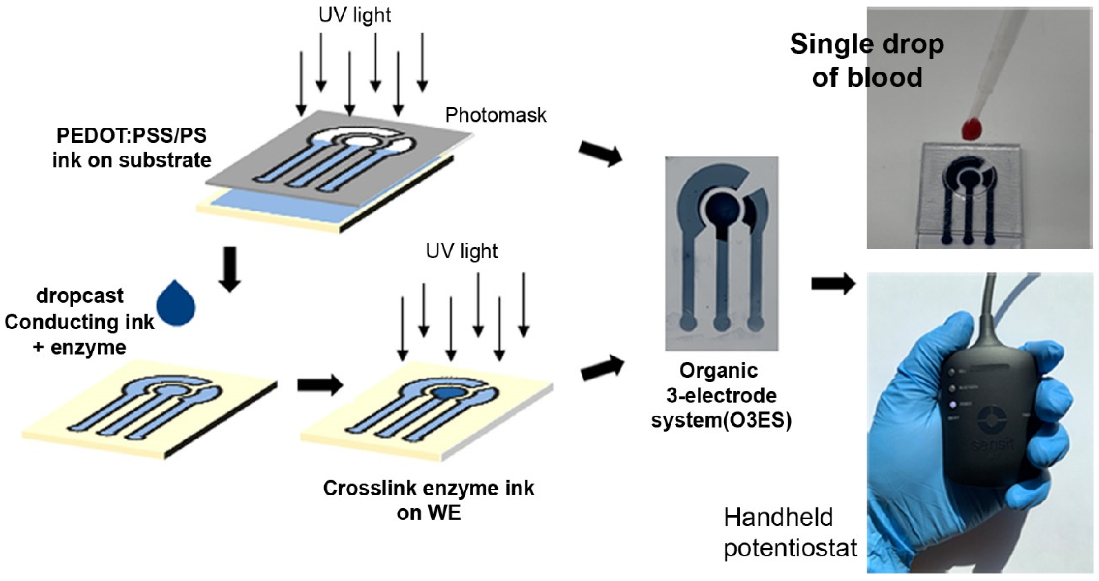
2.2. Fabrication of Disposable Biomimetic Sensors

Photoreactive silk protein inks were used to form the sensor substrates and electrodes. The substrate was prepared by casting a solution of 7.5% (w/v) PF and 1.5% photoinitiator in formic acid on clean glass slides. The solution was dried under ambient conditions and crosslinked using an OmniCure S1000 UV Spot Curing lamp (Lumen Dynamics, ON, Canada) (365 nm UV (20 mW cm−2) for 2 s). Electrodes were formed using a conductive ink composite comprising PS and the conducting polymer PEDOT:PSS. A 1% (w/w) dispersion of dry PEDOT:PSS pellets (Sigma-Aldrich, St. Louis, MO) in water was obtained by ultrasonicating for 30 min and filtering via a 0.25 µm syringe filter. The PS was mixed with the PEDOT:PSS dispersion (40 µL of PEDOT:PSS per mg of PS), with Irgacure 2959 as photoinitiator. 5% (v/v) DMSO is used to enhance conductivity and stability. To form the sensor, the ink was cast on PF substrates, dried under a chemical fume hood, and crosslinked under 365 nm UV light through a photomask for 2 s. The electrode patterns were developed using DI water to form the O3ES. The photolithographic fabrication of the sensor proposed in this work was carried out using a simple bench-top photolithography setup without any clean room requirements (Figure 1). The electrochemical characteristics, characterization, and patterning of the ink were earlier reported [40].

2.3. Blood Collection from Mice

Animal use was approved under the Virginia Commonwealth University’s Institutional Animal Care and Use Committee under protocol AD10000465. Target analytes were detected in whole blood collected from C57BL-6 mice (between the ages 2 and 22 months) obtained from Charles River Laboratories (Wilmington, MA, USA). The cavity in the intestinal area of the mouse was opened. Following this, the vena cava was located from which a 26-gauge needle was inserted, and the maximum amount of attainable blood was drawn. Once it was apparent that no more blood could be collected, the mouse was euthanized. The collected blood was quickly heparinized by shaking it in a heparin tube. All electrochemical experiments were performed within 5 h of blood collection. The sensing experiments consisted of directly applying one drop of blood (10 µL) directly to the well housing the sensor (Figure 1).

2.4. Electrochemical Characterization and Sensing

Electrochemical measurements were recorded using an extremely small footprint, wireless enabled handheld potentiostat (PalmSens Sensit BT potentiostat (PalmSens BV, Netherlands)). The potentiostat was connected wirelessly to an Android enabled cell phone which provided remote assessment of sensor performance, while being truly portable. Electrochemical characterization of the sensor in 0.1 M PBS and whole blood was carried out using Cyclic voltammetry (CV), Linear sweep voltammetry (LSV) and Electrochemical impedance spectroscopy (EIS). The CV scans were performed with a potential range of −1 V to 1.4 V at a scan rate of 100 mV/sec. Impedance spectra were recorded with an AC amplitude of 10 mV at a bias potential of 0 V, and the scanning frequency range of 0.1 Hz to 100 kHz. Detection of DA, UA, and AA in 0.1 M PBS and whole blood was carried out using Differential Pulse Voltammetry (DPV) with parameters: step size 5 mV, pulse size 50 mV, sample period 0.02 s, pulse time 0.2 s. The DPV responses for DA, UA, and AA were recorded. All experiments were conducted at room temperature. Each experiment was cross verified using at least three replicates. Following each experiment, the sensors were discarded. Limit of detection (LOD), limit of quantitation (LOQ) and sensitivity were calculated per ICH guidelines.

2.5. Proteolytic Degradation In Vitro

Micropatterned O3ES can be proteolytically degraded over time in the presence of enzymes, which simplifies their ease of disposal. In the present work, the degradation of O3ES in the presence of protease (Protease XIV from Streptomyces griseus, ≥3.5 U mg−1, Sigma Aldrich) was studied. Sensors were incubated in protease (1 U/mL) at 37 °C and their degradation was studied over a period of 1 month. To study the performance of the O3ES under degradation, the sensors were taken out from the protease solution each week, cleaned using DI water and dried using nitrogen gas. The sensors were then used for the detection of DA in PBS. The enzyme solution was replaced every 3 days to maintain the activity of protease. The performance of the sensor under degradation was evaluated as a function of sensitivity towards dopamine.

3. Results

3.1. Fabrication of Organic 3-Electrode Sensors (O3ES)

Silk protein photolithography was implemented for the fabrication of disposable, integrated, fully-organic, 3-electrode sensors (O3ES) [39,41,42]. Photofibroin (PF) forms the biodegradable sensor substrate on which the electrodes are fabricated. While such sensors are usable in flexible formats, here we report on their use in a “rigid” point-of-care device. To form precise micropatterned electrodes, the photoreactive silk inks were patterned using a facile and scalable, bench-top photolithography process. The active electrodes are formed using a water-based, photopatternable conductive ink comprising photosericin (PS) and PEDOT:PSS dispersion. A solution of 5% DMSO is used as a dopant to enhance the electrical properties. The optimization and characterization of the conductive ink was reported previously [39]. The conductive ink is crosslinked using UV light (365 nm) and developed using a biofriendly, fully aqueous process (Figure 1). The ink behaves as a negative-tone photoresist wherein the exposed area crosslinks and becomes insoluble in water. The electrode patterns are developed by removing uncrosslinked material using water.
The sensors have a small areal footprint (~2 cm2) with 1 mm line width and 2.54 mm connection pitch. Similar in design to screen-printed electrodes (SPEs), the O3ES configuration was earlier used for the detection of dopamine, ascorbic acid, and glucose in buffers and simulated biofluids (viz. urine, saliva, serum) [43]. Such systems are advantageous over conventional 3-electrode systems with Ag/AgCl and Pt reference and counter electrodes as they can be easily engineered into miniaturized and portable formats. These are particularly useful for POC applications as the single-use electrodes can be disposed after use, eliminating issues with sensor fouling and recalibration. The water-based ink coupled with aqueous processing under ambient conditions allows for the immobilization of biorecognition molecules such as enzymes and anti-bodies, thus adding to the versatility of the sensing platform. In the final step, a fluid chamber (well) was attached to the sensor, allowing for ultralow-volume detection of clinically important target analytes.

3.2. Electrochemical Characterization of the O3ES

The use of a handheld potentiostat coupled with the small footprint single-use sensor, enables the discussed POC device to be in a truly portable configuration. In order to understand and validate the electrochemical response in fresh blood, we used whole blood collected from mice as a surrogate. The objective was to show the efficacy of the proposed sensor in a complex biological milieu. It is important to note that quantification of the target biomarkers that may be already present in the mice blood was not attempted and beyond our current scope. The blood was drawn from standard C57BL-6 mice without any modification to induce production of target biomarkers.
The electrochemical impedance behavior of the O3ES in 0.1M PBS and in whole blood was measured (Figure 2a) using electrochemical impedance spectrometry (EIS). The PBS buffer (pH 7.4) was used to test the initial sensor response at physiological pH. The impedance at 1 kHz is an important electrochemical parameter as many biological activities occur at this frequency in physiological environments [44]. From the EIS data, a high signal to noise ratio was observed at ~1 kHz with a low impedance of 0.468 kΩ and 0.545 ΩK in PBS and blood respectively. This may be attributed to the porous nature of the soft, polymeric PEDOT:PSS/PS electrode surface, which creates excellent ion conduction properties that help to reduce the impedance at the electrode/electrolyte interface. The impedance was slightly higher in whole blood in comparison to PBS. This is presumably due to the various components of whole blood (blood cells, platelets, plasma etc.) at the electrode surface which can hinder electron transfer. Nonetheless, the low charge transfer resistance shows that the 3-electrode sensor developed can facilitate the biosensing of important biomolecules in both PBS buffer and whole blood. While this is an important step, the detection of analytes in clean buffers does not necessarily transfer to complex biological fluids such as blood, which are needed at POC applications.
The CV scans performed on the sensors revealed a wide stable potential window of −1.0 to 1.2 V in both 0.1M PBS and whole blood without any water splitting (Figure 2b). This indicates its potential use for sensing at different oxidation and reduction potentials. It should be noted, all the electrochemical experiments were performed in 10 µL of liquid sample (either PBS or blood), the equivalent of one drop of blood. Additionally, all the electrochemical measurements were performed within 2 min of introducing the liquid sample. This minimizes any effect of fouling (the small sample size may for instance, evaporate rapidly). Interestingly, the electrochemical behavior of the sensor as observed from CV in whole blood was identical to the behavior in 0.1M PBS. Further, we verified that dilution of blood did not affect the electrochemical behavior of the sensor. Whole blood, 5× diluted blood and 10× diluted blood (dilution in PBS) provided a response similar to 0.1M PBS (Figure 2c). The stability and anti-interference capability in whole blood, which is a much more complex environment when compared to PBS, is a critical attribute of the sensor developed in this work. When compared to PBS, 5× and 10× diluted blood, oxidation peaks around 0.3 V was observed in whole blood. This may be attributed to the presence of uric acid in whole blood. The sensors also demonstrated excellent stability towards redox cycling and retain electrochemical activity even after 100 redox cycles in whole blood (Figure 2d). This suggests that the proposed O3ES can be used for the detection of analytes in whole blood multiple times without any loss in electrochemical properties.
The electrochemical response of the O3ES to the target analytes was examined using Linear Sweep Voltammetry (LSV). Whole blood was spiked with 100 µM of DA, AA, and UA each and a LSV scan was performed in the range of 0 V–0.5 V (Figure 3a). Each biomolecule has a characteristic oxidation peak. The oxidation peaks of DA, UA, and AA were found to be at 0.28 V, 0.34 V, and 0.1 V, respectively (Figure 3b), in agreement with other reports in literature where electrochemical detection of AA, DA and UA was carried out using PEDOT:PSS based sensors [33,34]. The scan rate is another important parameter that affects the sensitivity, resolution, and peak intensity of the biosensor. When a faster scan rate is used, the oxidation peaks shift to a higher potential. It also results in a poor separation of the oxidation peaks. Because the oxidation peaks of AA, DA and UA are very close to each other, more well-defined peaks were obtained using a lower scan rate. A slow scan rate also allows the electro-oxidation of AA with a lower potential (~0.1 V), which results in a better separation of the AA and DA peaks. The sensitivity of AA is the lowest among the three analytes. This can be explained by the slow kinetics of the oxidation reaction; hence, a slow scan rate ensures adequate time for the reaction to occur at the specific oxidation potential resulting in a higher intensity and separation of the individual peaks. A scan-rate of 10 mV/s was found to be optimal for the best resolution. All detection experiments were carried out at this scan rate.

3.3. Electrochemical Detection of Biomolecules

The detection of AA, DA and UA was carried out via DPV using the O3ES. For each analyte, detection was initially tested in 0.1 M PBS (7.4 pH), followed by whole blood. The performance metrics of the O3ES (RE, CE, WE using PEDOT:PSS/PS ink) were previously shown to be very competitive in comparison to conventional 3-electrode cells that use PEDOT:PSS/PS as the WE, while employing Ag/AgCl and Pt wire as the RE and the CE, respectively [43]. In this study, it was found that the O3ES yielded a lower conductivity and sensor signal in comparison to a conventional system (Ag/AgCl, Pt). This may be attributed to a slower and limited charge injection using PEDOT:PSS versus Ag or Pt electrodes. The concentration range for each analyte was set to cover the characteristic physiological concentration ranges in blood (reference ranges for dopamine were 0.01–1 µM, for ascorbic acid were 25–75 µM and for uric acid were 100–450 µM) [22,23,24,25]. Each experiment was replicated using at least 3 different sensors.
The normalized current (I/I0) versus concentration response curves for DA, AA, and UA showed a linear range from 10 µM to 100 µM, 100 µM and 600 µM, respectively, in both PBS and whole blood (Figure 4a–c). The sensitivity of the sensor against all the analytes were found to be higher in PBS when compared to whole blood. On an average, the sensitivity of the sensor in whole blood was ~45% less than the sensitivity in PBS. As noted above, this is not unexpected, and is likely due to the cellular and plasma components in whole blood. As the detection of analytes in blood is carried out within 5 h of blood collection, the pre-existence of some of the analytes in whole blood may be expected. Dopamine has a short half-life of ~2 min as it is metabolized by neurotransmitter amine enzymes present in the peripheral circulation [45,46]. In contrast, uric acid and ascorbic acid have a longer half-life of 24 h and 10—20 days, respectively, and are solely eliminated by the kidneys [47,48]. However, the analytes already present in mice blood are oxidized in the initial blank run, thus eliminating their interference in subsequent sensing experiments. It is important to note that the electrodes reported here consist of the organic conductive ink as the sole electroactive material, without use of any other electroactive species in the sensor, or the electrolyte to amplify the response. Thus, the improvement of biofriendly characteristics (facile processing, green synthesis, biodegradation, and biocompatibility), come at the cost of a slight decrease in performance metrics, albeit well within the physiologically relevant regimes.
The sensitivity of the sensor was found to the highest for DA (both in PBS and blood), followed by UA and AA (Figure 4d). The higher sensitivity towards dopamine is explained by a “pre-concentration” effect in PEDOT:PSS systems [35]. Dopamine, being positively charged in PBS and blood, has an electrostatic attraction towards the anionic PSS surfactant in PEDOT:PSS. The PSS therefore facilitates electron transfer between dopamine and the electrode surface, resulting in a higher sensitivity over uric acid and ascorbic acid. In contrast, uric acid and ascorbic acid are present as negative charges in physiological solutions, which cause a repelling effect from the electrode surface, thus resulting in their relatively lower sensitivity. The repelling effect from the negatively charged PEDOT:PSS electrodes along with a slow oxidation reaction kinetics explains the low sensitivity of the O3ES towards AA.
Detection of DA, UA, and AA in whole blood was carried out in the presence of each other using the proposed sensor system (Figure 5a). Owing to the close oxidation potential of DA, AA, and UA, it is important to study their mutual interference. Detection of DA was carried out in the presence of 100 µM of AA and UA. A linear response curve of DA is seen in the presence of UA and AA. While the sensitivity towards DA was lower in the presence of UA and AA in comparison to DA alone, the order of sensitivity is the same in both cases. As a second verification of the versatility of the system, AA was detected in the presence of DA (100 µM) and UA (300 µM). The UA was increased in comparison to the prior experiment to see the effect of higher interference. A linear response was observed (Figure 5d). Interestingly, the sensitivity of the O3ES towards AA in the presence of DA and UA was found to be same as the sensitivity towards AA alone. This indicates that the microfabricated O3ES developed in this work can used for the efficient detection of DA and AA in whole blood in the presence of a large amounts of interferents.

3.4. Enzyme Based Amperometric Biosensing

The water-soluble nature of photosericin (PS) allows the aqueous processing of the conducting ink under ambient conditions. The ink provides a stable, biocompatible crosslinked matrix for the encapsulation of biomolecules. This is a critical characteristic of the O3ES system which allows the immobilization of specific recognition biomolecules such as enzymes and antibodies for biosensing. In the above experiments, the DA, UA, and AA were detected non-specifically by monitoring specific oxidation potentials. To demonstrate the feasibility of using the O3ES for specific sensing, the enzyme uricase was immobilized onto the working electrode. The uricase shows response to uric acid only. Detection of uric acid was conducted in the presence and absence of uricase enzyme initially in 0.1 M PBS via chronoamperometry (Figure 6a). Both the groups (+enzyme and -enzyme) yielded a linear response in the range of 10–300 µM. However, as expected, the sensitivity of the sensor towards UA is significantly higher (~90%) in the presence of uricase. Moreover, the current signal was found to be saturated at higher UA concentrations (~600 µM) in the absence of uricase. The chronoamperometric response of UA in the presence of uricase was then monitored in blood (Figure 6b). A linear response was observed over the entire range (0–600 µM) of UA. Expectedly, the sensor demonstrated a higher sensitivity towards UA in PBS in comparison to blood (~64%). This is again explained by the electrode inhibition in blood which hinders electron transfer between the enzyme and electrode redox center.

3.5. Detection of Dopamine under Degradation

The O3ES systems developed using protein-based inks on protein substrates provide a distinct advantage in terms of being bioresorbed under physiological conditions or biodegraded in nature. This allows their disposal without creating extra waste, for instance, single use sensor strips may be composted. Given the growing concerns surrounding disposal of abandoned electronics, this is an important contemporary challenge [49]. While the results in this work demonstrate their use in POC devices, we can potentially consider their utility for continuous detection in implantable or wearable applications, with degradation following stable operation lifetime. The silk-based devices can be controllably broken down in the presence of protease enzymes, with complete degradation over a period of several weeks [50,51,52]. We tested the O3ES system to understand their performance over time as transient devices in an enzymatic environment. The sensors were immersed in a protease solution (1 U/mL) at 37 °C which resulted in their degradation over a period of one month. The protease solution was replaced every 3 days to maintain the activity of enzyme. The detection of dopamine was studied during the degradation process and the sensitivity was calculated. The sensor yielded a linear response towards dopamine throughout the entire degradation process (Figure 7). Interestingly, the sensitivity of the sensor increased in the first two weeks of degradation. This is due to the degradation of silk sericin (PS) matrix, which increases the exposure of the electroactive PEDOT:PSS to the analyte, thus increasing sensitivity. However, the sensitivity dropped significantly in weeks 3 and 4, which is due to loss of structural integrity caused by the degradation of the electrode architecture. Thus, the working lifetime of the O3ES developed in this work can be specified as 2 weeks in physiological environments. This experiment provides utility in considering such transient devices in challenging biological environments.

4. Discussion

While various works have discussed the use of sensors for POC applications (optical or electrochemical), there continue to be challenges in aspects of portability, ease-of-use, and sampling. The 3-electrode design reported in this work utilizes the same material to fabricate the working (WE), reference (RE), and counter (CE) electrodes without the need for Ag/AgCl or Pt. This makes the fabrication of the sensors easy and low cost. Further, as shown by our group earlier, the fabrication as a fully-organic biosensor offers high biocompatibility and potential biodegradability. The connection is made to a handheld, wireless-enabled potentiostat with readout from a cellphone. These factors enhance the portability of the O3ES discussed. Finally, the ultralow blood sample of 10 µL provides the needed sample for the detection of the biomarkers of interest. A summary of the performance metrics of the O3ES is shown in Table 1.
The fully organic, portable biosensor is therefore capable of detecting analytes in a single drop of blood. These sensors are designed to be used in point-of-care settings for the detection of clinically relevant biomolecules. The low footprint of both the sensing electronics (handheld form factor), the sensor component itself (designed as miniaturized organic 3-electrode sensor) and the ultralow sampling volume (single drop of blood—10 µL) make it suitable to be envisioned for such applications. It may be noted that the blood volume (single drop 10 µL) is so low that it can evaporate within an hour, leaving behind cellular debris on the sensor. Therefore, larger blood volumes or other configurations may need to be explored for continuous detection. Detection can be accomplished using a range of electrochemical techniques including differential pulse voltammetry (DPV) and amperometry. We reported on three analytes of interest in a single drop of blood—dopamine, uric acid, and ascorbic acid that are typically studied together owing to their respective interferences. While the targets (DA, UA, and AA) are detected non-specifically, the methodology is versatile and can be easily extended to diverse targets. The linear ranges for AA and UA are well within the reference ranges for these analytes in blood. While the DA levels are higher than those found physiologically, the ability to detect it in the presence of interference in this system is noted. The sensor was successfully able to discriminate each of these analytes with high sensitivity and in the presence of each other. The performance metrics of the O3ES systems are competitive in relation to earlier reported works as shown in Table 2.
To extend the applicability of the O3ES system to be useful for enzymatic sensing, we showed the facile immobilization of the enzyme uricase in the electrode ink used to fabricate the working electrode. This further improves the range of targets that may be detected using this system (e.g., enzymatic detection). Similarly, in order to improve the detection limits of dopamine to be usable for physiological concentrations in the sub-micromolar ranges, it may be possible to optimize the sensors using an enzyme such as tyrosinase for the specific detection [53]. While shown for a POC application, we also show how these sensors can be designed for continuous, transient function, providing an operational lifetime of ~2 weeks prior to degradation. Thus, the biomimetic sensors reported herein, used with small form factor electronics therefore have the potential to form clinically translatable tools for improving healthcare outcomes. The low cost and ease-of-use, together with their biodegradability, make them useful for economical diagnostics in resource limited settings.

5. Conclusions

In summary, a fully organic, portable biosensor capable of detecting analytes in a single drop of blood is shown. These sensors are designed to be used in point-of-care settings for the detection of clinically relevant biomolecules with the sensing element designed to easily discarded and potentially biodegraded on disposal. The small blood volume required (single drop—10 µL), enables these single-use sensors to be coupled to low footprint electronics. The sensors can be used for non-specific detection of electroactive species, or coupled with biorecognition molecules (enzymes, antibodies, aptamers) for specific detection of analytes in blood. In this study, we reported on three analytes of interest in a single drop of blood—dopamine, uric acid, and ascorbic acid that are typically studied together owing to their respective interferences. A range of electrochemical techniques such as cyclic voltammetry, differential pulse voltammetry and amperometry can be used. This work demonstrates the potential for the realization of low-cost, high throughput POC devices capable of detecting important blood metabolites using ultralow sample volumes. Such POC diagnostics offer the possibility of providing rapid diagnostic results in a non-laboratory setting.

Author Contributions

Conceptualization, S.P., R.L.H. and V.K.Y.; methodology, S.P. and S.A.; formal analysis, S.P.; investigation, S.P. and S.A.; resources, S.A.; writing—original draft preparation, S.P. and V.K.Y.; writing—review and editing, S.P., R.L.H. and V.K.Y.; supervision, V.K.Y.; funding acquisition, R.L.H. and V.K.Y. All authors have read and agreed to the published version of the manuscript.

Funding

This research was supported in part by a grant from the Commonwealth Cybersecurity Initiative. S.P. was supported in part by a grant from VCU Innovation Gateway.

Institutional Review Board Statement

Animal use was approved under the Virginia Commonwealth University’s Institutional Animal Care and Use Committee under protocol AD10000465.

Informed Consent Statement

Not applicable.

Data Availability Statement

Relevant data are available upon request from the authors.

Conflicts of Interest

The authors declare no conflict of interest. The funders had no role in the design of the study; in the collection, analyses, or interpretation of data; in the writing of the manuscript, or in the decision to publish the results.

References

  1. Pappa, A.-M.; Parlak, O.; Scheiblin, G.; Mailley, P.; Salleo, A.; Owens, R.M. Organic Electronics for Point-of-Care Metabolite Monitoring. Trends Biotechnol. 2018, 36, 45–59. [Google Scholar] [CrossRef] [PubMed] [Green Version]
  2. Wang, S.; Chinnasamy, T.; Lifson, M.A.; Inci, F.; Demirci, U. Flexible Substrate-Based Devices for Point-of-Care Diagnostics. Trends Biotechnol. 2016, 34, 909–921. [Google Scholar] [CrossRef] [PubMed] [Green Version]
  3. Sekretaryova, A.N.; Eriksson, M.; Turner, A.P.F. Bioelectrocatalytic systems for health applications. Biotechnol. Adv. 2016, 34, 177–197. [Google Scholar] [CrossRef]
  4. Gray, M.; Meehan, J.; Ward, C.; Langdon, S.P.; Kunkler, I.H.; Murray, A.; Argyle, D. Implantable biosensors and their contribution to the future of precision medicine. Vet. J. 2018, 239, 21–29. [Google Scholar] [CrossRef] [PubMed]
  5. Weltin, A.; Kieninger, J.; Urban, G.A. Microfabricated, amperometric, enzyme-based biosensors for in vivo applications. Anal. Bioanal. Chem. 2016, 408, 4503–4521. [Google Scholar] [CrossRef] [Green Version]
  6. Hasanzadeh, M.; Shadjou, N. Electrochemical nanobiosensing in whole blood: Recent advances. TrAC Trends Anal. Chem. 2016, 80, 167–176. [Google Scholar] [CrossRef]
  7. Bonanno, L.M.; DeLouise, L.A. Whole blood optical biosensor. Biosens. Bioelectron. 2007, 23, 444–448. [Google Scholar] [CrossRef] [Green Version]
  8. Kuan, D.-H.; Huang, N.-T. Recent advancements in microfluidics that integrate electrical sensors for whole blood analysis. Anal. Methods 2020, 12, 3318–3332. [Google Scholar] [CrossRef]
  9. Jiang, C.; Wang, G.; Hein, R.; Liu, N.; Luo, X.; Davis, J.J. Antifouling Strategies for Selective In Vitro and In Vivo Sensing. Chem. Rev. 2020, 120, 3852–3889. [Google Scholar] [CrossRef]
  10. Lin, P.-H.; Li, B.-R. Antifouling strategies in advanced electrochemical sensors and biosensors. Analyst 2020, 145, 1110–1120. [Google Scholar] [CrossRef]
  11. Russo, M.J.; Han, M.; Desroches, P.E.; Manasa, C.S.; Dennaoui, J.; Quigley, A.F.; Kapsa, R.M.I.; Moulton, S.E.; Guijt, R.M.; Greene, G.W.; et al. Antifouling Strategies for Electrochemical Biosensing: Mechanisms and Performance toward Point of Care Based Diagnostic Applications. ACS Sens. 2021, 6, 1482–1507. [Google Scholar] [CrossRef] [PubMed]
  12. Song, Y.; Huang, Y.-Y.; Liu, X.; Zhang, X.; Ferrari, M.; Qin, L. Point-of-care technologies for molecular diagnostics using a drop of blood. Trends Biotechnol. 2014, 32, 132–139. [Google Scholar] [CrossRef] [PubMed] [Green Version]
  13. Rivnay, J.; Owens, R.M.; Malliaras, G.G. The Rise of Organic Bioelectronics. Chem. Mater. 2014, 26, 679–685. [Google Scholar] [CrossRef]
  14. Lipomi, D.J.; Bao, Z. Stretchable and ultraflexible organic electronics. MRS Bull. 2017, 42, 93–97. [Google Scholar] [CrossRef] [Green Version]
  15. Berggren, M.; Richter-Dahlfors, A. Organic bioelectronics. Adv. Mater. 2007, 19, 3201–3213. [Google Scholar] [CrossRef]
  16. Asplund, M.; Nyberg, T.; Inganas, O. Electroactive polymers for neural interfaces. Polym. Chem. 2010, 1, 1374–1391. [Google Scholar] [CrossRef]
  17. Wise, R.A. Dopamine, learning and motivation. Nat. Rev. Neurosci. 2004, 5, 483–494. [Google Scholar] [CrossRef]
  18. Moghimi, N.; Leung, K.T. FePt Alloy Nanoparticles for Biosensing: Enhancement of Vitamin C Sensor Performance and Selectivity by Nanoalloying. Anal. Chem. 2013, 85, 5974–5980. [Google Scholar] [CrossRef]
  19. Cai, W.; Lai, T.; Du, H.; Ye, J. Electrochemical determination of ascorbic acid, dopamine and uric acid based on an exfoliated graphite paper electrode: A high performance flexible sensor. Sens. Actuators B-Chem. 2014, 193, 492–500. [Google Scholar] [CrossRef]
  20. Piermarini, S.; Migliorelli, D.; Volpe, G.; Massoud, R.; Pierantozzi, A.; Cortese, C.; Palleschi, G. Uricase biosensor based on a screen-printed electrode modified with Prussian blue for detection of uric acid in human blood serum. Sens. Actuators B Chem. 2013, 179, 170–174. [Google Scholar] [CrossRef]
  21. Chen, J.-C.; Chung, H.-H.; Hsu, C.-T.; Tsai, D.-M.; Kumar, A.; Zen, J.-M. A disposable single-use electrochemical sensor for the detection of uric acid in human whole blood. Sens. Actuators B Chem. 2005, 110, 364–369. [Google Scholar] [CrossRef]
  22. Shashikumara, J.; Swamy, B.K.; Madhuchandra, H. Poly (amido black) modified carbon paste electrode sensor for dopamine in the presence of uric acid. Mater. Sci. Energy Technol. 2020, 3, 390–396. [Google Scholar] [CrossRef]
  23. Eddin, F.B.K.; Fen, Y.W. Recent advances in electrochemical and optical sensing of dopamine. Sensors 2020, 20, 1039. [Google Scholar] [CrossRef] [PubMed] [Green Version]
  24. da Silva, R.P.; Lima, A.W.O.; Serrano, S.H. Simultaneous voltammetric detection of ascorbic acid, dopamine and uric acid using a pyrolytic graphite electrode modified into dopamine solution. Anal. Chim. Acta 2008, 612, 89–98. [Google Scholar] [CrossRef]
  25. Rahman, M.M.; Lopa, N.S.; Ju, M.J.; Lee, J.-J. Highly sensitive and simultaneous detection of dopamine and uric acid at graphene nanoplatelet-modified fluorine-doped tin oxide electrode in the presence of ascorbic acid. J. Electroanal. Chem. 2017, 792, 54–60. [Google Scholar] [CrossRef]
  26. Shang, F.; Zhou, L.; Mahmoud, K.A.; Hrapovic, S.; Liu, Y.; Moynihan, H.A.; Glennon, J.D.; Luong, J.H. Selective nanomolar detection of dopamine using a boron-doped diamond electrode modified with an electropolymerized sulfobutylether-β-cyclodextrin-doped poly (N-acetyltyramine) and polypyrrole composite film. Anal. Chem. 2009, 81, 4089–4098. [Google Scholar] [CrossRef] [Green Version]
  27. da Silva, L.V.; Silva, F.A.S.; Kubota, L.T.; Lopes, C.B.; Lima, P.R.; Costa, E.O.; Pinho Júnior, W.; Goulart, M.O.F. Amperometric sensor based on carbon nanotubes and electropolymerized vanillic acid for simultaneous determination of ascorbic acid, dopamine, and uric acid. J. Solid State Electrochem. 2016, 20, 2389–2393. [Google Scholar] [CrossRef]
  28. Yang, Y.J. One-pot synthesis of reduced graphene oxide/zinc sulfide nanocomposite at room temperature for simultaneous determination of ascorbic acid, dopamine and uric acid. Sens. Actuators B Chem. 2015, 221, 750–759. [Google Scholar] [CrossRef]
  29. Huang, J.; Liu, Y.; Hou, H.; You, T. Simultaneous electrochemical determination of dopamine, uric acid and ascorbic acid using palladium nanoparticle-loaded carbon nanofibers modified electrode. Biosens. Bioelectron. 2008, 24, 632–637. [Google Scholar] [CrossRef]
  30. Sun, C.-L.; Lee, H.-H.; Yang, J.-M.; Wu, C.-C. The simultaneous electrochemical detection of ascorbic acid, dopamine, and uric acid using graphene/size-selected Pt nanocomposites. Biosens. Bioelectron. 2011, 26, 3450–3455. [Google Scholar] [CrossRef]
  31. Zhang, X.; Zhang, Y.-C.; Ma, L.-X. One-pot facile fabrication of graphene-zinc oxide composite and its enhanced sensitivity for simultaneous electrochemical detection of ascorbic acid, dopamine and uric acid. Sens. Actuators B Chem. 2016, 227, 488–496. [Google Scholar] [CrossRef]
  32. Manivel, P.; Dhakshnamoorthy, M.; Balamurugan, A.; Ponpandian, N.; Mangalaraj, D.; Viswanathan, C. Conducting polyaniline-graphene oxide fibrous nanocomposites: Preparation, characterization and simultaneous electrochemical detection of ascorbic acid, dopamine and uric acid. RSC Adv. 2013, 3, 14428–14437. [Google Scholar] [CrossRef]
  33. Gualandi, I.; Tonelli, D.; Mariani, F.; Scavetta, E.; Marzocchi, M.; Fraboni, B. Selective detection of dopamine with an all PEDOT:PSS Organic Electrochemical Transistor. Sci. Rep. 2016, 6, 35419. [Google Scholar] [CrossRef]
  34. Su, C.-H.; Sun, C.-L.; Liao, Y.-C. Printed Combinatorial Sensors for Simultaneous Detection of Ascorbic Acid, Uric Acid, Dopamine, and Nitrite. ACS Omega 2017, 2, 4245–4252. [Google Scholar] [CrossRef] [Green Version]
  35. Pal, R.K.; Kundu, S.C.; Yadavalli, V.K. Biosensing using photolithographically micropatterned electrodes of PEDOT:PSS on ITO substrates. Sens. Actuators B Chem. 2017, 242, 140–147. [Google Scholar] [CrossRef]
  36. Jeyalakshmi, S.R.; Kumar, S.S.; Mathiyarasu, J.; Phani, K.; Yegnaraman, V. Simultaneous determination of ascorbic acid, dopamine and uric acid using pedot polymer modified electrodes. IJCA 2007, 46, 957–961. [Google Scholar]
  37. Meng, S.; Liu, Y.; Wang, L.; Ji, X.; Chen, Y.; Zheng, T.; Yu, J.; Feng, H. Graphene-Based Flexible Sensors for Simultaneous Detection of Ascorbic Acid, Dopamine, and Uric Acid. Front. Bioeng. Biotechnol. 2021, 9, 726071. [Google Scholar] [CrossRef]
  38. Kurland, N.E.; Dey, T.; Kundu, S.C.; Yadavalli, V.K. Precise Patterning of Silk Microstructures Using Photolithography. Adv. Mater. 2013, 25, 6207–6212. [Google Scholar] [CrossRef]
  39. Pal, R.K.; Farghaly, A.A.; Collinson, M.M.; Kundu, S.C.; Yadavalli, V.K. Photolithographic Micropatterning of Conducting Polymers on Flexible Silk Matrices. Adv. Mater. 2016, 28, 1406–1412. [Google Scholar] [CrossRef]
  40. Rockwood, D.N.; Preda, R.C.; Yücel, T.; Wang, X.; Lovett, M.L.; Kaplan, D.L. Materials fabrication from Bombyx mori silk fibroin. Nat. Protoc. 2011, 6, 1612. [Google Scholar] [CrossRef]
  41. Pal, R.K.; Farghaly, A.A.; Wang, C.; Collinson, M.M.; Kundu, S.C.; Yadavalli, V.K. Conducting polymer-silk biocomposites for flexible and biodegradable electrochemical sensors. Biosens. Bioelectron. 2016, 81, 294–302. [Google Scholar] [CrossRef] [PubMed]
  42. Pradhan, S.; Yadavalli, V.K. Photolithographically Printed Flexible Silk/PEDOT: PSS Temperature Sensors. ACS Appl. Electron. Mater. 2020, 3, 21–29. [Google Scholar] [CrossRef]
  43. Xu, M.; Jiang, Y.; Pradhan, S.; Yadavalli, V.K. Use of silk proteins to form organic, flexible, degradable biosensors for metabolite monitoring. Front. Mater. 2019, 6, 331. [Google Scholar] [CrossRef]
  44. Cogan, S.F. Neural Stimulation and Recording Electrodes. Annu. Rev. Biomed. Eng. 2008, 10, 275–309. [Google Scholar] [CrossRef] [Green Version]
  45. Bhatt-Mehta, V.; Nahata, M.C. Dopamine and Dobutamine in Pediatric Therapy. Pharmacother. J. Hum. Pharmacol. Drug Ther. 1989, 9, 303–314. [Google Scholar] [CrossRef] [PubMed]
  46. Eisenhofer, G.; Kopin, I.J.; Goldstein, D.S. Catecholamine Metabolism: A Contemporary View with Implications for Physiology and Medicine. Pharmacol. Rev. 2004, 56, 331–349. [Google Scholar] [CrossRef]
  47. Linster, C.L.; Van Schaftingen, E. Vitamin C: Biosynthesis, recycling and degradation in mammals. FEBS J. 2007, 274, 1–22. [Google Scholar] [CrossRef]
  48. Burns, C.M.; Wortmann, R.L. Latest evidence on gout management: What the clinician needs to know. Ther. Adv. Chronic. Dis. 2012, 3, 271–286. [Google Scholar] [CrossRef] [Green Version]
  49. Irimia-Vladu, M. “Green” electronics: Biodegradable and biocompatible materials and devices for sustainable future. Chem. Soc. Rev. 2014, 43, 588–610. [Google Scholar] [CrossRef] [Green Version]
  50. Brenckle, M.A.; Cheng, H.; Hwang, S.; Tao, H.; Paquette, M.; Kaplan, D.L.; Rogers, J.A.; Huang, Y.; Omenetto, F.G. Modulated Degradation of Transient Electronic Devices through Multilayer Silk Fibroin Pockets. ACS Appl. Mater. Inter. 2015, 7, 19870–19875. [Google Scholar] [CrossRef]
  51. Horan, R.L.; Antle, K.; Collette, A.L.; Huang, Y.Z.; Huang, J.; Moreau, J.E.; Volloch, V.; Kaplan, D.L.; Altman, G.H. In vitro degradation of silk fibroin. Biomaterials 2005, 26, 3385–3393. [Google Scholar] [CrossRef] [PubMed]
  52. Xu, M.; Yadavalli, V.K. Flexible Biosensors for the Impedimetric Detection of Protein Targets Using Silk-Conductive Polymer Biocomposites. ACS Sens. 2019, 4, 1040–1047. [Google Scholar] [CrossRef] [PubMed]
  53. Liu, X.; Liu, J. Biosensors and sensors for dopamine detection. View 2021, 2, 20200102. [Google Scholar] [CrossRef]
  54. Li, Y.; Lin, H.; Peng, H.; Qi, R.; Luo, C. A glassy carbon electrode modified with MoS2 nanosheets and poly(3,4-ethylenedioxythiophene) for simultaneous electrochemical detection of ascorbic acid, dopamine and uric acid. Microchim. Acta 2016, 183, 2517–2523. [Google Scholar] [CrossRef]
  55. Aydoğdu Tığ, G.; Günendi, G.; Pekyardımcı, Ş. A selective sensor based on Au nanoparticles-graphene oxide-poly(2,6-pyridinedicarboxylic acid) composite for simultaneous electrochemical determination of ascorbic acid, dopamine, and uric acid. J. Appl. Electrochem. 2017, 47, 607–618. [Google Scholar] [CrossRef]
Figure 1. Schematic of fabrication of the organic 3-electrode sensors (O3ES) and testing of single drop of blood via a handheld, wireless enabled device.
Chemosensors 10 00263 g001
Figure 2. Electrochemical characterization of the O3ES. (a) Stable CV over a wide potential window; (b) effect of blood dilution on the electrochemical behavior of the sensor; (c) stability over 100 redox cycles in whole blood; and (d) EIS Bode plot showing impedance spectra.
Chemosensors 10 00263 g002
Figure 3. Oxidation peak determination. (a) Oxidation peaks of AA, DA and UA in whole blood using LSV; and (b) effect of scan rate. A scan rate of 10 mV/s was found to be optimal.
Chemosensors 10 00263 g003
Figure 4. Electrochemical response curves of (a) DA; (b) UA; and (c) AA in 0.1 M PBS and whole blood (from mice). (d) Sensitivity of the O3ES towards each individual analyte in PBS and whole blood.
Chemosensors 10 00263 g004
Figure 5. Detection of DA, AA, and UA in the presence of each other. (a) DPV response of DA in the presence of AA and UA; (b) Calibration plot showing sensor response to dopamine alone in the presence of AA and UA; (c) DPV response of AA in the presence of DA and UA; and (d) Calibration plot showing sensor response to AA alone in the presence of DA and UA.
Chemosensors 10 00263 g005
Figure 6. Enzymatic O3ES response via amperometry: (a) in the presence and absence of uricase enzyme in PBS; and (b) in the presence of uricase enzyme in PBS and whole blood.
Chemosensors 10 00263 g006
Figure 7. Electrochemical performance of the biosensor during degradation. (a) Electrochemical response of the O3ES towards DA over a period of 1 month in the presence of protease enzyme in PBS; and (b) sensitivity of the O3ES towards dopamine under ~1 month of degradation.
Chemosensors 10 00263 g007
Table 1. Analytical parameters of the micropatterned O3ES.
Detection
Method
Sensitivity
(µA/µM.cm2)
LOD (µM)LOQ (µM)
PBSBloodPBSBloodPBSBlood
Dopamine (DA)DPV0.2790.1608.249.4224.9728.54
Ascorbic acidDPV0.0640.04116.1424.5048.9274.25
Uric acidDPV0.0970.04569.8737.72211.73114.30
DA (in presence of AA/UA)DPVn/a0.077n/a8.16n/a24.75
AA (in presence of DA/AA)DPVn/a0.031n/a11.16n/a33.83
UA (enzymatic)DPV, amperometry0.4750.16918.0347.4054.64143.65
Table 2. Comparison of the detection limits of various electrochemical sensors to the targets discussed in comparison with the O3ES.
Sensor MaterialTechniqueMediumDetection Limit (µM)Reference
DAUAAA
PEDOT:PSSDPVPBS0.4480.0650.0097[33]
Graphene oxide
nanoribbons (GONRs) and PEDOT:PSS
DPV, amperometryPBS0.030.0110.041[34]
MoS2, PEDOT:PSSDPVPBS, urine0.520.955.83[54]
rGO-Ag/PANiDPVPBS, urine0.20.20.5[55]
rGO/ZnODPVPBS, calf plasma, urine1.080.333.17[31]
Silk sericin/ PEDOT:PSSDPV, amperometryPBS, mouse blood9.4237.7224.50This work
Publisher’s Note: MDPI stays neutral with regard to jurisdictional claims in published maps and institutional affiliations.

