Article

Solid-Phase Microextraction (SPME) and Gas Chromatographic/Mass Spectrometry in Chronic Obstructive Pulmonary Disease (COPD): A Chemometric Approach

by
Loukia Lypirou
1,
Christos Chronis
2,
Konstantinos Exarchos
2,
Konstantinos Kostikas
2 and
Vasilios Sakkas
1,*
1
Chemistry Department, University of Ioannina, 45510 Ioannina, Greece
2
Respiratory Medicine Department, School of Medicine, University of Ioannina, 45510 Ioannina, Greece
*
Author to whom correspondence should be addressed.
Chemosensors 2023, 11(10), 542; https://doi.org/10.3390/chemosensors11100542
Submission received: 7 September 2023 / Revised: 27 September 2023 / Accepted: 16 October 2023 / Published: 18 October 2023

Abstract

:
Chronic Obstructive Pulmonary Disease (COPD) is a chronic respiratory condition that often goes undiagnosed despite the availability of spirometry for diagnosis, and its exact prevalence remains uncertain. Exhaled breath has been proposed as a source of relevant health information, particularly Volatile Organic Compounds (VOCs), which can be easily obtained and applied in clinical practice. In this study, exhaled breath samples were collected from patients diagnosed with COPD of varying severity during their stable condition using specialized RTubeVOC tubes. Volatile compounds from the air samples were extracted using a 50/30 µm divinylbenzene/carboxen/polydimethylsiloxane (DVB/CAR/PDMS) fiber and the analysis was performed using gas chromatography/mass spectrometry (GC/MS) technique. The patients were divided into two groups based on their history of exacerbations, and the aim was to identify VOCs associated with the risk of future COPD exacerbation, thus allowing for more personalized and objective COPD treatment. Blood eosinophil content was also taken into consideration. A panel of distinguishing mass-spectral features was identified between the two patient groups. The discriminating exhaled molecules were heptane 2,2,4,6,6-pentamethyl, gamma-terpinene, 2-ethylhexanol, and undecane demonstrating the potential of analyzing VOCs in exhaled breath for the detection and management of COPD, offering a promising avenue to improve COPD management and treatment approaches.

1. Introduction

Chronic Obstructive Pulmonary Disease (COPD) is a chronic respiratory condition characterized by airway inflammation and destruction of lung parenchyma. Patients often experience symptoms such as dyspnea, cough, and sputum production. The development of COPD is strongly associated with gene-environment interplay, with tobacco smoking and inhalation of toxic particles being the main environmental factors. The diagnosis of COPD is usually confirmed through spirometry, which detects airflow obstruction (FEV1/FVC < 0.7 after bronchodilation), in the relevant clinical context. However, despite clear diagnostic criteria, COPD remains under-diagnosed, and its exact prevalence is elusive. Nevertheless, the global prevalence of COPD is estimated at 10.3% [1], and approximately three million deaths annually are attributed to the disease [2].
The natural progression of COPD involves acute respiratory events characterized by worsening symptoms that call for additional therapeutic measures. Exacerbations are classified as mild when treated with short-acting bronchodilators only, moderate when antibiotics and/or oral corticosteroids are required, and severe when hospitalization is necessary [3]. The debilitating nature of COPD, combined with its numerous comorbid conditions, imposes a significant economic and social burden primarily attributable to exacerbations. COPD exacerbations have long been recognized as crucial factors in determining a patient’s treatment plan and overall prognosis.
In recent literature, there has been growing interest in exploring biomarkers for COPD that can aid in treatment selection and objectively assess therapeutic response [4]. Currently, a blood eosinophil count ≥300 cells/μL is used to identify COPD patients likely to benefit from preventive treatment with inhaled corticosteroids. Several studies have also reported a correlation between increased blood eosinophils and future exacerbation risk [5,6], although the evidence remains inconclusive. Moreover, higher blood eosinophil contents are associated with elevated lung eosinophil numbers and the presence of type-2 inflammation in the airways [7], albeit through an indirect pathway that remains unresolved. Building upon the role of blood eosinophils, the Global Initiative for Chronic Obstructive Lung Disease (GOLD) has proposed a strategy called “Treatable traits” to classify COPD heterogeneity in clinical practice and integrate phenotypes with endotypes [8].
In the pursuit of patient-oriented COPD treatment supported by objective means, there is a pressing need for additional biomarkers that are easily obtained and applicable in clinical practice. Volatile Organic Compounds (VOCs) present a promising perspective in this regard. VOCs are a diverse group of carbon-based chemicals that are volatile at room temperature, and they offer a safe, easy-to-perform, and non-invasive biomarker. In respiratory conditions, particularly COPD, VOCs are expected to capture certain physiological and pathological processes, such as inflammation, occurring in the airways or alveoli and emitted in exhaled breath. While the literature has shown applications of VOCs in various health conditions, including breast cancer [9], asthma [10], pulmonary arterial hypertension [11], and interstitial lung diseases [12], none of these applications have been implemented in clinical practice. To achieve this, strict standardization is required in the steps involved in obtaining, manipulating, and analyzing VOCs. Solid-phase micro-extraction (SPME) has been successfully used to extract a series of volatile compounds from human breath, including aliphatic and aromatic hydrocarbons [13]. In contrast to alternative preconcentration methods, solid-phase microextraction (SPME) offers a straightforward, cost-effective, and solvent-free solution. It can be seamlessly automated without requiring thermal desorption equipment or modifications to the gas chromatography (GC) apparatus. Furthermore, SPME is universally compatible with all GC systems, rendering it accessible for use in virtually any laboratory setting. For the identification of volatile compounds in intricate mixtures, Gas Chromatography/Mass Spectrometry (GC/MS) has traditionally been the preferred technique.
In this study, we analyze the exhaled breath of patients diagnosed with COPD of varying severity during their steady state (i.e., not during an exacerbation), while also measuring their blood eosinophil count. Patients are divided into two groups based on their exacerbation history: those with ≥1 exacerbation in the past year and those without any exacerbations. Our objective is to identify VOCs associated with future COPD exacerbation risk and subsequently facilitate more personalized and objective COPD treatment.

2. Materials and Methods

2.1. Subjects

The study was conducted as a collaboration between the Chemistry Department of the University of Ioannina and the Pulmonary Clinic of the University Hospital of Ioannina, spanning from May 2021 to September 2021. It included men with mean age 66 years old who had been diagnosed with COPD. To ensure consistency in the experimental results, participants were instructed not to smoke or consume food or beverages three hours before the collection of breath samples.
A total of 27 patients with COPD were involved in this study, with 12 of them having experienced at least one exacerbation and 15 of them having no history of exacerbations during the last year. The study protocol was approved by the ethic committee of University of Ioannina and University Hospital. The trial was conducted according to the Declaration of Helsinki and written informed consent was obtained from all individuals. Table 1 provides an overview of the number of patients, their clinical status, and their age.

2.2. Exhaled Air Collection

The samples in this study were collected using specialized RTubeVOC tubes. These tubes are specifically designed as single exhalation devices and feature two one-way valves, ensuring that the airflow remains unidirectional throughout the breathing cycle when the individual exhales through the mouthpiece. With a capacity of 65 mL, these tubes are designed to capture the last fraction of exhaled air, which is representative of the air from inside the lungs, while expelling the initial fraction. It is important to note that air samples need to be processed within two hours of collection to prevent volatile compounds from depositing on the tube walls, which could lead to significant losses of these compounds (RTubeVOC End Tidal Air Collector|Respiratory Research).

2.3. Solid-Phase Microextraction (SPME)

The volatile compounds from the air samples were extracted using a 50/30 µm divinylbenzene/carboxen/polydimethylsiloxane (DVB/CAR/PDMS) fiber manufactured by Supelco, a division of Merck KGaA, located in Bellefonte, PA, USA. Before each use, the fiber was cleaned in the GC injection port for 1 min at 280 °C in order to release any contaminants, and then exposed to the RTubeVOC tube containing the exhaled air breath sample for a duration of 30 min at a temperature of 25 °C.
After this loading phase (extraction), the SPME fiber was thermally desorbed by introducing it into the gas chromatograph (GC) injection port. The desorption process was carried out at a temperature of 200 °C for a period of 5 min [13]. The SPME conditions were adopted from a well-established procedure of Wang et al., 2014 [13] used for VOCs determination in exhaled breaths from patients with breast cancer, cyclomastopathy and mammary gland fibroma. For confirmation purposes preliminary experiments were carried out to ensure a reliable and repeatable exhaled breath sampling procedure based on the respective GC/MS chromatographs obtained. Since SPME was employed for identification of significant metabolites or potential biomarkers and not for quantification purposes the method was not validated by studying analytical characteristics such a linear range, precision, limits of detection (LOD) and quantification (LOQ).

2.4. Gas Chromatography-Mass Spectrometry (GC/MS) Analysis

The analysis was conducted using a gas chromatography/mass spectrometry (GC/MS) system consisting of a Trace GC Ultra (Thermo Scientific, Waltham, MA, USA) and an RTX-5MS capillary column (30 m length, inner diameter of 0.25 mm, and a film thickness of 0.25µm). Manual injection of the samples was performed by directly placing the SPME fiber at the injection port of the GC/MS system. The injection was carried out in the splitless mode, with a splitless time of 1 min. The injector temperature was set to 200 °C, and helium (99.999%) was used as the carrier gas, with a constant flow rate of 2 mL/min. The temperature program used in the analysis involved a gradual increase in temperature. Initially, the column temperature was held at 40 °C for 2 min, followed by an increase of 7 °C per minute until reaching 200 °C. The column temperature was held at 200 °C for 1 min and was then increased at a rate of 20 °C per minute to 230 °C (held for 3 min). Then with the same gradient 20 °C/min, it reached 270 °C, where it was maintained for 5 min. The chromatographic run was completed in 37.37 min. The temperature of the interface system-transfer line was set to 285 °C. For mass spectrometry analysis, an ISQ Single Quadrupole instrument (Thermo Scientific) was used in full-scan mode. The scan time was set to 0.5 s, and the scan range covered from 35 to 200 atomic mass units (amu). The analytes’ ions were formed using a gas phase electron impact ion source (EI) with positive ionization. The accelerating voltage for the electrons was set to 70 eV, and the ion source temperature was maintained at 250 °C. A typical SPME/GC-MS chromatogram of an exhaled breath sample is provided in Figure S1.

2.5. GC-MS Data Preprocessing

The raw GC/MS data obtained from the analysis were converted into mzML format using the ProteoWizard 3.0.21260 64-bit program. This format conversion allowed for further processing and analysis of the data. The mzML data were then imported into the XCMS Online platform (Version 3.7.1), which is the online version of the XCMS software.
In XCMS Online, various data processing steps were performed. This included feature detection, where all the detectable characteristics in the data were identified. Additionally, retention time correction, statistical analysis, alignment, and normalization were conducted to ensure data quality and consistency. The processed data were then visualized for analysis purposes. The output of this process was a table in Excel format. The table contained information on all the detected characteristics, including their mass, retention time, p-value, and other relevant data. Any missing values in the table were corrected using the MetaboAnalyst 5.0 software.
Subsequently, the corrected table was imported into the SIMCA program (Sartorius Stedim Biotech GmbH, Göttingen, Germany) for further analysis and interpretation of the data. SIMCA is a multivariate analysis software commonly used for analyzing complex data sets in various fields, including metabolomics. In summary, the GC/MS data were converted into mzML format, processed, and analyzed using XCMS Online, corrected for missing values with MetaboAnalyst 5.0, and further analyzed in the SIMCA program.

2.6. Statistical Analysis

Multivariate analysis techniques, specifically principal component analysis (PCA), partial least squares analysis (PLS-DA), and orthogonal partial least squares analysis (OPLS-DA), were applied to the entire dataset using the SIMCA Software. These techniques are commonly used to analyze complex datasets and identify patterns, relationships, and differences between groups. To validate the models created during the analysis, a permutation test was performed with 100 iterations. This test helps assess the statistical significance and robustness of the models. Following the statistical analysis, it was important to identify the characteristics or variables that were primarily responsible for the separation observed between the groups. The Variable Importance in the Projection (VIP) diagram and S-plot were used for this purpose. The VIP diagram provides a measure of the importance of each variable in the model, while the S-plot helps visualize the relationship between the variables and the differences between the groups. Furthermore, the t-test was utilized as the final step in distinguishing the features that significantly differed between the two populations. A feature was considered significant if its p-value was less than 0.05. The t-test is a statistical test used to compare means between two groups and determine if the differences observed are statistically significant [14,15,16].

3. Results

The statistical analysis of the data using PCA and PLS-DA should be performed with two different scaling: UV scaling and Pareto scaling. These two scalings are commonly used in multivariate analysis to account for different variations and emphasize important features in the data. Once the PLS-DA stage is reached, it is important to conduct a permutation test for both UV scaling and Pareto scaling. The permutation test helps assess the statistical significance and predictive ability of the models generated with each scaling. By comparing the results of the permutation test, you can determine which scaling provides a better description and prediction of the data. After selecting the preferred scaling based on the results of the permutation test, the statistical analysis can continue with further techniques such as OPLS-DA, S-Plot, and VIP-Plot. OPLS-DA is an extension of PLS-DA that helps identify variables responsible for group separation while removing confounding variation. The S-Plot and VIP-Plot are graphical tools that aid in the interpretation of the models, highlighting important variables and their contributions to the separation between groups.

3.1. UV Scaling

The results obtained from the PCA score plot (Figure 1A) indicated that there was not a clear separation trend between samples of patients with exacerbations and those without exacerbations. This is expected as PCA is an unsupervised method that does not consider the predefined categories or classes. It primarily shows the relationship and patterns among the observations themselves. However, the PLS-DA score plot (Figure 1B) demonstrated a clear separation between the patient samples based on exacerbation status. This is because PLS-DA is a supervised method that incorporates the class information into the analysis, allowing for better discrimination between the groups of interest. In this case, since the categories of exacerbations and non-exacerbations were defined before applying PLS-DA, it was able to effectively differentiate the two groups. To further validate the PLS-DA model, the Permutation Test for UV Scaling was performed (Figure 1C). The validity criteria were evaluated based on the Q2 and R2 values. The Q2 values (blue points, left side of the plot) are lower than the original points (right side), indicating that the model has predictive ability. The blue dashed line of Q2 points intersects the vertical axis (on the left) below zero, further confirming the validity of the model. Additionally, the green R2 values (left side) are lower than the starting point on the right, supporting the robustness of the supervised model. These results demonstrate that the PLS-DA model with UV scaling provides a valid and reliable separation between patients with exacerbations and those without exacerbations, indicating its potential for predicting and distinguishing the two groups based on the given data.

3.2. Pareto Scaling

The results obtained from the PCA score plot (Figure 2A) did not show a clear separation trend between samples of patients with exacerbations and those without exacerbations. As mentioned earlier, PCA is an unsupervised method that primarily reveals the relationship between observations themselves rather than considering the predefined categories.
Similarly, the PLS-DA score plot (Figure 2B) did not demonstrate a clear separation between the patient samples based on exacerbation status, even though the categories were defined beforehand. This suggests that the PLS-DA model may not be effective in distinguishing between the two groups in this dataset. To validate the PLS-DA model, the Permutation test (Figure 2C) was performed using Pareto Scaling. However, the results of the Permutation test indicated that Pareto Scaling was over-fitted to the data. The R2 and Q2 values calculated from the permuted data were higher than the original values in the validation plot. This suggests that the model based on Pareto Scaling may not be reliable for further statistical analysis, as it does not accurately reflect the underlying patterns in the data.

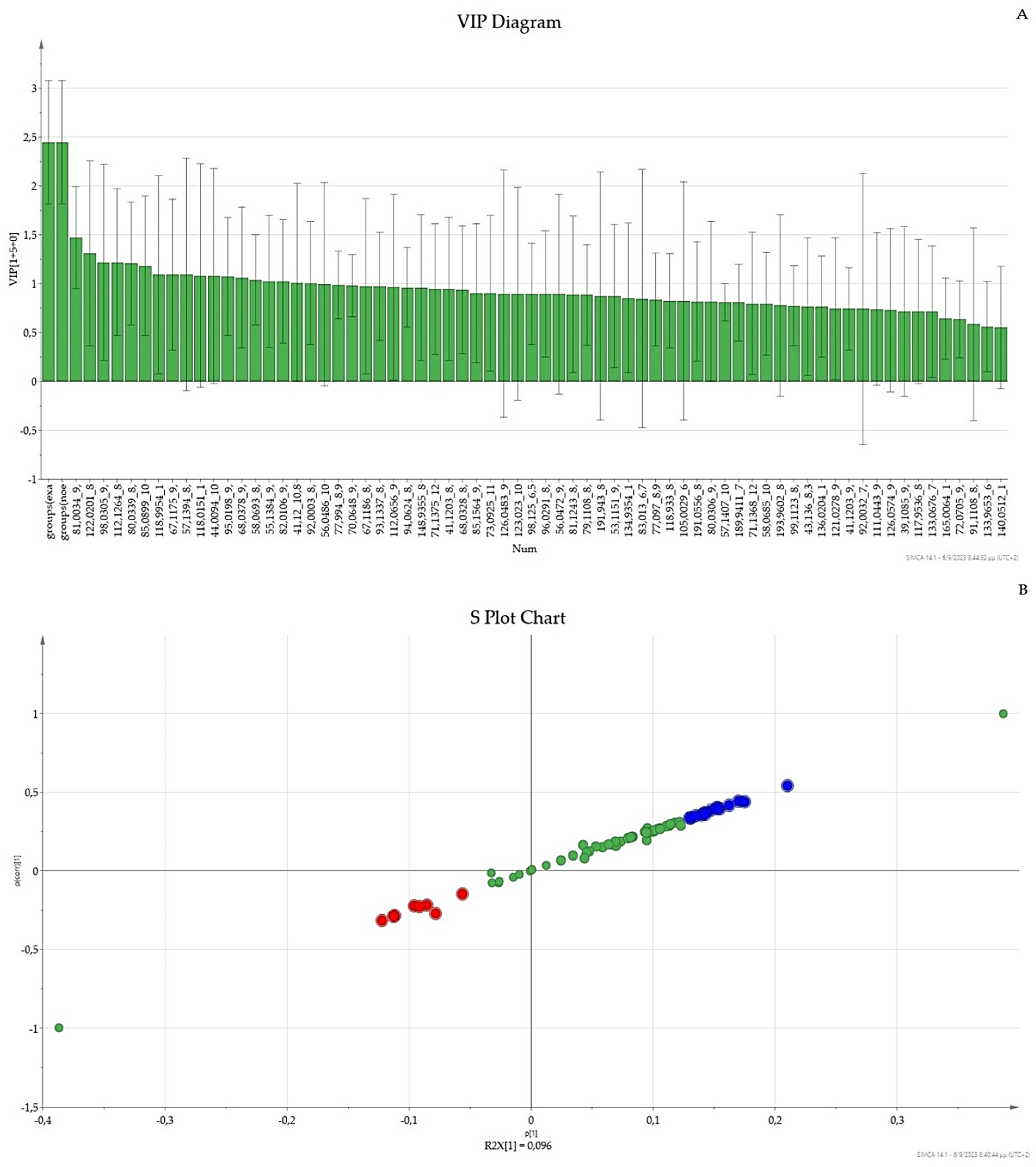
3.3. OPLS-DA UV Scaling, VIP, and S-Plot

In the OPLS-DA Score plot using UV scaling (Figure 3), the patient samples (exacerbations and non-exacerbations) were completely separated along the X-axis. This indicates that the OPLS-DA model was able to successfully differentiate between the two categories based on the defined exacerbation status. After obtaining the OPLS-DA results, the VIP (Variable Importance in the Projection) diagram was generated (Figure 4). The VIP chart provides a summary of the most important features that contribute to the separation observed in the OPLS-DA model. In this chart, features with VIP values greater than 1.0 are considered statistically significant, indicating their importance in distinguishing between the two categories. On the other hand, features with VIP values less than 0.5 are considered non-statistically significant and may exert a diminished impact on the separation. From the VIP diagram, a table with the VIP values and the MZ_RT (Table 2) was automatically generated. In addition to the VIP diagram, the S-plot was utilized to display the covariance and correlation of the characteristics. The S-plot helps identify the features that exhibit high differentiation and contribute the most to the separation observed in the OPLS-DA model. Features located at the edges of the S-plot, further away from the center, are the ones that show the greatest covariance and correlation with the defined categories. From the S-Plot, a table with the MZ_RT (Table 3) was automatically generated. The combination of two tables, namely Table 2 and Table 3, resulted in the creation of a third table, denoted as Table 4, that provides a summary of the characteristics derived from the sample analysis.
Based on the data in Table 2, a search was conducted in the mass spectrum using the corresponding retention times of each characteristic. The NIST library was utilized (a similarity score higher than 90% between the mass spectral data of the analyzed compound to the its closest hit in the library was used) to identify the compounds associated with these characteristics (Table 5). For each identified compound, a t-test was performed to determine if there was a statistically significant difference between the two groups (patients with exacerbations and patients without exacerbations). The t-test was conducted at a 95% confidence level, and a feature was considered significant if the p-value was less than 0.05.

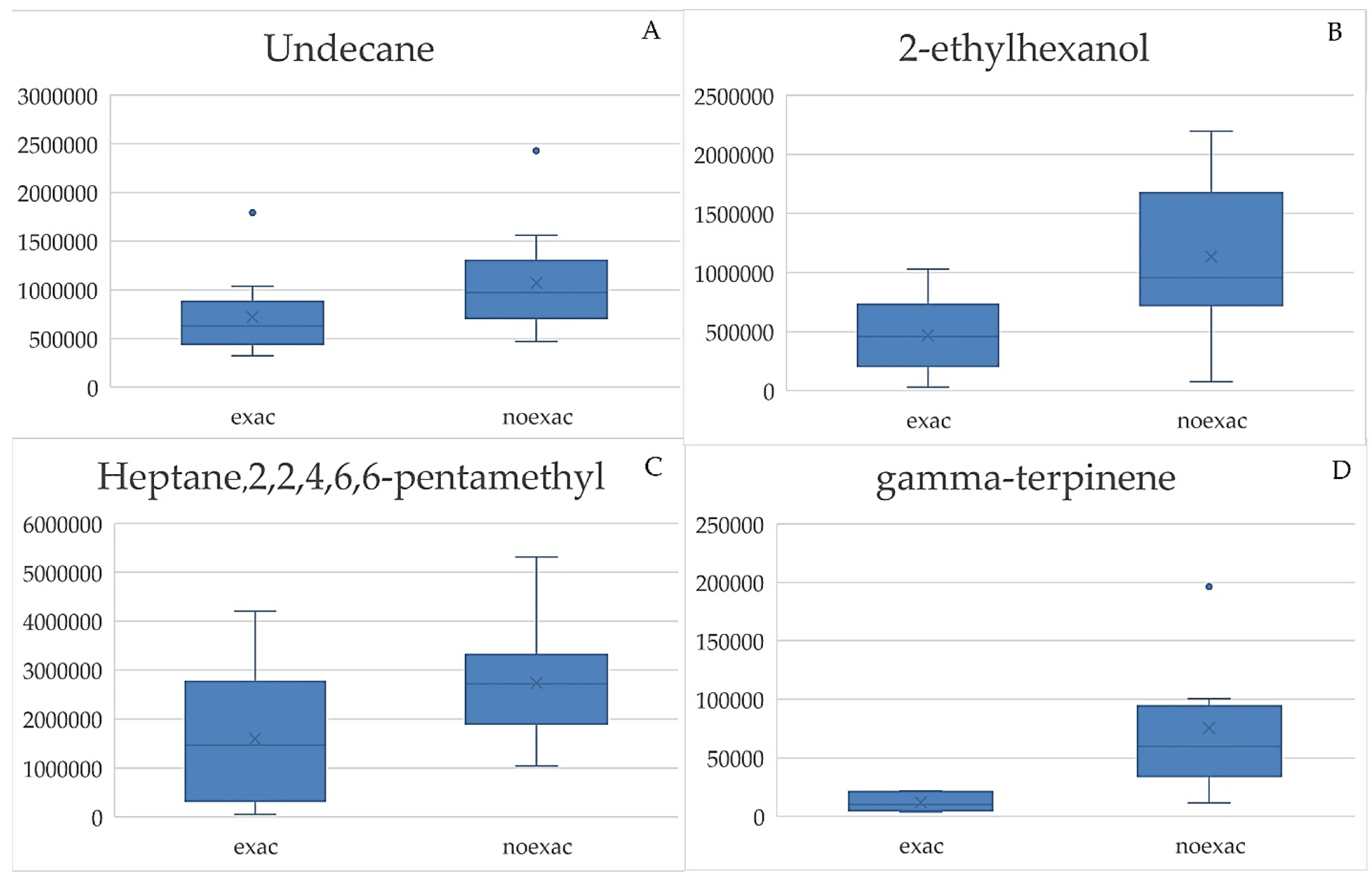
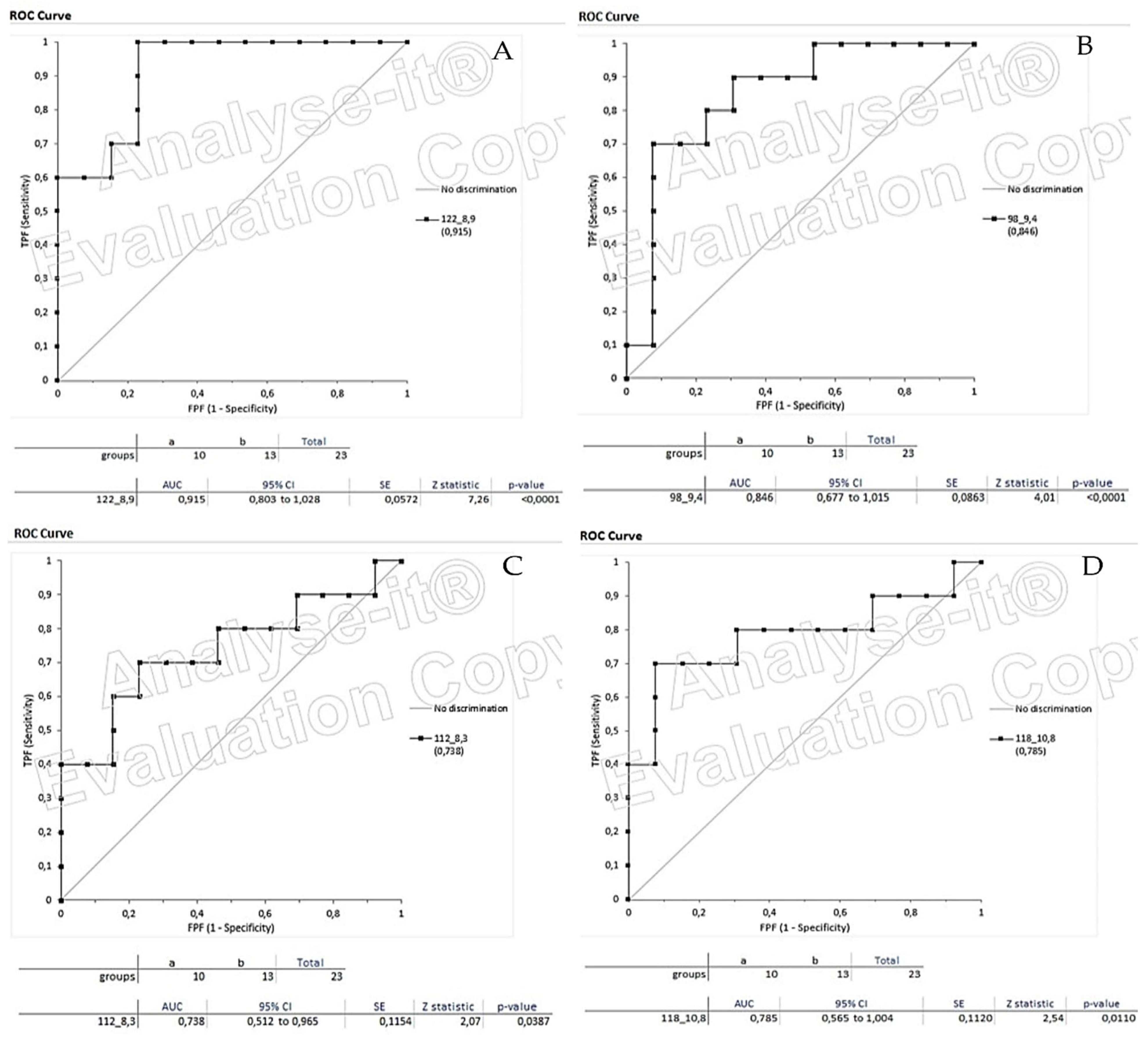
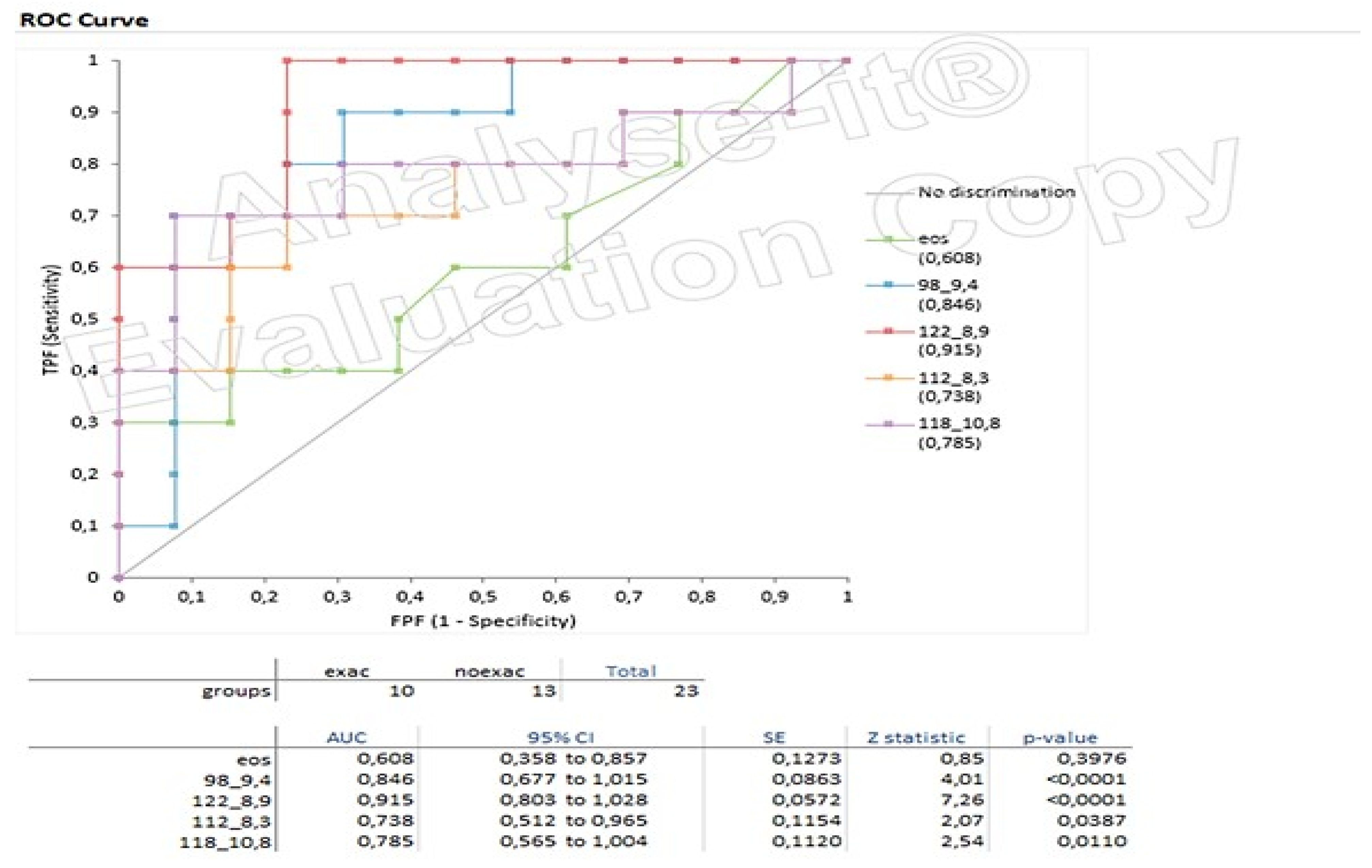
3.4. Box Plots and ROC Curves

Based on the box plots (Figure 5) for the identified compounds, it was observed that the mean values of patients without exacerbations (noexac) were higher than the mean values of patients with exacerbations (exac). The box plots also indicated that for the compounds gamma-terpinene (1,4-Cyclohexadiene, 1-methyl-4-(1-methylethylene)) with RT: 8.9 and 2-ethylhexanol with RT: 9.4, the overlap range between the two groups was very narrow. However, for the compounds Heptane,2,2,4,6,6-pentamethyl with RT: 8.3 and Undecane with RT: 10.8, there was some overlap between the two groups.
The ROC curves (Figure 6) for all the identified compounds showed that the area under the curve (AUC) values were greater than 0.7, indicating satisfactory to excellent separation between the two classes. Additionally, the p-values for all the curves were significantly lower than the required threshold of 0.05, indicating statistical significance and reliable results.
Furthermore, from the ROC curves (Figure 7), it appeared that the diagnostic performance of the four substances was better compared to Eosinophils (Eos) of the patients. These findings suggest that the identified compounds have potential as biomarkers for distinguishing between patients with exacerbations and those without exacerbations in COPD.

4. Discussion

The aim of the study was to identify potential metabolites that can differentiate between patients with different stages of COPD. We collected exhaled air samples from patients within the same age range, including both patients with exacerbations and patients without exacerbations. We successfully employed an analytical methodology that involved using RTubeVOC for air sample collection and solid phase microextraction (PDMS/CAR/DVB-30/50 um) for volatile compound extraction. Gas chromatography coupled with mass spectrometry was used for the analysis of the extracted compounds. To analyze the complex data obtained from the analysis, multivariate statistical analysis was performed. As a result, four compounds were identified: Heptane,2,2,4,6,6-pentamethyl, gamma-terpinene, 2-ethylhexanol, and Undecane. It is worth noting that 2-ethylhexanol belongs to the alcohol compound class, while the other three compounds belong to the hydrocarbon class. The identification of these compounds was done with a confidence level greater than 95%.
The study faced great challenges in collecting air samples from patients due to the coronavirus situation in Greece. With a larger number of samples, the results could potentially be more representative of the disease and provide a better understanding of COPD. It’s important to note that there are currently no studies available that specifically compare the two stages of COPD (patients with exacerbations and patients without exacerbations). Existing literature mainly focuses on comparisons between COPD patients and healthy subjects, lung cancer patients, or asthma patients [17].
Nevertheless, this study contributes valuable evidence by demonstrating differences in the breath profiles of patients with COPD and exacerbations compared to patients with COPD without exacerbations. Four VOCs were identified in the analysis. It was found through literature research that undecane and n-terpinene have been reported as identified biomarkers in online COPD databases [17,18]. The remaining two biomarkers are likely among the many yet to be certified biomarkers, as there are over 3000 metabolites in exhaled air, and approximately 500 have been identified so far. Our suggested approach addresses the primary limitation in the analysis of exhaled-breath condensate, which is likely linked to the challenge of standardizing sample collection. With respect to electronic nose, besides its portability, it is not equipped to offer structural elucidation for distinguishing VOCs within different groups. However, our method cannot be compared to the real-time techniques that provide minimized sample handling with low associated risk of sample contamination.

Supplementary Materials

The following supporting information can be downloaded at: https://www.mdpi.com/article/10.3390/chemosensors11100542/s1, Figure S1: A typical SPME/GC-MS chromatogram of an exhaled breath sample of patient (A) with exacerbations and (B) without exacerbations.

Author Contributions

Conceptualization: C.C., V.S. and K.K.; methodology, L.L., C.C., V.S. and K.K.; software, L.L. and K.E.; validation, L.L., C.C. and V.S.; formal analysis, L.L.; investigation, L.L., C.C. and K.E.; resources, K.K. and V.S.; data curation, L.L., K.E. and V.S.; writing—original draft preparation, L.L., C.C. and K.E.; writing—review and editing, L.L., C.C., K.E., K.K. and V.S.; visualization, L.L. and K.E.; supervision, K.K. and V.S.; project administration, V.S.; All authors have read and agreed to the published version of the manuscript.

Funding

This research received no external funding.

Institutional Review Board Statement

Not applicable.

Informed Consent Statement

Not applicable.

Data Availability Statement

Not applicable.

Conflicts of Interest

The authors declare no conflict of interest.

References

  1. Adeloye, D.; Song, P.; Zhu, Y.; Campbell, H.; Sheikh, A.; Rudan, I.; NIHR RESPIRE Global Respiratory Health Unit. Global, Regional, and National Prevalence of, and Risk Factors for, Chronic Obstructive Pulmonary Disease (COPD) in 2019: A Systematic Review and Modelling Analysis. Lancet Respir. Med. 2022, 10, 447–458. [Google Scholar] [CrossRef]
  2. Vos, T.; Barber, R.; Bell, B.; Bertozzi-Villa, A.; Biryukov, S.; Bolliger, I.; Charlson, F. Global, Regional, and National Incidence, Prevalence, and Years Lived with Disability for 301 Acute and Chronic Diseases and Injuries in 188 Countries, 1990–2013: A Systematic Analysis for the Global Burden of Disease Study 2013. Lancet 2015, 386, 743–800. [Google Scholar] [CrossRef]
  3. Kim, V.; Aaron, S.D. What Is a COPD Exacerbation? Current Definitions, Pitfalls, Challenges and Opportunities for Improvement. Eur. Respir. J. 2018, 52, 1801261. [Google Scholar] [CrossRef] [PubMed]
  4. Stockley, R.A.; Halpin, D.M.G.; Celli, B.R.; Singh, D. Chronic Obstructive Pulmonary Disease Biomarkers and Their Interpretation. Am. J. Respir. Crit. Care Med. 2019, 199, 1195–1204. [Google Scholar] [CrossRef] [PubMed]
  5. Signe, V.K.; Nielsen, S.F.; Lange, P.; Vestbo, J.; Nordestgaard, B.G. Blood Eosinophils and Exacerbations in Chronic Obstructive Pulmonary Disease. The Copenhagen General Population Study. Am. J. Respir. Crit. Care Med. 2016, 193, 965–974. [Google Scholar] [CrossRef]
  6. Yun, J.H.; Lamb, A.; Chase, R.; Singh, D.; Parker, M.; Saferali, A.; Vestbo, J. Blood Eosinophil Count Thresholds and Exacerbations in Patients with Chronic Obstructive Pulmonary Disease. J. Allergy Clin. Immunol. 2018, 141, 2037–2047. [Google Scholar] [CrossRef] [PubMed]
  7. Higham, A.; Beech, A.; Wolosianka, S.; Jackson, N.; Long, G.; Kolsum, U.; Southworth, T. Type 2 Inflammation in Eosinophilic Chronic Obstructive Pulmonary Disease. Allergy 2021, 76, 1861–1864. [Google Scholar] [CrossRef] [PubMed]
  8. Agusti, A.; Bel, E.; Thomas, M.; Vogelmeier, C.; Brusselle, G.; Holgate, S.; Humbert, M. Treatable Traits: Toward Precision Medicine of Chronic Airway Diseases. Eur. Respir. J. 2016, 47, 410–419. [Google Scholar] [CrossRef] [PubMed]
  9. Benet, J.G.; Seo, M.; Khine, M.; Padró, J.G.; Martnez, A.P.; Kurdahi, F. Breast Cancer Detection by Analyzing the Volatile Organic Compound (VOC) Signature in Human Urine. Sci. Rep. 2022, 12, 14873. [Google Scholar] [CrossRef] [PubMed]
  10. Schleich, F.N.; Zanella, D.; Stefanuto, P.R.; Bessonov, K.; Smolinska, A.; Dallinga, J.W.; Henket, M. Exhaled Volatile Organic Compounds Are Able to Discriminate between Neutrophilic and Eosinophilic Asthma. Am. J. Respir. Crit. Care Med. 2019, 200, 444–453. [Google Scholar] [CrossRef] [PubMed]
  11. Nakhleh, M.K.; Haick, H.; Humbert, M.; Cohen-Kaminsky, S. Volatolomics of Breath as an Emerging Frontier in Pulmonary Arterial Hypertension. Eur. Respir. J. 2017, 49, 1601897. [Google Scholar] [CrossRef] [PubMed]
  12. Plantier, L.; Smolinska, A.; Fijten, R.; Flamant, M.; Dallinga, J.; Mercadier, J.J.; Pachen, D.; d’Ortho, M.P.; van Schooten, F.J.; Crestani, B.; et al. The Use of Exhaled Air Analysis in Discriminating Interstitial Lung Diseases: A Pilot Study. Respir. Res. 2022, 23, 12. [Google Scholar] [CrossRef] [PubMed]
  13. Wang, C.; Sun, B.; Guo, L.; Wang, X.; Ke, C.; Liu, S.; Zhao, W.; Luo, S.; Guo, Z.; Zhang, Y.; et al. Volatile organic metabolites identify patients with breast cancer, cyclomastopathy, and mammary gland fibroma. Sci. Rep. 2014, 4, 5383. [Google Scholar] [CrossRef] [PubMed]
  14. Westerhuis, J.A.; Hoefsloot, H.C.J.; Smit, S.; Vis, D.J.; Smilde, A.K.; Velzen, E.J.J.; Duijnhoven, J.P.M.; Dorsten, F.A. Assessment of PLSDA cross validation. Metabolomics 2008, 4, 81–89. [Google Scholar] [CrossRef]
  15. Blasco, H.; Błaszczyński, J.; Billaut, J.C.; Nadal-Desbarats, L.; Pradat, P.F.; Devos, D.; Moreau, C.; Andres, C.R.; Emond, P.; Corcia, P.; et al. Comparative analysis of targeted metabolomics: Dominance-based rough set approach versus orthogonal partial least square-discriminant analysis. J. Biomed. Inform. 2015, 53, 291–299. [Google Scholar] [CrossRef] [PubMed]
  16. Farrés, M.; Platikanov, S.; Tsakovski, S.; Tauler, R. Comparison of the variable importance in projection (VIP) and of the selectivity ratio (SR) methods for variable selection and interpretation. J. Chemom. 2015, 29, 528–531. [Google Scholar] [CrossRef]
  17. Ratiu, I.A.; Ligor, T.; Bocos-Bintintan, V.; Mayhew, C.A.; Buszewski, B. Volatile organic compounds in exhaled breath as fingerprints of lung cancer, asthma, and COPD. J. Clin. Med. 2021, 10, 23. [Google Scholar] [CrossRef] [PubMed]
  18. Mule, N.M.; Patil, D.D.; Kaur, M. A comprehensive survey on investigation techniques of exhaled breath (EB) for diagnosis of diseases in human body. Inform. Med. Unlocked 2021, 26, 100715. [Google Scholar] [CrossRef]
Figure 1. (A) The PCA score plot in UV Scaling; (B) the PLS-DA score plot in UV Scaling; (C) The Permutation Test for UV Scaling, performed by SIMCA.
Chemosensors 11 00542 g001
Figure 2. (A) The PCA score plot in Pareto Scaling; (B) the PLS-DA score plot in Pareto Scaling; (C) The Permutation test for Pareto Scaling, performed by SIMCA.
Chemosensors 11 00542 g002
Figure 3. OPLS-DA Score plot in UV scaling performed by SIMCA program (* = explained variance).
Chemosensors 11 00542 g003
Figure 4. (A) VIP Plot chart; (B) S-Plot chart (both were generated by Simca program).
Chemosensors 11 00542 g004
Figure 5. (A) Box plot for Undecane (RT: 10.8); (B) Box plot for 2-ethylhexanol (RT: 9.4); (C) Box plot for Heptane,2,2,4,6,6-pentamethyl (RT: 8.3); (D) Box plot for gamma-terpinene (RT: 8.9).
Chemosensors 11 00542 g005
Figure 6. (A) ROC Curve for gamma-terpinene (RT: 8.9); (B) ROC Curve for 2-ethylhexanol (RT: 9.4); (C) Box plot for Heptane,2,2,4,6,6-pentamethyl (RT: 8.3); (D) ROC Curve for Undecane (RT: 10.8).
Chemosensors 11 00542 g006
Figure 7. A summary of ROC Curves for Heptane,2,2,4,6,6-pentamethyl (RT: 8.3), gamma-terpinene (RT: 8.9), 2-ethylhexanol (RT: 9.4), Undecane (RT: 10.8) compared to the ROC Curve of eosinophils (Eos) from each patient.
Chemosensors 11 00542 g007
Table 1. Patient information.
Patients CodeAgeNumber of ExacerbationsEOS Count per μL
exac1663450
noexac1660380
noexac260030
noexac3530160
noexac4770170
exac2792180
noexac5620150
noexac6770310
noexac7600210
noexac8780420
exac3773190
exac4702360
exac5704500
noexac969070
exac6582160
noexac10710170
exac7733120
exac8721150
exac9611310
noexac11690330
noexac12590180
noexac13730150
noexac1468060
noexac1570080
exac10704480
Table 2. VIP-Statistically Important Characteristics.
MZ_RTVIP ValuesMZ_RTVIP Values
81_9.41.6449658_8.31.12807
122_8.91.4097941_10.81.11455
98_9.41.3587855_9.41.09555
80_8.91.34396112_101.09417
112_8,31.3358982_9.41.09209
85_101.2934492_8.91.06967
55_6.51.2702467_8.31.06094
119_101.2499777_8.91.05121
57_8.31.2426493_8.91.05057
67_9.41.2333156_10.81.04641
118_10.81.2183394_8.91.04191
44_10.81.2041670_9.41.0409
95_9.41.1895771_121.03677
68_9.41.17337123_10.81.01216
Table 3. S-Plot-Statistically Significant Characteristic.
MZ_RTMZ_RT
98_9.467_9.4
80_8.9112_8.3
55_6.544_10.8
122_8.995_9.4
68_9.4118_10.8
85_10119_10
81_9.4
Table 4. Summary table of the characteristics obtained from the analysis of the samples.
RT (min)Characteristics (M/Z)
6.555
8.357, 58, 67, 112
8.977, 80, 92, 93, 94, 122
9.455, 67, 68, 70, 81, 82, 95, 98
1085, 112, 119
10.841, 44, 56, 118, 123
1271
Table 5. Metabolites identified with the NIST library.
RT (min)M/Zp-ValueVolatiles
8.357, 58, 67, 1120.0387Heptane,2,2,4,6,6-pentamethyl
8.977, 80, 92, 93, 94, 1220.0001gamma-terpinene(1-methyl-4-propan-2-ylcyclohexa-1,4-diene)
9.455, 67, 68, 70, 81, 82, 95, 980.00012-ethylhexanol
10.841, 44, 56, 118, 1230.0110Undecane
Disclaimer/Publisher’s Note: The statements, opinions and data contained in all publications are solely those of the individual author(s) and contributor(s) and not of MDPI and/or the editor(s). MDPI and/or the editor(s) disclaim responsibility for any injury to people or property resulting from any ideas, methods, instructions or products referred to in the content.

