Article

Chiral Recognition of Phenylglycinamide Enantiomer Based on Electrode Modified by Silver-Ammonia Ion-Functionalized Carbon Nanotubes Complex

1
Jiangsu Key Laboratory of Advanced Catalytic Materials and Technology, Changzhou University, Changzhou 214500, China
2
Changzhou University Huaide College, Jingjiang 214500, China
*
Authors to whom correspondence should be addressed.
Chemosensors 2023, 11(2), 86; https://doi.org/10.3390/chemosensors11020086
Submission received: 15 December 2022 / Revised: 18 January 2023 / Accepted: 19 January 2023 / Published: 22 January 2023

Abstract

:
Polyacrylic acid (PAA) chains were used to decorate the surface of multi-walled carbon nanotubes (MWCNTs) via in situ free radical polymerization, and sulfonated chitosan (SCS) was synthesized via a simple and environmental method. Silver-ammonia ions were introduced as the fixative with PAA-MWCNTs as the basic framework, and SCS was used to decorate the surface, thereby obtaining PAA-MWCNTs-Ag-SCS. The modified electrode exhibited excellent cyclic voltammogram (CV) stability after 100 cycles of scanning. According to differential pulse voltammetry (DPV), the peak current value was approximately 250 μA, exhibiting outstanding sensitivity to phenylglycinamide (Pen) enantiomers. The peak current ratio of D-Pen to L-Pen reached 2.16, showing excellent selectivity. The detection limit (DL) was calculated as 0.015 mM and 0.036 mM for L-Pen and D-Pen, respectively, using the signal-to-noise ratio (S/N = 3). This study provides a new idea for the construction of a chiral-sensing platform with outstanding sensitivity, superior stability, and excellent recognition efficiency.

1. Introduction

The reason why life needs chirality is one of the “125 cutting-edge scientific issues in the world” released by Science [1]. Although we do not have a sure answer for this issue, research into the chirality problem has a long history [2]. Ranging from Pasteur’s isolation of left-handed from right-handed sodium ammonium tartrate crystals [3] to Kelvin’s first definition of chirality [4], the growing interest in chirality has mainly stemmed from biological studies. As is well known, in achiral environments, chiral compounds exhibit no differences in physicochemical properties. However, because of the natural asymmetry in the basic substances of life, such as amino acids, sugars, proteins, and nucleic acids [5], chiral drugs often exhibit different biological activities in organisms [6]. In general, one configuration of a chiral enantiomer may be effective and safe, while the other may be ineffective or even toxic [7]. Thus, the recognition of chiral enantiomers is highly significant in the pharmaceutical and biochemical fields.
Up until now, various separation methods, including supercritical fluid chromatography [8], high-performance liquid chromatography [9], gas chromatography [8], exchange chromatography [10], thin-layer chromatography, ion-capillary electrophoresis [11], fluorescence spectroscopy [12], and capillary electrochromatography [13], have been evaluated for the separation and analysis of optically active compounds. Among the range of detection methods, electrochemical methods stand out [14] due to their advantages of being environmentally friendly, cost-effective, responsive, and easily miniaturized [15]. The focus in electrochemical chiral recognition research has been the construction of a chiral-sensing platform with outstanding recognition performance [16].
Chitosan (CS) possesses a double-helix structure similar to DNA, and it contains abundant chiral binding sites for enantiomeric recognition [13]. However, its practical application remains limited due to its relatively poor water solubility and low optical rotation [17,18]. Hence, in this work, we synthesized sulfonated chitosan (SCS) using a facile chemical procedure, which not only increased the water solubility of chitosan but also improved the optical rotation from 10° to 75°.
Multi-walled carbon nanotubes (MWCNTs) are macromolecules that consist of sp2-hybridized carbon atoms [19], which are connected in a planar hexagonal lattice [20]. They are widely used in many fields due to their superior physicochemical properties, such as a high specific surface area, favorable conductivity, long-term stability [21], improved electrode kinetics, and low over-voltage [22,23,24]. Although MWCNTs have outstanding mechanical characteristics, their weak dispersity in water derived from their structural properties gives rise to unsolvable inferiority [25]. Although the traditional acidizing method can increase the aqueous dispersion of MWCNTs, it may damage their structure and reduce their electronic conduction performance [26,27]. In this study, we encapsulated polyacrylic acid (PAA) chains onto the surface of MWCNTs using in situ free radical polymerization. This not only avoids disrupting the surface structure of multi-walled carbon nanotubes but also increases their dispersibility.
In an attempt to construct a stable electrochemical chiral-sensing platform, silver-ammonia ions were introduced. The prepared SCS was anchored on the surface of PAA-MWCNTs via the electrostatic interactions involving the sulfonic acid group of SCS, the carboxylic acid group from the surface of PAA-MWCNTs, and silver-ammonia ions, thus establishing a chiral surface material with PAA-MWCNTs as the basic framework and SCS decorating the surface. This material exhibited excellent cyclic voltammogram (CV) stability after 100 cycles of scanning. The differential pulse voltammetry (DPV) peak current ratio of D-Pen to L-Pen reached 2.16, and the peak current value was approximately 250 μA, exhibiting excellent selectivity and outstanding sensitivity to Pen enantiomers. The detection limit (DL) was calculated as 0.015 mM and 0.036 mM for L-Pen and D-Pen, respectively, using the signal-to-noise ratio (S/N = 3). This study provides a new idea for the construction of a chiral platform with outstanding sensitivity, superior stability, and excellent recognition efficiency.

2. Experimental

2.1. Reagents and Apparatus

L-phenylglycinamide and D-phenylglycinamide were obtained from Shanghai Sigma-Aldrich Corp (St. Louis, MO, USA). Multi-walled carbon nanotubes (MWCNTs) were purchased from Shanghai Aladdin Reagent. Propenoic acid (AA) (Shanghai, China), 2,2-azoisobutyronitrile, and 1,3-propane sultone salt were provided by Sigma Co., Ltd. Chitosan (CS) was provided by Shanghai Jin sui Biotechnology Co., Ltd. (Shanghai, China) Acetone, acetic acid, and methanol were received from Adamas. Silver nitrate, sodium hydroxide, and ammonia water were purchased from Jiangsu Xin nuo ke Catalyst Co., Ltd. (Zhangjiagang, China).
The electrochemical tests were operated in three-electrode cells (CHI-920C electrochemical workstation). A glassy carbon electrode was selected as the working electrode, modified with MWCNTs, SCS, PAA-MWCNTs, or PAA-MWCNTs-Ag-SCS composite. As the counter electrode, platinum foil (1 cm × 4 cm) was used; as the reference electrode, a saturated calomel electrode (SCE) was used. The morphological characterization of the modified electrodes was performed with a scanning electron microscope (SEM, SUPRA55, Carl Zeiss, Jena, Germany). The water contact angle of the complexes was measured with a Theta Flex water contact angle instrument (Da Chang Yang Hang (Shanghai, China) Co., Ltd.). The Fourier-transform infrared spectra of the MWCNTs, PAA-MWCNTs, SCS, and PAA-MWCNTs-Ag-SCS were recorded through a Nicolet FTIR-8400 S spectrometer (Shimadzu, Kyoto, Japan). The complexes were subjected to spectral analysis with a 756 S UV-Visible spectrophotometer. The X-ray photoelectron spectroscopy (XPS) of the modified electrodes was studied on a Thermo Scientific ESCALAB250Xi.

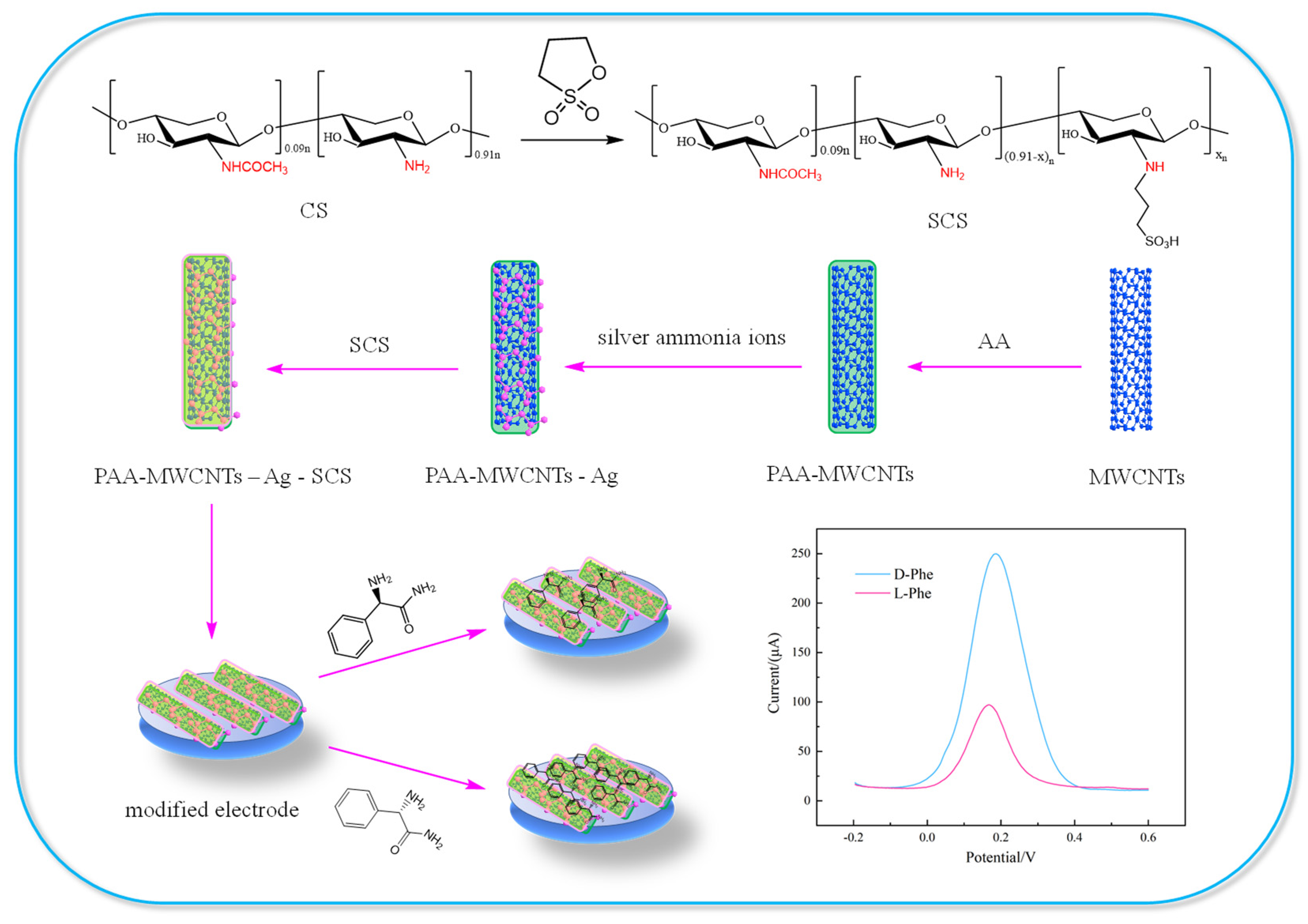
2.2. Preparation of SCS

The preparation process of the chiral-sensing platform and identification of the Pen enantiomers are shown in Scheme 1. As indicated by Rwei and Lien [28], CS was obtained by dissolving 1 g of chitosan in 2 wt % acetic acid aqueous solution. Under a nitrogen atmosphere, 1,3-propanesultone (2 g) was added slowly to the solution of CS, stirred for 6 h at 60 °C, and then poured into acetone to precipitate a white product. The white solid was dried in an oven at 60 °C for 4 h. After complete removal of the unreacted 1,3-propane-sultone, the resulting SCS was a white powder.

2.3. Preparation of PAA-MWCNTs

First, 0.1 g of MWNTs were dispersed in 100 mL of acetone and subjected to ultrasonication for 30 min. Subsequently, at a high agitation rate, 1 g of AA was added slowly. Then, polymerization was initiated by 0.03 g of AIBN. The reaction was maintained for 12 h at a temperature of 60 °C. The resulting black solid was collected by filtration, and all solid materials were washed with acetone and deionized water two more times to remove all impurities. The powder was finally dried in a vacuum oven at 70 °C [29].

2.4. Preparation of PAA-MWCNTs-Ag-SCS

To a test tube with 1 mL of 2% AgNO3 solution, two drops of NaOH solution were added, shaking the test tube to complete the reaction. Then, 2% dilute ammonia water was added drop-by-drop while shaking until complete dissolution of the precipitate, eventually yielding silver-ammonia solution. Subsequently, PAA-MWCNTs (3 mg) were dispersed in 1 mL of deionized water, forming black suspensions, after which silver-ammonia solution was added dropwise. Ultrasonication was applied for 0.5 h, yielding a suspension. A total of 3 mg of SCS was dissolved in 1 mL of deionized water and then added to the above suspension. After sonication for 1 h, a well-dispersed PAA-MWCNTs-Ag-SCS suspension was obtained.

2.5. Preparation of PAA-MWCNTs-Ag-SCS/GCE

Using alumina, the GCE was polished, sonicated in ethanol and ultrapure water, and thoroughly rinsed. Afterward, 5 μL of PAA-MWCNTs-Ag-SCS composite was dropped onto the pretreated GCE and then allowed to dry under an infrared lamp. The modified electrodes of MWCNTs/GCE, SCS/GCE, PAA-MWCNTs/GCE, and PAA-MWCNTs-Ag/GCE were prepared with the same procedure.

2.6. Electrochemical Chiral Discrimination of Pen Enantiomers

The prepared PAA-MWCNTs-Ag-SCS/GCE was put into 0.1 M PBS (pH = 7) solution containing 5 mM L- or D-Pen (6 min, 25 °C) to combine Pen isomers. After removal, the electrode was placed under an infrared lamp and allowed to dry off. The enantiomer recognition process of the modified electrodes was performed using differential pulse voltammetry (DPV) in 0.1 M KCl solution containing 5 mM [Fe(CN)6]3−/4− with a three-electrode system. The CV, EIS, and DPV tests were performed from −0.2 to 0.6 V at a pulse amplitude of 100 mV and pulse width of 0.1 s. The typical Nyquist plots of the modified electrodes with frequencies swept from 106 Hz to 1 Hz were recorded.

3. Results and Discussion

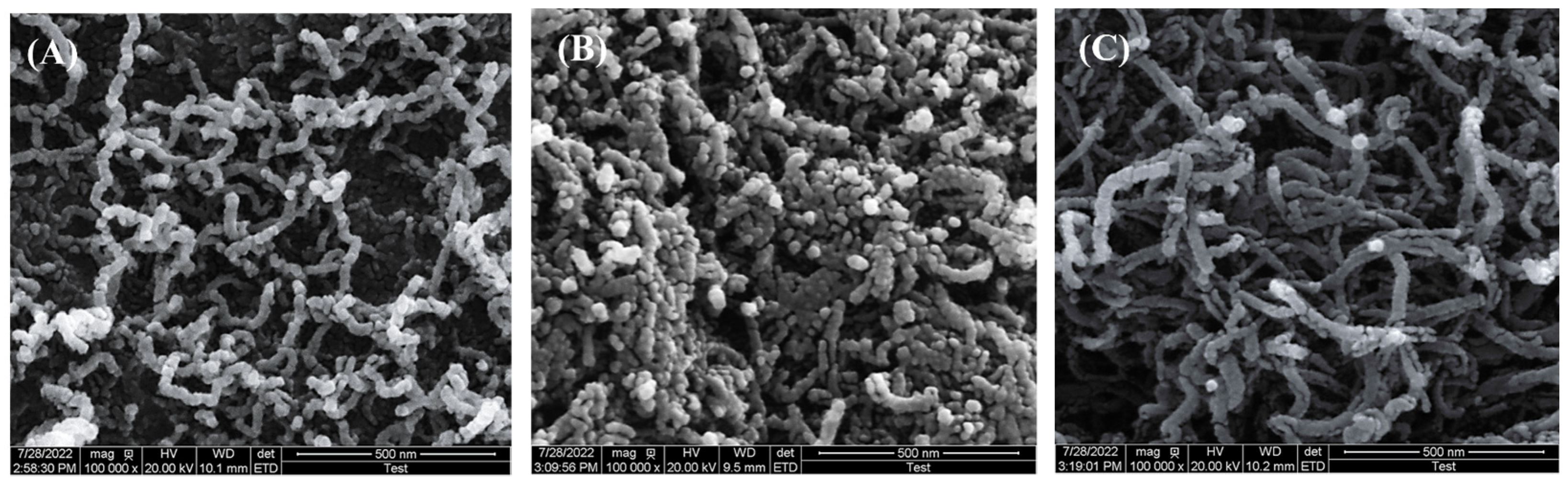
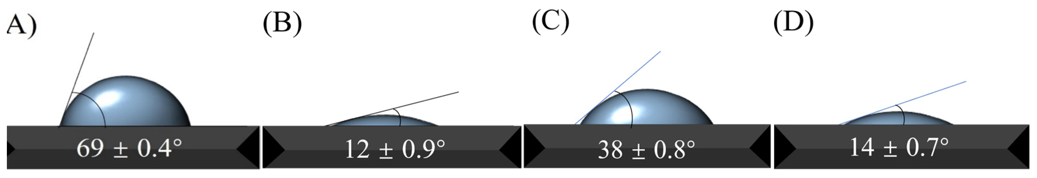
3.1. Characterization of Chiral Materials

The morphological characteristics of the composites were obtained on a scanning electron microscope (Figure 1). From Figure 1A, it can be observed that the MWCNTs displayed a smooth and well-defined rod-like morphology. Upon encapsulating the polyacrylic acid (PAA) chains onto the surface of the MWCNTs, the originally loose multi-walled carbon nanotubes began to aggregate (Figure 1B). This may be attributed to the interactions of the carboxyl groups on the surface of the PAA-MWCNTs [30]. Interestingly, the PAA-MWCNTs-Ag-SCS recovered the well-defined loose rod-like structures, which coarsened significantly (Figure 1C). This may have been due to the silver-ammonia ions generating an electrostatic effect with the sulfonic groups of SCS and the carboxylic moieties of the PAA-MWCNTs. This enabled the loading of SCS and silver-ammonia ions onto the surface of the PAA-MWCNTs, which increased their thickness while generating a loose structure.
Further bonding features of the PAA-MWCNTs-Ag-SCS were characterized using FT-IR spectroscopy (Figure 2A). For the MWCNTs (curve a), the characteristic peak at 3435 cm−1 was ascribed to the characteristic vibration of -OH, while the peaks at 1384 cm−1 and 1560 cm−1 belonged to the vibration absorption peak of C=C [31,32]. Compared with curve a, curve b showed an absorption peak at 1632–1712 cm−1, which could be ascribed to the interaction between PAA and the MWNTs [29]. For the CS (curve c), the characteristic peak at 3428 cm−1 was ascribed to the stretching vibration of -OH and -NH2, and the peaks at 1650 cm−1 and 1595 cm−1 were ascribed to the amide I band, which are considered key characteristics of chitosan. Furthermore, the characteristic peak of C-H was observed at 2867–2923 cm−1.
Compared with the curve of CS, curve d exhibited evident absorption peaks at 1042 cm−1 and 1193 cm−1 corresponding to the stretching vibration peak of S=O. The absorption peak at 1654 cm−1 was ascribed to the C=O stretching vibration of the amide group, and the peak at 1602 cm−1 was attributed to the bending vibration of the SCS branch [28]. All characteristic peaks were observed in curve e, indicating that PAA-MWCNTs-Ag-SCS was successfully prepared.
The chemical composition of the PAA-MWCNTs-Ag-SCS and the elemental valence state of the compound were studied using XPS [33]. The full XPS spectrum of the PAA-MWCNTs-Ag-SCS is shown in Figure 2B, which clearly demonstrates the presence of S 2p (165.08 eV), C 1s (284.08 eV), N 1s (399.08 eV), O 1s (531.08 eV), and Ag 3d (368.08 eV) [34,35]. Figure 2C–F exhibits the high-resolution XPS spectra of the C 1s, N 1s, O 1s, and Ag 3d regions, respectively. The high-resolution spectrum of C 1s exhibited four peaks, which could be assigned to C-C/C=C (284.08 eV), C-N (284.88 eV), C-O (286.28 eV), and C=O (287.38 eV) bonds. The high-resolution spectrum of N 1s exhibited four peaks [36], which could be assigned to Ag-N (397.28 eV), -NH2 (398.68 eV), C-N (399.58 eV), and -NH3+ (401.38 eV) bonds. The high-resolution spectrum of O 1s exhibited three peaks, which could be assigned to C=O (531.18 eV), C-OH (532.18 eV), and C-O-C (532.88 eV) bonds [37]. The high-resolution spectrum of Ag 3d exhibited two peaks, which could be assigned to Ag 3d5/2 (368.78 eV) and Ag 3d3/2 (374.08 eV) [38]. These results strongly illustrate the successful preparation of PAA-MWCNTs-Ag-SCS.
Following XPS analysis, we investigated the wettability of the samples by measuring the water contact angle. As shown in Figure 3, the contact angle for the PAA-MWCNTs was 11.1° (Figure 3B) and less than half of that for the MWCNTs (68.6°, Figure 3A). This could be a result of the introduction of a number of hydrophilic carboxylate groups via encapsulation of the polyacrylic acid (PAA) chains onto the surface of the MWCNTs. Although SCS showed a higher water contact angle (37.2°, Figure 3C), that of the PAA-MWCNTs-Ag-SCS (13.3°, Figure 3D) was obviously reduced, which may be attributable to the combination of SCS with the PAA-MWCNTs containing a strongly hydrophilic group.

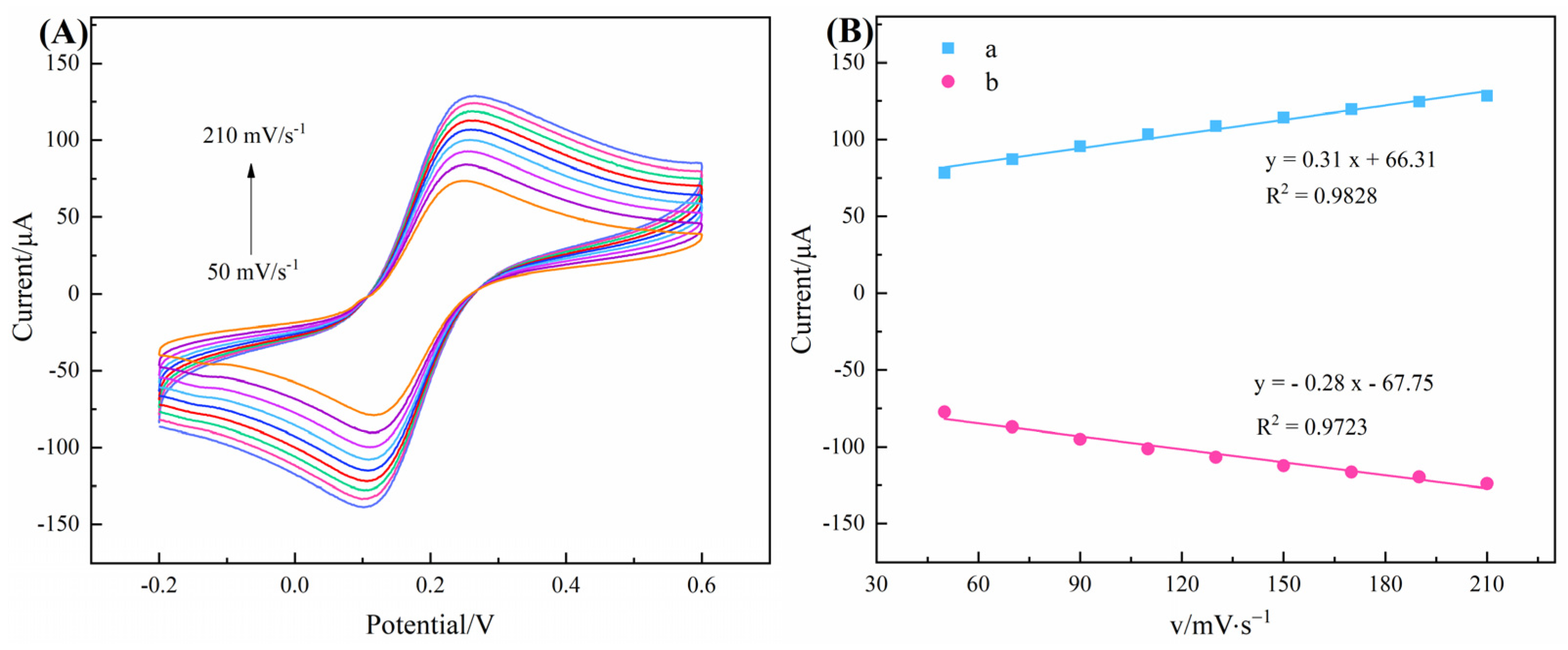
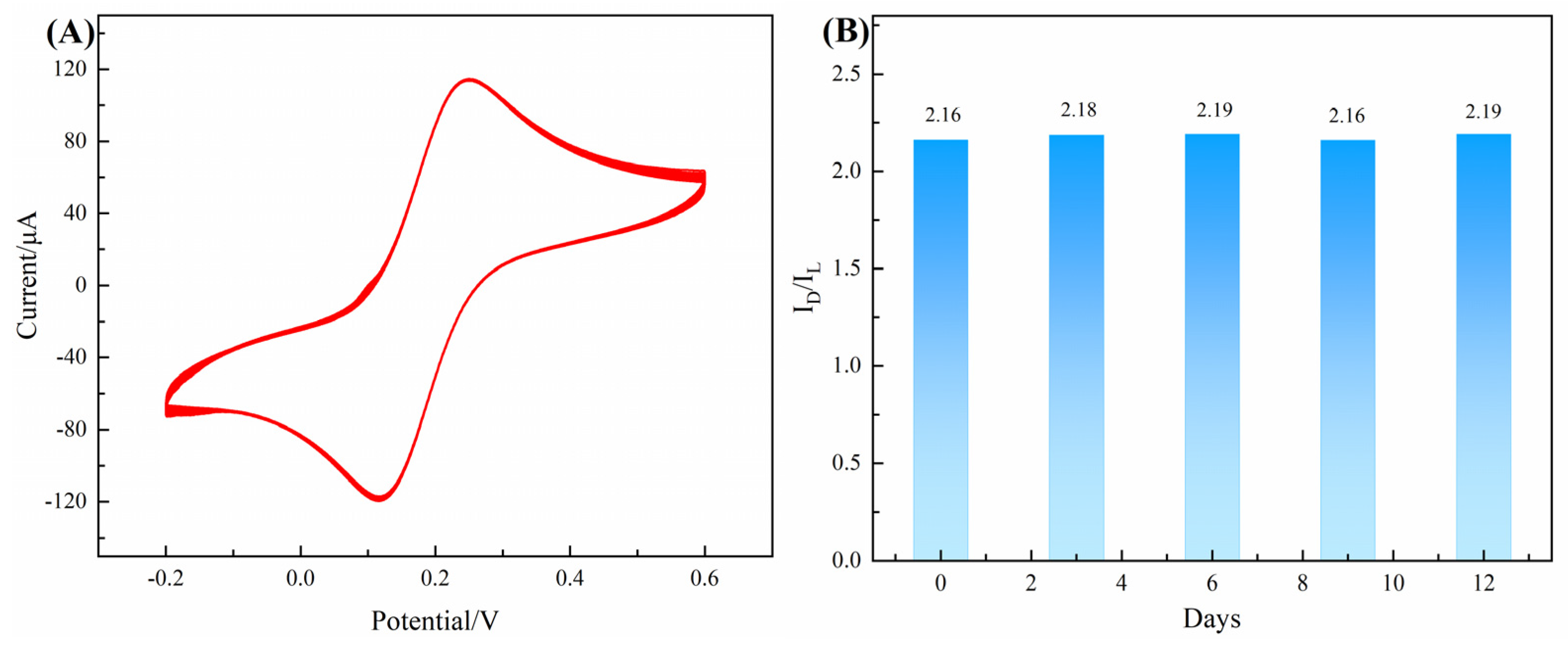
3.2. Electrochemical Properties of Chiral-Sensing Platform

The sensing performance of the modified electrodes was studied using CV. The results are illustrated in Figure 4A, revealing a couple of quasi-reversible redox peaks on the bare GCE. After loading the MWCNTs onto the surface of the GCE, an increase in the peak current was observed. Compared to the MWCNTs/GCE, there was a remarkable increase in the redox peak current of the PAA-MWCNTs/GCE, which could have resulted from the introduction of another electron conduction mechanism (i.e., -ionic conductivity) into the system during the encapsulation of the polyacrylic acid (PAA) chains onto the surface of the MWCNTs [23]. The peak current was strongly reduced upon introducing SCS and silver-ammonia ions into the complexes via electrostatic interactions, which may have been due to the poor electronic conductivity of SCS, hindering the transmission of electrons.
To further understand the electrochemical performance of the modified electrodes, EIS was performed to evaluate the interfacial electron conduction properties (Figure 4B). The electrolyte resistance (Rs) represents the bulk nature of the electrolyte solution, while the Warburg element (Zw) describes the diffusion of the redox probe. Ret and Cdl represent the transfer resistance of electrons from the liquid phase to the electrode and double-layer electric capacity, respectively. The impedance value of the MWCNTs/GCE was 27.49 Ω, which represents a marked decrease when compared with bare GCE (73.56 Ω), whereas the decline in the impedance value of the PAA-MWCNTs/GCE (9.85 Ω) was more obvious. In apparent contrast, the impedance value of the PAA-MWCNTs-Ag-SCS (153 Ω) was markedly improved, agreeing with the results obtained in the CV test.
The electrochemical kinetics of the prepared PAA-MWCNTs-Ag-SCS/GCE was studied using 0.1 M KCl solution containing 5 mM [Fe(CN)6]3−/4− at various scan rates. The CV curves of the PAA-MWCNTs-Ag-SCS/GCE at various scan rates are presented in Figure 5A. The results indicate that the chiral-sensing platform has good stability and reversibility, which is favorable for its practical application. The linear regression equations (Figure 5B) for the oxidation peak current and the reduction peak current can be described as y = 0.31x + 66.31 (R2 = 0.9828) and y = −0.28x − 67.75 (R2 = 0.9723), respectively. It can be determined from Figure 5B that the peak currents were proportionate to the scan rate, describing a typical adsorption-control process.
In order to evaluate the role played by different materials during the process of chiral recognition, SCS/GCE, PAA-MWCNTs/GCE, PAA-MWCNTs-Ag/GCE, and PAA-MWCNTs-Ag-SCS/GCE were fabricated and used as the working electrodes for Pen enantiomer determination. As can be seen in Figure 6, the modified electrodes of bare GCE (Figure 6A), PAA-MWCNTs/GCE (Figure 6C), and PAA-MWCNTs-Ag/GCE (Figure 6D) did not show a remarkable chiral recognition effect on the Pen enantiomer. This is despite the fact that the peak currents of the PAA-MWCNTs/GCE and PAA-MWCNTs-Ag/GCE significantly increased compared with the PAA-MWCNTs-Ag-SCS/GCE (Figure 6E). In sharp contrast, a large increase in the chiral recognition effect for SCS/GCE (Figure 6B) and PAA-MWCNTs-Ag-SCS/GCE was observed, which can only be explained by the introduction of sulfonated chitosan as the chiral selector. Compared with SCS/GCE, PAA-MWCNTs-Ag-SCS/GCE has a better chiral recognition effect. This may be due to the structural rigidity of the SCS in the PAA-MWCNTs-Ag-SCS/GCE, and the electrostatic interaction between the sulfonic acid group and Pen enantiomers is greatly reduced due to the massive combination of the sulfonic acid group and silver-ammonia ion. The sulfonic acid group of SCS could be immobilized on the PAA-MWCNTs-Ag surface; the amino group and hydroxyl group on the SCS are the main chiral bonding site. The L-configuration of the Pen enantiomer can present three substituents to match the PAA-MWCNTs-Ag-SCS’s three-point site. No matter how its mirror image rotates, the D-enantiomer can match a maximum of only two sites [39]. The crucial step for chiral recognition is the formation of diastereomeric complexes between the PAA-MWCNTs-Ag-SCS and the Pen enantiomer. The achievement of electrochemical chiral recognition is caused by the difference in the amount of the diastereomeric complexes formed [40], which cause the discrepancy of the DPV peak current signal between the L-Pen and D-Pen.
As can be seen from Table 1, compared with other chiral-sensing platforms, PAA-MWCNTs-Ag-SCS/GCE has a higher peak current intensity and more superior chiral recognition effect, that is to say that the sensing platform has higher sensitivity and superior chiral recognition effect.

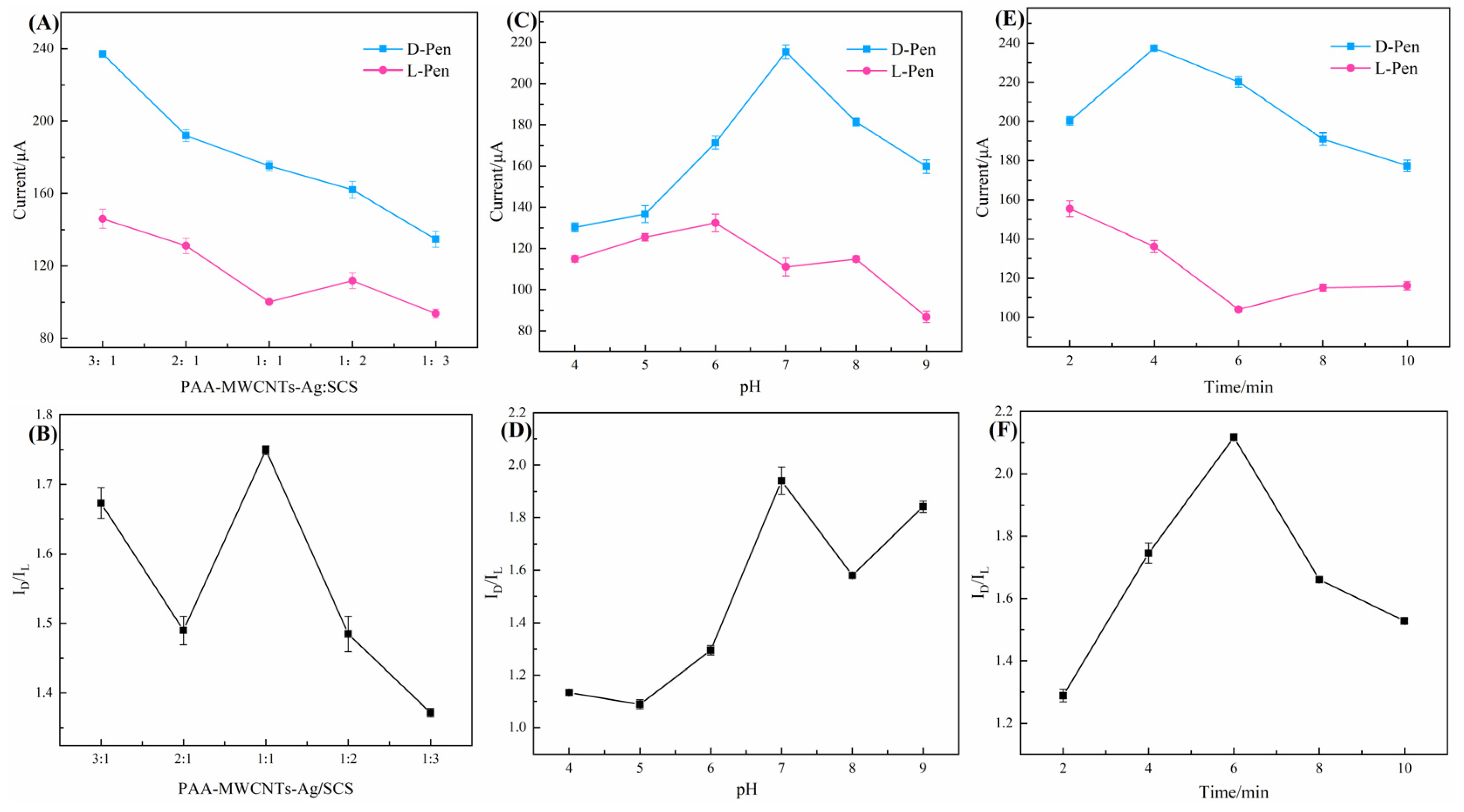
3.3. Optimization of Chiral Recognition Conditions

The loading capacity of SCS determines the spatial distribution shape, the number of chiral binding sites, and thus, the chiral recognition effect of the PAA-MWCNTs-Ag-SCS/GCE. Hence, optimizing the mass concentration ratio of the PAA-MWCNTs-Ag and SCS is important in promoting the chiral recognition effect. As can be seen from Figure 7A, too much SCS probably led to a decrease in the current response, while too little SCS probably resulted in a lack of sufficient binding sites for the recognition of the Pen enantiomers. Therefore, in the optimal formulation of PAA-MWCNTs-Ag-SCS, the weight ratio of PAA-MWCNTs-Ag to SCS was 1:1 (Figure 7B).
Solvation and desolvation processes play a key role in the process of chiral enantiomer recognition; therefore, pH is a nonnegligible factor in the process of chiral recognition. Figure 7D shows that the ratio of ID/IL increased at a pH value of 4.0 to 7.0 but decreased at a pH of 7.0 to 9.0; the peak current showed the same trend (Figure 7C). This may be due to the fact that electrostatic interactions dominated at pH < 7 and pH > 7, while hydrogen bond interactions dominated at pH = 7. However, more enantiomers were bound by electrostatic interactions than hydrogen bonding. Consequently, pH = 7 was selected as the best solvent state.
The enantiomeric action time is also a vital factor during the process of chiral recognition. An overly short action time would lead to too little binding of the enantiomers, whereas an excessively long action time would lead to a decrease in the peak current response at the surface of the modified electrode (Figure 7E). Thus, the optimal enantiomeric action time was chosen to be 6 min (Figure 7F).

3.4. Thermodynamic Characterization of Chiral Recognition Process

To 0.2 mM L-Pen or 0.2 mM D-Pen solutions, PAA-MWCNTs-Ag-SCS (0–6 mg/mL) was added at various concentrations, and the ultraviolet absorption spectra of the mixed solutions were obtained. This is illustrated in Figure 8A,B.
As the concentration of PAA-MWCNTs-Ag-SCS increased, the intensities of the maximum absorption peaks gradually increased, which suggests the existence of a supramolecular interaction between the Pen isomers and the chiral composite material, according to the classical Hidebrend–Benesi equation
1 A A 0 = 1 Δ ε × [ Pen ] + 1 ( Δ ε × [ Pen ] × K × [ PAA-MWCNTs-Ag-SCS ] n
where A is the absorbance of Pen at 256.8 nm under various concentrations of PAA-MWCNTs-Ag-SCS, A0 is the absorbance without PAA-MWCNTs-Ag-SCS, K is the stability constant, Δε is the change in the differential molar extinction coefficient of Pen in the absence and presence of PAA-MWCNTs-Ag-SCS, and n is the stoichiometric ratio. [PAA-MWCNTs-Ag-SCS]n represents the initial PAA-MWCNTs-Ag-SCS concentration, while [Pen] represents the initial concentration of Pen. The linear regression was best when n = 1 (Figure 8C,D), which confirmed the formation of a complex with 1:1 stoichiometry between PAA-MWCNTs-Ag-SCS and the Pen enantiomers. Dividing the intercept by the slope of the plot, the K values obtained were 294.11 and 367.76 for PAA-MWCNTs-Ag-SCS/L-Pen and PAA-MWCNTs-Ag-SCS/D-Pen, respectively. The larger K for PAA-MWCNTs-Ag-SCS/D-Pen implies that PAA-MWCNTs-Ag-SCS had a higher affinity for D-Pen than its isomer in agreement with the DPV results.

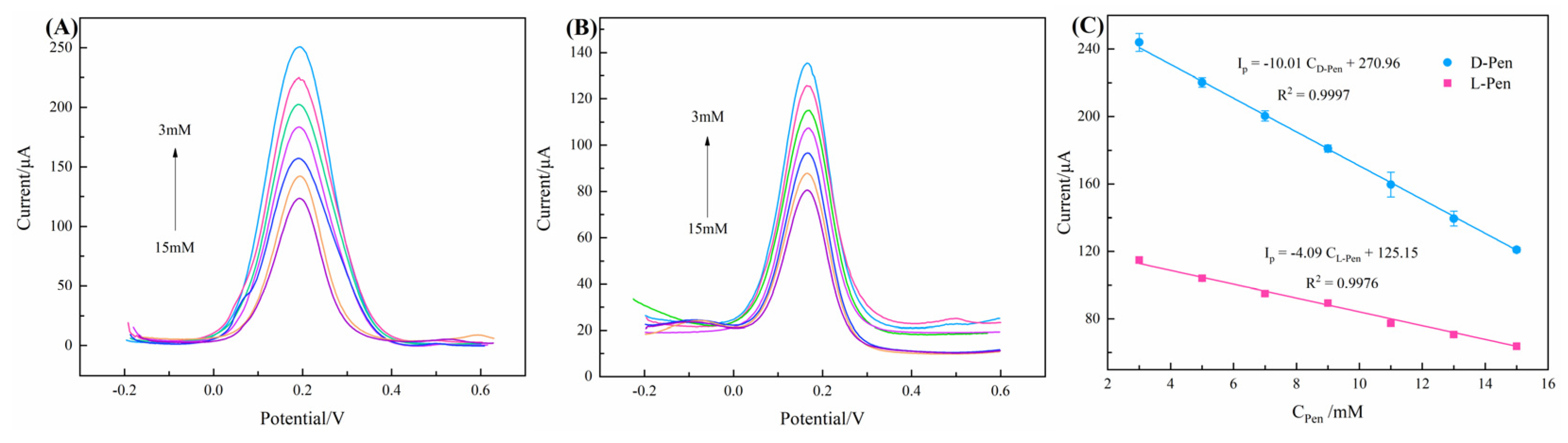
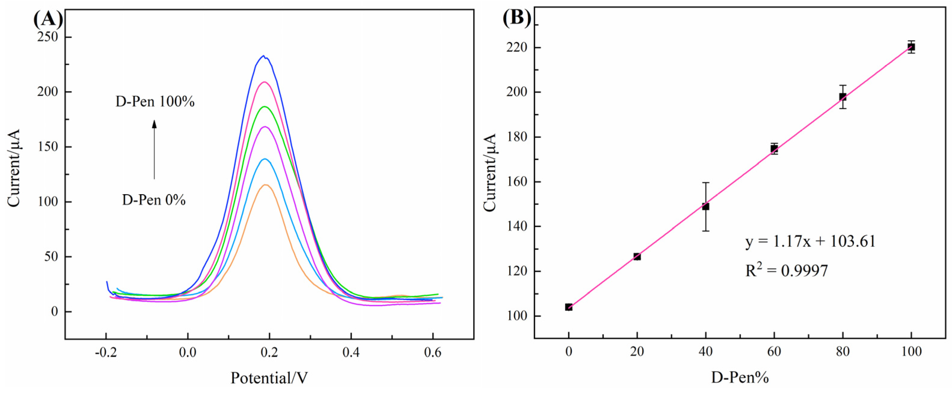
3.5. Practical Application of Chiral-Sensor Platform

The stability and reproducibility of PAA-MWCNTs-Ag-SCS/GCE in sensing 5 mM Pen enantiomer were investigated using DPV and CV at a scan rate of 100 mV/s in a 0.1 M KCl solution containing 5 mM [Fe(CN)6]4−/3−. The CV curves of PAA-MWCNTs-Ag-SCS/GCE (Figure 9A) were almost coincident after 100 cycles, demonstrating the high stability of the modified electrode. The repeatability of PAA-MWCNTs-Ag-SCS (Figure 9B) was assessed by measurements of the peak currents every 3 days, and the relative standard deviation of ID/IL was calculated as 7.37%. Accordingly, PAA-MWCNTs-Ag-SCS/GCE could be prepared using the aforementioned method with high reproducibility.
Furthermore, determining the detection limit for PAA-MWCNTs-Ag-SCS/GCE is vital for its practical application as a sensing platform. Figure 10 illustrates the DPV of D-Pen (Figure 10A) and L-Pen (Figure 10B) at various concentrations. There was a linear correlation between the Pen enantiomer concentration and oxidation peak current in the range of 3–15 mM (Figure 10C): Ip (μA) = −10.01 CD-Pen (mM) + 270.96 (R2 = 0.9997) and Ip (μA) = −4.09 CL-Pen (mM) + 125.15 (R2 = 0.9976). The detection limit (DL) was calculated to be 0.015 mM and 0.036 mM for L-Pen and D-Pen, respectively, using the signal-to-noise ratio (S/N = 3). As presented in Table 2, based on the above-mentioned results and previous work, it is not difficult to perceive that the PAA-MWCNTs-Ag-SCS/GCE exhibited a broader linear range and a lower limit of detection.
The ability to detect the ratio of L- to D-isomer in an enantiomeric mixed solution is essential for the practical application of a chiral-sensing platform. Thus, we recorded the DPV peak current values to the enantiomeric mixed solution (5 mM) of L-Pen and D-Pen using the constructed PAA-MWCNTs-Ag-SCS/GCE. It is noticeable from Figure 11A that the peak currents of L-Pen and D-Pen merged into one at the original position, whereas the peak currents and the percentage of D-Pen in the mixed solution showed a linear correlation (Figure 11B): Ip (μA) = 1.17x + 103.61 (R2 = 0.9997). This suggests that PAA-MWCNTs-Ag-SCS/GCE can be used to detect the percentage composition of one enantiomer (D-Trp%) in the enantio-enriched solution.
The selectivity of the PAA-MWCNTs-Ag-SCS/GCE was evaluated in the detection of other enantiomers (L-/D-Phe, L-/D-Pro, L-/D-Trp, and L-/D-MA) under the same conditions as used for the Pen isomers. As shown in Figure 12A, PAA-MWCNTs-Ag-SCS/GCE had a superior specific recognition effect for Pen enantiomers. According to Dacankov’s theory [52], the specific response of the chiral sensing platform could have resulted from the differences in the ability of L-Pen and D-Pen to form hydrogen bonds with PAA-MWCNTs-Ag-SCS/GCE. Moreover, PAA-MWCNTs-Ag-SCS/GCE also had a better recognition effect toward Phe and Trp enantiomers, which may be attributable to the structural similarity of L-/D-Phe and L-/D-Trp to Pen.
The anti-interference ability of the chiral-sensing platform was determined as follows: PAA-MWCNTs-Ag-SCS/GCE was put into 0.1 M PBS (pH = 7) solution containing 5 mM L- or D-Pen in the presence of Na+, K+, Ag+, Cu2+, or Zn2+. After removal, the electrode was placed under an infrared lamp and allowed to dry. The anti-interference ability of the modified electrodes was determined using differential pulse voltammetry (DPV) in 0.1 M KCl solution containing 5 mM [Fe(CN)6]4−/3− using the three-electrode system. As shown in Figure 12B, there was a slight influence on recognition efficiency derived from the metal ions Na+ and K+ with low coordination ability. In contrast, Ag+, Cu2+, and Zn2+ have strong coordination ability; therefore, their influence on the chiral identification effect was also found to be greater, which may be due to the substantial involvement of chiral ligand exchange recognition in the process of enantiomeric detection [53].

4. Conclusions

In conclusion, we encapsulated polyacrylic acid (PAA) chains onto the surface of MWCNTs using in situ free radical polymerization, and SCS was synthesized via a simple and environmentally conscious method. Silver-ammonia ions were introduced as the fixative with PAA-MWCNTs as the basic framework, and SCS was used to decorate the surface, thereby obtaining PAA-MWCNTs-Ag-SCS. The successful preparation of PAA-MWCNTs-Ag-SCS was established using SEM, FTIR, TGA, XPS, water contact angle, and correlation electrochemical characterization. Moreover, the DPV peak current ratio of D-Pen to L-Pen was 2.16 under the optimal experiment conditions. The detection limit (DL) was calculated as 0.015 mM and 0.036 mM for L-Pen and D-Pen, respectively, using the signal-to-noise ratio (S/N = 3). The chiral surface material PAA-MWCNTs-Ag-SCS developed in this article provides a new idea for the construction of a chiral-sensing platform with outstanding sensitivity, superior stability, and excellent recognition efficiency.

Author Contributions

Conceptualization, Y.J., W.Y., Y.K. and L.X.; methodology, Y.J., Y.K. and W.Y.; formal analysis, W.Y. and L.X.; investigation, Y.J., S.L. and L.X.; writing—original draft preparation, W.Y.; writing—review and editing, W.Y. and Y.K.; funding acquisition, Y.J. All authors have read and agreed to the published version of the manuscript.

Funding

Jiangsu Key Laboratory of Advanced Cata-lytic Materials and Technology, Changzhou University, Changzhou, China. Changzhou University Huaide College, Jing jiang, China.

Institutional Review Board Statement

Not applicable.

Informed Consent Statement

Not applicable.

Data Availability Statement

Not applicable.

Conflicts of Interest

The authors declare no conflict of interest.

References

  1. Globus, N.; Blandford, R.D. The chiral puzzle of life. Astrophys. J. 2020, 895, L11. [Google Scholar] [CrossRef]
  2. Lee, Y.J.; Kang, B.; Seo, J. Metalloporphyrin dimers bridged by a peptoid helix: Host-guest interaction and chiral recognition. Molecules 2018, 23, 2741. [Google Scholar] [CrossRef] [PubMed] [Green Version]
  3. Sung, B.; de La Cotte, A.; Grelet, E. Chirality-controlled crystallization via screw dislocations. Nat. Commun. 2018, 9, 1405. [Google Scholar] [CrossRef] [PubMed] [Green Version]
  4. Chen, Y.; Yang, X.; Gao, J. 3D Janus plasmonic helical nanoapertures for polarization-encrypted data storage. Light Sci. Appl. 2019, 8, 45. [Google Scholar] [CrossRef] [PubMed] [Green Version]
  5. Wang, Y.; Zhou, X.; Xu, C.; Jin, Y.; Li, B. Gold nanorods as visual sensing platform for chiral recognition with naked eyes. Sci. Rep. 2018, 8, 5296. [Google Scholar] [CrossRef] [Green Version]
  6. Managadze, G.G.; Engel, M.H.; Getty, S.; Wurz, P.; Brinckerhoff, W.B.; Shokolov, A.G.; Sholin, G.V.; Terent’ev, S.A.; Chumikov, A.E.; Skalkin, A.S.; et al. Excess of L-alanine in amino acids synthesized in a plasma torch generated by a hypervelocity meteorite impact reproduced in the laboratory. Planet Space Sci. 2016, 131, 70–78. [Google Scholar] [CrossRef] [Green Version]
  7. Hu, M.; Yuan, Y.X.; Wang, W.; Li, D.M.; Zhang, H.C.; Wu, B.X.; Liu, M.; Zheng, Y.S. Chiral recognition and enantiomer excess determination based on emission wavelength change of AIEgen rotor. Nat. Commun. 2020, 11, 2274. [Google Scholar] [CrossRef]
  8. Bicchi, C.; Amato, A.D.; Rubiolo, P. Cyclodextrin derivatives as chiral selectors for direct gas chromatographic separation of enantiomers in the essential oil aroma and flavour fields. J. Chromatogr. A 1999, 843, 99–121. [Google Scholar] [CrossRef]
  9. Evans, A.C.; Meinert, C.; Giri, C.; Goesmann, F.; Meierhenrich, U.J. Chirality photochemistry and the detection of amino acids in interstellar ice analogues and comets. Chem. Soc. Rev. 2012, 41, 5447–5458. [Google Scholar] [CrossRef]
  10. Sloboda, R.D. Separation of tubulin and microtubule-associated proteins by ion exchange chromatography. Cold Spring Harb. Protoc. 2015, 1, 78–81. [Google Scholar] [CrossRef]
  11. Zhang, Q.; Xue, S.; Li, A.; Ren, S. Functional materials in chiral capillary electrophoresis. Coord. Chem. Rev. 2021, 445, 214108. [Google Scholar] [CrossRef]
  12. Zhang, X.; Yin, J.; Yoon, J. Recent advances in development of chiral fluorescent and colorimetric sensors. Chem. Rev. 2014, 114, 4918–4959. [Google Scholar] [CrossRef] [PubMed]
  13. Ou, J.; Tao, Y.; Xue, J.; Kong, Y.; Dai, J.; Deng, L. Electrochemical enantiorecognition of tryptophan enantiomers based on graphene quantum dots-chitosan composite film. Electrochem. Commun. 2015, 57, 5–9. [Google Scholar] [CrossRef]
  14. Rahman, M.M.; Karim, M.R.; Alam, M.M.; Zaman, M.B.; Alharthi, N.; Alharbi, H.; Asiri, A.M. Facile and efficient 3-chloro phenol sensor development based on photolumenescent core-shell CdSe/ZnS quantum dots. Sci. Rep. 2020, 10, 557. [Google Scholar] [CrossRef] [PubMed] [Green Version]
  15. Mathew, G.; Narayanan, N.; Abraham, D.A.; De, M.; Neppolian, B. Facile green approach for developing electrochemically reduced graphene oxide-embedded platinum nanoparticles for ultrasensitive detection of nitric oxide. ACS Omega 2021, 6, 8068–8080. [Google Scholar] [CrossRef] [PubMed]
  16. Gaikwad, R.; Thangaraj, P.R.; Sen, A.K. Direct and rapid measurement of hydrogen peroxide in human blood using a microfluidic device. Sci. Rep. 2021, 11, 2960. [Google Scholar] [CrossRef]
  17. Li, Z.; Mo, Z.; Meng, S.; Gao, H.; Niu, X.; Guo, R.; Wei, T. The construction of electrochemical chiral interfaces using hydrox ypropyl chitosan. RSC Adv. 2017, 7, 8542–8549. [Google Scholar] [CrossRef] [Green Version]
  18. Lu, X.; Tang, S.; Huang, B.; Shen, X.; Hong, F. Preparation and characterization of bacterial cellulose/hydroxypropyl chitosan blend as-spun fibers. Fiber Polym. 2013, 14, 935–940. [Google Scholar] [CrossRef]
  19. Yeh, Y.T.; Lin, Z.; Zheng, S.Y.; Terrones, M. A carbon nanotube integrated microfluidic device for blood plasma extraction. Sci Rep. 2018, 8, 13623. [Google Scholar] [CrossRef] [Green Version]
  20. Corletto, A.; Shapter, J.G. Nanoscale patterning of carbon nanotubes: Techniques, applications, and future. Adv. Sci. 2021, 8, 2001778. [Google Scholar] [CrossRef]
  21. Zhang, A.; Pan, S.; Zhang, Y.; Chang, J.; Cheng, J.; Huang, Z.; Li, T.; Zhang, C.; de La Fuentea, J.M.; Zhang, Q.; et al. Carbon-gold hybrid nanoprobes for real-time imaging, photothermal/photodynamic and nanozyme oxidative therapy. Theranostics 2019, 9, 3443–3458. [Google Scholar] [CrossRef] [PubMed]
  22. Barsan, M.M.; Ghica, M.E.; Brett, C.M.A. Electrochemical sensors and biosensors based on redox polymer/carbon nanotube modified electrodes: A review. Anal. Chim. Acta. 2015, 881, 1–23. [Google Scholar] [CrossRef] [PubMed]
  23. Qian, J.; Yi, Y.; Zhang, D.; Zhu, G. Electrochemical recognition of tryptophan enantiomers using a multi-walled carbon nanotube@polydopamine composite loaded with copper (II). Microchim. Acta 2019, 186, 358. [Google Scholar] [CrossRef] [PubMed]
  24. Goulart, L.A.; Gonçalves, R.; Correa, A.A.; Pereira, E.C.; Mascaro, L.H. Synergic effect of silver nanoparticles and carbon nanotubes on the simultaneous voltammetric determination of hydroquinone, catechol, bisphenol a and phenol. Microchim. Acta 2018, 185, 12. [Google Scholar] [CrossRef]
  25. Yu, F.; Ma, J.; Han, S. Adsorption of tetracycline from aqueous solutions onto multi-walled carbon nanotubes with different oxygen contents. Sci. Rep. 2014, 4, 5326. [Google Scholar] [CrossRef] [Green Version]
  26. Hsiao, A.-E.; Tsai, S.-Y.; Hsu, M.W.; Chang, S.J. Decoration of multi-walled carbon nanotubes by polymer wrapping and its application in MWCNT/polyethylene composites. Nanoscale Res. Lett. 2012, 7, 240. [Google Scholar] [CrossRef] [PubMed] [Green Version]
  27. Aldalbahi, A.; Rahaman, M.; Almoiqli, M. Performance enhancement of modified 3D SWCNT/RVC electrodes using microwave-irradiated graphene oxide. Nanoscale Res Lett. 2019, 14, 351. [Google Scholar] [CrossRef]
  28. Sun, Z.; Shi, C.; Wang, X.; Fang, Q.; Huang, J. Synthesis, characterization and antimicrobial activities of sulfonated chitosan. Carbohydr. Polym. 2017, 155, 321–328. [Google Scholar] [CrossRef] [Green Version]
  29. Fu, H.; Du, Z.J.; Zou, W.; Li, H.Q.; Zhang, C. Enhanced electrical conductivity of sodium polyacrylate encapsulated multi-walled carbon nanotubes. Mater. Lett. 2012, 78, 54–57. [Google Scholar] [CrossRef]
  30. Al-Mutabagani, L.A.; Alshabanah, L.A.; Ahmed, H.A.; Hagar, M.; Abu Al-Ola, K.A. New symmetrical U-and wavy-shaped supramolecular H-bonded systems; geometrical and mesomorphic approaches. Molecules 2020, 25, 1420. [Google Scholar] [CrossRef] [PubMed]
  31. Upadhyay, S.S.; Srivastava, A.K. Hydroxypropyl β-cyclodextrin cross-linked multi-walled carbon nanotube-based chiral nanocomposite electrochemical sensors for the discrimination of multi-chiral drug atorvastatin isomers. New J. Chem. 2019, 43, 11178–11188. [Google Scholar] [CrossRef]
  32. Niu, X.; Yang, X.; Li, H.; Shi, Q.; Wang, K. Chiral voltammetric sensor for tryptophan enantiomers by using a self-assembled multiwalled carbon nanotubes/polyaniline/sodium alginate composite. Chirality 2021, 33, 248–260. [Google Scholar] [CrossRef] [PubMed]
  33. Bao, L.; Chen, X.; Yang, B.; Tao, Y.; Kong, Y. Construction of electrochemical chiral interfaces with integrated polysaccharides via amidation. ACS Appl. Mater. Interfaces 2016, 8, 21710–21720. [Google Scholar] [CrossRef]
  34. Maachou, H.; Genet, M.J.; Aliouche, D.; Dupont-Gillain, C.C.; Rouxhet, P.G. XPS Analysis of chitosan-hydroxyapatite biomaterials: From elements to compounds. Surf. Interface Anal. 2013, 45, 1088–1097. [Google Scholar] [CrossRef]
  35. Amaral, I.F.; Granja, P.L.; Barbosa, M.A. Chemical modification of chitosan by phosphorylation: An XPS, FT-IR and SEM study. J. Biomater. Sci. Polym. Ed. 2005, 16, 1575–1593. [Google Scholar] [CrossRef] [PubMed]
  36. Du, Y.; Mo, Z.; Wang, J.; Shuai, C.; Pei, H.; Chen, Y.; Yue, R. A novel chiral carbon nanocomposite based on cellulose gum modifying chiral tri-electrode system for the enantio-recognition of tryptophan. Electroanal. Chem. 2021, 895, 115390. [Google Scholar] [CrossRef]
  37. Zomorrodian, A.; Mesarwi, A.; Wu, N.J.; Ignatiev, A. XPS oxygen line broadening in lead zirconium titanate and related materials. Appl Surf Sci. 1995, 90, 343–348. [Google Scholar] [CrossRef]
  38. Mikhlin, Y.L.; Pal’yanova, G.A.; Tomashevich, Y.V.; Vishnyakova, E.A.; Vorobyev, S.A.; Kokh, K.A. XPS and Ag L3-Edge XANES Characterization of silver and silver–gold sulfoselenides. J. Phys. Chem. Solids 2018, 116, 292–298. [Google Scholar] [CrossRef]
  39. Berthod, A. Chiral recognition mechanism. Anal. Chem. 2006, 78, 2093–2099. [Google Scholar] [CrossRef]
  40. Niu, X.; Yang, X.; Li, H.; Liu, J.; Liu, Z.; Wang, K. Application of chiral materials in electrochemical sensors. Microchim. Acta 2020, 187, 676. [Google Scholar] [CrossRef]
  41. Gong, T.; Zhu, S.; Huang, S.; Gu, P.; Xiong, Y.; Zhang, J.; Jiang, X. A renewable electrochemical sensor based on a self-assembled framework of chiral molecules for efficient identification of tryptophan isomers. Anal. Chim. Acta 2022, 1191, 339276. [Google Scholar] [CrossRef] [PubMed]
  42. Zagitova, L.; Yarkaeva, Y.; Zagitov, V.; Nazyrov, M.; Gainanova, S.; Maistrenko, V. Voltammetric chiral recognition of naproxen enantiomers by N-tosylproline functionalized chitosan and reduced graphene oxide based sensor. Electroanal. Chem. 2022, 922, 116744. [Google Scholar] [CrossRef]
  43. Wang, L.; Gao, W.; Ng, S.; Pumera, M. Chiral protein-covalent organic framework 3D-printed structures as chiral biosensors. Anal. Chem. 2021, 93, 5277–5283. [Google Scholar] [CrossRef]
  44. Luo, H.; Li, H.; Ge, Q.; Cong, H.; Tao, Z.; Liu, M. An electrochemical sensor for enantio-recognition of tyrosine based on a chiral macrocycle functionalized rGO. Microchem. J. 2021, 164, 105949. [Google Scholar] [CrossRef]
  45. Zou, J.; Yu, J. Nafion-stabilized black phosphorus nanosheets-maltosyl-β-cyclodextrin as a chiral sensor for tryptophan enantiomers. Mat. Sci. Eng. R 2020, 112, 110910. [Google Scholar] [CrossRef] [PubMed]
  46. Zou, J.; Lan, X.; Zhao, G.; Huang, Z.; Liu, Y.; Yu, J. Immobilization of 6-O-α-maltosyl-β-cyclodextrin on the surface of black phosphorus nanosheets for selective chiral recognition of tyrosine enantiomers. Microchim. Acta 2020, 187, 636. [Google Scholar] [CrossRef] [PubMed]
  47. Jing, P.; Yin, Z.; Cai, W.; Li, J.; Wu, D.; Kong, Y. The hybrids of perylene tetracarboxylic acid functionalized multi-walled carbon nanotubes and chitosan for electrochemical chiral sensing of tryptophan enantiomers. Bioelectrochemistry 2022, 146, 108110. [Google Scholar] [CrossRef]
  48. Sun, X.; Fu, Z.; Zhang, M.; Fu, H.; Lin, C.; Kuang, J.; Zhang, H.; Hu, P. An electrochemical sensor employing β-cyclodextrin chiral cross-linked metal organic framework and graphene oxide for chiral enantiomer recognition. Microchem. J. 2022, 183, 108074. [Google Scholar] [CrossRef]
  49. Pei, H.; Wang, J.; Jin, X.; Zhang, X.; Liu, W.; Guo, R.; Liu, N.; Mo, Z. An electrochemical chiral sensor based on glutamic acid functionalized graphene-gold nanocomposites for chiral recognition of tryptophan enantiomers. Electroanal. Chem. 2022, 913, 116283. [Google Scholar] [CrossRef]
  50. Li, Z.; Xu, H.; Wu, D.; Zhang, J.; Liu, X.; Gao, S.; Kong, Y. Electrochemical chiral recognition of tryptophan isomers based on nonionic surfactant-assisted molecular imprinting sol-gel silica. ACS Appl. Mater. Interfaces 2019, 11, 2840–2848. [Google Scholar] [CrossRef]
  51. Yao, W.; Li, S.; Xie, L.; Jiang, Y. Chiral recognition of tryptophan enantiomer based on the electrode modified by polyaniline adsorption bovine serum albumin complex. Chirality 2022, 35, 129–144. [Google Scholar] [CrossRef] [PubMed]
  52. Dong, L.; Zhang, Y.; Duan, X.; Zhu, X.; Sun, H.; Xu, J. Chiral PEDOT-based enantioselective electrode modification material for chiral electrochemical sensing: Mechanism and model of chiral recognition. Anal. Chem. 2017, 89, 9695–9702. [Google Scholar] [CrossRef] [PubMed]
  53. Yang, X.; Niu, X.; Mo, Z.; Guo, R.; Liu, N.; Zhao, P.; Liu, Z. Electrochemical chiral interface based on the Michael addition/Schiff base reaction of polydopamine functionalized reduced graphene oxide. Electrochim. Acta 2019, 319, 705–715. [Google Scholar] [CrossRef]
Scheme 1. The preparation process of the chiral-sensing platform and identification of the phenylglycinamide enantiomer.
Chemosensors 11 00086 sch001
Figure 1. SEM images of MWCNTs (A), PAA-MWCNTs (B), and PAA-MWCNTs-Ag-SCS (C).
Chemosensors 11 00086 g001
Figure 2. (A) FT-IR spectra of MWCNTs (a), PAA-MWCNTs (b), CS (c), SCS (d), and PAA-MWCNTs-Ag-SCS (e); (B) XPS survey spectrum of PAA-MWCNTs-Ag-SCS; XPS C 1s (C), N 1s (D), O 1s (E), and Ag 3d (F) spectra for PAA-MWCNTs-Ag-SCS.
Chemosensors 11 00086 g002
Figure 3. Water contact angle photographs of MWCNTs (A), PAA-MWCNTs (B), SCS (C), and PAA-MWCNTs-Ag-SCS (D).
Chemosensors 11 00086 g003
Figure 4. CV curves (A) and EIS curves (B) of the GCE, MWCNTs/GCE, PAA-MWCNTs/GCE, and PAA-MWCNTs-Ag-SCS/GCE in 0.1 M KCl containing 5 mM [Fe(CN)6]4−/3−.
Chemosensors 11 00086 g004
Figure 5. (A) CV of PAA-MWCNTs-Ag-SCS/GCE at scan rates of 50, 70, 90, 110, 130, 150, 170, 190, and 210 mV/s. (B) Linear dependence between scan rate and peak current (anodic (a) and cathodic (b)).
Chemosensors 11 00086 g005
Figure 6. DPV curves of 5 mM L-Pen or D-Pen at the bare GCE (A), SCS/GCE (B), PAA-MWCNTs/GCE (C), PAA-MWCNTs-Ag/GCE (D), and PAA-MWCNTs-Ag-SCS/GCE (E) were recorded in 0.1 M KCl containing 5 mM [Fe(CN)6]4−/3−, respectively.
Chemosensors 11 00086 g006
Figure 7. Curves of response current versus each mass concentration ratio (A); peak current ratio of the Pen enantiomers at each mass concentration ratio (B); curves of response current at each pH (C); peak current ratio of the Pen enantiomers at each pH (D); curves of response current versus the enantiomeric action time (E); peak current ratio of the Pen enantiomers at each action time (F).
Chemosensors 11 00086 g007
Figure 8. UV spectra of 0.2 mM L-Pen (A) and D-Pen (B) after the addition of various PAA-MWCNTs-Ag-SCS concentrations (the amount-of-substance concentration of PAA-MWCNTs-Ag-SCS is replaced by the concentration of silver-ammonia ions). Double reciprocal plots of L-Pen (C) and D-Pen (D) (pH = 7).
Chemosensors 11 00086 g008
Figure 9. (A) CV of PAA-MWCNTs-Ag-SCS/GCE over 100 consecutive cycles. (B) Enantiomeric peak currents ratio for recognition of 5 mM L-Pen or D-Pen with different PAA-MWCNTs-Ag-SCS/GCEs prepared under identical conditions.
Chemosensors 11 00086 g009
Figure 10. DPV of D-Pen (A) and L-Pen (B) at various concentrations; (C) The linear association of the peak current values to concentrations of D-Pen and L-Pen in 0.1 M KCl containing 5 mM [Fe(CN)6]4−/3−.
Chemosensors 11 00086 g010
Figure 11. (A) DPV of 5 mM Pen enantiomeric mixed solution containing different contents of D-Pen (0%, 20%, 40%, 60%, 80%, and 100%) with PAA-MWCNTs-Ag-SCS/GCE; (B) Linear relationship between Ip and D-Pen% in the enantio-enriched solution.
Chemosensors 11 00086 g011
Figure 12. (A) Recognition efficiency of 6 mM L, D-phenylalanine (Phe); L, D-proline (Pro); L, D-tryptophan (Trp); and L, D-mandelic acid (MA) using PAA-MWCNTs-Ag-SCS/GCE. (B) Chiral recognition of Pen enantiomer in the presence of Na+, K+, Ag+, Cu2+, and Zn2+ (temperature, 25 °C; pH = 7). Error bars represent the standard deviation for three independent measurements.
Chemosensors 11 00086 g012
Table 1. A comparison of the developed electrochemical methods in the peak current interval and peak current ratio.
Chiral MaterialEnantiomerPeak Current
Interval/μA
Peak Current RatioReferences
Cu2-β-CD/NH2-CS-MWCNTstryptophan5.32–6.341.64[41]
rGO-TsPro-CSnaproxen7.34–10.831.60[42]
Fe3O4@COF@BSAtryptophan12.53–17.521.45[43]
rGO-CHMFtyrosine12–18.731.58[44]
NF/BPNSs-G2-β-CDtryptophan14–21.631.49[45]
Mal-βCD/BP NSstryptophan9.51–13.251.51[46]
CS/MWCNTs–PTCAtryptophan19–32.681.72[47]
PAA-MWCNTs-Ag-SCSphenylglycinamide222.42–102.912.16This work
Table 2. A comparison of the developed electrochemical methods in the limit of detection.
Chiral MaterialEnantiomerLimit of Detection (mM)References
GO-CLMOFmandelic acid (MA)0.091 (L-MA)0.15 (D-MA)[48]
RGO-Au/L-GluTryptophan (Trp)0.28 (L-Trp)0.86 (D-Trp)[49]
MISiO2/ITOTryptophan (Trp)0.11 (L-Trp)0.13 (D-Trp)[50]
APS-DPANI-BSATryptophan (Trp)0.071 (L-Trp)0.048 (D-Trp)[51]
CMC-CSTryptophan (Trp)0.041 (L-Trp)0.052 (D-Trp)[33]
PAA-MWCNTs-Ag-SCSPhenylglycinamide (Pen)0.015 (L-Trp)0.036 (D-Trp)This work
Disclaimer/Publisher’s Note: The statements, opinions and data contained in all publications are solely those of the individual author(s) and contributor(s) and not of MDPI and/or the editor(s). MDPI and/or the editor(s) disclaim responsibility for any injury to people or property resulting from any ideas, methods, instructions or products referred to in the content.

