1. Introduction
Alanine amino transaminase (ALT) is an enzyme that is found in the vital parts of the human body, primarily in the kidneys, skeletal muscles, serum, body tissues, and also in smaller amounts in the heart and highly concentrated amounts in the liver. The detection of ALT in human serum has a significant role in diagnosis and monitoring the progress of heart failure, liver diseases, and hepatocellular inflammation. High levels of ALT in serum represent a signal of severe liver diseases, specially hepatitis or toxic liver necrosis [
1]. The measurement of hepatic function is a fundamental for the diagnosis of several liver diseases, such as hepatitis A, B, C, steatosis, and hepatotoxicity caused by drugs.
ALT catalyzes the conversion of alanine and α-ketoglutarate to pyruvate and glutamate, participating in cellular nitrogen metabolism and liver gluconeogenesis. The ALT scale was reported to increase to five times its upper limit of normal during chronic hepatitis B virus (HBV) infections [
2]. The normal range of ALT in serum is 5–35 U/L, but for a damaged liver, enzyme levels can reach fifty times the normal range (250–1400 U/L) [
1]. Average ALT levels in serum are influenced by many factors, such as gender, body mass index, age, alcohol consumption, and medication use. The upper limit for normal ALT levels is 32.10 U/L and 23.15 U/L for men and women, respectively [
3].
A variety of methods have been reported in the literature for the detection of ALT, including colorimetric and spectrophotometric analysis [
4,
5], fluorescence [
6,
7], and chromatography [
8,
9]. Despite the availability of these conventional spectrophotometric assays in clinical laboratories for ALT detection, these methods lack sensitivity, are costly, and require complex reagents and trained operators. Thus, there is an increasing demand for the development of healthcare devices such as electrochemical biosensors, which have been found to be highly efficient and sensitive for such routine clinical applications [
10].
Biosensors are used in several applications, including quality control, food manufacturing, and biomedical fields [
11]. According to the literature, some biosensors were reported for ALT determination over the past two decades [
12,
13,
14,
15]. Monoclonal antibodies for human recombinant ALT [
16] and amperometric detection using platinum electrodes on the polydimethylsiloxane (PDMS) microchannel [
17] were also applied. Yet, none of the previously reported electrochemical methods involved the application of molecularly imprinted polymers (MIPs).
MIPs represent a class of artificial polymers developed through the last decades to be applied as synthetic alternatives for natural receptors because of their chemical and physical stability, in addition to their inexpensive manufacturing procedures [
18,
19]. MIPs offer a new principle for producing receptors by configuring specific cavities for binding with target analytes within the polymeric matrix. Subsequently, electrochemical biosensors based on MIP become highly attractive for many applications involving different classes of analytes, including pharmaceutical compounds [
20,
21], biological molecules as a virus [
22,
23], bacteria [
24,
25], and environmental hazardous pollutants [
26,
27].
On applying MIP for macromolecules and biological templates such as proteins, some challenges can be faced based on their fragility and the possibility of occurrence of any irreversible conformational changes upon polymerization [
28]. Thus, electropolymerization, in which the template is directly imprinted on the surface of the working electrode, is commonly used. This will aid in the avoidance of any possible denaturation or destruction of such molecules upon exposure to thermal initiation and the use of organic solvent in the case of traditional free radical initiation polymerization [
29,
30,
31,
32].
Electropolymerization is considered to be a green molecular imprinting strategy, following a range of green perspectives that aim to counteract or minimize any detrimental impacts of MIPS on human health when using bulk or precipitation polymerization that require a high consumption of organic solvents [
30].
The layer thickness can be controlled through the optimization of the electropolymerization parameters, such as the concentration of the monomer used and the number of electrodeposition cycles [
33]. The thickness of the film affects the template, its knocking out and rebinding efficiency. A very thin film may not be efficient in the rebinding process, and increasing the number of polymerization cycles might result in thick membranes that might hinder the template removal or act as an insulting layer, affecting the electrochemical signal [
33,
34,
35]. Another important consideration affecting the efficiency of MIP involves the successful removal of the template, without destruction of the polymeric deposited membrane, which is the main step in rendering efficient recognition cavities that are capable of rebinding to the target template [
36].
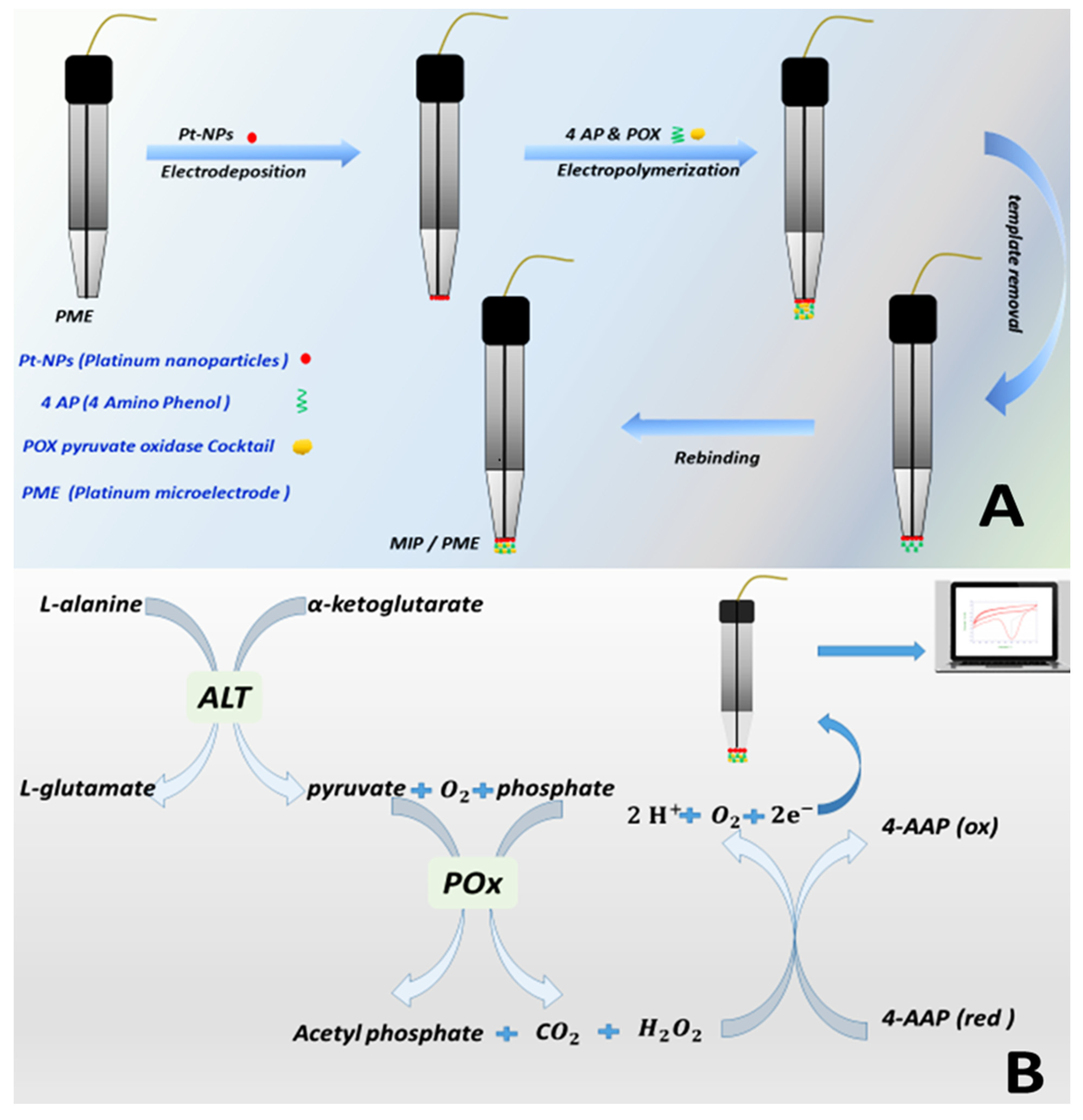
In this work, we report the development of the first microelectrode based on a molecularly imprinted pyruvate oxidase enzyme to be applied as an electrochemical biosensor for ALT detection. First, pyruvate oxidase is imprinted using 4-aminophenol (functional monomer) on a platinum microelectrode modified with platinum nanoparticles. Then, after the optimization of POx interaction conditions using sodium pyruvate, the sensor is applied to measure the pyruvate resulting from the reaction of ALT with its substrates, L-alanine and 15 mM α-ketoglutarate. The monomer 4-aminoantypirine is used as the electrochemical probe for tracking the progress of the enzymatic interactions between POx and pyruvate. This allows for the development of a sensitive, cost effective, miniaturized, and relatively inexpensive ALT biosensor that can be extended to develop many other important clinical and point-of-care biosensors.
2. Experimental
2.1. Reagents and Materials
All chemicals used in this work were of analytical grade without any further purification. These included 4 aminoantipyrine, 4-aminophenol, flavin adenine dinucleotide (FAD), thiamine pyrophosphate (TPP), pyruvate peroxidase (POx) enzyme, sodium pyruvate, platinum (IV) chloride 99%, reduced D.L-glutathione, alanine transaminase (ALT/GPT), L-alanine, alpha-ketoglutarate B-nicotinamide adenine dinucleotide, catalase, and glutathione enzymes purchased from Sigma-Aldrich (Schnelldorf, Germany); sodium monophosphate, diphosphate, hydroxide, chloride and acetate, magnesium sulfate heptahydrate, acetic, boric, phosphoric, and Sulfuric acids and methanol (99%) from the Al—Gomhoria Company for medicines and medical supplies (Cairo Egypt); and the alanine aminotransferase (ALT-GPT)—Ultimate Assay Kit from the Egyptian Company for Biotechnology (Cairo, Egypt). All the experiments were carried out at room temperature, and ultrapure water purified in prelab UHQ (ELGA) was used throughout this work.
2.2. Apparatus
All electrochemical measurements were recorded using a 620E electrochemical analyzer (CH Instruments, Bee Cave, TX, USA). A standard three-electrode cell was used; the working electrode was a 25 µm Pt wire sealed under vacuum in a borosilicate glass tube melting tube, kindly provided by the Institute of Analytical and Bioanalytical Chemistry, University of Ulm, Ulm, Germany in combination with an external Ag/AgCl as a reference electrode and a platinum counter electrode. A CH instruments Electrode Polishing Kit (carbimet disks, nylon polishing pads, and microcloth polishing pads) and 0.05 µm and 0.3 µm gamma alumina powder from CH Instruments Inc., USA were used for the electrode’s polishing. The pH measurements were carried out using a Jenway 3510 digital pH meter (Jenway Instruments, Staffordshire, England). The electrochemical impedance spectroscopy (EIS) was performed using Palmsense 4 EIS potentiostat/Galvanostat (palmsense BV, Houten, The Netherlands), and the platinum microelectrode surface was analyzed using scanning electron microscopy (SEM), (JSM 6360LV, JEOL/Akishima, Japan) at EPRI Egypt (Cairo, Egypt).
2.3. Preparation of the Working Solutions and Composites
Before its use, pyruvate peroxidase (POx) should first be activated by the reaction with its coenzymes TPP and FAD to form a completely active structure. The added coenzymes bind to the active sites of POx via noncovalent bonds. The activating solution was prepared by dissolving POx (100 U mL
−1) in 1 mL phosphate buffer pH 6.0 (0.01 M) with the cofactors (200 µM thiamine pyrophosphate, 25 µM of FAD, and 10 mM MgSO
4.7H
2O). Each cofactor was dissolved in 10 mL phosphate buffer with a pH 6.0 (0.01 M) [
37]. POx was mixed with the activating solution in a ratio of 3:1 in a dark tube and left to react for 1 h at 4 °C in the dark. The ALT enzyme solution was prepared in 0.01 M sodium phosphate buffer (pH 7.4). The substrates (600 mM L-alanine and 15 mM α-ketoglutarate) were dissolved in 0.01 M phosphate buffer (pH 7.4) [
37,
38].
For the electrodeposition of platinum nanoparticles, 5 mM H2PtCl6 dissolved in 0.5 M H2SO4 solution were used. Fifty mM 4-aminophenol dissolved in 0.5 M sulfuric acid was used as a functional monomer. Both solutions should be de-aerated with ultra-pure argon gas for 10 min, prior to the electropolymerization and molecular imprinting step. A series of concentrations of sodium pyruvate (0.1–150 mM) were freshly prepared in 20 mM 4-amino antipyrine solution (4-AAP), used as the active probe, previously prepared in (0.01 M) sodium phosphate buffer (pH 7.2) and degassed by nitrogen and stored at 4 °C.
2.4. Electrochemical Modification of the Platinum Microelectrode
Prior to electropolymerization, the platinum microelectrode surface was polished using alumina solution (0.05 µm and 0.3 µm gamma alumina powder), followed by cyclic voltammetry (CV) of the 0.5 M H2SO4 solution in the range 0.0–+1.0 V at a scan rate of 50 mV s until a stable response was reached, indicating a smooth and activated surface. Then, the electrode was washed with Milli-Q water, followed by methanol, and left to dry in air.
Electrochemical deposition of the platinum nanoparticles on the cleaned platinum microelectrode (working electrode) was performed by its immersion in 5 mM H
2PtCl
6 solution prepared in 0.5 M H
2SO
4 [
39] using CV for 30 cycles in the potential between −0.4–1.0 V at a scan rate of 0.05 V·s
−1. Finally, the electrode was washed with distilled water and dried at room temperature.
MIP was synthesized in situ on the electrode’s surface using CV. Non-conducting polymeric layers were electropolymerized on a platinum microelectrode using a solution composed of an enzyme cocktail containing 360 mM POx enzyme, 120 mM of the activating solution (TPP, FAD, and
), and 1 mL of 50 mM 4-aminophenol solution as a functional monomer. A total of 250 CV cycles were applied by a sweeping potential from 0.0 to +1.5 V, and the scan rate was 50 mV·s
−1. The non-imprinted electrode (NIP platinum microelectrode) was fabricated under the same conditions as the MIP platinum microelectrode, in absence of the POx enzyme in the electropolymerization medium. A schematic diagram for the microelectrode modifications steps is shown in
Figure 1A.
2.5. Application of the Fabricated Biosensor with Real Plasma Samples
ALT detection in human plasma samples was performed using the MIP-modified platinum microelectrode. Samples from anonymous volunteers were provided from a private clinical testing laboratory. All experimental procedures for dealing with and discarding the tested samples were carried out in compliance with the Ethics Committee of the Faculty of Science, Cairo University. Plasma samples were tested directly without any further treatment or protein precipitation.
The standard addition method was used, in which each 50 μL of human plasma in four tubes was mixed with 50 μL of different concentrations of the ALT enzyme (10, 25, 50, and 100 U/L) plus 950 μL of the substrate (L-alanine and α-ketoglutarate) on each tube. The mixture was maintained at 25 °C for 10 min prior to CV measurements, and the obtained data were reported with reference to the calibration curve and compared with the results obtained from the ALT activity kit.
2.6. Detection of ALT Activity in Real Plasma Samples Using Commercial Assay Kit
The Spectrum Diagnostics Ultimate ALT assay kit is intended for the in vitro quantitative and diagnostic determination of ALT in human plasma, in which ALT is used to catalyze the transfer of the amino group from alanine to oxoglutarate with the formation of glutamate and pyruvate. The latter is reduced to lactate by lactate dehydrogenase (LDH) in the presence of reduced nicotinamide adenine dinucleotide (NADH) as follows:
The working reagents were mixed with 100 μL of the human plasma samples at 30 °C; then, absorbance readings were recorded at 340 nm at different time intervals to determine the mean absorbance change per minute (∆A/min) using the AU chemistry analyzer and kinetic method according to the International Federation of Clinical Chemistry (IFCC).
3. Results and Discussion
3.1. POx Reaction Mechanism and Detection of ALT
Pyruvate oxidase is a homotetrameric flavoprotein. Every unit bonds with one FAD and one thiamine pyrophosphate (TPP) in the existence of Mg
2+ to catalyze the oxidative decarboxylation of pyruvate. The catalytic activity of the POx takes place in several consecutive steps [
40]. Firstly, the C2-H of TPP is depronated; then, pyruvate is connected with the C2 atom of enzyme-bound TPP. After that, decarboxylation of the pyruvate takes places to form hydroxyethyl-TPP, which leads to the oxidation of hydroxyethyl-TPP with FAD. Finally, the reoxidation of FAD by oxygen occurs.
According to the reaction scheme, ALT acts as an activation enzyme to generate L-glutamate and pyruvate. Then, pyruvate works as a substrate in the second reaction with the POx (pyruvate oxidase) enzyme cocktail (FAD, TPP, Mg
2+). Finally, POx in the presence of phosphate and oxygen participates in the conversion of pyruvate to acetyl phosphate, H
2O
2, and CO
2. The produced H
2O
2 works on oxidizing 4-AAP, as shown in
Figure 1B. Thus, the quantity of 4-AAP is directly proportional to pyruvate and, consequently, the reacted amount of ALT [
40].
The imprinted electrode, modified with nanoparticles, 4 AP, and POx, was used for the determination of ALT, utilizing 4-aminoantypirine as an indicator of the enzymatic reaction, as follows:
3.2. Electrode Surface Modification
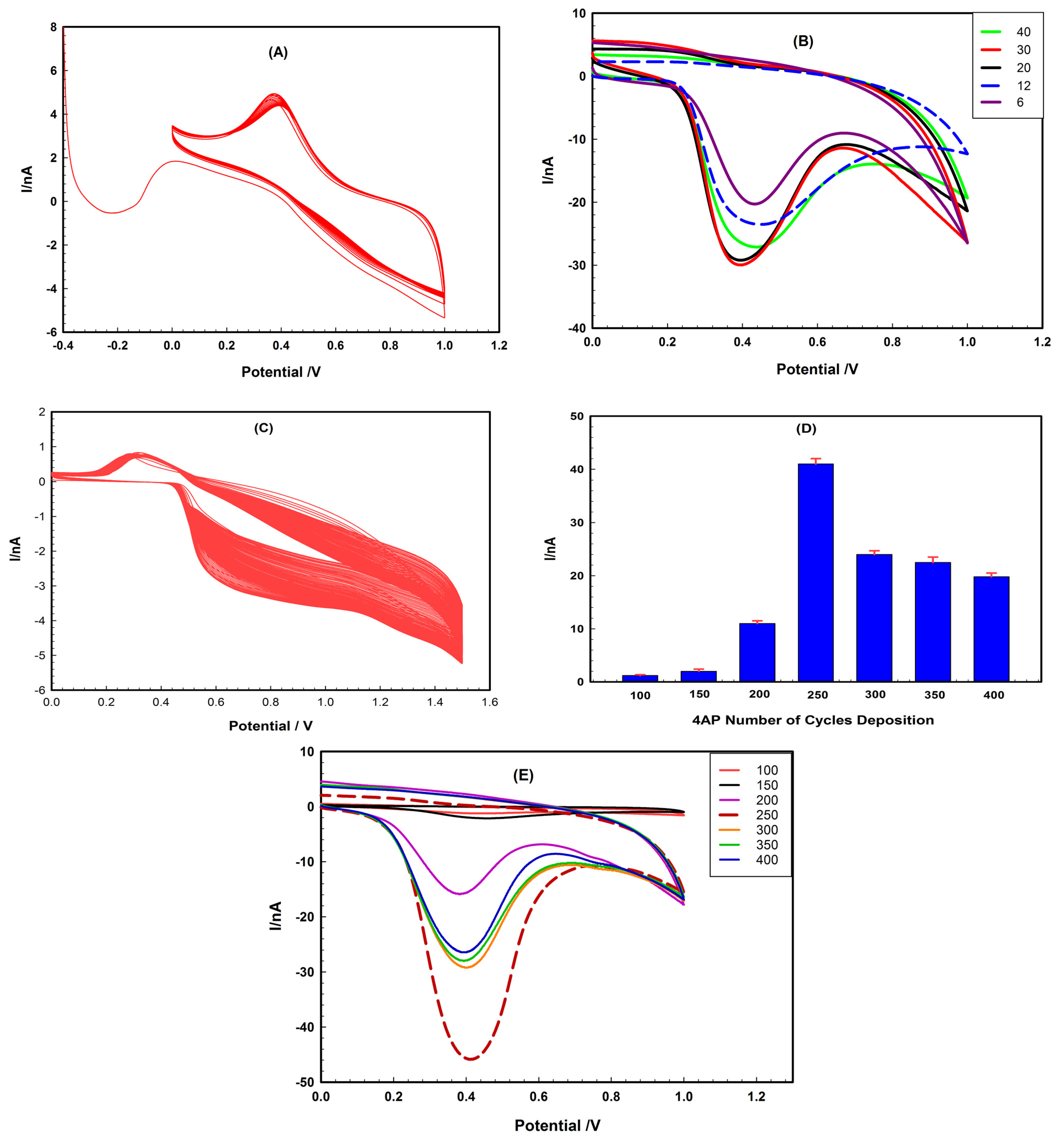
Platinum nanoparticles, due to their unique properties being of high surface area, high conductivity, chemical stability, good biocompatibility, and fast electron transfer ability, are reported to be frequently used in modifying many biosensors [
41]. The peak current of the biosensor was found to be enhanced after modification of the platinum microelectrode (PME) with a Pt nanoparticle. Different voltammetric deposition cycles, from 6 to 40 cycles, were applied under a potential between −0.4 and 1.0 V and a scan rate of 0.05 V·s
−1 (
Figure 2A,B). The cyclic voltammograms of 20 mM of 4-AAP showed that the highest current peak was recorded at 30 cycles, indicating that the required electrocatalytic properties of the sensor were reached [
32].
3.3. POx Imprinting on the Platinum Microelectrodes (PME)
Figure 2C represents the imprinting process of POx enzyme in the 4 AP polymeric matrix. The number of electropolymerization cycles is reported to affect the quality of the produced polymeric multi-layer films in terms of their stability, adsorption, and diffusion capacities. However, a thin-film layer was found to be sufficient for interaction with the target analyte, and an excessive increase in the thickness by depositing multi-layers may cause massive electrical resistance for electron transfer due to the formation of an insulating layer [
41].
Different number of CV cycles (100–400) was tested under a potential range between 0 and 1.0 V at a scan rate of 0.05 V·s
−1 for deposition of the imprinted polymeric films of 4 AP on PtNP-modified PME in the presence of the activated POx enzyme after its interaction with its coenzyme factors (TPP, FAD, and Mg
2+), in order to form the molecularly imprinted polymeric matrix (MIP). Then, the response of the imprinted PME was tested using 20 mM 4-AAP after knocking out the imprinted enzyme molecules. The current was found to increase until the number of cycles reached 250, then, a decrease was noticed at 300 cycles, as shown in
Figure 2D,E. This decrease indicated an increase in the thickness of the polymeric film, resulting in limited diffusion and electron transport on the surface of the modified PME [
42]. It was found that increasing the scan rate had a negative impact on the potential response, and the maximum current was attained at a scan rate of 50 mV s
−1, which was used for all further optimizations.
3.4. Optimization of the Electrochemical Parameters of the Biosensor
3.4.1. Effect of 4-Amino Antipyrine (4-AAP) Concentration
The electrocatalytic activity of the modified platinum microelectrode was tested using 4-AAP as an electroactive mediator over a wide range of concentrations (0.5, 1, 2, 5 10, 20, 25, and 30 mM) dissolved in 0.1 M phosphate buffer with a 7.2 pH, then mixed with 100 mM of sodium pyruvate. As shown in
Figure 3A,B, the peak current obtained was found to increase gradually upon increasing 4-AAP concentration. The maximum sensitivity was observed at 20 mM for 4-AAP and remained constant at 25 mM. Further increase in the 4-AAP concentration led to a negative effect on the PME response, as a result of the saturation of the electrode surface with 4-AAP. Thus, 20 mM 4-AAP was used in all upcoming experiments as the concentration of the electroactive mediator, as shown in
Figure 3C.
3.4.2. Influence of Sodium Pyruvate Concentration on the Response
The effect of pyruvate concentration on the modified PME with the 4 AP and POx enzyme was inspected at different sodium pyruvate concentrations in the range of 0.1–125 mM, as shown in
Figure 4A,B.
The peak current values were found to increase linearly with the sodium pyruvate concentration up to 100 mM, as shown in
Figure 4C. A further increase resulted in a decrease in the response due to the saturation of the biosensor surface with pyruvate, which resulted in the reduction of the catalytic activity of the imprinted POx [
42]. Thus, 100 mM sodium pyruvate solution was added to 4-AAP to act as the active probe in all the upcoming experiments involving the testing of POx activity to monitor the performance of the modified biosensor.
3.4.3. POx Removal from the MIP Matrix
The removal of the POx template from the imprinted matrix to create specific recognition sites was carried out by inserting the modified electrode PME into a continuously stirred 0.1 M phosphate buffer (pH 7.4) solution for time intervals ranging from 5 to 15 min, followed by washing with ultrapure distilled water. Ten minutes were found to be long enough for a stable 4-AAP response, indicating the efficient removal of the POx from the imprinted cavities and facilitating the diffusion of the active probe to the modified PME surface through these gates. Therefore, the peak current was increased, indicating successful removal of the POx template, as shown by the CV in
Figure 4D for the different stages of binding, washing, and rebinding of the template to the modified PME.
3.4.4. Incubation Time (Interaction Time between POx and ALT)
To determine the optimum reaction time, different amounts of the ALT enzyme (50 and 100 μL) were added to 950 μL of its substrate (600 mM L-alanine and 15 mM α-ketoglutarate) solution. The mixtures were then placed in a dark tube and left at room temperature for 5, 10, 15, 20, and 25 min. Then, each sample was mixed with 1 mL of 20 mM 4AAP solution, after which the mixture was applied to the PME to conduct cyclic voltammetry. As shown in
Figure 4E, the optimal reaction time for ALT detection was found to be 10 min.
3.4.5. Effect of Quiet Time
Quiet time represents the period that elapses while applying the initial potential to the working electrodes prior to potential scanning, which might affect the response of a biosensor. CV experiments were performed using 4-AAP within quiet time intervals from 1–25 s, as shown in
Figure 4F. A total of 20 s was found to be efficient for the interaction of POx enzyme and its substrate, whereas a further increase to 25 s resulted in a decrease in the current response. Thus, 20 s was employed as a quiet time in all the subsequent experiments.
3.4.6. pH Effect
The optimum pH of the operation using the POx MIP-modified PME should be defined to attain the maximum activity of the three-dimensional structure of the enzyme. Different types of buffers were tested, including 0.1 M phosphate buffer (pH values of 6.0, 6.5, 7.0, and 7.4), 0.1 M Britton–Robinson buffer (pH 2.0, 3.0, 4.0, 5.0, 6.0, 7.0, 7.4, and 8.0), and 0.1 M acetate buffer (pH 3.5, 4.5, and 5.6). The performance of the MIP-modified platinum microelectrode was investigated using cyclic voltammetric measurements of 20 mM 4-AAP dissolved in each buffer at the tested pH values, as shown in
Figure 5. It was noticed that the maximum current was reached upon using the phosphate buffer at pH 7.4 [
42]. This can be attributed to the similarity of the phosphate buffer to the pH value and nature of human blood; thus, it did not have a negative impact on the enzymatic activities.
3.5. Morphological Characterization of the Biosensor
Scanning electron microscopy images of bare PME, PtNP-modified PME, and POx MIP-modified PME after template removal are given in
Figure 6. The SEM images were used to characterize the different stages of PME fabrication and show the changes that occurred on the biosensor’ surface after each modification process, starting from electrodeposition of the PtNPs, imprinting of the POx enzyme into the 4AP polymer matrix, and for the NIP and the effect of POx removal from the MIP matrix after elution of the template agent.
Figure 6B clearly shows the spherical structure of Pt nanoparticles deposited homogenously onto the PME surface compared to the bare platinum electrode surface, shown in
Figure 6A.
Figure 6C demonstrates the difference between the smooth lining layer of the NIP-modified PME compared to the roughness of the same layer for the POx MIP-modified PME represented in
Figure 6D, in which the functional monomer 4AP was polymerized in the presence of the POx enzyme during the imprinting process. Meanwhile,
Figure 6E represents the roughness resulting from the knockout of the POX template from the POx MIP-modified PME surface with a different morphology and irregular structure after removal by the phosphate buffer when compared to both POx MIP-modified PME (shown in
Figure 6D) and the bare electrode showing the platinum microelectrode grain morphology (shown in
Figure 6A). This indicates the success of the imprinting and knocking-out processes of the POx template from the modified PME biosensor.
3.6. Selectivity Testing and Stability
The selectivity (cross-reactivity) of the POx MIP-modified PME for the POx enzyme was tested in the presence of other enzymes as possible interferents at different concentrations, including nicotinamide adenine dinucleotide (1000, 500, and 250 U/L), catalase (1000, 500, and 250 U/L), glutathione peroxidase (1000, 500, and 250 U/L), and reduced L-glutathione (1000, 500, and 250 U/L), all dissolved in 0.1 M phosphate buffer at pH 7.4. The current response of each enzyme was tested by the POx MIP-modified PME biosensor separately and compared to the current response of the electrode with 100 U/L of POx after knocking out POx, using the cyclic voltammetry (CV) technique in a solution of 20 mM 4 aminoantipyrine (4-AAP) containing 100 mM sodium pyruvate.
As shown in
Figure 7, POx showed a higher ΔI response compared to that obtained for the tested enzymes. This indicates a reduction in the active probe repose due to the reaction between POx and its substrate. Meanwhile, in the case of the other tested enzymes, the response was mainly due to the interaction of 4-AAP and the biosensor’s active surface through the recognition sites that were not occupied by the tested interferents, indicating the selectivity of the tested biosensor towards POx even in the presence of other active moieties [
40].
The stability of the POx MIP-modified PME was evaluated by detecting the peak current in 20 mM 4-AAP CV for one month when stored at 4 °C. Compared with the initial current of the imprinted biosensor, the electrode retained about 90% of its electro-activity, which indicates the durability of the tested biosensor [
40].
3.7. Calibration Curve
To evaluate the sensitivity and detection limit for MIP-modified PME, the electrochemical response of the biosensor, without knocking out POx, was tested for the detection of different ALT levels, as shown in
Figure 8A, utilizing 50 µL of ALT solutions (50–700 U/L) that were allowed to react with 950 µL of the corresponding substrate (600 mM L-alanine and 15 mM α-ketoglutarate) for 10 min at room temperature.
The cyclic voltammetric measurements were performed in the presence of 20 mM 4-AAP solution (dissolved in 100 mM sodium pyruvate) as a mediator.
As shown in
Figure 8B, the calibration curve of the ALT was found to be linear within the tested range, and its straight line can be represented as per the equation:
The peak current was found to increase with the ALT concentration due to the increase in the amounts of oxaloacetate and pyruvate that were generated with increased ALT concentrations.
The limit of detection (LOD) was calculated based on the signal-to-noise ratio (S/N = 3), where S is the measured ΔI of the lowest tested concentration of ALT, and N is the ΔI of the blank solution that is the PBS solution. The LOD was calculated to be 2.97 U/L, and the
value was 0.998. This result verified the applicability of the POx-imprinted PME biosensor for monitoring ALT activity levels in human plasma samples [
1,
37,
43].
3.8. Electrochemical Impedance Spectroscopy (EIS) Characterization of the POx MIP-Modified PME Biosensor
Electrochemical impedance spectroscopy (EIS) is one of the most efficient electrochemical techniques for examining the interfacial processes on an electrode’s surface and for monitoring the dynamics of bio-molecular interactions. EIS has a less negative effect on the measured biological interactions compared to other techniques, as it implements at the tight scale of small potentials [
41,
44].
To characterize the fabrication steps of the modified electrode, the electrochemical behavior of the POx MIP-modified PME at different fabrication stages was studied by EIS, using the Nyquist plots presented in
Figure 9. An electrical equivalent circuit that includes the resistance solution of FCN (R1), the constant phase element of the electrode surface outer layer (CPE1), the charge transfer resistance (R2) of the PtNPs layer, the constant phase element of the deposited polymeric film, the resistance of the embedded POx molecules (R3), and the short Warburg impedance (Ws1) display the diffusion of the [Fe(CN)
6]
−3/−4 (FCN) mediator that was used for circuit fitting.
The resistance of PME modified with PtNPs was found to be lower than that of the bare PME, which suggests the electrocatalytic activity and electron transfer enhancement of the deposited nanoparticles [
45]. The resistance increased dramatically after imprinting due to the entrapment of POx in the polymeric film on the surface, which hinders the electron transfer.
After extraction of the POx, the resistance of the polymeric film was found to decrease as a result of the vacant recognition cavities that allow for the passage of (FCN) into the conductive PtNPs layer [
46,
47]. Meanwhile, upon soaking the MIP electrode in the POx cocktail, the resistance was increased once more because of the rebinding of POx. Its recognition cavities in the film obstruct the electron transfer between FCN and the substrate, confirming that imprinted PME has a specific recognition toward POx molecules, which should originate from the imprinting effect.
3.9. Detection of ALT in Human Plasma Sample
A total of 12 serum samples from anonymous patients were included in this study. An ALT assay kit (Spectrum Diagnostics Ultimate ALT Kit, Cairo, Egypt) was utilized to determine ALT activity, which was used as a reference value for the samples to be tested with the application of the POx MIP-modified PME biosensor. The results of the samples were 8, 10, 17, 29, 38, 45, 51, 61, 69, 85, 121, and 160 U/L. As per the recommendations of clinical laboratory specialists, samples of higher values were not included in this study, as they are suspected to have hepatic infectious viruses and need special biosafety laboratories to be handled.
The POx MIP-modified PME was then applied for the cyclic voltammetric detection of ALT in human plasma samples, using the standard addition method at four different standard concentrations of Alt (10, 25, 50, and 100 U/L) spiked with the tested samples. The recovery of the ALT enzyme was then compared to the labeled values indicated by the reference kit.
As shown in
Table 1, the recovery values ranged from 99.80–103.82%, with RSD values 0.27–2.01%, indicating the accuracy of the presented biosensor compared to the reference sample. It did not require any complicated sampling procedures.
The results of the proposed biosensor were also compared with previously reported electrochemical, LSPR fluorescence, and spectrophotometric methods, as shown in
Table 2. Although the linearity range of the sensor is within 25–700 U/L, it can still be used for the detection of lower concentrations based on its limit of detection of 2.97 U/L, which is lower than other reported methods. Apart from having a fast response, the sensor has a considerably wide range of applicability, which represents a promising approach for its use in clinical testing and point-of-care diagnostics for the ALT enzyme.
4. Conclusions
A new biosensor was developed for the detection of the ALT enzyme based on a molecularly imprinted pyruvate oxidase enzyme on a micro platinum electrode modified with a platinum nanoparticle and 4 aminoantipyrine/sodium pyruvate as the active probe. Platinum nanoparticles were deposited using 30 CV cycles at a potential range of −0.4–1.0 V, and the imprinting of POx required 250 cycles in the presence of 4-aminophenol (4AP) as a functional monomer. The biosensor was tested in terms of applicability to detect sodium pyruvate as a substrate for POx, and then the application was extended for ALT detection within a linear range of 25–700 U/L and a limit of detection of 2.97 U/L.
The biosensor showed high selectivity towards ALT in the presence of other tested enzymes, including nicotinamide adenine dinucleotide, catalase, glutathione peroxidase, and L- and glutathione-reduced enzymes. It was efficiently applied for the assay of ALT in spiked plasma samples, with recovery values ranging from 99.80–103.82% and RSD values of 0.27–2.01% of the data attained upon applying the reference diagnostic.
Despite the multiple modification steps involved in the development of the presented sensor, it is still a promising approach for applications involving other enzymes related to the POx cycle, considering that sodium pyruvate can be replaced by the specific substrate in the active probe in order to assure the selectivity of the sensor towards the selected enzyme of interest. The proposed biosensor was found to require a short response time, and no complicated protein extraction or filtration procedures were necessary, which indicates the possibility of its efficient application for point-of-care clinical diagnostics of liver function and online monitoring of ALT levels in hospitalized patients, without the need for withdrawing samples.


 ,
, 


