Review

A Mini-Review on Metal Oxide Semiconductor Gas Sensors for Carbon Monoxide Detection at Room Temperature

by
Yaoyi He
1,2 and
Mingzhi Jiao
1,2,3,*
1
Tiandi (Changzhou) Automation Co., Ltd., Changzhou 213000, China
2
CCTEG Changzhou Research Institute, Changzhou 213000, China
3
National and Local Joint Engineering Laboratory of Internet Application Technology on Mine, China University of Mining and Technology, Xuzhou 221116, China
*
Author to whom correspondence should be addressed.
Chemosensors 2024, 12(4), 55; https://doi.org/10.3390/chemosensors12040055
Submission received: 21 February 2024 / Revised: 18 March 2024 / Accepted: 20 March 2024 / Published: 6 April 2024
(This article belongs to the Special Issue Chemical Sensors for Volatile Organic Compound Detection, 2nd Edition)

Abstract

:
Carbon monoxide can cause severe harm to humans even at low concentrations. Metal Oxide Semiconductor (MOS) carbon monoxide gas sensors have excellent sensing performance regarding sensitivity, selectivity, response speed, and stability, making them very desirable candidates for carbon monoxide monitoring. However, MOS gas sensors generally work at temperatures higher than room temperature, and need a heating source that causes high power consumption. High power consumption is a great problem for long-term portable monitoring devices for point-of-care or wireless sensor nodes for IoT application. Room-temperature MOS carbon monoxide gas sensors can function well without a heater, making them rather suitable for IoT or portable applications. This review first introduces the primary working mechanism of MOS carbon monoxide sensors and then gives a detailed introduction to and analysis of room-temperature MOS carbon monoxide sensing materials, such as ZnO, SnO2, and TiO2. Lastly, several mechanisms for room-temperature carbon monoxide sensors based on MOSs are discussed. The review will be interesting to engineers and researchers working on MOS gas sensors.

1. Introduction

Carbon monoxide (CO) can cause headaches, discomfort, and the possibility of collapse when humans are exposed to it for two hours at a concentration of 200 ppm. At 3000 ppm, CO will cause human death after exposure for 20 min [1,2,3,4,5]. Thus, convenient and precise monitoring of CO is crucial for safety reasons. Electrochemical sensors, infrared sensors, and semiconductor sensors are several common sensors for measuring CO concentration. Electrochemical CO sensors have a short lifetime and high cost. Infrared CO sensors have a large volume and high price. Compared with their counterpart like an electrochemical CO sensor, semiconductor CO sensors have several advantages such as low cost, high robustness, and a long lifetime, thus are widely applied in CO monitoring [6,7,8,9,10,11,12,13,14,15,16,17,18,19,20,21,22]. Many semiconductors such as carbon nanotubes, metal oxides, and organic compounds have been studied for use in CO sensors [23,24,25,26]. Among all the semiconductor sensing materials, metal oxides are the cheapest and most stable ones [6,7,8,9,10,11,12,13,14,15,16,17,18,19,20,21,22]. It is a trend to incorporate MOS CO sensors into mobile phones or other portable devices to facilitate CO measurement [27]. MOS CO sensors employ metal oxides like SnO2 as sensing elements. SnO2 CO sensors usually work at temperatures higher than room temperature, so a heater is necessary to provide the optimum working temperature. The heater will consume great amounts of electrical power, increasing the overall power consumption of the CO sensors. High power consumption gives rise to a problem for portable and IoT applications of MOS CO sensors, since a large lithium battery will be needed for the power supply.
Room-temperature MOS CO gas sensors are promising for IoT applications due to the removal of the heater and reduction in power consumption. Recent years have witnessed many findings regarding MOS room-temperature CO sensing. SnO2, ZnO, and TiO2 composites, especially with tailored nanostructures and doping elements, can respond to CO with high sensitivity and selectivity at room temperature. No specific review on room-temperature MOS CO sensors has been published yet, although some reviews on CO sensors above room temperature can be found [28]. This review will introduce the basic concept of sensor parameters and the sensing model first. Then, the performance and sensing mechanisms of different MOS materials are presented. A discussion of the sensing mechanisms is given after the materials section. Lastly, the conclusion with future perspectives is provided. Advances in room-temperature CO sensors will promote the application of flexible sensors since these typically need to work at room temperature on flexible substrates. Flexible sensors will be very useful in healthcare, environmental monitoring, and industrial safety [29].

2. General Definition and Sensing Model

The most critical parameters of MOS CO sensors are sensitivity, selectivity, response time, and recovery time [30]. The value of response/sensitivity is larger than 1. It is defined as Rair/Rgas or Rgas/Rair, where Rair is the resistance of the sensor in pure air, and Rgas refers to the resistance of the sensor in the measured gas. Selectivity between two gases is defined as the ratio between two responses for different gases. Response time is the period for a sensor to reach 90% of the step value between Rair and Rgas. Recovery time is the timeframe for a gas sensor to change back to 90% of the step between Rgas and Rair.
The traditional sensing model of MOS CO sensors can be explained in terms of three different aspects, i.e., receptor function, transducer function, and utility factor [31], as seen in Figure 1. The receptor function means an intraparticle model, which is in close relationship to the adsorption and reaction of CO molecules with the oxygen species that is adsorbed onto the surface of the MOS, inducing variation in the width of the space charge layer of the MOS.
The possible CO sensing mechanism of the MOS is as follows: The oxygen species can be O2−, O, or O2−, determined by the working temperatures [32]. The following possible reactions between CO and the adsorbed oxygen species will change the resistance of the sensing particles (Table 1). Meanwhile, the transducer function, i.e., interparticle model, is connected to the change in the height of the double Schottky barrier, causing changes in the resistance of the sensors. In addition, the utility factor is an assembly model, which suggests that the sensing characteristics of the MOS are also related to the pore configurations and film thickness of the MOS that determine the diffusion length of CO in the MOS materials. Moreover, water molecules are believed to interact with CO during the room-temperature sensing process as well, which has been studied by Diffuse Reflectance Infrared Fourier Transform Spectroscopy. This will be discussed further below in relation to the SnO2-Pd samples.
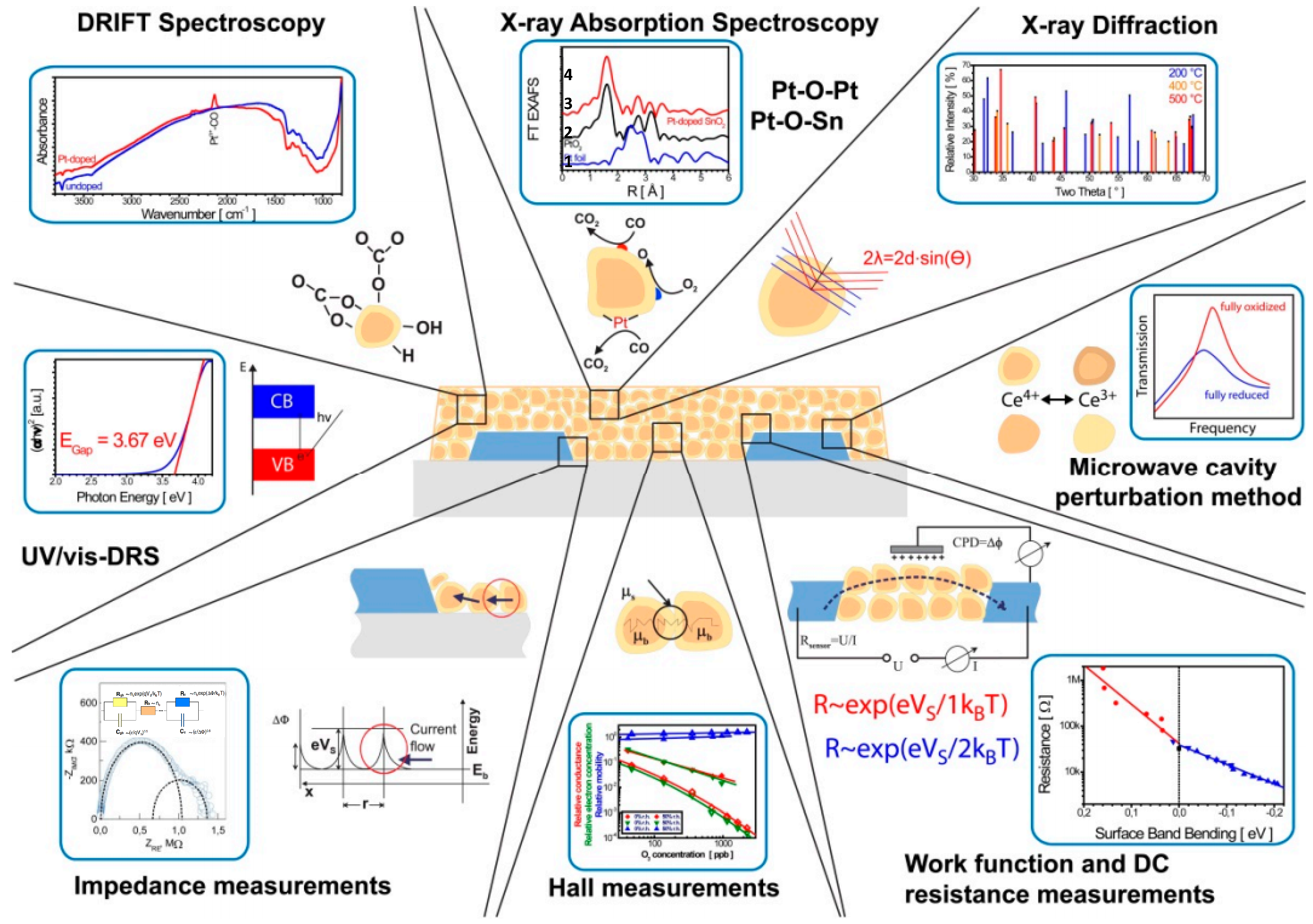
In situ measurement is very powerful for the investigation of the detailed mechanism of CO sensing [33], as shown in Figure 2. X-ray diffraction and X-ray absorption spectroscopies can be employed to study the structure information of the sensing materials. Diffuse Reflectance Infrared Fourier Transform Spectroscopy (DRIFTS) or Raman spectroscopy can be used to study the surface chemical information of the sensing materials. DRIFTS has been shown to be very effective for studying the sensing mechanism of CO gas based on a MOS. Its advantages include easy operation, direct usage of the sample, and simple sample preparation. Several examples of mechanism study using DRIFTS can be found in the next section.

3. MOS Sensing Materials

3.1. SnO2

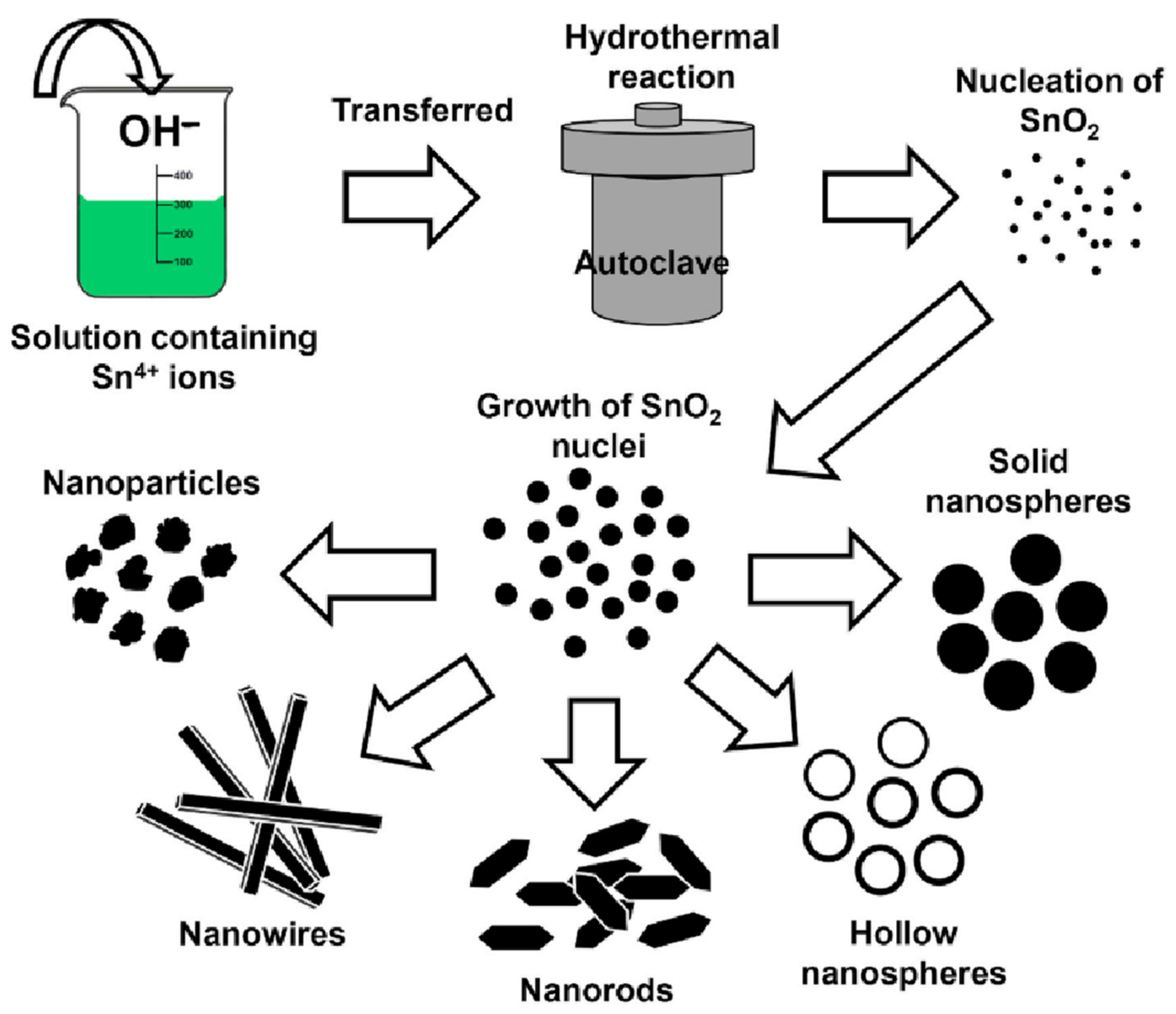
SnO2 is the most commonly employed metal oxide for CO detection because of its excellent self-catalysis effect due to its dual valences +2, +4. Its band gap is 3.6 eV. The Fermi level of SnO2 is −4.7 eV [34]. Generally, it can be produced through the wet or dry method (sputtering or chemical vapor deposition) [32,35,36,37,38,39,40]. The wet method is more popular than the sputtering method due to its ability to adjust morphologies in a simple way. For example, the hydrothermal method can produce various morphologies of SnO2, adjusting the CO sensing performance in a large range [41], as seen in Figure 3. At first, Sn4+ ions and OH react with each other and form precipitation. Some SnO2 nuclei will form from dehydration of the Sn4+ ions. Next, the small SnO2 nuclei will grow bigger. Nanoparticles, nanospheres, nanowires, or nanorods can be obtained by controlling the reactant and additives.
However, pure SnO2 is seldom reported to be sensitive to CO at room temperature. Au, Pt, or Pd are usually added to the surface of SnO2 to promote room-temperature CO sensing. Table 2 summarizes the comparison of the performance of SnO2 composites for room-temperature CO sensing. Nanosized SnO2 can be obtained by the hydrothermal method by employing poly ethylene glycol (PEG-6000) [42]. Au impregnation was performed with gold chloride (HAuCl4·3H2O). The optimum Au-SnO2 sample can respond to 500 ppm CO with a response of about 50 in 20 s. The humidity effect was checked, and the optimum Au-SnO2 response was almost the same under RH 55% and RH 70%. The mechanisms of room-temperature CO sensing using Au-SnO2 can be attributed to two aspects: First, Au has a catalytic effect through chemical sensitization via the spillover effect. Second, Au will increase the surface resistance of the Au-SnO2 composite through the Schottky barrier effect, as discussed by Yamazoe [43].
A comparison of the performance of SnO2 composites is shown in Table 2. Pt decoration has been shown to enhance the sensitivity of SnO2 to CO, showing a response value of 64.5 to 100 ppm at room temperature [43]. It can be confirmed that both components and structures of SnO2 composites can influence the sensing performance of the composites. For example, Pt-doped SnO2 porous nanosolid shows a much higher response than that of Pt-doped SnO2 nanoparticles [44,45]. The inclusion of perovskite and Pd-Au not only enhances the response of SnO2, but also shortens the response time and recovery time to around 30 s [46].
Table 2. Comparison of performance of different SnO2 composites for CO sensing at room temperature.
Sensing MaterialCO Concentration (ppm)ResponseResponse
Time (s)
Recovery
Time (s)
Reference
Au-SnO2500~5020 (50 °C)NA[42]
Pt- SnO2 nanoparticle50003.57~720NA[45]
Pt-SnO2 porous nanosolid10064.5144882[44]
Pd-SnO2 nanoparticle50~52040[47]
Polyaniline-Pd-SnO230048862[48]
CH3NH3SnI3/SnO2/Pd/Au50682532[46]
CNT-Co3O4− SnO210001.46 (Va/Vg)120150[2]
SnO2 can be doped with Pt by mixing hexachloro-platinic acid (H2PtCl6) with rutile SnO2 powder [44]. The samples were sintered at 600, 700, and 800 °C. It was found that heat-treating at 800 °C will lead to grain growth and surface area reduction, thus lowering the sensitivity. The SnO2-Pt sample annealed at 700 °C has the highest sensitivity among all the samples. Moreover, 1 wt% Pt-doped SnO2 powder can respond to 5000 ppm CO with a sensitivity of 3.57. H2PtCl6 was loaded onto SnO2 porous nanosolid (PNS) as well. The mixture was sintered in nitrogen at a temperature ranging from 400 to 600 °C. It was found that 500 °C is the optimum calcining temperature.
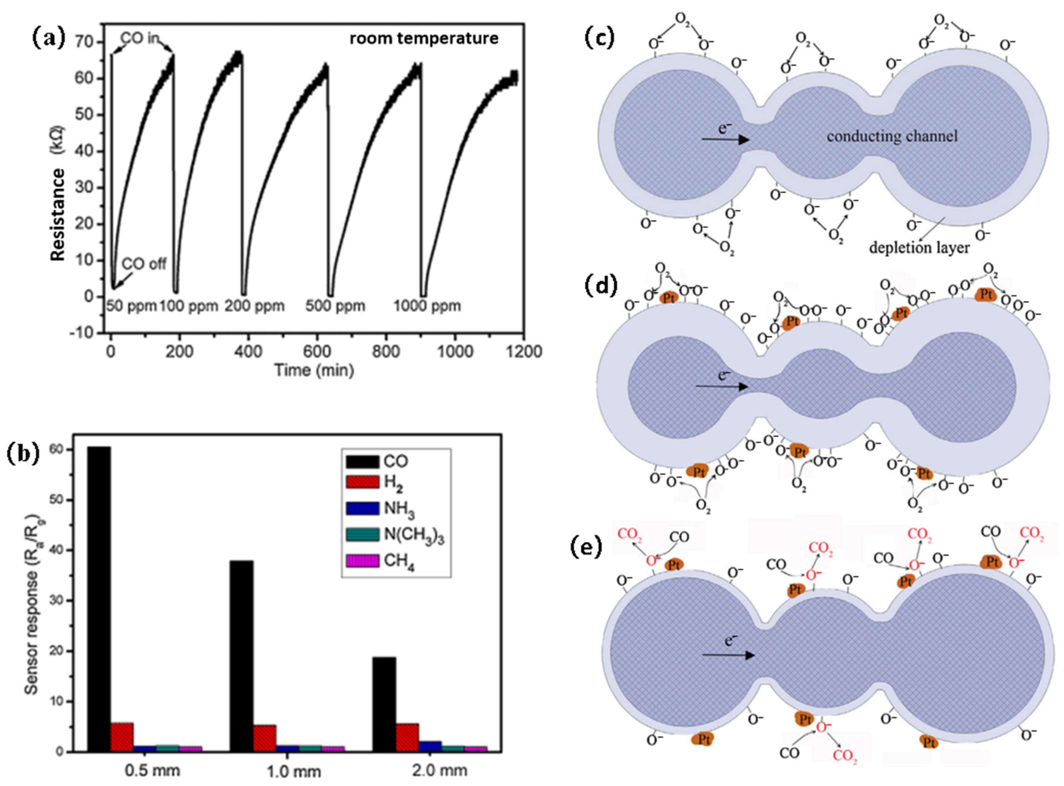
Doped SnO2 PNS with a 0.5 mm thickness can respond to 100 ppm CO with a sensitivity of 64.5, as shown in Figure 4a. The sensor has excellent selectivity to CO compared to H2 or CH4, as seen in Figure 4b. The mechanism of the excellent CO sensing performance of the Pt-loaded SnO2 PNS can be explained by the electrical and chemical effects. The electrical effect was further studied by Hall effect measurement. The electron concentration n and mobility µ before and after exposure to 1000 ppm CO were compared. It was found that the carrier concentration of the best sample increased by almost 50 times after exposure. Meanwhile, the carrier mobility of the best sample increased by 4.76 times after exposure. This phenomenon is in line with the neck control model, i.e., the carrier concentration is determined by the depletion layer and the mobility is controlled by the width of the conduction channel at the grain neck.
As seen in Figure 4c, surface-adsorbed oxygen molecules will change into oxygen ions by drawing electrons from SnO2. The oxygen ions will form a depletion layer on the surface of SnO2. Pt decoration will promote dissociation of the oxygen molecules, increasing the width of the surface depletion layer, as shown in Figure 4d. When exposed to CO, surface Pt clusters will catalyze the oxidation reaction between the CO and surface-adsorbed oxygen species. The reaction will cause the re-injection of electrons into the SnO2 nanoparticles. The resistance of the materials will decrease since the width of the conduction channel will be expanded.
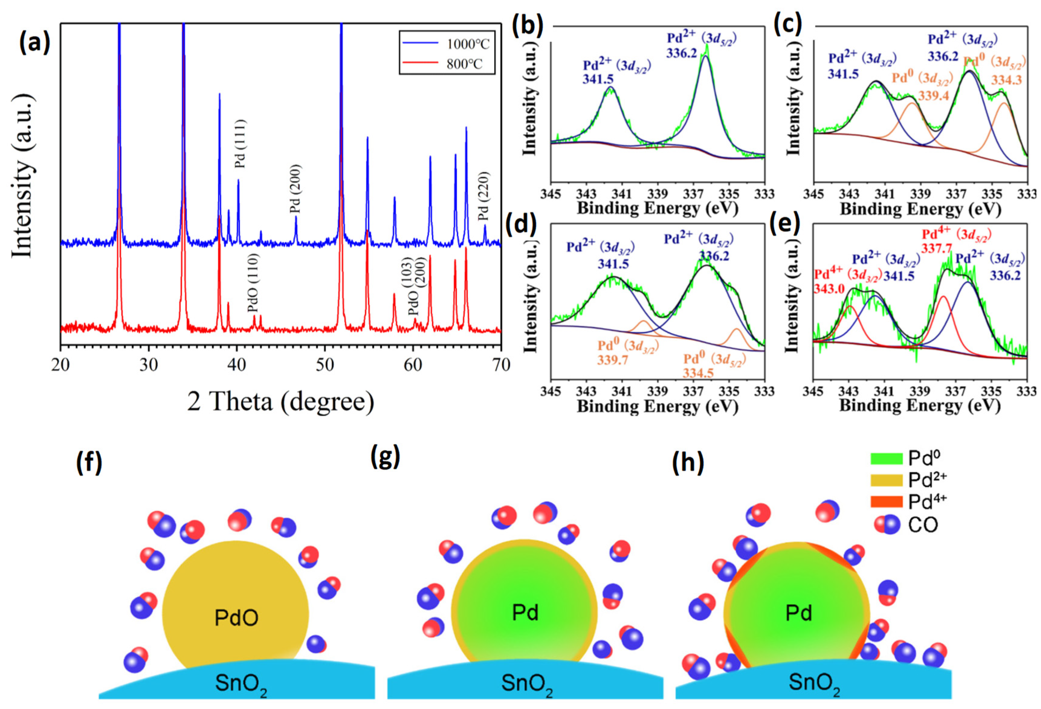
Pd-SnO2 was an excellent candidate for room-temperature CO sensing as well [47]. The sample with a Pd content less than or equal to 2% and heating temperature higher than 1000 °C can respond to 500 ppm CO in 30 s with a response value of 5. The response of the 1 wt% Pd sample heated at 1000 °C to 0.04% CO-N2 is ten times larger than that to 0.04% CO-20% O2-N2. This is because that the chemisorption of CO onto SnO2 will transfer two electrons to SnO2. Chemisorption of CO onto SnO2 will be hindered heavily when oxygen species are present on the surface of SnO2. It was confirmed by XRD analysis that PdO forms when the samples are heat-treated at 800 °C, while metallic Pd forms when the samples are heat-treated at 1000 °C instead, as seen in Figure 5a. Furthermore, the XPS results in Figure 5b show that metallic Pd is in the Pd2+ state for the 5 wt% Pd sample heat-treated at 1000 °C. Figure 5c proves that Pd2+ and Pd0 can be found in Pd nanoparticles. Figure 5d shows a pronounced decrease in the Pd0 ratio in all Pd species, which may be due to electron transfer from Pd to SnO2 at room temperature. Figure 5e shows that Pd2+ and Pd4+ can be observed in the sample of 1 wt% Pd heat-treated at 1000 °C. It can be concluded from Figure 5b–e that the ratio of Pd to SnO2 is crucial for the surface states of Pd. The configurations of the Pd-SnO2 species are visualized in Figure 5f–h. Three cases are presented: (1) Pd-SnO2 heated at a temperature lower than 900 °C: PdO was formed on the surface of SnO2, with no response to CO at room temperature; (2) Pd-SnO2 with Pd content > 2 wt% heated at 1000 °C: Pd was formed on the surface of SnO2, with no response to CO at room temperature; and (3) Pd-SnO2 with Pd content ≤ 2 wt% heated at 1000 °C: Pd was formed on the surface of SnO2 with Pd4+ states, with a response to CO at room temperature. The room-temperature sensing mechanisms of Pd-SnO2 may be the chemisorption of CO on the Pd nanoparticles at the Pd4+ sites and the spillover effect of CO toward SnO2.
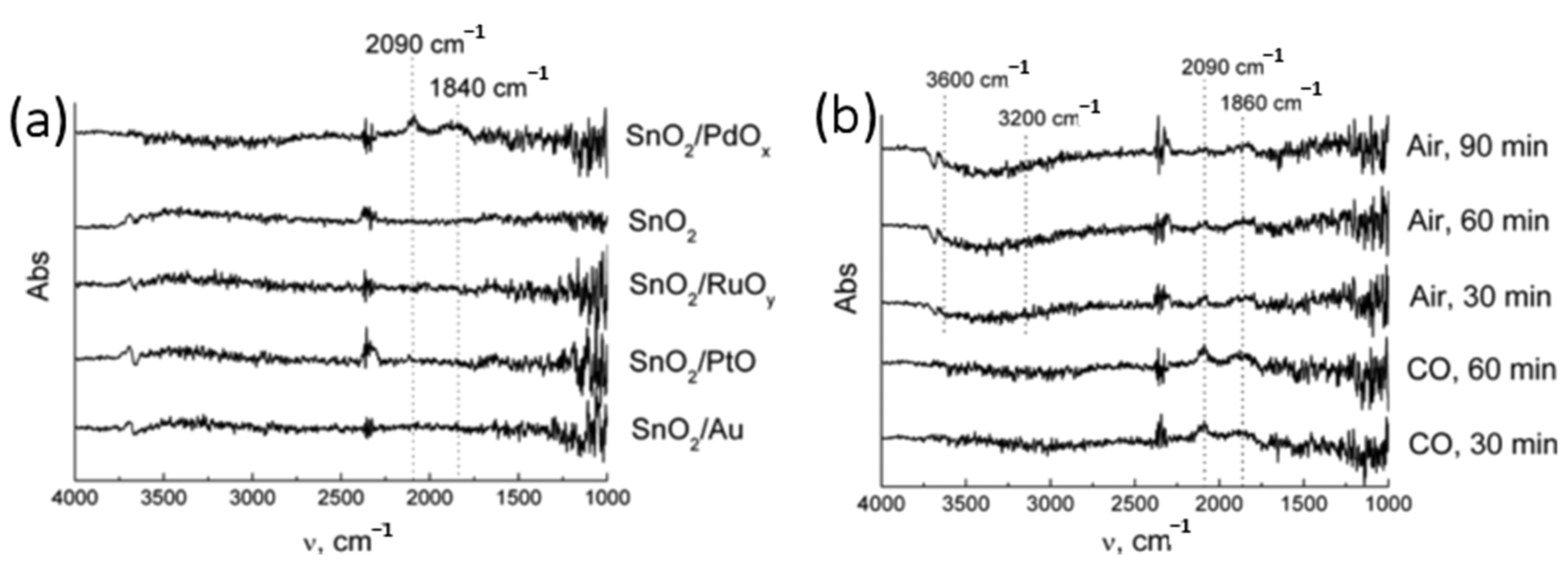
The room-temperature CO sensing mechanisms of PdO-modified SnO2 have also been investigated by the DRIFT method. As seen in Figure 6a, the peaks at 2090 cm−1 and 1840 cm−1 only appear for the SnO2/PdOx sample [49]. The peaks confirmed the occurrence of CO chemisorption on the reduced Pd species. The first peak refers to linear carbonyl binding to metallic sites with π-back donation from a metal, i.e., Pd0-bound CO. The other peak corresponds to carbonyls bound to Pd atoms in a bridging configuration. After CO is substituted by air, the strength of the OH stretching band at 3600–3200 cm−1 declines. This phenomenon is due to the hydrogen bonds between the OH groups. OH species will interact with CO at room temperature following the equation below:
CO + OH → CO2 + H+ + e
As shown in Figure 7a, first tin dioxide nanoparticles were obtained by the hydrothermal and drying method [50]. Then, graphene in different amounts was dispersed in ethanol and then sonicated. Then, 1 g tin oxide was added into the dispersion solution. The 0.5 wt% graphene-decorated SnO2 has the highest response to 40 ppm CO, as shown in Figure 7b. The 0.5 wt% graphene-decorated SnO2 can also respond to and recover from 40 ppm CO in less than 45 s, as shown in Figure 7c.

3.2. ZnO

ZnO is one of the most commonly employed CO sensing materials because of its variable morphologies and easy doping processes [51]. Compared to other semiconductor materials, ZnO has many excellent properties, such as a simple synthesis, wide bandgap of 3.37 eV, and binding energy of exciton of 60 meV [52,53,54]. ZnO can be grown in a controlled manner by the sputtering, CVD, or solution method, of which the solution method is the most popular [54,55,56]. For example, ZnO nanorods can be formed using  Zn ( OH ) 4 2  and CTAB aqueous solutions, in which CTAB functions as a structure director [57]. A ZnO nanorod array was realized on a (100) silicon wafer on a 5 nm thick ZnO seed layer by sputtering using ammonia and zinc chloride solution [58]. ZnO hierarchical nanostructures can be grown using sequential nucleation and growth methods [59,60]. The comparison of the performance of various ZnO materials is shown in Table 2. For example, ZnO nanocomb can be synthesized by chemical vapor deposition first. Then, the nanocomb can be drop-casted on SiO2-p-Si substrate with a patterned Ti/Au electrode array on top. The sample can respond to CO at room temperature [61]. In addition, ZnO thin films and nanoneedles are reported to be able to respond to CO at room temperature as well [62]. Au decoration with gold chloride solution, HAuCl4, is an effective way to synthesize Au-ZnO composites for room-temperature CO sensing. Table 3 shows that the base material, ZnO, can finally determine the sensing performance of two similar samples, such as the response time after a comparison of two Au-ZnO samples in [63,64].
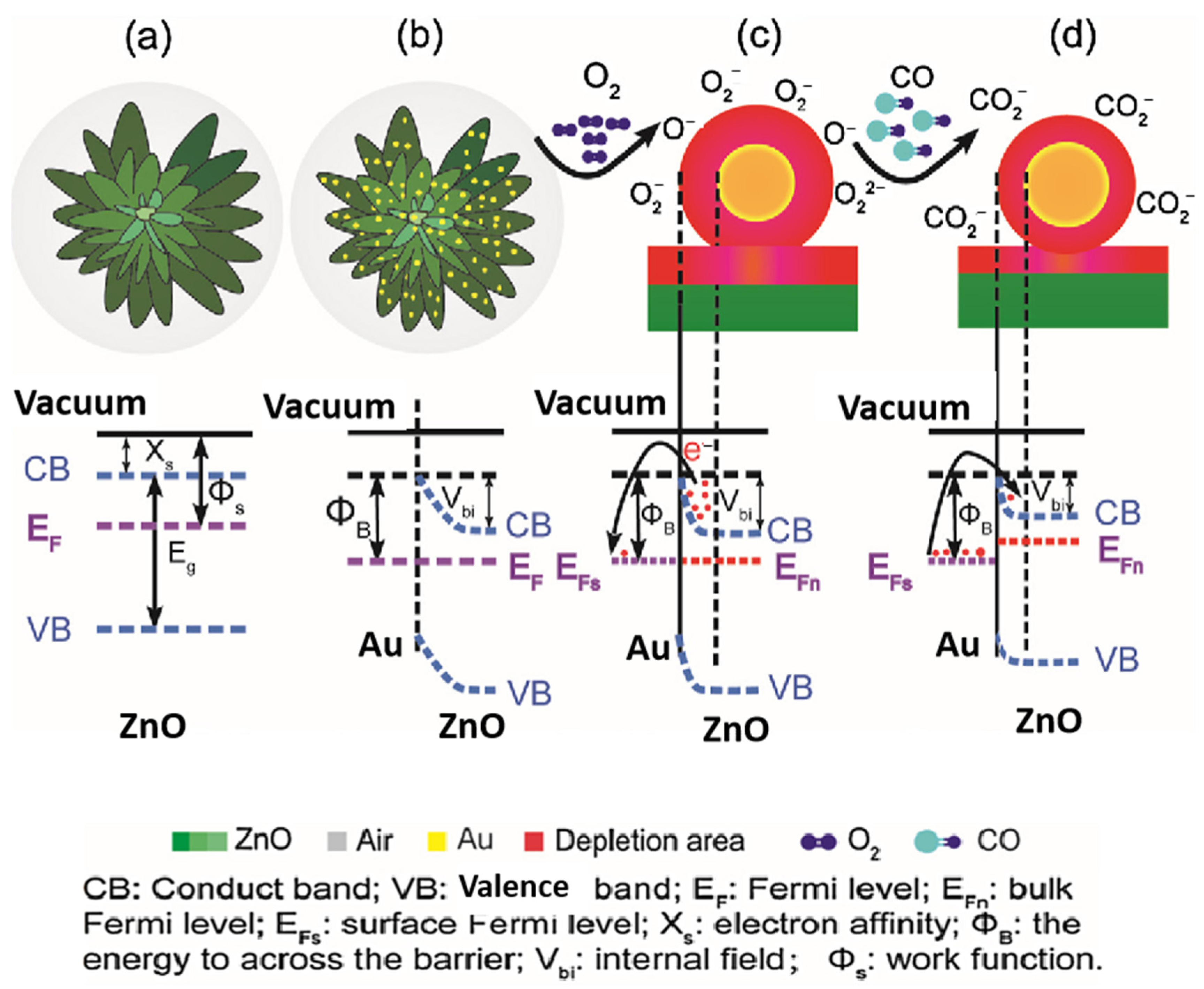
Notably, Au-ZnO nanostars can respond to 500 ppm CO in 41 s with a response value over 55 under dynamic mode, which is the highest response value of ZnO room-temperature sensing materials [64]. This high response is due to three aspects (Figure 8): Firstly, the ZnO nanostars are composed of many ultrafine nanoparticles with a typical size of around 20 nm, which offers more reaction sites for adsorption of oxygen. Secondly, the spillover effect of gold nanoparticles can facilitate dissociation of oxygen molecules over ZnO, decreasing the CO sensing temperature. It is believed that this chemical sensitization function should be the predominant mechanism for room-temperature CO sensing of Au-ZnO. The third effect is the work function modulation through the creation of a nanoscopic depletion region at the Au-ZnO surface, altering the Schottky barrier height, as seen in Figure 8b. In addition, ZnO-SnSe2 was reported to sense CO at room temperature with excellent resistance to humidity interference [66]. The author also found that UV illumination can improve the sensitivity of the material. This phenomenon is similar to that of the MoS2 composite, which will be discussed further below.

3.3. Other Metal Oxides

Besides SnO2 and ZnO, CuO, doped TiO2, and other metal oxides have been reported to respond to CO at room temperature. TiO2 is another interesting sensing material for room-temperature CO sensing. TiO2 was widely employed for photocatalysis at the beginning. Solution growth of TiO2 can be well controlled through the organic capping method [68]. Later, TiO2 nanotubes were synthesized by electrochemical anodization and employed for sensing CO, ethanol, and hydrogen [69]. Moreover, porous TiO2 was reported to be suitable for chemical sensors in cyber chemical systems [70]. The latest progress in TiO2 nanostructures for gas sensing can be found in another review [71]. WO3 is an excellent material for CO sensing as well [72,73]. It was reported that CeO2-WO3 can respond to CO very well at 430 °C. The response and recovery time of the sensor is less than 60 s [74]. Nanocomposites of graphene and WO3 can respond to 10 ppm CO at room temperature with a low response value of 1.03. The graphene–WO3 nanocomposites can respond to 100 ppm CO at 300 °C with a response value of 21.5 under dynamic mode [75]. The comparison of the performance of temperature sensors based on other metal oxides is shown in Table 4.
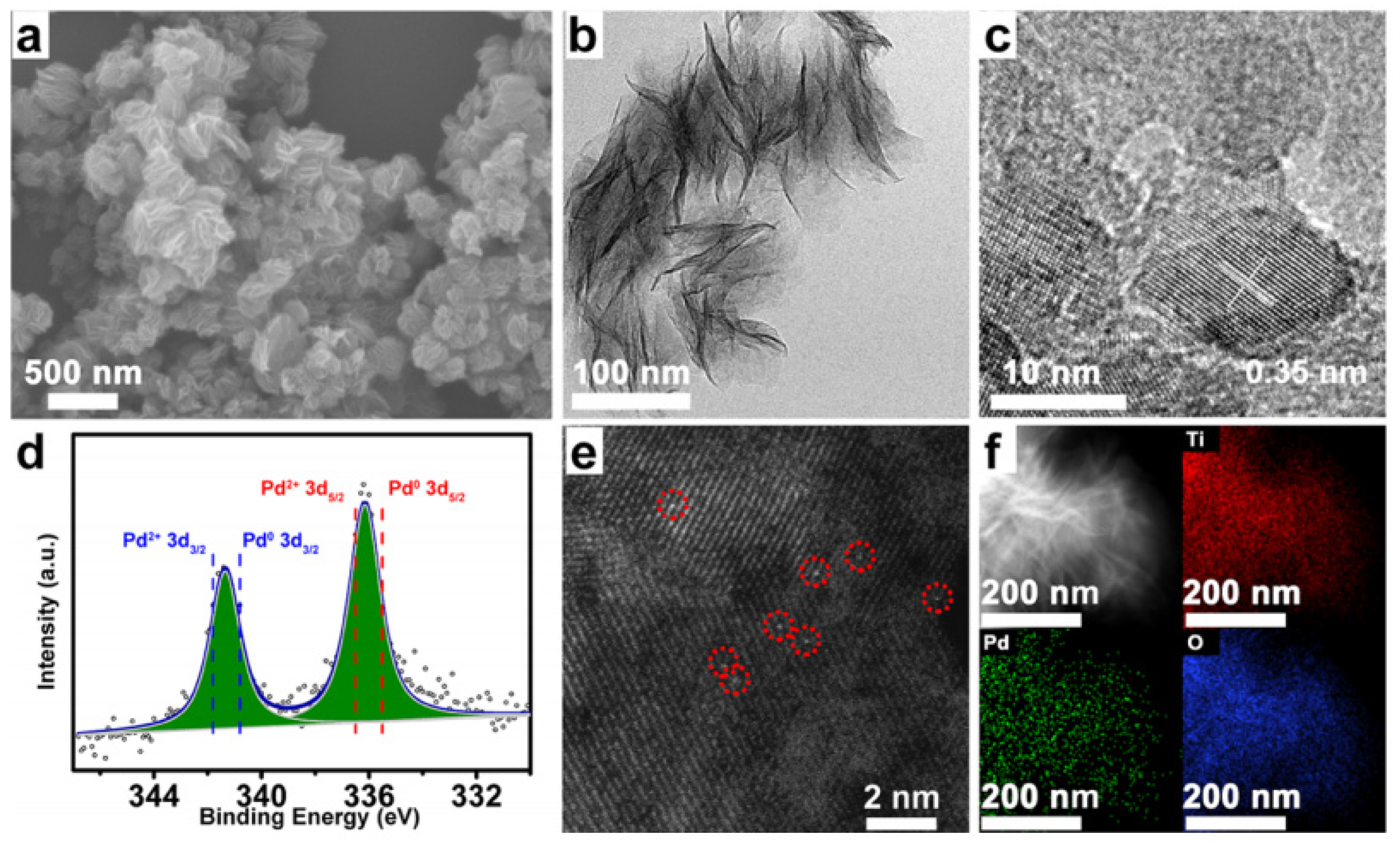
Recently, Xu synthesized atomically dispersed Pd over TiO2 nanoflower through a simple and mild photochemical method at room temperature [79]. SEM of Pd1-TiO2 shows a nanoflower morphology, as seen in Figure 9a. Figure 9b shows that no Pd nanoparticles can be found in Pd1-TiO2. An interplanar spacing of 0.35 nm is assigned to (100) of TiO2. As seen in Figure 9d, 336.2 and 341.4 eV of Pd1-TiO2 belong to Pd2+ and Pd0, indicating partial oxidation of Pd atoms. Figure 9e suggests that the Pd species is in the form of isolated single atoms. Figure 9f shows the uniform distribution of Ti, O, and Pd on nanoflowers.
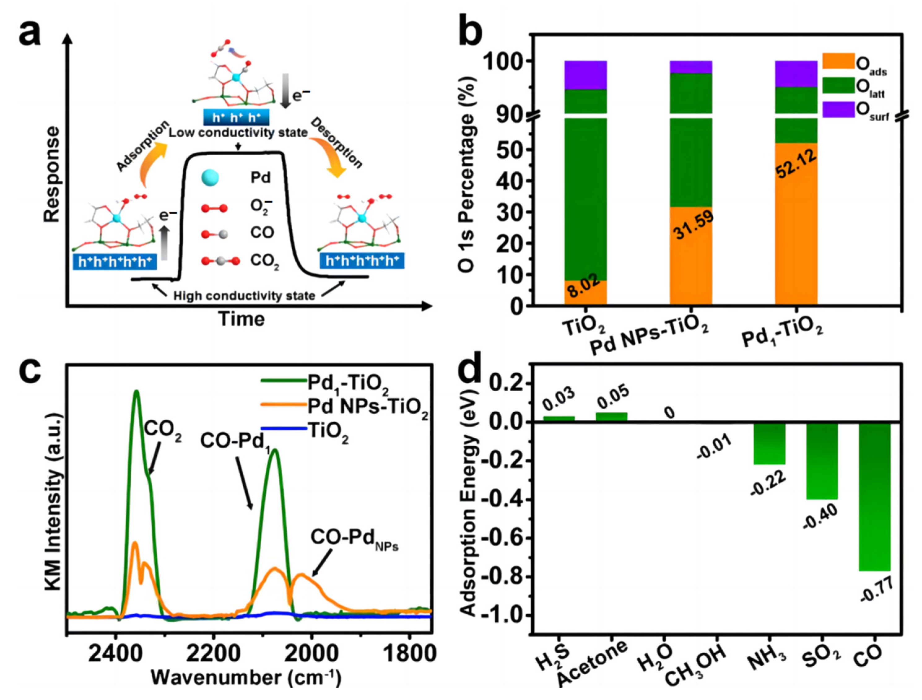
The TiO2, Pd NPs-TiO2, and Pd1-TiO2 samples all presented a P-type nature according to the analysis results from the UPS and UV-vis spectra. Thus, the oxygen molecules adsorbed on these materials will increase the densities of holes and increase the conductivity. When exposed to CO as a reductive analyte, free electrons will be injected into the materials and decrease the conductivity. Oads refers to the adsorbed active oxygen species, i.e., mainly  O 2  at room temperature. The relative percentage of Oads increased from 8.02% to 52.12% for TiO2 and Pd1-TiO2 (Figure 10b). Figure 10c shows that the CO adsorption ability is in the order TiO2 < Pd NPs-TiO2 < Pd1-TiO2. Meanwhile, the adsorption intensity of CO2 production of Pd1-TiO2 is greater than that of Pd NPs-TiO2, revealing higher room-temperature CO oxidation efficiency. Furthermore, the adsorption energy of CO on Pd1-TiO2 is -0.77 eV, which is the lowest among all the calculated gases, and accounts for the high selectivity of the sample to CO (Figure 10d).

3.4. Metal Oxide–2D Material Composites

Two-dimensional materials are very absorbing due to their unique structures and electronic properties. MoS2 is widely applied in room-temperature gas sensing materials [85,86]. It has many excellent properties, such as the very high luminescence quantum efficiency of single-layer MoS2. MoS2 has already been used for room-temperature sensing H2S, NO2, and SO2 [87,88,89,90,91]. Recently, several works have been reported regarding room-temperature CO sensing based on MoS2-MOS composites. For example, Ag-ZnO-MoS2 was prepared by the layer-by-layer (LBL) self-assembly method [92]. The sample can respond to 100 ppm CO with a response value of 1.05. Both the response and recovery processes take less than 60 s. This fast process under room temperature is due to the catalytic effect of Ag and synthetic effect of ZnO and MoS2. Co-In2O3-MoS2 can be prepared by LBL self-assembly as well [93]. The sample can achieve a response value of 1.08 to 10 ppm CO in 39 s. It is the Co2+ doping and heterojunction formation from both Co-In2O3 and MoS2 that contributes to the excellent CO sensing behavior. Moreover, SnO2-MoS2 was reported to respond to 40 ppm CO at room temperature with a response value of 4.97 under UV light illumination [94]. The sensing mechanism is shown in Figure 11. First, MoS2 can prevent interparticle aggregation of SnO2 nanoparticles and provide more sites for the adsorption of CO molecules from SnO2, thus increasing the sensitivity. Moreover, MoS2 provides direct conduction paths for charge carriers, because of its high charge carrier mobility. Moreover, MoS2 and SnO2 can form a p-n junction that can improve the electron–hole separation under UV irradiation.
When UV light is employed, the photogenerated electron–hole pairs are excited and separated by the built-in electric fields in SnO2. The photogenerated electrons on the conduction band of MoS2 can readily be transferred to SnO2, and the photogenerated holes on the valence band of SnO2 can be transferred to MoS2. The photogenerated electrons will then be captured by O2 and yield new O2−. Simultaneously, the reaction of the photogenerated holes with the adsorbed oxygen ions on the surface of the sensing material occurs according to Equations (5) and (6) shown below:
hυ → h+ + e
O2 + e (hυ) → O2 (hυ)
h+ (hυ) + O2 (ads) → O2 (gas)
The photogenerated electron hole can be separated efficiently at the interface, preventing recombination and enhancing the sensing performance. Once SnO2 is exposed to CO gas, CO adsorbed on the surface of SnO2 can also react with photogenerated electrons, as seen in Equation (8):
2CO (gas) + O2 (hυ) → 2CO2 (gas) + e
In addition, a SnO2-MoSe2 nanoflower was synthesized through the hydrothermal method. The composite can respond to 1 ppm CO very quickly, with higher sensitivity than the pristine MoSe2 or SnO2. The sensing mechanism may be the formation of n-n junctions between n-type SnO2 and n-type MoSe2 [95]. A comparison of MoS2 composites for room-temperature CO sensors can be found in Table 5.

4. Discussion of Sensing Mechanism

The CO sensing mechanisms of MOSs at temperatures higher than room temperature have been reported in several papers already [96,97,98]. While a precise understanding of the CO sensing mechanisms of MOSs at room temperature is still in its early stages, a general model can be deduced from the works reported already. Below are some critical factors:
(1)
Structures of MOS nanostructures
The structure (crystal plane, grain size, morphology, and so on) of the sensing materials will significantly influence the sensing performance. On one hand, MOSs with smaller grain size will usually have a higher response to CO at room temperature, which is in line with the grain size sensing model in Figure 3. For example, the ZnO thin films with the smallest grain size among all three types of samples have the highest response to CO at room temperature [65]. In addition, specific crystal planes and more active defects can enhance CO sensing at room temperature. For instance, it was reported that CuO nanosheets with exposed (111) crystal facets and more oxygen vacancies have a higher response to and lower detection limit of CO at room temperature.
(2)
Surface modification of MOSs
There are two main surface modification methods for MOSs, i.e., doping of noble metals or formation of heterojunctions. Doping of noble metals is the most common method to enhance room-temperature CO sensing based on MOSs. The chemical sensitization effect is the main contributing factor in lowering the working temperature. The chemical states and dispersion states of noble metals on the surface of a MOS play a vital role in its CO sensing performance at room temperature, as shown in the case of the Pd-MOS system [47]. In addition, a heterojunction of a MOS will enhance the room-temperature CO sensing at both the receptor and transducer point. For example, CuO-TiO2 composite can respond to CO at room temperature [82]. CuO materials have excellent CO adsorption properties, enhancing the receptor performance of the composites. Moreover, CuO-TiO2 can form a p-n junction, creating a space charge region at the interface of the two materials.
(3)
Annealing effect
The annealing effect, like the annealing atmosphere and annealing temperature, can also influence the room-temperature CO sensing performance of the MOS material. Considering SnO2-Pt composites, the SnO2-Pt composite annealed in nitrogen has a much higher response than that annealed in air [44]. The reason is twofold: First, the atmosphere can determine the chemical state of the loaded Pt. More Pt2+ with a much higher catalyzing activity will be present in SnO2-Pt annealed in nitrogen. Second, SnO2-Pt annealed in nitrogen has more oxygen vacancies that can be sites for chemisorption of oxygen. Furthermore, higher annealing temperatures can increase the crystallinity of the sensing material, as they did in Au-In2O3 [99]. However, too high a temperature, like 600 °C, may destroy the nanorod and make Au agglomerate in Au-In2O3.
(4)
UV activation
UV activation on the photosensitive materials will generate more electrons, thus forming more  O 2  at the surface, enhancing the reaction between oxygen species and CO at room temperature. For example, ZnO and TiO2 are very sensitive to UV light. MoS2 and SnSe2 have higher sensing performance under UV as well [95].

5. Conclusions

In this review, the typical studies of MOSs for room-temperature CO sensors have been summarized and discussed. For example, noble metal-doped SnO2 can function well for room-temperature CO sensing. ZnO with smaller diameters can sense CO at room temperature. Self-doped or Pd-doped TiO2 can respond to CO very well at room temperature as well. The room-temperature CO sensing mechanism of MOSs can be categorized into four parts: the structures of MOSs, surface modification effects, UV activation, and the annealing effect. MOSs with a smaller diameters usually have better CO sensing performance at room temperature. A noble metal or heterojunction effect at the surface of MOSs can facilitate room-temperature CO sensing. Moreover, UV activation can promote room-temperature CO sensing in some materials such as ZnO, TiO2, or MoS2. Both the annealing atmosphere and temperature can effect room-temperature CO performance. The future characteristics of room-temperature CO sensors will be higher sensitivity, a lower detection limit and cost, and a smaller size for integration and portable devices. Combining material engineering and MEMS technology is another promising field. This review will be of interest to many researchers and engineers working on sensor materials or in the safety monitoring sensor field.

Author Contributions

Conceptualization, Y.H. and M.J.; validation, Y.H.; formal analysis, Y.H.; investigation, M.J.; resources, Y.H.; data curation, M.J.; writing—original draft preparation, M.J.; writing—review and editing, Y.H.; visualization, M.J.; supervision, M.J.; funding acquisition, Y.H. All authors have read and agreed to the published version of the manuscript.

Funding

This research was funded by the International Science and Technology Cooperation Project of Tiandi Technology Co., Ltd. (No. 021-2-GH003), Key Technology and Standards of Mine Internet of Things of Tiandi Technology Co., Ltd. (No. 2019-TD-ZD007), and National Science Foundation Council (62204260).

Institutional Review Board Statement

Not applicable.

Informed Consent Statement

Not applicable.

Data Availability Statement

No new data were created or analyzed in this study. Data sharing is not applicable to this article.

Conflicts of Interest

Author Yaoyi He employed by the company Tiandi (Changzhou) Automation Co., Ltd. The remaining authors declare that the research was conducted in the absence of any commercial or financial relationships that could be construed as a potential conflict of interest.

References

  1. Choi, S.W.; Kim, J.; Lee, J.H.; Byun, Y.T. Remarkable Improvement of CO-Sensing Performances in Single-Walled Carbon Nanotubes Due to Modification of the Conducting Channel by Functionalization of Au Nanoparticles. Sens. Actuators B Chem. 2016, 232, 625–632. [Google Scholar] [CrossRef]
  2. Wu, R.J.; Wu, J.G.; Yu, M.R.; Tsai, T.K.; Yeh, C.T. Promotive Effect of CNT on Co3O4-SnO2 in a Semiconductor-Type CO Sensor Working at Room Temperature. Sens. Actuators B Chem. 2008, 131, 306–312. [Google Scholar] [CrossRef]
  3. Roy, A.; Ray, A.; Sadhukhan, P.; Naskar, K.; Lal, G.; Bhar, R.; Sinha, C.; Das, S. Polyaniline-Multiwalled Carbon Nanotube (PANI-MWCNT): Room Temperature Resistive Carbon Monoxide (CO) Sensor. Synth. Met. 2018, 245, 182–189. [Google Scholar] [CrossRef]
  4. Wang, H. Research Progress of Low-Power Carbon Monoxide Sensor. Ind. Mine Autom. 2021, 47, 16–23. [Google Scholar] [CrossRef]
  5. Dong, K.N.; Yang, J.F. Development of Mine-Used Infrared Carbon Monoxide Sensor Based on Mini Pump Suctio. Ind. Mine Autom. 2021, 47, 128–132. [Google Scholar]
  6. Shrivastava, S.; Mahana, D.; Nehra, S.; Gangwar, S.; Singh, S.; Yadav, C.S.; Muthusamy, S.K.; Dogra, A. CO Gas Sensing Characteristics of BaSnO3 Epitaxial Films Prepared by PLD: The Effect of Film Thickness. Sens. Actuators B Chem. 2024, 400, 134882. [Google Scholar] [CrossRef]
  7. Ani, A.; Poornesh, P.; Antony, A.; Chattopadhyay, S. Synergism of Spray-Pyrolyzed Aliovalent Ga (Iii) Ions and ZnO Nanostructures for Selective Sensing of Hazardous CO Gas towards Low Ppm Levels. Sens. Actuators B Chem. 2024, 399, 134827. [Google Scholar] [CrossRef]
  8. Zegebreal, L.T.; Tegegne, N.A.; Hone, F.G. Recent Progress in Hybrid Conducting Polymers and Metal Oxide Nanocomposite for Room-Temperature Gas Sensor Applications: A Review. Sens. Actuators A Phys. 2023, 359, 114478. [Google Scholar] [CrossRef]
  9. Desimone, P.M.; Zonta, G.; Giulietti, G.; Ortega, P.P.; Aldao, C.M.; Simões, A.Z.; Moura, F.; Ponce, M.A.; Foschini, C.R. Towards Carbon Monoxide Detection Based on ZnO Nanostructures. Mater. Sci. Eng. B 2024, 299, 117003. [Google Scholar] [CrossRef]
  10. Sadredini, A.R.; Nikfarjam, A.; Naeimi Pour, M.; Nazeri, N. Intensity Modulation of UV Light in Gas Sensor Array to Discriminate Several Analytes at Room Temperature. IET Sci. Meas. Technol. 2023, 18, 65–73. [Google Scholar] [CrossRef]
  11. Tang, M.; Zhang, D.; Sun, Y.; Chen, Q.; Chen, Y.; Xi, G.; Wang, Z.; Shao, X. CVD-Fabricated Co3O4-Co3S4 Heterojunction for Ultra-Sensitive Detection of CO in SF6 Discharge Decomposition Products. Sens. Actuators B Chem. 2024, 401, 134968. [Google Scholar] [CrossRef]
  12. Chen, M.; Li, X.; Li, Y.; Li, Y.; Qin, Z.; Wang, Q. MOF-Derived Co3O4 Nanoparticles over Direct Grown ZnO Nanoflower on Ceramic for CO Sensor with High Selectivity. Sens. Actuators B Chem. 2024, 401, 134951. [Google Scholar] [CrossRef]
  13. de Araújo, E.P.; Paiva, M.P.; Moisés, L.A.; Santo, G.S.D.E.; Blanco, K.C.; Chiquito, A.J.; Amorim, C.A. Improving Hazardous Gas Detection Behavior with Palladium Decorated SnO2 Nanobelts Networks. Sensors 2023, 23, 4783. [Google Scholar] [CrossRef] [PubMed]
  14. Zhao, F.; Cao, W.; Wang, P.H.; Wang, J.; Yu, L.; Qiao, Z.; Ding, Z.J. Fast and Sensitive Detection of CO by Bi-MOF-Derived Porous In2O3/Fe2O3 Core-Shell Nanotubes. ACS Sens. 2023, 8, 4577–4586. [Google Scholar] [CrossRef] [PubMed]
  15. Ali, Z.M.; Murshed, M.N.; El Sayed, M.E.; Samir, A.; Alsharabi, R.M.; Farea, M.O. A New Fabrication Strategy of Ag2O Doped PANI as a Highly Stable and Room Temperature Operable Carbon Monoxide Gas Sensor. Opt. Mater. 2023, 144, 114324. [Google Scholar] [CrossRef]
  16. Al-Maamori, M.H.; Al-Jamal, A.N.; Habeeb, S.A.; Hassan, A.S.; Majdi, H.S. Al3+-Modified ZnO Thin Film Sensor Fabricated by the Sputtering Method: Characterization and a Carbon Monoxide Gas Detection Study. J. Chem. Res. 2024, 48, 17475198231221453. [Google Scholar] [CrossRef]
  17. Meng, F.J.; Guo, X.M. Co/Au Bimetal Synergistically Modified SnO2-In2O3 Nanocomposite for Efficient CO Sensing. Ceram. Int. 2023, 49, 15979–15989. [Google Scholar] [CrossRef]
  18. You, S.; Li, G.; Fan, Z.; Li, X.; Fu, L.; Wu, W. Nanotechnology-Assisted Sensors for the Detection of Carbon Monoxide: A Review. Int. J. Electrochem. Sci. 2023, 18, 100314. [Google Scholar] [CrossRef]
  19. Qin, C.; Wei, Z.; Wang, B.; Wang, Y. Sn and Mn Co-Doping synergistically promotes the sensing properties of Co3O4 sensor for high-sensitive CO detection. Sens. Actuators B Chem. 2023, 390, 133930. [Google Scholar] [CrossRef]
  20. Pandit, N.A.; Ahmad, T. ZrO2/CeO2-Heterostructured Nanocomposites for Enhanced Carbon Monoxide Gas Sensing. ACS Appl. Nano Mater. 2023, 6, 7299–7309. [Google Scholar] [CrossRef]
  21. Baier, D.; Priamushko, T.; Weinberger, C.; Kleitz, F.; Tiemann, M. Selective Discrimination between CO and H2 with Copper-Ceria-Resistive Gas Sensors. ACS Sens. 2023, 8, 1616–1623. [Google Scholar] [CrossRef] [PubMed]
  22. Agrawal, J.; Shukla, M.; Singh, V. Investigation of the Effect of ZnO Film Thickness over the Gas Sensor Developed for Sensing Carbon Monoxide. Phys. Status Solidi (A) Appl. Mater. Sci. 2023, 220, 2300047. [Google Scholar] [CrossRef]
  23. Zhao, W.; Fam, D.W.H.; Yin, Z.; Sun, T.; Tan, H.T.; Liu, W.; Tok, A.I.Y.; Boey, Y.C.F.; Zhang, H.; Hng, H.H.; et al. A Carbon Monoxide Gas Sensor Using Oxygen Plasma Modified Carbon Nanotubes. Nanotechnology 2012, 23, 425502. [Google Scholar] [CrossRef] [PubMed]
  24. Afrin, R.; Shah, N.A. Room Temperature Gas Sensors Based on Carboxyl and Thiol Functionalized Carbon Nanotubes Buckypapers. Diam. Relat. Mater. 2015, 60, 42–49. [Google Scholar] [CrossRef]
  25. Panda, D.; Nandi, A.; Datta, S.K.; Saha, H.; Majumdar, S. Selective Detection of Carbon Monoxide (CO) Gas by Reduced Graphene Oxide (RGO) at Room Temperature. RSC Adv. 2016, 6, 47337–47348. [Google Scholar] [CrossRef]
  26. Simões, A.N.; Lustosa, G.M.M.M.; de Souza Morita, E.; de Souza, A.N.; Torres, F.; Bizzo, W.A.; Mazon, T. Room-Temperature SnO2-Based Sensor with Pd-Nanoparticles for Real-Time Detection of CO Dissolved Gas in Transformer Oil. Mater. Chem. Phys. 2024, 311, 128576. [Google Scholar] [CrossRef]
  27. Fort, A.; Landi, E.; Mugnaini, M.; Parri, L.; Pozzebon, A.; Vignoli, V. A LoRaWAN Carbon Monoxide Measurement System with Low-Power Sensor Triggering for the Monitoring of Domestic and Industrial Boilers. IEEE Trans. Instrum. Meas. 2021, 70, 5500609. [Google Scholar] [CrossRef]
  28. Mahajan, S.; Jagtap, S. Metal-Oxide Semiconductors for Carbon Monoxide (CO) Gas Sensing: A Review. Appl. Mater. Today 2020, 18, 100483. [Google Scholar] [CrossRef]
  29. Lawaniya, S.D.; Kumar, S.; Yu, Y.; Rubahn, H.G.; Mishra, Y.K.; Awasthi, K. Functional Nanomaterials in Flexible Gas Sensors: Recent Progress and Future Prospects. Mater. Today Chem. 2023, 29, 101428. [Google Scholar] [CrossRef]
  30. Jiao, M.Z.; Chen, X.Y.; Hu, K.X.; Qian, D.Y.; Zhao, X.H.; Ding, E.J. Recent Developments of Nanomaterials-Based Conductive Type Methane Sensors. Rare Met. 2021, 40, 1515–1527. [Google Scholar] [CrossRef]
  31. Yamazoe, N.; Shimanoe, K. New Perspectives of Gas Sensor Technology. Sens. Actuators B Chem. 2009, 138, 100–107. [Google Scholar] [CrossRef]
  32. Blackman, C. Do We Need “Ionosorbed” Oxygen Species? (Or, “a Surface Conductivity Model of Gas Sensitivity in Metal Oxides Based on Variable Surface Oxygen Vacancy Concentration”). ACS Sens. 2021, 6, 3509–3516. [Google Scholar] [CrossRef] [PubMed]
  33. Degler, D. Trends and Advances in the Characterization of Gas Sensing Materials Based on Semiconducting Oxides. Sensors 2018, 18, 3544. [Google Scholar] [CrossRef] [PubMed]
  34. Chen, Y.; Meng, Q.; Zhang, L.; Han, C.; Gao, H.; Zhang, Y.; Yan, H. SnO2-Based Electron Transporting Layer Materials for Perovskite Solar: A Review of Recent Progress. J. Energy Chem. 2019, 35, 144–167. [Google Scholar] [CrossRef]
  35. Zhang, Y.; Cui, S.; Chang, J.; Ocola, L.E.; Chen, J. Highly Sensitive Room Temperature Carbon Monoxide Detection Using SnO2 Nanoparticle-Decorated Semiconducting Single-Walled Carbon Nanotubes. Nanotechnology 2013, 24, 025503. [Google Scholar] [CrossRef] [PubMed]
  36. Bai, M.; Chen, M.; Li, X.; Wang, Q. One-Step CVD Growth of ZnO Nanorod/SnO2 Film Heterojunction for NO2 Gas Sensor. Sens. Actuators B Chem. 2022, 373, 132738. [Google Scholar] [CrossRef]
  37. Yang, A.; Tao, X.; Wang, R.; Lee, S.; Surya, C. Room Temperature Gas Sensing Properties of Sn O2 /Multiwall-Carbon-Nanotube Composite Nanofibers. Appl. Phys. Lett. 2007, 91, 133110. [Google Scholar] [CrossRef]
  38. Shojaee, M.; Nasresfahani, S.; Sheikhi, M.H. Hydrothermally Synthesized Pd-Loaded SnO2/Partially Reduced Graphene Oxide Nanocomposite for Effective Detection of Carbon Monoxide at Room Temperature. Sens. Actuators B Chem. 2018, 254, 457–467. [Google Scholar] [CrossRef]
  39. Nguyen, V.D.; Duong, T.T.; Dang, T.T.; Chu, M.H.; Matteo, T.; Hugo, N.; Nguyen, D.H. Enhancement of NH3 Gas Sensing with Ag-Pt Co-Catalyst on SnO2 Nanofilm towards Medical Diagnosis. Thin Solid Films 2023, 767, 139682. [Google Scholar] [CrossRef]
  40. Nguyen, V.D.; Nguyen, X.T.; Trinh, M.N.; Dang, T.T.; Chu, M.H.; Hugo, N.; Matteo, T.; Nguyen, V.H.; Nguyen, D.H. Design and Fabrication of Effective Gradient Temperature Sensor Array Based on Bilayer SnO2/Pt Gas Classification. Sens. Actuators B Chem. 2022, 351, 130979. [Google Scholar] [CrossRef]
  41. Emanuel, P.N.; Hellen, C.T.F.; Gelmires, A.N.; Romualdo, R.M. A Review of Recent Developments in Tin Dioxide Nanostructured Materials for Gas Sensors. Ceram. Int. 2022, 48, 7405–7440. [Google Scholar] [CrossRef]
  42. Manjula, P.; Arunkumar, S.; Manorama, S.V. Au/SnO2 an Excellent Material for Room Temperature Carbon Monoxide Sensing. Sens. Actuators B Chem. 2011, 152, 168–175. [Google Scholar] [CrossRef]
  43. Matsushima, S.; Teraoka, Y.; Norio, M.; Yamazoe, N. Electronic Interaction between Metal Additives and Tin Dioxide in Tin Dioxide-Based Gas Sensors. Jpn. J. Appl. Phys. 1988, 27, 1798–1802. [Google Scholar] [CrossRef]
  44. Wang, K.; Zhao, T.; Lian, G.; Yu, Q.; Luan, C.; Wang, Q.; Cui, D. Room Temperature CO Sensor Fabricated from Pt-Loaded SnO2 Porous Nanosolid. Sens. Actuators B Chem. 2013, 184, 33–39. [Google Scholar] [CrossRef]
  45. Kang, K.-S.; Lee, S.P. CO Gas Sensors Operating at Room Temperature. J. Mater. Sci. 2003, 38, 4319–4323. [Google Scholar] [CrossRef]
  46. Liu, H.; Chen, Y.; Dong, Z.; Wang, X.; Xu, J. The Preparation of CH3NH3SnI3/SnO2/Pd/Au Gas Sensor Material for Detecting CO and the Function of Each Component. J. Mater. Sci. Mater. Electron. 2022, 33, 7463–7476. [Google Scholar] [CrossRef]
  47. Zhu, S.; Liu, Y.; Wu, G.; Fei, L.; Zhang, S.; Hu, Y.; Yan, Z.; Wang, Y.; Gu, H.; Chen, W. Mechanism Study on Extraordinary Room-Temperature CO Sensing Capabilities of Pd-SnO2 Composite Nanoceramics. Sens. Actuators B Chem. 2019, 285, 49–55. [Google Scholar] [CrossRef]
  48. Kishnani, V.; Verma, G.; Pippara, R.K.; Yadav, A.; Chauhan, P.S.; Gupta, A. Highly Sensitive, Ambient Temperature CO Sensor Using Tin Oxide Based Composites. Sens. Actuators A Phys. 2021, 332, 113111. [Google Scholar] [CrossRef]
  49. Marikutsa, A.; Rumyantseva, M.; Gaskov, A. Specific Interaction of PdOx- and RuOy-Modified Tin Dioxide with CO and NH3 Gases: Kelvin Probe and DRIFT Studies. J. Phys. Chem. C 2015, 119, 24342–24350. [Google Scholar] [CrossRef]
  50. Li, L.; Wang, C.; Ying, Z.; Wu, W.; Hu, Y.; Yang, W.; Xuan, W.; Li, Y.; Wen, F. SnO2/Graphene Nanocomposite for Effective Detection of CO at Room Temperature. Chem. Phys. Lett. 2023, 830, 140803. [Google Scholar] [CrossRef]
  51. Pineda-Reyes, A.M.; Herrera-Rivera, M.R.; Rojas-Chávez, H.; Cruz-Martínez, H.; Medina, D.I. Recent Advances in ZnO-Based Carbon Monoxide Sensors: Role of Doping. Sensors 2021, 21, 4425. [Google Scholar] [CrossRef]
  52. Heo, Y.W.; Norton, D.P.; Tien, L.C.; Kwon, Y.; Kang, B.S.; Ren, F.; Pearton, S.J.; Laroche, J.R. ZnO Nanowire Growth and Devices. Mater. Sci. Eng. R Rep. 2004, 47, 1–47. [Google Scholar] [CrossRef]
  53. Schmidt-Mende, L.; Macmanus-Driscoll, J.L. ZnO-Nanostructures, Defects, and Devices. Mater. Today 2007, 10, 40–48. [Google Scholar] [CrossRef]
  54. Zhu, L.; Zeng, W. Room-Temperature Gas Sensing of ZnO-Based Gas Sensor: A Review. Sens. Actuators A Phys. 2017, 267, 242–261. [Google Scholar] [CrossRef]
  55. Huang, M.H.; Wu, Y.; Feick, H.; Tran, N.; Weber, E.; Yang, P. Catalytic Growth of Zinc Oxide Nanowires by Vapor Transport. Adv. Mater. 2001, 13, 113–116. [Google Scholar] [CrossRef]
  56. Xu, S.; Wang, Z.L. One-Dimensional ZnO Nanostructures: Solution Growth and Functional Properties. Nano Res. 2011, 4, 1013–1098. [Google Scholar] [CrossRef]
  57. Wang, L.; Kang, Y.; Liu, X.; Zhang, S.; Huang, W.; Wang, S. ZnO Nanorod Gas Sensor for Ethanol Detection. Sens. Actuators B Chem. 2012, 162, 237–243. [Google Scholar] [CrossRef]
  58. Wang, J.X.; Sun, X.W.; Yang, Y.; Huang, H.; Lee, Y.C.; Tan, O.K.; Vayssieres, L. Hydrothermally Grown Oriented ZnO Nanorod Arrays for Gas Sensing Applications. Nanotechnology 2006, 17, 4995–4998. [Google Scholar] [CrossRef]
  59. Yuan, Z.; Jiaqiang, X.; Qun, X.; Hui, L.; Qingyi, P.; Pengcheng, X. Brush-like Hierarchical Zno Nanostructures: Synthesis, Photoluminescence and Gas Sensor Properties. J. Phys. Chem. C 2009, 113, 3430–3435. [Google Scholar] [CrossRef]
  60. Alenezi, M.R.; Henley, S.J.; Emerson, N.G.; Silva, S.R.P. From 1D and 2D ZnO Nanostructures to 3D Hierarchical Structures with Enhanced Gas Sensing Properties. Nanoscale 2014, 6, 235–247. [Google Scholar] [CrossRef]
  61. Pan, X.; Zhao, X. Ultra-High Sensitivity Zinc Oxide Nanocombs for on-Chip Room Temperature Carbon Monoxide Sensing. Sensors 2015, 15, 8919–8930. [Google Scholar] [CrossRef] [PubMed]
  62. Jamshidi Bandari, A.; Nasirian, S. Carbon Monoxide Gas Sensing Features of Zinc Oxide Nanoneedles: Practical Selectivity and Long-Term Stability. J. Mater. Sci. Mater. Electron. 2019, 30, 10073–10081. [Google Scholar] [CrossRef]
  63. Joshi, R.K.; Hu, Q.; Alvi, F.; Joshi, N.; Kumar, A. Au Decorated Zinc Oxide Nanowires for CO Sensing. J. Phys. Chem. C 2009, 113, 16199–16202. [Google Scholar] [CrossRef]
  64. Arunkumar, S.; Hou, T.; Kim, Y.B.; Choi, B.; Park, S.H.; Jung, S.; Lee, D.W. Au Decorated ZnO Hierarchical Architectures: Facile Synthesis, Tunable Morphology and Enhanced CO Detection at Room Temperature. Sens. Actuators B Chem. 2017, 243, 990–1001. [Google Scholar] [CrossRef]
  65. Schipani, F.; Villegas, E.A.; Ramajo, L.A.; Parra, R. Zinc Oxide Thin Films for a Room Temperature Dual Carbon Dioxide and Carbon Monoxide Sensor. J. Mater. Sci. Mater. Electron. 2023, 34, 1092. [Google Scholar] [CrossRef]
  66. Wang, D.; Zhang, D.; Pan, Q.; Wang, T.; Chen, F. Gas Sensing Performance of Carbon Monoxide Sensor Based on Rod-Shaped Tin Diselenide/MOFs Derived Zinc Oxide Polyhedron at Room Temperature. Sens. Actuators B Chem. 2022, 371, 132481. [Google Scholar] [CrossRef]
  67. Yu, M.R.; Wu, R.J.; Chavali, M. Effect of “Pt” Loading in ZnO-CuO Hetero-Junction Material Sensing Carbon Monoxide at Room Temperature. Sens. Actuators B Chem. 2011, 153, 321–328. [Google Scholar] [CrossRef]
  68. Cozzoli, P.D.; Kornowski, A.; Weller, H. Low-Temperature Synthesis of Soluble and Processable Organic-Capped Anatase TiO2 Nanorods. J. Am. Chem. Soc. 2003, 125, 14539–14548. [Google Scholar] [CrossRef]
  69. Galstyan, V.; Comini, E.; Faglia, G.; Sberveglieri, G. TiO2 Nanotubes: Recent Advances in Synthesis and Gas Sensing Properties. Sensors 2013, 13, 14813–14838. [Google Scholar] [CrossRef]
  70. Galstyan, V. Porous TiO2-Based Gas Sensors for Cyber Chemical Systems to Provide Security and Medical Diagnosis. Sensors 2017, 17, 2947. [Google Scholar] [CrossRef]
  71. Kumarage, G.W.C.; Hakkoum, H.; Comini, E. Recent Advancements in TiO2 Nanostructures: Sustainable Synthesis and Gas Sensing. Nanomaterials 2023, 13, 1424. [Google Scholar] [CrossRef] [PubMed]
  72. Wu, R.J.; Chang, W.C.; Tsai, K.M.; Wu, J.G. The Novel CO Sensing Material CoOOH-WO3 with Au and SWCNT Performance Enhancement. Sens. Actuators B Chem. 2009, 138, 35–41. [Google Scholar] [CrossRef]
  73. Bittencourt, C.; Felten, A.; Espinosa, E.H.; Ionescu, R.; Llobet, E.; Correig, X.; Pireaux, J.J. WO3 Films Modified with Functionalised Multi-Wall Carbon Nanotubes: Morphological, Compositional and Gas Response Studies. Sens. Actuators B Chem. 2006, 115, 33–41. [Google Scholar] [CrossRef]
  74. Al-Kuhaili, M.F.; Durrani, S.M.A.; Bakhtiari, I.A. Carbon Monoxide Gas-Sensing Properties of CeO2-WO3 Thin Films. Mater. Sci. Technol. 2010, 26, 726–731. [Google Scholar] [CrossRef]
  75. Estananto; Septiani, N.L.W.; Iqbal, M.; Suyatman; Nuruddin, A.; Yuliarto, B. Nanocomposite of Graphene and WO3 Nanowires for Carbon Monoxide Sensors. Nanocomposites 2021, 7, 225–236. [Google Scholar] [CrossRef]
  76. Chen, K.W.; Liu, J.P.; Hsu, Y.S.; Liu, C.H.; Pai, Y.H.; Chen, C.H. Controlled Synthesis of Pt and Co3O4 Dual-Functionalized In2O3 Nanoassemblies for Room Temperature Detection of Carbon Monoxide. New J. Chem. 2018, 42, 16478–16482. [Google Scholar] [CrossRef]
  77. Geng, B.; Zhan, F.; Jiang, H.; Xing, Z.; Fang, C. Facile Production of Self-Assembly Hierarchical Dumbbell-like CoOOH Nanostructures and Their Room-Temperature CO-Gas-Sensing Properties. Cryst. Growth Des. 2008, 8, 3497–3500. [Google Scholar] [CrossRef]
  78. Kumar, M.; Kumar, D.; Gupta, A.K. Fe-Doped TiO2 Thin Films for CO Gas Sensing. J. Electron. Mater. 2015, 44, 152–157. [Google Scholar] [CrossRef]
  79. Ye, X.L.; Lin, S.J.; Zhang, J.W.; Jiang, H.J.; Cao, L.A.; Wen, Y.Y.; Yao, M.S.; Li, W.H.; Wang, G.E.; Xu, G. Boosting Room Temperature Sensing Performances by Atomically Dispersed Pd Stabilized via Surface Coordination. ACS Sens. 2021, 6, 1103–1110. [Google Scholar] [CrossRef]
  80. Su, J.; Zou, X.X.; Zou, Y.C.; Li, G.D.; Wang, P.P.; Chen, J.S. Porous Titania with Heavily Self-Doped Ti3+ for Specific Sensing of CO at Room Temperature. Inorg. Chem. 2013, 52, 5924–5930. [Google Scholar] [CrossRef]
  81. Kumar, M.; Gupta, A.K.; Kumar, D. Mg-Doped TiO2 Thin Films Deposited by Low Cost Technique for CO Gas Monitoring. Ceram. Int. 2016, 42, 405–410. [Google Scholar] [CrossRef]
  82. Wei, W.; Zhang, H.; Tao, T.; Xia, X.; Bao, Y.; Lourenço, M.; Homewood, K.; Huang, Z.; Gao, Y. A CuO/TiO2 Heterojunction Based CO Sensor with High Response and Selectivity. Energy Environ. Mater. 2023, 6, e12570. [Google Scholar] [CrossRef]
  83. Wu, Y.; Li, J.; Lv, M.; Zhang, X.; Gao, R.; Guo, C.; Cheng, X.; Zhou, X.; Xu, Y.; Gao, S.; et al. Selective Detection of Trace Carbon Monoxide at Room Temperature Based on CuO Nanosheets Exposed to (111) Crystal Facets. J. Hazard. Mater. 2023, 442, 130041. [Google Scholar] [CrossRef] [PubMed]
  84. Adeyemo, A.; Hunter, G.; Dutta, P.K. Interaction of CO with Hydrous Ruthenium Oxide and Development of a Chemoresistive Ambient CO Sensor. Sens. Actuators B Chem. 2011, 152, 307–315. [Google Scholar] [CrossRef]
  85. Li, H.; Wu, J.; Yin, Z.; Zhang, H. Preparation and Applications of Mechanically Exfoliated Single-Layer and Multilayer MoS2 and WSe2 Nanosheets. Acc. Chem. Res. 2014, 47, 1067–1075. [Google Scholar] [CrossRef] [PubMed]
  86. Perkins, F.K.; Friedman, A.L.; Cobas, E.; Campbell, P.M.; Jernigan, G.G.; Jonker, B.T. Chemical Vapor Sensing with Monolayer MoS2. Nano Lett. 2013, 13, 668–673. [Google Scholar] [CrossRef] [PubMed]
  87. Sadaf, S.; Zhang, H.; Akhtar, A. MoS 2–NiO Nanocomposite for H 2 S Sensing at Room Temperature. RSC Adv. 2023, 13, 28564–28575. [Google Scholar] [CrossRef] [PubMed]
  88. Kumar, A.; Sanger, A.; Kang, S.B.; Chandra, R. Interface Engineering-Driven Room-Temperature Ultralow Gas Sensors with Elucidating Sensing Performance of Heterostructure Transition Metal Dichalcogenide Thin Films. ACS Sens. 2023, 8, 3824–3835. [Google Scholar] [CrossRef] [PubMed]
  89. Piosik, E.; Szary, M.J. Development of MoS2 Doping Strategy for Enhanced SO2 Detection at Room Temperature. Appl. Surf. Sci. 2023, 638, 158013. [Google Scholar] [CrossRef]
  90. Choi, W.; Choudhary, N.; Han, G.H.; Park, J.; Akinwande, D.; Lee, Y.H. Recent Development of Two-Dimensional Transition Metal Dichalcogenides and Their Applications. Mater. Today 2017, 20, 116–130. [Google Scholar] [CrossRef]
  91. Li, H.; Yin, Z.; He, Q.; Li, H.; Huang, X.; Lu, G.; Fam, D.W.H.; Tok, A.I.Y.; Zhang, Q.; Zhang, H. Fabrication of Single- and Multilayer MoS2 Film-Based Field-Effect Transistors for Sensing NO at Room Temperature. Small 2012, 8, 63–67. [Google Scholar] [CrossRef] [PubMed]
  92. Zhang, D.; Sun, Y.; Jiang, C.; Yao, Y.; Dongyue, W.; Zhang, Y. Room-Temperature Highly Sensitive CO Gas Sensor Based on Ag-Loaded Zinc Oxide/Molybdenum Disulfide Ternary Nanocomposite and Its Sensing Properties. Sens. Actuators B Chem. 2017, 253, 1120–1128. [Google Scholar] [CrossRef]
  93. Zhang, D.; Wu, J.; Cao, Y. Cobalt-Doped Indium Oxide/Molybdenum Disulfide Ternary Nanocomposite toward Carbon Monoxide Gas Sensing. J. Alloys Compd. 2019, 777, 443–453. [Google Scholar] [CrossRef]
  94. Li, Y.; Song, X.; Li, L.; Wu, W.; Tao, K.; Ying, Z.; Hu, Y.; Zhou, Y.; Zhang, R.; Wang, G.; et al. Low Concentration CO Gas Sensor Constructed from MoS2 Nanosheets Dispersed SnO2 Nanoparticles at Room Temperature under UV Light. Ceram. Int. 2023, 49, 10249–10254. [Google Scholar] [CrossRef]
  95. Yang, Z.; Zhang, D.; Wang, D. Carbon Monoxide Gas Sensing Properties of Metal-Organic Frameworks-Derived Tin Dioxide Nanoparticles/Molybdenum Diselenide Nanoflowers. Sens. Actuators B Chem. 2020, 304, 127369. [Google Scholar] [CrossRef]
  96. Staerz, A.; Weimar, U.; Barsan, N. Current State of Knowledge on the Metal Oxide Based Gas Sensing Mechanism. Sens. Actuators B Chem. 2022, 358, 131531. [Google Scholar] [CrossRef]
  97. Degler, D.; Weimar, U.; Barsan, N. Current Understanding of the Fundamental Mechanisms of Doped and Loaded Semiconducting Metal-Oxide-Based Gas Sensing Materials. ACS Sens. 2019, 4, 2228–2249. [Google Scholar] [CrossRef] [PubMed]
  98. Boehme, I.; Weimar, U.; Barsan, N. Unraveling the Surface Chemistry of CO Sensing with In2O3 Based Gas Sensors. Sens. Actuators B Chem. 2021, 326, 129004. [Google Scholar] [CrossRef]
  99. Fu, H.; Hou, C.; Gu, F.; Han, D.; Wang, Z. Facile Preparation of Rod-like Au/In2O3 Nanocomposites Exhibiting High Response to CO at Room Temperature. Sens. Actuators B Chem. 2017, 243, 516–524. [Google Scholar] [CrossRef]
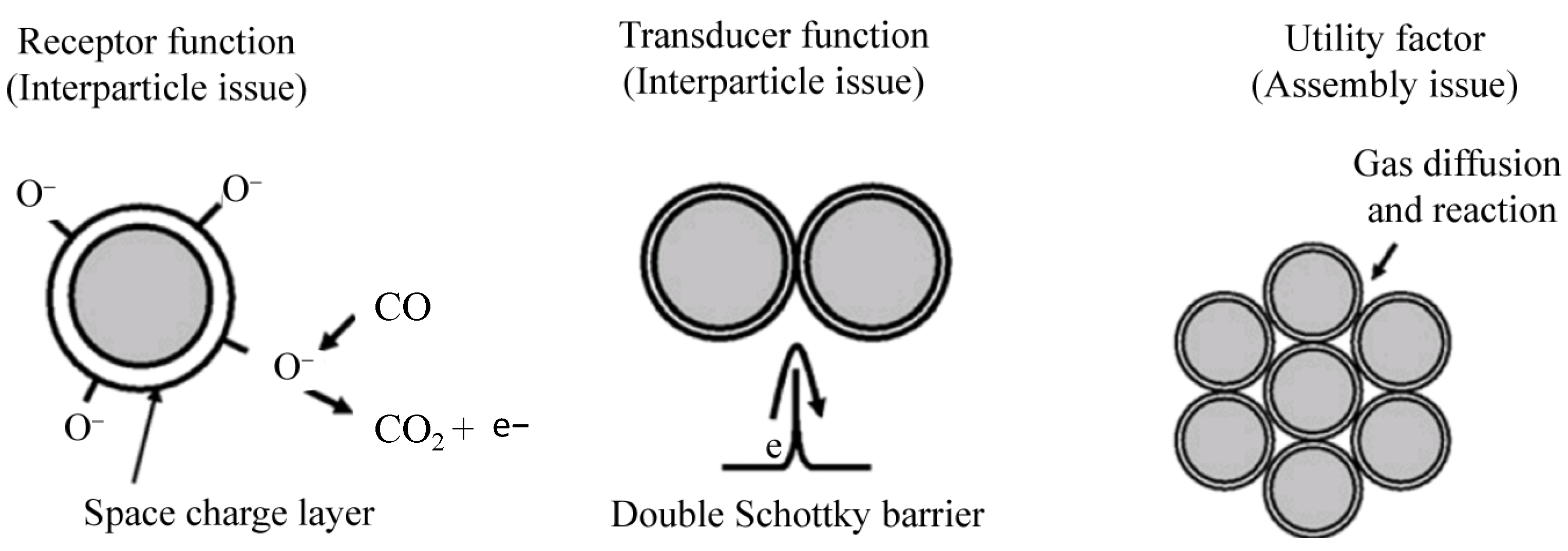
Figure 1. Basic sensing model of MOS CO gas sensors. Revised with permission from Ref. [31].
Chemosensors 12 00055 g001
Figure 2. Illustration of in situ spectroscopy for studying the working mechanisms of a MOS for CO sensing [33].
Chemosensors 12 00055 g002
Figure 3. Illustration of various SnO2 nanostructures using the hydrothermal method [41].
Chemosensors 12 00055 g003
Figure 4. (a) Dynamic response curve of the SnO2:Pt PNS sensor to CO at room temperature; thickness of PNS is 0.5 mm. (b) Comparison of response of different SnO2:Pt PNS sensors to 100 ppm CO at room temperature; thickness of PNS is 0.5 mm, 1.0 mm, or 2.0 mm. (ce) Schematic diagram of the mechanism of the spillover effect that leads to enhancement of the sensing performance of SnO2 PNS sensors by Pt-loading: (c) conduction model of SnO2 PNS sensor in air; (d) conduction model of SnO2: Pt PNS sensor in air; (e) conduction model of SnO2: Pt PNS sensor in CO [44].
Chemosensors 12 00055 g004
Figure 5. (a) XRD patterns for Pd-SnO2 of 10 wt% Pd heat-treated at 800 °C and 1000 °C, separately. (be) XPS spectra of Pd3d5/2, 3d3/2 electrons in different samples: (b) 5 wt% Pd-doped SnO2 heat-treated at 1000 °C, (c) pure Pd nanoparticles without SnO2, (d) a dried nanomixture of both Pd and SnO2 containing 2 wt% Pd in powder form, (e) 1 wt% Pd-doped SnO2 heat-treated at 1000 °C. (fh) Three configurations for Pd-SnO2 composites and their interactions with CO molecules at room temperature: (f) PdO-SnO2 composites with no interactions with CO molecules; (g) Pd-SnO2 composites, whose Pd nanoparticles are in Pd2+ states on the surface, with no interactions with CO; (h) Pd-SnO2 composites, whose Pd nanoparticles are in the states of Pd2+ and Pd4+ on the surface; in this case, CO molecules are chemisorbed on both Pd4+ sites of Pd nanoparticles and surface of SnO2 [47].
Chemosensors 12 00055 g005
Figure 6. DRIFT spectra of SnO2 and its composites exposed to CO at 25 °C: (a) nanocrystalline SnO2/PdOx, SnO2, SnO2/RuOy, SnO2/PtO, and SnO2/Au powders exposed to 100 ppm of CO for 1 h. (b) SnO2/PdOx powder exposed to 100 ppm of CO and air in sequence for different periods [49].
Chemosensors 12 00055 g006
Figure 7. (a) The procedure of the fabrication process of CO sensors based on tin dioxide–graphene composites; (b) response of different tin dioxide samples to 40 ppm CO, different colours corresponds to different samples; (c) response and recovery curves of tin dioxide–graphene 0.5 wt% to 40 ppm CO at 25 °C [50].
Chemosensors 12 00055 g007
Figure 8. Energy band diagram of ZnO and its composites: (a,b) before exposure to oxygen: (a) ZnO nanostars, (b) ZnO nanostars with Au nanoparticles; (c) ZnO nanostars with Au nanoparticles exposed to oxygen; (d) ZnO nanostars with Au nanoparticles with surface-adsorbed oxygen species exposed to CO [64].
Chemosensors 12 00055 g008
Figure 9. Characterization results of the Pd1-TiO2 sample: (ac) SEM, TEM, and high-resolution TEM images; (d) deconvoluted narrow-scan Pd 3d XPS spectrum; (e) ac-HAADF-STEM image, where the red circle shows that Pd species are in the form of isolated single stoms; and (f) STEM-EDS elemental mapping images [79].
Chemosensors 12 00055 g009
Figure 10. (a) Proposed CO sensing mechanism of Pd1-TiO2. (b) Relative percentages of O species from O 1 s spectra of TiO2 nanoflowers, Pd NPs-TiO2, and Pd1-TiO2. (c) DRIFT spectra of TiO2 nanoflowers, Pd NPs-TiO2, and Pd1-TiO2. (d) Calculated adsorption energies of selected gases on Pd1-TiO2 [79].
Chemosensors 12 00055 g010
Figure 11. Illustration of the working mechanism of MoS2/SnO2 response to CO under UV light. (a) Changes in surface element during the sensing step with or without UV; (b) band diagram between p-MoS2 and n-SnO2 [94].
Chemosensors 12 00055 g011
Table 1. Reaction between CO and the oxygen species adsorbed on the surface of MOS at elevated temperatures.
Possible ReactionTemperature Range
2CO + O2 (ads) → 2CO2 + eT < 150 °C(1)
CO + O (ads) → CO2 + e150 °C ≤ T ≤ 300 °C(2)
CO + O2− (ads) → CO2 + 2eT > 300 °C(3)
Table 3. Comparison of performance of ZnO composites for room-temperature CO sensing.
Sensing MaterialCO Concentration (ppm)ResponseResponse
Time (s)
Recovery
Time (s)
Reference
ZnO nanocomb2507.2220050[61]
ZnO thin films501.10~180-[65]
ZnO nanoneedles3751.5118638[62]
Au-ZnO nanowires100~5--[63]
Au-ZnO nanostars50055.34140[64]
SnSe2-ZnO polyhedron2001.171913[66]
Pt-ZnO-CuO10002.648181[67]
Table 4. Comparison of performance of composites of other materials for room-temperature CO sensing.
Sensing MaterialCO Concentration (ppm)ResponseResponse
Time (s)
Recovery
Time (s)
Reference
Pt-Co3O4-In2O354NANA[76]
Dumbbell CoOOH nanostructures50NA~20~18[77]
Fe-TiO2100~4.84325[78]
Atomically dispersed Pd-TiO2100125.492870[79]
Self-doped Ti3+-porous TiO25000~2~10~30[80]
Mg-TiO2 thin films1208.40(CO+Ar)6230[81]
CuO-TiO2 heterojunction1~2.2NANA[82]
CuO (111) nanosheets10039.610072.4[83]
RuOx (OH)y250~2NANA[84]
Table 5. Comparison of performance of composites of MoS2 for room-temperature CO sensing.
Sensing MaterialCO Concentration (ppm)ResponseResponse
Time (s)
Recovery
Time (s)
Reference
Ag-ZnO-MoS21001.054540[92]
Co-In2O3-MoS2 101.08~39~18[93]
SnO2-MoS2 UV404.974336[94]
Disclaimer/Publisher’s Note: The statements, opinions and data contained in all publications are solely those of the individual author(s) and contributor(s) and not of MDPI and/or the editor(s). MDPI and/or the editor(s) disclaim responsibility for any injury to people or property resulting from any ideas, methods, instructions or products referred to in the content.