Review

2,1,3-Benzothiadiazoles Are Versatile Fluorophore Building Blocks for the Design of Analyte-Sensing Optical Devices

by
Gleiston Gonçalves Dias
*,
Francielly Thaís Souto
and
Vanderlei Gageiro Machado
*
Departamento de Química, Universidade Federal de Santa Catarina—UFSC, Florianópolis 88040-900, Brazil
*
Authors to whom correspondence should be addressed.
Chemosensors 2024, 12(8), 156; https://doi.org/10.3390/chemosensors12080156
Submission received: 25 June 2024 / Revised: 21 July 2024 / Accepted: 1 August 2024 / Published: 7 August 2024
(This article belongs to the Special Issue The Recent Progress and Applications of Optical Chemical Sensors)

Abstract

:
BTDs (2,1,3-benzothiadiazoles) are fluorescent heterocycles widely used in different applications, including biomarkers, sensing optical devices, OLEDs, organic transistors, and solar cells. This review mainly focuses on the current progress in the design of compounds derived from the BTD core, aiming for their use as chromogenic and/or fluorogenic devices for detecting anionic, cationic, and neutral analytes. Reactions and synthetic strategies that show the synthetic versatility of BTDs are initially presented, to provide a better understanding regarding the assembly of optical detection systems. The photophysical mechanisms of the detection are also described. A discussion is also presented on the target analytes for which the optical detection devices based on BTD were planned. The examples discussed here will offer the sensors community perspectives for developing new optical detection devices based on BTD for different types of analytes of importance for the most diverse areas of knowledge.

1. Introduction

The development of chemical assays for identifying and quantifying analytes in different matrices represents an ongoing area of research in chemistry. Numerous efforts have been devoted to discovering more efficient strategies for investigating the chemical composition of samples of broad interest. These strategies typically employ simple methods capable of selectively identifying analytes in a rapid response, regardless of the presence of possible interferents.
In many cases, even low concentrations (ppm or ppb) of certain chemical species can produce several damages. For instance, potentially toxic metals and pesticides present in drinking water, food, and the air may pose risks to human health, contributing to various diseases. In such scenarios, the chemical analysis must accurately indicate the presence and quantity of these chemical species, necessitating methods of analysis.
Some of these instrumental methods include atomic absorption spectrometry [1], inductively coupled plasma mass spectrometry [2,3], ion chromatography [4], ion-selective electrodes [5,6,7], ultraviolet-visible (UV-vis) spectrophotometry [8], surface-enhanced Raman scattering [9,10,11], nuclear magnetic resonance (NMR) [12,13], electrochemical techniques [14,15], and others [16,17]. Despite their widespread uses and efficiency, these approaches often require laborious experimental procedures and show drawbacks, such as high-cost analysis and sophisticated instrumentation.
In the context of analyzing analytes at low concentrations, there is a critical need for analytical methodologies that offer low quantification limits (below parts per million). To address this need, optical strategies have been increasingly employed to develop such methodologies. Consequently, optical devices capable of detecting analytes in minute quantities have been the subject of extensive investigation. Ideally, these systems should adhere to the principles outlined by the World Health Organization’s conception of point-of-care diagnostic devices, being ASSURED (Affordable, Sensitive, Specific, User-friendly, Rapid/Robust, Equipment-free, and Deliverable) [18,19,20].
While many traditional assays serve as inspiration for designing analytical devices capable of detecting single and multiple analytes [21,22], the investigation of novel strategies for detecting diverse chemical species has emerged as a significant challenge in recent years.
The advancements in supramolecular chemistry and photochemistry over recent decades, coupled with the development of new materials and nanotechnological processes, have spurred a rapid expansion in the design of molecular and supramolecular systems for detecting single and multi-analytes across various applications. These endeavors have contributed to the emergence of a vast multidisciplinary research domain named by Anslyn as Supramolecular Analytical Chemistry [23]. Examples of optical detection devices encompass chemosensors and chemodosimeters (Scheme 1), among other designated classes.
Chemosensors (Scheme 1a) are optical devices that interact with an analyte through reversible processes [22,24]. They consist of two integrated parts: recognition and signaling units. The recognition unit is responsible for identifying the desired analyte within a multi-analyte array. Designing selective receptor sites for a specific analyte is crucial to enhance assay specificity, thereby reducing the likelihood of false positives.
The signaling unit communicates with the recognition unit through various strategies—covalent or non-covalent—and is responsible for signaling the presence of the analyte. In optical devices, signaling units typically consist of chromophore or fluorophore groups. Signal transduction occurs when the analyte interacts with the recognition site, resulting in a change in the medium’s color or the system’s fluorescence capability, in the case of chromogenic or fluorogenic chemosensors, respectively.
The simplest examples of optical chemosensors employ acid–base strategies involving hydrogen bonding via proton donor groups (NH, OH, and SH) within the chemosensor [25,26]. The presence of the basic analyte leads to its interaction with the recognition unit in the chemosensor, yielding changes in color and/or fluorescence emission [27,28].
On the contrary, chemodosimeters exhibit irreversible behavior under kinetic control. These optical molecular devices are based on various strategies but share a common systems principle: irreversible chemical reactions with a specific analyte produce product species with an optical behavior vastly distinct from the reactants (Scheme 1b). These strategies are determined by the nature of the interaction between the analyte and the device, resulting in chemodosimeters, where:
(I) The analyte covalently binds to the chromogenic or fluorogenic chemodosimeter, causing a variation in the absorption or the fluorescence emission band.
(II) The analyte interacts with the chemodosimeter and catalyzes a chemical reaction that leads to an optical response.
(III) The analyte reacts with the chemodosimeter, releasing a chromogenic and/or fluorogenic leaving group.
As simple examples, organic functional groups, associated with chromophores/fluorophores, are potential electrophilic centers in the design of efficient chemodosimeters. These organic groups can react with nucleophilic species, such as cyanide (CN) and fluoride (F) [29,30,31]. These chemical transformations result in new covalent bonds, which induce changes in the color and/or fluorescence emission of the system. This optical modification provides valuable information regarding the detection of chemical species [32].
Various chromophores and fluorophores have been used as optical signaling units, coupled with recognition moieties capable of interacting with an analyte. Numerous chromophore and fluorophore units have been described as, for instance (Scheme 2), acridine (1) [32,33,34,35,36,37,38], anthracene (2) [39,40,41,42,43], BODIPy (3) [44,45,46,47,48,49], coumarin (4) [50,51,52,53], fluorescein (5) [54,55,56,57,58,59,60,61], benzimidazole (6) [62,63,64,65,66,67,68], naphthalene (7), [69,70,71,72,73], pyrazine (8), [74,75,76,77], pyrene (9) [26,30,78,79,80,81], pyridinium-N-phenolate betaine (10) [82,83,84,85], stilbene (11) [86,87,88], Brooker’s merocyanine (12) [89,90,91], quinoline (13) [92,93,94,95,96], rhodamine (14) [97,98,99,100,101], and quinoxaline (15) [102,103,104].
The 2,1,3-benzothiadiazole (16, BTD, Scheme 3) is a fluorescent core, which has been used to design compounds with optical properties. These compounds hold potential for application as devices for detecting analytes.
The interest in developing BTD-based systems stems from their notable features, including:
1. The photostability of BTDs makes them suitable for applications requiring prolonged use, especially when exposed to light. Examples of such applications include their utilization in solar cells and bioimaging studies [105,106].
2. The electronic deficiency of the BTD nucleus is particularly interesting in developing a fluorophore because it allows a molecular design featuring electronic donor–acceptor groups attached to BTDs. Molecules with this electronic architecture tend to exhibit enhanced stability. Furthermore, this design may be associated with the optical response upon interaction with an analyte, as illustrated in various examples within this review [78,107].
3. Numerous studies have demonstrated that BTDs exhibit remarkable thermal stability, a crucial characteristic in developing compounds for electronic and solar cell applications [108,109,110].
4. Typically, BTDs exhibit large Stokes shifts, which prevent undesired background interference and prevent the detection of the excitation wavelength in a spectroscopic measurement by using appropriate filters. This feature is essential in developing probes for bioimaging assays [111,112,113].
5. The expansion of C-C and C-N coupling reactions, including Sonogashira, Suzuki, Heck, Hartwig–Buchwald, and Stille reactions [66,114,115,116,117,118], associated with the ready availability of key synthetic precursors (arylamines, terminal alkenes, alkynes, and aryl boronic acids, among others) [119,120,121,122,123,124], has allowed access to a wide range of BTD derivatives.
These aspects have propelled the use of BTD compounds across a broad spectrum of applications (Scheme 3). The most prevalent uses for this heterocyclic include the development of polymers [125,126,127,128,129,130,131], biomarkers [132,133,134,135,136,137,138,139,140,141,142,143,144,145], organic light-emitting diodes (OLEDs) [146,147,148,149,150,151,152,153], liquid crystals [154,155,156,157], organic transistors [80,158,159,160,161,162], solar cells [163,164,165,166,167,168,169], phototheranostic systems [170], enzyme sensors [105,106,171,172,173], and optical devices for analytical purposes, among others [143,173,174,175,176,177,178,179]. Despite the extensive research on BTDs in recent years, no review article specifically addressing these applications has been published.
Therefore, this review is dedicated to exploring this class of compounds as chromogenic and fluorogenic devices for detecting ionic and neutral analytes. Emphasis will be given to the synthetic strategies employed in obtaining these compounds.
While many BTD-based polymers are reported in the literature for similar purposes [64,180,181,182,183,184,185], they are not discussed herein, as they represent a distinct category. Additionally, the discussion focuses on small molecules as optical devices for analyte sensing. Before examining the examples of BTD-based analyte sensing strategies outlined in the literature, we will initially delineate the primary mechanisms underlying the optical response of the chemosensors, identify the target analytes for which the optical detection devices featured in this review were designed, and elucidate the principal strategies in the synthesis and modification of BTD derivatives. A glossary of terms and abbreviations used in this review is provided in the Abbreviations Section.

2. Fluorescence Response Mechanisms in Analyte Sensing Optical Devices

Various photophysical mechanisms involving fluorophores can be employed in optical devices for analytes’ detection and some of them are represented in a generalized form in Scheme 4 [186,187]. Photoinduced electron transfer (PET) is a particularly significant mechanism for modulating the output optical signal through fluorescence (Scheme 4a). This photophysical process involves an electron transfer to (or from) an electronically excited state of a molecule. Put simply, an orbital within a segment of the molecule (or from an external molecule) may possess energy levels between those of the Highest Occupied Molecular Orbital (HOMO) and the Lowest Unoccupied Molecular Orbital (LUMO) of the fluorophore [188,189,190].
Some mechanisms can be considered as classes of PET [187], such as:
(a) Internal (or intramolecular) charge transfer (ICT), where charge transfer (CT) occurs from an electron-rich donor moiety to an electron-poor acceptor moiety, both residing within the same molecule [191].
(b) Ligand−metal charge transfer (LMCT), primarily observed in inorganic complexes, where if the molecular orbitals of the ligand are occupied, CT can occur from the molecular orbitals of the ligand to the vacant or partially filled metallic d orbitals [192].
(c) Twisted internal (or intramolecular) charge transfer (TICT): a donor–acceptor complex is formed wherein an electron can move directly from the donor to the acceptor group [193].
In the fluorescence resonance energy transfer (FRET, Scheme 4b), the donor and acceptor groups are considered as two interacting dipoles, wherein only the absorption of photons emitted from the donor to the acceptor occurs (unlike PET, no electron transfer occurs) [194].
In the electron exchange (Scheme 4c), an electron is transferred from the LUMO of the excited donor to the LUMO of the acceptor group. Simultaneously, an electron is transferred from the LUMO of the acceptor to that of the donor [195].
Another important mechanism is excited-state intramolecular proton transfer (ESIPT). This process occurs in molecules containing proton donor and acceptor fragments, facilitating the exchange of the hydrogen atom when excited by radiation, causing a change in their photophysical properties. It is commonly associated with isomer formation, such as the tautomeric equilibrium shown in Scheme 4d [196].
Mechanisms involving aggregation are also considered in the design of chemosensors. One such example is aggregation-caused quenching (ACQ), in which the aggregation of fluorescent molecular structures causes their emission suppression [197]. This phenomenon contrasts with aggregation-induced emission (AIE), where weakly fluorescent chromogens are induced to emit by the formation of aggregates [198,199,200]. These effects may be associated with other phenomena that limit non-radiative relaxation through mechanical and thermal pathways [201], including restriction of intramolecular rotation (RIR), J-aggregate formation (JAF), and even the phenomena already discussed: TICT and ESIPT [202].
Two-photon (TP) photophysical processes represent yet another strategy for optical response mechanisms in analyte sensing. The TP fluorescence phenomenon occurs when a fluorophore emits photons nanoseconds after the simultaneous absorption of two less energetic photons with a femtosecond interval [79,203,204].

3. Target Analytes for Detection Using BTD-Based Optical Devices

A wide range of analytes are targets for detection, and various strategies can be devised for their identification. However, our focus will be narrowed to the analytes specifically targeted by sensing BTD-based optical systems. Therefore, the emphasis will be placed on cationic (Hg2+, Al3+, Cu2+, Ni2+, Co2+, Cr3+, and Na+), anionic (F and CN), and neutral compounds (DNA, SO2, amines, cysteine, homocysteine, hydrazine, nitroaromatic compounds, oxalyl chloride, and ethanol).

3.1. Cationic Species

The identification and quantification of cations, by optical devices, represent an important field of research. One of the primary motivations for this is that several metals cause various environmental problems and pose risks to human health. It is noteworthy to mention that while the term “heavy metal” continues to be used in the literature, some authors have recommended its discontinuation [205,206]. The International Union of Pure and Applied Chemistry (IUPAC) has described the term “heavy metal” as imprecise, meaningless, and misleading [207]. Therefore, herein, we refrain from using the term ‘heavy metal’.
Mercury stands out as one of the most extensively investigated cationic analytes targeted by BTD. This toxic metal ranks among the primary agents posing significant threats to public health [208]. It occurs naturally in elemental form, as well as in inorganic (Hg2+ salts) and organic (alkyl mercuric compounds) forms, and is naturally found in the earth’s crust [209]. However, natural processes, such as erosion and volcanic eruptions [210], alongside anthropogenic activities [211,212,213], contribute to environmental mercury contamination. In humans, even in low doses, mercury can severely affect the nervous system in children and adults, causing damage to the renal, cardiorespiratory, immune, and reproductive systems, and impairing motor function due to reduced muscle strength [214]. Mercury’s extreme toxicity is attributed to its strong affinity for thiol groups in proteins and enzymes, leading to the accumulation of derived species in biological systems [215]. Hg2+ is categorized as a soft acid, which readily binds to soft donor atoms, such as thiols. Strategies based on this thiophilic affinity are generally effective in detecting Hg2+, with resulting chemosensors exhibiting selectivity toward other potentially toxic metals [216].
Aluminum, one of the most plentiful metals in the earth’s crust [217], has an important role in human life, such as in utensils, electronic and electrical components of various appliances [218], vaccines [219], wastewater treatment [62], and in the aerospace industry [220]. However, despite its widespread use, aluminum is recognized as a neurotoxin implicated in disorders such as Alzheimer’s disease [62], osteomalacia [221], and breast cancer [222].
Cu2+, Cr3+, and Ni2+ are trace elements, which play crucial roles in various biological processes [223]. However, their excessive accumulation in the human body can be toxic [224,225,226,227]. For instance, while Cu2+ is involved in essential redox activities, its overabundance may lead to amyloidal precipitation, Wilson’s disease, prion disease, and Parkinson’s disease [228,229]. Cr3+ is important in the metabolism of carbohydrates, proteins, nucleic acids, and lipids [230]. Conversely, Cr6+ exists as a toxic form capable of causing allergic dermatitis and carcinogenic effects [231]. Ni2+ is important for biosynthesis and metabolism [232], but in high levels may trigger conditions such as dermatitis, asthma, pneumonitis, central nervous system disorders, and cancer [233].
Sodium ion (Na+) is a vital cation found in biological organisms, playing a pivotal role in maintaining overall health. Its significance extends to regulating acid–based homeostasis and the body fluid volume, participating in numerous signal transduction pathways, and serving as an electrolyte for physiological functions [234]. However, excessive levels of Na+ in the human body are linked to conditions such as retention and hypertension [235,236].

3.2. Anionic Species

The advancement of optical molecular devices for determining anionic analytes advanced later comparable studies involving cations. This delay can be attributed to the more complex topology of some anions due to a wide variety of geometries, charge distribution, and size. Additionally, some anionic species are pH-dependent, further complicating the design of efficient molecular systems [237,238]. Nonetheless, the development of such devices is crucial considering the extensive array of anions and their indispensable roles in various chemical, biological, and environmental processes [239].
Cyanides encompass chemical species with varying degrees of chemical complexity, all featuring CN portion. CN is frequently used in organic synthesis, polymers, metallurgy, pesticides, and extraction of gold, making it one of the most studied anions due to its adverse effects on living organisms [240]. It occurs in natural sources, in foods such as cassava, cabbage, spinach, mustard, cherries, and almonds [241]. This anion also occurs in tobacco, associated with respiratory issues [242]. CN exposure can cause cardiovascular, respiratory, and central nervous system diseases [243]. Moreover, its toxicity can become chronic depending on industrial, environmental, and dietary factors related to the exposed individual [243]. The high toxicity and prevalence of cyanides stem from their ability to form complexes with Fe3+ present in the active sites of certain enzymes, such as cytochrome oxidase [244]. The binding of CN to the enzyme site disrupts the electron transport chain, impairing tissue’s ability to utilize oxygen, resulting in cell dysfunction and death. According to the World Health Organization (WHO), the maximum acceptable concentration level of CN in drinking water is 0.2 ppm [245].
Another noteworthy anion with significant implications for healthcare is F, employed in the clinical treatment of osteoporosis and prophylaxis of dental caries. F is widely utilized as an additive in toothpaste, mouthwashes, water, and in some countries, such as China, Bulgaria, and Russia, in milk to prevent tooth decay and enamel demineralization [246,247]. However, excessive intake of F can cause dental or skeletal fluorosis, kidney stones, and even death [248]. The World Health Organization (WHO) has established a maximum permissible concentration of F in drinking water at 1.5 mg L−1 [249].

3.3. Neutral Analytes

The recognition of neutral analytes necessitates a meticulous examination of the molecular structure of the analyte, relying on intermolecular interactions, such as hydrogen bonds, van der Waals interactions, or hydrophobic effects. The practical use of these features for constructing optical devices for detecting such analytes remains challenging, primarily due to the limited availability of receptors and molecules possessing optical properties capable of recognizing and detecting neutral species.
Thiols, commonly referred to as mercaptans, due to their ability to react with mercury, are important molecules in the environment and biological processes [250]. Cysteine (Cys), homocysteine (HCy), glutathione (GSH), and hydrogen sulfide (H2S) stand out as pivotal antioxidants that protect cells from oxidative injury. Moreover, these compounds are also involved in metabolic regulation, signal transduction, and regulation of gene expression [251,252,253,254]. Considering the critical biological functions associated with thiols, the development of optical chemosensors for their detection has emerged as a burgeoning area of research [255].
α-Ketoglutarate (α-KG), in its acid form as α-ketoglutaric acid (α-KA), is a metabolite of the citric acid cycle (Krebs cycle), important in energy metabolism [256]. This carboxylic acid can be converted into 2-hydroxyglutarate (2-HG), which has been implicated in various diseases [257,258], including acute myeloid leukemia (AML) [259]. Here, 2-HG has been considered a biomarker for diagnosing and classifying tumor-derived IDH1 and IDH2 mutations [256]. Consequently, this metabolite has been the focus of efforts to develop sensors for diagnostic purposes [81,260,261].
Hydrazine has several uses, but it is highly toxic and a potential environmental pollutant [262]. The damage of this compound to the human body is due to the trapping of important biological compounds via the formation of hydrazone [263].
Ethanol is one of the most widely used chemicals, finding applications in various industrial processes and alcoholic beverages consumed worldwide [264]. Moreover, considering its utility as a fuel and antiseptic, evaluating ethanol levels in different mixtures holds significant importance [265].
Oxalyl chloride and phosgene find extensive use in industrial chemical processes across multiple applications [266,267,268]. However, these chemicals are notorious for their toxic properties [269,270]. According to The National Institute for Occupational Safety and Health (NIOSH), exposure to a phosgene concentration of 17 ppm for just 30 min is enough to cause death [271]. Phosgene was also employed as a feared chemical weapon during World War I [272]. Oxalyl chloride, which is highly toxic and irritating to human skin and eyes, can induce respiratory distress and vision impairment, potentially leading to fatality [273].
Another category of neutral analytes that warrants public safety concern encompasses nitroaromatic compounds. The rapid detection of trace amounts of nitroaromatic is imperative due to their potential application in the preparation of explosives, underscoring the importance of homeland security [274,275]. Additionally, explosives exhibit toxic, mutagenic, and carcinogenic effects in humans and animals [276].
Chemosensors capable of detecting amines are paramount due to public concern in health, food safety, and environmental monitoring [277]. For instance, N,N,N′,N′-tetramethylethylenediamine (TMEDA) is a toxic amine that can induce eye, skin, and respiratory irritations in humans. [278]. Biogenic amines [279], such as cadaverine, produced from lysine degradation, can inhibit the action of enzymes, compromising the metabolism of histamine, whose excess causes histamine toxicity [280].
It is pertinent to discuss the primary synthetic strategies employed for obtaining and modifying this class of compounds before delving into the examples of BTD sensors for detecting these analytes. These strategies play a pivotal role in the design of optical detection devices.

4. Synthesis of BTD-Based Compounds

The primary synthetic strategy to obtain BTDs involves the reaction of ortho-phenylenediamine derivatives with SOCl2 (Scheme 5). Therefore, various ortho-phenylenediamine derivatives have been planned for the synthesis of BTDs. For instance, this approach has been applied to the synthesis of BTDs bearing 5,6-difluorine (16) [65,281,282] and 5,6-dichlorine (17) [283] substituents, which serve as precursors for synthesizing compounds with semiconducting properties [284]. Further, 5,6-dicyano-BTD (18) has been employed for preparing polymer semiconductors using a similar approach [285]. Riant et al. synthesized 6-esther-BTD (19) as the precursor for compounds with potential cytotoxic activity [286]. Milata and Vaculka described the BTD (20) as the substrate for the Gould–Jacobs reaction [287]. Other substituted aromatic systems, such as naphthalene (21) [288], phenanthrene (22) [289], and pyrene (23) [290] bearing diamine moieties, have been reported in strategies to obtain highly conjugated BTDs.
Substituents on the ortho-phenylenediamine core enable sequential modifications upon BTD synthesis. Such transformations generally involve replacing halogens with other groups to enhance the conjugation of the system. For example, Lee et al. employed aromatic substitution to replace fluorine for alkyloxy groups (Scheme 6a) [291,292]. Although the authors have described that 16 was purchased, it can also be synthesized, as previously described. The monomers 25 achieved were used to prepare polymers with alkyl linear and branched chains for optoelectronic and photovoltaic applications.
Another example includes the reaction of precursor 26 with SOCl2 to provide 27, which can be further modified via Suzuki [293,294,295] and Sonogashira [296] reactions to insert aryl (28) and aryl acetylene (29) groups (Scheme 6b). Similarly, butterfly-shaped systems, such as 33, can be achieved from BTD 31 via the Sonogashira reaction, as described by Wang et al. (Scheme 6c) [297]. Another feasible modification of 31 was described by Han et al. via a Suzuki reaction followed by an oxidative cyclization using FeCl3 to obtain 36 for photovoltaic application (Scheme 6d) [298]. A similar product, 39, was obtained from the diamine compound 38 using the well-established BTD synthesis (Scheme 6e), starting from compound 35 [299]. Despite the consistency in this synthetic route’s modifications, this class of compounds has been described as part of an important strategy for synthesizing polymers based on thiophene-fused BTD [300,301].
The compound 4,5-dimethyl-ortho-phenylenediamine (40) serves as a valuable substrate for preparing BTDs, as the modification on methyl enables access to various derivatives (Scheme 7a). One of the early examples of such transformations was reported by Neidlein and Knecht in 1987 [302,303]. Compounds 42 and 43 were prepared via radical bromination of methyl groups at 41 (derived from 40), employing a stoichiometric amount of N-bromosuccinimide (NBS). Subsequently, these products were treated with potassium carbonate in water to yield 44 and 45 [302]. In another study, the authors reported the oxidation of a methyl group of 41 to a formyl using SeO2 resulting in a 30% yield of 46 [303].
In 2018, Iyer et al. modified BTD 41 to synthesize D–π–A-conjugated copolymer 48 for application in bulk-heterojunction solar cells (Scheme 7b) [304]. One year later, the same group reported similar polymers using non-conjugated ester groups as side chains for the same application [305]. A similar approach was also adopted by Karpagam et al. for the synthesis of polymers for hole-transporting materials in perovskite solar cells [306].
Vanelle et al. described the synthesis of 4-methyl-BTD (51) from 50, as a precursor of different BTD derivatives (Scheme 8) [307]. The radical bromination of the methyl group at 51 yielded intermediates 52 and 53, used to prepare the 4-formyl-BTD (54). The reaction of 52 with LiCMe2NO2 or between 53 and HCOOH led to the formation of 54, bearing a formyl group at position 4. These examples, involving classical modification of aromatic methyl, hold strategic significance in affording a BTD core with a methyl group, thereby offering opportunities for further chemical transformations.

4.1. BTD-Br2 and Their Modifications

One of the most significant modifications of BTDs was reported in 1970 by Pilgram et al., involving the bromination of positions 4 and 7 (Scheme 9a) [308]. According to the authors, the dropwise addition of bromine to a BTD in 47% hydrobromic acid led to BTD-Br2 (55) in nearly quantitative yield. The absence of bromination at 5 and 6 positions of BTD was attributed to an energetic addition of Br+ at position 4 (or 7), via an σ-bond intermediate, over position 5 (or 6). Compound BTD-Br2 (55) is a strategic substrate in BTD chemistry, used in various synthetic routes for further modification (Scheme 9b). Generally, four approaches could be addressed to modify BTD-Br2:
(I) Extrusion of sulfur via a deprotection reaction to afford 3,6-dibromo-diamino derivatives (63), used for other reactions, as mentioned previously.
(II) Replacement of bromine to other molecular groups, such as aryl ethenyls (57), aryl (58), aryl ethynyl (59), and anilines (60), via Heck, Sonogashira, Suzuki (or Stille), and Buchwald reactions, respectively.
(III) Substitution of one bromine atom for a nitro group to afford 61.
(IV) Nitration of the remaining free positions 5 and 6, yielding derivative 62 for further modifications.

4.1.1. Extrusion

In contrast to approaches discussed so far, BTDs can undergo extrusion reactions (reductive N–S bond cleavage) to yield ortho-phenylenediamines (Scheme 10a). This reaction can be facilitated by various agents, such as SmI2 [309], LiAlH4 [310], Zn [311], Hg2Cl2/Al, or Mg [312]. One of the most commonly used methodologies to promote such a reaction involves NaBH4 under CoCl2.6H2O catalysis [313]. In a broader context, the synthesis of BTDs can be viewed as a protective reaction of the diamino group to reduce the electronic density of the system, thereby preventing undesired reactions. Subsequently, the extrusion can be considered a deprotection step to regenerate the diamine group with the modified aromatic system (Scheme 10b). A classic example of this strategy involves the synthesis of BTD (15, protection step) and bromination (modification) to achieve BTD-Br2 (55), followed by extrusion (deprotection) to afford 3,6-dibromo-ortho-phenylenediamine (63; Scheme 10c). Direct bromination of ortho-phenylenediamine (64) is not employed due to the formation of undesired products from highly reactive 63. Subsequently, substrate 63 can provide several nitrogenous heterocycles containing two bromine atoms in the ortho position.
Furthermore, bromine atoms can be substituted by other groups through classical coupling reactions to prepare more conjugated heterocycles, such as phenazines (65) [314,315], imidazoles (66) [316,317], triazoles (67) [318], and others [319] (Scheme 11a). Another feasible and similar approach involves replacing the bromine atoms in BTD-Br2 with other groups, followed by extrusion to form ortho-modified diamines, which allow access to other heterocycles. For instance, Dupont et al. have noted that extending the π-system at the 5 and 8 positions of the quinoxalines via classical Suzuki and Sonogashira reactions is challenging (Scheme 11b) [320]. However, such modifications can be achieved by replacing the bromine of BTD-Br2 with an aryl group, followed by sulfur extrusion and cyclization with a 1,2-dicarbonyl compound. Examples involving modifications of BTD-Br2 derivatives via classical cross-coupling reactions are discussed in the following section.

4.1.2. Replacement of Bromine of BTD-Br2

The replacement of the bromine atoms in BTD-Br2 (55) by other molecular groups represents one of the primary strategies in preparing BTDs. Suzuki, Heck, Sonogashira, and Buchwald–Hartwig coupling reactions are commonly employed for the structural diversification of this class of compounds, as illustrated in Scheme 9. Since the BTD nucleus itself is electronically deactivated, these modifications enable modulation of the optical properties of these compounds. This is achieved by altering the molecular electronic density through the insertion of donor and/or acceptor units, as well as by extending the conjugated system. Herein, we will demonstrate that such synthetic approaches are widely used to incorporate molecular groups capable of recognizing anionic and neutral species.
The replacement of the halogen atoms for alkynyl groups can be achieved through Sonogashira reactions, facilitating the extension of the π-system via an alkyne bridge. Similarly, aryl groups can be attached to BTD via Heck reactions with an alkenyl group serving as a bridge between the BTD core and the aryl group. Buchwald–Hartwig amination of BTD-Br2, although less investigated than other cross-coupling protocols, has been employed as a protocol to introduce an aniline group at the BTD core and, with the NH group, works as a bridge.
On the other hand, arylation of BTD-Br2 without a connector (amine, alkenyl, or alkene) is feasible via Suzuki or Stille protocols. Furthermore, protocols involving direct arylation on BTD, without the bromine at 53, have been developed via the C–H activation procedure. However, those protocols are still under investigation for BTDs [321,322,323,324].
The replacement of BTD bromine atoms by other groups, via palladium-catalyzed coupling reactions, is one of the most common synthetic strategies for obtaining BTD derivatives. Examples of the use of this strategy are illustrated in Scheme 12 [112,126,139,141,143,146,157,165,166], and several other examples are cited throughout this review. It is worth noting that this synthetic approach is frequently employed in the design of BTD-based chemosensors, as will be discussed later.

4.1.3. Synthesis and Modification of 4-Bromo-7-nitrobenzo[c][1,2,5]thiadiazole (61)

In 1971, Pilgram et al. reported that refluxing BTD-Br2 (55) in 69% HNO3 resulted in the change of one bromine atom to a nitro group, yielding 61 in 40% yield (Scheme 13a) [325]. The molecular architecture of 61 is interesting because substituting bromine for other molecular groups offers various possible synthetic possibilities. Additionally, the electron-withdrawing effect of the nitro group, in combination with substituting bromine for an electron-donating group, such as an amine, reinforces the donor–acceptor (D–A) molecular system. This system may lead to the formation of red emissive compounds.
For instance, in 2011, Li et al. conducted a Sonogashira reaction on 61 to provide the D-A molecular system 79, followed by a reaction with tetracyanoethylene (TCNE, 80) or 7,7,8,8-tetracyanoquinodimethane (TCNQ, 81) to afford 82 and 83, respectively (Scheme 13b) [326]. In 2013, Li et al. described the insertion of carbazole as the donating group at 61, using Suzuki and Sonogashira protocols, to obtain products 86 and 87 (Scheme 13c) [327]. These compounds exhibited large, third-order, nonlinear absorption effects and showed AIE properties. Jones et al. described the substitution of bromine at 61 for a 3-(aminopropyl)triethoxysilane (APTES) group to obtain 88, which was employed to prepare silica nanoparticles for super-resolution microscopy applications (Scheme 13d) [328].
In 1975, Komin and Carmack described that treating BTD (15) with a mixture of nitric and sulfuric acid at 0–10 °C led to mononitration, yielding compound 89 in 95% yield (Scheme 14) [329]. The reduction in the nitro group at 92 has been investigated because the 4-NH2-BTD (93) finds various applications, not only in further chemical modification but also in altering the electronic properties of the compounds. In this reduction, the electron-withdrawing effect of the NO2 group is replaced by the electron-donating properties of NH2, resulting in oppositive electron flow change within the molecule. This phenomenon can lead to various modifications in the optical properties of the molecules.
Several reductive approaches have been investigated to promote this reaction, including cobalt [330,331,332] and iron [333,334,335] nano-catalysts, activated iron [336], Fe [337], Cu/Co [338], Zn [339], and Fe/Pd phthalocyanines [340], polystyrene-supported rhodium [341], molybdenum sulfide clusters [342], and also in metal-free conditions by N-doped carbon nanotubes [343].
The 4-amine group attached at BTD opens up avenues for possible complexation with different metals [344,345,346,347,348,349,350] and for modification to prepare derivatives, such as phthalimides [351], ureas [352,353], thioureas [354], amides [355,356,357,358,359,360,361], alkylamines [362,363,364], azides [365], imines [366,367], N-arylpiperazines [368], and others [63,369,370,371,372]. Furthermore, 4-amine-BTD (94) has been used as a directing group in C−H bond activation reactions [373,374,375].

4.2. The Strategical Synthesis of 5,6-Dinitro-BTD Derivatives

Nitration of 4,7-dihydro-BTDs with fuming nitric acid leads to 5,6-dinitro-BTDs (Scheme 15), compounds used to prepare different heterocycles. Three synthetic strategies involving the reactivity of the nitro groups may be employed to modify 5,6-dinitro-BTD into three classes:
Class I—annulation reactions with aromatic substituents at positions 4 and 7 to afford highly conjugated, fused heterocycles.
Class II—synthesis of benzobisthiadiazoles.
Class III—reductions in nitro moieties leading to 5,6-diamino-BTDs, which can be used to synthesize heterocycles (phenazines, quinoxalines, and imidazoles).
BTD-Br2 (55) is highly considered to carry out 5,6-dinitration because the bromine can be further substituted with aryl groups to enhance the conjugation of the derivatives. This substitution allows for modulation of the electronic and optical properties of the compounds. It makes them suitable for synthesizing various heterocycles, such as phenazines, quinoxalines, imidazoles, and benzobisthiadiazoles.
In 2012, Nakamura et al. described the cyclization of 5,6-dinitro-BTD derivatives 97 and 100, bearing thiophene and phenyl substituents, respectively, at 4 and 7 positions (Scheme 16). These reactions, carried out in the presence of PPh3 and o-dichlorobenzene (o-DCB) in reflux conditions, resulted in π-extended BTD fused with thienopyrrole (98) or indole (101) [376]. Recently, this synthetic strategy involving a thienopyrrole scaffold has been used to prepare different compounds for photovoltaic applications [377,378,379,380,381,382,383,384,385,386].
A different modification of BTD-NO2 (62) consists of submitting it to a reduction of the nitro group, followed by a reaction with N-sulfinylaniline and trimethylsilyl chloride (TMSCl) to produce benzobisthiadiazoles (Scheme 17a). A remarkable feature of this heterocycle is the emission at the second near-infrared window (NIR-II, 1000−1700 nm), making this class of compounds a promising fluorophore for biological applications. The NIR-II region holds particular interest because it is considered a “biologically transparent” window, as tissue photon scattering/absorption and naturally biological fluorescence are considerably significantly reduced [387,388,389]. These attributes facilitate deeper tissue penetration and provide imaging with a reduced background compared to visible and first near-infrared (NIR-I) optical imaging (400–900 nm). For instance, in 2018, Tang et al. described the synthesis of 104 (Scheme 17b) associated with amphiphilic polymers, affording a nanoparticle for NIR-II fluorescence imaging and photothermal therapy of bladder tumors in vivo [390]. Similar compounds have been prepared using this strategy [391,392], typically associated with different matrices through nanoparticle formation for bioimaging applications [393,394,395,396,397,398,399,400,401,402].

4.3. 5,6-Diamino-BTD and Its Derivatives

The reduction in nitro groups of 5,6-dinitro-BTDs, usually achieved using iron in acetic acid, enables access to 5,6-diamino-BTDs, a pivotal strategy for modification of BTDs (Scheme 18a). These 5,6-diamino-BTDs serve as a precursor for synthesizing phenazines, quinoxalines, and imidazoles fused to the BTD unit (Scheme 18b). These hybrid heterocycles possess rigid structures with highly conjugated systems, making them valuable for optical and electronic applications. Moreover, BTDs bearing bromine at the 4 and 7 positions are commonly employed in this context, as other aryl groups can substitute these halogens to further extend the conjugation of the π-system.
The condensation between 5,6-diamino-BTDs and cyclic-1,2-dicarbonyl compounds results in the formation of phenazines (Scheme 19a). For instance, in 2015, Mateo-Alonso et al. described the synthesis of compound 105, followed by condensation with pyrene-4,5-dione (106) to afford 107, a phenazine-coupled to BTD with electron-conducting properties (Scheme 19b) [403]. Similarly, one year later, He et al. employed a comparable strategy to produce the highly conjugated and symmetric phenazine 108 for solar cell applications (Scheme 19c) [404]. Numerous other examples using similar methodologies have been documented to prepare phenazines associated with BTD for varied optical and electronic applications [159,405,406,407].
Quinoxaline is a compound similar to phenazine but it exhibits lower structural rigidity due to the free rotation of aryl groups. In this sense, treatment of 5,6-diamine-BTDs with non-fused 1,2-dicarbonyl compounds leads to quinoxaline-BTDs (Scheme 20a). An illustrative example of this synthetic approach was provided by Tang et al., in 2018, where the condensation of 5,6-diamine-BTD (109) with benzyl (111) yielded the quinoxaline-BTD 111 (Scheme 20b) [408]. The resulting compound exhibited NIR-I emission properties suitable for ultradeep, intravital, and two-photon microscopy applications. This synthetic design has been employed to afford compounds for similar applications [409,410,411,412]. Additionally, quinoxaline-BTD compounds have also been designed for applications in photodynamic therapy [413] and for developing materials with amplified spontaneous emission (ASE) properties [414].
Nakamura et al. investigated the conversion of quinoxaline into phenazine [415]. In 2017, the authors modified the quinoxaline 114 to the phenazine 115 upon treatment with vanadium (V) oxytrifluoride in CH2Cl2 (Scheme 21). Once again, bromine atoms present in the compounds are strategically positioned for subsequent group replacement. In this case, Sonogashira reactions were carried out to introduce alkenyl benzene groups to afford 116 with potential application in organic electronics. The increased structural rigidity of phenazines, attributed to the fusion of benzene rings, renders them more suitable for investigation in optical and electronic applications compared with quinoxalines.
Another approach to modifying 5,6-diamino-BTDs involves condensing them with aldehydes to provide imidazole-fused BTDs (Scheme 22a). Zhang et al., in 2015, demonstrated this transformation through the reduction of 94, followed by condensation with 2,3,4,5,6-pentafluorobenzaldehyde (117), to provide the imidazole-BTD hybrid 118, a precursor for polymer synthesis (Scheme 22b) [416,417]. The halogenated group at 119 was designed to intensify the electron-withdrawing properties of the BTD in polymers, thereby augmenting the D–A conjugated system. Nurulla et al. described the synthesis of molecule 121, bearing imidazole-BTD units, which exhibited electrochromic properties (Scheme 22c) [418].
So far, we have discussed the main synthetic strategies used for synthesizing and modifying BTDs. The reactions outlined here illustrate various methodologies to tune the optical and electronic properties of these compounds. These strategies have been extensively employed in designing BTDs capable of recognizing ionic and molecular species with distinct responses.

5. Design and Sensing Fluorescence Mechanisms in BTD-Based Optical Molecular Devices

Typically, modifications of BTD involve molecular groups designed to interact with a specific analyte, thereby altering electronic density modification and consequently modifying their optical properties, such as color and fluorescence. These alterations are often associated with red- or blue-shift band modification of absorption and/or emission spectra. Additionally, the intensity of emission, usually measured by fluorescence quantum yield (Φ), may be modified upon interaction with an analyte. Weak emissive (low Φ) BTDs may exhibit a significant enhancement in emission intensity (high Φ) in the presence of the analyte, leading to an optical device classified as off–on. Conversely, BTDs that display fluorescence quenching (decrease in Φ) upon interaction with an analyte are termed on–off. These optical responses in the presence of an analyte may be attributed to modification of the electronic properties of the system. As previously mentioned, such modifications include PET, ICT, and ESIPT. Herein, we discuss different synthetic methodologies to design fluorescent and chromogenic BTD-based chemosensors and chemodosimeters for detecting ionic and neutral analytes.
Cation recognition, tracing back to the early days of supramolecular chemistry [419,420,421,422], has inspired the development of strategies based on the optical chemosensors capable of detecting specific cations in biological and environmental processes. Over the last five decades, advances in this field have been achieved through the advancement of optical chemosensors. These molecules include a receptor designed for selective binding to a specific cation, paired with a signaling group that alters its spectral characteristics (UV-vis and fluorescence) upon interacting with the target cation.

6. Optical Sensing of Cation Based on BTD

In 2020, Zhao et al. [423] reported the synthesis of BTD S1 (Scheme 23a), designed with a D–A structure comprising triphenylamine as an electron-donating group and rhodanine-3-acetic acid, which acts as an electron-accepting group and as the recognition site for the analyte. This molecular architecture enabled a NIR fluorogenic chemosensor for detecting Hg2+. The weak emission of S1 enhanced at 675 nm upon adding Hg2+, irrespective of the presence of other cations. Furthermore, a color change occurred under UV irradiation from reddish to dark red, indicating S1 as a naked-eye chemosensor to Hg2+. The relative Φ of S1 was 5%, and the addition of 3.4 equiv. of Hg2+ increased the Φ of S1-Hg2+ to 39%, with a Limit of Detection (LOD) of 13.1 nmol L−1. The sensing mechanism of S1 toward Hg2+ was investigated through 1H NMR titration, HRMS, and theoretical calculations, revealing an interaction between S1 and Hg2+, as depicted in Scheme 23b.
Incubation of A549 cells with 10 μmol L−1 of the probe S1 for 30 min initially resulted in a weak fluorescence emission in the intracellular region. However, the emission intensified upon treatment with 25 μmol L−1 Hg2+ for 30 min. Subsequently, the fluorescence intensity was further increased upon incubating with 50 μmol L−1 Hg2+. A similar experiment using 50 μmol L−1 Hg2+ yielded a similar outcome. These findings suggested that S1 could be used to detect Hg2+ within cells. Furthermore, when subjected to zebrafish incubation for 1 h, S1 exhibited weak emission. However, after 1 h of exposure to Hg2+, red fluorescence emission was described in the digestive system of the zebrafish larvae, indicating that S1 could permeate organisms and detect Hg2+ within living bodies.
Still exploring the rhodanine-3-acetic acid group as a receptor for Hg2+ and BTD as a signaling unit, Zhao et al. [424] introduced the symmetric chemosensor S2 (Scheme 24a). Initially, S2 exhibited a weak fluorescence (Φ = 1.55%) at 537 nm, which was increased (Φ = 40.21%) upon adding Hg2+, accompanied by a color change, under UV irradiation, from light green to dark green. This phenomenon was attributed to a strong ICT process after forming a S2-(Hg2+)2 complex (Scheme 24b), supported by DFT studies. Notably, other cations (Ag+, Al3+, Ba2+, Ca2+, Cd2+, Cr3+, Co2+, Cu2+, Fe3+, K+, Mg2+, Mn2+, Ni2+, Pb2+, Sn2+, Sr2+, and Zn2+) did not induce a significant fluorescence change or interfere with Hg2+ recognition in competitive experiments. S2 exhibited a LOD of 0.393 μmol L−1 and a binding constant of 1.13 × 10⁸ L2 mol−2. Job’s plots, NMR, and DFT studies led the authors to propose the sensing mechanism related to the coordination of Hg2+ with sulfur and oxygen atoms from the rhodanine-3-acetic unit, as depicted in Scheme 24b. Bioimaging assays revealed low cytotoxicity of S2 for MCF-7 cells, with weak fluorescence emission. However, an increase in fluorescence emission was observed upon treating these cells with Hg2+, indicating that S2 can detect Hg2+ in cells. Furthermore, this chemosensor can detect Hg2+ in zebrafish larvae, with accumulation in the digestive system.
An example of a fluorogenic chemodosimeter for Hg2+ detection was described by Shen et al. [425] based on the D–A–D structure of triphenylamine and BTD, featuring the dithioacetal group as a recognition unit (Scheme 25a). A formyl group as an electron acceptor attached at 134 intensified the ICT phenomenon. Consequently, upon removing the thioethoxy groups of S3, triggered by the Hg2+, the enhancement of ICT from S3 to 135 may induce optical changes. Indeed, upon adding Hg2+ solution to S3, a visible color change from yellow to red occurred, corresponding to a fluorescence displacement from 600 nm to 650 nm (near-infrared). No significant changes in the optical properties of the S3 solutions were observed upon adding other cations, indicating that this chemodosimeter could effectively identify and quantify Hg2+ with LOD = 0.089 µmol L−1. The authors proposed a sensing mechanism, as described in Scheme 25b, based on NMR studies and similar systems previously reported.
Subsequently, Shen et al. [426] introduced a similar compound, S4 (Scheme 26a), with additional thiophenyl groups compared to the previous S3. This heterocyclic system was planned to tune the energy gap of the D-π-A-π-D, extending the π conjugation of the system and stabilizing the quinoid structure. Indeed, S4 exhibited emission at 656 nm, whereas the earlier compound, S3 [425], exhibited emission at 600 nm. Upon addition of Hg2+ solution to S4, a color change from red (associated with 489 nm absorption) to violet (associated with 503 nm absorption) occurred due to the removal of the thioethoxy groups (Scheme 26b). Furthermore, the fluorescence emission was observed in a bathochromic shift from 656 nm to 723 nm (centered in the NIR region). Unlike Hg2+, other metals did not induce significant changes in the optical properties of S4, with LOD = 0.36 µmol L−1 for Hg2+.
Compounds S5 and S6 represent additional examples of BTD-based chemodosimeters for detecting Hg2+ and CH3Hg+, described by Zou and Tian (Scheme 27) [427]. Photophysical studies revealed that S5 and S6 exhibited absorption bands centered at 405 nm and 402 nm, respectively. These bands decreased when Hg2+ and CH3Hg+ were added, and new absorption bands appeared at 315 nm for S5 and 358 nm for S6.
Adding Hg2+ to S5 and S6 caused an induced intramolecular guanylation triggered by forming HgS. Consequently, the emission of S5 in solution was quenched, attributed to an increase in the PET process from the aniline moiety to the BTD unit as they approached each other. Conversely, adding Hg2+ to the S6 solution led to a blue shift in the maximum emission due to the formation of 151. This spectral change was attributed to a decrease in the ICT of the BTD core caused by the imidazoline unit. The LOD values for Hg2+ with S5 and S6 were estimated to be 1.6 × 10⁻⁷ and 5.0 × 10⁻⁷ mol L−1, respectively.
When CH3Hg+ was added to S5, emission intensity increased with a hypsochromic shift from 528 nm to 522 nm, followed by emission intensity decreasing with the gradually increasing concentration of CH3Hg+; however, with greater amounts of CH3Hg+ (6 equiv.) than Hg2+ (1 equiv.). According to the authors, the spectral difference in S5 between these two chemical species may be attributed to the weaker affinity of CH3Hg+ with sulfur than Hg2+. This requires more CH3Hg+ species and more time to process the sensing reaction described in Scheme 27. Like Hg2+, new fluorescence emission was also observed at 470 nm due to the formation of 151. The detection limits for CH3Hg+ with chemodosimeters S5 and S6 were 8.0 × 10⁻⁷ and 1.0 × 10⁻⁶ mol L−1, respectively.
Furthermore, the addition of Ag+ to S5 resulted in emission enhancement, with a bathochromic shift from 525 to 540 nm, accompanied by a naked-eye color change from yellow–green to bright yellow under excitation with 365 nm light, followed by strong fluorescence quenching after 1 h. The fluorescence quenching by Hg2+ led to the fluorescence color changing from yellow–green to dark green. The chemodosimeter S5 exhibited higher selectivity for Hg2+ and CH3Hg+ over other cations, with the fluorescence color changing from yellow–green to blue upon irradiation by a UV lamp (365 nm).
In 2020, Gao et al. described the modification of the BTD 126 by inserting two thioacetal units to afford S7 (Scheme 28) [428]. Photophysical studies revealed that S6 exhibited a weak fluorescence emission in solution. However, upon adding Hg2+, the hydrolysis of this chemodosimeter occurred, recovering the aldehyde unit and an intense emission at 613 nm. This phenomenon was attributed to a strong electron-withdrawing effect of the aldehyde function at 126, favoring the ICT. Dynamic light scattering (DLS) studies indicated that the S7 solution formed exhibited nanoparticles of around 8.2 nm, while adding Hg2+ produced nanoaggregates around 227.9 nm. These findings suggested the aggregation of 126 in an aqueous solution, leading to an intense red emission. The LOD for Hg2+ using S7 was calculated to be 90 nmol L−1, significantly lower than other dithioacetal-based probes described in the literature. Furthermore, cell imaging experiments demonstrated that S7 could monitor Hg2+ ions in living cells.
In 2019, Liu et al. modified the BTD 55 via a Cu-catalyzed cross-coupling reaction to insert an imidazole ring, to afford S8 (Scheme 29) [429]. This compound displayed fluorescence emission centered at 538 nm, sharply quenching upon adding Cu2+. Other cations, such as Cd2+, Zn2+, Cr3+, Al3+, Co2+, Ni2+, Ag+, and Fe3+, caused a weak reduction in fluorescence intensity, whereas Li+, Na+, Ca2+, Mg2+, and K+ did not cause significant modification on optical properties of S8. However, the authors did not describe the effect of Hg2+. LOD was determined to be 0.11 µmol L−1, with a fast response time. The sequential addition of CO32 confirmed the reversibility of the S8 binding. Furthermore, the authors speculated that the sensing mechanism involves the transference of electrons from imidazole to the empty d-orbital of Cu2+, resulting in fluorescence quenching. Imaging experiments demonstrated that S8 could detect Cu2+ in living cells via fluorescent quenching.
Recently, Huang et al. described the same chemosensor (S8) [430], using a C-N coupling via Pd(PPh3)4 catalysis (Scheme 30a). The detection experiments were carried out in water, deemed more suitable for environmental and biological applications. In an aqueous solution, S8 displayed weak fluorescence (Φ = 0.72%) at 384/527 nm. However, upon adding Hg2+, fluorescence emission increased (Φ = 23.0%), with a bathochromic shift to 464 nm. This phenomenon was attributed to an ICT process induced by mercury ion coordination, resulting in a turn-on response. Other cations, including Cu2+, did not significantly modify the emission. Liu et al. [429] reported that S8 displayed an intense emission in DMF (Scheme 29), which was quenched upon adding Cu2+, although no experiment for Hg2+ was described. These two papers show the versatility of S8 due to two different mechanisms of operation: detecting Cu2+ in DMF via a turn-off response or Hg2+ in water via a turn-on response.
BTD S8 exhibited a LOD of 0.93 nmol L−1, with an association constant (Ka) equal to 4.93 × 10⁸ L−2 mol−2. Job’s plots and NMR experiments led the authors to propose that the sensing mechanism involves the coordination of Hg2+ with the imidazole nitrogen (Scheme 30b). The addition of CN to a highly emissive solution of S8-Hg2+ led to an emission quenching, representing an off–on–off fluorescence transformation with reversibility implemented at least five times. In this sense, an INHIBIT molecular logic gate was described using Hg2+ (input 1) and CN (input 2) as input signals and the emission intensity at 464 nm as output (Scheme 30c). It is worth noting that molecular logic gates [431,432] based on BTD remain unexplored, signaling a promising avenue for future research and development. While notable progress has been made in the design and application of BTD-based chemosensors, their use in molecular logic gates is still in its infancy and requires significant advancements.
In 2017, Hua et al. [433] described the synthesis of S9, which acts as an off–on chemosensor based on the AIE functionality of the BTD moiety (Scheme 31). Additionally, the carboxyl group was strategically planned for a recognition site for Al3+ and to increase the solubility of S9. Initially, the chemosensor exhibited weak fluorescence in pure THF. However, upon adding water, emission notably increased at 650 nm. In 50% of water/THF, fluorescence became detectable, with the maximum emission intensity in 80% water/THF. This phenomenon was attributed to the AIE of S9.
Similarly, this compound exhibited almost no emission in a DMSO/HEPES mixture (containing 50 vol% DMSO, pH = 7.0). Nevertheless, upon adding Al3+, the fluorescence increased at 580 nm, exhibiting ten-fold enhancement when the concentration of Al3+ reached 100 mmol L−1, with LOD = 1.5 × 10⁻⁷ mol L−1. Nanoaggregates were further analyzed using SEM and DLS measurements. In addition, biological assays demonstrated that S9 could be satisfactorily used for imaging detection and real-time monitoring of Al3+ in living HeLa cells.
In 2008, Xie et al. [434] synthesized a multichromophoric α-cyclodextrin (α-CyD) S10, bearing benzothiadiazolyl-triazole units obtained via a click reaction between acetyl-protected per-(6-azido)-α-CyD (166) and TMS-ethynyl-BTD (163; Scheme 32). Compound S10 exhibited emission at 468 nm and Φ = 0.242. No significant fluorescence emission of S10 was observed upon adding Ca2+, Mn2+, Sr2+, Ba2+, Cd2+, and Pb2+, whereas a slight quenching by Fe2+ and Zn2+ was reported. However, adding Co2+, Cu2+, and Hg2+ induced a significant reduction in emission intensity, while a complete quenching occurred with Ni2+. A selectivity toward Ni2+ was achieved by adding 50 equiv. of this cation into the competing metal ion–S10 mixtures (competition experiment), resulting in a fluorescence change in the presence of only Ni2+. This phenomenon was attributed to the PET or CT mechanism caused by the paramagnetic Ni2+.
The authors determined the stability constant (log K) of the Ni2+-S10 complex to be 7.46 ± 0.53, indicating strong binding affinity. A comparison with the model compound 165 revealed differences in metal ion interactions. While 165 showed minimal response to Mn2+, Cd2+, and Pb2+, it exhibited slight quenching with Fe2+, Co2+, Zn2+, and Hg2+, and significant quenching with Cu2+ and Ni2+. This comparison highlighted S10’s superior binding affinity toward Co2+ and Hg2+. Titration experiments with Ni2+ further elucidated the binding properties, demonstrating that the monomer 165 exhibited weaker binding (log K = 4.48 ± 0.03) compared to the macrocycle α-CyD S10, attributed to a loss of cooperativity.
Despite the enhanced selectivity achieved by the multichromophoric system described earlier, the authors noted significant challenges in dissolving BTD compounds in water. In 2010, the same research group addressed this issue by appending amino acid groups to the BTD core via a triazole unit [435], to improve water solubility and enable selective metal ion sensing in aqueous environments [436]. The amino acid alanine derivative S11 and lysine derivative S12 were expected to facilitate metal complexation (Scheme 33). These compounds exhibited absorption bands centered at 384 and 391 nm in the HEPES buffer solution. S12 demonstrated sensitivity to pH, likely due to the possible protonation of the nitrogen atoms, which could compete with metal ions. As pH increased from 3.0 to 6.2, the absorption of S12 shifted from 404 to 392 nm, accompanied by an enhancement and a red shift of maximum emission from 529 to 552 nm. In pH values higher than 6.2, no further changes in emission intensity were observed. However, S11 showed no significant alteration in optical properties over the pH range of 3.0 to 11.5, attributed to the intramolecular hydrogen bonding between the amino acid group and the chromophore.
Adding Cu2+ to S11 and S12 solutions led to fluorescence quenching. No significant changes in fluorescence emission were induced by Hg2+, Mn2+, Mg2+, Ba2+, Cd2+, Co2+, Zn2+, and Pb2+, while a slight quenching occurred with Fe2+, Ag+, and Ni2+ for both compounds. Competition experiments were carried out by adding 20 equiv. of Cu2+ to the competing metal ion–ligand solution, and fluorescence quenching by Cu2+ was described in all cases. Furthermore, S12 showed more sensitivity than S11, attributed to the more flexible amino acid moiety in S12 for Cu2+ complexation. According to the authors, the different selectivity observed between S10 and products S11 and S12 for Cu2+ underscores the influence of connected functional groups and their cooperativity on the selectivity of BTD compounds.
In 2023, Zeng et al. reported a supramolecular turn-on chemosensor synthesized through condensation of 4,4′-(benzothiadiazole-4,7-diyl)dibenzaldehyde (128) and tris(2-aminoethyl)amine (167) in trichloromethane, as depicted in Scheme 34 [437]. The resulting cage S13 was successfully used to coordinate cadmium ions. The addition of Ag+ or Cu2+ resulted in a slight quenching of the fluorescence of S13. Conversely, Co2+, Ba2+, Pb2+, Mg2+, Zn2+, Fe2+, Ni2+, or Cd2+ enhanced the fluorescence of cage S13. Although these cations produced only a small increase in fluorescence emission intensity, adding Cd2+ (at 10 equiv.) increased fluorescence by almost seven-fold. According to the authors, this result is useful for producing sensors for petrochemical industries.
Coincidentally, S13 was utilized as a chemosensor for Hg2+ by another research group. Guo et al. [438] reported that the proposal for the cage structure was grounded in its potential AIE properties. During their investigation, the authors noted that when changing the S13 medium, the resulting solutions presented emissions with different intensities and wavelengths alongside the anticipated AIE effect in an undissolved solvent. This property was attributed to different stacking patterns and degrees of aggregation. Upon adding successive aliquots of Hg2+ to the chemosensor in trichloromethane, the authors reported the gradual suppression of fluorescence emission, attributed to three factors: (1) the affinity of this metal with the N and S atoms in the structure of the central BTDs in the cage, (2) the influence of Hg as a heavy atom in the structure, and (3) the comprehensive charge transfer effect generated in the complexation process.
In 2017, Moser et al. [439] coupled the BTDs 161, 169, and 170 with the 15-crown-5 derivative (168) through direct C–H heteroarylation to obtain chemosensors S14, S15, and S16, respectively (Scheme 35). The 15-crown-5 is classically recognized as a Na+ receptor [440,441] and the chemosensors synthesized are classified as chromoionophores [442] (from Greek: χρώμα: color, ιόν: to go, and φόρος: carrier). In this class of chemosensors, a chromophore is covalently linked to a receptor, such as a crown ether or a cryptand. Analogously, the use of a fluorophore provides fluoroionophores. Chemosensors S14, S15, and S16 exhibited absorption maxima at 506, 588, and 593 nm, respectively. Upon adding Na+, these absorptions were blue-shifted to 416, 460, and 463 nm, respectively, accompanied by an increase in absorbance intensity at the shorter band and a decrease in their longest wavelength absorption bands. The authors attributed this modification to a conformational twist in the bithiophene backbone of the sensors induced by Na+ chelation, leading to a decrease in the conjugation and an increase in the bandgap of the system. Chemosensors S14, S15, and S16 showed, for Na+, the LODs of 14.6, 17.2, and 21.7 µmol L−1, respectively, and limits of quantification of 4.9, 57.4, and 72.0 µmol L−1, respectively. Other cations, such as Li+, K+, or Ca2+, did not cause significant interference with the optical properties of the chemosensors. Although the BTD core suggests a potential fluorescence for the compounds, their emissive properties were not discussed.
In 2020, Xia et al. [443] reported the chemosensor S17 with D–A–D architecture through functionalization of 55 with pyrrole groups (Scheme 36). Various analyses were carried out to understand the electronic properties of S17, such as potential energy surface analysis (PES), electron localization function (ELF), localized orbital locator (LOL), and electrostatic potential (ESP) analysis, as well as XRD, FT-IR, and field emission scanning electron microscopy (FESEM) analysis. S17 exhibited bright-yellow fluorescence emission at 553 nm, which decreased to 60% and 73% upon adding Fe3+ and Cr2O72−, respectively. Additionally, Co2+, Ni2+, and Cu2+ could weaken the emission of S17, albeit to a lesser extent. The chemosensor showed LOD = 3.04 μmol L−1 for Fe3+ and 43.5 nmol L−1 for Cr2O72−. FT-IR spectra and DFT calculations proposed that the fluorescence sensing mechanism involves a polymerization of S17 induced by Fe3+ and Cr2O72−. DFT calculation indicated that the HOMO level was increased, while the LUMO level and corresponding bandgap were all decreased upon S17 polymerization. The thermodynamic stability of the formed rigid chain limited the rotation of the pyrrole moiety, resulting in fluorescence quenching. Paper test studies were performed with S17, indicating visible fluorescence quenching when soaked and dried with S17 on filter papers treated with 2.0 μmol L−1 Fe3+ or 0.2 μmol L−1 Cr2O72− solutions. This outcome underscores the potential of this system for application in real environmental detection scenarios.
In 2013, Xie et al. [444] utilized azide-alkyne cycloaddition to modify BTD 164, employing Cu+ catalysis for the synthesis of 1,4-disubstituted 1,2,3-triazolyl BTD, as previously reported in [435,436,439,443,444,445], and 1,5-disubstituted 1,2,3-triazolyl BTD via Ru2+ catalysis (Scheme 37). This synthetic strategy aimed to extend conjugation and enable nitrogen binding to cations, resulting in the development of fluorogenic chemosensors.
The transformation of the alkyne-BTD 175 to the triazole S20 resulted in a red shift from 347 to 361 nm in absorption and fluorescence emission from 427 nm to 469 nm, attributed to the extension of the aromatic conjugation. Similar spectral changes were observed between S18 and the more π-extended compound S20 bearing two triazole units. Notably, 1,4- and 1,5-regioisomers (S20 and S23) exhibited distinct optical properties. Compound S24 showed an absorption band centered at 354 nm and an emission band centered at 475 nm with a fluorescence Φ = 0.71. Conversely, compound S20 exhibited absorption and emission bands with the maximum at 401 and 522 nm, respectively, representing significant redshifts. Molecular modeling studies indicated that the spectral differences could be attributed to a large dihedral angle between the triazole unit and BTD in the 1,5-regioisomer (S23), resulting in a less conjugated system.
Upon addition of various cations to the S18 solution, fluorescence quenching occurred for Co2+, Cu2+, Hg2+, and Ni2+ cations in the following order: Ni2+ > Cu2+ > Hg2+ > Co2+. The authors attributed this sensing mechanism to a rotation of the triazole unit driving its N3 syn to the nitrogen of BTD, potentially facilitating metal coordination between these molecular units. Compound S20 showed similar sensing selectivity to S18, as did S21, with fluorescence emission quenching observed only by adding Ni2+, Hg2+, Cu2+, and Co2+. However, compound S22 displayed broader activity against different cations, exhibiting low selectivity. Furthermore, the S23 solution exhibited fluorescence quenching with a slight blue shift only after adding Hg2+. Subsequent studies led the authors to conclude that the binding ability of triazolyl-BTD chemosensors to cations depends mostly on the triazole-BTD binding cleft, almost independent of the number of separated binding sites and protected amino acid parts.
In 2013, Zhu et al. [221] reported the synthesis of indoline−BTD donor−acceptor systems S24 and S25 (Scheme 38), as an interesting approach of chemosensors based on the generation of radicals via Cu2+/Cu+ redox coupling. Chemosensor S24 displayed a broad absorption band centered at 512 nm due to the ICT process. The addition of Cu(ClO4)2 to the S24 solution led to new absorbance bands in the Vis and NIR regions centered at 400, 532, 633, 872, and 959 nm (396, 646, and 1042 nm for S25), coinciding with a decrease in the ICT band with a maximum at 512 nm (490 nm for S25). As a result, a color change occurred from red–violet to blue. The authors attributed this phenomenon of NIR absorption of S24 and S25 after adding Cu2+ to the generation of corresponding radical cations S24+• and S25+•.
Furthermore, S24 and S25 exhibited fluorescence emission centered at 750 and 720 nm, respectively, in the NIR region, owing to the strong ICT process associated with the D−A architecture of indoline−BTD. Adding Cu2+ led to almost complete fluorescence quenching, attributed to the radical cation formation. Electron paramagnetic resonance (EPR) and cyclic voltammetry (CV) measurements indicated the ability of S24 and S25 to generate corresponding radical cations with the interaction of Cu2+ with the sensors. Moreover, the aldehyde unit in S24 provided S24+• with much better stability than S25+• because of the extension of spin density and change in the charge distribution. Other ions did not cause significant changes in the optical properties of S24 and S25 solutions.
In 2015, Dinçalp et al. [446] reported the synthesis of BTD S26 via a Suzuki reaction between 2-(2-hydroxyphenyl)-7-phenyl-1H-benzimidazole-4-boronic acid (181) and 4,7-dibromo-BTD (55; Scheme 39a). Photophysical studies have indicated that S26 can detect Co2+ and Cu2+, trace elements important in different biological processes [447,448,449]. The emission intensity of S26, centered at 474 nm, was systematically decreased upon adding Co2+. Regarding Cu2+, initially, the addition of the metal ion led to the increase in emission of S26, which was attributed to a conformational change in this chemosensor from a twisted to a rigid form, reducing the non-radiative decay in the molecule. Subsequently, a drastic reduction in the emission intensity was described upon adding Cu2+, explained by emission quenching between S26 and paramagnetic Cu2+ ions. The sequential addition of S26-Co2+ and S26-Cu2+ systems to CN and EDTA solutions led to fluorescence recovery, indicating the strong affinity of these anionic species for Co2+ and Cu2+. The LOD values of S26 for Co2+ and Cu2+ ions in ethanol were 4.07 × 10⁻⁷ and 5.50 × 10⁻⁷ mol L−1, respectively, and 5.50 × 10⁻⁷ and 3.70 × 10⁻⁶ mol L−1 in PhCN, respectively. The authors proposed the sensing mechanism for Co2+ and Cu2+ ions, as demonstrated in Scheme 39b.
In 2022, Choe and Kim reported the synthesis of S27 through condensation of julolidine derivative 183, selected as the donor group, and BTD unit 182 as the acceptor group (Scheme 40) [450]. Compound S27 was designed to be a useful chromogenic chemosensor for sequential detection of Cu2+ and H2S. S27 exhibited a color change in the presence of Cu2+ from orange to violet in MeOH/PBS solution (9:1, v/v), in a 1:1 binding ratio. Subsequently, the formed S27-Cu2+ complex could detect H2S by releasing CuS and restoring the colorimetric properties of the free metal system. The authors determined LOD to be 0.55 and 0.45 μmol L−1 for Cu2+ and H2S, respectively. The system shows potential for reuse in multiple cycles, making it an interesting candidate for logical interpretation.
In 2013, Moro et al. described the BTD-triazole-linked glycoconjugates S28S31 (Scheme 41) [451]. These compounds exhibited absorption bands within the same region (268–412 nm), suggesting, according to the authors, that the triazole does not significantly interfere with the electronic conjugation in the ground state of the products. The emission spectra indicated that S30 and S31 show red-shifted emission bands (~537 nm) compared to S28 and S29 (~508 nm). This difference was attributed to the ICT related to the electron-donating methoxy group and the electron-withdrawing effect of the BTD core. Adding Ni2+ to S28S31 solutions led to significant emission quenching, while Co2+ induced a slight reduction in the emission intensity. Other cations, such as Zn2+, Cd2+, Ag+, and Mg2+, did not cause discernible modification in optical properties. Compound S28 exhibited the highest fluorescence quenching effect among the investigated compounds.
In 2013, Bunz et al. described the bis-triazolyl-BTD S32 (Scheme 42a) [452]. The design of this chemosensor was based on a click chemistry reaction to afford a bis-triazole, capable of binding metal, bearing oligo(ethylene glycol) groups, to improve the solubility in water. Indeed, S32 was demonstrated to be a water-soluble sensor capable of metal sensing via fluorescence quenching upon adding Cu2+, Ni2+, and Ag+. The sensing mechanism proposed by the authors involves the coordination of the cation with nitrogen from triazole and nitrogen from the BTD core, as illustrated in Scheme 42b.
Recently, Zhao et al. proposed a naphthol hydrazone Schiff-based BTD (S33) as a chemosensor for Fe3+ in PC3 cells [453]. As represented in Scheme 43a, the synthesis of S33 involved two steps. Initially, 3-hydroxy-2-naphthaldehyde (186) and hydrazine hydrate were condensed, forming the intermediate 187 in 75% yield. Subsequently, 187 was mixed with BTD 54 in an acid medium, resulting in S33 as a yellow powder with a 79% yield.
Firstly, photophysical studies were conducted in 0.2 mol L−1 HEPES buffer solution of pH 7.2 in 2 vol% DMSO, revealing a suppression of emission fluorescence in the presence of Fe3+. According to the DFT calculations, the electron density distribution of S33 suggests a possible occurrence of ICT between the BTD and the naphthol groups. Notably, upon the introduction of Fe3+, a significant shift in the spectroscopic characteristics of the probe was described. Furthermore, the HOMO and LUMO energy levels of S33 both upshifted toward the vacuum energy level after the coordination. This resulted in an enhanced delocalization due to a decrease in ΔE from the molecule to the complex S33-Fe3+. NMR experiments indicated that the CHEQ sensing mechanism involves the coordination of Fe3+ with the binding sites on the naphthol unit (the oxygen anion after deprotonation) and the imine nitrogen on the hydrazone bridge, as illustrated in Scheme 43b. LOD of 0.036 µmol L−1 was determined from the emission titration curve. The authors stated that incorporating a BTD unit, possessing a potent electron-withdrawing ability, balances the molecular structure, as naphthalene can function as both an electron-donating and an electron-withdrawing group. They highlighted that this modification also facilitates intramolecular electron transfer from the naphthol group to the BTD unit, thereby increasing the chemosensor fluorescence.

7. Optical Sensing of Anion Based on BTD

Various optical devices for CN and F detection are constructed bearing acid groups (–NH, –SH, and –OH) in their molecular structures (Scheme 44). Based on an acid–base strategy (or hydrogen bonding), chemosensors bind F and CN, which disturb their photophysical properties [454,455].
Qu et al. synthesized the BTD S34, which bears two triphenylamine groups as electron donor (D) units, the BTD core as an electron-withdrawing group (A), and acrylonitrile as the π-linkage moiety and as a recognition unit (Scheme 45a) [456]. The AIE properties of this chemosensor were initially investigated to evaluate the optical response upon adding water to an S34 solution in DMF. In DMF, S34 exhibited orange–red fluorescence centered at 621 nm, which was almost quenched with a slight red shift when the water volume fraction (fw) reached 20%, attributed to TICT caused by the more polar solvent. Further increasing fw from 20% to 60% resulted in a fluorescence enhancement and a blue-shifted emission from 626 nm to 565 nm, likely due to aggregate formation of S30 owing to poor solubility in high water fractions, leading to restriction of intramolecular motion and activation of the AIE enhancement. The emission intensity decreased again with a red shift from 565 to 596 nm with an increase in fw from 60 to 99%, possibly due to the autofluorescence absorption phenomenon of the numerous aggregates and the change in aggregate morphology [456].
The addition of CN led to emission enhancement at 596 nm with LOD = 0.35 μmol L−1, whereas other anions caused no significant change in the optical properties of the S34 solution. Furthermore, upon adding 4 equiv. of CN, the absorption band of S34 at 448 nm increased with an apparent red shift to 490 nm. NMR studies indicated that the sensing mechanism is related to the nucleophilic addition of CN to the vinylic linkage in S34 and the consequent interruption of the ICT process (Scheme 45b).
The practicality of this chemosensor for detecting CN was investigated using paper strips loaded with S34. These strips exhibited a yellow color and dark-red fluorescence. The yellow paper changed to orange and emitted brighter fluorescence upon soaking in cyanide-containing water. Moreover, S34 showed relatively low toxicity, good cell membrane permeability, and the ability to detect CN in living cells. Additionally, S34 in PBS solution was injected into nude mice for in vivo imaging studies. Results demonstrated that the fluorescence emission at the lung and liver and the total emission intensity at 30 min post-administration reached the maximum, indicating that S34 was mainly accumulated in those organs. After 150 min, the fluorescence almost disappeared, attributed to the metabolism.
Zhao et al. synthesized the symmetrical BTD derivative S35 via the Suzuki reaction (Scheme 46) [457]. The conception of this compound was rationalized considering the π-π* conjugation between the vacant p orbital on the boron with the π* orbital of the conjugated molecule. S35 exhibited two absorption bands with maxima at 322 nm and 392 nm, and emission with a maximum of 478 nm, which decreased upon adding F. This fluorescence quenching was attributed to the complexation of F with the boron center, inducing ICT from the four-coordinate borate moiety to the central BTD core [458,459].
In 2016, Wang et al. synthesized S36 and S37, as described in Scheme 47a [460]. Photophysical studies revealed that S36 exhibited absorption bands centered at 400 and 470 nm, whereas S37 showed absorption bands with maxima at 400 and 480 nm. Adding F led to a new absorption at λmax = 628 nm (S36) and 640 nm (S37), corresponding to the visual change from red to dark green. Furthermore, S36 exhibited emission with a maximum of 626 nm, which was quenched upon adding F, simultaneously with the appearance of new emission bands centered at 460 and 525 nm. Conversely, the emission intensity of S37 at 505 nm increased upon adding F. NMR studies indicated that the sensing mechanism may be attributed to the NH deprotonation of S36 and S37 caused by F. Indeed, adding tetrabutylammonium hydroxide to S36 and S37 solutions resulted in similar NMR spectra to those with TBAF. The sharp changes in the optical properties of S36 and S37 upon adding F may disrupt a potential ESIPT process between the NH group and the nitrogen from the BTD unit (Scheme 47b) via proton abstraction. This hypothesis was supported by adding iodoethane to the system, which caused a color change from dark green to red. The LOD values of S36 and S37 were 0.86 and 4.25 µmol L−1, respectively. Adding other anions did not result in significant changes in the optical properties of these chemosensors.
In 2015, Yu and Dong [461] described the synthesis of BTD 205, bearing a hydroxyl group, which was silylated by the insertion of the tert-butyldiphenylsilyl group to afford S38 (Scheme 48a). The design of this chemodosimeter was based on the recognized strong affinity between F with the silicon center [462], which may lead to the release of the chromophore/fluorophore group, thereby modifying optical properties. Indeed, the absorption band of S38 centered at 370 nm was decreased upon adding F, accompanied by the appearance of a new absorption band at 519 nm. This optical modification corresponds to a naked-eye color change from colorless to pink. Furthermore, the emission of S38 with a maximum of 493 nm was quenched upon adding F. This system worked as an on–off fluorogenic chemodosimeter for F, with LOD = 1.7 µmol L−1. Selectivity and competitivity studies showed that other ions did not cause a significant optical response, indicating the high selectivity of S38 to F. 1H NMR studies were also performed, evidencing the cleavage of Si–O bonds of S38 induced by F (Scheme 48b).
In 2016, Shen et al. designed BTD S39 with molecular architecture based on donor–acceptor–donor (D–A–D), bearing triphenylamine groups and the dicyanovinyl moiety as an acceptor (Scheme 49a) [463]. Adding CN to compound S39 solution reduced the absorption band at 521 nm and the appearance of a new absorption at 450 nm. This optical modification corresponded to a visible color change of the solution from purple to yellow. Furthermore, S39 exhibited fluorescence emission at 627 nm (orange–red color), which was increased upon the addition of CN. The LOD of the fluorescent modification for this anion was determined to be 0.014 µmol L−1. Selectivity and competitivity studies indicated that other anions did not cause significant changes in the optical response of S39. The sensing mechanism proposed by the authors involved Michael’s addition of CN to the dicyanovinyl moiety of S39 (Scheme 49b). This reaction caused the interruption of the electron-withdrawing effect of the dicyanovinyl groups and consequent obstruction in the ICT, which resulted in significant changes in the absorption and emission properties.
This strategy, based on nucleophilic addition of CN to the β-position of dicyanovinyl groups, was also explored in 2019 by Wu et al. [464] with the chemosensor BTD-based S40 (Scheme 50a), which bears a triphenylamine moiety. The authors have also designed a similar structure, 212, without the BTD unit as a model compound (Scheme 50b). The addition of CN to the solution of 212 led to an optical response via an on–off fluorescence response (Scheme 50c). Compound S40 exhibited a significant red shift compared to 212, providing an efficient π-conjugation and increasing electrophilic nature toward CN due to the electron-deficient BTD core.
Adding CN to 212 in solution decreased the absorption at 438 nm and the appearance of a new band centered at 332 nm. Conversely, CN in S40 solution caused an increase in the absorption band at λmax = 310 nm and a decrease in the band centered at 367 nm, in addition to a significant blue shift at λmax = 466 nm. Fluorescence studies showed that 212 displayed an intense fluorescence emission at λmax = 608 nm, whereas S40 exhibited a weak fluorescence emission. The authors attributed this optical difference to enhanced π-conjugation by the electron-deficient BTD unit. This enhancement improved the ICT transition between the triphenylamine-BTD and dicyanovinyl units, resulting in the compound exhibiting weak emissive properties.
The addition of CN to 212 led to an on–off process without any shift in the emission wavelength. Conversely, S40 exhibited an off–on response with a blue-shift emission from 608 to 597 nm upon adding the anion, associated with a naked-eye color alteration in the solution from brown to bright yellow. According to the authors, these results indicated that the nucleophilic addition of CN to the β-position of the dicyanovinyl group broke the π-conjugation in the molecular structure of S40, causing a reduction in the ICT transition between triphenylamine-BTD and dicyanovinyl units. The sensing mechanism was studied via NMR spectrometry (Scheme 50c). It is important to note that the detection mechanisms of the two chemosensors are the same but with different optical responses. S40 could be applied for in vivo detection of CN in BALB/C mice and in sprouting potatoes, cassava, bitter apricot seeds, and apple seeds.
Recently, Wu et al. [465] improved their previous work by developing three new fluorescent compounds, based on alkoxy chains, of different sizes, into the triphenylamine-based donor moiety with an electron-deficient BTD core for selective detection of CN in solution. Compounds S41S43 were synthesized in a similar route to compound S40 (described in the previous work), in which the functionalized triphenylamine group was introduced to the BTD-Br (55) through Suzuki coupling and the aldehyde group via reaction with (4-formylphenyl)boronic acid, as illustrated in Scheme 51. Preliminary DFT studies indicated robust π-conjugation systems among the triphenylamine, BTD, and dicyanovinyl units, while also confirming the hypothesis of the detection mechanism via ICT suppression. Before interacting with CN, the electron density in the HOMOs was concentrated in the triphenylamine moieties, and the LUMOs were predominantly in the dicyanovinyl unit. However, a distribution in the BTD core also occurred in both cases. The overlap between the HOMO and LUMO facilitated ICT from the electron donor to the acceptor, thereby resulting in the observed fluorescence quenching. When CN was introduced, the LUMO electrons were primarily distributed in the BTD unit. This suggests that their conjugation systems may have been disrupted due to the nucleophilic addition of CN. The detection mechanism was also analyzed using 1H NMR, UV-vis, and fluorescence techniques.
Regarding the photophysical behavior (in THF) of compounds containing the alkoxy chain (S41S43) compared to compound S40, the absorption exhibited a red shift, as anticipated, owing to the presence of alkoxy groups. Additionally, the fluorescence emission displayed a notable variation, ranging from 610 nm for S40 to 578 nm for S41, 567 nm for S42, and 575 nm for S43. According to the authors, these results suggest that the alkoxy chains increased steric effects and induced a more pronounced ICT capacity, reducing emission from the source sensors and facilitating the “turn-on” response to CN.
The authors observed a 30-, 23-, and 20-fold improvement in the on/off ratio ((F-F0)/F0) between the fluorescence intensities of S41S43. A complementary proposal was to evaluate the sensors’ response to CN in aqueous media. With up to 70% water in THF, it is possible to observe that the F/F0 ratio could increase to 17.4-fold at fw 90%. These results indicate that solutions with fw of 70–90% aggregated in an ordered pattern and with more intense emission, and above 90%, the compound S41-CN agglomerated into a type of amorphous particles, forming, in a random manner, less emissive particles. For analytical purposes, S41’s response to CN in a mixture of THF/water (v/v, 1/9) was evaluated. A good linear range, from 0.2 to 40 μmol L−1 (R2 = 0.9945), and a low LOD for CN (0.077 μmol L−1) indicated that S41 can be used for quantitative purposes. The CN concentration in food and water samples, including sprouting potatoes, bitter almonds, and cassava, could be accurately determined using S41.
In 2020, Gao et al. described the condensation reaction between the BTD 126 and 3-ethyl-2-methylbenzo[d]thiazol-3-ium bromide (216) to provide the non-emissive compound S44 (Scheme 52a) [466]. The addition of CN ion to the S44 solution led to the disappearance of the absorption bands centered at 403 nm and 469 nm, forming a new band centered at 431 nm. This optical response corresponded to a visible color change from orange–yellow to light yellow. Additionally, the weak emissive sensor became highly emissive at 589 nm, with an almost 7-fold enhancement in emission. DLS studies revealed the formation of nanoaggregates with an average size of 257.9 nm. The emission intensity enhancement was attributed to the AIE mechanism caused by the formation of product 217. The LOD of S44 toward CN ion was 1.34 × 10⁻⁷ mol L−1. MS spectrometry, 1H NMR, and 13C NMR studies of the solid formed upon the mixture of S44 and CN indicated that the sensing mechanism involves the nucleophilic addition of the ion on the benzothiazolium moiety (Scheme 52b).
In 2018, Saravanan et al. described the synthesis of the BTD derivatives S45, S46, and S47 (Scheme 53a) [467]. The addition of F led to color changes for all compounds. Thus, adding F to S45 showed an orange color (485 nm), modified for blue, while the pink color of S46 and S47 changed to colorless. These color changes, illustrated in Scheme 53b,c, indicate the potential of these molecules as naked-eye chemosensors. 1H-NMR titration experiments for the compounds suggested the deprotonation of the N-H by F. The electron-withdrawing nitro group of S45 stabilized the deprotonated form of the compound. For compounds S46 and S47, the latter having the electron-donating methoxy group, destabilizing the deprotonated form resulted in its decomposition, which was prevented by Cu2+ ion addition, via the formation of complexes. Furthermore, the practical application for detecting F in an aqueous solution was investigated by developing a color paper strip containing these compounds. The S45 strip did not show a significant color change in the presence of F, while the strips containing S46 and S47 changed to black. S45 exhibited an emission band centered at 640 nm with a shoulder at 680 nm, both completely quenched upon adding F. In contrast, another ion did not cause a significant change in the fluorescent properties. Then, the authors proposed that suitable electron-deficient groups may lead to the deprotonation of the diarylamines, and that this strategy could be used to develop similar fluorogenic chemosensors.
In 2013, Xie et al. conducted the deprotection of BTD 175 followed by a click chemistry reaction to afford the chemosensor S48 (Scheme 54a) [468]. Additionally, the azido-functionalized dicyanomethylene-4H-pyran derivative 225, also a chemosensor but not bearing a BTD unit, was synthesized in two steps. Subsequently, the click chemistry reaction between 225, obtained from 175, and 225 led to the formation of chemosensor S49. Photophysical studies with all compounds were carried out in acetonitrile.
The addition of F to compounds S48 and S49 induced a slight fluorescence quenching, with a small blue shift from 496 to 489 nm in the case of compound S49. This phenomenon was attributed to the deprotection of the TMS group induced by F. No significant modification at optical properties of S48, 225, and S49 was observed upon adding other anions. Adding Cu2+ or Ni2+ to S48 solutions resulted in a significant decrease in the fluorescence emission, with a lesser extent observed for Hg2+, Co2+, and Fe2+. Conversely, Cu2+ led to the complete quenching of 225, while Fe2+ and Hg2+ decreased the emission intensity to approximately 75% and 60% of their initial intensity, respectively.
Regarding S49, the addition of Cu2+ caused a discoloration of the solution accompanied by a new emission centered at 490 nm, characteristic of BTD fluorescence. The same cation led to the disappearance of the absorption bands of 225 and S49 at 457 nm. The most notable fluorescent quenching for the other cations studied was observed with Ni2+, Fe2+, and Hg2+, resulting in an emission decrease of 50–70%.
Subsequently, the effect of the combination of cations and anions on the optical properties of S48, 225, and S49 was investigated. Cu2+ was selected as a model due to its modification of the emission properties of the chemosensors. Additionally, F was chosen for its ability to deprotect the TMS group, while Br was selected for being inert regarding the chemosensors studied.
By adding 1 equiv. of Cu2+ to S48 resulted in an approximately two-fold decrease in the emission intensity, and a sequential reduction in the intensity was observed upon an increment of another equivalent of Cu2+. However, upon adding 1 equiv. F or Br, after adding 1 equiv. Cu2+, the emission intensity was recovered to approximately 80–90% of its initial level. Interestingly, the addition of halide before Cu2+ did not significantly change the emission. This phenomenon was attributed to Cu2+ complexation with a nitrogen atom of BTD and N(3) of the triazole unit, forming the [S48.Cu] complex (Scheme 54b). Subsequent anion addition led to the formation of the [S48.CuX]+ complex, with de-coordination of one of the N-atoms. Another halide increment recovered S48 with the formation of species [Cu(X)n](2−n)+. The initial addition of the anions (before Cu2+) inhibited the formation of [S45.Cu] (via formation of [Cu(X)n](2−n)+).
By adding 1 equiv. Cu2+ to S49 in solution followed by F led to a complete emission quenching, while Br did not change the BTD emission around 490 nm. On the other hand, the addition of F before Cu2+ slightly reduced the emission at approximately 600 nm, while the addition of Cu2+ after Br produced a partial reduction of fluorescence intensity of dicyanomethylene-4H-pyran unit, with simultaneous emission of the BTD moiety around 490 nm. The addition of Br after Cu2+ showed no significant influence compared to Cu2+ ion alone, whereas F resulted in a complete quenching of fluorescence of the BTD core. Compound S49 showed a LOD for Cu2+ of 1.31 (λem = 608/486 nm) to 2.28 ppb (λem = 486/608 nm) in acetonitrile, and for Br (1 equiv.) with Cu2+ exhibited a LOD of 69.0 (λem = 605/490 nm) to 88.2 ppb (λem = 490/605 nm). Furthermore, S49 showed a LOD for F that reached 0.13 ppb with the prior addition of 1 equiv. Cu2+.
In 2019, Liu et al. described the synthesis of BTD S50, inserting a pyrazole unit via a Cu-catalyzed cross-coupling reaction (Scheme 55), as part of the same study previously described (Scheme 29) [429]. S50 displayed a yellow fluorescence emission centered at 550 nm. The addition of HO resulted in a complete fluorescence quenching, whereas other anions (HPO42−, HCO3, H2PO4, SO42−, CO32−, F, Cl, Br, I, AcO, NO3, OCN, SCN, and WO42−) did not show a similar effect. The chemosensor exhibited LOD = 55 µmol L−1. Electron transfer and hydrogen bonding interactions were suggested to explain the sensing mechanism between S50 and HO.

8. Optical Sensing of Neutral Analytes Based on BTD

The biological relevance of Cys, HCy, and GSH (Scheme 56a) has inspired chemists to design optical devices for thiol sensing [255]. Several BTD-based optical devices have been described for detecting these analytes [469,470,471]. Those chemodosimeters operate similarly, relying on aromatic nucleophilic substitution. The BTD bears a leaving group (an aryl-thioether, Cl, or bromine) attached to a carbon susceptible to a nucleophilic attacked Cys, Hcy, and GSH (Scheme 56b). Additionally, a withdrawing group (such as 1,2-naphthoquinone or nitro) was installed at those chemodosimeters to enhance the electronic deficiency of the BTD core and facilitate the aromatic nucleophilic substitution. Thus, the replacement of the leaving group by a thiol derivative may modify the electronic properties of the chemodosimeter, resulting in an optical response.
Cys, Hcy, and GSH bear amino and thiol groups as nucleophilic centers. Typically, the amine group, more nucleophilic than the thiol group, attacks the carbon bearing the leaving group. Subsequently, an intramolecular nucleophilic aromatic ipso substitution reaction, known as Smiles rearrangement, occurs, leading to the exchange of the amino-thiol moiety attached to an aromatic system (Scheme 56c) [472,473]. Below, we will discuss three examples based on this strategy.
In 2015, Yoon et al. [469] synthesized BTD S51 through the reaction between p-aminothiophenol (232) and 4-chloro-7-nitro-BTD (231; Scheme 57). The p-aminophenylthioether group was designed as a sensing unit for detecting Cys and HCy. Indeed, photophysical studies revealed that S51 reacts with Cys under pH 6.0, yielding the product S51-NHR. This reaction is accompanied by a red shift in its maximum absorption from 430 to 475 nm, with a fluorescence enhancement at 535 nm. Conversely, both Cys and HCy react with S51 at pH 7.4, resulting in a similar optical response, and S51 remains unchanged upon the addition of GSH. Moreover, this chemodosimeter has demonstrated applicability in identifying thiol levels in living cells.
In 2018, Liu et al. [470] utilized compound S52 for the fluorogenic detection of GSH, Cys, and HCy (Scheme 58). The rationale behind this design was based on the possible ICT interruption caused by the electron-withdrawing effect of chlorine, which results in a compound with weak fluorescence emission. The subsequent nucleophilic substitution of chlorine by the sulfhydryl group of biothiols (Cys, HCy, and GSH) could trigger the ICT, thereby increasing the fluorescence emission of S52. Indeed, S52 initially exhibited almost no fluorescence at 558 nm, which increased 17-fold upon the addition of GSH, although no significant alteration was described upon the addition of Cys, HCy, and NaSH. This off–on fluorescent probe showed a low LOD of 89 nmol L−1.
Other amino acids (Ala, Arg, Asp, His, Ile, Leu, Lys, Met, Phe, Pro, Ser, Thr, Trp, Tyr, Val, Glu, Gly, Cys, and HCy), anions (HS, Br, Cl, F, HPO42–, NO3, and SO42−), cations (Al3+, Fe3+, K+, Mg2+, Na+, and Zn2+), and reactive oxygen species (ROS), H2O2, and ClO, caused no significant effect on the emission intensity of S52. According to the authors, this probe is promising due to its ability to distinguish GSH from Cys and HCy, despite their similar molecular structure and chemical reactivity. Additionally, bioimaging studies demonstrated that S52 exhibited excellent cell permeability and could detect GSH in living MCF-7 cells.
In 2020, Wang et al. [471] synthesized S53 in a single step via bromide substitution in BTD 55 by a nitro group (Scheme 59a). The weak fluorescence of S53 at 555 nm, attributed to the obstruction of the ICT process, was enhanced upon the addition of Cys. The sensing mechanism for this system, investigated using the ESI-MS technique, is based on the substitution of bromine by the sulfur group of Cys, followed by S-N rearrangement (Scheme 59b). This process led to the formation of 236, which exhibited intense blue emission because of ICT. Other species, such as Cys, GSH, HCy, Al3+, Ca2+, Co2+, Cu2+, Mg2+, S2−, SO32−, Ni2+, and Zn2+, did not significantly modify the optical properties of S53, not even S2− and SO32−, which are reagents containing a sulfur atom. These results led to the evaluation of the applicability of this strategy for in vitro and in vivo systems. Bioimaging studies have demonstrated that the off–on fluorescence in S53 could monitor Cys in living HeLa cells, over other analytes, especially HCy and GSH.
In 2021, Gao et al. synthesized the A–A–A (acceptor–acceptor–acceptor) two-photon fluorescent probe S54, bearing symmetrically two β-chlorovinyl aldehyde units to act as reactive groups to recognize SO2 (Scheme 60a) [474]. These groups contribute to the weak emission at 520 nm of S54 due to their weak electron-withdrawing effect. Chlorine atoms positioned at the conjugated ortho-position were strategically placed to prevent Michael’s addition from other thiols and thiol-containing analogs. The nucleophilic addition of SO2 to the aldehyde groups in phosphate buffer (10 mmol L−1, pH 7.4, containing 5% DMSO) resulted in the formation of a derivative, which exhibited a donor–(acceptor)–acceptor–(acceptor)–donor (D–(A)–A–(A)–D) structure. This activation of ICT then led to an increase in the emission intensity of S57 at 520 nm (Scheme 60b). The sensing mechanism was investigated via 1H NMR, HR-MS, and DFT. The evidence suggested that S54 showed excellent two-photon properties, including large two-photon absorption cross-sections (TPA) of 91 GM and photostability. S54 exhibited a LOD toward SO2 of 190 nmol L−1. Furthermore, this system was applied to the two-photon imaging of exogenous and endogenous SO2 derivatives under different physiological processes in HeLa cells and zebrafish.
In 2015, Guo et al. designed compound S55, bearing a hydrazine moiety for molecular recognition of α-ketoglutarate (240, α-KA; Scheme 61) [475]. The rationale behind this design involved the hydrazine group as an efficient α-nucleophile attacking the carbonyl of α-KA, resulting in the hydrazone formation. To enhance the fluorescence response of S55 for α-KA, different optimization studies were carried out involving variations of solvent, pH, temperature, concentration, and response time. The sensing process led to a 75-fold fluorescence enhancement of the emission band with a maximum at 560 nm, attributed to the formation of the hydrazone 241 between S55 and α-KA, as depicted in Scheme 61. Additionally, the authors described a blue-shift absorption from λmax = 475 to 400 nm.
Considering the potential reactivity of S55 with other carbonylated compounds or carboxylic acids, various amino acids (Pro, Leu, Asn, Lle, Glu, Phe, Met, Thr, Val, His, Asp, Ala, Trp, Sar, Ser, Cys, Gln, Arg, and Ami) were evaluated as well, and no significant fluorescence response was observed. Moreover, potential ROS generators (glyoxal (GO), hydrogen peroxide (H2O2), sodium pyruvate (PAS), phenylglyoxal (PGO), methylglyoxal (MGO), and phenylpyruvic acid (PPA)) did not interfere in the analysis. PAS and PPA exhibited minor responses to some extent. Additionally, S58 was effectively applied to detect α-KA, under optimum conditions in a serum environment.
Other examples of BTD-based systems for the detection of amines are the isomers S56 and S57 [476], reported by Shaw et al. (Scheme 62) [477]. Particular attention was paid to biogenic amines, which are significant in biological systems but can indicate diseases or food spoilage when present in high amounts [478,479]. The optical properties of S56 and S57 were evaluated in the presence of 1,5-diaminopentane (246), or cadaverine, a recognized spoilage indicator in the food industry.
Upon adding cadaverine (246) at S56 in dichloromethane, a decrease in the absorbance band centered at 458 nm simultaneously occurred with an increase in the absorbance band at 300 nm. Over time (from 5 s to overnight), the S56-cadaverine solution exhibited a blue-shift absorption, corresponding to the absorption at 300 nm, and a new absorption band centered at 400 nm. Similarly, the absorbances for the band at λmax = 408 nm of S57 decreased with an increase in the absorbances around 300 nm. After overnight reaction, there was a decrease in absorption of S57 at λmax = 408 nm and the formation of new absorption bands at λmax = 328 and 378 nm.
Adding cadaverine to S56 resulted in a weak emission for a band around 500 nm upon 30 min. On the other hand, S57 exhibited an 8-fold increase in emission intensity for a band around 500 nm after an overnight reaction. Based on synthetic and photophysical data, a sensing mechanism was proposed involving the formation of imines 247 and 248 from S56 and S57 with cadaverine (Scheme 62b).
Transparent spin-coated films of S56 and S57 were prepared and exposed to primary, secondary, and tertiary amines as vapors. Emission quenching was observed via the photoinduced hole transfer (PHT) mechanism, enabling rapid and sensitive detection of amine vapors. The films containing S56 and S57 exhibited an LOD for cadaverine vapor of 130 ppb and 610 ppb, respectively.
Recently, Loch, Burn, and Shaw introduced compounds S58S61 to identify effectively illicit drug vapors, facilitating a PHT process [480]. These compounds were designed to possess a high ionization potential, making them valuable in identifying compounds with low electron affinities. The synthesis of these compounds involved a Suzuki coupling reaction, using compound 241 and Br-BTDs as starting materials, as described in Scheme 63.
Thin films were prepared by coating fused silica substrates with the solution of the compound and exposing them to the equilibrium headspace vapor of each free-base drug. Compounds containing electron-withdrawing groups, S60 and S61, demonstrated promising results in detecting vapors of the four tested drugs: fentanyl (249), cocaine (250), (±)-3,4-methylenedioxyamphetamine (251), and (+)-methamphetamine (252), observed by photoluminescence quenching. S60 and S61 exhibited superior performance compared to S58, which failed to detect cocaine and (±)-3,4-methylenedioxyamphetamine, and S59, which was only able to detect fentanyl. This outcome is particularly intriguing because it indicates that while the correct ionization potential is necessary to allow the transfer of photoinduced holes, it is not always sufficient for detection, as fentanyl and cocaine are primary amines, and the compound was able to detect only the first of them.
In 2017, Wang et al. synthesized S62, which contains α-diamine groups (Scheme 64a) [481]. These groups were designed to provide a rapid and effective acylation by oxalyl chloride or phosgene, leading to the formation of a piperazine-2,3-dione (257) or a 2-imidazolidinone (258). The cyclization reaction inhibited the ICT process originally verified from two amino groups to the BTD center in S62, leading to an optical response. Indeed, S62 exhibited a weak fluorescence in DCM due to the strong ICT; however, 257 and 258 (Scheme 64b) displayed intense emission with bands centered at 516 and 508 nm, respectively (28-fold and 8-fold higher than that of S62). This phenomenon was attributed to a reduction in the ICT process, caused by carbamylation of α-diamine groups, which weakened their electron-donating ability. Indeed, the sequential addition of oxalyl chloride or phosgene (generated in situ by the decomposition of triphosgene in the presence of triethylamine) enhanced fluorescence. In this regard, S62 was demonstrated to be a naked-eye off–on optical device for both analytes, with LODs of 3 and 20 nmol L−1, respectively. The similar compounds, diethyl chlorophosphate (DCP), thionyl chloride (SOCl2), sulfuryl chloride (SO2Cl2), phosphorus oxychloride (POCl3), acetyl chloride (CH3COCl), tosyl chloride (TsCl), benzene sulfonyl chloride (BsCl), and benzoyl chloride (BzCl), did not exhibit significant changes in optical properties of S62.
Test strips with immobilizing S62 on filter paper by polystyrene were fabricated and applied to detect oxalyl chloride and phosgene in the gas. Under exposure to a UV lamp (365 nm), the weak blue–green fluorescence emission of the test strips became bright yellow–green in the presence of different amounts of oxalyl chloride vapor (0–20 ppm), with a response under 1 ppm of the analyte. Similarly, the paper test was submitted to phosgene (0–50 ppm), resulting in yellow–green emission. Vapors of other analytes (DCP, SOCl2, SO2Cl2, POCl3, CH3COCl, TsCl, BsCl, and BzCl) did not cause modification in the fluorescence properties of these paper tests.
In 2016, Mahapatra et al. modified the amino-BTD 91 by incorporating a phthalimide group, to achieve compound S63 (Scheme 65a) [482]. This chemodosimeter was rationalized for the detection of hydrazine, NH2NH2, with the sensing mechanism being based on the ability of the phthalimide moiety to undergo simultaneous substitution–elimination, resulting in the formation of phthalhydrazide (261) and the amino-BTD 54. Initially, S63 exhibited low emission intensity (Φ = 0.067) attributed to the PET process from the fluorophore to the electron-acceptor phthalimide moiety. However, upon treating S63 with hydrazine, an enhancement of its fluorescence emission (Φ = 0.704) was observed (Scheme 65b). This phenomenon occurred due to the suppression of the PET-induced fluorescence quenching. The optical changes were visually observed through a chromogenic change from colorless to yellow and a fluorogenic color change from colorless to green, resulting in a 9.05-fold increase for the emission band centered at 498 nm. The LOD value for S63 toward hydrazine was 8.47 × 10⁻⁸ mol L−1 (2.9 ppb), lower than that of the threshold limit value (TLV; 10 ppb) recommended by the EPA and WHO. Various anions, cations, and amines did not cause significant modifications in the optical properties of S63. S63-loaded silica gel TLC plates were prepared, and they could detect hydrazine in solution through changes in the emission color from dark to green. Furthermore, S63 detected hydrazine vapor with a LOD as low as 0.1%. Additionally, S63 was applied as a biological imaging chemodosimeter for detecting hydrazine in living cells.
Another example of a BTD-based sensor for hydrazine detection was recently described by Neto et al. via a donor–acceptor molecular architecture (Scheme 66) [483]. Initially, BTD 263 was synthesized in quantitative yield by treating S64 with aqueous hydrazine solution. Both compounds exhibited large Stokes shifts, suggesting an ICT. Photophysical studies indicated that adding hydrazine to the S64 solution led to an increase in emission associated with the formation of product 263. S67 showed LOD and LOQ values of 340 nmol L−1 (10 ppb) and 1 μmol L−1 (37 ppb). The author aimed to develop a chemosensor capable of detecting intracellular hydrazine. Thus, after verifying the photostability, bioimaging assays confirmed the capability of S64 to selectively stain lipid droplets (LDs) and detect hydrazine inside these organelles. According to the authors, S64 displayed higher fluorescence intensity and LD selectivity than the commercially available dyes. Furthermore, hydrazine was also detected by S64 in vivo, using zebrafish (Danio rerio) as the living model.
Recently, Limberger et al. [484] synthesized two D–A–D styryl-BTD derivatives (S65 and S66 in Scheme 67). Both symmetric and nonsymmetrical derivatives displayed absorption in the blue region and fluorescence emission bands located between 531 and 556 nm for S65 and 522 and 560 nm for S66. Moderate to high Stokes shifts (84–158) and positive solvatochromism were observed, suggesting an excited-state ICT mechanism in which the peripheral styryl and aryl groups act as electron-donating groups to the BTD acceptor core. DFT calculations showed that S66 exhibited a higher LUMO distribution over the acceptor BTD unit, which can be related to a higher sensitivity to changes in the polarity of the medium by this compound, in agreement with the results obtained from the Lippert plots. Photophysical studies have demonstrated that S66 has the potential for sensing ethanol in hydroalcoholic solutions, offering good linearity between emission intensity and ethanol content in the range from 40 to 90%. Additionally, S66 demonstrated an analytical response to the presence of ethanol in the form of increased emission. This is an advantage over systems usually reported in the literature, based on emission quenching, because it provides analytical results with less uncertainty.
Kumar and Iyer recently reported on the tetraphenyl-imidazole-appended BTD-based chemosensor S67 featuring a D−A−D architecture, designed for the selective detection of picric acid (PA; S67) [485]. S67 exhibits an absorption band centered at 390 nm, which shifts to 370 nm upon adding PA. In contrast, other nitrocompounds, such as m-nitrobenzaldehyde (MNB), p-nitrophenol (PNP), dichloronitrobenzene (DCNB), nitrobenzene (NB), p-nitrochlorobenzene (PNCB), dinitrobenzene (DNB), p-nitrobenzaldehyde (PNBA), p-nitrotoluene (PNT), dinitrochlorobenzene (DNCB), difluoronitrobenzene (DFNB), and dinitrophenol (DNP), did not induce similar changes. Moreover, adding PA to an emissive solution of S67 at 596 nm resulted in a new emission band at 481 nm, exhibiting a blue shift of 115 nm and achieving an LOD of 7.89 nmol L−1. Notably, the nitrocompounds did not alter the emission behavior of the chemosensor. A paper strip test was conducted by impregnating S67 into Whatman paper, followed by exposure to nitrocompounds. Only PA caused the S67-fabricated strip to change color to green, while other analytes did not significantly affect the color. The authors proposed the sensing mechanism shown in Scheme 68, based on NMR, HRMS, and theoretical calculations.
In 2015, Lu et al. described highly conjugated branched BTD-oligomers S68 and S69, which bear terminal carbazoles (Scheme 69) [486]. Weak π–π stacking in the solid states was observed due to the site isolation effect of the branched structure. S68 exhibited a strong emission at λmax = 604 nm and a weak emission band centered at 444 nm. Conversely, only a red-shifted emission to λmax = 636 nm was observed in films of S68 prepared by the spin-coating method. Similarly, S69 exhibited a strong emission with a maximum of 600 nm and a weak emission at 473 nm, with only one emission at λmax = 629 nm in the film. Those emissions were attributed to the ICT mechanism. The gradual addition of DNT and TNT to S68 and S69 in toluene led to a progressive emission quenching. The LOD values for S68 toward TNT and DNT were 1.3 × 10⁻⁵ and 2.0 × 10⁻⁵ mol L−1, and for S69 were 1.5 × 10⁻⁵ and 1.8 × 10⁻⁵ mol L−1, respectively. The films of S68 and S69 could also detect vapors of DNT and TNT via fluorescence quenching. The fluorescence response of S68 and S69 toward DNT and TNT resulted from the PET mechanism. Furthermore, the emission of both films could be recovered to some extent via blowing with a dryer, suggesting that this chemosensor for nitrocompounds could be applied repeatedly.
Optical devices for detecting biomacromolecules, such as DNA, RNA, and proteins, are important biological investigation tools [487,488,489]. In this sense, Neto et al. [490] synthesized the BTD derivatives S70S72, which bear geometry and electronic properties desirable as DNA duplex intercalators (Scheme 70). At a concentration of 10 µmol L−1, S70S72 could detect even 1 ppm of DNA in phosphate buffer solutions, with increased fluorescence intensity and a red shift (1–5 nm). The authors emphasized the importance of the triple-bond as a spacer to facilitate the intercalation binding between the dyes and DNA duplex. Furthermore, the 4-MeOPh group on compounds S70S72 increased the thermal, electrochemical, and excited-state stability and acted as an electron density donor for the BTD core.
In 2021, Iglesias, Alves et al. prepared a series of bis-triazolylcalcogenium-BTDs (S73S80) via copper(I)-catalyzed azide-alkyne cycloaddition (CuAAC) reactions of arylcalcogenides azides and bis-alkynyl BTD 184 under mild reaction conditions, as depicted in Scheme 71 [491]. In spectroscopic analyses, an increase in intensity at 250−450 nm followed by a small bathochromic shift was observed when these compounds were introduced into solutions of calf-thymus DNA (CT-DNA) or bovine serum albumin (BSA) compounds. Theoretical studies have demonstrated that chalcogen-containing compounds exhibit significant affinity for biomolecules, such as CT-DNA and BSA. Among the compounds tested, S73 showed the highest affinity with DNA regarding energy, forming two hydrogen bonds, four π–anion interactions, and one sulfur hydrophobic effect (−10.8 kcal/mol). In terms of interaction with BSA, this compound demonstrated high binding affinity with BSA (−10.4 kcal mol−1), forming two conventional hydrogen bonds with TYR147 and ARG196, as well as π–alkyl interactions with LYS465, LYS242, and ILE202, π–anion interactions with GLU464, and a π–π stacking interaction with HIS246. Similarly, compound S78 exhibited strong affinity (−10.6 kcal mol−1) through two hydrogen bonds with SER192 and ARG185, as well as π–alkyl interactions with ALA193, ARG196, LEU189, and LYS114, and π–anion interactions with GLU424. Additionally, two π–cation interactions with ARG458 were observed. In general, good LOD values, obtained through UV-vis titrations, were achieved, indicating the potential for application of the synthesized compounds as optical detection devices.
Recently, Moro et al. described the synthesis of the BTDs S50, S81, S82, S83, and S84 (Scheme 72) via cross-coupling reactions [492]. They investigated the UV-vis absorption, steady-state fluorescence emission, and theoretical calculations of these compounds. According to the authors, these chemosensors exhibited absorptions in the violet to blue region and fluorescence emission in the range of 499 to 570 nm, both dependent on the structure and the solvent used.
Initially, the compounds were studied as sensors for water detection in acetone using fluorescence spectroscopy. Generally, the increase in the amount of water in acetone with the chemosensors S50, S81, S82, and S83 led to a reduction in fluorescence intensity. On the other hand, the emission of BTD S84 in acetone showed a slight increase in the presence of water, with no discernible trend in photometric titrations.
The authors described that, at a water fraction of 60% v/v, the fluorescence of BTD S50 was almost completely suppressed in a linear correlation within the 0–50% v/v water range, like BTD S82. Conversely, the fluorescence emission of BTD S81 was quenched in two distinct linear regions: the first within the 0–30% v/v water range, and the second within the 30–60% range. Upon the addition of water, BTD S83 exhibited an exponential decrease in fluorescence emission when excited at 430 nm.
Similar experiments were conducted in dry toluene, dichloromethane, and THF solvents for compounds S50, S81, S82, and S83, with an increase in water from 0.1 to 1.0% v/v leading to a consequent increase in fluorescence emission with different behaviors. In conclusion, the authors developed fluorescent sensors based on BTD for water detection in organic solvents.

9. Optical Sensing of Multi-Analytes Based on BTD Core

So far, we have discussed examples of traditional optical devices with unique binding/recognition functions for a specific analyte. These systems can efficiently perform their detection and quantification functions, finding applications as a cellular substructure locator, to evaluate complex biological events, phototherapy, optical imaging, quantifying microenvironment parameters, antibacterial therapy, etc. [493]. However, many biological processes involve simultaneous events with two or more species [494]. In recent years, the development of optical detection devices capable of generating two or even more optical responses that allow ratiometric and accurate measurements has gained great interest [495,496].
In 2014, Molina et al. synthesized heteroaryl 5-substituted imidazo-BTDs S85 and S86 for selective sensing for cations, anions, and nitroaromatic compounds (Scheme 73) [497]. Upon addition of Hg2+ to S85 in MeCN solution, the absorption band centered at 391 nm disappeared, and, simultaneously, a blue-shifted absorption band at 325 nm appeared. Furthermore, the emission at 555 nm was blue-shifted to 512 nm with a significant decrease in the emission intensity. The multifunction features of S86 were observed upon the addition of AcO, resulting in an absorption band red shift (Δλmax = +17 nm), while CN, HP2O73−, and F led to deprotonation. Compound S86 could also selectively detect Hg2+ via a decrease in the absorption band centered at 350 nm, with a simultaneous new band with λmax = 367 nm. Similar to S85, the emission intensity of S86 was reduced upon adding Hg2+, and its absorption band was red-shifted upon the addition of AcO. Additionally, 4-nitrophenol (NP) and 2,4-dinitrophenol (DNP) led to a highly fluorescent quenching of S85 and S86. NMR studies have indicated that the phenol group in the nitroaromatic compounds engaged in hydrogen-bonding interactions with the nitrogen atoms in the structures of the chemosensors. This interaction triggered the fluorescence quenching mechanism.
The broad applications of amine compounds in chemical fields, coupled with their environmental implications, along with potential damage to human health, inspired Zheng et al. to develop amine chemosensors based on the BTD core [498]. In this sense, the BTD 55 was modified via Cu-catalyzed cross-coupling reactions to yield the chemosensors S87 and S50 (Scheme 74). The fluorescence emission of S87 and S50 in different solvents and amines was investigated. Results revealed that triethylamine (TEA), ethylenediamine (EDA), and TMEDA caused fluorescence quenching in S87 of 84.2%, 83.1%, and 79.4%, with LODs of 0.129, 0.635, and 0.320 µmol L−1, respectively. Similarly, S50 in TEA and TMEDA caused strong fluorescence quenching by 67.3 and 72.6%, with a LOD of 0.400 and 0.278 µmol L−1, respectively. The sensing mechanism was attributed to the aggregation of the amines, which can significantly reduce the fluorescence intensity of S87 and S50. According to the authors, both compounds were the first fluorogenic chemosensors for the selective detection of TMEDA.
Although most studies involving BTD chemosensors for anions have focused on CN and F behaving as basic species, an example of this fluorescent core for detecting other anions has also been described. For example, the high-oxidant MnO4 and ClO species are widely used in different processes in laboratories and industries. Both compounds can cause damage to health, mainly due to the formation of reactive oxygen species. In this context, Zheng et al. also applied chemosensors S87 and S50 to detect MnO4 and ClO [498]. The latter species led to fluorescence quenching of S87 of 90.0 and 94.6%, respectively, whereas other anions (Cr2O72−, SCN, PO43−, SO42−, AcO, Br, CO32−, HO, CrO42−, NO3, NCO, H2PO4, Cl, F, HPO42−, HCO3, I, and WO42−) did not cause significant modification of the optical properties of S87. The LOD values for MnO4 and ClO with S87 were 9.820 and 12.740 µmol L−1. The quenching mechanism of MnO4 was attributed to static and dynamic on–off processes, whereas the formation of hydrogen bond interactions between ClO and S90 was the reason for the emission quenching observed.

10. Conclusions and Perspectives

The BTD-based chemosensors from the literature described in this review are detailed in Table 1 with their respective properties. These examples and the synthetic strategies discussed throughout this review highlight the versatility of modifying BTDs to incorporate groups capable of recognizing ionic and neutral species. This recognition causes optical changes in the system, through different photophysical mechanisms.
Despite numerous applications in BTD-based analyte detection devices, this class of compounds remains relatively underexplored in the frontier of chemosensor development. Traditionally utilized in the design of organic compounds for optical and electronic applications, the limited exploration of BTDs in molecular and supramolecular optical devices for analytical purposes may stem from initial synthetic challenges, particularly relying on palladium-catalyzed coupling reactions.
However, advancements in synthetic methodologies and a deeper understanding of the structure–property relationships of BTD derivatives offer substantial potential for expanding their role in chemosensor design. Further research and innovation in this area could lead to novel BTD-based chemosensor platforms with enhanced sensitivity, selectivity, and versatility across a broad range of analytical applications
Currently, optical devices based on the BTD nucleus for detecting cationic species, such as K+, Li+, Ca2+, Pd2+, and various anions beyond F and CN are not known. However, the diverse array of potential analytes that BTD-based optical systems can detect—including ions such as Hg2+, Al3+, Co2+, Ni2+, and Cu2+; anionic species, such as phosphate, chloride, iodide, nitrate, sulfate, carboxylates, and nucleic acids; neutral analytes, such as hydrazine, thiols, alcohols, nitrocompounds, and amines—demonstrate the versatility of BTD-containing compounds.
This analysis underscores the need for further exploration to advance the state-of-the-art in developing optical devices for analyte sensing using BTDs, thereby contributing to the advancement of the field. Current trends in optical chemosensors include the development of multi-functional devices, categorized into multi-analyte detection chemosensors and integrated function sensors capable of simultaneous detection and other functionalities.
Examples of BTD-based compounds capable of sequential or simultaneous detection of two analytes are currently non-existent. In addition, the BTD-based systems discussed in this review may inspire research toward designing molecular logic gates, which operate according to binary logic, with detectable signals (outputs) depending on the analytes (inputs). Moreover, strategies such as competition-based chemosensors and chromo- and fluoro-reactants have yet to be applied to BTDs.
In conclusion, there remains significant potential for advancing BTD-based optical devices. This review aimed to inspire researchers to push the boundaries of this field and explore new avenues for the development of innovative BTD-based optical devices capable of detecting species of chemical, biochemical, environmental, and industrial interest.

Author Contributions

G.G.D., conceptualization, writing—review and editing; F.T.S., writing and editing; V.G.M., conceptualization, writing—review and editing. All authors have read and agreed to the published version of the manuscript.

Funding

This research was supported by the Brazilian Conselho Nacional de Desenvolvimento Científico e Tecnológico (CNPq—Process Numbers 407774/2021-1 and 311387/2023-3) and Coordenação de Aperfeiçoamento de Pessoal de Nível Superior (CAPES—Finance Code 001). G.G.D. thanks CNPq for the postdoctoral scholarship received (Process Number 162828/2020-9).

Institutional Review Board Statement

Not applicable.

Informed Consent Statement

Not applicable.

Data Availability Statement

No new data were created or analyzed in this study. Data sharing does not apply to this article.

Acknowledgments

The financial support of the Brazilian CNPq, CAPES, and UFSC is gratefully acknowledged.

Conflicts of Interest

The authors declare no conflicts of interest.

Abbreviations

2-HG2-Hydroxyglutarate
A–A–AAcceptor–acceptor–acceptor
AIEAggregation-induced emission
AMLAcute myeloid leukemia
ASEAmplified spontaneous emission
APTES3-(Aminopropyl)triethoxysilane
ArGeneric aryl group
BOCtert-Butyloxycarbonyl
BODIPY4,4-Difluoro-4-bora-3a,4a-diaza-s-indacene
BSABovine serum albumin
BTD2,1,3-Benzothiadiazole
CHEFChelation-enhanced fluorescence
CHEQChelation-enhanced fluorescence quenching
COD1,5-Cyclooctadiene
Cp*1,2,3,4,5-Pentamethylcyclopentadiene
CTCharge transfer
CTABCetyltrimethylammonium bromide
CVCyclic voltammetry
CysCysteine
D-(A)–A–(A)-DDonor (acceptor)–acceptor–(acceptor) donor
D–ADonor−acceptor
dbaDibenzylideneacetone
DCE1,2-Dichloroethane
DFTDensity functional theory
DLSDynamic light scattering
DMAN,N-dimethylacetamide
DMFN,N-dimethylformamide
DMSODimethyl sulfoxide
DNADeoxyribonucleic acid
DNT2,4-Dinitrotoluene
dppf1,1′-Bis(diphenylphosphino)ferrocene
EDAEthylenediamine
ELFElectron localization function
EPRElectron paramagnetic resonance
ESIPTExcited-state intramolecular proton transfer
ESPElectrostatic potential analysis
EEElectron exchange
FESEMField emission scanning electron microscopy
FRETFluorescence resonance energy transfer
FT-IRFourier-transform infrared spectroscopy
fwVolume fraction
GSHGlutathione
GOGlyoxal
HEPES4-(2-Hydroxyethyl)-1-piperazineethanesulfonic acid
HCyHomocysteine
HRMSHigh-resolution mass spectrometry
ICTInternal (or intramolecular) charge transfer
IRRIntramolecular rotations
JAFJ-aggregate formation
LODLimit of detection
LOLLocalized orbital locator
LUMOLowest Unoccupied Molecular Orbital
MGOMethylglyoxal
MLCTMetal-to-ligand charge transfer
MWMicrowave
NIRNear-infrared
NIR-IFirst near-infrared (400–900 nm)
NIR-IISecond near-infrared (1000−1700 nm)
NMRNuclear magnetic resonance
NPp-Nitrophenol
NBSN-Bromosuccinimide
o-DBC1,2-Dichlorobenzene
OLEDOrganic light-emitting diodes
o-tolo-Tolyl
PAPicric acid
PASSodium pyruvate
PBSPhosphate-buffered saline
PESPotential energy surface analysis
PETPhotoinduced electron transfer
Phen1,10-Phenanthroline
PHTPhotoinduced hole transfer
PGOPhenylglyoxal
PPAPhenylpyruvic acid
ppbPart per billion
ppmPart per million
R2Coefficient of determination
rtRoom temperature
RIRRestriction of intramolecular rotation
ROSReactive oxygen species
SEMScanning electron microscopy
TBAFTetra-n-butylammonium fluoride
TBDPD-Cltert-Butyl(chloro)diphenylsilane
TCNETetracyanoethylene
TCNQ7,7,8,8-Tetracyanoquinodimethane
TEATriethanolamine
TICTTwisted internal (or intramolecular) charge transfer
THFTetrahydrofuran
TLVThreshold limit value
TMEDATetramethylethylenediamine
TMSTetramethylsilane
TMSClTrimethylsilyl chloride
TNT2,4,6-Trinitrotoluene
TPATwo-photon absorption cross-section
WHOWorld Health Organization
UVUltraviolet
XRDX-ray diffraction
α-CyDα-Cyclodextrin
α-KAα-Ketoglutarate
λmaxMaximum wavelength
λemMaximum emission wavelength
λabsMaximum absorption wavelength
ΦFluorescence quantum yield

References

  1. Mohammed, E.; Mohammed, T.; Mohammed, A. Optimization of Instrument Conditions for the Analysis for Mercury, Arsenic, Antimony and Selenium by Atomic Absorption Spectroscopy. MethodsX 2018, 5, 824–833. [Google Scholar] [CrossRef] [PubMed]
  2. Wysocka, I. Determination of Rare Earth Elements Concentrations in Natural Waters—A Review of ICP-MS Measurement Approaches. Talanta 2021, 221, 121636. [Google Scholar] [CrossRef] [PubMed]
  3. Ammann, A.A. Inductively Coupled Plasma Mass Spectrometry (ICP MS): A Versatile Tool. J. Mass Spectrom. 2007, 42, 419–427. [Google Scholar] [CrossRef] [PubMed]
  4. Michalski, R. Ion Chromatography Applications in Wastewater Analysis. Separations 2018, 5, 16. [Google Scholar] [CrossRef]
  5. Dimeski, G.; Badrick, T.; St John, A. Ion Selective Electrodes (ISEs) and interferences—A review. Clin. Chim. Acta 2010, 411, 309–317. [Google Scholar] [CrossRef] [PubMed]
  6. Shao, Y.; Ying, Y.; Ping, J. Recent Advances in Solid-Contact Ion-Selective Electrodes: Functional Materials, Transduction Mechanisms, and Development Trends. Chem. Soc. Rev. 2020, 49, 4405–4465. [Google Scholar] [CrossRef] [PubMed]
  7. Suman, S.; Singh, R. Anion Selective Electrodes: A Brief Compilation. Microchem. J. 2019, 149, 104045. [Google Scholar] [CrossRef]
  8. Guo, Y.; Liu, C.; Ye, R.; Duan, Q. Advances on Water Quality Detection by UV-Vis Spectroscopy. Appl. Sci. 2020, 10, 6874. [Google Scholar] [CrossRef]
  9. Xu, G.; Song, P.; Xia, L. Examples in the Detection of Heavy Metal Ions Based on Surface-Enhanced Raman Scattering Spectroscopy. Nanophotonics 2021, 10, 4419–4445. [Google Scholar] [CrossRef]
  10. Khongkaew, P.; Phechkrajang, C.; Cruz, J.; Cárdenas, V.; Rojsanga, P. Quantitative Models for Detecting the Presence of Lead in Turmeric Using Raman Spectroscopy. Chemom. Intell. Lab. Syst. 2020, 200, 103994. [Google Scholar] [CrossRef]
  11. Ji, W.; Li, L.; Zhang, Y.; Wang, X.; Ozaki, Y. Recent Advances in Surface-Enhanced Raman Scattering-Based Sensors for the Detection of Inorganic Ions: Sensing Mechanism and Beyond. J. Raman Spectrosc. 2021, 52, 468–481. [Google Scholar] [CrossRef]
  12. Monakhova, Y.B.; Kuballa, T.; Tschiersch, C.; Diehl, B.W.K. Rapid NMR Determination of Inorganic Cations in Food Matrices: Application to Mineral Water. Food Chem. 2017, 221, 1828–1833. [Google Scholar] [CrossRef] [PubMed]
  13. Hafer, E.; Holzgrabe, U.; Kraus, K.; Adams, K.; Hook, J.M.; Diehl, B. Qualitative and Quantitative 1H NMR Spectroscopy for Determination of Divalent Metal Cation Concentration in Model Salt Solutions, Food Supplements, and Pharmaceutical Products by Using EDTA as Chelating Agent. Magn. Reson. Chem. 2020, 58, 653–665. [Google Scholar] [CrossRef] [PubMed]
  14. Lu, Y.; Liang, X.; Niyungeko, C.; Zhou, J.; Xu, J.; Tian, G. A Review of the Identification and Detection of Heavy Metal Ions in the Environment by Voltammetry. Talanta 2018, 178, 324–338. [Google Scholar] [CrossRef] [PubMed]
  15. Bansod, B.K.; Kumar, T.; Thakur, R.; Rana, S.; Singh, I. A Review on Various Electrochemical Techniques for Heavy Metal Ions Detection with Different Sensing Platforms. Biosens. Bioelectron. 2017, 94, 443–455. [Google Scholar] [CrossRef] [PubMed]
  16. Malik, L.A.; Bashir, A.; Qureashi, A.; Pandith, A.H. Detection and Removal of Heavy Metal Ions: A Review. Environ. Chem. Lett. 2019, 17, 1495–1521. [Google Scholar] [CrossRef]
  17. Bacon, J.R.; Butler, O.T.; Cairns, W.R.L.; Cavoura, O.; Cook, J.M.; Davidson, C.M.; Mertz-Kraus, R. Atomic Spectrometry Update-a Review of Advances in Environmental Analysis. J. Anal. At. Spectrom. 2021, 36, 10–55. [Google Scholar] [CrossRef]
  18. Kettler, H.; White, K.; Hawkes, S. Mapping the Landscape of Diagnostics for Sexually Transmitted Infections: Key Findings and Recommandations; Unicef/Undp/World Bank/Who: Geneva, Switzerland, 2004; pp. 1–44. [Google Scholar]
  19. Peeling, R.W.; Holmes, K.K.; Mabey, D.; Ronald, A. Rapid Tests for Sexually Transmitted Infections (STIs): The Way Forward. Sex Transm. Infect. 2006, 82, 1–6. [Google Scholar] [CrossRef]
  20. Martinez, A.W.; Phillips, S.T.; Whitesides, G.M.; Carrilho, E. Diagnostics for the Developing World: Microfluidic Paper-Based Analytical Devices. Anal. Chem. 2010, 82, 3–10. [Google Scholar] [CrossRef]
  21. Jungreis, E. Spot Test Analysis: Clinical, Environmental, Forensic, and Geochemical Applications, 2nd ed.; Wiley-Blackwell: New York, NY, USA, 1997. [Google Scholar]
  22. Edwards, R. Immunodiagnostics: A Practical Approach, 1st ed.; Oxford University Press: Oxford, UK, 1999; ISBN 0199635889. [Google Scholar]
  23. Anslyn, E.V. Supramolecular Analytical Chemistry. J. Org. Chem. 2007, 72, 687–699. [Google Scholar] [CrossRef]
  24. Wang, B.; Anslyn, E.V. (Eds.) Chemosensors: Principles, Strategies, and Applications; Wiley: Hoboken, NJ, USA, 2011; ISBN 0854046356. [Google Scholar]
  25. Boiocchi, M.; Boca, L.D.; Esteban-Gómez, D.; Fabbrizzi, L.; Licchelli, M.; Monzani, E. Anion-Induced Urea Deprotonation. Chem.—Eur. J. 2005, 11, 3097–3104. [Google Scholar] [CrossRef] [PubMed]
  26. Buske, J.L.O.; Nicoleti, C.R.; Cavallaro, A.A.; Machado, V.G. 4-(Pyren-1-Ylimino)Methylphenol and Its Silylated Derivative as Chromogenic Chemosensors Highly Selective for Fluoride or Cyanide. J. Braz. Chem. Soc. 2015, 26, 2507–2519. [Google Scholar] [CrossRef]
  27. Nandi, L.G.; Nicoleti, C.R.; Marini, V.G.; Bellettini, I.C.; Valandro, S.R.; Cavalheiro, C.C.S.; Machado, V.G. Optical Devices for the Detection of Cyanide in Water Based on Ethyl(Hydroxyethyl)Cellulose Functionalized with Perichromic Dyes. Carbohydr. Polym. 2017, 157, 1548–1556. [Google Scholar] [CrossRef] [PubMed]
  28. Nicoleti, C.R.; Garcia, D.N.; Da Silva, L.E.; Begnini, I.M.; Rebelo, R.A.; Joussef, A.C.; Machado, V.G. Synthesis of 1,8-Naphthyridines and Their Application in the Development of Anionic Fluorogenic Chemosensors. J. Fluoresc. 2012, 22, 1033–1046. [Google Scholar] [CrossRef] [PubMed]
  29. Schramm, A.D.S.; Nicoleti, C.R.; Stock, R.I.; Heying, R.S.; Bortoluzzi, A.J.; Machado, V.G. Anionic Optical Devices Based on 4-(Nitrostyryl)Phenols for the Selective Detection of cyanide in Acetonitrile and Cyanide in Water. Sens. Actuators B Chem. 2017, 240, 1036–1048. [Google Scholar] [CrossRef]
  30. Ferreira, N.L.; de Cordova, L.M.; Schramm, A.D.S.; Nicoleti, C.R.; Machado, V.G. Chromogenic and Fluorogenic Chemodosimeter Derived from Meldrum’s Acid Detects Cyanide and Sulfide in Aqueous Medium. J. Mol. Liq. 2019, 282, 142–153. [Google Scholar] [CrossRef]
  31. Souto, F.T.; Jonatan, J.L.; Nicoleti, C.R.; Dreyer, J.P.; da, S. Heying, R.; Bortoluzzi, A.J.; Machado, V.G. Chromogenic Chemodosimeter Based on a Silylated Azo Compound Detects Cyanide in Water and Cassava. Spectrochim. Acta A Mol. Biomol. Spectrosc. 2021, 260, 119950. [Google Scholar] [CrossRef] [PubMed]
  32. Quang, D.T.; Kim, J.S. Fluoro- and Chromogenic Chemodosimeters for Heavy Metal Ion Detection in Solution and Biospecimens. Chem. Rev. 2010, 110, 6280–6301. [Google Scholar] [CrossRef] [PubMed]
  33. Chemate, S.; Erande, Y.; Mohbiya, D.; Sekar, N. Acridine Derivative as a “Turn on” Probe for Selective Detection of Picric Acid: Via PET Deterrence. RSC Adv. 2016, 6, 84319–84325. [Google Scholar] [CrossRef]
  34. Dai, Q.; Gao, C.; Liu, Y.; Liu, H.; Xiao, B.; Chen, C.; Chen, J.; Yuan, Z.; Jiang, Y. Highly Sensitive and Selective “Naked Eye” Sensing of Cu(II) by a Novel Acridine-Based Sensor Both in Aqueous Solution and on the Test Kit. Tetrahedron 2018, 74, 6459–6464. [Google Scholar] [CrossRef]
  35. Lee, S.C.; Park, S.; So, H.; Lee, G.; Kim, K.-T.; Kim, C. An Acridine-Based Fluorescent Sensor for Monitoring ClO in Water Samples and Zebrafish. Sensors 2020, 20, 4764. [Google Scholar] [CrossRef] [PubMed]
  36. Carlos, F.d.S.; da Silva, L.A.; Zanlorenzi, C.; Nunes, F.S. A Novel Macrocycle Acridine-Based Fluorescent Chemosensor for Selective Detection of Cd2+ in Brazilian Sugarcane Spirit and Tobacco Cigarette Smoke Extract. Inorg. Chim. Acta. 2020, 508, 119634. [Google Scholar] [CrossRef]
  37. Carlos, F.d.S.; Monteiro, R.F.; da Silva, L.A.; Zanlorenzi, C.; Nunes, F.S. A Highly Selective Acridine-Based Fluorescent Probe for Detection of Al3+ in Alcoholic Beverage Samples. Spectrochim. Acta A Mol. Biomol. Spectrosc. 2020, 231, 118119. [Google Scholar] [CrossRef]
  38. Nunes, M.C.; Carlos, F.; Fuganti, O.; Galindo, D.D.M.; De Boni, L.; Abate, G.; Nunes, F.S. Turn-on Fluorescence Study of a Highly Selective Acridine-Based Chemosensor for Zn2+ in Aqueous Solutions. Inorg. Chim. Acta 2020, 499, 119191. [Google Scholar] [CrossRef]
  39. Attia, G.; Rahali, S.; Teka, S.; Fourati, N.; Zerrouki, C.; Seydou, M.; Chehimi, S.; Hayouni, S.; Mbakidi, J.P.; Bouquillon, S.; et al. Anthracene Based Surface Acoustic Wave Sensors for Picomolar Detection of Lead Ions. Correlation between Experimental Results and DFT Calculations. Sens. Actuators B Chem. 2018, 276, 349–355. [Google Scholar] [CrossRef]
  40. Kaur, N.; Kaur, B. Recent Development in Anthracene Possessing Chemosensors for Cations and Anions. Microchem. J. 2020, 158, 105131. [Google Scholar] [CrossRef]
  41. Prusti, B.; Chakravarty, M. Electron-Rich Anthracene-Based Twisted π-System as a Highly Fluorescent Dye: Easy Recognition of Solvents and Volatile Organic Compounds. Dyes Pigm. 2020, 181, 108543. [Google Scholar] [CrossRef]
  42. Tümay, S.O.; Irani-nezhad, M.H.; Khataee, A. Development of Dipodal Fluorescence Sensor of Iron for Real Samples Based on Pyrene Modified Anthracene. Spectrochim. Acta A Mol. Biomol. Spectrosc. 2021, 261, 120017. [Google Scholar] [CrossRef] [PubMed]
  43. Kaur, B.; Gupta, A.; Kaur, N. A Novel, Anthracene-Based Naked Eye Probe for Detecting Hg2+ Ions in Aqueous as Well as Solid State Media. Microchem. J. 2020, 153, 104508. [Google Scholar] [CrossRef]
  44. Kim, D.; Yamamoto, K.; Ahn, K.H. A BODIPY-Based Reactive Probe for Ratiometric Fluorescence Sensing of Mercury Ions. Tetrahedron 2012, 68, 5279–5282. [Google Scholar] [CrossRef]
  45. Boens, N.; Leen, V.; Dehaen, W. Fluorescent Indicators Based on BODIPY. Chem. Soc. Rev. 2012, 41, 1130–1172. [Google Scholar] [CrossRef]
  46. Kaur, P.; Singh, K. Recent Advances in the Application of BODIPY in Bioimaging and Chemosensing. J. Mater. Chem. C Mater. 2019, 7, 11361–11405. [Google Scholar] [CrossRef]
  47. Wang, L.; Ding, H.; Ran, X.; Tang, H.; Cao, D. Recent Progress on Reaction-Based BODIPY Probes for Anion Detection. Dyes Pigm. 2020, 172, 107857. [Google Scholar] [CrossRef]
  48. Shi, W.J.; Huang, Y.; Liu, W.; Xu, D.; Chen, S.T.; Liu, F.; Hu, J.; Zheng, L.; Chen, K. A BODIPY-Based “OFF-ON” Fluorescent Probe for Fast and Selective Detection of Hypochlorite in Living Cells. Dyes Pigm. 2019, 170, 107566. [Google Scholar] [CrossRef]
  49. Li, Q.; Guo, Y.; Shao, S. A BODIPY Based Fluorescent Chemosensor for Cu(II) Ions and Homocysteine/Cysteine. Sens. Actuators B Chem. 2012, 171–172, 872–877. [Google Scholar] [CrossRef]
  50. Cao, D.; Liu, Z.; Verwilst, P.; Koo, S.; Jangjili, P.; Kim, J.S.; Lin, W. Coumarin-Based Small-Molecule Fluorescent Chemosensors. Chem. Rev. 2019, 119, 10403–10519. [Google Scholar] [CrossRef]
  51. Devendhiran, T.; Kumarasamy, K.; Lin, M.C.; Yang, Y.X. Synthesis and Physical Studies of Coumarin-Based Chemosensor for Cyanide Ions. Inorg. Chem. Commun. 2021, 134, 108951. [Google Scholar] [CrossRef]
  52. Sun, X.Y.; Liu, T.; Sun, J.; Wang, X.J. Synthesis and Application of Coumarin Fluorescence Probes. RSC Adv. 2020, 10, 10826–10847. [Google Scholar] [CrossRef] [PubMed]
  53. Janeková, H.; Gašpar, J.; Gáplovský, A.; Stankovičová, H. Selective Fluoride Chemosensors Based on Coumarin Semicarbazones. J. Photochem. Photobiol. A Chem. 2021, 410, 113168. [Google Scholar] [CrossRef]
  54. Şenol, A.M.; Onganer, Y.; Meral, K. An Unusual “off-on” Fluorescence Sensor for Iron(III) Detection Based on Fluorescein–Reduced Graphene Oxide Functionalized with Polyethyleneimine. Sens. Actuators B Chem. 2017, 239, 343–351. [Google Scholar] [CrossRef]
  55. Liu, D.; Wang, Y.; Wang, R.; Wang, B.; Chang, H.; Chen, J.; Yang, G.; He, H. Fluorescein-Based Fluorescent Sensor with High Selectivity for Mercury and Its Imaging in Living Cells. Inorg. Chem. Commun. 2018, 89, 46–50. [Google Scholar] [CrossRef]
  56. Keerthana, S.; Sam, B.; George, L.; Sudhakar, Y.N.; Varghese, A. Fluorescein Based Fluorescence Sensors for the Selective Sensing of Various Analytes. J. Fluoresc. 2021, 31, 1251–1276. [Google Scholar] [CrossRef]
  57. Rathod, R.V.; Bera, S.; Maity, P.; Mondal, D. Mechanochemical Synthesis of a Fluorescein-Based Sensor for the Selective Detection and Removal of Hg2+ Ions in Industrial Effluents. ACS Omega 2020, 5, 4982–4990. [Google Scholar] [CrossRef] [PubMed]
  58. Rajasekar, M. Recent Development in Fluorescein Derivatives. J. Mol. Struct. 2021, 1224, 129085. [Google Scholar] [CrossRef]
  59. Das, B.; Jana, A.; Das Mahapatra, A.; Chattopadhyay, D.; Dhara, A.; Mabhai, S.; Dey, S. Fluorescein Derived Schiff Base as Fluorimetric Zinc (II) Sensor via ‘Turn on’ Response and Its Application in Live Cell Imaging. Spectrochim. Acta A Mol. Biomol. Spectrosc. 2019, 212, 222–231. [Google Scholar] [CrossRef] [PubMed]
  60. Jiao, Y.; Liu, X.; Zhou, L.; He, H.; Zhou, P.; Duan, C.; Peng, X. A Fluorescein Derivative-Based Fluorescent Sensor for Selective Recognition of Copper(II) Ions. J. Photochem. Photobiol. A Chem. 2018, 355, 67–71. [Google Scholar] [CrossRef]
  61. Hou, L.; Feng, J.; Wang, Y.; Dong, C.; Shuang, S.; Wang, Y. Single Fluorescein-Based Probe for Selective Colorimetric and Fluorometric Dual Sensing of Al3+ and Cu2+. Sens. Actuators B Chem. 2017, 247, 451–460. [Google Scholar] [CrossRef]
  62. Ciardelli, G.; Ranieri, N. The Treatment and Reuse of Wastewater in the Textile Industry by Means of Ozonation and Electroflocculation. Water Res. 2001, 35, 567–572. [Google Scholar] [CrossRef] [PubMed]
  63. Chulanova, E.A.; Irtegova, I.G.; Vasilieva, N.V.; Bagryanskaya, I.Y.; Gritsan, N.P.; Zibarev, A.V. Novel Long-Lived π-Heterocyclic Radical Anion: A Hybrid of 1,2,5-Thiadiazo- and 1,2,3-Dithiazolidyls. Mendeleev Commun. 2015, 25, 336–338. [Google Scholar] [CrossRef]
  64. Prakash, S.M.; Jayamoorthy, K.; Srinivasan, N.; Dhanalekshmi, K.I. Fluorescence tuning of 2-(1H-Benzimidazol-2-yl)phenol-ESIPT process. J. Lumin. 2016, 172, 304–308. [Google Scholar] [CrossRef]
  65. Cho, N.; Song, K.; Lee, J.K.; Ko, J. Facile Synthesis of Fluorine-Substituted Benzothiadiazole-Based Organic Semiconductors and Their Use in Solution-Processed Small-Molecule Organic Solar Cells. Chem.—Eur. J. 2012, 18, 11433–11439. [Google Scholar] [CrossRef] [PubMed]
  66. Chinchilla, R.; Nájera, C. Recent Advances in Sonogashira Reactions. Chem. Soc. Rev. 2011, 40, 5084–5121. [Google Scholar] [CrossRef] [PubMed]
  67. Jindal, G.; Vashisht, P.; Kaur, N. Benzimidazole appended optical sensors for ionic species: Compilation of literature reports from 2017 to 2022. Results Chem. 2022, 4, 100551. [Google Scholar] [CrossRef]
  68. Dias, G.G.; Rodrigues, M.O.; Paz, E.R.S.; Nunes, M.P.; Araujo, M.H.; Rodembusch, F.S.; Da Silva Júnior, E.N. Aryl-Phenanthro[9,10-d]Imidazole: A Versatile Scaffold for the Design of Optical-Based Sensors. ACS Sens. 2022, 7, 2865–2919. [Google Scholar] [CrossRef] [PubMed]
  69. Padghan, S.D.; Wang, C.Y.; Liu, W.C.; Sun, S.S.; Liu, K.M.; Chen, K.Y. A Naphthalene-Based Colorimetric and Fluorometric Dual-Channel Chemodosimeter for Sensing Cyanide in a Wide pH Range. Dyes Pigm. 2020, 183, 108724. [Google Scholar] [CrossRef]
  70. Muniyasamy, H.; Chinnadurai, C.; Nelson, M.; Chinnamadhaiyan, M.; Ayyanar, S. Triazole-Naphthalene Based Fluorescent Chemosensor for Highly Selective Naked Eye Detection of Carbonate Ion and Real Sample Analyses. Inorg. Chem. Commun. 2021, 133, 108883. [Google Scholar] [CrossRef]
  71. Zhang, X.; Chen, S.; Jin, S.; Zhang, Y.; Chen, X.; Zhang, Z.; Shu, Q. Naphthalene Based Lab-on-a-Molecule for Fluorimetric and Colorimetric Sensing of F and CN and Nitroaromatic Explosives. Sens. Actuators B Chem. 2017, 242, 994–998. [Google Scholar] [CrossRef]
  72. Xiao, N.; Zhang, C. Selective Monitoring of Cu(II) with a Fluorescence–on Naphthalene–Based Probe in Aqueous Solution. Inorg. Chem. Commun. 2019, 107, 107467. [Google Scholar] [CrossRef]
  73. Zhang, W.; Zhao, X.; Gu, W.; Cheng, T.; Wang, B.; Jiang, Y.; Shen, J. A Novel Naphthalene-Based Fluorescent Probe for Highly Selective Detection of Cysteine with a Large Stokes Shift and Its Application in Bioimaging. New J. Chem. 2018, 42, 18109–18116. [Google Scholar] [CrossRef]
  74. Hagimori, M.; Taniura, M.; Mizuyama, N.; Karimine, Y.; Kawakami, S.; Saji, H.; Mukai, T. Synthesis of a Novel Pyrazine-Pyridone Biheteroaryl-Based Fluorescence Sensor and Detection of Endogenous Labile Zinc Ions in Lung Cancer Cells. Sensors 2019, 19, 2049. [Google Scholar] [CrossRef]
  75. Dhanushkodi, M.; Vinoth Kumar, G.G.; Balachandar, B.K.; Sarveswari, S.; Gandhi, S.; Rajesh, J. A Simple Pyrazine Based Ratiometric Fluorescent Sensor for Ni2+ Ion Detection. Dyes Pigm. 2020, 173, 107897. [Google Scholar] [CrossRef]
  76. Shi, F.; Cui, S.; Liu, H.; Pu, S. A High Selective Fluorescent Sensor for Cu2+ in Solution and Test Paper Strips. Dyes Pigm. 2020, 173, 107914. [Google Scholar] [CrossRef]
  77. Guo, F.-F.; Wang, B.-B.; Wu, W.-N.; Bi, W.-Y.; Xu, Z.-H.; Fan, Y.-C.; Bian, L.-Y.; Wang, Y. A Pyrazine-Containing Hydrazone Derivative for Sequential Detection of Al3+ and F. J. Mol. Struct. 2022, 1251, 132073. [Google Scholar] [CrossRef]
  78. Prabakaran, G.; David, C.I.; Nandhakumar, R. A review on pyrene based chemosensors for the specific detection on d-transition metal ions and their various applications. J. Environ. Chem. Eng. 2023, 11, 109701. [Google Scholar] [CrossRef]
  79. Kowser, Z.; Rayhan, U.; Akther, T.; Redshaw, C.; Yamato, T. A brief review on novel pyrene based fluorometric and colorimetric chemosensors for the detection of Cu2+. Mater. Chem. Front. 2021, 5, 2173–2200. [Google Scholar] [CrossRef]
  80. Kinik, F.P.; Ortega-Guerrero, A.; Ongari, D.; Ireland, C.P.; Smit, B. Pyrene-based metal organic frameworks: From synthesis to applications. Chem. Soc. Rev. 2021, 50, 3143–3177. [Google Scholar] [CrossRef] [PubMed]
  81. Srivastava, A.; Mishra, G.; Pathak, A.K.; Pandey, S.; Awasthi, C.; Pandey, M.D.; Behera, K. Pyrene-Appended Luminescent Probes for Selective Detection of Toxic Heavy Metals and Live Cell Applications. ChemistrySelect 2024, 9, e202303914. [Google Scholar] [CrossRef]
  82. Zimmermann-Dimer, L.M.; Reis, D.C.; Machado, C.; Machado, V.G. Chromogenic Anionic Chemosensors Based on Protonated Merocyanine Solvatochromic Dyes in Trichloromethane and in Trichloromethane-Water Biphasic System. Tetrahedron 2009, 65, 4239–4248. [Google Scholar] [CrossRef]
  83. Zimmermann-Dimer, L.M.; Machado, V.G. Chromogenic Anionic Chemosensors Based on Protonated Merocyanine Solvatochromic Dyes: Influence of the Medium on the Quantitative and Naked-Eye Selective Detection of Anionic Species. Dyes Pigm. 2009, 82, 187–195. [Google Scholar] [CrossRef]
  84. Guerra, J.P.T.A.; Lindner, A.; Nicoleti, C.R.; Marini, V.G.; Silva, M.; Machado, V.G. Synthesis of Anionic Chemodosimeters Based on Silylated Pyridinium N-Phenolate Betaine Dyes. Tetrahedron Lett. 2015, 56, 4733–4736. [Google Scholar] [CrossRef]
  85. Machado, V.G.; Stock, R.I.; Reichardt, C. Pyridinium N-Phenolate Betaine Dyes. Chem. Rev. 2014, 114, 10429–10475. [Google Scholar] [CrossRef] [PubMed]
  86. Nicoleti, C.R.; Nandi, L.G.; Ciancaleoni, G.; Machado, V.G. Spectrometric and Kinetics Studies Involving Anionic Chromogenic Chemodosimeters Based on Silylated Imines in Acetonitrile or Acetonitrile-Water Mixtures. RSC Adv. 2016, 6, 101853–101861. [Google Scholar] [CrossRef]
  87. Huang, C.; Fan, J.; Peng, X.; Lin, Z.; Guo, B.; Ren, A.; Cui, J.; Sun, S. Highly Selective and Sensitive Twin-Cyano-Stilbene-Based Two-Photon Fluorescent Probe for Mercury (II) in Aqueous Solution with Large Two-Photon Absorption Cross-Section. J. Photochem. Photobiol. A Chem. 2008, 199, 144–149. [Google Scholar] [CrossRef]
  88. Zhu, M.Q.; Gu, Z.; Zhang, R.; Xiang, J.N.; Nie, S. A Stilbene-Based Fluoroionophore for Copper Ion Sensing in Both Reduced and Oxidized Environments. Talanta 2010, 81, 678–683. [Google Scholar] [CrossRef]
  89. Stock, R.I.; Dreyer, J.P.; Nunes, G.E.; Bechtold, I.H.; Machado, V.G. Optical Chemosensors and Chemodosimeters for Anion Detection Based on Merrifield Resin Functionalized with Brooker’s Merocyanine Derivatives. ACS Appl. Polym. Mater. 2019, 1, 1757–1768. [Google Scholar] [CrossRef]
  90. Dreyer, J.P.; Stock, R.I.; Nandi, L.G.; Bellettini, I.C.; Machado, V.G. Electrospun Blends Comprised of Poly(Methyl Methacrylate) and Ethyl(Hydroxyethyl)Cellulose Functionalized with Perichromic Dyes. Carbohydr. Polym. 2020, 236, 115991. [Google Scholar] [CrossRef] [PubMed]
  91. Nandi, L.G.; Nicoleti, C.R.; Bellettini, I.C.; Machado, V.G. Optical Chemosensor for the Detection of Cyanide in Water Based on Ethyl(Hydroxyethyl)Cellulose Functionalized with Brooker’s Merocyanine. Anal. Chem. 2014, 86, 4653–4656. [Google Scholar] [CrossRef] [PubMed]
  92. Zhu, Q.; Li, Z.; Mu, L.; Zeng, X.; Redshaw, C.; Wei, G. A Quinoline-Based Fluorometric and Colorimetric Dual-Modal pH Probe and Its Application in Bioimaging. Spectrochim. Acta A Mol. Biomol. Spectrosc. 2018, 188, 230–236. [Google Scholar] [CrossRef] [PubMed]
  93. Mikata, Y.; Nodomi, Y.; Kizu, A.; Konno, H. Quinoline-Attached Triazacyclononane (TACN) Derivatives as Fluorescent Zinc Sensors. Dalton Trans. 2014, 43, 1684–1690. [Google Scholar] [CrossRef]
  94. Loya, M.; Hazarika, S.I.; Pahari, P.; Atta, A.K. Fluorometric Detection of Cu2+ and Ni2+ by a Quinoline-Based Glucopyranose Derivative via the Excimer of Quinoline Subunit. J. Mol. Struct. 2021, 1241, 130634. [Google Scholar] [CrossRef]
  95. Ranee, S.J.; Sivaraman, G.; Pushpalatha, A.M.; Muthusubramanian, S. Quinoline Based Sensors for Bivalent Copper Ions in Living Cells. Sens. Actuators B Chem. 2018, 255, 630–637. [Google Scholar] [CrossRef]
  96. Paisuwan, W.; Rashatasakhon, P.; Ruangpornvisuti, V.; Sukwattanasinitt, M.; Ajavakom, A. Dipicolylamino Quinoline Derivative as Novel Dual Fluorescent Detecting System for Hg2+ and Fe3+. Sens. Biosens. Res. 2019, 24, 100283. [Google Scholar] [CrossRef]
  97. Yang, Y.; Gao, C.Y.; Liu, J.; Dong, D. Recent Developments in Rhodamine Salicylidene Hydrazone Chemosensors. Anal. Methods 2016, 8, 2863–2871. [Google Scholar] [CrossRef]
  98. Jiao, Y.; Zhou, L.; He, H.; Yin, J.; Gao, Q.; Wei, J.; Duan, C.; Peng, X. A Novel Rhodamine B-Based “off-on’’ Fluorescent Sensor for Selective Recognition of Copper (II) Ions. Talanta 2018, 184, 143–148. [Google Scholar] [CrossRef] [PubMed]
  99. Majumdar, A.; Lim, C.S.; Kim, H.M.; Ghosh, K. New Six-Membered pH-Insensitive Rhodamine Spirocycle in Selective Sensing of Cu2+ through C-C Bond Cleavage and Its Application in Cell Imaging. ACS Omega 2017, 2, 8167–8176. [Google Scholar] [CrossRef] [PubMed]
  100. Arumugaperumal, R.; Srinivasadesikan, V.; Lin, M.C.; Shellaiah, M.; Shukla, T.; Lin, H.C. Facile Rhodamine-Based Colorimetric Sensors for Sequential Detections of Cu(II) Ions and Pyrophosphate (P2O74−) Anions. RSC Adv. 2016, 6, 106631–106640. [Google Scholar] [CrossRef]
  101. Cheng, J.; Yang, E.; Ding, P.; Tang, J.; Zhang, D.; Zhao, Y.; Ye, Y. Two Rhodamine Based Chemosensors for Sn4+ and the Application in Living Cells. Sens. Actuators B Chem. 2015, 221, 688–693. [Google Scholar] [CrossRef]
  102. da Silva, L.C.; Machado, V.G.; Menezes, F.G. Quinoxaline-Based Chromogenic and Fluorogenic Chemosensors for the Detection of Metal Cations. Chem. Pap. 2021, 75, 1775–1793. [Google Scholar] [CrossRef]
  103. Dey, S.K.; Al Kobaisi, M.; Bhosale, S.V. Functionalized Quinoxaline for Chromogenic and Fluorogenic Anion Sensing. ChemistryOpen 2018, 7, 934–952. [Google Scholar] [CrossRef]
  104. Ebrahimzadeh, M.A.; Hashemi, Z.; Biparva, P. A Multifunctional Quinoxaline-Based Chemosensor for Colorimetric Detection of Fe3+ and Highly Selective Fluorescence Turn-off Response of Cu2+ and Their Practical Application. Spectrochim. Acta A Mol. Biomol. Spectrosc. 2023, 302, 123092. [Google Scholar] [CrossRef]
  105. Neto, B.A.D.; Lapis, A.A.M.; Mancilha, F.S.; Batista, E.L., Jr.; Netz, P.A.; Rominger, F.; Basso, L.A.; Santos, D.S.; Dupont, J. On the Selective Detection of Duplex Deoxyribonucleic Acids by 2,1,3-Benzothiadiazole Fluorophores. Mol. Biosyst. 2010, 6, 967. [Google Scholar] [CrossRef] [PubMed]
  106. Neto, B.A.D.; Correa, J.R.; Spencer, J. Fluorescent Benzothiadiazole Derivatives as Fluorescence Imaging Dyes: A Decade of New Generation Probes. Chem.—Eur. J. 2022, 28, e202103262. [Google Scholar] [CrossRef] [PubMed]
  107. Zheng, P.; Xu, J.; Peng, F.; Peng, S.; Liao, J.; Zhao, H.; Li, L.; Zeng, X.; Yu, H. Novel Dual Acceptor (D-D′-A′-π-A) Dye-Sensitized Solar Cells Based on the Triarylamine Structure and Benzothiadiazole Double Electron Withdrawing Unit. New J. Chem. 2021, 45, 4443–4452. [Google Scholar] [CrossRef]
  108. Fang, H.; Gao, H.; Wang, T.; Zhang, B.; Xing, W.; Cheng, X. Benzothiadiazole-Based D-π-A-π-D Fluorophores: Synthesis, Self-Assembly, Thermal and Photophysical Characterization. Dyes Pigm. 2017, 147, 190–198. [Google Scholar] [CrossRef]
  109. Frizon, T.E.A.; Valdivia Martínez, J.C.; Westrup, J.L.; Duarte, R.d.C.; Zapp, E.; Domiciano, K.G.; Rodembusch, F.S.; Dal-Bó, A.G. 2,1,3-Benzothiadiazole-Based Fluorophores. Synthesis, Electrochemical, Thermal and Photophysical Characterization. Dyes Pigm. 2016, 135, 26–35. [Google Scholar] [CrossRef]
  110. Aguiar, L.d.O.; Regis, E.; Tuzimoto, P.; Girotto, E.; Bechtold, I.H.; Gallardo, H.; Vieira, A.A. Investigation of Thermal and Luminescent Properties in 4,7-Diphenylethynyl-2,1,3-Benzothiadiazole Systems. Liq. Cryst. 2018, 45, 49–58. [Google Scholar] [CrossRef]
  111. Casey, A.; Ashraf, R.S.; Fei, Z.; Heeney, M. Thioalkyl-Substituted Benzothiadiazole Acceptors: Copolymerization with Carbazole Affords Polymers with Large Stokes Shifts and High Solar Cell Voltages. Macromolecules 2014, 47, 2279–2288. [Google Scholar] [CrossRef]
  112. Ishi-I, T.; Sakai, M.; Shinoda, C. Benzothiadiazole-Based Dyes That Emit Red Light in Solution, Solid, and Liquid State. Tetrahedron 2013, 69, 9475–9480. [Google Scholar] [CrossRef]
  113. Patalag, L.J.; Werz, D.B. Benzothiadiazole Oligoene Fatty Acids: Fluorescent Dyes with Large Stokes Shifts. Beilstein J. Org. Chem. 2016, 12, 2739–2747. [Google Scholar] [CrossRef]
  114. Thomas, A.M.; Sujatha, A.; Anilkumar, G. Recent Advances and Perspectives in Copper-Catalyzed Sonogashira Coupling Reactions. RSC Adv. 2014, 4, 21688–21698. [Google Scholar] [CrossRef]
  115. Jagtap, S. Heck Reaction—State of the Art. Catalysts 2017, 7, 267. [Google Scholar] [CrossRef]
  116. Carsten, B.; He, F.; Son, H.J.; Xu, T.; Yu, L. Stille Polycondensation for Synthesis of Functional Materials. Chem. Rev. 2011, 111, 1493–1528. [Google Scholar] [CrossRef] [PubMed]
  117. Bellina, F.; Carpita, A.; Rossi, R. Palladium Catalysts for the Suzuki Cross-Coupling Reaction: An Overview of Recent Advances. Synthesis 2004, 2419–2440. [Google Scholar] [CrossRef]
  118. Dias, G.G.; Pinho, P.V.B.; Duarte, H.A.; Resende, J.M.; Rosa, A.B.B.; Correa, J.R.; Neto, B.A.D.; Da Silva Júnior, E.N. Fluorescent Oxazoles from Quinones for Bioimaging Applications. RSC Adv. 2016, 6, 76053–76063. [Google Scholar] [CrossRef]
  119. Srivastav, N.; Singh, R.; Kaur, V. Carbastannatranes: A Powerful Coupling Mediators in Stille Coupling. RSC Adv. 2015, 5, 62202–62213. [Google Scholar] [CrossRef]
  120. Martín-Matute, B.; Szabó, K.J.; Mitchell, T.N. Organotin Reagents in Cross-Coupling Reactions. In Metal Catalyzed Cross-Coupling Reactions and More; de Meijere, A., Bräse, S., Oestreich, M., Eds.; Wiley-VCH: Weinheim, Germany, 2013; Volume 1, pp. 423–474. [Google Scholar] [CrossRef]
  121. Levashov, A.S.; Buryi, D.S.; Goncharova, O.V.; Konshin, V.V.; Dotsenko, V.V.; Andreev, A.A. Tetraalkynylstannanes in the Stille Cross Coupling Reaction: A New Effective Approach to Arylalkynes. New J. Chem. 2017, 41, 2910–2918. [Google Scholar] [CrossRef]
  122. Hall, D.G. Structure, Properties, and Preparation of Boronic Acid Derivatives. Overview of Their Reactions and Applications. In Boronic Acids: Preparation and Applications in Organic Synthesis and Medicine; Hall, D.G., Ed.; Wiley-VCH: Weinheim, Germany, 2005; ISBN 9783527606542. [Google Scholar]
  123. Lennox, A.J.J.; Lloyd-Jones, G.C. Selection of Boron Reagents for Suzuki-Miyaura Coupling. Chem. Soc. Rev. 2014, 43, 412–443. [Google Scholar] [CrossRef] [PubMed]
  124. Shaw, R.; Elagamy, A.; Althagafi, I.; Pratap, R. Synthesis of Alkynes from Non-Alkyne Sources. Org. Biomol. Chem. 2020, 18, 3797–3817. [Google Scholar] [CrossRef]
  125. Li, J.; Meng, J.; Huang, X.; Cheng, Y.; Zhu, C. A Highly Selective Fluorescent Sensor for Hg2+ Based on the Water-Soluble Poly(p-Phenyleneethynylene). Polymer 2010, 51, 3425–3430. [Google Scholar] [CrossRef]
  126. Huang, X.; Xu, Y.; Zheng, L.; Meng, J.; Cheng, Y. A Highly Selective and Sensitive Fluorescence Chemosensor Based on Optically Active Polybinaphthyls for Hg2+. Polymer 2009, 50, 5996–6000. [Google Scholar] [CrossRef]
  127. Pu, K.Y.; Liu, B. Fluorescence Turn-on Responses of Anionic and Cationic Conjugated Polymers toward Proteins: Effect of Electrostatic and Hydrophobic Interactions. J. Phys. Chem. B 2010, 114, 3077–3084. [Google Scholar] [CrossRef] [PubMed]
  128. Jiang, Q.J.; Lin, J.Y.; Hu, Z.J.; Hsiao, V.K.S.; Chung, M.Y.; Wu, J.Y. Luminescent Zinc(II) Coordination Polymers of Bis(Pyridin-4-Yl)Benzothiadiazole and Aromatic Polycarboxylates for Highly Selective Detection of Fe(III) and High-Valent Oxyanions. Cryst. Growth Des. 2021, 21, 2056–2067. [Google Scholar] [CrossRef]
  129. Dey, S.; Singh, B.; Dasgupta, S.; Dutta, A.; Indra, A.; Lahiri, G.K. Ruthenium-Benzothiadiazole Building Block Derived Dynamic Heterometallic Ru-Ag Coordination Polymer and Its Enhanced Water-Splitting Feature. Inorg. Chem. 2021, 60, 9607–9620. [Google Scholar] [CrossRef] [PubMed]
  130. Tian, Y.; Wu, W.C.; Chen, C.Y.; Strovas, T.; Li, Y.; Jin, Y.; Su, F.; Meldrum, D.R.; Jen, A.K.Y. 2,1,3-Benzothiadiazole (BTD)-Moiety-Containing Red Emitter Conjugated Amphiphilic Poly(Ethylene Glycol)-Block-Poly(ε-Caprolactone) Copolymers for Bioimaging. J. Mater. Chem. 2010, 20, 1728–1736. [Google Scholar] [CrossRef]
  131. Harris, J.D.; Stihl, M.; Schmidt, H.W.; Carter, K.R. Dithienobenzimidazole-Containing Conjugated Donor–Acceptor Polymers: Synthesis and Characterization. J. Polym. Sci. A Polym. Chem. 2019, 57, 60–69. [Google Scholar] [CrossRef]
  132. Wang, L.; Xia, Q.; Liu, R.; Qu, J. Real-Time Imaging of Cancer Cell Generations and Monitoring Tumor Growth Using a Nucleus-Targeted Red Fluorescent Probe. J. Mater. Chem. B 2018, 6, 2340–2346. [Google Scholar] [CrossRef]
  133. Wang, L.; Xia, Q.; Hou, M.; Yan, C.; Xu, Y.; Qu, J.; Liu, R. A Photostable Cationic Fluorophore for Long-Term Bioimaging. J. Mater. Chem. B 2017, 5, 9183–9188. [Google Scholar] [CrossRef]
  134. Souza, V.S.; Corrêa, J.R.; Carvalho, P.H.P.R.; Zanotto, G.M.; Matiello, G.I.; Guido, B.C.; Gatto, C.C.; Ebeling, G.; Gonçalves, P.F.B.; Dupont, J.; et al. Appending Ionic Liquids to Fluorescent Benzothiadiazole Derivatives: Light up and Selective Lysosome Staining. Sens. Actuators B Chem. 2020, 321, 128530. [Google Scholar] [CrossRef]
  135. Raitz, I.; De Souza Filho, R.Y.; De Andrade, L.P.; Correa, J.R.; Neto, B.A.D.; Pilli, R.A. Preferential Mitochondrial Localization of a Goniothalamin Fluorescent Derivative. ACS Omega 2017, 2, 3774–3784. [Google Scholar] [CrossRef]
  136. Qin, W.; Li, K.; Feng, G.; Li, M.; Yang, Z.; Liu, B.; Tang, B.Z. Bright and Photostable Organic Fluorescent Dots with Aggregation-Induced Emission Characteristics for Noninvasive Long-Term Cell Imaging. Adv. Funct. Mater. 2014, 24, 635–643. [Google Scholar] [CrossRef]
  137. Pennakalathil, J.; Jahja, E.; Özdemir, E.S.; Konu, Ö.; Tuncel, D. Red Emitting, Cucurbituril-Capped, pH-Responsive Conjugated Oligomer-Based Nanoparticles for Drug Delivery and Cellular Imaging. Biomacromolecules 2014, 15, 3366–3374. [Google Scholar] [CrossRef] [PubMed]
  138. Feng, G.; Liu, J.; Zhang, R.; Liu, B. Cell Imaging Using Red Fluorescent Light-up Probes Based on an Environment-Sensitive Fluorogen with Intramolecular Charge Transfer Characteristics. Chem. Commun. 2014, 50, 9497–9500. [Google Scholar] [CrossRef] [PubMed]
  139. Mota, A.A.R.; Correa, J.R.; De Andrade, L.P.; Assumpção, J.A.F.; De Souza Cintra, G.A.; Freitas-Junior, L.H.; Da Silva, W.A.; De Oliveira, H.C.B.; Neto, B.A.D. From Live Cells to Caenorhabditis Elegans: Selective Staining and Quantification of Lipid Structures Using a Fluorescent Hybrid Benzothiadiazole Derivative. ACS Omega 2018, 3, 3874–3881. [Google Scholar] [CrossRef] [PubMed]
  140. Palamà, I.; Di Maria, F.; Viola, I.; Fabiano, E.; Gigli, G.; Bettini, C.; Barbarella, G. Live-Cell-Permeant Thiophene Fluorophores and Cell-Mediated Formation of Fluorescent Fibrils. J. Am. Chem. Soc. 2011, 133, 17777–17785. [Google Scholar] [CrossRef] [PubMed]
  141. Oliveira, F.F.D.; Santos, D.C.B.D.; Lapis, A.A.M.; Corrêa, J.R.; Gomes, A.F.; Gozzo, F.C.; Moreira, P.F.; De Oliveira, V.C.; Quina, F.H.; Neto, B.A.D. On the Use of 2,1,3-Benzothiadiazole Derivatives as Selective Live Cell Fluorescence Imaging Probes. Bioorganic Med. Chem. Lett. 2010, 20, 6001–6007. [Google Scholar] [CrossRef] [PubMed]
  142. Passos, S.T.A.; Souza, G.C.; Brandão, D.C.; Machado, D.F.S.; Grisolia, C.K.; Correa, J.R.; da Silva, W.A.; Neto, B.A.D. Plasma Membrane Staining with Fluorescent Hybrid Benzothiadiazole and Coumarin Derivatives: Tuning the Cellular Selection by Molecular Design. Dyes Pigm. 2021, 186, 109005. [Google Scholar] [CrossRef]
  143. Carvalho, P.H.P.R.; Correa, J.R.; Guido, B.C.; Gatto, C.C.; De Oliveira, H.C.B.; Soares, T.A.; Neto, B.A.D. Designed Benzothiadiazole Fluorophores for Selective Mitochondrial Imaging and Dynamics. Chem.—Eur. J. 2014, 20, 15360–15374. [Google Scholar] [CrossRef] [PubMed]
  144. Hu, Z.; Feng, L.; Yang, P. 2, 1, 3-Benzothiadiazole Derivative Small Molecule Fluorophores for NIR-II Bioimaging. Adv. Funct. Mater. 2024, 34, 2310818. [Google Scholar] [CrossRef]
  145. Neto, B.A.D.; Carvalho, P.H.P.R.; Correa, J.R. Benzothiadiazole Derivatives as Fluorescence Imaging Probes: Beyond Classical Scaffolds. Acc. Chem. Res. 2015, 48, 1560–1569. [Google Scholar] [CrossRef]
  146. Yu, Y.; Xu, Z.; Zhao, Z.; Zhang, H.; Ma, D.; Lam, J.W.Y.; Qin, A.; Tang, B.Z. In Situ Generation of Red-Emissive AIEgens from Commercial Sources for Nondoped OLEDs. ACS Omega 2018, 3, 16347–16356. [Google Scholar] [CrossRef]
  147. Zhang, Y.; Song, J.; Qu, J.; Qian, P.-C.; Wong, W.-Y. Recent progress of electronic materials based on 2,1,3-benzothiadiazole and its derivatives: Synthesis and their application in organic light-emitting diodes. Sci. China Chem. 2021, 64, 341–357. [Google Scholar] [CrossRef]
  148. Ni, F.; Wu, Z.; Zhu, Z.; Chen, T.; Wu, K.; Zhong, C.; An, K.; Wei, D.; Ma, D.; Yang, C. Teaching an Old Acceptor New Tricks: Rationally Employing 2,1,3-Benzothiadiazole as Input to Design a Highly Efficient Red Thermally Activated Delayed Fluorescence Emitter. J. Mater. Chem. C Mater. 2017, 5, 1363–1368. [Google Scholar] [CrossRef]
  149. Liu, T.; Chen, X.; Zhao, J.; Wei, W.; Mao, Z.; Wu, W.; Jiao, S.; Liu, Y.; Yang, Z.; Chi, Z. Hybridized Local and Charge-Transfer Excited State Fluorophores Enabling Organic Light-Emitting Diodes with Record High Efficiencies Close to 20%. Chem. Sci. 2021, 12, 5171–5176. [Google Scholar] [CrossRef] [PubMed]
  150. Liu, T.; Xie, G.; Zhong, C.; Gong, S.; Yang, C. Boosting the Efficiency of Near-Infrared Fluorescent OLEDs with an Electroluminescent Peak of Nearly 800 Nm by Sensitizer-Based Cascade Energy Transfer. Adv. Funct. Mater. 2018, 28, 1706088. [Google Scholar] [CrossRef]
  151. Gudeika, D.; Miasojedovas, A.; Bezvikonnyi, O.; Volyniuk, D.; Gruodis, A.; Jursenas, S.; Grazulevicius, J.V. Differently Substituted Benzothiadiazoles as Charge-Transporting Emitters for Fluorescent Organic Light-Emitting Diodes. Dyes Pigm. 2019, 166, 217–225. [Google Scholar] [CrossRef]
  152. Collier, G.S.; Brown, L.A.; Boone, E.S.; Kaushal, M.; Ericson, M.N.; Walter, M.G.; Long, B.K.; Kilbey, S.M. Linking Design and Properties of Purine-Based Donor-Acceptor Chromophores as Optoelectronic Materials. J. Mater. Chem. C Mater. 2017, 5, 6891–6898. [Google Scholar] [CrossRef]
  153. Li, Y.; Yao, J.; Wang, C.; Zhou, X.; Xu, Y.; Hanif, M.; Qiu, X.; Hu, D.; Ma, D.; Ma, Y. Highly Efficient Deep-Red/near-Infrared D-A Chromophores Based on Naphthothiadiazole for OLEDs Applications. Dyes Pigm. 2020, 173, 107960. [Google Scholar] [CrossRef]
  154. Zhang, X.; Yamaguchi, R.; Moriyama, K.; Kadowaki, M.; Kobayashi, T.; Ishi-i, T.; Thiemann, T.; Mataka, S. Highly Dichroic Benzo-2,1,3-Thiadiazole Dyes Containing Five Linearly π-Conjugated Aromatic Residues, with Fluorescent Emission Ranging from Green to Red, in a Liquid Crystal Guest-Host System. J. Mater. Chem. 2006, 16, 736–740. [Google Scholar] [CrossRef]
  155. Zhang, X.; Gorohmaru, H.; Kadowaki, M.; Kobayashi, T. Benzo-2,1,3-Thiadiazole-Based, Highly Dichroic Fluorescent Dyes for Fluorescent Host–Guest Liquid Crystal Displays. J. Mater. Chem. 2004, 14, 1901–1904. [Google Scholar] [CrossRef]
  156. Vieira, A.A.; Cristiano, R.; Bortoluzzi, A.J.; Gallardo, H. Luminescent 2,1,3-Benzothiadiazole-Based Liquid Crystalline Compounds. J. Mol. Struct. 2008, 875, 364–371. [Google Scholar] [CrossRef]
  157. Benevides, T.O.; Regis, E.; Nicoleti, C.R.; Bechtold, I.H.; Vieira, A.A. Phase-Dependent Photoluminescence of Non-Symmetric 2,1,3-Benzothiadiazole Liquid Crystals. Dyes Pigm. 2019, 163, 300–307. [Google Scholar] [CrossRef]
  158. Zhao, Z.; Yin, Z.; Chen, H.; Zheng, L.; Zhu, C.; Zhang, L.; Tan, S.; Wang, H.; Guo, Y.; Tang, Q.; et al. High-Performance, Air-Stable Field-Effect Transistors Based on Heteroatom-Substituted Naphthalenediimide-Benzothiadiazole Copolymers Exhibiting Ultrahigh Electron Mobility up to 8.5 Cm V−1 S−1. Adv. Mater. 2017, 29, 1602410. [Google Scholar] [CrossRef] [PubMed]
  159. Gu, P.Y.; Zhang, J.; Long, G.; Wang, Z.; Zhang, Q. Solution-Processable Thiadiazoloquinoxaline-Based Donor-Acceptor Small Molecules for Thin-Film Transistors. J. Mater. Chem. C Mater. 2016, 4, 3809–3814. [Google Scholar] [CrossRef]
  160. Shi, K.; Zhang, W.; Zhou, Y.; Wei, C.; Huang, J.; Wang, Q.; Wang, L.; Yu, G. Chalcogenophene-Sensitive Charge Carrier Transport Properties in A-D-A′′-D Type NBDO-Based Copolymers for Flexible Field-Effect Transistors. Macromolecules 2018, 51, 8662–8671. [Google Scholar] [CrossRef]
  161. Bulumulla, C.; Gunawardhana, R.; Kularatne, R.N.; Hill, M.E.; McCandless, G.T.; Biewer, M.C.; Stefan, M.C. Thieno[3,2- b] Pyrrole-Benzothiadiazole Banana-Shaped Small Molecules for Organic Field-Effect Transistors. ACS Appl. Mater. Interfaces 2018, 10, 11818–11825. [Google Scholar] [CrossRef] [PubMed]
  162. Baek, N.S.; Hau, S.K.; Yip, H.-L.; Acton, O.; Chen, K.-S.; Jen, A.K.-Y. High Performance Amorphous Metallated π-Conjugated Polymers for Field-Effect Transistors and Polymer Solar Cells. Chem. Mater. 2008, 20, 5734–5736. [Google Scholar] [CrossRef]
  163. Livi, F.; Gobalasingham, N.S.; Thompson, B.C.; Bundgaard, E. Analysis of Diverse Direct Arylation Polymerization (DArP) Conditions toward the Efficient Synthesis of Polymers Converging with Stille Polymers in Organic Solar Cells. J. Polym. Sci. A Polym. Chem. 2016, 54, 2907–2918. [Google Scholar] [CrossRef]
  164. Heuvel, R.; van Franeker, J.J.; Janssen, R.A.J. Energy Level Tuning of Poly(Phenylene-Alt-Dithienobenzothiadiazole)s for Low Photon Energy Loss Solar Cells. Macromol. Chem. Phys. 2017, 218, 1600502. [Google Scholar] [CrossRef] [PubMed]
  165. Gobalasingham, N.S.; Ekiz, S.; Pankow, R.M.; Livi, F.; Bundgaard, E.; Thompson, B.C. Carbazole-Based Copolymers via Direct Arylation Polymerization (DArP) for Suzuki-Convergent Polymer Solar Cell Performance. Polym. Chem. 2017, 8, 4393–4402. [Google Scholar] [CrossRef]
  166. Gautam, P.; Misra, R.; Siddiqui, S.A.; Sharma, G.D. Unsymmetrical Donor-Acceptor-Acceptor-π-Donor Type Benzothiadiazole-Based Small Molecule for a Solution Processed Bulk Heterojunction Organic Solar Cell. ACS Appl. Mater. Interfaces 2015, 7, 10283–10292. [Google Scholar] [CrossRef]
  167. Du, C.; Li, W.; Li, C.; Bo, Z. Ethynylene-Containing Donor-Acceptor Alternating Conjugated Polymers: Synthesis and Photovoltaic Properties. J. Polym. Sci. A Polym. Chem. 2013, 51, 383–393. [Google Scholar] [CrossRef]
  168. Cariello, M.; Ahn, S.; Park, K.W.; Chang, S.K.; Hong, J.; Cooke, G. An Investigation of the Role Increasing π-Conjugation Has on the Efficiency of Dye-Sensitized Solar Cells Fabricated from Ferrocene-Based Dyes. RSC Adv. 2016, 6, 9132–9138. [Google Scholar] [CrossRef]
  169. Ashraf, R.S.; Gilot, J.; Janssen, R.A.J. Fused Ring Thiophene-Based Poly(Heteroarylene Ethynylene)s for Organic Solar Cells. Sol. Energy Mater. 2010, 94, 1759–1766. [Google Scholar] [CrossRef]
  170. Gu, H.; Liu, W.; Li, H.; Sun, W.; Du, J.; Fan, J.; Peng, X. 2,1,3-Benzothiadiazole Derivative AIEgens for Smart Phototheranostics. Coord. Chem. Rev. 2022, 473, 214803. [Google Scholar] [CrossRef]
  171. Li, H.; Lin, H.; Lv, W.; Gai, P.; Li, F. Equipment-Free and Visual Detection of Multiple Biomarkers via an Aggregation Induced Emission Luminogen-Based Paper Biosensor. Biosens. Bioelectron. 2020, 165, 112336. [Google Scholar] [CrossRef] [PubMed]
  172. Liu, X.; Yu, B.; Shen, Y.; Cong, H. Design of NIR-II High Performance Organic Small Molecule Fluorescent Probes and Summary of Their Biomedical Applications. Coord. Chem. Rev. 2022, 468, 214609. [Google Scholar] [CrossRef]
  173. Galdino, N.M.; Souza, V.S.; Rodembusch, F.S.; Bussamara, R.; Scholten, J.D. Biosensors Based on Graphene Oxide Functionalized with Benzothiadiazole-Derived Ligands for the Detection of Cholesterol. ACS Appl. Biol. Mater. 2023, 6, 2651–2666. [Google Scholar] [CrossRef] [PubMed]
  174. Liu, S.; He, F.; Yao, L.; Gu, C.; Xu, H.; Xie, Z.; Wu, H.; Ma, Y. Chemistry and Materials Based on 5,5′-Bibenzo[c][1,2,5]Thiadiazole. Chem. Commun. 2013, 49, 5730–5732. [Google Scholar] [CrossRef]
  175. Misra, R.; Gautam, P.; Mobin, S.M. Aryl-Substituted Unsymmetrical Benzothiadiazoles: Synthesis, Structure, and Properties. J. Org. Chem. 2013, 78, 12440–12452. [Google Scholar] [CrossRef]
  176. Kato, S.I.; Matsumoto, T.; Shigeiwa, M.; Gorohmaru, H.; Maeda, S.; Ishi-i, T.; Mataka, S. Novel 2,1,3-Benzothiadiazole-Based Red-Fluorescent Dyes with Enhanced Two-Photon Absorption Cross-Sections. Chem.—Eur. J. 2006, 12, 2303–2317. [Google Scholar] [CrossRef]
  177. Hamer, S.; Röhricht, F.; Jakoby, M.; Howard, I.A.; Zhang, X.; Näther, C.; Herges, R. Synthesis of Dipolar Molecular Rotors as Linkers for Metal-Organic Frameworks. Beilstein J. Org. Chem. 2019, 15, 1331–1338. [Google Scholar] [CrossRef]
  178. Krüger, R.; Iepsen, B.; Larroza, A.M.E.; Fronza, M.G.; Silveira, C.H.; Bevilacqua, A.C.; Köhler, M.H.; Piquini, P.C.; Lenardão, E.J.; Savegnago, L.; et al. Symmetrical and Unsymmetrical 4,7-Bis-Arylvinyl-Benzo-2,1,3-Chalcogenodiazoles: Synthesis, Photophysical and Electrochemical Properties and Biomolecular Interaction Studies. Eur. J. Org. Chem. 2020, 2020, 348–361. [Google Scholar] [CrossRef]
  179. Behramand, B.; Molin, F.; Gallardo, H. Dyes and Pigments Synthesis, Characterization and Photophysical/Electrochemical Properties. Dyes Pigm. 2012, 95, 600–605. [Google Scholar] [CrossRef]
  180. Xu, B.; Wu, X.; Li, H.; Tong, H.; Wang, L. Selective Detection of TNT and Picric Acid by Conjugated Polymer Film Sensors with Donor-Acceptor Architecture. Macromolecules 2011, 44, 5089–5092. [Google Scholar] [CrossRef]
  181. Malik, A.H.; Hussain, S.; Iyer, P.K. Aggregation-Induced FRET via Polymer-Surfactant Complexation: A New Strategy for the Detection of Spermine. Anal. Chem. 2016, 88, 7358–7364. [Google Scholar] [CrossRef] [PubMed]
  182. Zhan, R.; Liu, B. Benzothiadiazole-Containing Conjugated Polyelectrolytes for Biological Sensing and Imaging. Macromol. Chem. Phys. 2015, 216, 131–144. [Google Scholar] [CrossRef]
  183. Dong, Y.; Koken, B.; Ma, X.; Wang, L.; Cheng, Y.; Zhu, C. Polymer-Based Fluorescent Sensor Incorporating 2,2′-Bipyridyl and Benzo[2,1,3]Thiadiazole Moieties for Cu2+ Detection. Inorg. Chem. Commun. 2011, 14, 1719–1722. [Google Scholar] [CrossRef]
  184. Bouffard, J.; Swager, T.M. Fluorescent Conjugated Polymers That Incorporate Substituted 2,1,3-Benzooxadiazole and 2,1,3-Benzothiadiazole Units. Macromolecules 2008, 41, 5559–5562. [Google Scholar] [CrossRef]
  185. Wang, D.Y.; Wang, W.J.; Wang, R.; Xi, S.C.; Dong, B. A Fluorescent Covalent Triazine Framework Consisting of Donor–Acceptor Structure for Selective and Sensitive Sensing of Fe3+. Eur. Polym. J. 2021, 147, 110297. [Google Scholar] [CrossRef]
  186. Udhayakumari, D. Mechanistic Innovations in Fluorescent Chemosensors for Detecting Toxic Ions: PET, ICT, ESIPT, FRET and AIE Approaches. J. Fluoresc. 2024, 1–30. [Google Scholar] [CrossRef]
  187. Mako, T.L.; Racicot, J.M.; Levine, M. Supramolecular Luminescent Sensors. Chem. Rev. 2019, 119, 322–477. [Google Scholar] [CrossRef] [PubMed]
  188. Daly, B.; Ling, J.; De Silva, A.P. Current Developments in Fluorescent PET (Photoinduced Electron Transfer) Sensors and Switches. Chem. Soc. Rev. 2015, 44, 4203–4211. [Google Scholar] [CrossRef] [PubMed]
  189. Escudero, D. Revising Intramolecular Photoinduced Electron Transfer (PET) from First-Principles. Acc. Chem. Res. 2016, 49, 1816–1824. [Google Scholar] [CrossRef]
  190. Martínez-Máñez, R.; Sancenón, F. Fluorogenic and Chromogenic Chemosensors and Reagents for Anions. Chem. Rev. 2003, 103, 4419–4476. [Google Scholar] [CrossRef] [PubMed]
  191. Pal, A.; Karmakar, M.; Bhatta, S.R.; Thakur, A. A Detailed Insight into Anion Sensing Based on Intramolecular Charge Transfer (ICT) Mechanism: A Comprehensive Review of the Years 2016 to 2021. Coord. Chem. Rev. 2021, 448, 214167. [Google Scholar] [CrossRef]
  192. Thompson, D.W.; Ito, A.; Meyer, T.J. [Ru(Bpy)3]2+* and Other Remarkable Metal-to-Ligand Charge Transfer (MLCT) Excited States. Pure Appl. Chem. 2013, 85, 1257–1305. [Google Scholar] [CrossRef]
  193. Sasaki, S.; Drummen, G.P.C.; Konishi, G.I. Recent Advances in Twisted Intramolecular Charge Transfer (TICT) Fluorescence and Related Phenomena in Materials Chemistry. J. Mater. Chem. C Mater. 2016, 4, 2731–2743. [Google Scholar] [CrossRef]
  194. Yuan, L.; Lin, W.; Zheng, K.; Zhu, S. FRET-Based Small-Molecule Fluorescent Probes: Rational Design and Bioimaging Applications. Acc. Chem. Res. 2013, 46, 1462–1473. [Google Scholar] [CrossRef] [PubMed]
  195. Fabbrizzi, L.; Licchelli, M.; Pallavicini, P.; Parodi, L.; Taglietti, A. Fluorescent Sensors for and with Transition Metals. In Perspectives in Supramolecular Chemistry; Sauvage, J.P., Ed.; John Wiley & Sons, Ltd.: Hoboken, NJ, USA, 1999; pp. 93–134. [Google Scholar]
  196. Sedgwick, A.C.; Wu, L.; Han, H.H.; Bull, S.D.; He, X.P.; James, T.D.; Sessler, J.L.; Tang, B.Z.; Tian, H.; Yoon, J. Excited-State Intramolecular Proton-Transfer (ESIPT) Based Fluorescence Sensors and Imaging Agents. Chem. Soc. Rev. 2018, 47, 8842–8880. [Google Scholar] [CrossRef] [PubMed]
  197. Mei, J.; Hong, Y.; Lam, J.W.Y.; Qin, A.; Tang, Y.; Tang, B.Z. Aggregation-Induced Emission: The Whole Is More Brilliant than the Parts. Adv. Mater. 2014, 26, 5429–5479. [Google Scholar] [CrossRef]
  198. Mei, J.; Leung, N.L.C.; Kwok, R.T.K.; Lam, J.W.Y.; Tang, B.Z. Aggregation-Induced Emission: Together We Shine, United We Soar! Chem. Rev. 2015, 115, 11718–11940. [Google Scholar] [CrossRef] [PubMed]
  199. Wang, D.; Tang, B.Z. Aggregation-Induced Emission Luminogens for Activity-Based Sensing. Acc. Chem. Res. 2019, 52, 2559–2570. [Google Scholar] [CrossRef] [PubMed]
  200. Gao, M.; Tang, B.Z. Fluorescent Sensors Based on Aggregation-Induced Emission: Recent Advances and Perspectives. ACS Sens. 2017, 2, 1382–1399. [Google Scholar] [CrossRef] [PubMed]
  201. Wang, H.; Zhao, E.; Lam, J.W.Y.; Tang, B.Z. AIE Luminogens: Emission Brightened by Aggregation. Mater. Today 2015, 18, 365–377. [Google Scholar] [CrossRef]
  202. Alam, P.; Leung, N.L.C.; Zhang, J.; Kwok, R.T.K.; Lam, J.W.Y.; Tang, B.Z. AIE-Based Luminescence Probes for Metal Ion Detection. Coord. Chem. Rev. 2021, 429, 213693. [Google Scholar] [CrossRef]
  203. Kim, D.; Ryu, H.G.; Ahn, K.H. Recent Development of Two-Photon Fluorescent Probes for Bioimaging. Org. Biomol. Chem. 2014, 12, 4550–4566. [Google Scholar] [CrossRef] [PubMed]
  204. Kim, H.M.; Cho, B.R. Two-Photon Fluorescent Probes for Metal Ions. Chem. Asian J. 2011, 6, 58–69. [Google Scholar] [CrossRef] [PubMed]
  205. Pourret, O. On the Necessity of Banning the Term “Heavy Metal” from the Scientific Literature. Sustainability 2018, 10, 2879. [Google Scholar] [CrossRef]
  206. Pourret, O.; Hursthouse, A. It’s Time to Replace the Term “Heavy Metals” with “Potentially Toxic Elements” When Reporting Environmental Research. Int. J. Environ. Res. Public Health 2019, 16, 4446. [Google Scholar] [CrossRef]
  207. Duffus, J.H. “Heavy Metals”—A Meaningless Term? (IUPAC Technical Report). Pure Appl. Chem. 2002, 74, 793–807. [Google Scholar] [CrossRef]
  208. Bradley, M.; Barst, B.; Basu, N. A Review of Mercury Bioavailability in Humans and Fish. Int. J. Environ. Res. Public Health 2017, 14, 169. [Google Scholar] [CrossRef] [PubMed]
  209. Egeland, G.M.; Middaugh, J.P. Balancing Fish Consumption Benefits with Mercury Exposure. Science 1997, 278, 1904–1905. [Google Scholar] [CrossRef] [PubMed]
  210. Oregaard, G.; Sørensen, S.J. High Diversity of Bacterial Mercuric Reductase Genes from Surface and Sub-Surface Floodplain Soil (Oak Ridge, USA). ISME J. 2007, 1, 453–467. [Google Scholar] [CrossRef] [PubMed]
  211. Wang, S.; Zhang, L.; Wang, L.; Wu, Q.; Wang, F.; Hao, J. A Review of Atmospheric Mercury Emissions, Pollution and Control in China. Front. Environ. Sci. Eng. 2014, 8, 631–649. [Google Scholar] [CrossRef]
  212. Wang, Q.; Kim, D.; Dionysiou, D.D.; Sorial, G.A.; Timberlake, D. Sources and Remediation for Mercury Contamination in Aquatic Systems—A Literature Review. Environ. Pollut. 2004, 131, 323–336. [Google Scholar] [CrossRef] [PubMed]
  213. Liu, S.; Wang, X.; Guo, G.; Yan, Z. Status and Environmental Management of Soil Mercury Pollution in China: A Review. J. Environ. Manag. 2021, 277, 111442. [Google Scholar] [CrossRef] [PubMed]
  214. Bernhoft, R.A. Mercury Toxicity and Treatment: A Review of the Literature. J. Environ. Public Health 2012, 2012, 1–10. [Google Scholar] [CrossRef]
  215. Harris, H.H.; Pickering, I.J.; George, G.N. The Chemical Form of Mercury in Fish. Science 2003, 301, 1203. [Google Scholar] [CrossRef] [PubMed]
  216. Nolan, E.M.; Lippard, S.J. Tools and Tactics for the Optical Detection of Mercuric Ion. Chem. Rev. 2008, 108, 3443–3480. [Google Scholar] [CrossRef]
  217. Klein, G.L. Aluminum Toxicity to Bone: A Multisystem Effect? Osteoporos. Sarcopenia 2019, 5, 2–5. [Google Scholar] [CrossRef]
  218. Greger, J.L.; Sutherland, J.E. Aluminum Exposure and Metabolism. Crit. Rev. Clin. Lab. Sci. 1997, 34, 439–474. [Google Scholar] [CrossRef] [PubMed]
  219. Principi, N.; Esposito, S. Aluminum in Vaccines: Does It Create a Safety Problem? Vaccine 2018, 36, 5825–5831. [Google Scholar] [CrossRef] [PubMed]
  220. Miller, W.S.; Zhuang, L.; Bottema, J.; Wittebrood, A.J.; De Smet, P.; Haszler, A.; Vieregge, A. Recent Development in Aluminium Alloys for the Automotive Industry. Mater. Sci. Eng. A. 2000, 280, 37–49. [Google Scholar] [CrossRef]
  221. Wu, X.; Guo, Z.; Wu, Y.; Zhu, S.; James, T.D.; Zhu, W. Near-Infrared Colorimetric and Fluorescent Cu2+ Sensors Based on Indoline-Benzothiadiazole Derivatives via Formation of Radical Cations. ACS Appl. Mater. Interfaces 2013, 5, 12215–12220. [Google Scholar] [CrossRef] [PubMed]
  222. Darbre, P.D. Aluminium, Antiperspirants and Breast Cancer. J. Inorg. Biochem. 2005, 99, 1912–1919. [Google Scholar] [CrossRef] [PubMed]
  223. Al-fartusie, F.S.; Mohssan, S.N. Essential Trace Elements and Their Vital Roles in Human Body. Indian J. Adv. Chem. Sci. 2017, 5, 127–136. [Google Scholar] [CrossRef]
  224. Gaetke, L.M.; Chow-Johnson, H.S.; Chow, C.K. Copper: Toxicological Relevance and Mechanisms. Arch. Toxicol. 2014, 88, 1929–1938. [Google Scholar] [CrossRef] [PubMed]
  225. Sun, H.; Brocato, J.; Costa, M. Oral Chromium Exposure and Toxicity. Curr. Environ. Health Rep. 2015, 2, 295–303. [Google Scholar] [CrossRef] [PubMed]
  226. Franco, L.C.; Steinbeisser, S.; Zane, G.M.; Wall, J.D.; Fields, M.W. Cr(VI) Reduction and Physiological Toxicity Are Impacted by Resource Ratio in Desulfovibrio Vulgaris. Appl. Microbiol. Biotechnol. 2018, 102, 2839–2850. [Google Scholar] [CrossRef]
  227. Genchi, G.; Carocci, A.; Lauria, G.; Sinicropi, M.S.; Catalano, A. Nickel: Human Health and Environmental Toxicology. Int. J. Environ. Res. Public Health 2020, 17, 679. [Google Scholar] [CrossRef]
  228. Choudhury, N.; Saha, B.; De, P. Recent Progress in Polymer-Based Optical Chemosensors for Cu2+ and Hg2+ Ions: A Comprehensive Review. Eur. Polym. J. 2021, 145, 110233. [Google Scholar] [CrossRef]
  229. Domaille, D.W.; Que, E.L.; Chang, C.J. Synthetic Fluorescent Sensors for Studying the Cell Biology of Metals. Nat. Chem. Biol. 2008, 4, 168–175. [Google Scholar] [CrossRef]
  230. Kumawat, L.K.; Mergu, N.; Asif, M.; Gupta, V.K. Novel Synthesized Antipyrine Derivative Based “Naked Eye” Colorimetric Chemosensors for Al3+ and Cr3+. Sens. Actuators B Chem. 2016, 231, 847–859. [Google Scholar] [CrossRef]
  231. Bagchi, D.; Stohs, S.J.; Downs, B.W.; Bagchi, M.; Preuss, H.G. Cytotoxicity and Oxidative Mechanisms of Different Forms of Chromium. Toxicology 2002, 180, 5–22. [Google Scholar] [CrossRef] [PubMed]
  232. Denkhaus, E.; Salnikow, K. Nickel Essentiality, Toxicity, and Carcinogenicity. Crit. Rev. Oncol. Hematol. 2002, 42, 35–56. [Google Scholar] [CrossRef] [PubMed]
  233. Kumar, A.; Chae, P.S.; Kumar, S. A Dual-Responsive Anthrapyridone-Triazole-Based Probe for Selective Detection of Ni2+ and Cu2+: A Mimetic System for Molecular Logic Gates Based on Color Change. Dyes Pigm. 2020, 174, 108092. [Google Scholar] [CrossRef]
  234. Gao, G.; Cao, Y.; Liu, W.; Li, D.; Zhou, W.; Liu, J. Fluorescent Sensors for Sodium Ions. Anal. Methods 2017, 9, 5570–5579. [Google Scholar] [CrossRef]
  235. Jaitovich, A.; Bertorello, A.M. Intracellular Sodium Sensing: SIK1 Network, Hormone Action and High Blood Pressure. Biochim. Biophys. Acta Mol. Basis Dis. 2010, 1802, 1140–1149. [Google Scholar] [CrossRef]
  236. Grant, F.D.; Romero, J.R.; Jeunemaitre, X.; Hunt, S.C.; Hopkins, P.N.; Hollenberg, N.H.; Williams, G.H. Low-Renin Hypertension, Altered Sodium Homeostasis, and an α-Adducin Polymorphism. Hypertension 2002, 39, 191–196. [Google Scholar] [CrossRef]
  237. Gale, P.A.; Caltagirone, C. Anion Sensing by Small Molecules and Molecular Ensembles. Chem. Soc. Rev. 2015, 44, 4212–4227. [Google Scholar] [CrossRef]
  238. Zimmermann-Dimer, L.M.; Machado, V.G. Chromogenic and Fluorogenic Chemosensors for Detection of Anionic Analites. Quim. Nova 2008, 31, 2134–2146. [Google Scholar] [CrossRef]
  239. Figueroa, L.E.S.; Moragues, M.E.; Climent, E.; Agostini, A.; Martínez-Máñez, R.; Sancenón, F. Chromogenic and Fluorogenic Chemosensors and Reagents for Anions. A Comprehensive Review of the Years 2010-2011. Chem. Soc. Rev. 2013, 42, 3489–3613. [Google Scholar] [CrossRef]
  240. Chakraborty, S.; Paul, S.; Roy, P.; Rayalu, S. Detection of Cyanide Ion by Chemosensing and Fluorosensing Technology. Inorg. Chem. Commun. 2021, 128, 108562. [Google Scholar] [CrossRef]
  241. Wang, F.; Wang, L.; Yoon, J. Recent Progress in the Development of Fluorometric and Colorimetric Chemosensors for Detection of Cyanide Ions. Chem. Soc. Rev. 2014, 43, 4312–4324. [Google Scholar] [CrossRef]
  242. Vogel, S.N.; Sultan, T.R. Cyanide Poisoning. Clin. Toxicol. 1964, 8, 622–624. [Google Scholar] [CrossRef] [PubMed]
  243. Ma, J.; Dasgupta, P.K. Analytica Chimica Acta Recent Developments in Cyanide Detection: A Review. Anal. Chim. Acta 2010, 673, 117–125. [Google Scholar] [CrossRef] [PubMed]
  244. Gupta, N.; Balomajumder, C.; Agarwal, V.K. Enzymatic Mechanism and Biochemistry for Cyanide Degradation: A Review. J. Hazard. Mater. 2010, 176, 1–13. [Google Scholar] [CrossRef]
  245. Pati, P.B. Organic Chemodosimeter for Cyanide: A Nucleophilic Approach. Sens. Actuators B Chem. 2016, 222, 374–390. [Google Scholar] [CrossRef]
  246. Clarkson, J.J.; McLoughlin, J. Role of Fluoride in Oral Health Promotion. Int. Dent. J. 2000, 50, 119–128. [Google Scholar] [CrossRef]
  247. Pollick, H. The Role of Fluoride in the Prevention of Tooth Decay. Pediatr. Clin. N. Am. 2018, 65, 923–940. [Google Scholar] [CrossRef]
  248. Gai, L.; Mack, J.; Lu, H.; Nyokong, T.; Li, Z.; Kobayashi, N.; Shen, Z. Organosilicon Compounds as Fluorescent Chemosensors for Fluoride Anion Recognition. Coord. Chem. Rev. 2015, 285, 24–51. [Google Scholar] [CrossRef]
  249. Zhou, Y.; Zhang, J.F.; Yoon, J. Fluorescence and Colorimetric Chemosensors for Fluoride-Ion Detection. Chem. Rev. 2014, 114, 5511–5571. [Google Scholar] [CrossRef] [PubMed]
  250. Peng, H.; Chen, W.; Cheng, Y.; Hakuna, L.; Strongin, R.; Wang, B. Thiol Reactive Probes and Chemosensors. Sensors 2012, 12, 15907–15946. [Google Scholar] [CrossRef] [PubMed]
  251. Lipton, S.A.; Choi, Y.B.; Takahashi, H.; Zhang, D.; Li, W.; Godzik, A.; Bankston, L.A. Cysteine Regulation of Protein Function—As Exemplified by NMDA-Receptor Modulation. Trends Neurosci. 2002, 25, 474–480. [Google Scholar] [CrossRef] [PubMed]
  252. Forman, H.J.; Zhang, H.; Rinna, A. Glutathione: Overview of Its Protective Roles, Measurement, and Biosynthesis. Mol. Asp. Med. 2009, 30, 1–12. [Google Scholar] [CrossRef] [PubMed]
  253. Munday, R.; Winterbourn, C.C. Reduced Glutathione in Combination with Superoxide Dismutase as an Important Biological Antioxidant Defence Mechanism. Biochem. Pharmacol. 1989, 38, 4349–4352. [Google Scholar] [CrossRef] [PubMed]
  254. Szabõ, C. Hydrogen Sulphide and Its Therapeutic Potential. Nat. Rev. Drug Discov. 2007, 6, 917–935. [Google Scholar] [CrossRef] [PubMed]
  255. Chen, X.; Zhou, Y.; Peng, X.; Yoon, J. Fluorescent and Colorimetric Probes for Detection of Thiols. Chem. Soc. Rev. 2010, 39, 2120–2135. [Google Scholar] [CrossRef] [PubMed]
  256. He, W.; Miao, F.J.P.; Lin, D.C.H.; Schwandner, R.T.; Wang, Z.; Gao, J.; Chen, J.L.; Tlan, H.; Ling, L. Citric Acid Cycle Intermediates as Ligands for Orphan G-Protein-Coupled Receptors. Nature 2004, 429, 188–193. [Google Scholar] [CrossRef]
  257. Heiden, M.G.V.; Cantley, L.C.; Thompson, C.B. Understanding the Warburg Effect: The Metabolic Requirements of Cell Proliferation. Science 2009, 324, 1029–1033. [Google Scholar] [CrossRef]
  258. Fu, X.; Chin, R.M.; Vergnes, L.; Hwang, H.; Deng, G.; Xing, Y.; Pai, M.Y.; Li, S.; Ta, L.; Fazlollahi, F.; et al. 2-Hydroxyglutarate Inhibits ATP Synthase and MTOR Signaling. Cell Metab. 2015, 22, 508–515. [Google Scholar] [CrossRef] [PubMed]
  259. Ward, P.S.; Patel, J.; Wise, D.R.; Abdel-Wahab, O.; Bennett, B.D.; Coller, H.A.; Cross, J.R.; Fantin, V.R.; Hedvat, C.V.; Perl, A.E.; et al. The Common Feature of Leukemia-Associated IDH1 and IDH2 Mutations Is a Neomorphic Enzyme Activity Converting α-Ketoglutarate to 2-Hydroxyglutarate. Cancer Cell 2010, 17, 225–234. [Google Scholar] [CrossRef]
  260. Das, A.; Alam, M.; Gogoi, C.; Dalapati, R.; Biswas, S. Rational Design of a Functionalized Aluminum Metal-Organic Framework as a Turn-off Fluorescence Sensor for α-Ketoglutaric Acid. Dalton Trans. 2020, 49, 16928–16934. [Google Scholar] [CrossRef] [PubMed]
  261. Gan, L.L.; Chen, L.H.; Nan, F.J. Discovery of a Novel Calcium-Sensitive Fluorescent Probe for α-Ketoglutarate. Acta Pharmacol. Sin. 2017, 38, 1683–1690. [Google Scholar] [CrossRef] [PubMed]
  262. Nguyen, K.H.; Hao, Y.; Chen, W.; Zhang, Y.; Xu, M.; Yang, M.; Liu, Y.N. Recent Progress in the Development of Fluorescent Probes for Hydrazine. Luminescence 2018, 33, 816–836. [Google Scholar] [CrossRef] [PubMed]
  263. Zhang, X.Y.; Yang, Y.S.; Wang, W.; Jiao, Q.C.; Zhu, H.L. Fluorescent Sensors for the Detection of Hydrazine in Environmental and Biological Systems: Recent Advances and Future Prospects. Coord. Chem. Rev. 2020, 417, 213367. [Google Scholar] [CrossRef]
  264. Hu, X.; Cao, H.; Dong, W.; Tang, J. Ratiometric Fluorescent Sensing of Ethanol Based on Copper Nanoclusters with Tunable Dual Emission. Talanta 2021, 233, 122480. [Google Scholar] [CrossRef] [PubMed]
  265. Huang, R.; Liu, K.; Liu, H.; Wang, G.; Liu, T.; Miao, R.; Peng, H.; Fang, Y. Film-Based Fluorescent Sensor for Monitoring Ethanol-Water-Mixture Composition via Vapor Sampling. Anal. Chem. 2018, 90, 14088–14093. [Google Scholar] [CrossRef]
  266. Da Rocha, D.R. Oxalyl Chloride: A Versatile Reagent in Organic Synthesis. Synlett 2007, 2007, 1172–1173. [Google Scholar] [CrossRef]
  267. Boersch, C.; Merkul, E.; Müller, T.J.J. Catalytic Syntheses of N-Heterocyclic Ynones and Ynediones by in Situ Activation of Carboxylic Acids with Oxalyl Chloride. Angew. Chem. Int. Ed. 2011, 50, 10448–10452. [Google Scholar] [CrossRef]
  268. Gupta, N.K.; Pashigreva, A.; Pidko, E.A.; Hensen, E.J.M.; Mleczko, L.; Roggan, S.; Ember, E.E.; Lercher, J.A. Bent Carbon Surface Moieties as Active Sites on Carbon Catalysts for Phosgene Synthesis. Angew. Chem. Int. Ed. 2016, 128, 1760–1764. [Google Scholar] [CrossRef]
  269. Rosenthal, T.; Baum, G.L.; Frand, U.; Molho, M. Poisoning Caused by Inhalation of Hydrogen Chloride, Phosphorus Oxychloride, Phosphorus Pentachloride, Oxalyl Chloride, and Oxalic Acid. Chest 1978, 73, 623–626. [Google Scholar] [CrossRef] [PubMed]
  270. Barbee, S.J.; Stone, J.J.; Hilaski, R.J. Acute Inhalation Toxicology of Oxalyl Chloride. Am. Ind. Hyg. Assoc. J. 1995, 56, 74–76. [Google Scholar] [CrossRef]
  271. Gangopadhyay, A.; Ali, S.S.; Kumar, A. A Powerful Turn-On Fluorescent Probe for Phosgene: A Primary Amide Strategically Attached to an Anthracene Fluorophore. ChemistrySelect 2019, 4, 8968–8972. [Google Scholar] [CrossRef]
  272. Tan, J.; Li, Z.; Lu, Z.; Chang, R.; Sun, Z.; You, J. Recent Progress in the Development of Chemodosimeters for Fluorescence Visualization of Phosgene. Dyes Pigm. 2021, 193, 109540. [Google Scholar] [CrossRef]
  273. Tang, L.; Sun, Y.; Zhong, K.; Jin, L. A TCF-Based Colorimetric and Fluorescent Probe for Highly Selective Detection of Oxalyl Chloride. Tetrahedron Lett. 2020, 61, 152470. [Google Scholar] [CrossRef]
  274. Verbitskiy, E.V.; Rusinov, G.L.; Chupakhin, O.N.; Charushin, V.N. Design of Fluorescent Sensors Based on Azaheterocyclic Push-Pull Systems towards Nitroaromatic Explosives and Related Compounds: A Review. Dyes Pigm. 2020, 180, 108414. [Google Scholar] [CrossRef]
  275. Paquin, F.; Rivnay, J.; Salleo, A.; Stingelin, N.; Silva, C. Multi-Phase Semicrystalline Microstructures Drive Exciton Dissociation in Neat Plastic Semiconductors. J. Mater. Chem. C 2015, 3, 10715–10722. [Google Scholar] [CrossRef]
  276. Akhgari, F.; Fattahi, H.; Oskoei, Y.M. Recent Advances in Nanomaterial-Based Sensors for Detection of Trace Nitroaromatic Explosives. Sens. Actuators B Chem. 2015, 221, 867–878. [Google Scholar] [CrossRef]
  277. Fu, Y.; Xu, W.; He, Q.; Cheng, J. Recent Progress in Thin Film Fluorescent Probe for Organic Amine Vapour. Sci. China Chem. 2016, 59, 3–15. [Google Scholar] [CrossRef]
  278. Ahmed, M.; Latif, N.; Khan, R.A.; Ahmad, A.; Schetinger, M.R.; Rocha, J.B. Toxicological Effect of N, N, N′, N′-Tetramethylethylene on Rat Brain Acetylcholinesterase. Toxicol. Ind. Health 2014, 30, 415–420. [Google Scholar] [CrossRef] [PubMed]
  279. Danchuk, A.I.; Komova, N.S.; Mobarez, S.N.; Doronin, S.Y.; Burmistrova, N.A.; Markin, A.V.; Duerkop, A. Optical Sensors for Determination of Biogenic Amines in Food. Anal. Bioanal. Chem. 2020, 412, 4023–4036. [Google Scholar] [CrossRef] [PubMed]
  280. Kaur, N.; Chopra, S.; Singh, G.; Raj, P.; Bhasin, A.; Sahoo, S.K.; Kuwar, A.; Singh, N. Chemosensors for Biogenic Amines and Biothiols. J. Mater. Chem. B 2018, 6, 4872–4902. [Google Scholar] [CrossRef] [PubMed]
  281. Shen, P.; Bin, H.; Zhang, Y.; Li, Y. Synthesis and Optoelectronic Properties of New D-A Copolymers Based on Fluorinated Benzothiadiazole and Benzoselenadiazole. Polym. Chem. 2014, 5, 567–577. [Google Scholar] [CrossRef]
  282. Cevher, S.C.; Hizalan, G.; Alemdar Yilmaz, E.; Cevher, D.; Udum Arslan, Y.; Toppare, L.; Yıldırım, E.; Cirpan, A. A Comprehensive Study: Theoretical and Experimental Investigation of Heteroatom and Substituent Effects on Frontier Orbitals and Polymer Solar Cell Performances. J. Polym. Sci. 2020, 58, 2792–2806. [Google Scholar] [CrossRef]
  283. Bin, H.; Angunawela, I.; Ma, R.; Nallapaneni, A.; Zhu, C.; Leenaers, P.J.; Saes, B.W.H.; Wienk, M.M.; Yan, H.; Ade, H.; et al. Effect of Main and Side Chain Chlorination on the Photovoltaic Properties of Benzodithiophene: Alt -Benzotriazole Polymers. J. Mater. Chem. C Mater. 2020, 8, 15426–15435. [Google Scholar] [CrossRef]
  284. Mo, D.; Lin, L.; Chao, P.; Lai, H.; Zhang, Q.; Tian, L.; He, F. Chlorination: Vs. Fluorination: A Study of Halogenated Benzo [c] [1,2,5]Thiadiazole-Based Organic Semiconducting Dots for near-Infrared Cellular Imaging. New J. Chem. 2020, 44, 7740–7748. [Google Scholar] [CrossRef]
  285. Shi, S.; Wang, H.; Chen, P.; Uddin, M.A.; Wang, Y.; Tang, Y.; Guo, H.; Cheng, X.; Zhang, S.; Woo, H.Y.; et al. Cyano-Substituted Benzochalcogenadiazole-Based Polymer Semiconductors for Balanced Ambipolar Organic Thin-Film Transistors. Polym. Chem. 2018, 9, 3873–3884. [Google Scholar] [CrossRef]
  286. Macé, Y.; Bony, E.; Delvaux, D.; Pinto, A.; Mathieu, V.; Kiss, R.; Feron, O.; Quetin-Leclercq, J.; Riant, O. Cytotoxic Activities and Metabolic Studies of New Combretastatin Analogues. Med. Chem. Res. 2015, 24, 3143–3156. [Google Scholar] [CrossRef]
  287. Milata, V.; Vaculka, M. 4-Amino and 5-Aminobenzothiadiazoles in Gould–Jacobs Reaction. Monatsh. Chem. 2019, 150, 711–719. [Google Scholar] [CrossRef]
  288. Kim, J.; Yun, M.H.; Kim, G.H.; Kim, J.Y.; Yang, C. Replacing 2,1,3-Benzothiadiazole with 2,1,3-Naphthothiadiazole in PCDTBT: Towards a Low Bandgap Polymer with Deep HOMO Energy Level. Polym. Chem. 2012, 3, 3276–3281. [Google Scholar] [CrossRef]
  289. Doyranlı, C.; Çolak, B.; Lacinel, G.; Can, M.; Koyuncu, F.B.; Koyuncu, S. Effect of the Planar Center Moiety for a Donor-Acceptor Polymeric Electrochrome. Polymer 2017, 108, 423–431. [Google Scholar] [CrossRef]
  290. Xiao, J.; Xiao, X.; Zhao, Y.; Wu, B.; Liu, Z.; Zhang, X.; Wang, S.; Zhao, X.; Liu, L.; Jiang, L. Synthesis, Physical Properties and Self-Assembly Behavior of Azole-Fused Pyrene Derivatives. Nanoscale 2013, 5, 5420–5425. [Google Scholar] [CrossRef] [PubMed]
  291. Kini, G.P.; Lee, S.K.; Shin, W.S.; Moon, S.J.; Song, C.E.; Lee, J.C. Achieving a Solar Power Conversion Efficiency Exceeding 9% by Modifying the Structure of a Simple, Inexpensive and Highly Scalable Polymer. J. Mater. Chem. A Mater. 2016, 4, 18585–18597. [Google Scholar] [CrossRef]
  292. Kini, G.P.; Oh, S.; Abbas, Z.; Rasool, S.; Jahandar, M.; Song, C.E.; Lee, S.K.; Shin, W.S.; So, W.W.; Lee, J.C. Effects on Photovoltaic Performance of Dialkyloxy-Benzothiadiazole Copolymers by Varying the Thienoacene Donor. ACS Appl. Mater. Interfaces 2017, 9, 12617–12628. [Google Scholar] [CrossRef] [PubMed]
  293. Yang, Y.; Xie, Y.; Zhang, Q.; Nakatani, K.; Tian, H.; Zhu, W. Aromaticity-Controlled Thermal Stability of Photochromic Systems Based on a Six-Membered Ring as Ethene Bridges: Photochemical and Kinetic Studies. Chem.—Eur. J. 2012, 18, 11685–11694. [Google Scholar] [CrossRef] [PubMed]
  294. Toscani, A.; Marín-Hernández, C.; Robson, J.A.; Chua, E.; Dingwall, P.; White, A.J.P.; Sancenón, F.; de la Torre, C.; Martínez-Máñez, R.; Wilton-Ely, J.D.E.T. Highly Sensitive and Selective Molecular Probes for Chromo-Fluorogenic Sensing of Carbon Monoxide in Air, Aqueous Solution and Cells. Chem.—Eur. J. 2019, 25, 2069–2081. [Google Scholar] [CrossRef]
  295. Larsen, C.B.; Van Der Salm, H.; Shillito, G.E.; Lucas, N.T.; Gordon, K.C. Tuning the Rainbow: Systematic Modulation of Donor-Acceptor Systems through Donor Substituents and Solvent. Inorg. Chem. 2016, 55, 8446–8458. [Google Scholar] [CrossRef] [PubMed]
  296. Qin, X.; Si, Y.; Wu, Z.; Zhang, K.; Li, J.; Yin, Y. Alkyne/Ruthenium(Ii) Complex-Based Ratiometric Surface-Enhanced Raman Scattering Nanoprobe for in Vitro and Ex Vivo Tracking of Carbon Monoxide. Anal. Chem. 2020, 92, 924–931. [Google Scholar] [CrossRef]
  297. Wang, Z.; Peng, Z.; Huang, K.; Lu, P.; Wang, Y. Butterfly-Shaped π-Extended Benzothiadiazoles as Promising Emitting Materials for White OLEDs. J. Mater. Chem. C Mater. 2019, 7, 6706–6713. [Google Scholar] [CrossRef]
  298. Nakanishi, T.; Shirai, Y.; Han, L. Synthesis and Optical Properties of Photovoltaic Materials Based on the Ambipolar Dithienonaphthothiadiazole Unit. J. Mater. Chem. A Mater. 2015, 3, 4229–4238. [Google Scholar] [CrossRef]
  299. Efrem, A.; Lim, C.J.; Lu, Y.; Ng, S.C. Synthesis and Characterization of Dithienobenzothiadiazole-Based Donor-Acceptor Conjugated Polymers for Organic Solar Cell Applications. Tetrahedron Lett. 2014, 55, 4849–4852. [Google Scholar] [CrossRef]
  300. Arroyave, F.A.; Richard, C.A.; Reynolds, J.R. Efficient Synthesis of Benzo[1,2- b:6,5- b’]Dithiophene-4,5-Dione (BDTD) and Its Chemical Transformations into Precursors for π-Conjugated Materials. Org. Lett. 2012, 14, 6138–6141. [Google Scholar] [CrossRef] [PubMed]
  301. Wang, J.; Zhao, C.; Zhou, L.; Liang, X.; Li, Y.; Sheng, G.; Du, Z.; Tang, J. An Effective Strategy to Design a Large Bandgap Conjugated Polymer by Tuning the Molecular Backbone Curvature. Macromol. Rapid Commun. 2021, 42, 2000757. [Google Scholar] [CrossRef] [PubMed]
  302. Neidlein, R.; Knecht, D. Bromierung Methylsubstituierter 2,1,3-Benzothiadiazole Und 2,1,3-Benzoselenadiazole. Chem. Ber. 1987, 120, 1593–1595. [Google Scholar] [CrossRef]
  303. Neidlein, R.; Knecht, D. Oxidationsreaktionen methyl-substituierter 2,1,3-Benzothiadiazole und 2,1,3-Benzoselenadiazole mit Selen-dioxid. Helv. Chim. Acta 1987, 70, 997–1000. [Google Scholar] [CrossRef]
  304. Ratha, R.; Singh, A.; Raju, T.B.; Iyer, P.K. Insight into the Synthesis and Fabrication of 5,6-Alt-Benzothiadiazole-Based D–π–A-Conjugated Copolymers for Bulk-Heterojunction Solar Cell. Polym. Bull. 2018, 75, 2933–2951. [Google Scholar] [CrossRef]
  305. Ratha, R.; Afroz, M.A.; Gupta, R.K.; Iyer, P.K. Functionalizing Benzothiadiazole with Non-Conjugating Ester Groups as Side Chains in a Donor-Acceptor Polymer Improves Solar Cell Performance. New J. Chem. 2019, 43, 4242–4252. [Google Scholar] [CrossRef]
  306. Mahesh, K.; Karpagam, S.; Putnin, T.; Le, H.; Bui, T.T.; Ounnunkad, K.; Goubard, F. Role of Cyano Substituents on Thiophene Vinylene Benzothiadiazole Conjugated Polymers and Application as Hole Transporting Materials in Perovskite Solar Cells. J. Photochem. Photobiol. A Chem. 2019, 371, 238–247. [Google Scholar] [CrossRef]
  307. Vanelle, P.; Liegeois, C.T.; Meuche, J.; Maldonado, J.; Crozet, M.P. An Original Way for Synthesis of New Nitro-Benzothiadiazole Derivatives. Heterocycles 1997, 45, 955–962. [Google Scholar] [CrossRef]
  308. Pilgram, K.; Zupan, M.; Skiles, R. Bromination of 2,1,3-benzothiadiazoles. J. Heterocycl. Chem. 1970, 7, 629–633. [Google Scholar] [CrossRef]
  309. Fan, X.; Zhang, X.; Zhang, Y. Samarium Diiodide Mediated Regeneration of 1,2-Benzenediamine and Preparation of Benzimidazolin-2-Ones from 2,1,3-Benzothiadiazoles. J. Chem. Res. 2005, 2005, 21–22. [Google Scholar] [CrossRef]
  310. Biegger, P.; Schaffroth, M.; Tverskoy, O.; Rominger, F.; Bunz, U.H.F. A Stable Bis(Benzocyclobutadiene)-Annelated Tetraazapentacene Derivative. Chem.—Eur. J. 2016, 22, 15896–15901. [Google Scholar] [CrossRef] [PubMed]
  311. Wagner, C.; Kreis, F.; Popp, D.; Hübner, O.; Kaifer, E.; Himmel, H.J. 1,2,4,5-Tetrakis(Tetramethylguanidino)-3,6-Diethynyl-Benzenes: Fluorescent Probes, Redox-Active Ligands and Strong Organic Electron Donors. Chem.—Eur. J. 2020, 26, 10336–10347. [Google Scholar] [CrossRef] [PubMed]
  312. Prashad, M.; Liu, Y.; Repic, O. A Practical Method for the Reduction of 2,1,3-Benzothiadiazoles to 1,2-Benzenediamines with Magnesium and Methanol. Tetrahedron Lett. 2001, 42, 2277–2279. [Google Scholar] [CrossRef]
  313. Neto, B.A.D.S.; Lopes, A.S.; Wüst, M.; Costa, V.E.U.; Ebeling, G.; Dupont, J. Reductive Sulfur Extrusion Reaction of 2,1,3-Benzothiadiazole Compounds: A New Methodology Using NaBH4/CoCl2·6H2O(Cat) as the Reducing System. Tetrahedron Lett. 2005, 46, 6843–6846. [Google Scholar] [CrossRef]
  314. Zhang, Y.; Shi, J.; He, X.; Tu, G. All-Thiophene-Substituted: N-Heteroacene Electron-Donor Materials for Efficient Organic Solar Cells. J. Mater. Chem. A Mater. 2016, 4, 13519–13524. [Google Scholar] [CrossRef]
  315. de Moliner, F.; King, A.; Dias, G.G.; de Lima, G.F.; de Simone, C.A.; Júnior, E.N.d.S.; Vendrell, M. Quinone-Derived π-Extended Phenazines as New Fluorogenic Probes for Live-Cell Imaging of Lipid Droplets. Front. Chem. 2018, 6, 339. [Google Scholar] [CrossRef]
  316. Ileri, M.; Hacioglu, S.O.; Toppare, L. The Effect of Para- and Meta-Substituted Fluorine on Optical Behavior of Benzimidazole Derivatives. Electrochim. Acta 2013, 109, 214–220. [Google Scholar] [CrossRef]
  317. Kwan, C.S.; Wang, T.; Chan, S.M.; Cai, Z.; Leung, K.C.F. Selective Detection of Sulfide in Human Lung Cancer Cells with a Blue-Fluorescent “ON-OFF-ON” Benzimidazole-Based Chemosensor Ensemble. Dalton Trans. 2020, 49, 5445–5453. [Google Scholar] [CrossRef]
  318. Keshtov, M.L.; Kuklin, S.A.; Ostapov, I.E.; Chen, F.C.; Khokhlov, A.R. Novel Regular D–A-Conjugated Polymers Based on 2,6-Bis(6-Fluoro-2-Hexyl-2H-Benzotriazol-4-Yl)-4,4-Bis(2-Ethylhexyl)-4H-Silolo[3,2-b:4,5-B′]Dithiophene Derivatives: Synthesis, Optoelectronic, and Electrochemical Properties. Dokl. Chem. 2016, 470, 274–278. [Google Scholar] [CrossRef]
  319. Fu, Y.; Qiu, F.; Zhang, F.; Mai, Y.; Wang, Y.; Fu, S.; Tang, R.; Zhuang, X.; Feng, X. A Dual-Boron-Cored Luminogen Capable of Sensing and Imaging. Chem. Commun. 2015, 51, 5298–5301. [Google Scholar] [CrossRef] [PubMed]
  320. Mancilha, F.S.; Da Silveira Neto, B.A.; Lopes, A.S.; Moreira, P.F.; Quina, F.H.; Gonçalves, R.S.; Dupont, J. Are Molecular 5,8-π-Extended Quinoxaline Derivatives Good Chromophores for Photoluminescence Applications? Eur. J. Org. Chem. 2006, 2006, 4924–4933. [Google Scholar] [CrossRef]
  321. Shome, S.; Singh, S.P. Access to Small Molecule Semiconductors: Via C-H Activation for Photovoltaic Applications. Chem. Commun. 2018, 54, 7322–7325. [Google Scholar] [CrossRef] [PubMed]
  322. He, C.Y.; Wu, C.Z.; Qing, F.L.; Zhang, X. Direct (Het)Arylation of Fluorinated Benzothiadiazoles and Benzotriazole with (Het)Aryl Iodides. J. Org. Chem. 2014, 79, 1712–1718. [Google Scholar] [CrossRef] [PubMed]
  323. Zhang, J.; Chen, W.; Rojas, A.J.; Jucov, E.V.; Timofeeva, T.V.; Parker, T.C.; Barlow, S.; Marder, S.R. Controllable Direct Arylation: Fast Route to Symmetrical and Unsymmetrical 4,7-Diaryl-5,6-Difluoro-2,1,3-Benzothiadiazole Derivatives for Organic Optoelectronic Materials. J. Am. Chem. Soc. 2013, 135, 16376–16379. [Google Scholar] [CrossRef] [PubMed]
  324. Zhang, J.; Parker, T.C.; Chen, W.; Williams, L.R.; Khrustalev, V.N.; Jucov, E.V.; Barlow, S.; Timofeeva, T.V.; Marder, S.R. C-H-Activated Direct Arylation of Strong Benzothiadiazole and Quinoxaline-Based Electron Acceptors. J. Org. Chem. 2016, 81, 360–370. [Google Scholar] [CrossRef] [PubMed]
  325. Pilgram, K.; Zupan, M. Anomalous Nitration in the 2,1,3-Benzothiadiazole Series. J. Org. Chem. 1971, 36, 207–209. [Google Scholar] [CrossRef]
  326. Chen, S.; Li, Y.; Liu, C.; Yang, W.; Li, Y. Strong Charge-Transfer Chromophores from [2+2] Cycloadditions of TCNE and TCNQ to Peripheral Donor-Substituted Alkynes. Eur. J. Org. Chem. 2011, 2011, 6445–6451. [Google Scholar] [CrossRef]
  327. Chen, S.; Qin, Z.; Liu, T.; Wu, X.; Li, Y.; Liu, H.; Song, Y.; Li, Y. Aggregation-Induced Emission on Benzothiadiazole Dyads with Large Third-Order Optical Nonlinearity. Phys. Chem. Chem. Phys. 2013, 15, 12660–12666. [Google Scholar] [CrossRef]
  328. Jenkinson, D.R.; Cadby, A.J.; Jones, S. The Synthesis and Photophysical Analysis of a Series of 4-Nitrobenzochalcogenadiazoles for Super-Resolution Microscopy. Chem.—Eur. J. 2017, 23, 12585–12592. [Google Scholar] [CrossRef] [PubMed]
  329. Komin, A.P.; Carmack, M. The Chemistry of 1,2,5-thiadiazoles V. Synthesis of 3,4-diamino-1,2,5-thiadiazole and [1,2,5] Thiadiazolo[3,4-b ]Pyrazines. J. Heterocycl. Chem. 1976, 13, 13–22. [Google Scholar] [CrossRef]
  330. Jagadeesh, R.V.; Banerjee, D.; Arockiam, P.B.; Junge, H.; Junge, K.; Pohl, M.M.; Radnik, J.; Brückner, A.; Beller, M. Highly Selective Transfer Hydrogenation of Functionalised Nitroarenes Using Cobalt-Based Nanocatalysts. Green Chem. 2015, 17, 898–902. [Google Scholar] [CrossRef]
  331. Formenti, D.; Ferretti, F.; Topf, C.; Surkus, A.E.; Pohl, M.M.; Radnik, J.; Schneider, M.; Junge, K.; Beller, M.; Ragaini, F. Co-Based Heterogeneous Catalysts from Well-Defined A-Diimine Complexes: Discussing the Role of Nitrogen. J. Catal. 2017, 351, 79–89. [Google Scholar] [CrossRef]
  332. Li, W.; Artz, J.; Broicher, C.; Junge, K.; Hartmann, H.; Besmehn, A.; Palkovits, R.; Beller, M. Superior Activity and Selectivity of Heterogenized Cobalt Catalysts for Hydrogenation of Nitroarenes. Catal. Sci. Technol. 2019, 9, 157–162. [Google Scholar] [CrossRef]
  333. Jagadeesh, R.V.; Surkus, A.E.; Junge, H.; Pohl, M.M.; Radnik, J.; Rabeah, J.; Huan, H.; Schünemann, V.; Brückner, A.; Beller, M. Nanoscale Fe2O3-Based Catalysts for Selective Hydrogenation of Nitroarenes to Anilines. Science 2013, 342, 1073–1076. [Google Scholar] [CrossRef] [PubMed]
  334. Jagadeesh, R.V.; Stemmler, T.; Surkus, A.E.; Junge, H.; Junge, K.; Beller, M. Hydrogenation Using Iron Oxide-Based Nanocatalysts for the Synthesis of Amines. Nat. Protoc. 2015, 10, 548–557. [Google Scholar] [CrossRef] [PubMed]
  335. Jagadeesh, R.V.; Natte, K.; Junge, H.; Beller, M. Nitrogen-Doped Graphene-Activated Iron-Oxide-Based Nanocatalysts for Selective Transfer Hydrogenation of Nitroarenes. ACS Catal. 2015, 5, 1526–1529. [Google Scholar] [CrossRef]
  336. Liu, Y.; Lu, Y.; Prashad, M.; Repič, O.; Blacklock, T.J. A Practical and Chemoselective Reduction of Nitroarenes to Anilines Using Activated Iron. Adv. Synth. Catal. 2005, 347, 217–219. [Google Scholar] [CrossRef]
  337. Sharma, U.; Verma, P.K.; Kumar, N.; Kumar, V.; Bala, M.; Singh, B. Phosphane-Free Green Protocol for Selective Nitro Reduction with an Iron-Based Catalyst. Chem.—Eur. J. 2011, 17, 5903–5907. [Google Scholar] [CrossRef]
  338. Sharma, U.; Kumar, P.; Kumar, N.; Kumar, V.; Singh, B. Highly Chemo- and Regioselective Reduction of Aromatic Nitro Compounds Catalyzed by Recyclable Copper(II) as Well as Cobalt(II) Phthalocyanines. Adv. Synth. Catal. 2010, 352, 1834–1840. [Google Scholar] [CrossRef]
  339. Sharma, U.; Kumar, N.; Verma, P.K.; Kumar, V.; Singh, B. Zinc Phthalocyanine with PEG-400 as a Recyclable Catalytic System for Selective Reduction of Aromatic Nitro Compounds. Green Chem. 2012, 14, 2289–2293. [Google Scholar] [CrossRef]
  340. Verma, P.K.; Bala, M.; Thakur, K.; Sharma, U.; Kumar, N.; Singh, B. Iron and Palladium(II) Phthalocyanines as Recyclable Catalysts for Reduction of Nitroarenes. Catal. Lett. 2014, 144, 1258–1267. [Google Scholar] [CrossRef]
  341. Sharma, S.; Bhattacherjee, D.; Das, P. Supported Rhodium Nanoparticles Catalyzed Reduction of Nitroarenes, Arylcarbonyls and Aryl/Benzyl Sulfoxides Using Ethanol/Methanol as In Situ Hydrogen Source. Adv. Synth. Catal. 2018, 360, 2131–2137. [Google Scholar] [CrossRef]
  342. Pedrajas, E.; Sorribes, I.; Gushchin, A.L.; Laricheva, Y.A.; Junge, K.; Beller, M.; Llusar, R. Chemoselective Hydrogenation of Nitroarenes Catalyzed by Molybdenum Sulphide Clusters. ChemCatChem 2017, 9, 1128–1134. [Google Scholar] [CrossRef]
  343. Li, G.; Zheng, S.; Wang, L.; Zhang, X. Metal-Free Chemoselective Hydrogenation of Nitroarenes by N-Doped Carbon Nanotubes via in Situ Polymerization of Pyrrole. ACS Omega 2020, 5, 7519–7528. [Google Scholar] [CrossRef] [PubMed]
  344. Bashirov, D.A.; Sukhikh, T.S.; Kuratieva, N.V.; Chulanova, E.A.; Yushina, I.V.; Gritsan, N.P.; Konchenko, S.N.; Zibarev, A.V. Novel Applications of Functionalized 2,1,3-Benzothiadiazoles for Coordination Chemistry and Crystal Engineering. RSC Adv. 2014, 4, 28309–28316. [Google Scholar] [CrossRef]
  345. Sukhikh, T.S.; Khisamov, R.M.; Bashirov, D.A.; Komarov, V.Y.; Molokeev, M.S.; Ryadun, A.A.; Benassi, E.; Konchenko, S.N. Tuning of the Coordination and Emission Properties of 4-Amino-2,1,3-Benzothiadiazole by Introduction of Diphenylphosphine Group. Cryst. Growth Des. 2020, 20, 5796–5807. [Google Scholar] [CrossRef]
  346. Sukhikh, T.S.; Komarov, V.Y.; Konchenko, S.N.; Benassi, E. The Hows and Whys of Peculiar Coordination of 4-Amino-2,1,3-Benzothiadiazole. Polyhedron 2018, 139, 33–43. [Google Scholar] [CrossRef]
  347. Sukhikh, T.S.; Bashirov, D.A.; Shuvaev, S.; Komarov, V.Y.; Kuratieva, N.V.; Konchenko, S.N.; Benassi, E. Noncovalent Interactions and Photophysical Properties of New Ag(I) Complexes with 4-Amino-2,1,3-Benzothiadiazole. Polyhedron 2018, 141, 77–86. [Google Scholar] [CrossRef]
  348. Sukhikh, T.S.; Ogienko, D.S.; Bashirov, D.A.; Kuratieva, N.V.; Komarov, V.Y.; Rakhmanova, M.I.; Konchenko, S.N. New Red-Luminescent Cadmium Coordination Polymers with 4-Amino-2,1,3-Benzothiadiazole. J. Coord. Chem. 2016, 69, 3284–3293. [Google Scholar] [CrossRef]
  349. Khisamov, R.; Sukhikh, T.; Bashirov, D.; Ryadun, A.; Konchenko, S. Structural and Photophysical Properties of 2,1,3-Benzothiadiazole Based Phosph(III)Azane and Its Complexes. Molecules 2020, 25, 2428. [Google Scholar] [CrossRef] [PubMed]
  350. Hanson, R.H.; Meloan, C.E. A Study of the Metal Complexes of 2,1,3-Benzoselenadiazole, 2,1,3-Benzothiadiazole and Their Derivatives—II. Inorg. Nucl. Chem. Lett. 1971, 7, 467–472. [Google Scholar] [CrossRef]
  351. Nagarajan, S.; Majumder, S.; Sharma, U.; Rajendran, S.; Kumar, N.; Chatterjee, S.; Singh, B. Synthesis and Anti-Angiogenic Activity of Benzothiazole, Benzimidazole Containing Phthalimide Derivatives. Bioorganic Med. Chem. Lett. 2013, 23, 287–290. [Google Scholar] [CrossRef] [PubMed]
  352. Tichenor, M.S.; Keith, J.M.; Jones, W.M.; Pierce, J.M.; Merit, J.; Hawryluk, N.; Seierstad, M.; Palmer, J.A.; Webb, M.; Karbarz, M.J.; et al. Heteroaryl Urea Inhibitors of Fatty Acid Amide Hydrolase: Structure-Mutagenicity Relationships for Arylamine Metabolites. Bioorganic Med. Chem. Lett. 2012, 22, 7357–7362. [Google Scholar] [CrossRef] [PubMed]
  353. Keith, J.M.; Apodaca, R.; Xiao, W.; Seierstad, M.; Pattabiraman, K.; Wu, J.; Webb, M.; Karbarz, M.J.; Brown, S.; Wilson, S.; et al. Thiadiazolopiperazinyl Ureas as Inhibitors of Fatty Acid Amide Hydrolase. Bioorganic Med. Chem. Lett. 2008, 18, 4838–4843. [Google Scholar] [CrossRef] [PubMed]
  354. Ghorab, M.M.; Alsaid, M.S.; El-Gaby, M.S.A.; Safwat, N.A.; Elaasser, M.M.; Soliman, A.M. Biological Evaluation of Some New N-(2,6-Dimethoxypyrimidinyl) Thioureido Benzenesulfonamide Derivatives as Potential Antimicrobial and Anticancer Agents. Eur. J. Med. Chem. 2016, 124, 299–310. [Google Scholar] [CrossRef] [PubMed]
  355. Laurent, A.D.; Houari, Y.; Carvalho, P.H.P.R.; Neto, B.A.D.; Jacquemin, D. ESIPT or Not ESIPT? Revisiting Recent Results on 2,1,3-Benzothiadiazole under the TD-DFT Light. RSC Adv. 2014, 4, 14189–14192. [Google Scholar] [CrossRef]
  356. Suzuki, T.; Tsuji, T. Preparation, Structure, and Amphoteric Redox Properties of p-Phenylenediamine-Type Dyes Fused with a Chalcogenadiazole Unit. J. Org. Chem. 2001, 4, 8954–8960. [Google Scholar] [CrossRef]
  357. Brzozowski, Z.; Saączewski, F.; Gdaniec, M. Synthesis, Structural Characterization and in Vitro Antitumor Activity of 4-Dimethylaminopyridinium (6-Chloro-1,1-Dioxo-1,4,2-Benzodithiazin-3-yl) Methanides. Eur. J. Med. Chem. 2003, 38, 991–999. [Google Scholar] [CrossRef]
  358. Brzozowski, Z.; Saączewski, F. A New Type of Mixed Anhydride and Its Applications to the Synthesis of 7-Substituted 8-Chloro-5,5-Dioxoimidazo[1,2-b][1,4,2]Benzodithiazines with in Vitro Antitumor Activity. J. Med. Chem. 2002, 45, 430–437. [Google Scholar] [CrossRef] [PubMed]
  359. Brzozowski, Z.; Saączewski, F.; Neamati, N. Synthesis, Antitumor and Anti-HIV Activities of Benzodithiazine-Dioxides. Bioorganic Med. Chem. 2006, 14, 2985–2993. [Google Scholar] [CrossRef] [PubMed]
  360. Aronov, A.M.; Munagala, N.R.; Ortiz De Montellano, P.R.; Kuntz, I.D.; Wang, C.C. Rational Design of Selective Submicromolar Inhibitors of Tritrichomonas Foetus Hypoxanthine-Guanine-Xanthine Phosphoribosyltransferase. Biochemistry 2000, 39, 4684–4691. [Google Scholar] [CrossRef]
  361. Robertson, L.A.; Shkrob, I.A.; Agarwal, G.; Zhao, Y.; Yu, Z.; Assary, R.S.; Cheng, L.; Moore, J.S.; Zhang, L. Fluorescence-Enabled Self-Reporting for Redox Flow Batteries. ACS Energy Lett. 2020, 5, 3062–3068. [Google Scholar] [CrossRef]
  362. Neumann, J.; Elangovan, S.; Spannenberg, A.; Junge, K.; Beller, M. Improved and General Manganese-Catalyzed N-Methylation of Aromatic Amines Using Methanol. Chem. Eur. J. 2017, 23, 5410–5413. [Google Scholar] [CrossRef] [PubMed]
  363. Bala, M.; Verma, P.K.; Sharma, U.; Kumar, N.; Singh, B. Iron Phthalocyanine as an Efficient and Versatile Catalyst for N-Alkylation of Heterocyclic Amines with Alcohols: One-Pot Synthesis of 2-Substituted Benzimidazoles, Benzothiazoles and Benzoxazoles. Green Chem. 2013, 15, 1687–1693. [Google Scholar] [CrossRef]
  364. Ferraro, V.; Girotto, M.; Bortoluzzi, M. N,N-Dimethyl-4-amino-2,1,3-benzothiadiazole: Synthesis and Luminescent Solvatochromism. Chem. Proc. 2022, 8, 87. [Google Scholar] [CrossRef]
  365. Bertrand, H.C.; Clède, S.; Guillot, R.; Lambert, F.; Policar, C. Luminescence Modulations of Rhenium Tricarbonyl Complexes Induced by Structural Variations. Inorg. Chem. 2014, 53, 6204–6223. [Google Scholar] [CrossRef] [PubMed]
  366. Radhakrishnan, S.; Somanathan, N. Poly(Thiophenes) Functionalised with Thiazole Heterocycles as Electroluminescent Polymers. J. Mater. Chem. 2006, 16, 2990–3000. [Google Scholar] [CrossRef]
  367. Sokolova, A.S.; Yarovaya, O.I.; Shernyukov, A.V.; Gatilov, Y.V.; Razumova, Y.V.; Zarubaev, V.V.; Tretiak, T.S.; Pokrovsky, A.G.; Kiselev, O.I.; Salakhutdinov, N.F. Discovery of a New Class of Antiviral Compounds: Camphor Imine Derivatives. Eur. J. Med. Chem. 2015, 105, 263–273. [Google Scholar] [CrossRef]
  368. Gao, R.; Canney, D.J. A Versatile and Practical Microwave-Assisted Synthesis of Sterically Hindered N-Arylpiperazines. J. Org. Chem. 2010, 75, 7451–7453. [Google Scholar] [CrossRef] [PubMed]
  369. Mundla, S.R.; Wilson, L.J.; Klopfenstein, S.R.; Seibel, W.L.; Nikolaides, N.N. A Novel Method for the Efficient Synthesis of 2-Arylamino-2-Imidazolines. Tetrahedron Lett. 2000, 41, 6563–6566. [Google Scholar] [CrossRef]
  370. Tortolani, D.R.; Poss, M.A. A Convenient Synthesis to N-Aryl-Substituted 4-Piperidones. Org. Lett. 1999, 1, 1261–1262. [Google Scholar] [CrossRef]
  371. Wu, T.Y.; Li, J.L. Electrochemical Synthesis, Optical, Electrochemical and Electrochromic Characterizations of Indene and 1,2,5-Thiadiazole-Based Poly(2,5-Dithienylpyrrole) Derivatives. RSC Adv. 2016, 6, 15988–15998. [Google Scholar] [CrossRef]
  372. Broncová, G.; Shishkanova, T.V.; Dendisová, M.; Člupek, M.; Kubáč, D.; Matějka, P. Poly(4-Amino-2,1,3-Benzothiadiazole) Films: Preparation, Characterization and Applications. Chem. Pap. 2017, 71, 359–366. [Google Scholar] [CrossRef]
  373. Al Mamari, H.H.; Al Awaimri, N.; Al Lawati, Y. N-Benzo[c][1,2,5]Thiazol-4-Yl-3-Trifluoromethylbenzamide Hamad. Molbank 2019, 2019, M1075. [Google Scholar] [CrossRef]
  374. Dalal, A.; Singh, P.; Babu, S.A. One-Pot, Solvent-Free Pd(II)-Catalyzed Direct β-C-H Arylation of Carboxamides Involving Anhydrides as Substrates via in Situ Installation of Directing Group. Tetrahedron 2019, 75, 1246–1257. [Google Scholar] [CrossRef]
  375. Reddy, C.; Bisht, N.; Parella, R.; Babu, S.A. 4-Amino-2,1,3-Benzothiadiazole as a Removable Bidentate Directing Group for the Pd(II)-Catalyzed Arylation/Oxygenation of Sp2/Sp3 β-C-H Bonds of Carboxamides. J. Org. Chem. 2016, 81, 12143–12168. [Google Scholar] [CrossRef]
  376. Kato, S.I.; Furuya, T.; Kobayashi, A.; Nitani, M.; Ie, Y.; Aso, Y.; Yoshihara, T.; Tobita, S.; Nakamura, Y. π-Extended Thiadiazoles Fused with Thienopyrrole or Indole Moieties: Synthesis, Structures, and Properties. J. Org. Chem. 2012, 77, 7595–7606. [Google Scholar] [CrossRef]
  377. Mo, D.; Chen, H.; Zhou, J.; Tang, N.; Han, L.; Zhu, Y.; Chao, P.; Lai, H.; Xie, Z.; He, F. Alkyl Chain Engineering of Chlorinated Acceptors for Elevated Solar Conversion. J. Mater. Chem. A Mater. 2020, 8, 8903–8912. [Google Scholar] [CrossRef]
  378. Luo, Z.; Ma, R.; Xiao, Y.; Liu, T.; Sun, H.; Su, M.; Guo, Q.; Li, G.; Gao, W.; Chen, Y.; et al. Conformation-Tuning Effect of Asymmetric Small Molecule Acceptors on Molecular Packing, Interaction, and Photovoltaic Performance. Small 2020, 16, 2001942. [Google Scholar] [CrossRef] [PubMed]
  379. Bhanvadia, V.J.; Machhi, H.K.; Soni, S.S.; Zade, S.S.; Patel, A.L. Design and Development of Dithienopyrrolobenzothiadiazole (DTPBT)-Based Rigid Conjugated Polymers with Improved Hole Mobilities. Polymer 2020, 211, 123089. [Google Scholar] [CrossRef]
  380. Zhang, J.; Han, Y.; Zhang, W.; Ge, J.; Xie, L.; Xia, Z.; Song, W.; Yang, D.; Zhang, X.; Ge, Z. High-Efficiency Thermal-Annealing-Free Organic Solar Cells Based on an Asymmetric Acceptor with Improved Thermal and Air Stability. ACS Appl. Mater. Interfaces 2020, 12, 57271–57280. [Google Scholar] [CrossRef] [PubMed]
  381. Gao, W.; Ma, X.; An, Q.; Gao, J.; Zhong, C.; Zhang, F.; Yang, C. An Asymmetrical Fused-Ring Electron Acceptor Designed by a Cross-Conceptual Strategy Achieving 15.6% Efficiency. J. Mater. Chem. A Mater. 2020, 8, 14583–14591. [Google Scholar] [CrossRef]
  382. Cheung, A.M.H.; Yu, H.; Luo, S.; Wang, Z.; Qi, Z.; Zhou, W.; Arunagiri, L.; Chang, Y.; Yao, H.; Ade, H.; et al. Incorporation of Alkylthio Side Chains on Benzothiadiazole-Based Non-Fullerene Acceptors Enables High-Performance Organic Solar Cells with over 16% Efficiency. J. Mater. Chem. A Mater. 2020, 8, 23239–23247. [Google Scholar] [CrossRef]
  383. Chai, G.; Chang, Y.; Peng, Z.; Jia, Y.; Zou, X.; Yu, D.; Yu, H.; Chen, Y.; Chow, P.C.Y.; Wong, K.S.; et al. Enhanced Hindrance from Phenyl Outer Side Chains on Nonfullerene Acceptor Enables Unprecedented Simultaneous Enhancement in Organic Solar Cell Performances with 16.7% Efficiency. Nano Energy 2020, 76, 105087. [Google Scholar] [CrossRef]
  384. Lin, F.; Jiang, K.; Kaminsky, W.; Zhu, Z.; Jen, A.K.Y. A Non-Fullerene Acceptor with Enhanced Intermolecular π-Core Interaction for High-Performance Organic Solar Cells. J. Am. Chem. Soc. 2020, 142, 15246–15251. [Google Scholar] [CrossRef]
  385. Xiao, J.; Yan, T.; Lei, T.; Li, Y.; Han, Y.; Cao, L.; Song, W.; Tan, S.; Ge, Z. Organic Solar Cells Based on Non-Fullerene Acceptors of Nine Fused-Ring by Modifying End Groups. Org. Electron. 2020, 81, 105662. [Google Scholar] [CrossRef]
  386. Xu, Y.; Yao, H.; Ma, L.; Wu, Z.; Cui, Y.; Hong, L.; Zu, Y.; Wang, J.; Woo, H.Y.; Hou, J. Organic Photovoltaic Cells with High Efficiencies for Both Indoor and Outdoor Applications. Mater. Chem. Front. 2021, 5, 893–900. [Google Scholar] [CrossRef]
  387. Zhao, J.; Zhong, D.; Zhou, S. NIR-I-to-NIR-II Fluorescent Nanomaterials for Biomedical Imaging and Cancer Therapy. J. Mater. Chem. B 2018, 6, 349–365. [Google Scholar] [CrossRef]
  388. Hong, G.; Antaris, A.L.; Dai, H. Near-Infrared Fluorophores for Biomedical Imaging. Nat. Biomed. Eng. 2017, 1, 0010. [Google Scholar] [CrossRef]
  389. He, S.; Song, J.; Qu, J.; Cheng, Z. Crucial Breakthrough of Second Near-Infrared Biological Window Fluorophores: Design and Synthesis toward Multimodal Imaging and Theranostics. Chem. Soc. Rev. 2018, 47, 4258–4278. [Google Scholar] [CrossRef] [PubMed]
  390. Alifu, N.; Zebibula, A.; Qi, J.; Zhang, H.; Sun, C.; Yu, X.; Xue, D.; Lam, J.W.Y.; Li, G.; Qian, J.; et al. Single-Molecular Near-Infrared-II Theranostic Systems: Ultrastable Aggregation-Induced Emission Nanoparticles for Long-Term Tracing and Efficient Photothermal Therapy. ACS Nano 2018, 12, 11282–11293. [Google Scholar] [CrossRef] [PubMed]
  391. Zhang, L.; Liu, C.; Zhou, S.; Wang, R.; Fan, Q.; Liu, D.; Wu, W.; Jiang, X. Improving Quantum Yield of a NIR-II Dye by Phenylazo Group. Adv. Healthc. Mater. 2020, 9, 1901470. [Google Scholar] [CrossRef] [PubMed]
  392. Qian, K.; Qu, C.; Ma, X.; Chen, H.; Kandawa-Schulz, M.; Song, W.; Miao, W.; Wang, Y.; Cheng, Z. Tuning the near Infrared II Emitting Wavelength of Small Molecule Dyes by Single Atom Alteration. Chem. Commun. 2020, 56, 523–526. [Google Scholar] [CrossRef] [PubMed]
  393. Zhang, L.; Zhang, Z.; Liu, C.; Zhang, X.; Fan, Q.; Wu, W.; Jiang, X. NIR-II Dye-Labeled Cylindrical Polymer Brushes for in Vivo Imaging. ACS Macro Lett. 2019, 8, 1623–1628. [Google Scholar] [CrossRef] [PubMed]
  394. Zhang, R.; Wang, Z.; Xu, L.; Xu, Y.; Lin, Y.; Zhang, Y.; Sun, Y.; Yang, G. Rational Design of a Multifunctional Molecular Dye with Single Dose and Laser for Efficiency NIR-II Fluorescence/Photoacoustic Imaging Guided Photothermal Therapy. Anal. Chem. 2019, 91, 12476–12483. [Google Scholar] [CrossRef] [PubMed]
  395. Wu, W.; Yang, Y.; Yang, Y.; Yang, Y.; Zhang, K.; Guo, L.; Ge, H.; Chen, X.; Liu, J.; Feng, H. Molecular Engineering of an Organic NIR-II Fluorophore with Aggregation-Induced Emission Characteristics for In Vivo Imaging. Small 2019, 15, 1805549. [Google Scholar] [CrossRef] [PubMed]
  396. Teng, L.; Song, G.; Liu, Y.; Han, X.; Li, Z.; Wang, Y.; Huan, S.; Zhang, X.B.; Tan, W. Nitric Oxide-Activated “Dual-Key-One-Lock” Nanoprobe for in Vivo Molecular Imaging and High-Specificity Cancer Therapy. J. Am. Chem. Soc. 2019, 141, 13572–13581. [Google Scholar] [CrossRef]
  397. Li, Q.; Ding, Q.; Li, Y.; Zeng, X.; Liu, Y.; Lu, S.; Zhou, H.; Wang, X.; Wu, J.; Meng, X.; et al. Novel Small-Molecule Fluorophores for: In Vivo NIR-IIa and NIR-IIb Imaging. Chem. Commun. 2020, 56, 3289–3292. [Google Scholar] [CrossRef]
  398. Li, S.; Deng, Q.; Zhang, Y.; Li, X.; Wen, G.; Cui, X.; Wan, Y.; Huang, Y.; Chen, J.; Liu, Z.; et al. Rational Design of Conjugated Small Molecules for Superior Photothermal Theranostics in the NIR-II Biowindow. Adv. Mater. 2020, 32, 2001146. [Google Scholar] [CrossRef] [PubMed]
  399. Sun, Y.; Ding, F.; Zhou, Z.; Li, C.; Pu, M.; Xu, Y.; Zhan, Y.; Lu, X.; Li, H.; Yang, G.; et al. Rhomboidal Pt(II) Metallacycle-Based NIR-II Theranostic Nanoprobe for Tumor Diagnosis and Image-Guided Therapy. Proc. Natl. Acad. Sci. USA 2019, 116, 1968–1973. [Google Scholar] [CrossRef] [PubMed]
  400. Ge, X.; Lou, Y.; Su, L.; Chen, B.; Guo, Z.; Gao, S.; Zhang, W.; Chen, T.; Song, J.; Yang, H. Single Wavelength Laser Excitation Ratiometric NIR-II Fluorescent Probe for Molecule Imaging in Vivo. Anal. Chem. 2020, 92, 6111–6120. [Google Scholar] [CrossRef] [PubMed]
  401. Gao, S.; Wei, G.; Zhang, S.; Zheng, B.; Xu, J.; Chen, G.; Li, M.; Song, S.; Fu, W.; Xiao, Z.; et al. Albumin Tailoring Fluorescence and Photothermal Conversion Effect of Near-Infrared-II Fluorophore with Aggregation-Induced Emission Characteristics. Nat. Commun. 2019, 10, 2206. [Google Scholar] [CrossRef] [PubMed]
  402. Ding, F.; Chen, Z.; Kim, W.Y.; Sharma, A.; Li, C.; Ouyang, Q.; Zhu, H.; Yang, G.; Sun, Y.; Kim, J.S. A Nano-Cocktail of an NIR-II Emissive Fluorophore and Organoplatinum(II) Metallacycle for Efficient Cancer Imaging and Therapy. Chem. Sci. 2019, 10, 7023–7028. [Google Scholar] [CrossRef]
  403. Marco, A.B.; Cortizo-Lacalle, D.; Gozalvez, C.; Olano, M.; Atxabal, A.; Sun, X.; Melle-Franco, M.; Hueso, L.E.; Mateo-Alonso, A. An Electron-Conducting Pyrene-Fused Phenazinothiadiazole. Chem. Commun. 2015, 51, 10754–10757. [Google Scholar] [CrossRef] [PubMed]
  404. Zhang, Y.; Ren, F.; Li, Q.; Zhang, Z.; He, X.; Chen, Z.; Shi, J.; Tu, G. Novel N-Heteroacene Small Molecules as Electron Donors for Organic Bulk Heterojunction Photovoltaics. Org. Electron. 2018, 57, 93–97. [Google Scholar] [CrossRef]
  405. Simayi, R.; Murat, A.; Imerhasan, M.; Mijit, M.; Mahmut, M. Low Band Gap Polymers Based on the Electrochemical Polymerization of Phenazine: Studies on the Color Changing Ability in near-Infrared Region. J. Polym. Res. 2020, 27, 293. [Google Scholar] [CrossRef]
  406. Lu, Y.; Sun, Q.; Zhang, Z.; Tang, L.; Shen, X.; Xue, S.; Yang, W. New Frog-Type Dibenzo[a,c][1,2,5]Thiadiazolo[3,4-i]Phenazine Heterocyclic Derivatives with Aggregation-Enhanced One- and Two-Photon Excitation NIR Fluorescence. Dyes Pigm. 2018, 153, 233–240. [Google Scholar] [CrossRef]
  407. Brownell, L.V.; Robins, K.A.; Jeong, Y.; Lee, Y.; Lee, D.C. Highly Systematic and Efficient HOMO-LUMO Energy Gap Control of Thiophene-Pyrazine-Acenes. J. Phys. Chem. C 2013, 117, 25236–25247. [Google Scholar] [CrossRef]
  408. Qi, J.; Sun, C.; Li, D.; Zhang, H.; Yu, W.; Zebibula, A.; Lam, J.W.Y.; Xi, W.; Zhu, L.; Cai, F.; et al. Aggregation-Induced Emission Luminogen with near-Infrared-II Excitation and near-Infrared-I Emission for Ultradeep Intravital Two-Photon Microscopy. ACS Nano 2018, 12, 7936–7945. [Google Scholar] [CrossRef] [PubMed]
  409. Li, S.; Cheng, T.; Yin, C.; Zhou, S.; Fan, Q.; Wu, W.; Jiang, X. Phenothiazine versus Phenoxazine: Structural Effects on the Photophysical Properties of NIR-II AIE Fluorophores. ACS Appl. Mater. Interfaces 2020, 12, 43466–43473. [Google Scholar] [CrossRef] [PubMed]
  410. Li, S.; Yin, C.; Wang, R.; Fan, Q.; Wu, W.; Jiang, X. Second Near-Infrared Aggregation-Induced Emission Fluorophores with Phenothiazine Derivatives as the Donor and 6,7-Diphenyl-[1,2,5]Thiadiazolo[3,4-g]Quinoxaline as the Acceptor for in Vivo Imaging. ACS Appl. Mater. Interfaces 2020, 12, 20281–20286. [Google Scholar] [CrossRef] [PubMed]
  411. Yin, C.; Wen, G.; Liu, C.; Yang, B.; Lin, S.; Huang, J.; Zhao, P.; Wong, S.H.D.; Zhang, K.; Chen, X.; et al. Organic Semiconducting Polymer Nanoparticles for Photoacoustic Labeling and Tracking of Stem Cells in the Second Near-Infrared Window. ACS Nano 2018, 12, 12201–12211. [Google Scholar] [CrossRef] [PubMed]
  412. Wang, S.; Chen, H.; Liu, J.; Chen, C.; Liu, B. NIR-II Light Activated Photosensitizer with Aggregation-Induced Emission for Precise and Efficient Two-Photon Photodynamic Cancer Cell Ablation. Adv. Funct. Mater. 2020, 30, 2002546. [Google Scholar] [CrossRef]
  413. Li, X.; Zhang, L.P.; Kang, L.; Zhao, Y. Novel Mitochondrial-Targeted Thiadiazolo[3,4-g] Quinoxaline Dyes as Efficient Photosensitizers for Ultra-Low Dose Operable Photodynamic Therapy. Chem. Commun. 2019, 55, 11390–11393. [Google Scholar] [CrossRef] [PubMed]
  414. Martín, R.; Prieto, P.; Carrillo, J.R.; Rodríguez, A.M.; De Cozar, A.; Boj, P.G.; Díaz-García, M.A.; Ramírez, M.G. Design, Synthesis and Amplified Spontaneous Emission of 1,2,5-Benzothiadiazole Derivatives. J. Mater. Chem. C Mater. 2019, 7, 9996–10007. [Google Scholar] [CrossRef]
  415. Kato, S.I.; Watanabe, K.; Tamura, M.; Ueno, M.; Nitani, M.; Ie, Y.; Aso, Y.; Yamanobe, T.; Uehara, H.; Nakamura, Y. Tetraalkoxyphenanthrene-Fused Thiadiazoloquinoxalines: Synthesis, Electronic, Optical, and Electrochemical Properties, and Self-Assembly. J. Org. Chem. 2017, 82, 3132–3143. [Google Scholar] [CrossRef] [PubMed]
  416. Hu, B.; Li, M.; Chen, W.; Wan, X.; Chen, Y.; Zhang, Q. Novel Donor-Acceptor Polymers Based on 7-Perfluorophenyl-6H-[1,2,5]Thiadiazole[3,4-g]Benzoimidazole for Bulk Heterojunction Solar Cells. RSC Adv. 2015, 5, 50137–50145. [Google Scholar] [CrossRef]
  417. Hu, B.; Wang, C.; Zhang, J.; Qian, K.; Lee, P.S.; Zhang, Q. Organic Memory Effect from Donor-Acceptor Polymers Based on 7-Perfluorophenyl-6H-[1,2,5]Thiadiazole[3,4-g]Benzoimidazole. RSC Adv. 2015, 5, 77122–77129. [Google Scholar] [CrossRef]
  418. Mahmut, M.; Awut, T.; Nurulla, I.; Mijit, M. Synthesis and Spectroelectrochemical Investigation of Low-Bandgap Polymer: Integrated Quinoxaline and Benzimidazole in One Electron Acceptor Unit. J. Appl. Polym. Sci. 2014, 131, 1–7. [Google Scholar] [CrossRef]
  419. Dietrich, B.; Viout, P.; Lehn, J.-M. Macrocyclic Chemistry: Aspects of Organic and Inorganic Supramolecular Chemistry; VCH: Weinheim, Germany, 1993; ISBN 3-527-28330-7. [Google Scholar]
  420. Lehn, J. Supramolecular Chemistry: Concepts and Perspectives, 1st ed.; VCH: Weinheim, Germany; New York, NY, USA; Basel, Switzerland; Cambridge, UK; Tokyo, Japan, 1995; ISBN 978-3-527-29311-7. [Google Scholar]
  421. Fitzpatrick, D.W.; Ulrich, H.J. Macrocyclic Chemistry: New Research Developments, 1st ed.; Nova Science Publishers: New York, NY, USA, 2010; ISBN 1608768961. [Google Scholar]
  422. Gokel, G.W.; Korzeniowski, S.H. Macrocyclic Polyether Syntheses; Springer: Berlin/Heidelberg, Germany, 1982; ISBN 3540113177. [Google Scholar]
  423. He, H.; Meng, X.; Deng, L.; Sun, Q.; Huang, X.; Lan, N.; Zhao, F. A Novel Benzothiadiazole-Based and NIR-Emissive Fluorescent Sensor for Detection of Hg2+ and Its Application in Living Cell and Zebrafish Imaging. Org. Biomol. Chem. 2020, 18, 6357–6363. [Google Scholar] [CrossRef] [PubMed]
  424. Meng, X.; Wang, J.; Li, X.; Sun, Q.; Tu, Q.; Liu, X.; He, H.; Zhao, F. A Large Stokes Shift Probe Based Enhanced ICT Strategy for Hg2+-Detection in Cancer Cells and Zebrafish. Microchem. J. 2021, 169, 106551. [Google Scholar] [CrossRef]
  425. Zhang, Q.; Li, Y.; Zuo, H.; Wang, C.; Shen, Y. A New Colorimetric and Ratiometric Chemodosimeter for Mercury (II) Based on Triphenylamine and Benzothiadiazolenovel. J. Photochem. Photobiol. A Chem. 2017, 332, 293–298. [Google Scholar] [CrossRef]
  426. Zhang, Q.; Zhang, J.; Zuo, H.; Wang, C.; Shen, Y. A Novel Near-Infrared Chemosensor for Mercury Ion Detection Based on D-A Structure of Triphenylamine and Benzothiadiazole. Tetrahedron 2017, 73, 2824–2830. [Google Scholar] [CrossRef]
  427. Zou, Q.; Tian, H. Chemodosimeters for Mercury(II) and Methylmercury(I) Based on 2,1,3-Benzothiadiazole. Sens. Actuators B Chem. 2010, 149, 20–27. [Google Scholar] [CrossRef]
  428. Liu, B.; Liu, J.; He, J.; Zhang, J.; Zhou, H.; Gao, C. A Novel Red-Emitting Fluorescent Probe for the Highly Selective Detection of Hg2+ Ion with AIE Mechanism. Chem. Phys. 2020, 539, 110944. [Google Scholar] [CrossRef]
  429. Tian, X.M.; Yao, S.L.; Wu, J.; Xie, H.; Zheng, T.F.; Jiang, X.J.; Wu, Y.; Mao, J.; Liu, S.J. Two Benzothiadiazole-Based Fluorescent Sensors for Selective Detection of Cu2+ and OH Ions. Polyhedron 2019, 171, 523–529. [Google Scholar] [CrossRef]
  430. Yu, B.; Yuan, R.; He, T.; Liang, L.; Huang, K. A Benzothidiazole-Based Reversible Fluorescent Probe with Excellent Performances in Selectively and Sensitively Sensing of Hg2+ in Water. J. Fluoresc. 2022, 32, 2077–2086. [Google Scholar] [CrossRef]
  431. Souto, F.T.; Dias, G.G. Input Selection Drives Molecular Logic Gate Design. Analytica 2023, 4, 456–499. [Google Scholar] [CrossRef]
  432. Dias, G.G.; Souto, F.T. Architecture of Molecular Logic Gates: From Design to Application as Optical Detection Devices. Organics 2024, 5, 114–162. [Google Scholar] [CrossRef]
  433. Lu, N.; Jiang, T.; Tan, H.; Hang, Y.; Yang, J.; Wang, J.; Qu, X.; Hua, J. A Red Fluorescent Turn-on Chemosensor for Al3+ Based on a Dimethoxy Triphenylamine Benzothiadiazole Derivative with Aggregation-Induced Emission. Anal. Methods 2017, 9, 2689–2695. [Google Scholar] [CrossRef]
  434. Maisonneuve, S.; Fang, Q.; Xie, J. Benzothiadiazoyl-Triazoyl Cyclodextrin: A Selective Fluoroionophore for Ni(II). Tetrahedron 2008, 64, 8716–8720. [Google Scholar] [CrossRef]
  435. Li, C.; Tang, J.; Xie, J. Synthesis of Crosslinking Amino Acids by Click Chemistry. Tetrahedron 2009, 65, 7935–7941. [Google Scholar] [CrossRef]
  436. Ruan, Y.B.; Maisonneuve, S.; Li, C.; Tang, J.; Xie, J. Cooperative Recognition of Cu2+ Based on Amino Acids Tethered Benzothiadiazoyl-Bistriazoles. Front. Chem. 2010, 5, 208–213. [Google Scholar] [CrossRef]
  437. Wang, Z.C.; Tan, Y.Z.; Yu, H.; Bao, W.H.; Tang, L.L.; Zeng, F. A Benzothiadiazole-Based Self-Assembled Cage for Cadmium Detection. Molecules 2023, 28, 1841. [Google Scholar] [CrossRef] [PubMed]
  438. Song, H.; Li, T.Y.; Pan, Y.; Han, X.; Guo, Y.; Shi, L.; Song, M.-P. Covalent Organic Nanocage with Aggregation Induced Emission Property and Detection for Hg2+ as Fluorescence Sensors. Dyes Pigm. 2023, 219, 111584. [Google Scholar] [CrossRef]
  439. Moser, M.; Thorley, K.J.; Moruzzi, F.; Ponder, J.F.; Maria, I.P.; Giovannitti, A.; Inal, S.; McCulloch, I. Highly Selective Chromoionophores for Ratiometric Na+ Sensing Based on an Oligoethyleneglycol Bridged Bithiophene Detection Unit. J. Mater. Chem. C Mater. 2019, 7, 5359–5365. [Google Scholar] [CrossRef]
  440. Yoshio, M.; Noguchi, H. Crown Ethers for Chemical Analysis: A Review. Anal. Lett. 1982, 15, 1197–1276. [Google Scholar] [CrossRef]
  441. Tsukanov, A.V.; Dubonosov, A.D.; Bren, V.A.; Minkin, V.I. Organic Chemosensors with Crown-Ether Groups (Review). Chem. Heterocycl. Compd. 2008, 44, 899–923. [Google Scholar] [CrossRef]
  442. Lohr, H.G.; Vögtle, F. Chromo- and Fluoroionophores. A New Class of Dye Reagents. Acc. Chem. Res. 1985, 18, 65–72. [Google Scholar] [CrossRef]
  443. Qian, J.; Huang, N.; Lu, Q.; Wen, C.; Xia, J. A Novel D-A-D-Typed Rod-like Fluorescent Material for Efficient Fe(Ⅲ) and Cr(Ⅵ) Detection: Synthesis, Structure and Properties. Sens. Actuators B Chem. 2020, 320, 128377. [Google Scholar] [CrossRef]
  444. Ruan, Y.B.; Yu, Y.; Li, C.; Bogliotti, N.; Tang, J.; Xie, J. Triazolyl Benzothiadiazole Fluorescent Chemosensors: A Systematic Investigation of 1,4- or 1,5-Disubstituted Mono- and Bis-Triazole Derivatives. Tetrahedron 2013, 69, 4603–4608. [Google Scholar] [CrossRef]
  445. Li, C.; Henry, E.; Mani, N.K.; Tang, J.; Brochon, J.C.; Deprez, E.; Xie, J. Click Chemistry to Fluorescent Amino Esters: Synthesis and Spectroscopic Studies. Eur. J. Org. Chem. 2010, 2010, 2395–2405. [Google Scholar] [CrossRef]
  446. Çimen, O.; Dinçalp, H.; Varlikli, C. Studies on UV-Vis and Fluorescence Changements in Co2+ and Cu2+ Recognition by a New Benzimidazole-Benzothiadiazole Derivative. Sens. Actuators B Chem. 2015, 209, 853–863. [Google Scholar] [CrossRef]
  447. Uauy, R.; Olivares, M.; Gonzalez, M. Essentiality of Copper in Humans. Am. J. Clin. Nutr. 2018, 67, 952–959. [Google Scholar] [CrossRef] [PubMed]
  448. Little, C.; Aakre, S.E.; Rumsby, M.G.; Gwarsha, K. Effect of Co2+-Substitution on the Substrate Specificity of Phospholipase C from Bacillus Cereus during Attack on Two Membrane Systems. Biochem. J. 1982, 207, 117–121. [Google Scholar] [CrossRef] [PubMed]
  449. Li, C.Y.; Zhang, X.B.; Jin, Z.; Han, R.; Shen, G.L.; Yu, R.Q. A Fluorescent Chemosensor for Cobalt Ions Based on a Multi-Substituted Phenol-Ruthenium(II) Tris(Bipyridine) Complex. Anal. Chim. Acta 2006, 580, 143–148. [Google Scholar] [CrossRef]
  450. Choe, D.; Kim, C. A Benzothiadiazole-Based Colorimetric Chemosensor for Detecting Cu2+ and Sequential H2S in Practical Samples. Inorg. Chim. Acta 2022, 543, 121180. [Google Scholar] [CrossRef]
  451. Moro, A.V.; Ferreira, P.C.; Migowski, P.; Rodembusch, F.S.; Dupont, J.; Lüdtke, D.S. Synthesis and Photophysical Properties of Fluorescent 2,1,3- Benzothiadiazole-Triazole-Linked Glycoconjugates: Selective Chemosensors for Ni(II). Tetrahedron 2013, 69, 201–206. [Google Scholar] [CrossRef]
  452. Bryant, J.J.; Lindner, B.D.; Bunz, U.H.F. Water-Soluble Bis-Triazolyl Benzochalcogendiazole Cycloadducts as Tunable Metal Ion Sensors. J. Org. Chem. 2013, 78, 1038–1044. [Google Scholar] [CrossRef] [PubMed]
  453. Musikavanhu, B.; Zhu, D.; Tang, M.; Xue, Z.; Wang, S.; Zhao, L. A Naphthol Hydrazone Schiff Base Bearing Benzothiadiazole Unit for Fluorescent Detection of Fe3+ in PC3 Cells. Spectrochim. Acta A Mol. Biomol. Spectrosc. 2023, 289, 122242. [Google Scholar] [CrossRef]
  454. Udhayakumari, D. Detection of Toxic Fluoride Ion via Chromogenic and Fluorogenic Sensing. A Comprehensive Review of the Year 2015–2019. Spectrochim. Acta A Mol. Biomol. Spectrosc. 2020, 228, 117817. [Google Scholar] [CrossRef] [PubMed]
  455. Xu, Z.; Chen, X.; Kim, H.N.; Yoon, J. Sensors for the Optical Detection of Cyanide Ion. Chem. Soc. Rev. 2010, 39, 127–137. [Google Scholar] [CrossRef]
  456. Deng, K.; Wang, L.; Xia, Q.; Liu, R.; Qu, J. A Turn-on Fluorescent Chemosensor Based on Aggregation-Induced Emission for Cyanide Detection and Its Bioimaging Applications. Sens. Actuators B Chem. 2019, 296, 126645. [Google Scholar] [CrossRef]
  457. Wang, B.; Pan, H.; Jia, J.; Ge, Y.Q.; Cai, W.Q.; Wang, J.W.; Zhao, C.H. Highly Emissive Dimesitylboryl-Substituted 2,1,3-Benzothiadiazole Derivatives: Photophysical Properties and Efficient Fluorescent Sensor for Fluoride Anions. Tetrahedron 2014, 70, 5488–5493. [Google Scholar] [CrossRef]
  458. Zhao, Y.H.; Pan, H.; Fu, G.L.; Lin, J.M.; Zhao, C.H. A Highly Emissive Cruciform Triarylborane as a Ratiometric and Solid State Fluorescence Sensor for Fluoride Ions. Tetrahedron Lett. 2011, 52, 3832–3835. [Google Scholar] [CrossRef]
  459. Pan, H.; Fu, G.L.; Zhao, Y.H.; Zhao, C.H. Through-Space Charge-Transfer Emitting Biphenyls Containing a Boryl and an Amino Group at the o, o ′-Positions. Org. Lett. 2011, 13, 4830–4833. [Google Scholar] [CrossRef] [PubMed]
  460. Wu, J.; Lai, G.; Li, Z.; Lu, Y.; Leng, T.; Shen, Y.; Wang, C. Novel 2,1,3-Benzothiadiazole Derivatives Used as Selective Fluorescent and Colorimetric Sensors for Fluoride Ion. Dyes Pigm. 2016, 124, 268–276. [Google Scholar] [CrossRef]
  461. Yu, Y.; Dong, C. An Efficient Colorimetric and Fluorescent Probe for the Detection of Fluoride Ions Based on a Benzothiadiazole Derivative. Anal. Methods 2015, 7, 9604–9608. [Google Scholar] [CrossRef]
  462. Chen, P.; Bai, W.; Bao, Y. Fluorescent Chemodosimeters for Fluoride Ions via Silicon-Fluorine Chemistry: 20 Years of Progress. J. Mater. Chem. C Mater. 2019, 7, 11731–11746. [Google Scholar] [CrossRef]
  463. Zhang, Q.; Zhang, J.; Zuo, H.; Wang, C.; Shen, Y. A Novel Colorimetric and Fluorescent Sensor for Cyanide Anions Detection Based on Triphenylamine and Benzothiadiazole. Tetrahedron 2016, 72, 1244–1248. [Google Scholar] [CrossRef]
  464. Wu, H.; Chen, M.; Xu, Q.; Zhang, Y.; Liu, P.; Li, W.; Fan, S. Switching to a “Turn-on” Fluorescent Probe for Selective Monitoring of Cyanide in Food Samples and Living Systems. Chem. Commun. 2019, 55, 15137–15140. [Google Scholar] [CrossRef] [PubMed]
  465. Xie, T.; Li, Y.; Zhang, M.; Wang, L.; Hu, Y.; Yin, K.; Fan, S.; Wu, H. Aggregation-Induced Emission Activity of Sensor TBM-C1 Hybrid of Methoxy-Triphenylamine (OMe-TPA) and Dicyanovinyl for Cyanide Detection in Aqueous THF: Mechanistic Insights and Potential Applications. Spectrochim. Acta A Mol. Biomol. Spectrosc. 2024, 312, 124058. [Google Scholar] [CrossRef] [PubMed]
  466. Zhai, B.; Hu, Z.; Peng, C.; Liu, B.; Li, W.; Gao, C. Rational Design of a Colorimetric and Fluorescence Turn-on Chemosensor with Benzothiazolium Moiety for Cyanide Detection in Aqueous Solution. Spectrochim. Acta A Mol. Biomol. Spectrosc. 2020, 224, 117409. [Google Scholar] [CrossRef] [PubMed]
  467. Elavarasan, K.; Saravanan, C.; Selvam, N.P.; Easwaramoorthi, S. Benzothiadiazole-Based Diarylamines as a Fluoride Sensor: Prevention of Fluoride Induced Decomposition of Receptor Molecule by Complex Formation with Cu2+. ChemistrySelect 2018, 3, 10085–10090. [Google Scholar] [CrossRef]
  468. Yu, Y.; Bogliotti, N.; Maisonneuve, S.; Tang, J.; Xie, J. Fluorescent Dyad for Cooperative Recognition of Copper Cation and Halogen Anion. Tetrahedron Lett. 2013, 54, 1877–1883. [Google Scholar] [CrossRef]
  469. Lee, D.; Kim, G.; Yin, J.; Yoon, J. An Aryl-Thioether Substituted Nitrobenzothiadiazole Probe for the Selective Detection of Cysteine and Homocysteine. Chem. Commun. 2015, 51, 6518–6520. [Google Scholar] [CrossRef]
  470. Chen, F.; Zhang, J.; Qu, W.; Zhong, X.; Liu, H.; Ren, J.; He, H.; Zhang, X.; Wang, S. Development of a Novel Benzothiadiazole-Based Fluorescent Turn-on Probe for Highly Selective Detection of Glutathione over Cysteine/Homocysteine. Sens. Actuators B Chem. 2018, 266, 528–533. [Google Scholar] [CrossRef]
  471. Yang, C.F.; Zeng, L.Y.; Ning, B.K.; Wang, J.Y.; Zhang, H.; Zhang, Z.H. Development of a Fast-Responsive and Turn on Fluorescent Probe with Large Stokes Shift for Specific Detection of Cysteine in Vivo. Spectrochim. Acta A Mol. Biomol. Spectrosc. 2020, 225, 117482. [Google Scholar] [CrossRef]
  472. Whalley, D.M.; Greaney, M.F. Recent Advances in the Smiles Rearrangement: New Opportunities for Arylation. Synthesis 2022, 54, 1908–1918. [Google Scholar] [CrossRef]
  473. Truce, W.E.; Kreider, E.M.; Brand, W.W. The Smiles and Related Rearrangements of Aromatic Systems. In Organic Reactions; Wiley: Hoboken, NJ, USA, 2011; pp. 99–215. [Google Scholar]
  474. Zhang, Q.; Hu, X.; Dai, X.; Sun, J.; Gao, F. A Photostable Reaction-Based A-A-A Type Two-Photon Fluorescent Probe for Rapid Detection and Imaging of Sulfur Dioxide. J. Mater. Chem. B 2021, 9, 3554–3562. [Google Scholar] [CrossRef] [PubMed]
  475. He, Y.; Guo, Z.; Jin, P.; Jiao, C.; Tian, H.; Zhu, W. Optimizing the Chemical Recognition Process of a Fluorescent Chemosensor for α-Ketoglutarate. Ind. Eng. Chem. Res. 2015, 54, 2886–2893. [Google Scholar] [CrossRef]
  476. Gui, K.; Mutkins, K.; Schwenn, P.E.; Krueger, K.B.; Pivrikas, A.; Wolfer, P.; Stingelin Stutzmann, N.; Burn, P.L.; Meredith, P. A Flexible N-Type Organic Semiconductor for Optoelectronics. J. Mater. Chem. 2012, 22, 1800–1806. [Google Scholar] [CrossRef]
  477. Zhang, G.; Loch, A.S.; Kistemaker, J.C.M.; Burn, P.L.; Shaw, P.E. Dicyanovinyl-Based Fluorescent Sensors for Dual Mechanism Amine Sensing. J. Mater. Chem. C Mater. 2020, 8, 13723–13732. [Google Scholar] [CrossRef]
  478. Silla Santos, M.H. Biogenic Amines: Their Importance in Foods. Int. J. Food Microbiol. 1996, 29, 213–231. [Google Scholar] [CrossRef]
  479. Shalaby, A.R. Significance of Biogenic Amines to Food Safety and Human Health. Food Res. Int. 1996, 29, 675–690. [Google Scholar] [CrossRef]
  480. Loch, A.S.; Burn, P.L.; Shaw, P.E. Fluorescent Sensors for the Detection of Free-Base Illicit Drugs—Effect of Tuning the Electronic Properties. Sens. Actuators B Chem. 2023, 387, 133766. [Google Scholar] [CrossRef]
  481. Zhang, W.Q.; Cheng, K.; Yang, X.; Li, Q.Y.; Zhang, H.; Ma, Z.; Lu, H.; Wu, H.; Wang, X.J. A Benzothiadiazole-Based Fluorescent Sensor for Selective Detection of Oxalyl Chloride and Phosgene. Org. Chem. Front. 2017, 4, 1719–1725. [Google Scholar] [CrossRef]
  482. Maji, R.; Mahapatra, A.K.; Maiti, K.; Mondal, S.; Ali, S.S.; Sahoo, P.; Mandal, S.; Uddin, M.R.; Goswami, S.; Quah, C.K.; et al. A Highly Sensitive Fluorescent Probe for Detection of Hydrazine in Gas and Solution Phases Based on the Gabriel Mechanism and Its Bioimaging. RSC Adv. 2016, 6, 70855–70862. [Google Scholar] [CrossRef]
  483. Santos, C.O.; Passos, S.T.A.; Sorto, J.E.P.; Machado, D.F.S.; Correa, J.R.; da Silva Júnior, E.N.; Rodrigues, M.O.; Neto, B.A.D. Sensitive Hydrazine Detection and Quantification with a Fluorescent Benzothiadiazole Sensor: Selective Lipid Droplets and in Vivo Imaging. Org. Biomol. Chem. 2023, 21, 4606–4619. [Google Scholar] [CrossRef] [PubMed]
  484. Fiuza, R.M.d.; Padilha, J.; Maqueira, L.; Aucélio, R.Q.; Limberger, J. Synthesis and Application of a Highly Fluorescent Styryl-Benzothiadiazole Derivative as a Chemosensor for Ethanol in Hydroalcoholic Solutions. Spectrochim. Acta A Mol. Biomol. Spectrosc. 2022, 271, 120913. [Google Scholar] [CrossRef] [PubMed]
  485. Kumar, S.M.; Iyer, S.K. D–π–A–π–D-Configured Imidazole-Tethered Benzothiadiazole-Based Sensor for the Ratiometric Discrimination of Picric Acid: Applications in Latent Fingerprint Imaging. J. Org. Chem. 2024, 89, 5392–5400. [Google Scholar] [CrossRef] [PubMed]
  486. Peng, J.; Ye, K.; Sun, J.; Zhan, Y.; Jia, J.; Xue, P.; Zhang, G.; Zhang, Z.; Lu, R. Branched Benzothiadiazole-Cored Oligomers with Terminal Carbazoles: Synthesis and Fluorescence Probing Nitroaromatics. Dyes Pigm. 2015, 116, 36–45. [Google Scholar] [CrossRef]
  487. Prentø, P. Staining of Macromolecules: Possible Mechanisms and Examples. Biotech. Histochem. 2009, 84, 139–158. [Google Scholar] [CrossRef] [PubMed]
  488. Dias, G.G.; King, A.; De Moliner, F.; Vendrell, M.; Da Silva Júnior, E.N. Quinone-Based Fluorophores for Imaging Biological Processes. Chem. Soc. Rev. 2018, 47, 12–27. [Google Scholar] [CrossRef]
  489. Cheng, Z.; Valença, W.O.; Dias, G.G.; Scott, J.; Barth, N.D.; de Moliner, F.; Souza, G.B.P.; Mellanby, R.J.; Vendrell, M.; da Silva Júnior, E.N. Natural Product-Inspired Profluorophores for Imaging NQO1 Activity in Tumour Tissues. Bioorganic Med. Chem. 2019, 27, 3938–3946. [Google Scholar] [CrossRef] [PubMed]
  490. Neto, B.A.D.; Lapis, A.A.M.; Mancilha, F.S.; Vasconcelos, I.B.; Thum, C.; Basso, L.A.; Santos, D.S.; Dupont, J. New Sensitive Fluorophores for Selective DNA Detection. Org. Lett. 2007, 9, 4001–4004. [Google Scholar] [CrossRef]
  491. Krüger, R.; Larroza, A.; Fronza, M.G.; Tisoco, I.; Savegnago, L.; Reis, J.S.; Back, D.F.; Iglesias, B.A.; Alves, D. Bis-Triazolylchalcogenium-Functionalized Benzothiadiazole Derivatives as Light-up Sensors for DNA and BSA. J. Org. Chem. 2021, 86, 17866–17883. [Google Scholar] [CrossRef]
  492. Ebersol, C.P.; Debia, N.P.; Zimba, H.C.; Moraes, E.S.; Lüdtke, D.S.; Rodembusch, F.S.; Moro, A.V. Photoactive Benzothiadiazole-N-Heterocycle Derivatives: Synthesis, Photophysics and Water Sensing in Organic Solvents. New J. Chem. 2024, 48, 4680–4689. [Google Scholar] [CrossRef]
  493. Qi, Y.L.; Chen, L.L.; Guo, L.; Cao, Y.Y.; Wang, H.R.; Yang, Y.S.; Lu, Y.D.; Zhu, H.L. Multifunctional Fluorescent Probes “Killing Two Birds with One Stone”—Recent Progress and Outlook. Appl. Mater. Today 2020, 21, 100877. [Google Scholar] [CrossRef]
  494. Kolanowski, J.L.; Liu, F.; New, E.J. Fluorescent Probes for the Simultaneous Detection of Multiple Analytes in Biology. Chem. Soc. Rev. 2018, 47, 195–208. [Google Scholar] [CrossRef] [PubMed]
  495. Sun, Z.; Cui, G.; Li, H.; Liu, Y.; Tian, Y.; Yan, S. Multifunctional Optical Sensing Probes Based on Organic-Inorganic Hybrid Composites. J. Mater. Chem. B 2016, 4, 5194–5216. [Google Scholar] [CrossRef] [PubMed]
  496. Nawaz, H.; Zhang, X.; Chen, S.; You, T.; Xu, F. Recent Studies on Cellulose-Based Fluorescent Smart Materials and Their Applications: A Comprehensive Review. Carbohydr. Polym. 2021, 267, 118135. [Google Scholar] [CrossRef] [PubMed]
  497. Alfonso, M.; Espinosa, A.; Tárraga, A.; Molina, P. Multifunctional Benzothiadiazole-Based Small Molecules Displaying Solvatochromism and Sensing Properties toward Nitroarenes, Anions, and Cations. ChemistryOpen 2014, 3, 242–249. [Google Scholar] [CrossRef]
  498. Qiu, C.Q.; Li, L.Q.; Yao, S.L.; Liu, S.J.; Xu, H.; Zheng, T.F. Two Benzothiadiazole-Based Compounds as Multifunctional Fluorescent Sensors for Detection of Organic Amines and Anions. Polyhedron 2021, 199, 115100. [Google Scholar] [CrossRef]
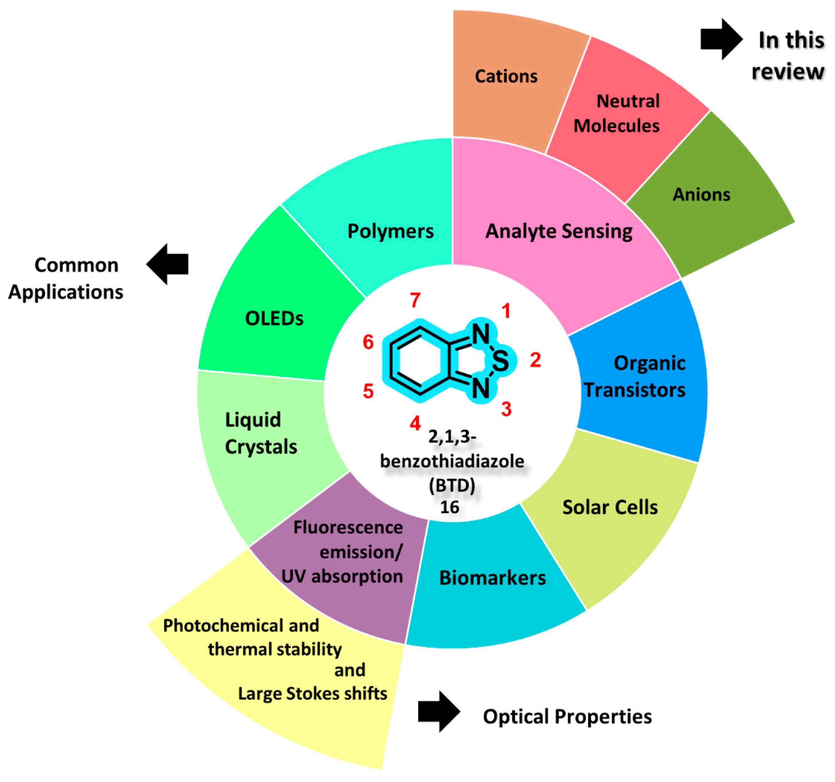
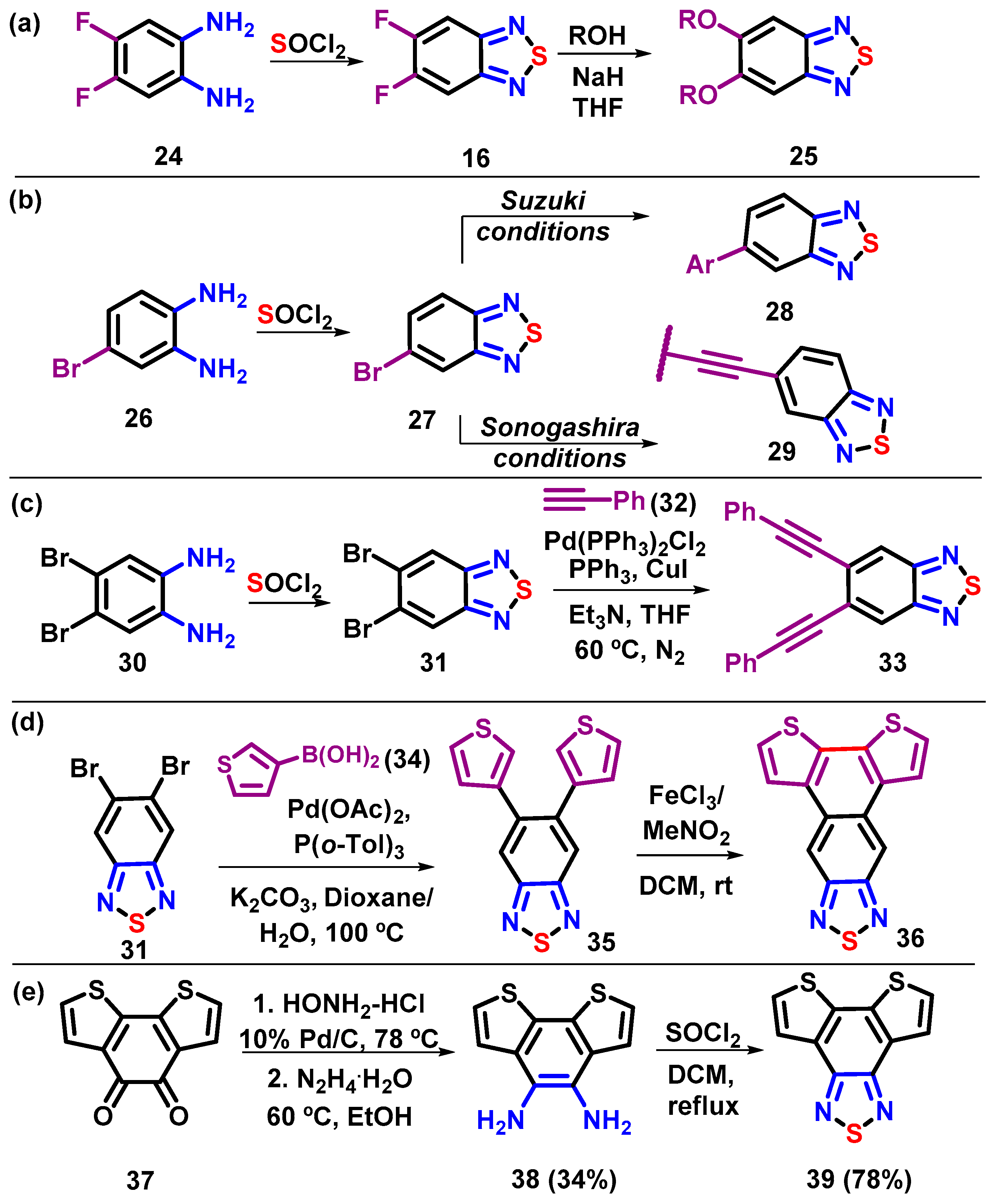
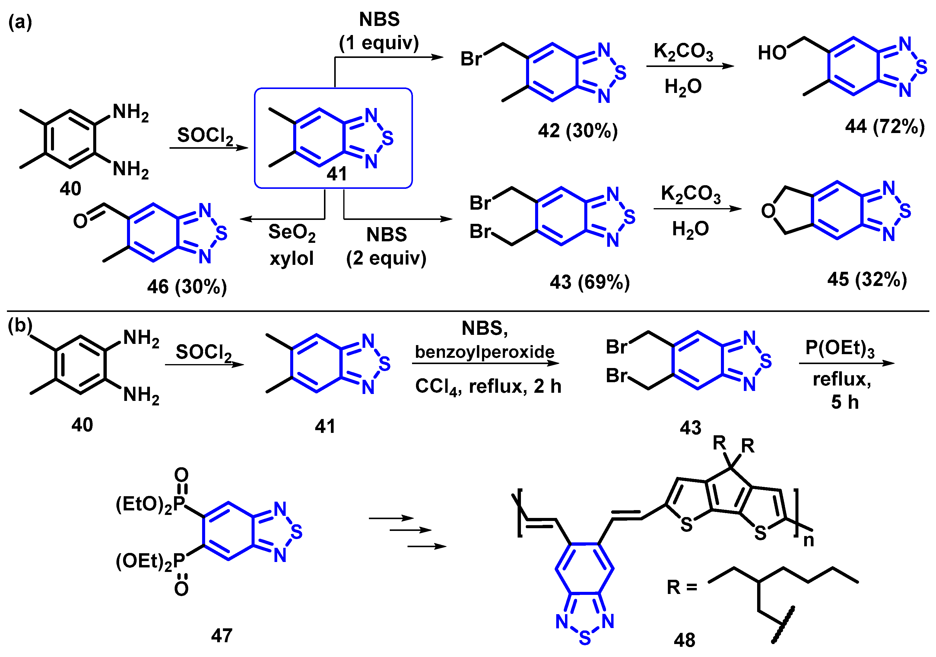
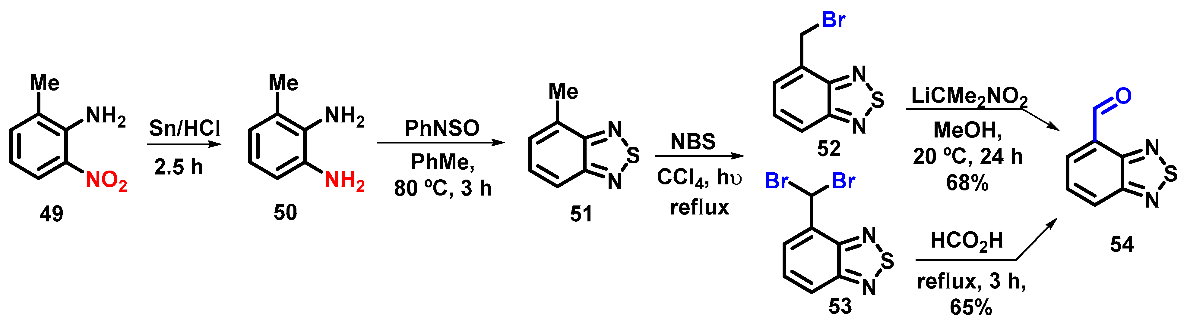
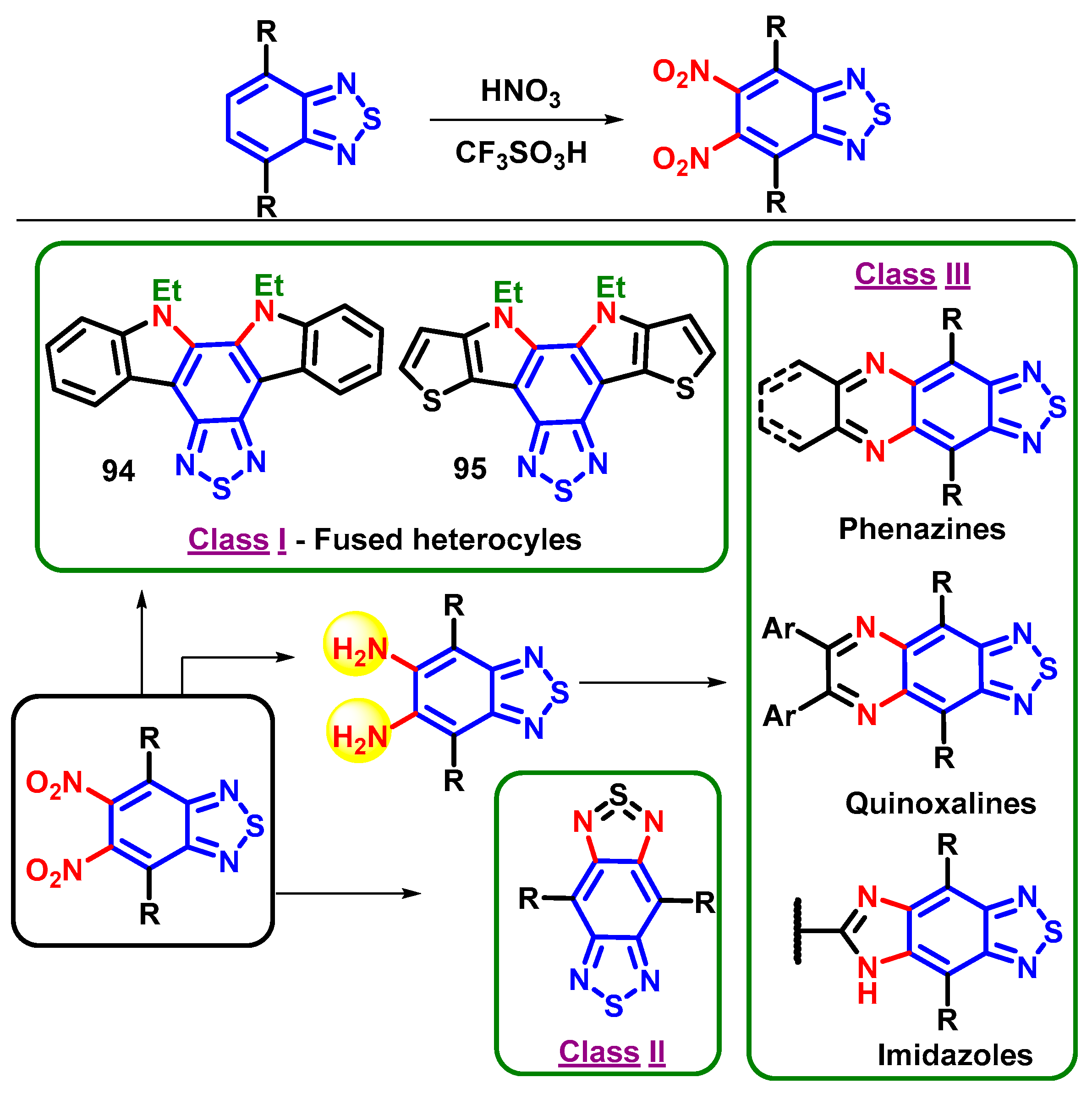
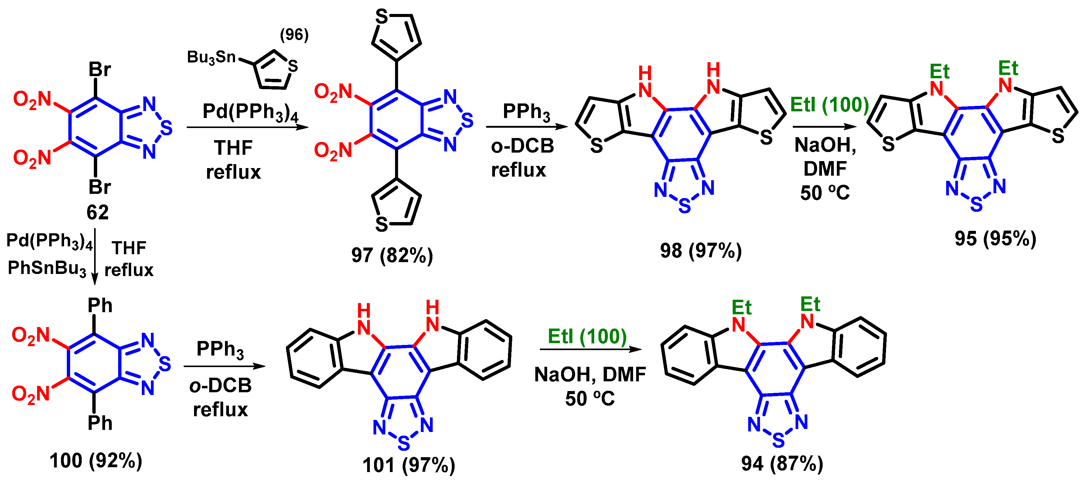
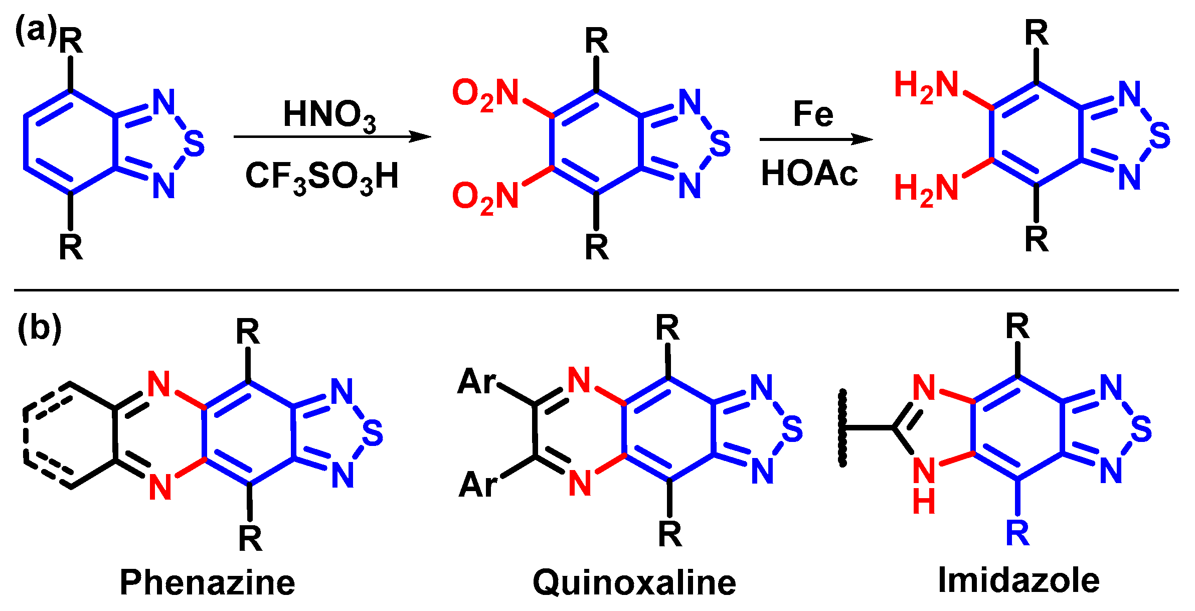
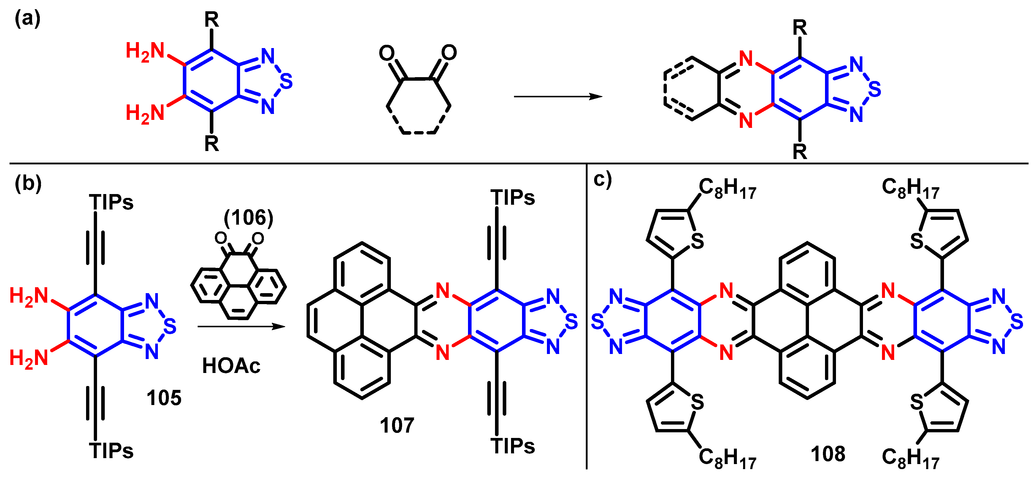
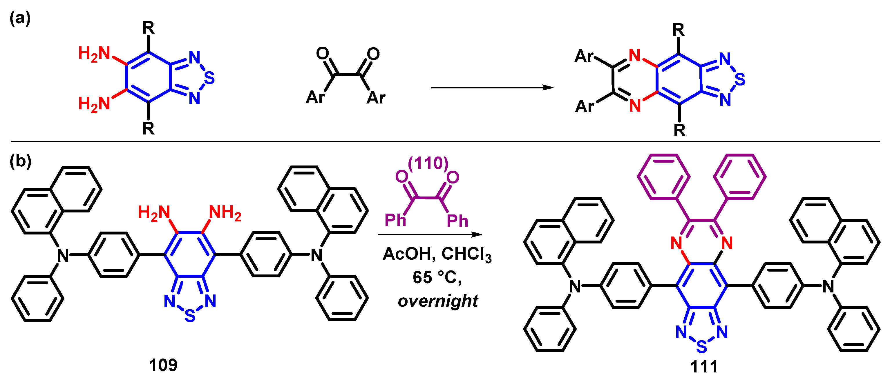
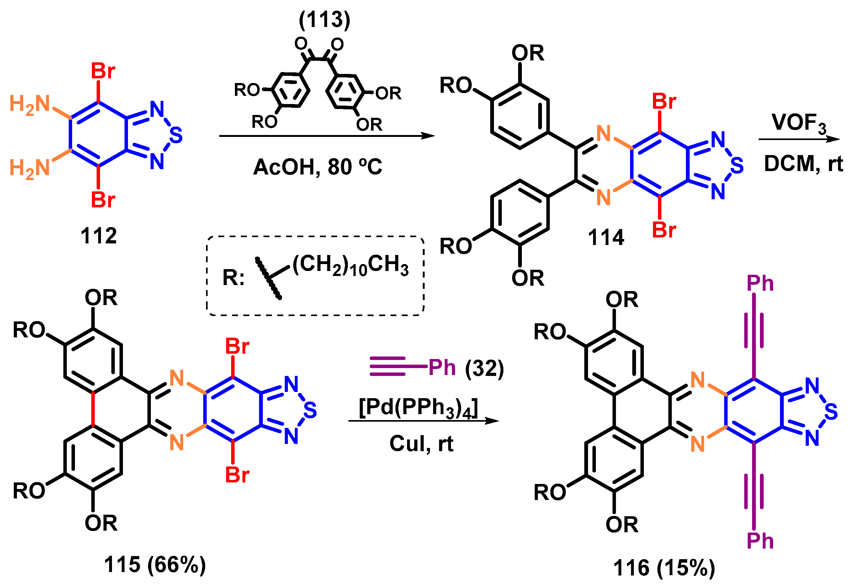
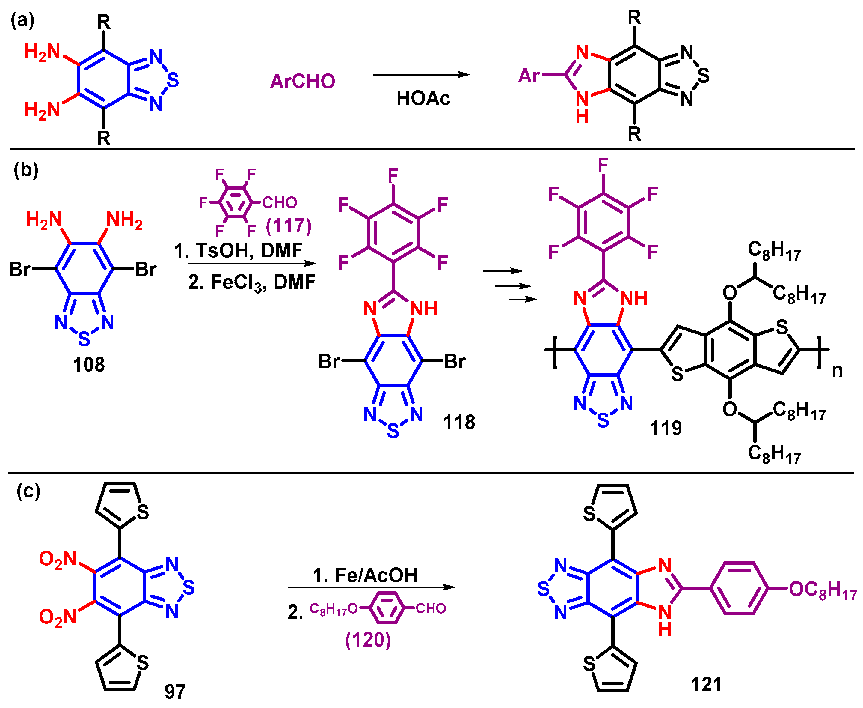
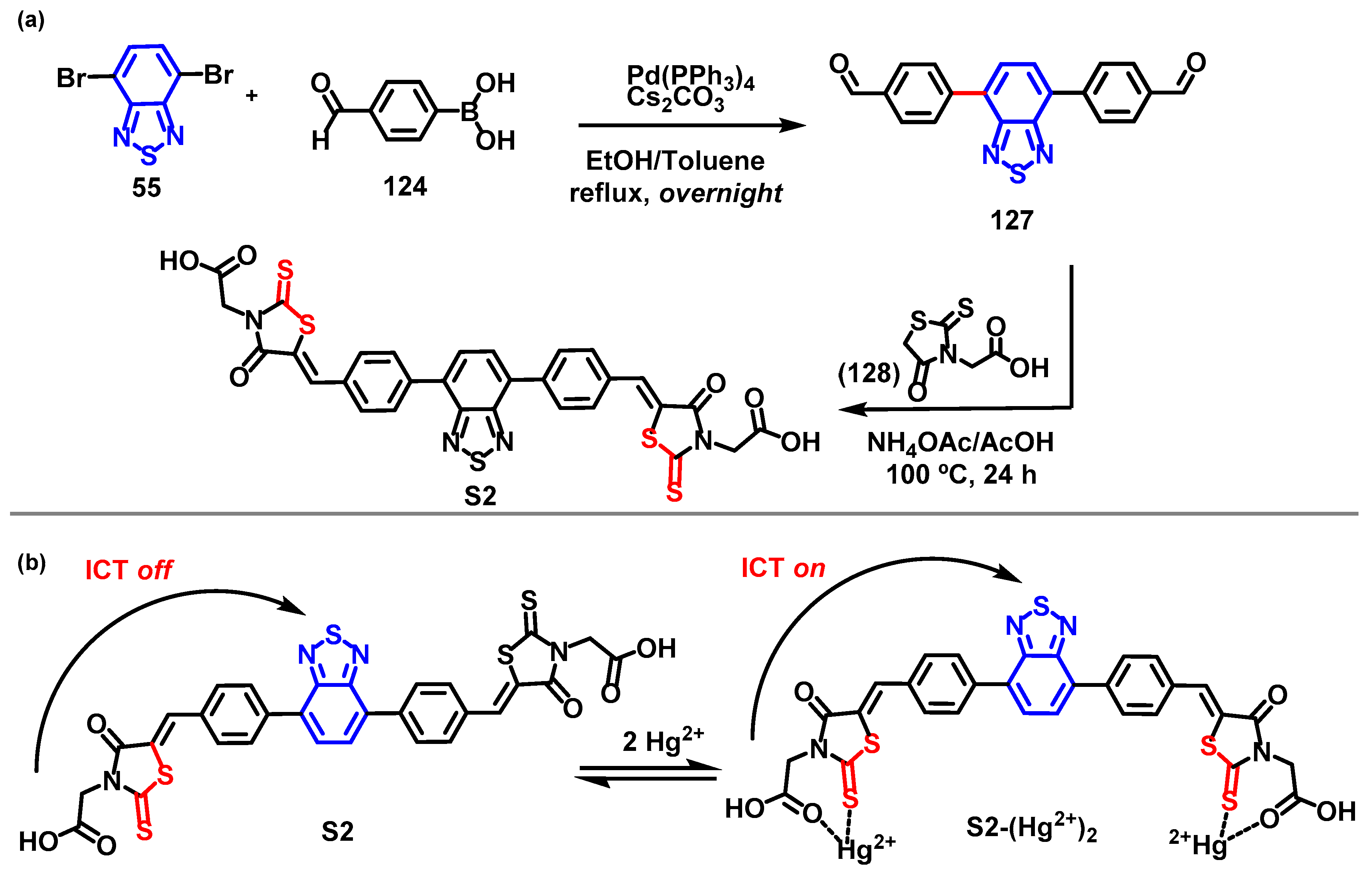
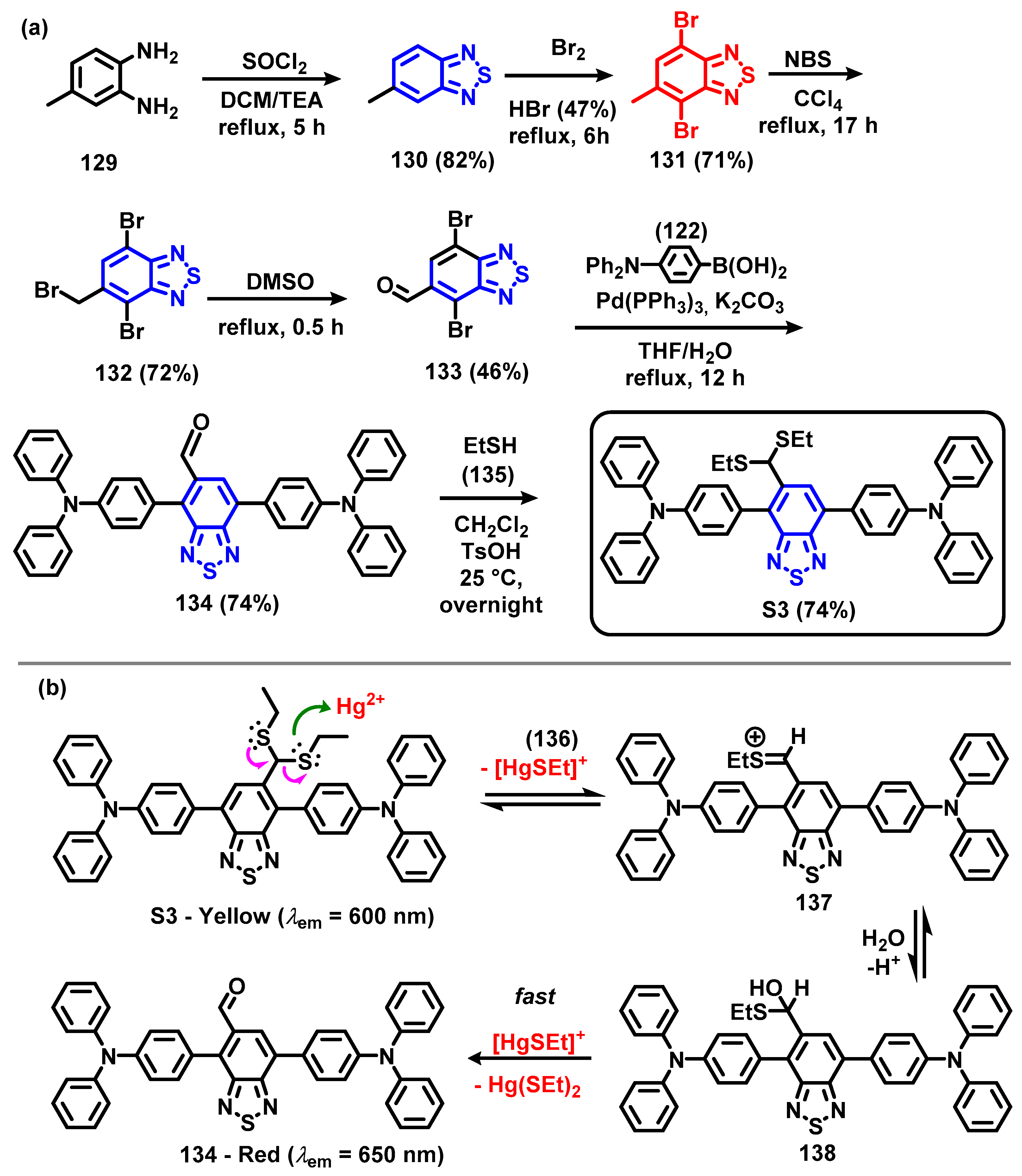
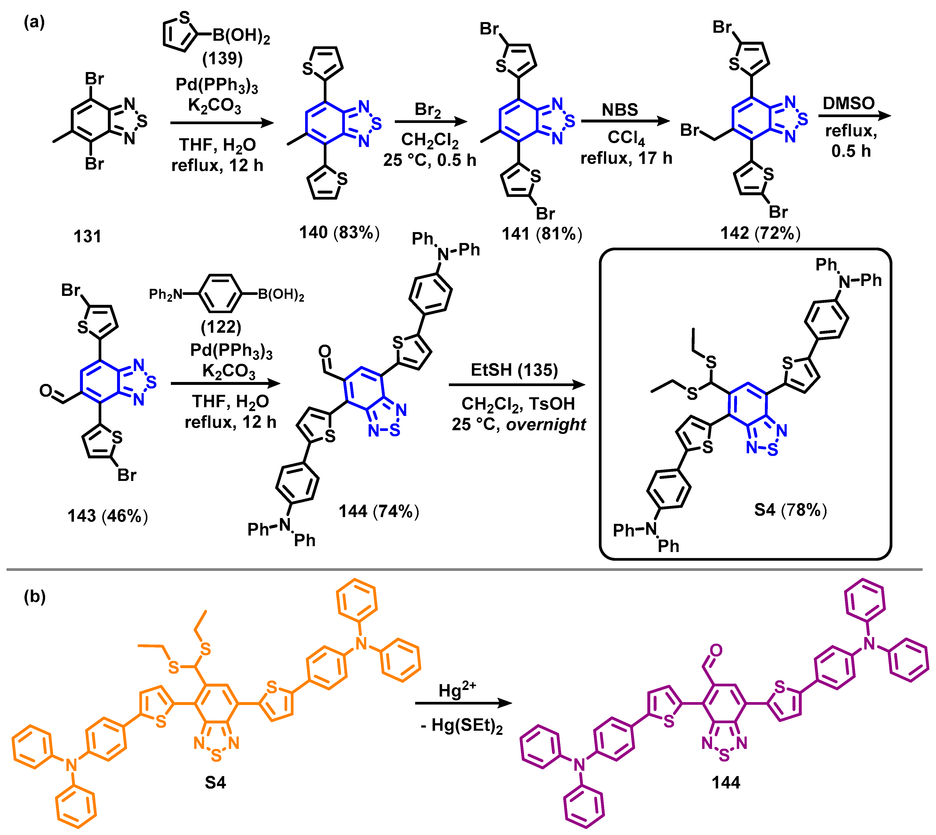
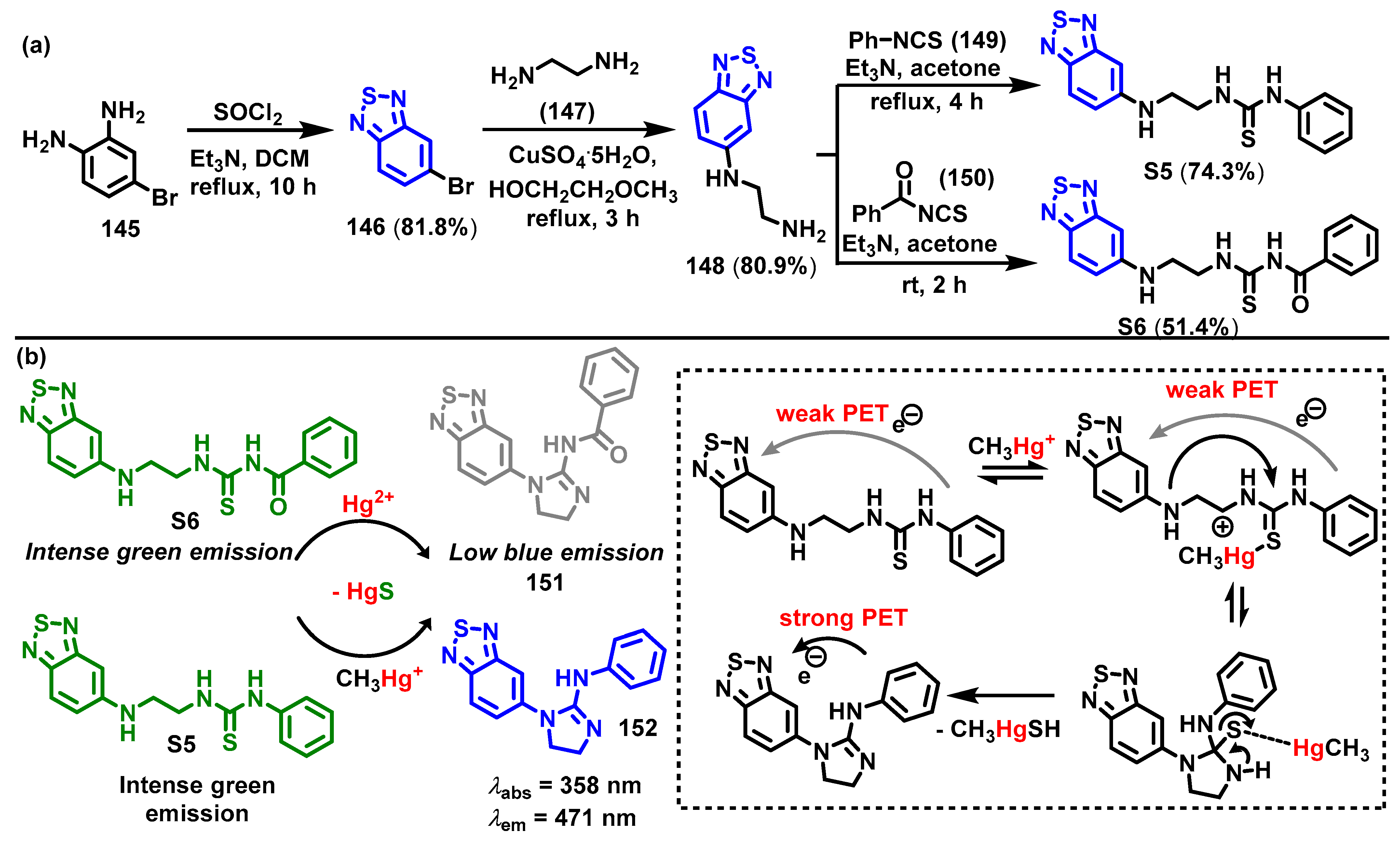
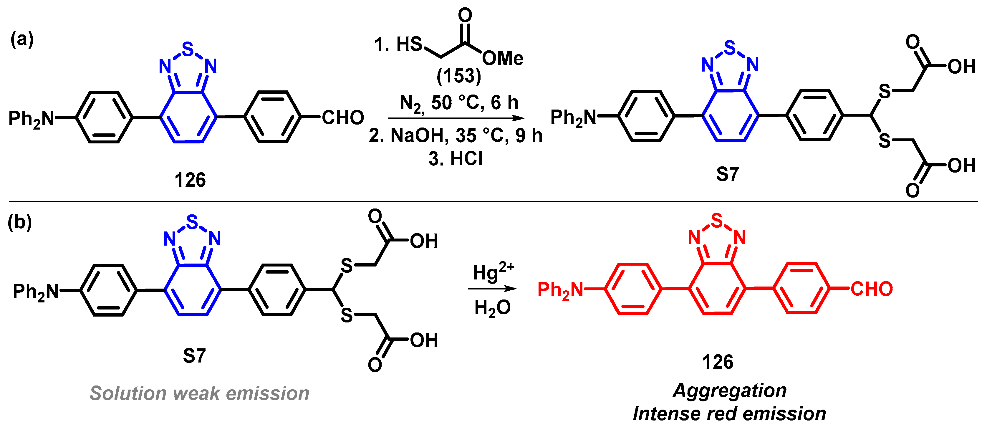
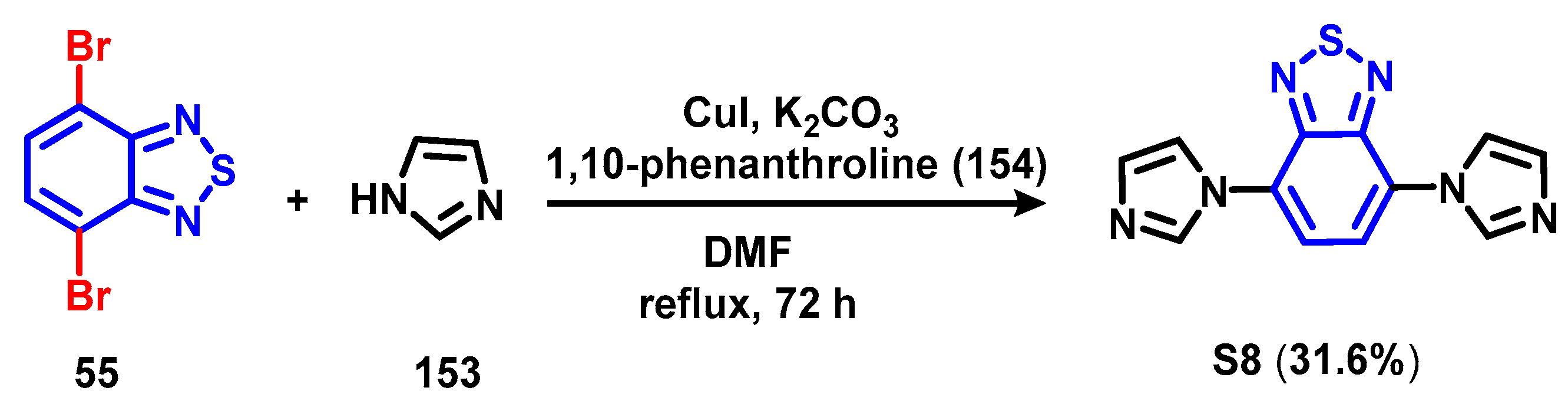
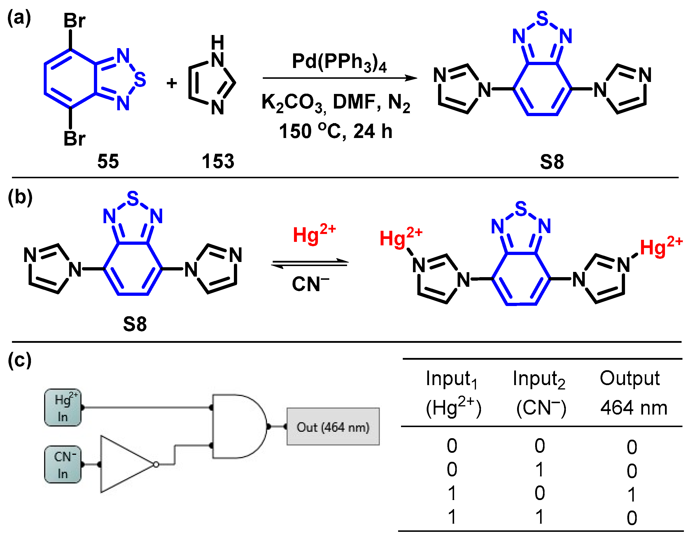
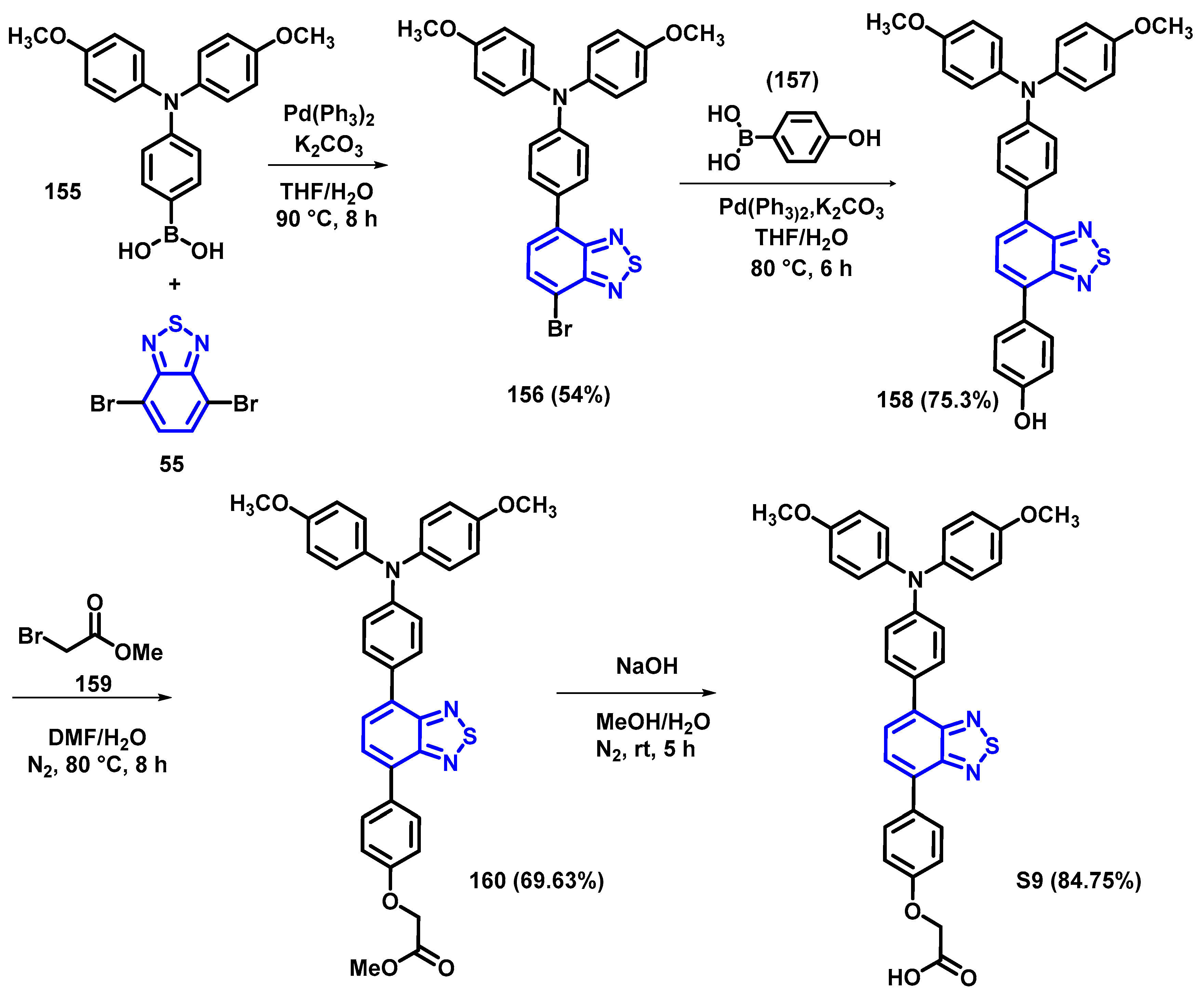
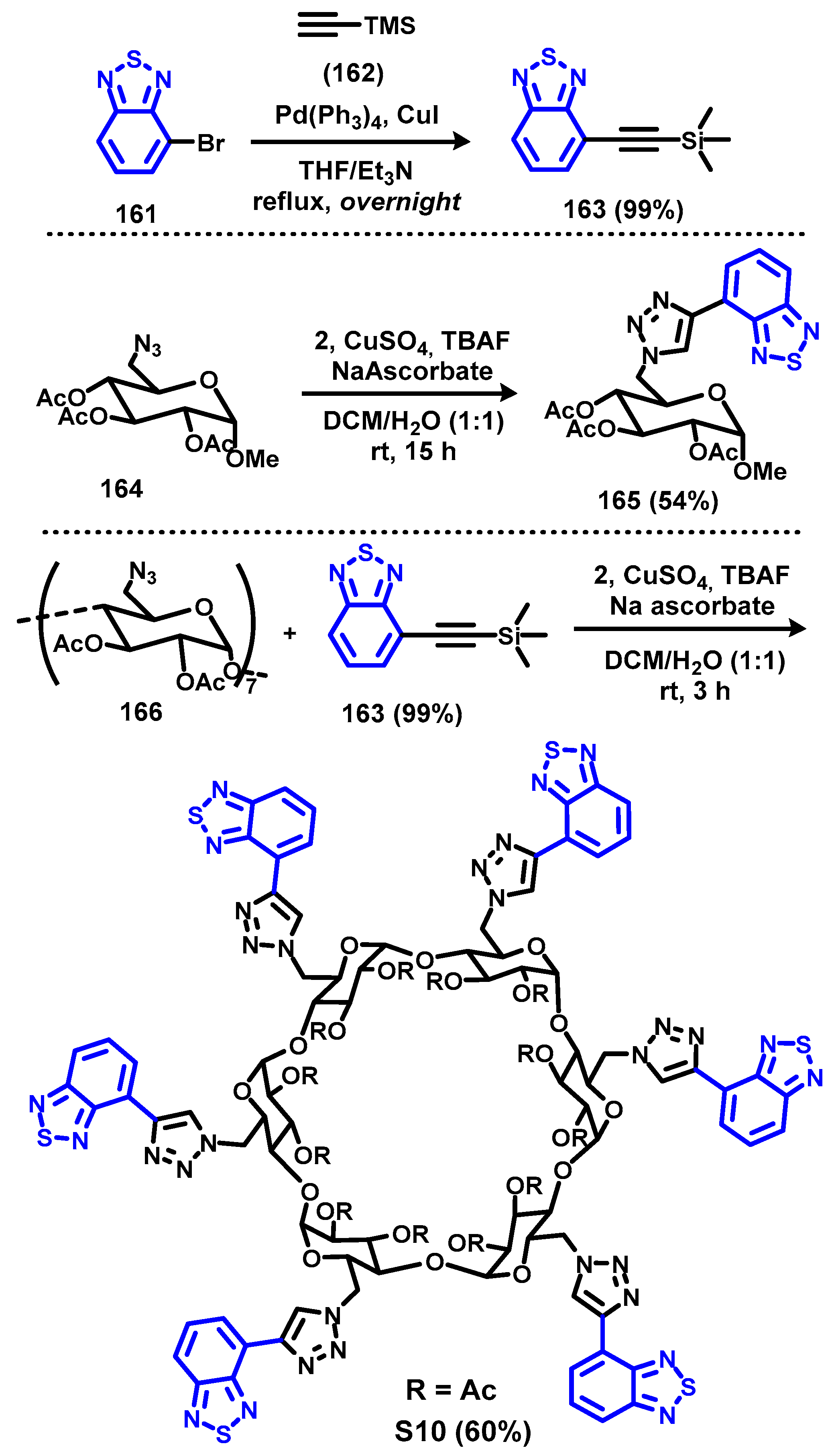
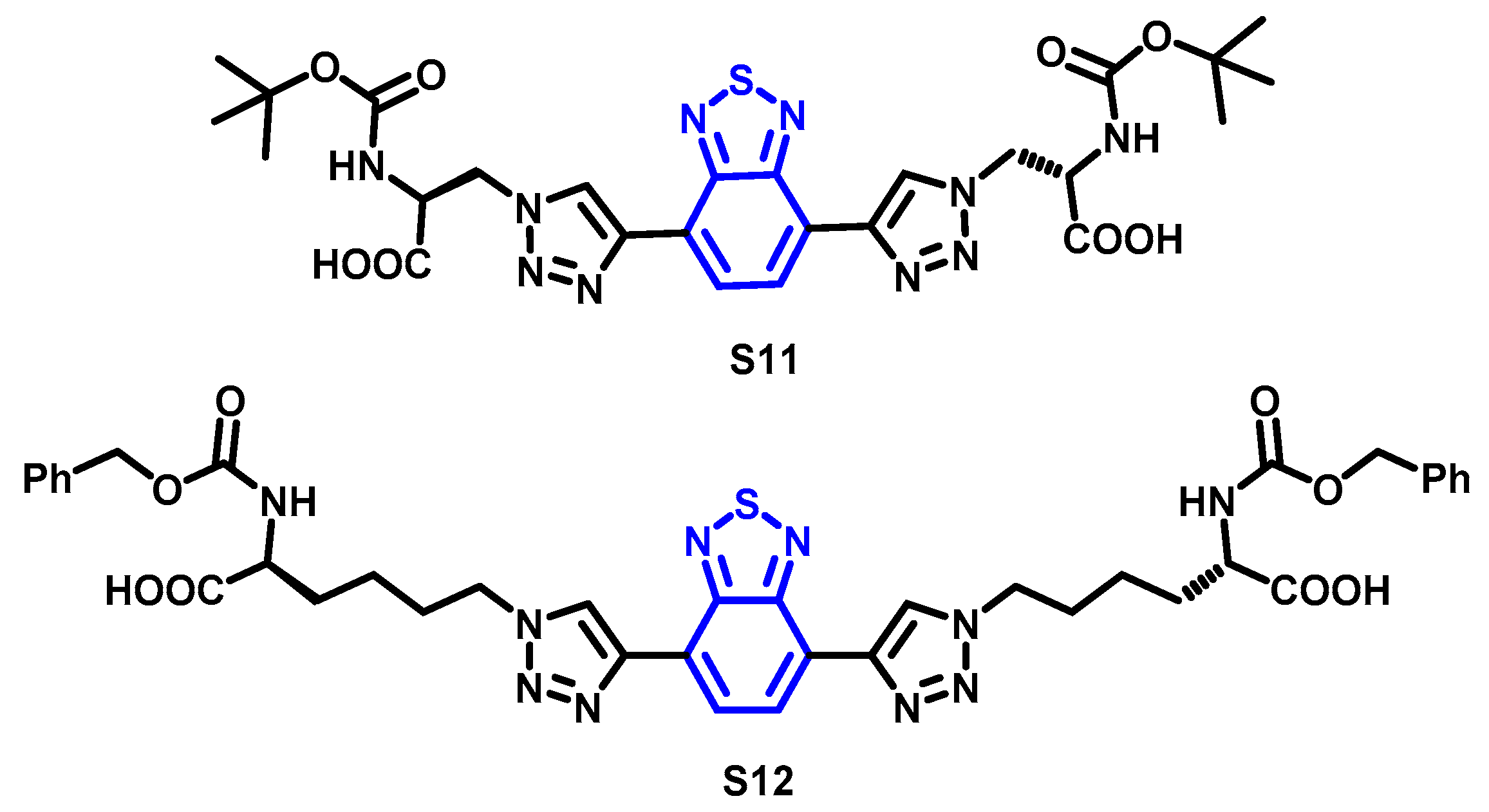
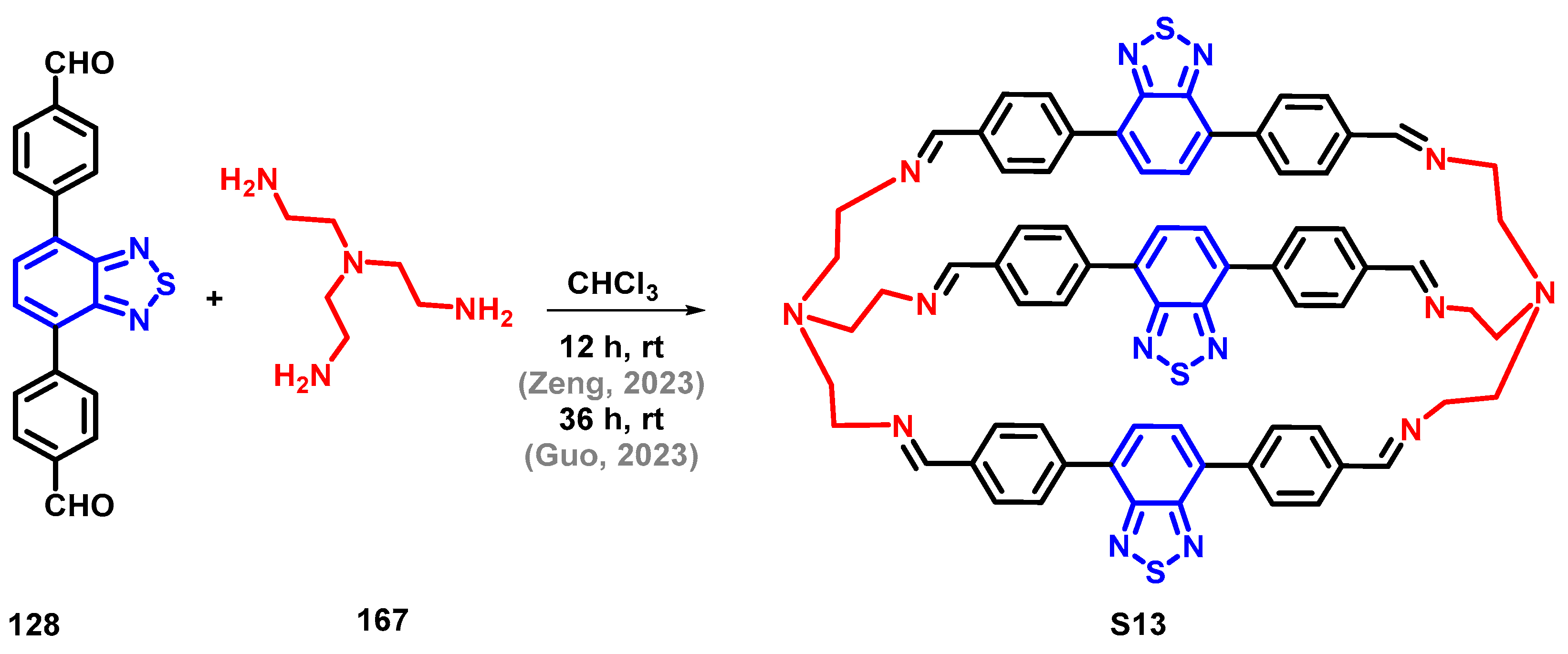
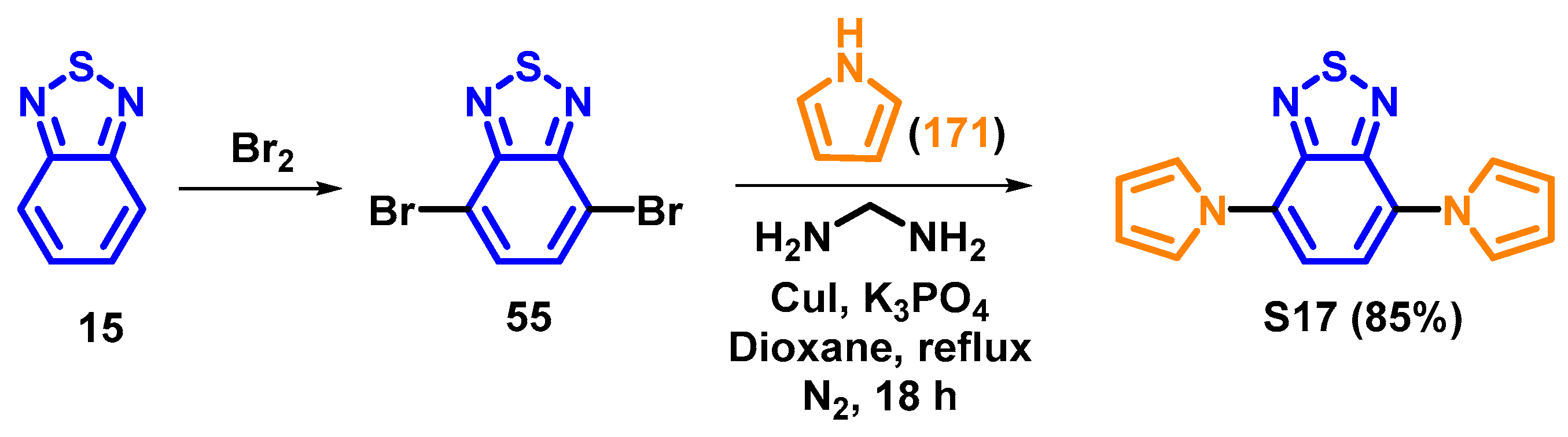
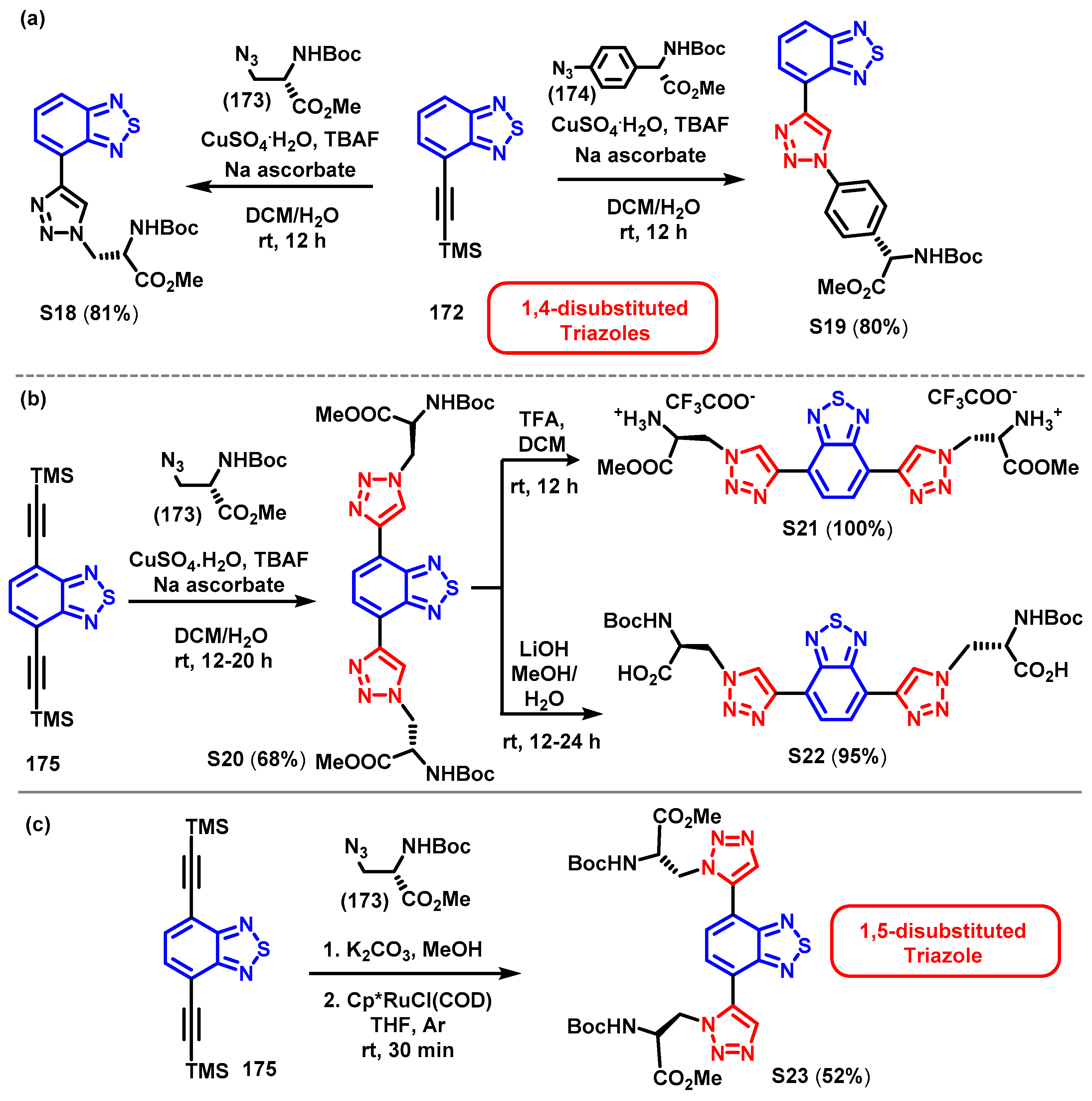
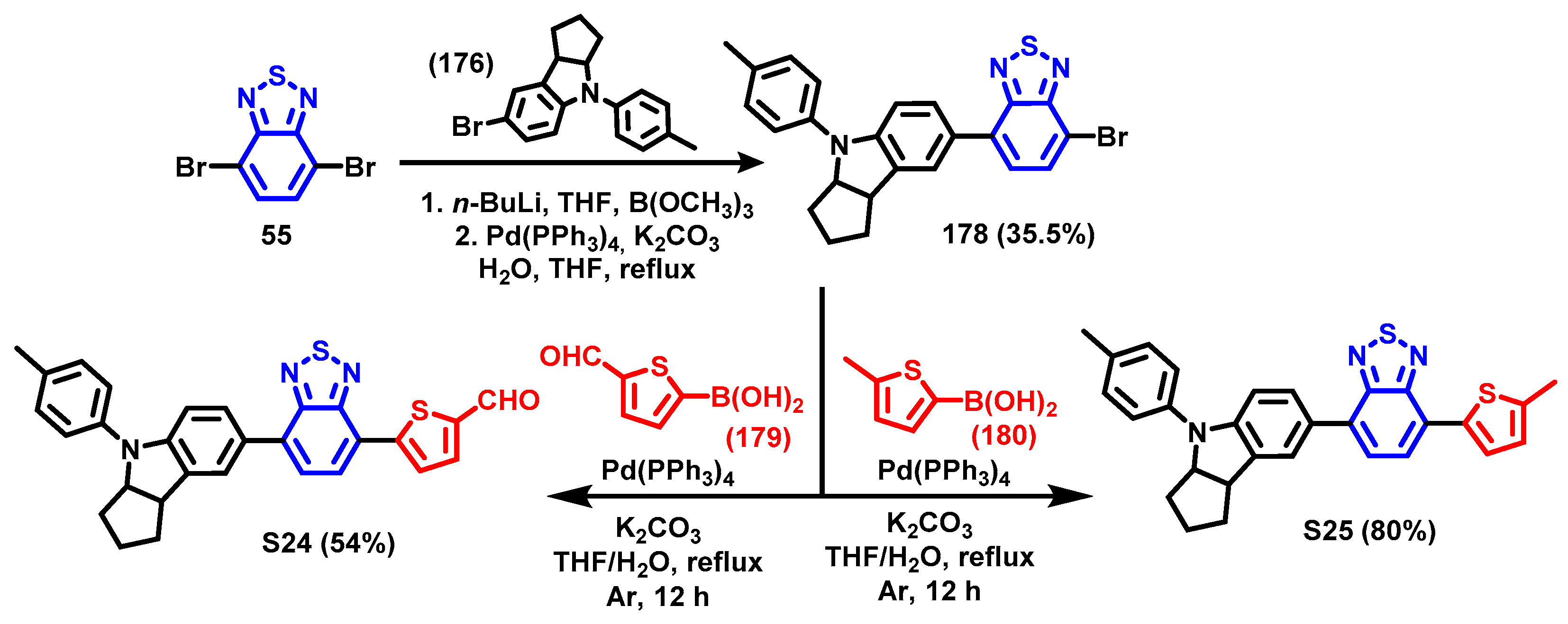
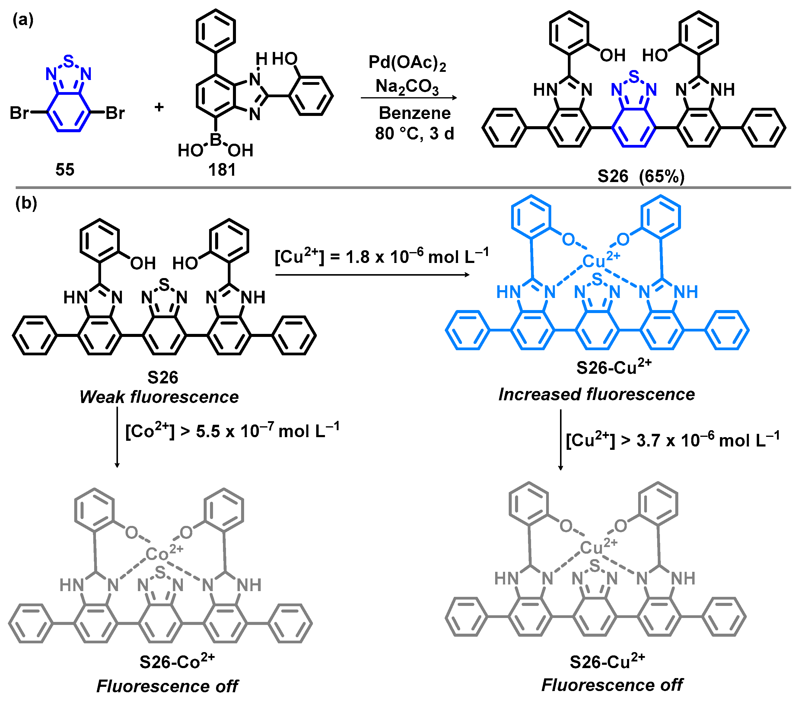
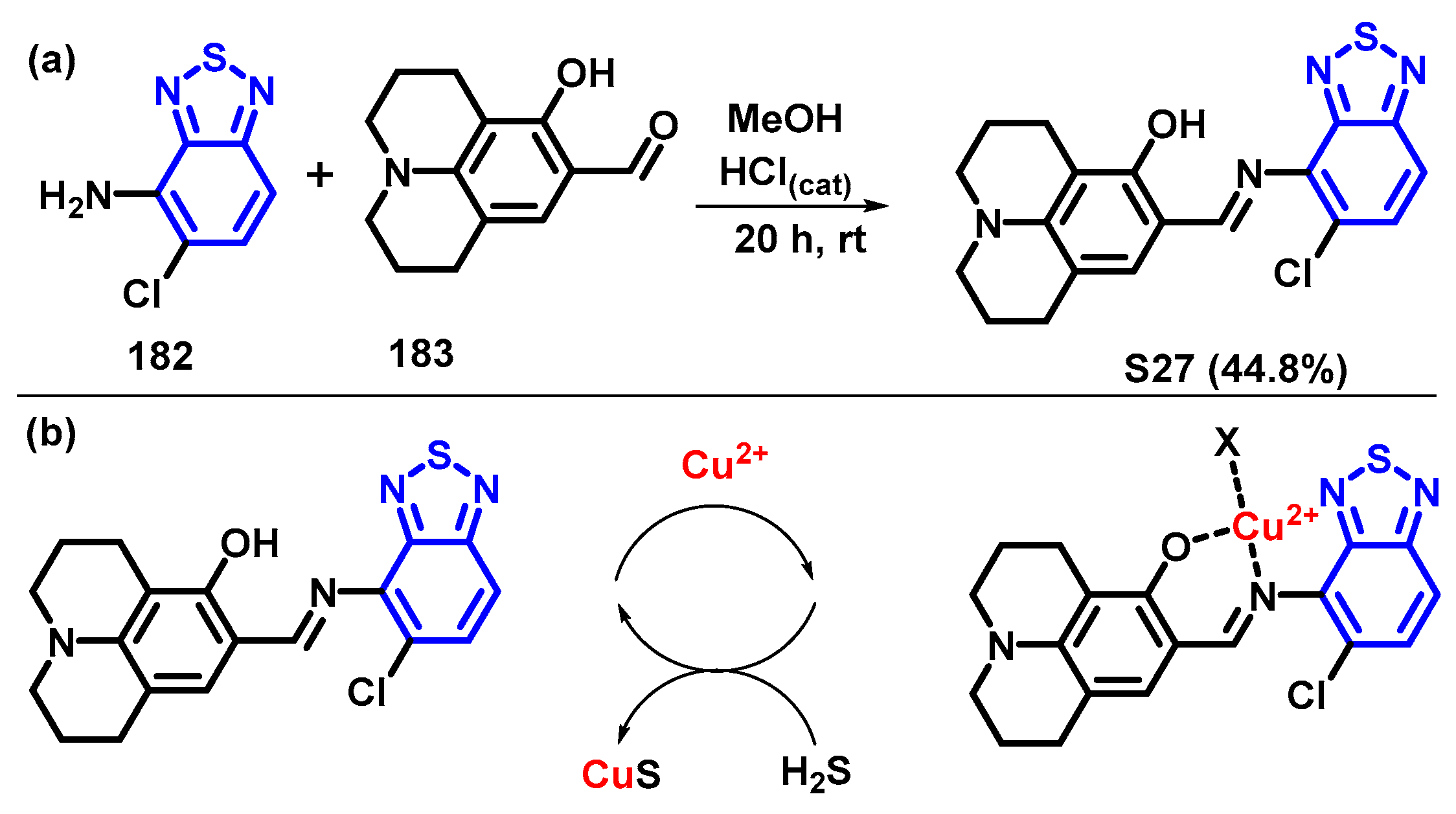
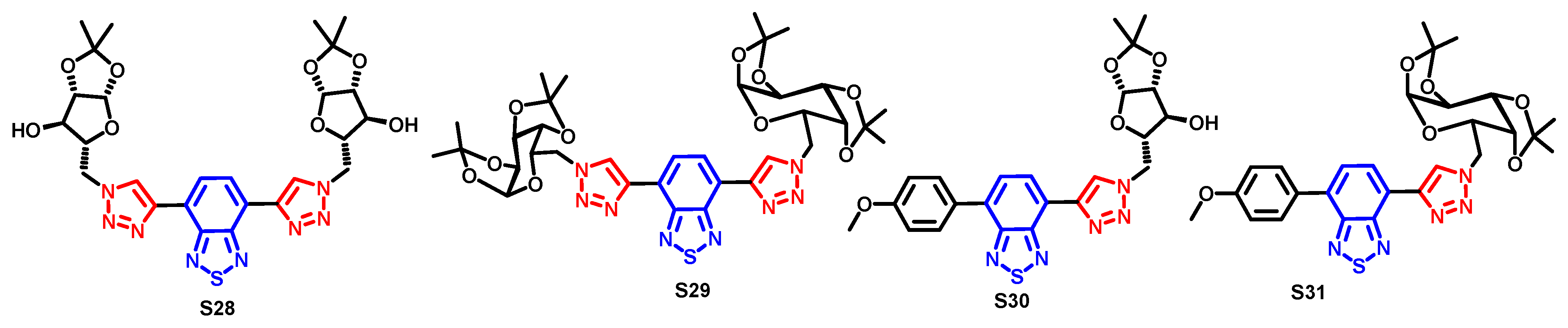
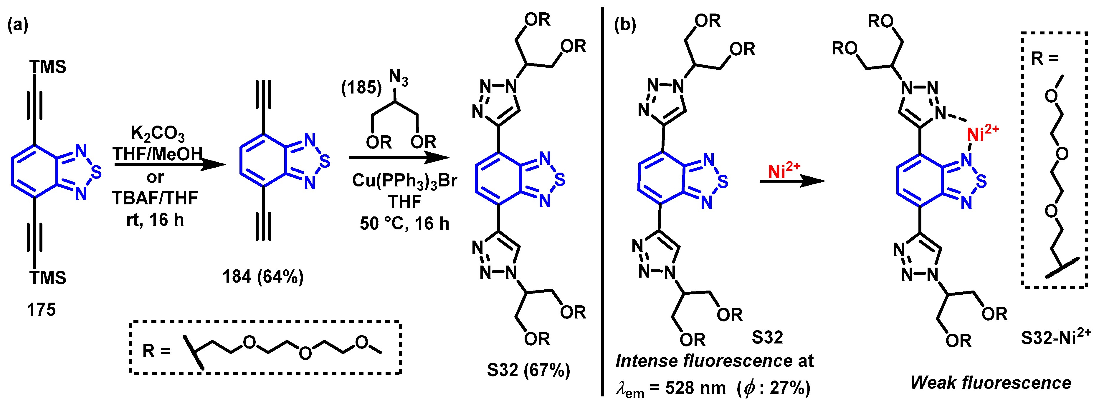
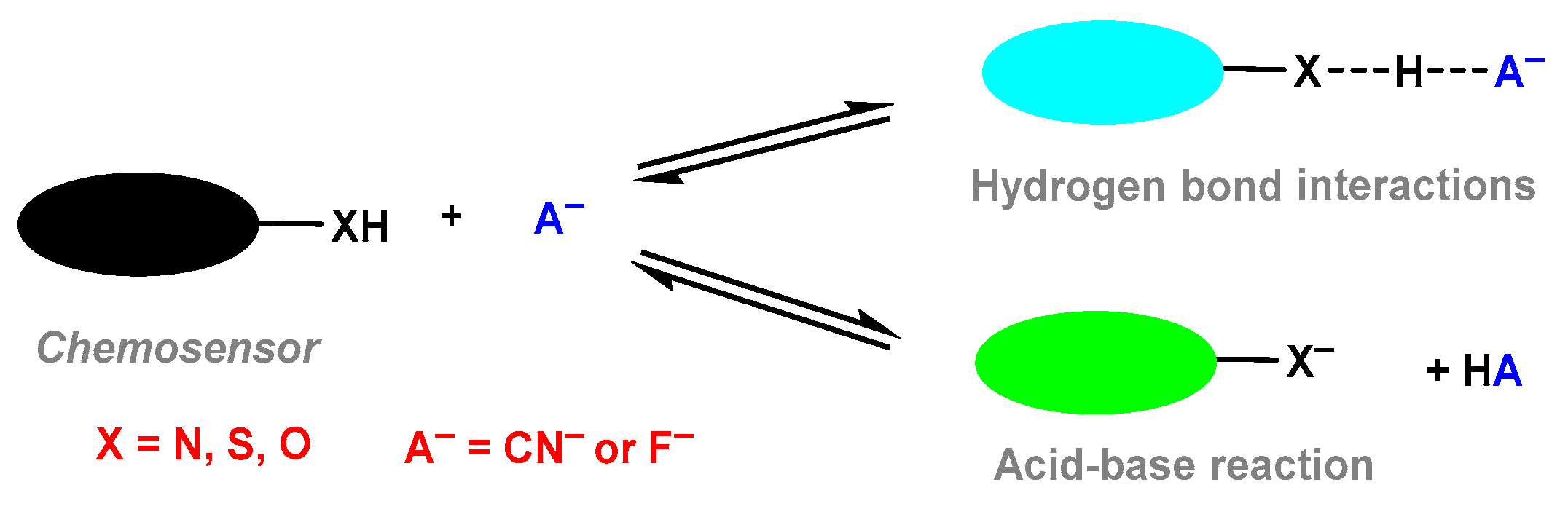
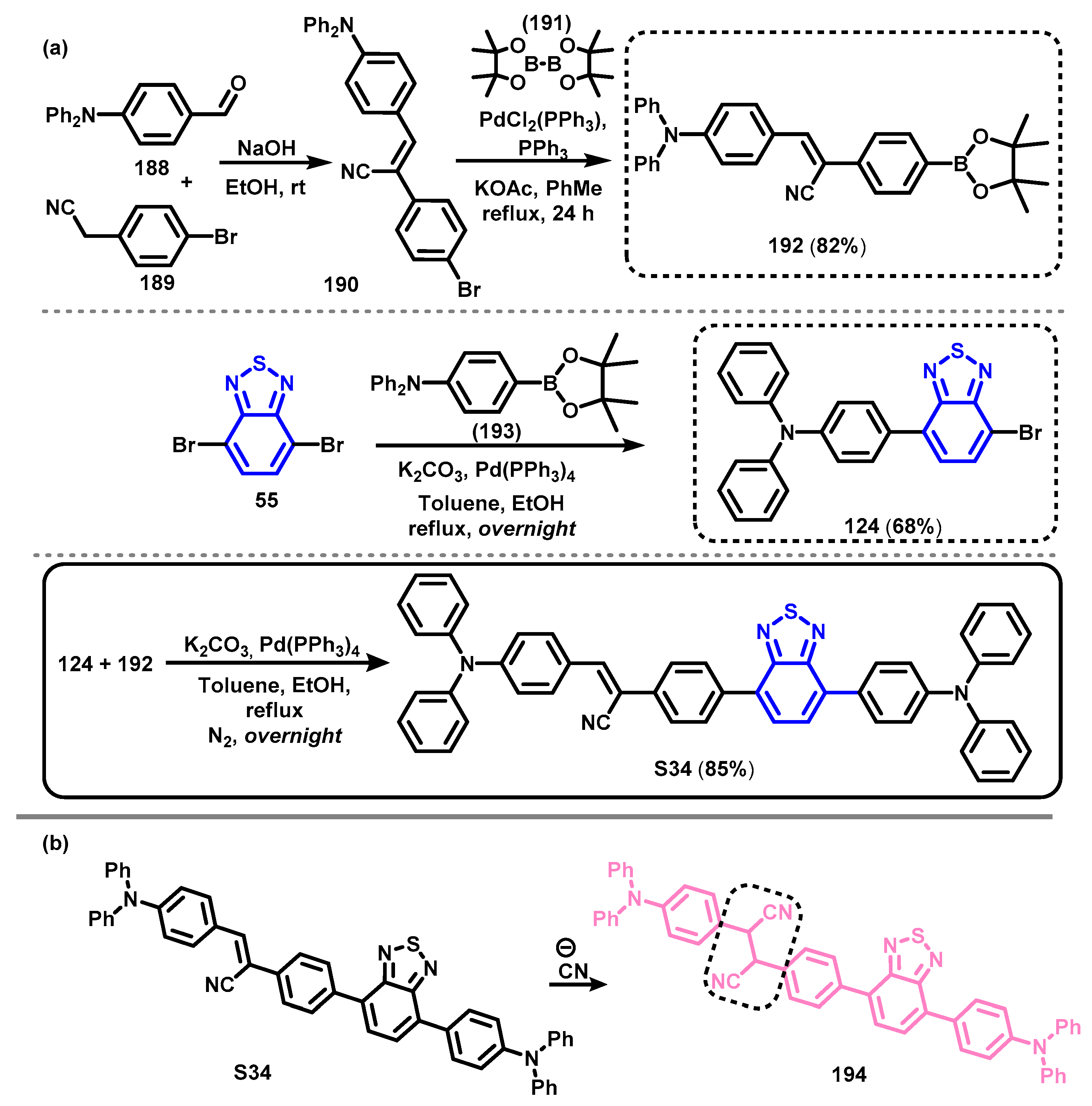
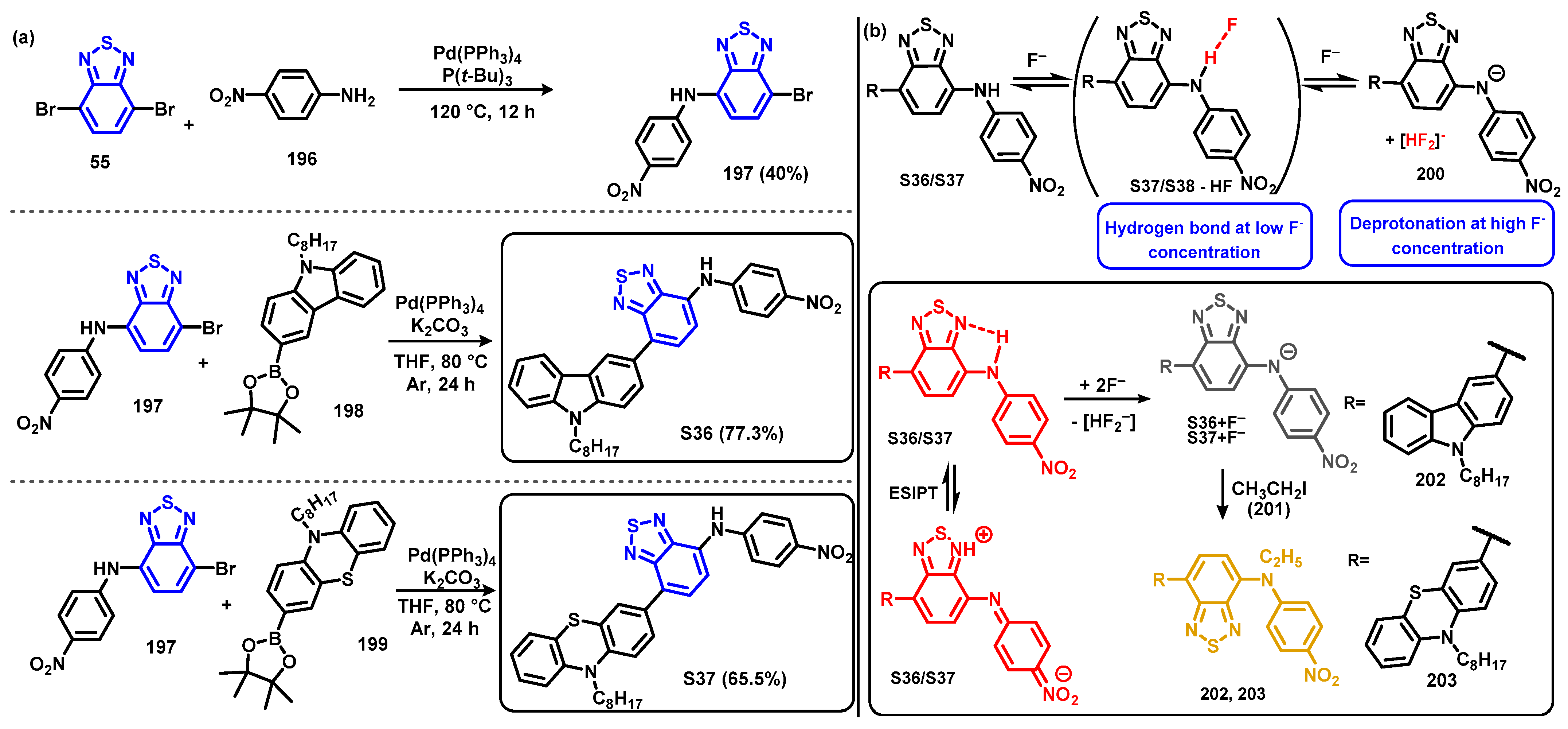
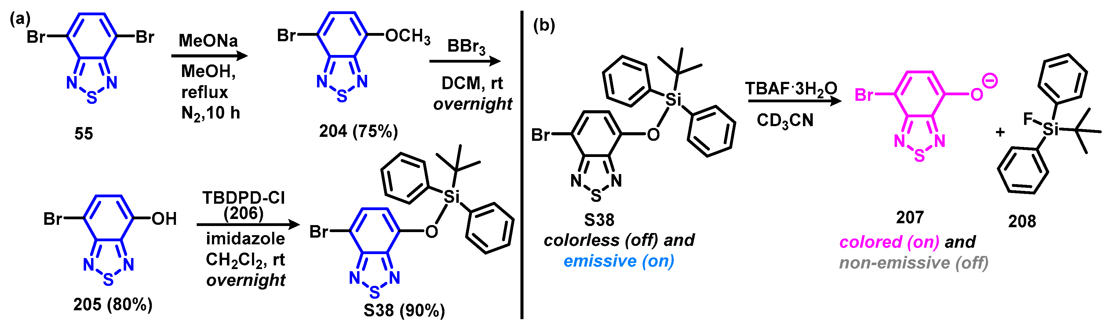
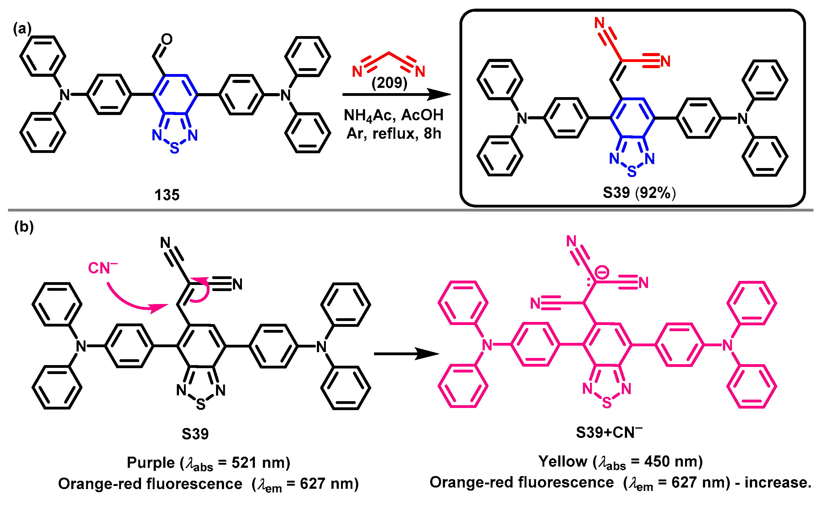
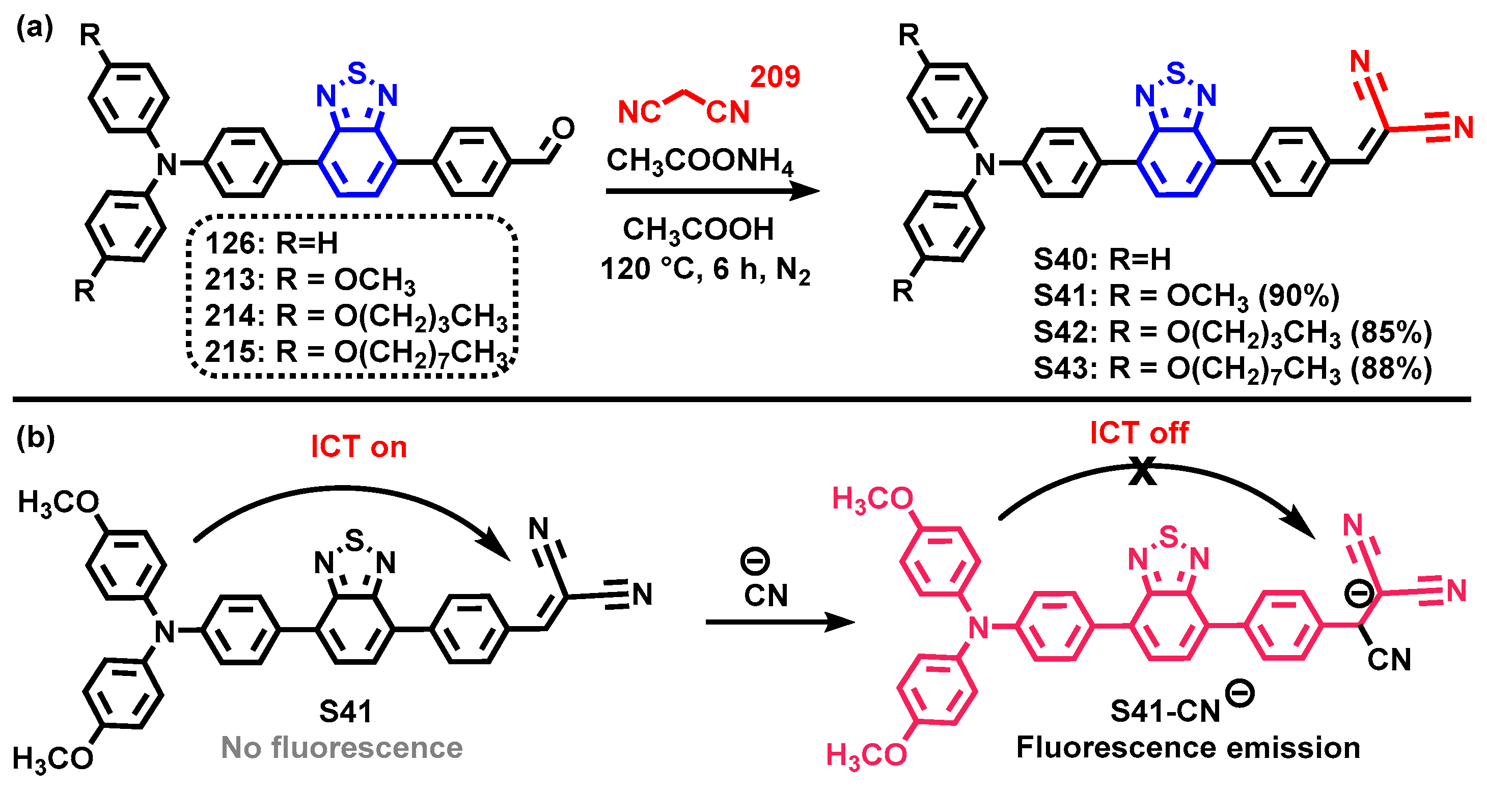
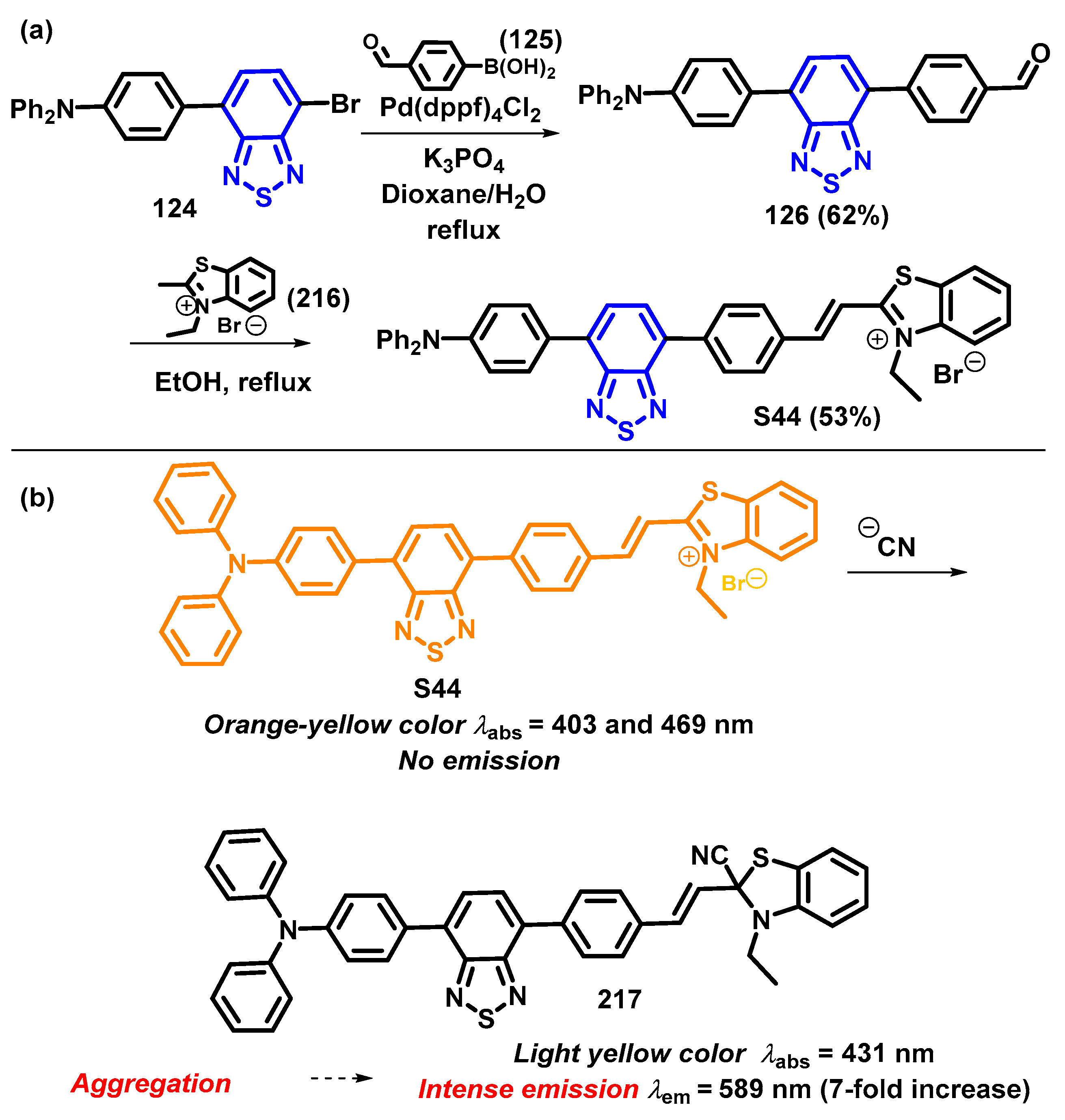
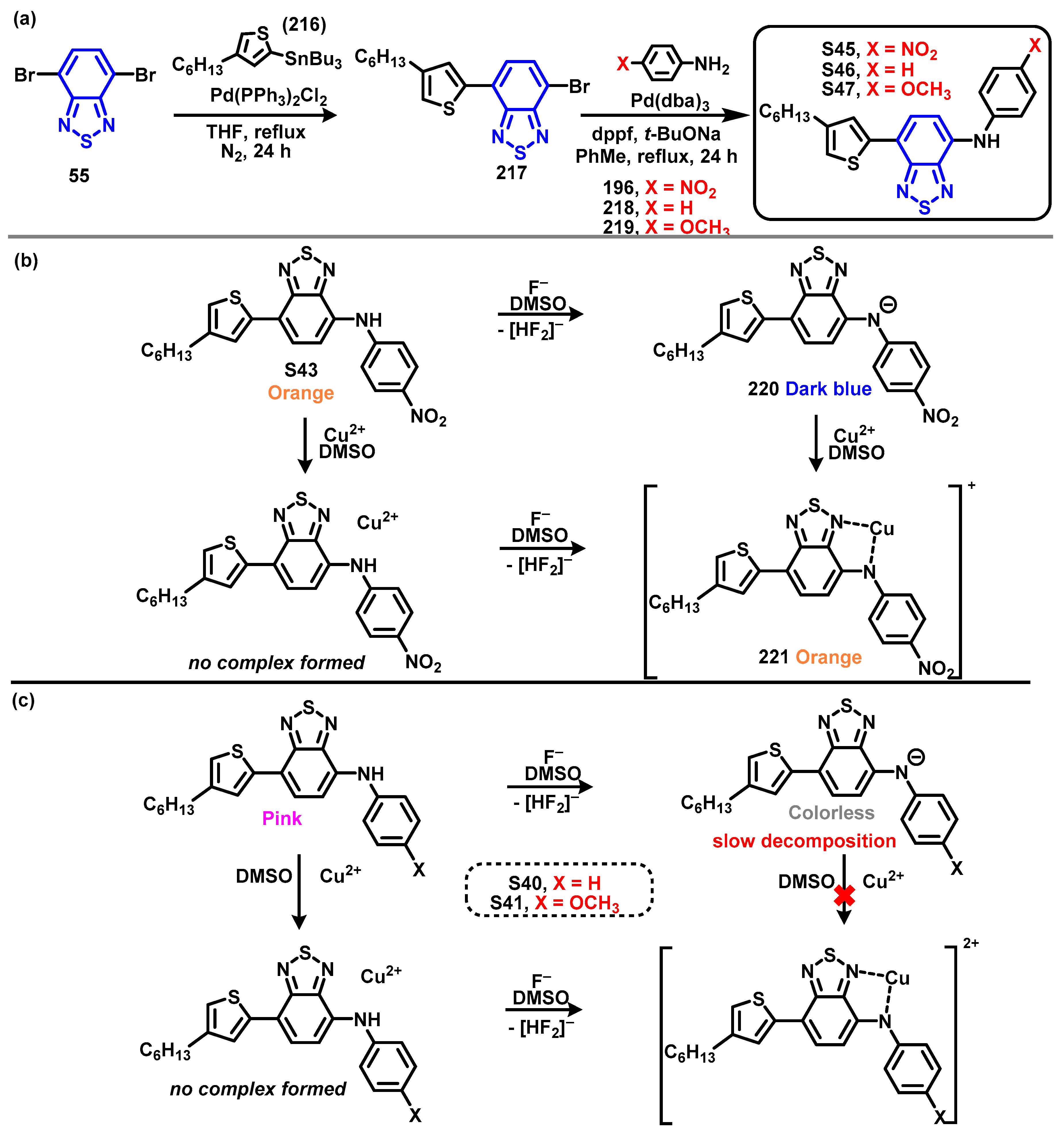
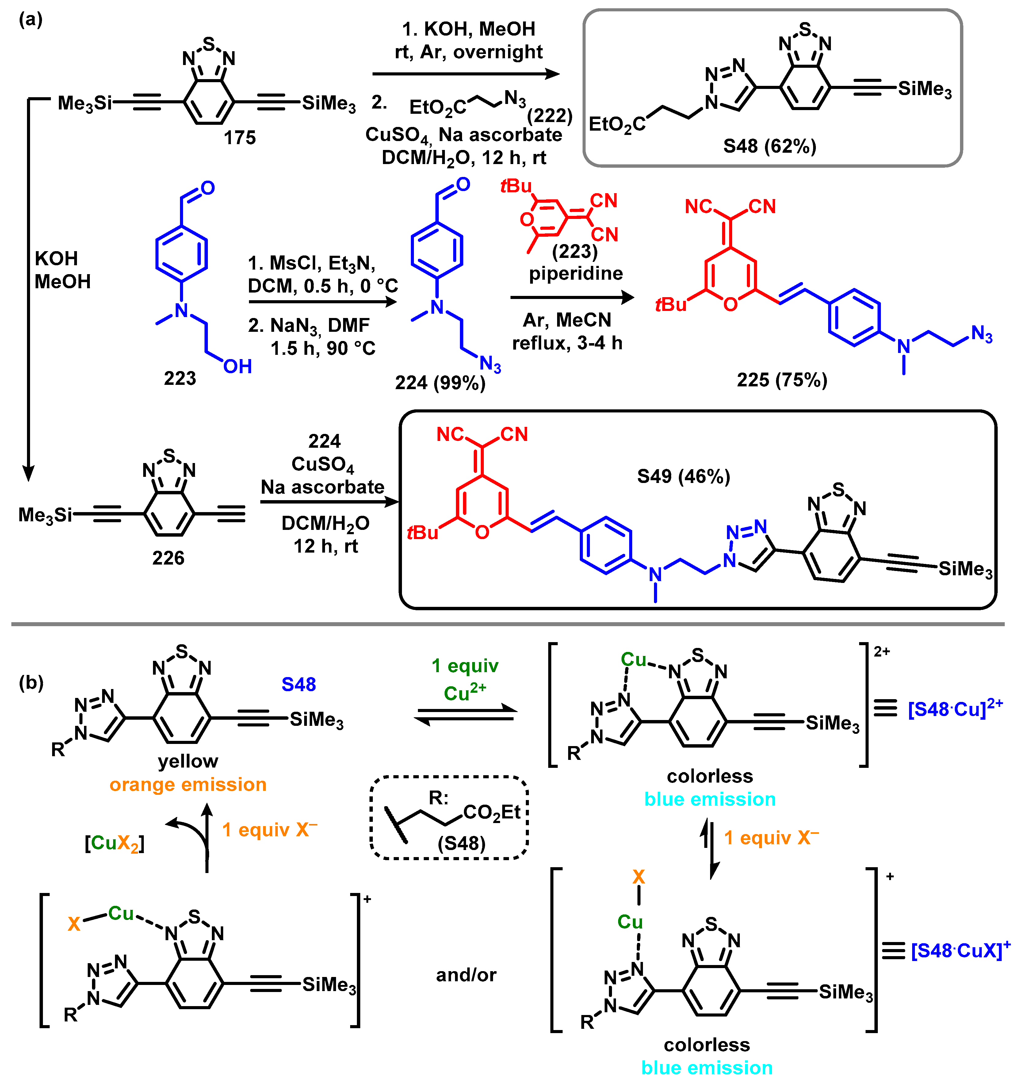
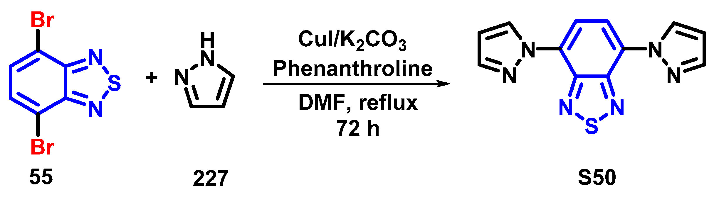
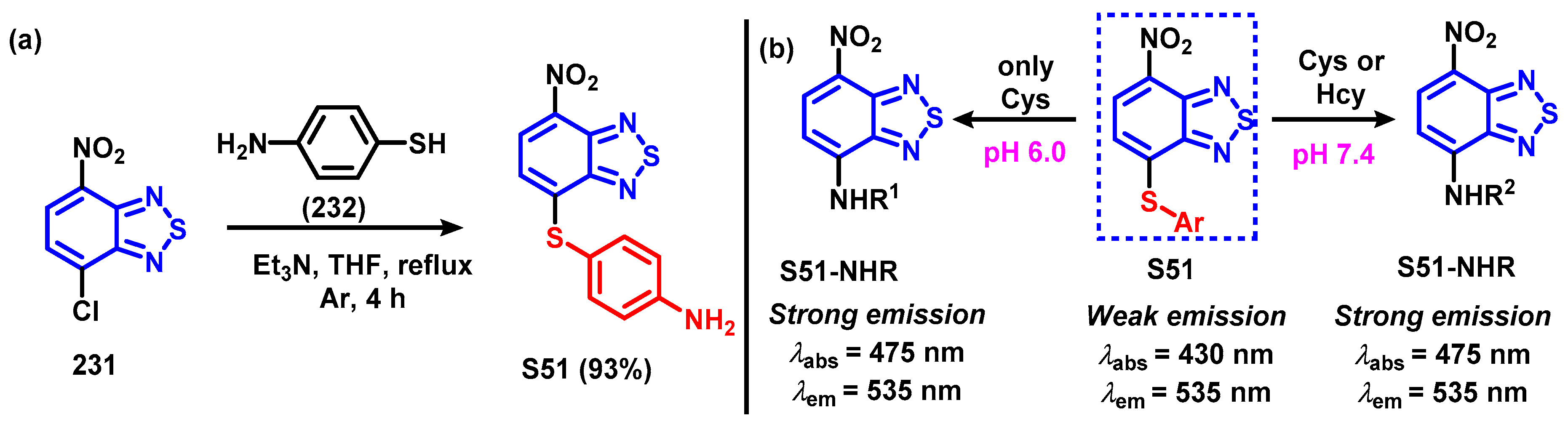
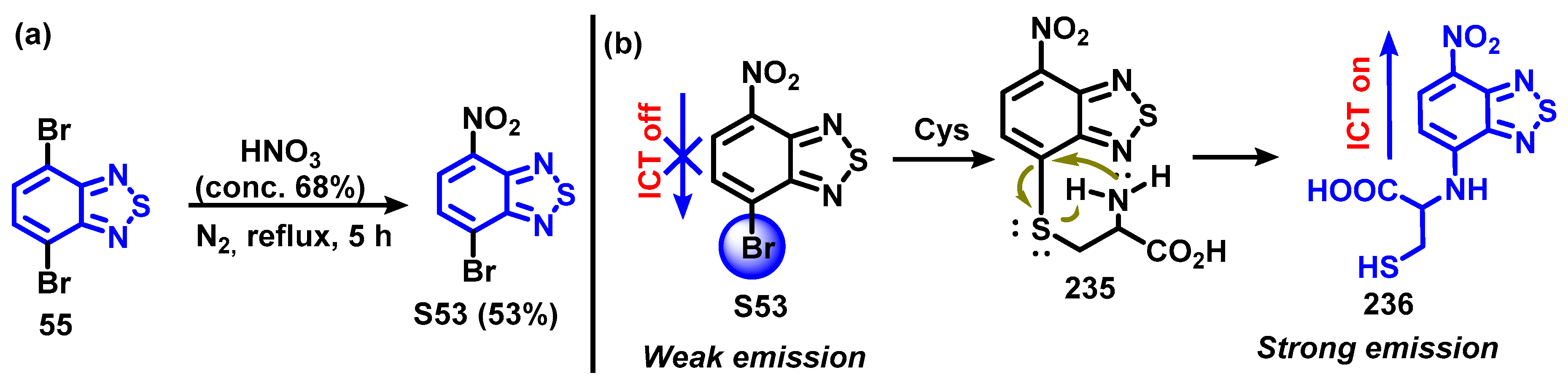
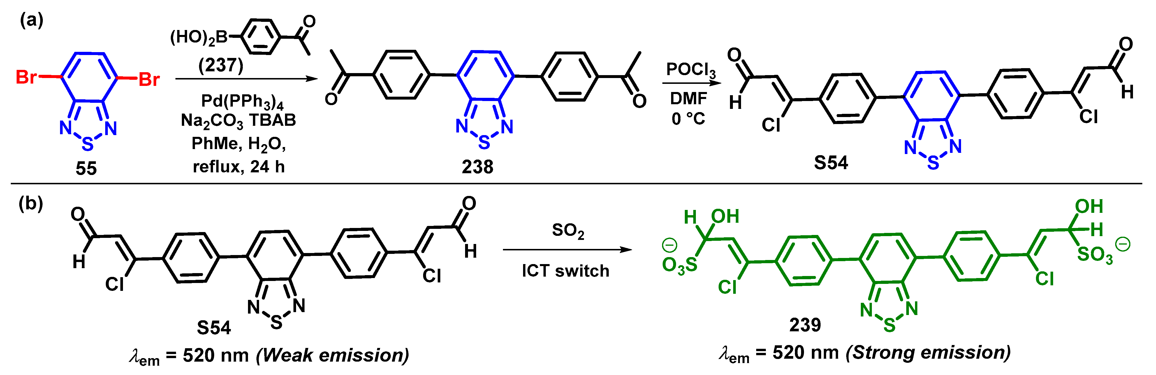
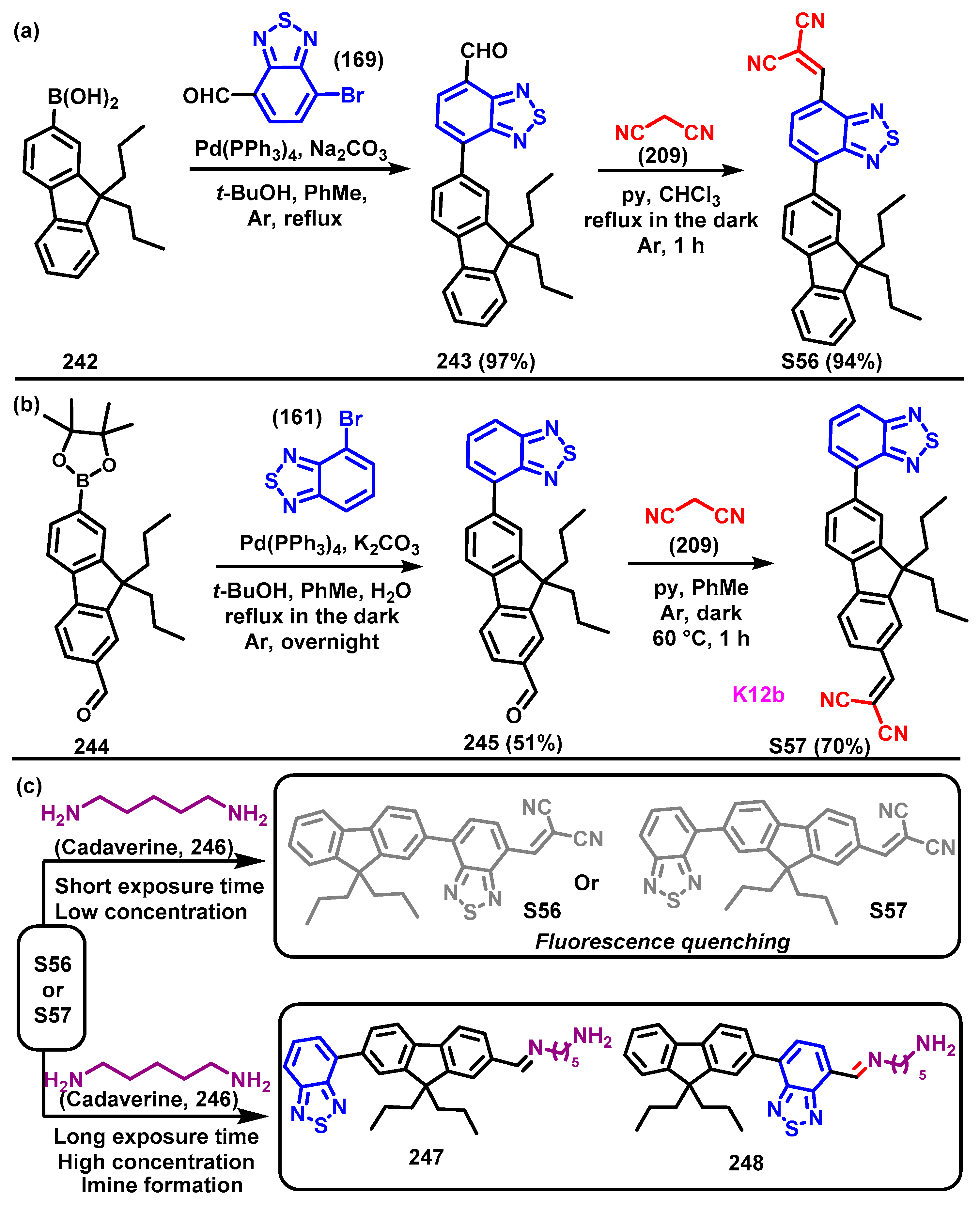
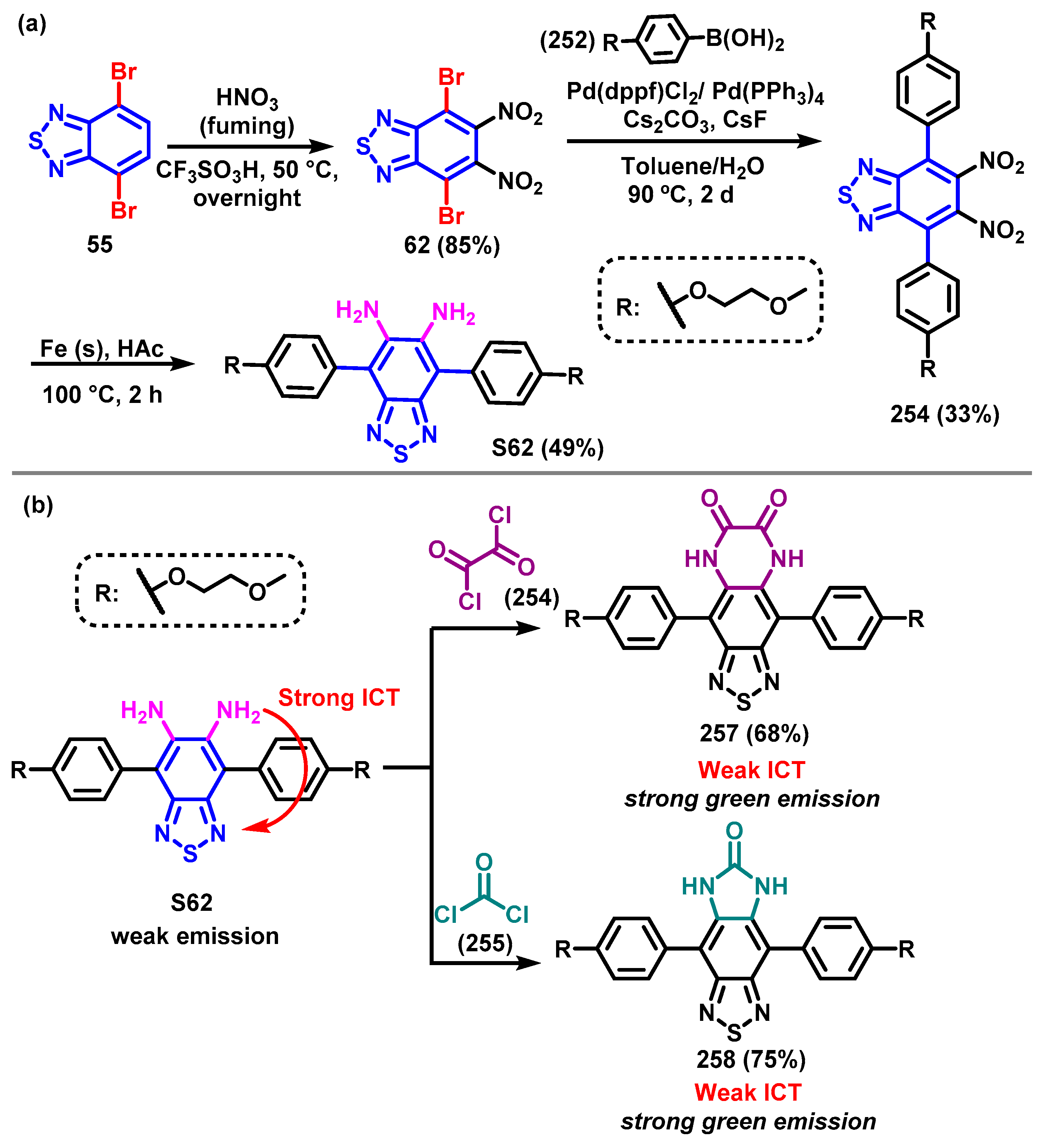
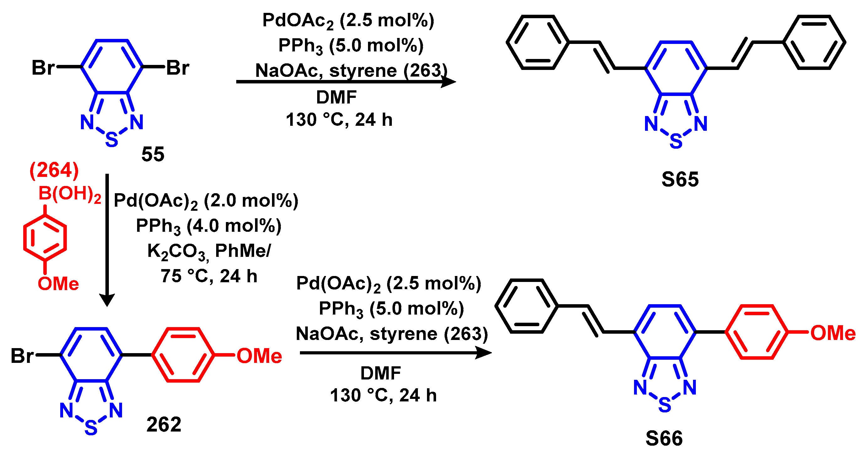
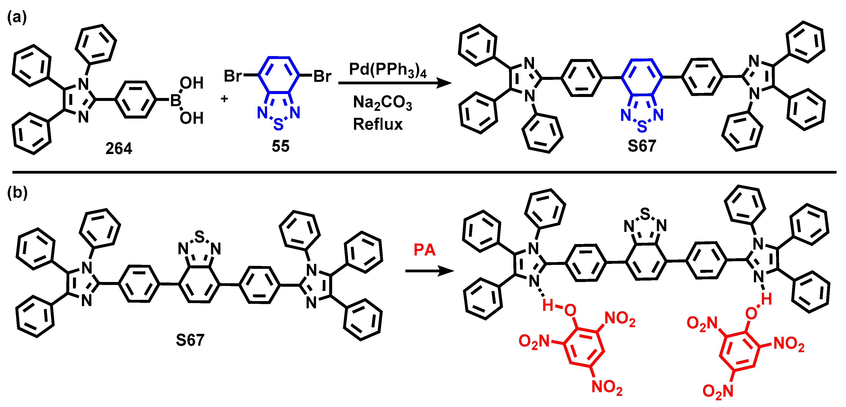
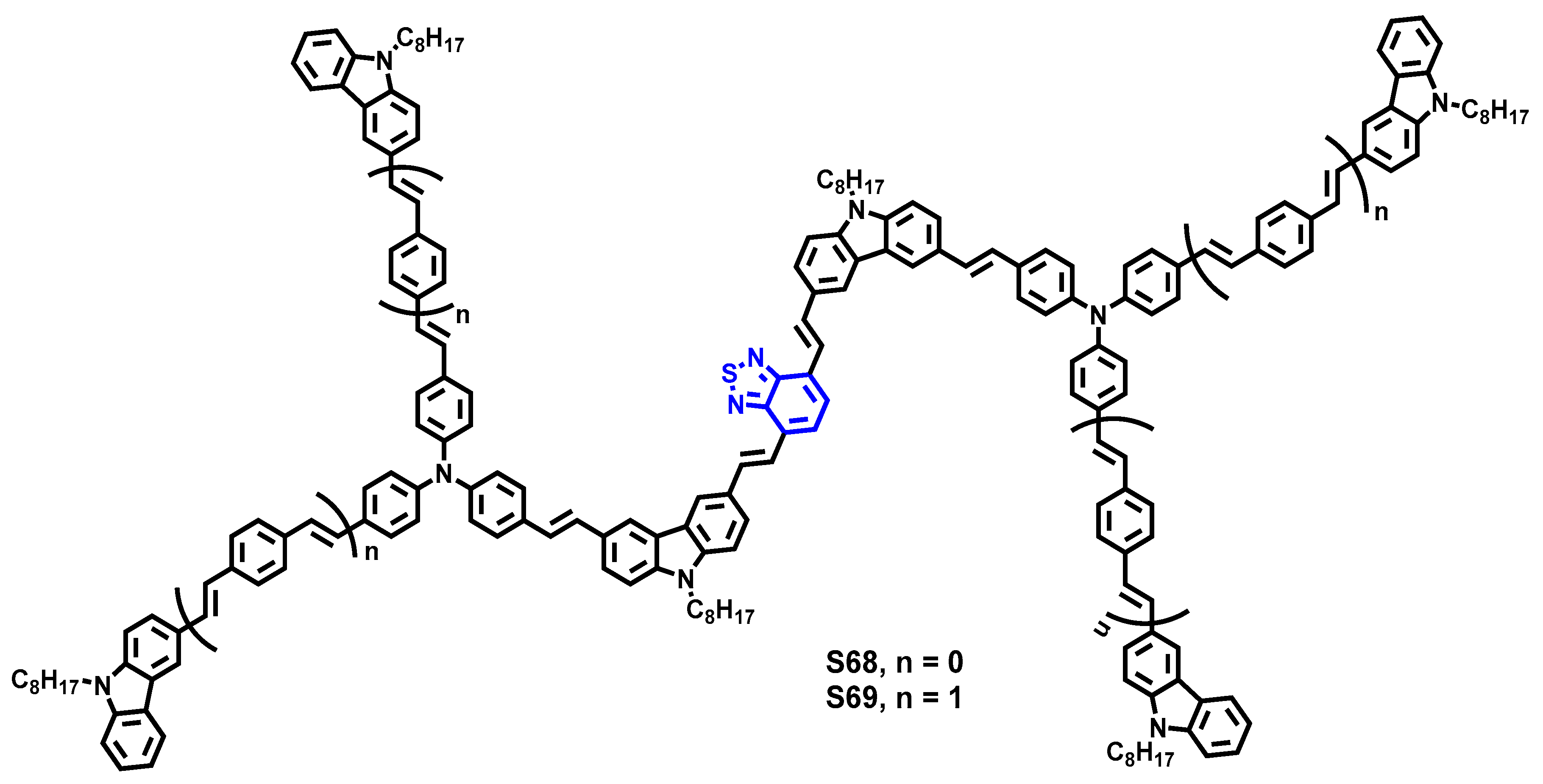
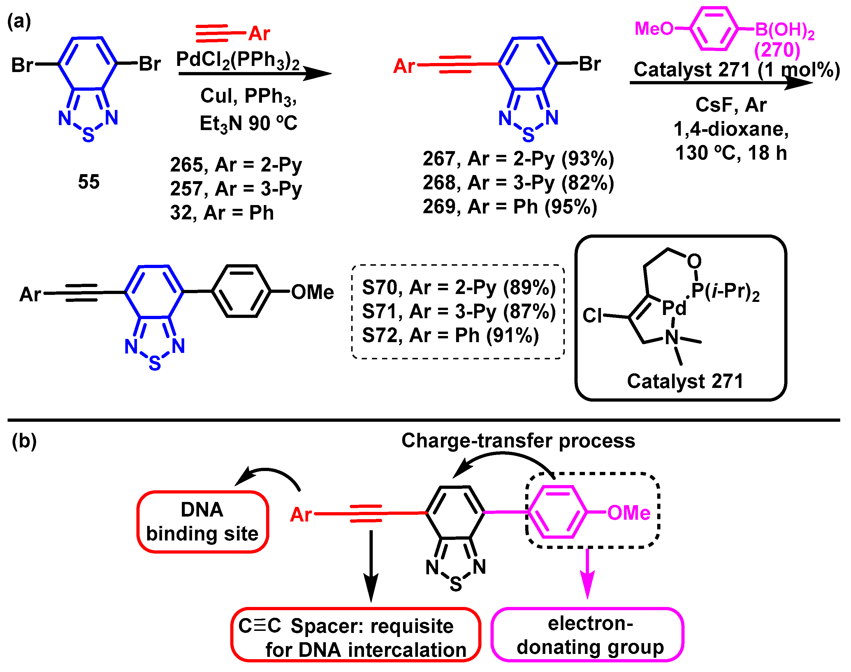
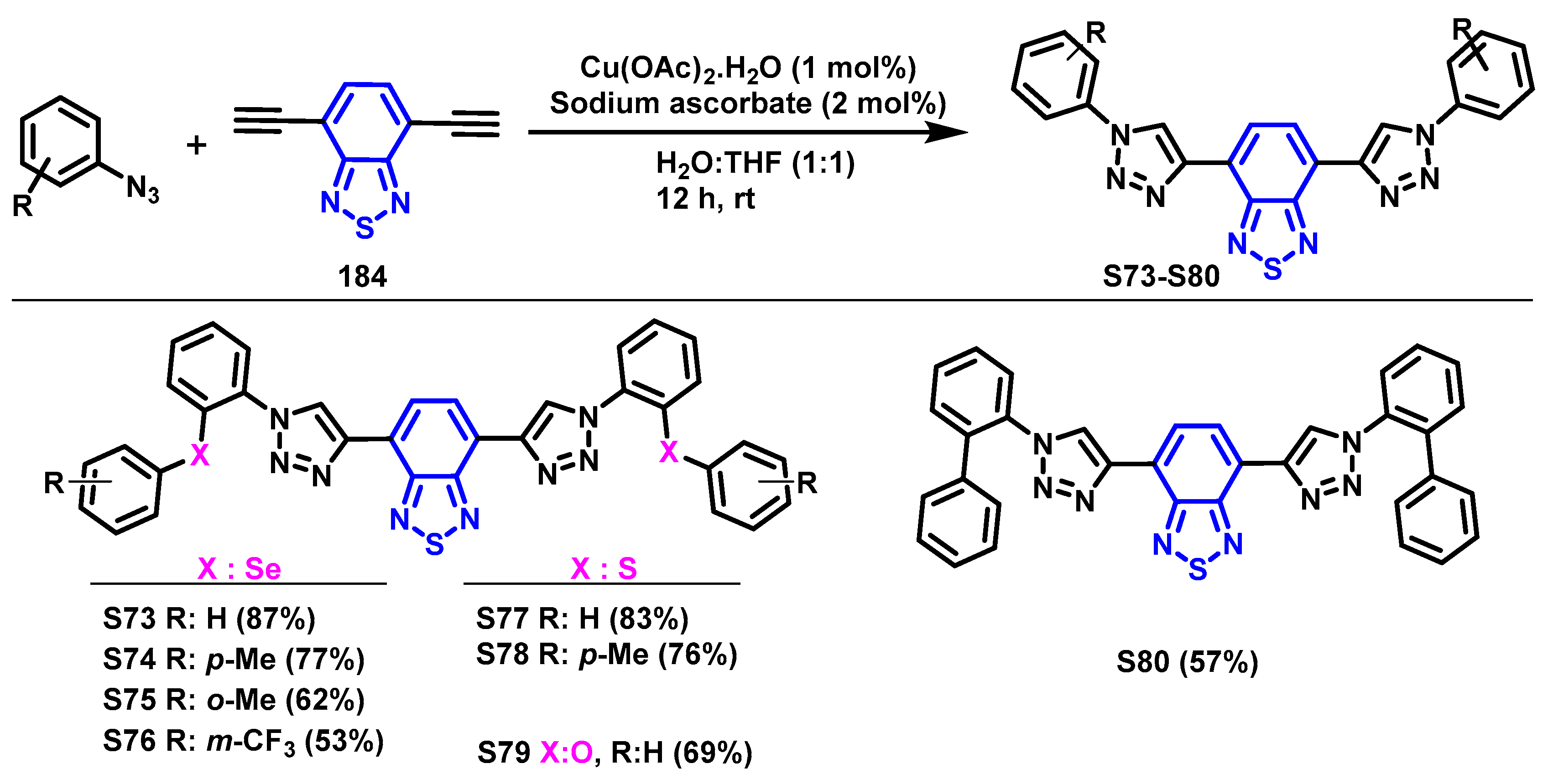
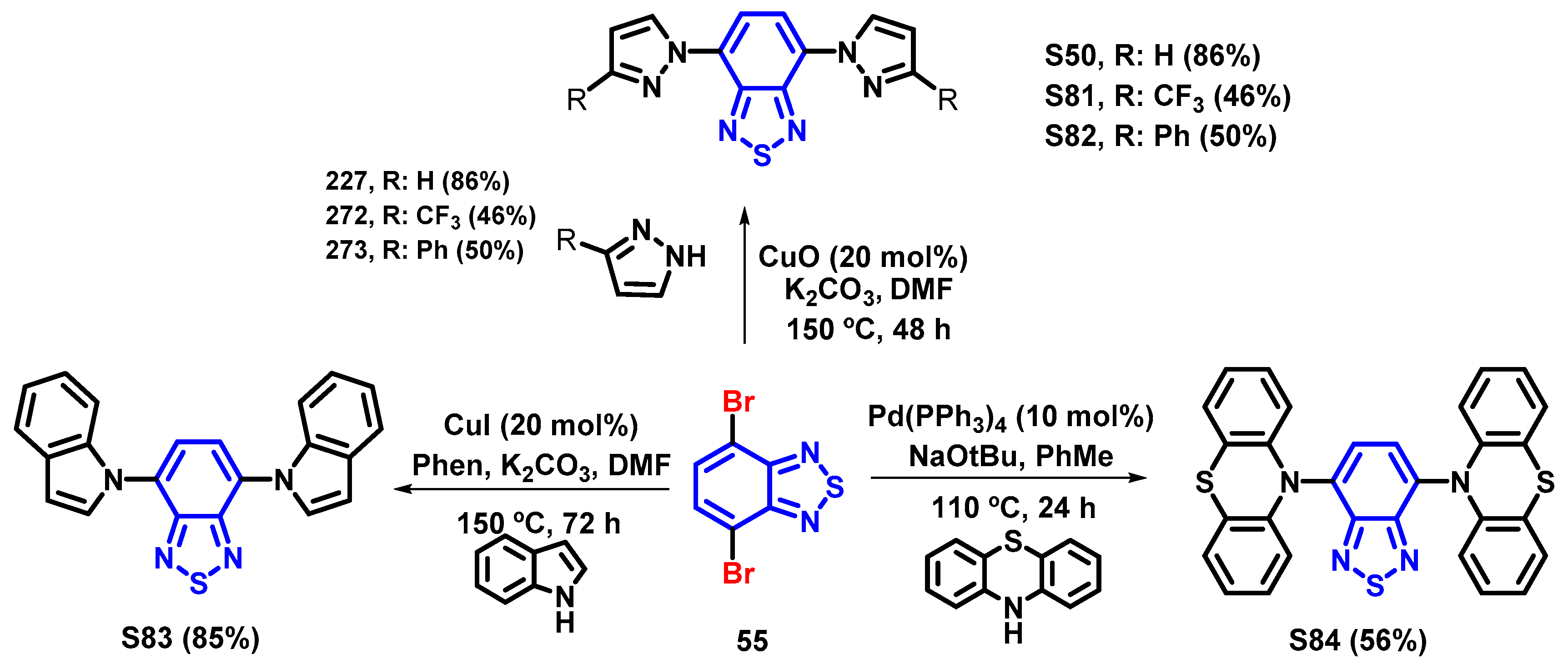
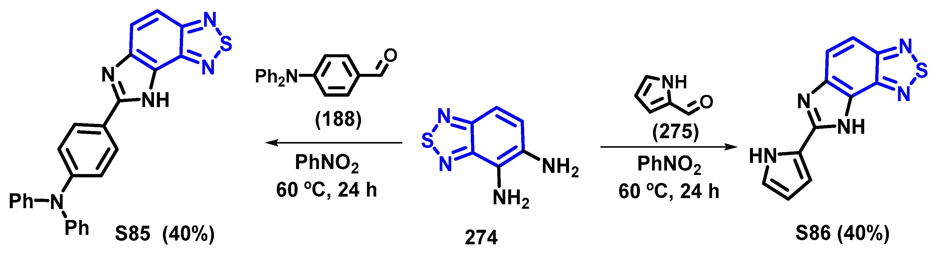
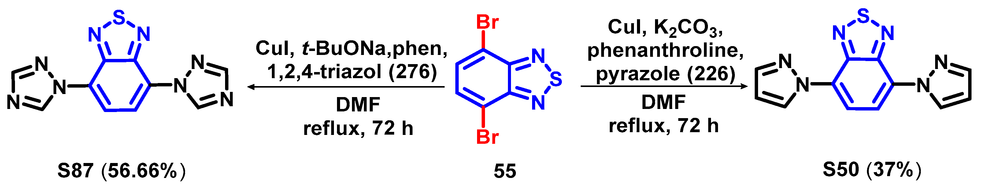
Scheme 1. General representations for (a) an optical chemosensor and (b) a chemodosimeter selective for a specific analyte. In (I), the analyte forms a covalent bond with the chromogenic or fluorogenic chemodosimeter, resulting in a shift in the absorption or emission spectra. In (II), the analyte interacts with the chemodosimeter, initiating a chemical reaction that produces an optical signal. In (III), the analyte reacts with the chemodosimeter, leading to the release of a chromogenic or fluorogenic moiety.
Chemosensors 12 00156 sch001
Scheme 2. Examples of fluorophore/chromophore cores (in blue) as signaling units in the design of fluorogenic and/or chromogenic chemosensors and chemodosimeters for detecting analytes [26,29,36,40,44,51,59,64,70,77,84,90,92,101,104].
Chemosensors 12 00156 sch002
Scheme 3. Representation for the molecular structure of 2,1,3-benzothiadiazole (BTD) and general applications for BTD compounds.
Chemosensors 12 00156 sch003
Scheme 4. Representative molecular orbital diagram of the mechanisms of (a) photoinduced electron transfer (PET) between LUMOs and HOMOs, (b) fluorescence resonance energy transfer (FRET), (c) electron exchange (EE), and (d) excited-state intramolecular proton transfer (ESIPT).
Chemosensors 12 00156 sch004
Scheme 5. Examples of BTD derivatives obtained from the corresponding ortho-phenylenediamine via treatment with thionyl chloride.
Chemosensors 12 00156 sch005
Scheme 6. Examples of synthesis of BTD derivatives from ortho-phenylenediamines and their further modification: (a) Aromatic substitution to replace fluorine for alkyloxy groups described by Lee et al. [291,292]. (b) Synthesis of 27 followed by Suzuki [293,294,295] and Sonogashira [296] reactions. (c) Synthesis of butterfly-shaped systems 33 via the Sonogashira reaction described by Wang et al. [297]. (d) Suzuki reaction followed by an oxidative cyclization using FeCl3 to obtain 36 described by Han et al. [298] and (e) a similar product, 39, obtained from 38 [299].
Chemosensors 12 00156 sch006
Scheme 7. (a) Synthesis of dimethyl-BTD 41 and its possible chemical modification on methyl moieties [302,303]. (b) Modification of BTD 41 to synthesize D–π–A-conjugated copolymer 48 for application in bulk-heterojunction solar cells described by Iyer et al. [304,305].
Chemosensors 12 00156 sch007
Scheme 8. Synthesis of 4-formyl-BTD described by Vanelle et al. [307].
Chemosensors 12 00156 sch008
Scheme 9. (a) Synthesis of BTD-Br2 (55) and (b) examples of more common modifications of 55 via replacement of bromine.
Chemosensors 12 00156 sch009
Scheme 10. (a) General representation of BTD extrusion. (b) Synthesis of BTDs as protective/deprotective approaches to modify o-phenylenediamines. (c) Synthesis of BTD (15, protection step) and bromination (modification) to achieve BTD-Br2 (55), followed by extrusion (deprotection) to afford 3,6-dibromo-ortho-phenylenediamine.
Chemosensors 12 00156 sch010
Scheme 11. (a) Synthesis of BTDs as protective/deprotective approaches to modify o-phenylenediamines exemplified by phenazine [314,315], imidazole [316,317] and triazole [318]. (b) Extending the π-system at the 5 and 8 positions of the quinoxalines via classical Suzuki and Sonogashira reactions is challenging [320].
Chemosensors 12 00156 sch011
Scheme 12. Replacement of bromine at BTD-Br2 (55) for other groups, representation of possible connectors, and some literature examples of systems synthesized by this strategy, with their corresponding applications [112,126,139,141,143,146,157,165,166].
Chemosensors 12 00156 sch012
Scheme 13. (a) Synthesis of 4-bromo-7-nitro-BTD (61). (bd) show some examples of coupling reactions using 61 as a synthetic intermediate [325,326,327,328].
Chemosensors 12 00156 sch013
Scheme 14. Synthesis of 4-nitro-BTD (92) and its reduction to 4-amino-BTD (93).
Chemosensors 12 00156 sch014
Scheme 15. Synthesis of 5,6-dinitro-BTD and its further transformation into three classes of compounds: Class I: fused heterocycles, Class II: benzobisthiadiazoles, and Class III: phenazines, quinolines, and imidazoles.
Chemosensors 12 00156 sch015
Scheme 16. Modification of 5,6-dinitro derivatives via intramolecular cyclization to afford heterocycles fused to BTD [376].
Chemosensors 12 00156 sch016
Scheme 17. (a) General representation of synthesis of benzobisthiadiazoles from 5,6-dinitro-BTD (62) and (b) synthesis of compound 104, bearing a benzobisthiadiazole unit, for NIR-II fluorescence imaging and photothermal therapy applications [390].
Chemosensors 12 00156 sch017
Scheme 18. (a) Synthesis of 5,6-dinitro-BTDs followed by a reduction in nitro groups to afford the 5,6-diamino-BTDs. (b) Phenazines, quinoxalines, and imidazoles obtained from 5,6-diamino-BTD.
Chemosensors 12 00156 sch018
Scheme 19. Synthesis of phenazines via condensation between 5,6-diamino-BTDs and cyclic-1,2-dicarbonyl compounds [403,404].
Chemosensors 12 00156 sch019
Scheme 20. (a) General synthesis of quinoxaline and (b) an exemple of synthesis of quinoxaline fused to BTD via condensation between a 5,6-diamino-BTD and an acyclic 1,2-dicarbonyl compound [408].
Chemosensors 12 00156 sch020
Scheme 21. Synthesis of quinoxaline-fused BTDs, followed by their oxidation to phenazine-fused BTDs, as described by Nakamura et al. [415].
Chemosensors 12 00156 sch021
Scheme 22. (a) Synthesis of imidazole-fused BTDs via condensation between a 5,6-diamino-BTD and an aldehyde. (b) Synthesis of the imidazole-BTD hybrid 118, a precursor for polymer synthesis, as described by Zhang et al. [416,417]. (c) Synthesis of imidazole-BTD 121, as described by Nurulla et al. [418].
Chemosensors 12 00156 sch022
Scheme 23. (a) The synthetic pathways of chemosensor S1 and (b) its sensing mechanism, proposed by Zhao et al. [423].
Chemosensors 12 00156 sch023
Scheme 24. (a) Synthesis of S2 according to Zhao et al. [424]. (b) Fluorescence response of S2 with Hg2+ ion in the coexistence of other metal ions.
Chemosensors 12 00156 sch024
Scheme 25. (a) Synthetic route of chemodosimeter S3 designed by Shen et al. [425] and (b) the proposed mechanism for its reaction with Hg2+.
Chemosensors 12 00156 sch025
Scheme 26. (a) Synthetic route of chemodosimeter S4 presented by Shen et al. [426] and (b) its reaction with Hg2+.
Chemosensors 12 00156 sch026
Scheme 27. (a) Synthetic route for chemodosimeters S5 and S6 and (b) mechanistic proposal for their reaction with Hg2+ and CH3Hg+ according to Zou and Tian [427].
Chemosensors 12 00156 sch027
Scheme 28. (a) Synthetic route of chemodosimeter S7 according to Guo et al. [428] and (b) its proposed reaction with Hg2+.
Chemosensors 12 00156 sch028
Scheme 29. Synthetic route of chemosensor S8, described by Liu et al. [429].
Chemosensors 12 00156 sch029
Scheme 30. (a) Synthesis of S8 according to Huang et al. [430]. (b) Suggested sensing mechanism of S8 toward Hg2+. (c) Representation of an INHIBIT logic gate and INHIBIT truth table.
Chemosensors 12 00156 sch030
Scheme 31. Route for the synthesis of chemosensor S9 presented by Hua et al. [433].
Chemosensors 12 00156 sch031
Scheme 32. Synthetic route of chemosensor S10, designed by Xie et al. [434].
Chemosensors 12 00156 sch032
Scheme 33. Structures of compounds S11 and S12, synthesized by Xie et al. [436].
Chemosensors 12 00156 sch033
Scheme 34. The synthetic route for S13 involves condensing 128 with the trisamino linker tris(2-aminoethyl)amine (167) in CDCl3 for 12 h [437] and 36 h [438].
Chemosensors 12 00156 sch034
Scheme 35. Synthetic scheme illustrating the direct C–H heteroarylation of 17-crown-5 (168) with different BTD-based auxophores to afford S14, S15, and S16, designed by Moser et al. [439].
Chemosensors 12 00156 sch035
Scheme 36. Synthetic route of chemosensor S17, developed by Xia et al. [443].
Chemosensors 12 00156 sch036
Scheme 37. Structure and synthesis of (a) 1,4-disubstituted triazolyl BTD derivatives S18S19, (b) synthesis of S20S22, and (c) synthesis of 1,5-disubstituted triazolyl BTD derivative S23 reported by Xie et al. [444].
Chemosensors 12 00156 sch037
Scheme 38. Synthetic route proposed by Zhu et al. [221] of S24 and S25.
Chemosensors 12 00156 sch038
Scheme 39. (a) Schematic illustration of the synthetic route of S26 and (b) proposed sensing mechanism for the chemosensor in ethanol in the presence of Co2+ and Cu2+ by Dinçalp et al. [446].
Chemosensors 12 00156 sch039
Scheme 40. (a) Synthesis of chemosensor S27 [450]. (b) Sensing mechanism for S27 and Cu2+.
Chemosensors 12 00156 sch040
Scheme 41. Compounds S28S31, synthesized by Moro et al. [451].
Chemosensors 12 00156 sch041
Scheme 42. (a) Synthesis of S32 described by Bunz et al. [452]. (b) Schematic binding mode of S32 with Ni2+ salts.
Chemosensors 12 00156 sch042
Scheme 43. (a) Synthesis of chemosensor S33 proposed by Zhao et al. [453]. (b) Sensing mechanism for S33 and Fe3+.
Chemosensors 12 00156 sch043
Scheme 44. General representation of possible interactions of an optical chemosensor with CN and F, based on an acid–base reaction or HB interactions.
Chemosensors 12 00156 sch044
Scheme 45. (a) Synthetic route of S34 by Qu et al. [456]. (b) Schematic representation for the interaction of S34 with CN.
Chemosensors 12 00156 sch045
Scheme 46. The synthesis of BTD derivative S35, described by Zhao et al. [457].
Chemosensors 12 00156 sch046
Scheme 47. (a) Synthetic route of chemosensors S36 and S37. (b) Interaction mechanism between S36 and S37 and F [460].
Chemosensors 12 00156 sch047
Scheme 48. (a) Synthetic route of fluorescent presented by Yu and Dong [461] for chemodosimeter S38 and (b) mechanism of cleavage of Si-O induced by F.
Chemosensors 12 00156 sch048
Scheme 49. (a) Synthetic route of S39 and (b) the proposed mechanism for the detection of CN [463].
Chemosensors 12 00156 sch049
Scheme 50. (a) Synthesis of the chemodosimeter S40, (b) synthesis of S40, (c) CN detection reaction by 212, and (d) CN detection reaction by S40, according to Wu et al. [464].
Chemosensors 12 00156 sch050
Scheme 51. (a) Synthesis of chemodosimeters S41S43. (b) CN detection mechanism in S41 [465].
Chemosensors 12 00156 sch051
Scheme 52. (a) Synthetic route of S44. (b) Sensing mechanism of S44 on detection of CN [466].
Chemosensors 12 00156 sch052
Scheme 53. (a) Synthesis of S45S47. (b) Graphical representations of the interaction of S45S47 with F and Cu2+ in DMSO. (c) Process of Cu2+ and F detection [467].
Chemosensors 12 00156 sch053
Scheme 54. (a) Synthesis of fluorophores S48 and S49 and (b) the sensing mechanism of detection of Cu2+ [468].
Chemosensors 12 00156 sch054
Scheme 55. Synthetic route of S50, reported by Liu et al. [429].
Chemosensors 12 00156 sch055
Scheme 56. (a) Molecular structures of cysteine (Cys), homocysteine (HCy), and glutathione (GSH). (b) Chemodosimeters S51, S52, and S53. (c) Representation of Smiles rearrangement.
Chemosensors 12 00156 sch056
Scheme 57. (a) Synthetic route for S51, described by Yoon et al. [469]. (b) Sensing reaction between S54 and Cys or HCy.
Chemosensors 12 00156 sch057
Scheme 58. (a) Synthetic route of S52 and (b) mechanism involved in the interaction between S52 and biothiols, proposed by Liu et al. [470].
Chemosensors 12 00156 sch058
Scheme 59. (a) Synthesis of S53. (b) Sensing mechanism of S53 with Cys, proposed by Wang et al. [471].
Chemosensors 12 00156 sch059
Scheme 60. (a) Synthetic route proposed for compound S54 by Gao et al. [474]. (b) Reaction of detection of SO2.
Chemosensors 12 00156 sch060
Scheme 61. (a) Synthesis of compound S55. (b) Reaction of S55 with α-KA, demonstrated by Guo et al. [475].
Chemosensors 12 00156 sch061
Scheme 62. Synthetic routes of chemosensors (a) S56 and (b) S57. (c) Illustration of the sensing mechanisms in S56 and S57 films with cadaverine (246). The emission is quenched (illustrated by the grey boxes) with a short exposure time or low cadaverine concentration. At longer exposure times or high cadaverine (246) concentrations, an aza-Michael addition occurs with subsequent imine formation, changing the emission color and PL intensity (illustrated by the colored boxes on the right) [477].
Chemosensors 12 00156 sch062
Scheme 63. (a) Synthesis of compounds S58S61, described by Loch, Burn, and Shaw [480]. (b) Structures of analytes tested in their work.
Chemosensors 12 00156 sch063
Scheme 64. (a) Synthetic route of S62. (b) Chemical structures of the weakly fluorescent S62 and its highly fluorescent products 257 and 258, obtained from the reaction with oxalyl chloride and phosgene, respectively [481].
Chemosensors 12 00156 sch064
Scheme 65. (a) Synthetic route of S63 and (b) sensing mechanism on detection of hydrazine [482].
Chemosensors 12 00156 sch065
Scheme 66. (a) Synthesis of S64 via Suzuki reaction between 262 and 125, reported by Neto et al. [483]. (b) Sensing reaction for hydrazine detection.
Chemosensors 12 00156 sch066
Scheme 67. Synthetic route of S65 and S66, described by Limberger et al. [484].
Chemosensors 12 00156 sch067
Scheme 68. (a) Synthetic route of the chemosensor S67. (b) Sensing mechanism of S67 and PA according to Iyer et al. [485].
Chemosensors 12 00156 sch068
Scheme 69. Representation of the molecular structures of S68 and S69 [486].
Chemosensors 12 00156 sch069
Scheme 70. (a) Synthesis of BTDs S70S72. (b) General architecture of BTDs S70S72, synthesized by Neto et al. [490].
Chemosensors 12 00156 sch070
Scheme 71. Synthesis of bis-triazolyl-BTDs S73S80 via CuAAC [491].
Chemosensors 12 00156 sch071
Scheme 72. Synthesis of BTDs S50, S81, S82, S83, and S84, described by Moro et al. [492].
Chemosensors 12 00156 sch072
Scheme 73. Synthetic routes of S85 and S86, described by Molina et al. [497].
Chemosensors 12 00156 sch073
Scheme 74. Synthetic route of chemosensors S87 and S50 [498].
Chemosensors 12 00156 sch074
Table 1. Properties of BTD-based fluorogenic/chromogenic chemosensors and chemodosimeters for detection of ionic and molecular analytes.
MoleculeAnalyteSolvent MediumMechanismLODRef.
S1Hg2+THF/H2O (9:1, v/v)S,O-chelation
D–A structure
0.0131 µmol L−1[423]
S2Hg2+Acetone-H2O (8:2, v/v)S,O-chelation
D–A structure
0.393 µmol L−1[424]
S3Hg2+THF/H2O (99:1, v/v)ICT
D–A–D structure
0.089 µmol L−1[425]
S4Hg2+THF/H2O (99:1, v/v)ICT
D-π-A-π-D structure
0.36 µmol L−1[426]
S5Hg2+
CH3Hg+
CH3CN/H2O (1:1, v/v)PET0.16 µmol L−1
0.8 µmol L−1
[427]
S6Hg2+
CH3Hg+
CH3CN/H2O (1:1, v/v)PET0.5 µmol L−1
1.0 µmol L−1
[427]
S7Hg2+HEPES buffer
(10 mmol L−1, pH = 7.4,
with 3% DMSO)
ICT and AIE0.090 µmol L−1[428]
S8Cu2+DMFTransference of electrons from imidazole to the empty d-orbital of Cu2+0.11 µmol L−1[429]
S8Hg2+WaterICT0.00093 µmol L−1[430]
S9Al3+HEPES buffer (50 vol% DMSO, pH = 7.0)AIE0.15 µmol L−1[433]
S10Ni2+CH3CNPET or CT-[434]
S11Cu2+HEPES buffer (pH 7.4)Metal complexation-[436]
S12Cu2+HEPES buffer pH 7.4Metal complexation-[436]
S13Cd2+CHCl3/MeCN (10:1) --[437]
S13Hg2+TrichloromethaneMetal complexation
AIE
-[438]
S14Na+THF:CH3CN (1:1, v/v)Conformational twist14.6 µmol L−1[439]
S15Na+THF:CH3CN (1:1, v/v)Conformational twist17.2 µmol L−1[439]
S16Na+THF:CH3CN (1:1, v/v)Conformational twist21.7 µmol L−1[439]
S17Fe3+
Cr6+
THFThermodynamic
stability of the newly rigid chain
D–A–D structure
3.04 μmol L−1
0.0435 µmol L−1
[443]
S18Ni2+, Hg2+, Cu2+, Co2+CH3CNMetal complexation-[444]
S19Cu2+HEPES buffer (pH 7.4)Metal complexation-[444]
S20Ni2+, Hg2+
Cu2+, Co2+
CH3CNMetal complexation-[444]
S21Ni2+, Hg2+ Cu2+, Co2+CH3CNMetal complexation-[444]
S22Not selectiveCH3CNMetal complexation-[444]
S23Hg2+CH3CNMetal complexation [444]
S24Cu2+CH3CNICT-[221]
S25Cu2+CH3CNICT-[221]
S26Co2+
Cu2+
Co2+
Cu2+
Co2+–F
EtOH
PhCN
Metal-to-ligand
charge transfer
(MLCT)
0.407 µmol L−1
0.550 µmol L−1
0.550 µmol L−1
3.70 µmol L−1
[446]
S27Cu2+
H2S
MeOH/PBS solution (9:1, v/v)-0.55 μmol L−1
0.55 μmol L−1
[450]
S28Ni2+CH3CNPET or CT-[451]
S29Ni2+CH3CNPET or CT-[451]
S30Ni2+CH3CNPET or CT-[451]
S31Ni2+CH3CNPET or CT-[451]
S32Ag+
Cu2+
Ni2+
H2OMetal complexation
3.80 μmol L−1
0.27 μmol L−1
0.56 μmol L−1
[452]
S33Fe3+0.2 mol L−1 HEPES buffer/pH 7.2 in 2 vol% DMSOICT0.036 μmol L−1[453]
S34CNPBS solutionAIE and ICT0.35 μmol L−1[456]
S35FTHFIntramolecular CT-[457]
S36FDMSOESIPT0.86 µmol L−1[460]
S37FDMSOESIPT4.25 µmol L−1[460]
S38FCH3CN/Tris–HCl buffer (9:1, v/v, pH 7.5)Cleavage of Si–O bonds1.7 µmol L−1[461]
S39CNTHF/H2O (99:1, v/v)ICT0.014 µmol L−1[463]
S40CNTHFICT 0.087 μmol L−1[464]
S41CNTHF/H2O (v/v, 1/9)ICT0.077 μmol L−1[465]
S42CNTHFICT-[465]
S43CNTHFICT-[465]
S44CNTHF/H2O (2:8, v/v)AIE0.134 µmol L−1[466]
S45FDMSODeprotonation-[467]
S46FDMSODeprotonation-[467]
S47FDMSODeprotonation-[467]
S48Cu2+
F
Br
CH3CNDeprotection TMS group-[468]
S49Cu2+
F
Br
CH3CNDeprotection TMS groupCu2+: 1.31 (λem 608/486 nm) to 2.28 ppb (λem 486/608 nm);
Br: 69.0 (λem 605/490 nm) to 88.2 ppb (λem 490/605 nm);
F: 0.13 ppb
[468]
S50HODMFElectron transfer–hydrogen bonding interaction55 µmol L−1[429]
S50TEA
TMEDA
DMFAggregation
Aggregation
0.400 µmol L−1
0.278 µmol L−1
[498]
S51Cys
Hcy
HEPES (0.01 mol L−1, pH 7.4)
/1% DMSO
PET0.1 µmol L−1
0.1 µmol L−1
[469]
S52GSH
Cys
Hcy
PBS solution (10 mmol L−1, pH 7.4/1 mmol L−1 CTAB)ICT0.089 µmol L−1
-
-
[470]
S53CysPBS solution (containing 20% DMSO, pH 7.4)ICT-[471]
S54SO2PBS solution (pH 7.4) containing
5% DMSO
ICT0.190 µmol L−1[474]
S55α-KAPBS solution (pH 5.7,
1 mmol L−1 CTAB)
ICT-[475]
S56Biogenic
amines
(cadaverine)
DCM
Thin films
PHT-
130 ppb
[477]
S57Biogenic amines
(cadaverine)
DCM
Thin films
PHT-
610 ppb
[477]
S58(+)-Methamphetamine
and fentanyl
FilmsPHT-[480]
S59FentanylFilmsPHT-[480]
S60(+)-Methamphetamine,
(±)-3,4-methylenedioxy-amphetamine, cocaine, and fentanyl
FilmsPHT-[480]
S61(+)-Methamphetamine, (±)-3,4-methylenedioxy-amphetamine, cocaine, and fentanylFilmsPHT-[480]
S62Oxalyl chloride
Phosgene
DCMICT0.003 µmol L−1
0.020 µmol L−1
[481]
S63HydrazineH2O/DMSO (4:6, v/v) solution (10 mmol L−1 HEPES
buffer, pH 7.4)
PET0.0847 µmol L−1[482]
S64HydrazineWaterICT0.340 µmol L−1[483]
S65Not tested---[484]
S66EtOH in waterEtOH/waterICT [484]
S67PAACN/H2O (8:2 v/v)ICT
D–π–A–π–D structure
0.0079 nmol L−1[485]
S68TNT
DNT
TolueneICT13 µmol L−1
20 µmol L−1
[486]
S69TNT
DNT
TolueneICT15 µmol L−1
18 µmol L−1
[486]
S70DNAPBS solution (pH 7.0)ICT1 ppm[490]
S71DNAPBS solution (pH 7.0)ICT1 ppm[490]
S72DNAPBS solution (pH 7.0)ICT1 ppm[490]
S73CT-DNA
BSA
DMSO (5%)/Tris-HCl buffer (pH 7.4)-1.05 µmol L−1
7.10 µmol L−1
[491]
S74CT-DNA
BSA
DMSO (5%)/Tris-HCl buffer (pH 7.4)-9.45 µmol L−1
7.30 µmol L−1
[491]
S75CT-DNA
BSA
DMSO (5%)/Tris-HCl buffer (pH 7.4)-7.65 µmol L−1
0.10 µmol L−1
[491]
S76CT-DNA
BSA
DMSO (5%)/Tris-HCl buffer (pH 7.4)-7.72 µmol L−1
7.27 µmol L−1
[491]
S77CT-DNA
BSA
DMSO (5%)/Tris-HCl buffer (pH 7.4)-0.30 µmol L−1
0.65 µmol L−1
[491]
S78CT-DNA
BSA
DMSO (5%)/Tris-HCl buffer (pH 7.4)-2.52 µmol L−1
7.52 µmol L−1
[491]
S79CT-DNA
BSA
DMSO (5%)/Tris-HCl buffer (pH 7.4)-6.60 µmol L−1
6.55 µmol L−1
[491]
S80CT-DNA
BSA
DMSO (5%)/Tris-HCl buffer (pH 7.4)-1.40 µmol L−1
2.22 µmol L−1
[491]
S81Water in acetoneWater/acetoneCT
ligand-to-ligand charge
transfer (LLCT)
-[492]
S82Water in acetoneWater/acetoneCT
LLCT
-[492]
S83Water in acetoneWater/acetoneCT
LLCT
-[492]
S84Water in acetoneWater/acetoneCT
LLCT
-[492]
S85Hg2+
AcO
p-nitrophenol
picric acid
CH3CNCoordination bond interaction
Hydrogen-bonding interactions
0.13 µg mL−1
1.07 mg mL−1
-
[497]
S86Hg2+
AcO
p-nitrophenol
picric acid
CH3CNCoordination bond interaction
Coordination bond interaction
Hydrogen-bonding interactions
159 µg mL−1
-
-
317 µg mL−1
[497]
S87TEA
EDA
TMEDA
MnO4
ClO
DMFAggregation
Aggregation
Aggregation
Hydrogen bond
Hydrogen bond
0.129 µmol L−1
0.635 µmol L−1
0.320 µmol L−1
9.820 µmol L−1
12.740 µmol L−1
[498]
Disclaimer/Publisher’s Note: The statements, opinions and data contained in all publications are solely those of the individual author(s) and contributor(s) and not of MDPI and/or the editor(s). MDPI and/or the editor(s) disclaim responsibility for any injury to people or property resulting from any ideas, methods, instructions or products referred to in the content.

