Article

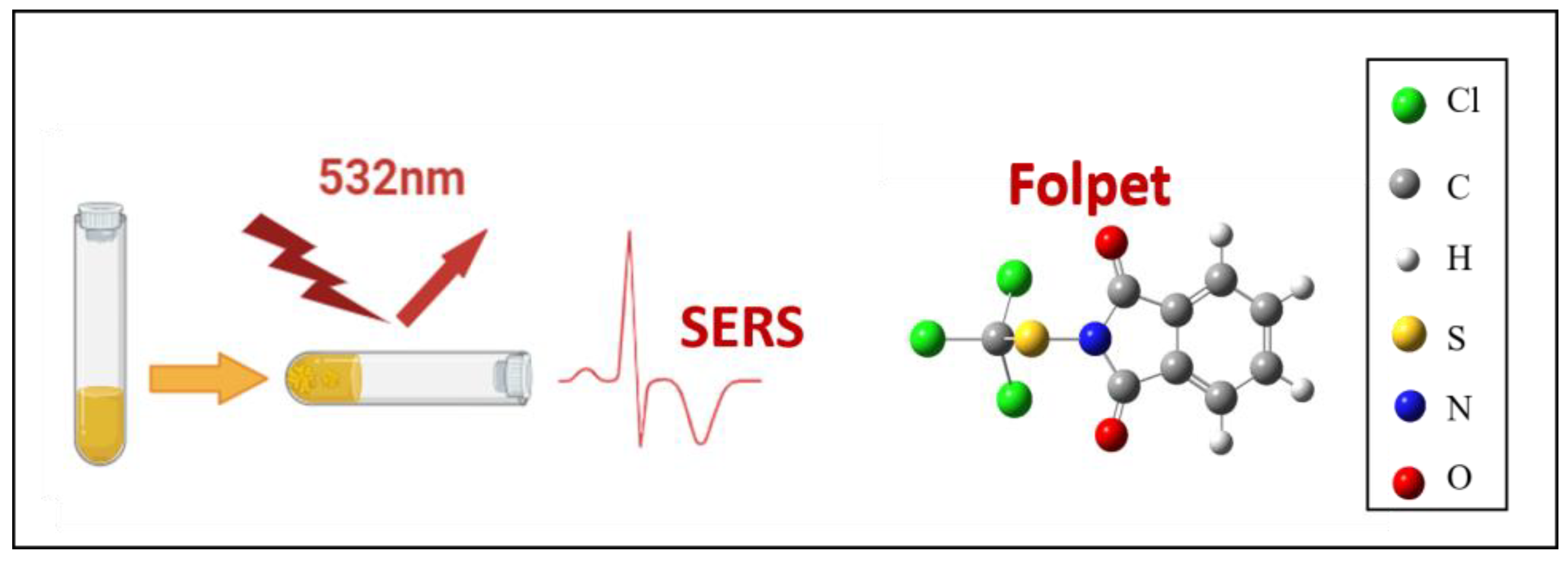
Sensitive Detection of Fungicide Folpet by Surface-Enhanced Raman Scattering: Experimental and Theoretical Approach

by
Oumaima Douass
1,
Bousselham Samoudi
1 and
Santiago Sanchez-Cortes
2,*
1
Intelligent System Design Laboratory, Research Team: Optics, Materials and Systems, Department of Physics, Faculty of Sciences, Abdelmalek Essâadi University, M’ Hannech II, P.O. Box 2121, Tétouan 93030, Morocco
2
Instituto de Estructura de la Materia, Consejo Superior de Investigaciones Científicas (IEM-CSIC), 28006 Madrid, Spain
*
Author to whom correspondence should be addressed.
Chemosensors 2024, 12(9), 186; https://doi.org/10.3390/chemosensors12090186
Submission received: 5 August 2024 / Revised: 28 August 2024 / Accepted: 6 September 2024 / Published: 12 September 2024

Abstract

:
In this work, Surface-Enhanced Raman Spectroscopy (SERS) was employed as an effective detection technique for folpet, characterized by its notable specificity and sensitivity. The investigation involved the use of UV–Vis, Raman, and SERS spectroscopy of folpet at different concentrations for a comprehensive study of plasmon-driven effects such as plasmon resonance, plasmon hybridization, and electric field enhancement resulting in the SERS’ intensification. Specifically, SERS detection of folpet solutions at concentrations below 100 µM is presented in detail by using Ag nanoparticles prepared with hydroxylamine reduction. The experimentation encompassed diverse conditions to optimize the detection process, with Raman spectra acquired for both folpet powder and aqueous solution of folpet at the natural pH. SERS analyses were conducted across a concentration range of 9.5 × 10⁻⁸ to 1.61 × 10⁻⁴ M, employing 532 nm excitation. The differences in the spectral profiles observed for folpet Raman powder and SERS are ascribed to N–S cleavage; these changes are attributed to plasmon catalysis induced by the used Ag nanoparticles. Transmission electron microscopy (TEM) was also important in the present analysis to better understand which mechanism of nanoparticles aggregation is more favorable for the SERS detection regarding the formation of hot spots in the suspension. Complementing the experimental data, the molecular structure and theoretical Raman spectra of the folpet molecule were calculated through density functional theory (DFT) methods. The outcomes of these calculations were crucial in the elucidation of folpet’s vibrational modes. The culmination of this research resulted in the successful detection of folpet, achieving a notable limit of detection at 4.78 × 10⁻⁸ M. This comprehensive approach amalgamates experimental and theoretical methodologies, offering significant insights into the detection capabilities and molecular characteristics of folpet via SERS analysis.
Keywords:
SERS; folpet; DFT; degradation

1. Introduction

Folpet is a fungicide belonging to the phthalimide family with the chemical formula (C9H4Cl3NO2S). It is mainly used in plant protection products and phytopharmaceutical specialties, which may contain folpet in combination with other fungicides. It is also found in certain types of biocides, such as protective products for coatings, paints, plastics, and buildings [1]. In the pursuit of effective and precise pesticide detection, the convergence of experimental and theoretical methodologies has become pivotal [2]. Surface-enhanced Raman Spectroscopy (SERS), Raman spectroscopy, and density functional theory (DFT) analysis collectively form a robust triad for investigating folpet detection, which contributes to advancements in environmental monitoring, food safety, and pesticide management [3]. SERS has revolutionized the landscape of molecular detection by offering exceptional sensitivity and specificity. SERS is a technique that leads to a huge, amplified Raman signal [4] and which relies on plasmonic effects derived from the specific light-matter interaction taking place in metal nanostructures of certain metals. In particular, the localized surface plasmon resonance (LSPR) effect is considered the plasmon-driven effect responsible for the enhancement of the electric field on metal nanoparticles, leading to spectacular increase in the Raman signal and the luminescence quenching [5]. Other important plasmon effects that we have also considered in this work were the shift of plasmon resonance, the hybridization of plasmons between nanoparticles leading to the appearance of new plasmon resonances in the extinction spectra in the presence of the pesticide, and the plasmon-driven catalysis induced by plasmon excitation on folpet.
The distinct advantage of SERS lies in its ability to enhance weak Raman signals, thereby improving the detection limits and facilitating the identification of folpet [6]. Conventional Raman spectroscopy, while lacking the signal enhancement observed in SERS, remains a powerful and widely used technique for molecular characterization. It relies on inelastic scattering of monochromatic light, revealing vibrational information about chemical bonds [7]. In the context of folpet detection, Raman spectroscopy provides a straightforward and non-destructive tool for the identification of characteristic vibrational modes. However, its sensitivity may be limited, especially when dealing with low concentrations [2]. The utilization of the density functional theory (DFT) in theoretical exploration stands as a crucial complement to experimental methodologies. DFT serves as a computational tool that enables the calculation of the essential molecular features, electronic configuration, and vibrational frequencies of folpet, eliminating the necessity for immediate experimentation [8]. Through the application of DFT, the objective of this work is to elucidate the theoretical Raman spectra of the folpet molecule, thereby deepening our comprehension of its vibrational modes [9], as well as the assignment of the experimental vibrational spectra to better analyze the Raman and SERS spectra.
In the present work, UV–Vis, Raman, and SERS spectroscopies were used in the detection and structural characterization of folpet. The analysis of the pesticide degradation was also carried out on the basis of the spectral modifications in the SERS spectra. The folpet degradation can be attributed to the plasmon catalysis process [10], a plasmon-driven effect occurring due to the decay of surface plasmons, which excites ‘hot carriers’ that are able to favor chemical reactions of adsorbates linked to the surface by decreasing the necessary activation energy [11]. The degradation of folpet seems be a process favorable for the detection of the pesticide on silver surfaces by SERS due to the higher SERS activity of the resulting products. In previous works, we have demonstrated that the existence of Ag clusters on the surface is a key factor that fosters these catalytic processes [12] because the probability of hot carrier transfer is intensified in these adatom structures [13]. In addition, the formation of surface Ag adatoms is increased when using the Ag nanoparticles synthetized by using hydroxylamine hydrochlorate (AgH NPs) due to the effect of chloride on the surface [14]. For this reason, we have employed this substrate in the SERS analysis of folpet (Figure 1).
Theoretical DFT calculation of folpet was also performed and used as an aid for the vibrational analysis and following the chemical degradation of folpet. Remarkably, no prior investigations have been conducted on the Surface-Enhanced Raman Spectroscopy (SERS) or the density functional theory (DFT) of folpet up to the present moment. Numerous articles address the detection of pesticides, employing similar methodologies, including a study conducted by R.J.G. Rubira et al. [15] entitled “SERS Detection of Prometryn Herbicide Based on Its Optimized Adsorption on Ag Nanoparticles” and other studies by Marcelo J.S. Oliveira et al. [16,17] entitled “Detection of Thiabendazole Fungicide/Parasiticide by SERS: Quantitative Analysis and Adsorption Mechanism” and “Surface-Enhanced Raman Scattering of Thiram: Quantitative and Theoretical Analyses.”

2. Experimental and Theoretical Methods

2.1. Materials

AgNO3, hydroxylamine, NaOH, potassium nitrate (KNO3), ethanol, and folpet were purchased from Sigma-Aldrich (analytical reagent). All aqueous solutions used for the silver colloid synthesis were prepared using Millipore MilliQ water.

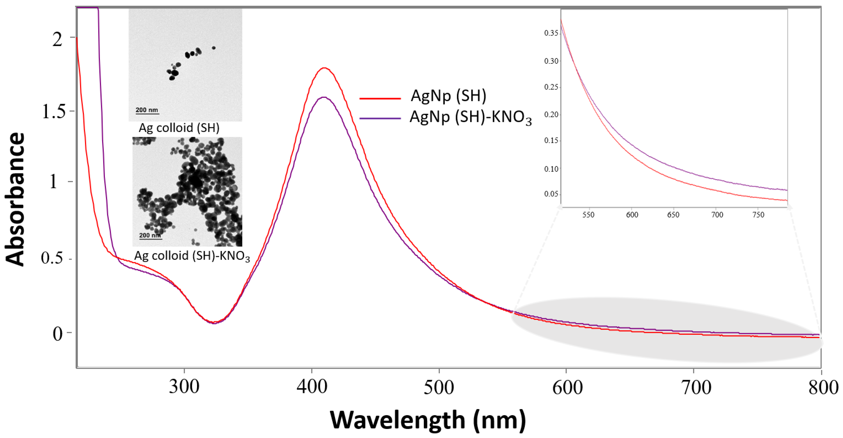
2.2. Preparation of Ag Nanoparticles

AgNPs were prepared by the chemical reduction method, employing hydroxylamine as the reducer [18]. The method consists of adding 300 µL of sodium hydroxide (1 M) to 90 mL of hydrochloride hydroxylamine aqueous solution (1.66 × 10 −3 M) under stirring. Then, 10 mL of Ag nitrate aqueous solution (1 × 10⁻² M) was added to this mixture, drop by drop. Finally, the colloidal suspension remained 15 min under stirring and then kept at room temperature for at least 24 h before starting the experiments. The suspension of Ag nanoparticles (AgH colloid) showed a grayish-yellow color.

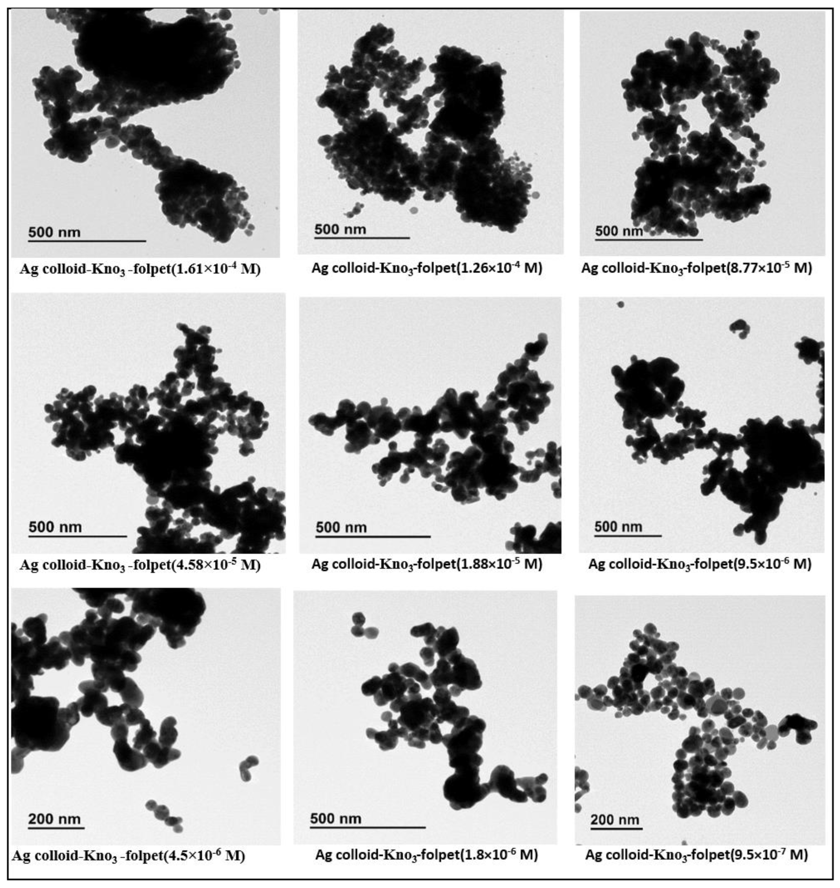
2.3. Characterization of AgNPs: UV-Vis and TEM Images

The UV-Vis absorption spectrum of neat AgNPs showed the plasmon of the Ag colloid with a maximum at 410 nm. The obtained AgNPs were characterized by transmission electron microscopy (TEM) [19] to determine their morphology. The TEM images showed that the main part of the hydroxylamine AgNPs colloidal suspension (AgH) has an almost spherical shape.

2.4. Preparation of Samples

The stock solution of the folpet prepared at a concentration of 10−3 M was prepared in ethanol to perform the UV-vis experiments. The sample for UV-vis spectroscopy of the colloidal suspension of AgNPs was prepared by solving 500 µL of the colloid in 2 mL of water. The UV-vis spectra were also obtained for the colloidal suspensions aggregated by the addition of potassium nitrate (KNO3), because all samples for SERS were activated by adding 20 µL of a 0.5 M potassium nitrate solution to 500 µL of colloids. This activation method gives rise to a homogeneous aggregation of Ag nanoparticles, as demonstrated by the TEM micrographs. The samples for the UV-vis spectroscopy of the AgNP suspension containing folpet were prepared by adding different amounts of the 10−3 M folpet solution to 500 µL of the aggregated colloid (after the addition of nitrate). Quartz cells of 1 cm optical path length were used.
A folpet solution with a concentration of 10−2 M was prepared in ethanol to perform the normal Raman analysis of the pesticide solution, while the Raman spectrum of the powder was registered on the solid microcrystal deposited on a quartz slide.
The samples for the SERS analysis were prepared by adding different aliquots of initial folpet 10−3, 10−4, and 10−5 M stock ethanol solutions to 500 µL of the AgH colloid, to which 20 µL of 0.5 M KNO3 were previously added to activate the suspension. By this method, the following final concentrations of folpet were prepared: 1.61 × 10⁻⁴, 1.26 × 10⁻⁴, 8.77 × 10⁻⁵, 4.58 × 10⁻⁵, 1.88 × 10⁻⁵, 9.5 × 10⁻⁶. 4.5 × 10⁻⁶, 1.8 × 10⁻⁶, 9.5 × 10⁻⁷, 1.8 × 10⁻⁷, and 9.5 × 10⁻⁸ M.
The external standard method [20] involves adding a specific amount of folpet to an AgNP colloid solution with 20 µL of KNO3 to prepare a control sample. This sample is then processed and analyzed for detection. For a detailed explanation, refer to Scheme S1 and Tables S2 and S3 of the external standard method in the Supporting Information File.

2.5. Instrumentation

The UV/VIS/NIR absorbance spectra of the folpet solutions and of the colloidal dispersion were obtained with a Shimadzu 3600 UV/VIS/NIR spectrometer equipped with 2H and W lamps as sources and a photomultiplier (UV/VIS), with a InGaAs and a PbS (NIR) as detectors. The baseline reference sample was prepared with 2 mL of MilliQ water. UV Probe software was used to launch the measurements. The Raman and SERS spectra were measured with a Renishaw inVia Raman spectrometer coupled with an optical microscope (Leica) equipped with Peltier-cooled CCD detector. All the Raman and SERS spectra were measured by using a laser at 532 nm (frequency-doubled Nd-Yag laser) as the excitation line, using an edge filter and 1800 L/mm grating working at 532 nm. Spectra were collected in the range from 2000–100 cm−1. The software WIRE 5.6 was used to launch the acquisitions. The TEM studies were conducted using a JEM 1400 PLUS JEOL 120 KV TEM, with a filament LaB6 to generate the electron beam. The images were recorded using a Gatan OneView CMOS camera.

2.6. DFT Calculations

The DFT calculation was performed using the Gaussian 09 program [21]. The molecule was constructed and visualized using the GaussView 6 program. Geometry optimization is a standard chemistry–physical calculation used to find the lowest energy or the largest relaxed conformation for a molecule; it is performed by finding the first derivative of the energy with respect to the distance between different atoms, known as the gradient. At the stationary point, this gradient is zero; because the gradient is the negative of the force, the forces are also zero at such point. The optimization structures of all molecules were obtained with the DFT method using the three-parameter hybrid-functional of Becker (B3LYP), with a 6-311++G (d,p) basis set [22]; this process was used for folpet and degraded folpet. Additionally, for the optimization structure of the degraded folpet complexes with silver atoms, the same functional and basis sets were used for C, H, N, and O atoms and LANL2DZ for the Ag atoms. The optimized geometries were confirmed to be minima on the Potential Energy Surface (PES), with all vibrational frequencies being positive. The Raman activity was calculated using optimized geometry.

3. Results and Discussion

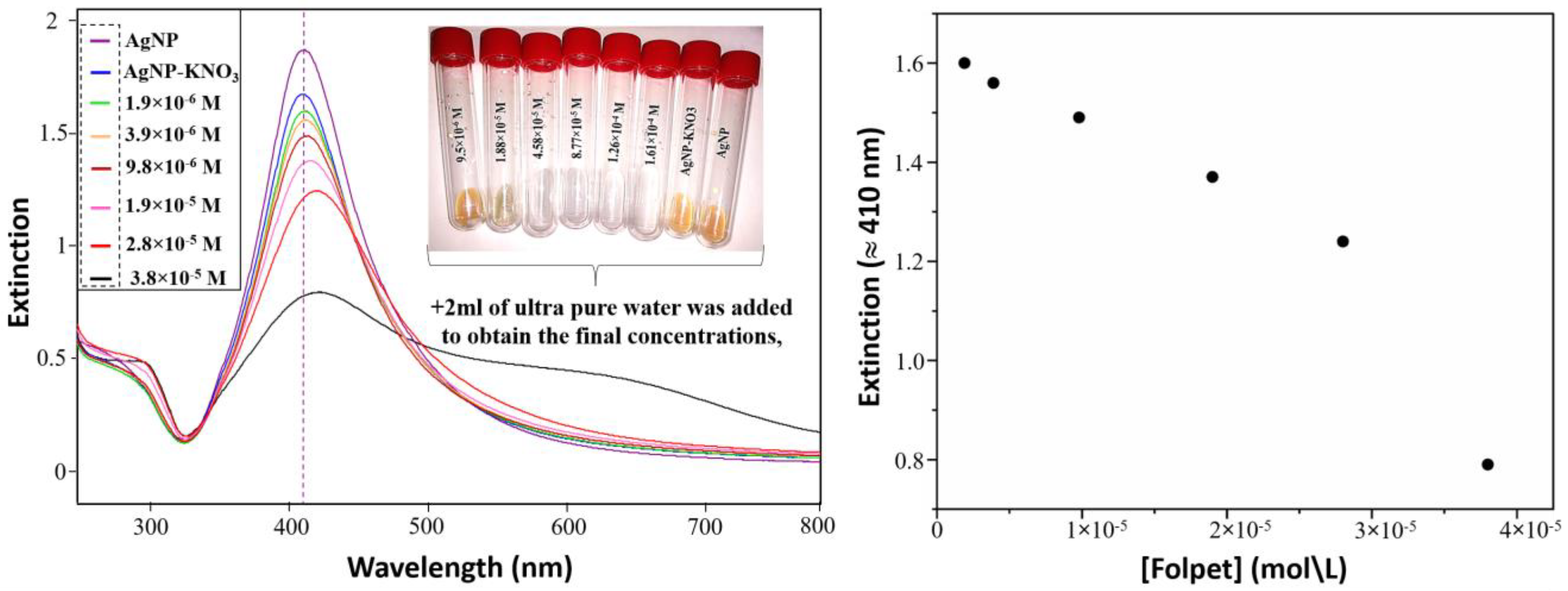
3.1. Extinction Measurements (External Standard Method)

Figure 2 exhibits the extinction spectra of the Ag colloid with and without KNO3. The plasmon resonance spectrum of the AgH colloid presents a maximum at 410 nm, which is due to the collective oscillations of electrons of Ag, which lead to localized surface plasmon resonances (LSPR). The TEM images of AgH in the absence of KNO3 (Figure 3) show predominant spherical or quasi-spherical nanoparticles. The addition of nitrate induces a significant aggregation, as shown in Figure 2, with an absorbance decrease of 0.20 absorbance unit in the plasmon resonance of the non-aggregated AgH nanoparticles, while the absorbance significantly increases in the 530–800 nm region (inset) because of the partial aggregation that induces a certain hybridization of plasmons in close nanoparticles. The addition of folpet into the AgNPs suspension induced significant changes in the UV–vis spectrum, which depend on the concentration of the added fungicide. These changes are associated with three important plasmonic effects: the modification of the LSPR resonance by the adsorption of the analyte on the surface; the variation in the intensity of the maximum at 410 nm, which decreases on raising the folpet concentration; and the enhancement of a secondary band in the 600–700 nm interval due to the plasmon hybridization. In fact, the shift from 410 to 421 nm of the maximum is clearly due to the variation in the LSPR upon the adsorption of folpet, while the variation in the intensity of the two LSPR resonances observed (410–420 nm, and 600–700 nm) are attributed to both the decrease in the individual nanoparticles and the hybridization between the plasmon oscillations in the nanoparticles induced by the aggregation of the colloidal AgNPs [23], respectively. In summary, within the range of 1.9 × 10⁻⁶ to 9.8 × 10⁻⁶, a linear decrease in the intensity of the AgNP plasmon band at 410 nm was seen. At 9.8 × 10⁻⁶, the plasmon band intensity exhibited a decrease at this concentration, shifting from 410 to 413 nm. For concentrations greater than 9.8 × 10⁻⁶, there was noticeable decrease in the plasmon band intensity, which shifted from 410 to 421 nm; this decrease was associated with the adsorption of folpet on the silver nanoparticles, which modifies the surrounding refraction index leading to an LSPR modification. On the other hand, the Ag nanoparticles’ aggregation lead to Ag colloid precipitation [24] and a change in the suspension color (see inset in Figure 3).

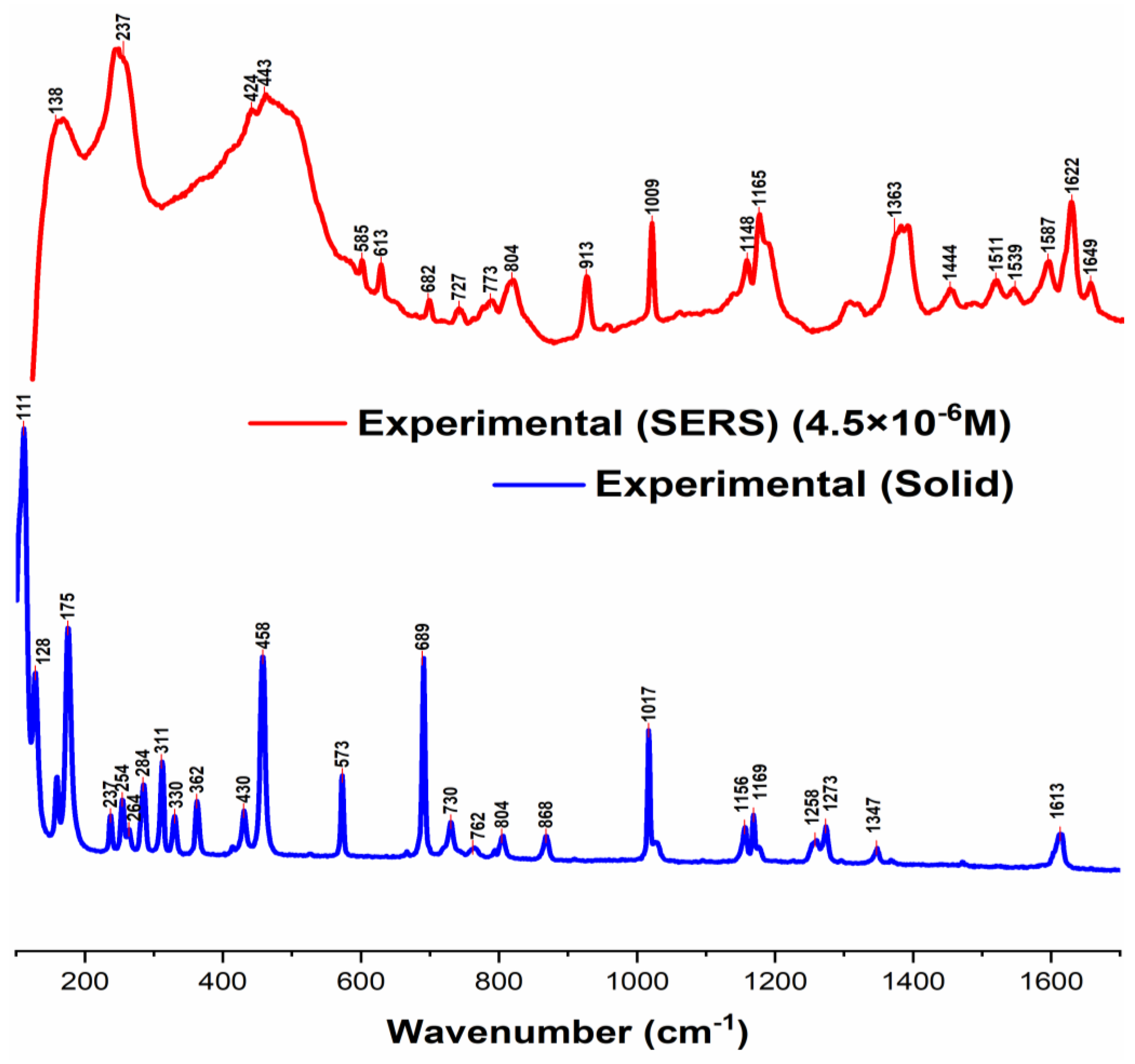
3.2. Raman and SERS Measurements of Folpet in AgH Colloid

Figure 4 depicts the Raman spectrum of the folpet solid (powder) compared with the Surface-Enhanced Raman Scattering (SERS) spectrum for folpet in Ag colloid (4.5 × 10⁻⁶ M). It is striking to observe that strong differences were noticed in the spectral profiles between the normal Raman and SERS. This effect can be attributed to a deep structural change in the folpet. In a previous work [25], it was reported that the chemical breakdown of the N–S bond can occur in folpet. In support of the cleavage of the N-S bond, several changes are evident in Figure 4. For instance, the bands at 111 cm−1 and 458 cm−1, corresponding to N-S-CCl3 bending, vanish entirely in the SERS spectrum. The same is true of the 175 cm−1 assigned to S-Cl bending appears only in the Raman spectrum. On the other hand, the band related to 237 cm−1 is found in both Raman and SERS spectrum, but in the latest spectrum, it widens significantly. The band at 237 cm−1 can be assigned to the Ag-Cl stretching produced by the chloride ions adsorbed on the surface of the metal. The band at 284 cm−1, assigned to C-Cl stretching and observed in the Raman powder spectrum, completely vanished in the SERS spectrum. Moreover, the bands 330, 362 and 762 cm−1 correspond to S-C-Cl stretching, C-C-O bending, and S-CCl stretching, respectively. All of them are absent in SERS spectrum. All bands below 600 cm−1 are presented in the supporting information (Figure S1) for a better display. The band at 689 cm−1 assigned to the C-S bond dominates the Raman powder spectrum, although it is seen at 682 cm−1 in the SERS spectrum with a significant reduction. Furthermore, the band at 868 cm−1 assigned to the N-S stretching appears in the Raman spectra and completely disappears in the SERS spectra and is strong evidence of the cleavage of the N–S bond. The strong band at 913 cm−1 is likely attributed to the C-COO stretching in carboxylates, while the band at 1017 cm−1, corresponding to the ring-breathing mode, persists in the SERS spectrum, indicating the presence of the aromatic ring in the adsorbed species, although the spectra shift suggests a structural change. Additionally, the broad bands at 1165 and 1372 cm−1 in the SERS spectrum are attributed to carboxylates and possibly amides, along with new bands between 1500 and 1650 cm−1, indicating the presence of amides and amines in the adsorbed species. The band at 1613 cm−1 is assigned to ring-stretching motions, with possible coupling with the NH2 bending. The latter band appears in both the Raman and SERS spectrum, while in the Raman spectrum this band is weak compared to SERS, which is considered to be the strongest band. These changes lead us to assume that folpet is degraded upon its adsorption on the surface, and it appears in new species such as phthalimide [26,27], terephthalic [28], phthalimidic acid, and thiophosgene, which seem to interact with a different affinity when adsorbed onto the silver surface, thus affecting the Raman signals. To clarify the above deductions, we used theoretical calculations of the substances that probably appear as products (those indicated below Figure 5) to complete and explain the results obtained in the SERS spectrum.

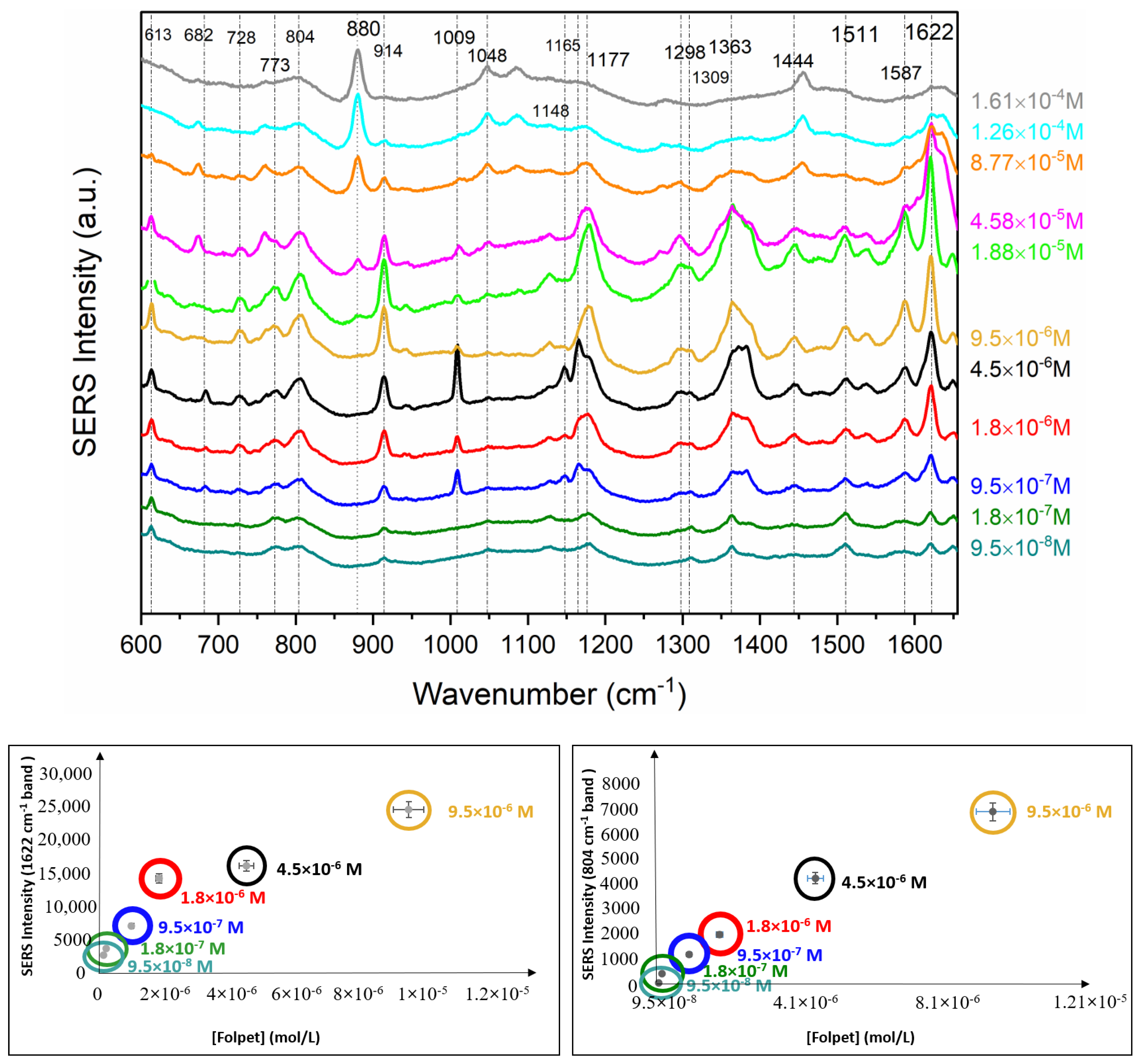
3.3. Quantitative Analysis

Quantitative analysis was performed in a range of concentration from 1.6 × 10⁻⁴–9.5 × 10⁻⁸ M (Figure 6), highlighting the frequency range between 600 and 1650 cm−1. The quantitative analysis was made by using the 804, 914, and 1622 cm−1 bands as reference. As can be seen, a sigmoidal increase was observed for the intensity of the latter from 9.5 × 10⁻⁸ to 1.8 × 10⁻⁶ M, showing a linear-like increase in the 10−8–10−6 M interval. A maximum for the SERS intensity was observed at a concentration of 9.5 × 10⁻⁶ M. Furthermore, for concentrations higher than 9.5 × 10⁻⁶ M, a decrease in the SERS intensity was observed for the bands 804, 914, and 1622 cm−1. Additionally, a loss of linearity for concentrations above 10−6 M can be observed, which is associated with changes in the electric charge of the Ag nanoparticles, leading to an extensive colloid aggregation. We suggest that the concentration at 10−5 M represents a threshold above which the Ag nanoparticles undergo a full covering, leading to dramatic changes in the electric and dynamical properties. The consequence of these effects is a strong shift of the LSPR plasmon resonance, a strong aggregation, and a consequent decrease in the SERS intensity. This aggregation can be followed by the TEM images obtained at different concentrations (Figure 7). High folpet concentrations lead to the formation of very compact aggregates (1.61 × 10⁻⁴ M). In these kinds of nanoparticles, the possibility of having hot spots for electric field enhancement and SERS intensification is very limited, thus accounting for the lowering of SERS signals above 10−5 M. However, as the concentration of the pesticide rises, the aggregates appear to be looser, generating more interparticle spaces where hot spots can be created.

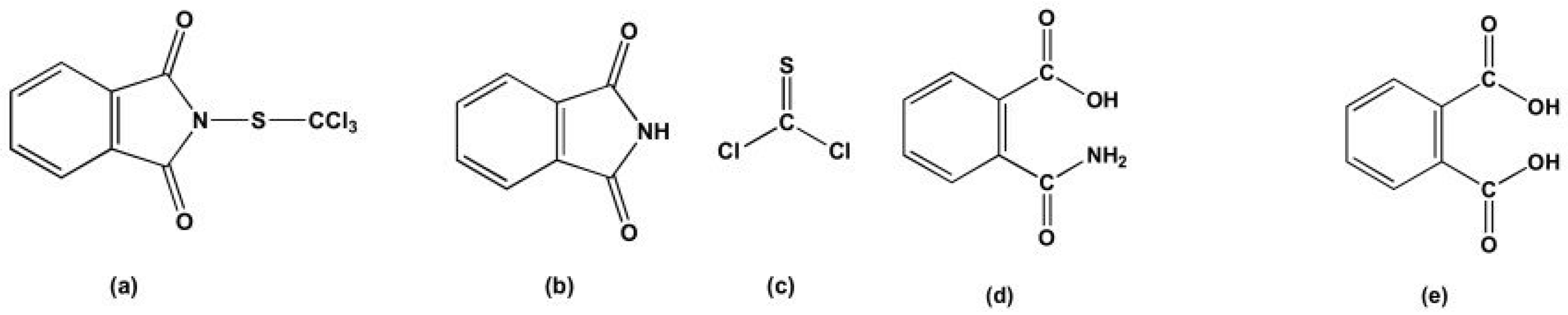
3.4. Degradation of Folpet Revealed by SERS Spectra

SERS spectra display narrower bands at 914, 1009, and 1622 cm−1 that can be attributed to the aromatic moieties of folpet and the degradation products. This degradation is attributed to plasmonic catalysis occurring on the surface of the Ag NPs obtained by hydroxylamine hydrochloride. After conducting the theoretical spectrum analysis of the molecules resulting from the folpet degradation, it is noteworthy that both the phthalimidic acid and terephthalic acid present bands that correlate quite well with those observed in the SERS spectra, as presented in the supporting information (Table S1) and in Figure 8. The prominent band seen at 1622 cm−1 in the SERS is absent in the theoretical spectrum of the terephthalic acid, but it is clearly visible in the theoretical spectrum of the phthalimidic acid, as illustrated in the Figure 8 and Figure 9 and in the supporting information (Figure S4). To validate these findings, we conducted spectral calculations for the complexes’ phthalimidic acid-Agn (n = 1–3). The results highlight the fact that the most intense band at 1622 cm−1 also dominates in the theoretical spectra. In addition, the SERS bands at 914, 1444, 1511, 1587, and 1649 cm−1 are also predicted in the theoretical spectra of phthalimidic acid at 906, 1444, 1511, 1609, and 1636 cm−1. After performing the DFT calculations, a good fit was obtained between the experimental and calculated spectra. This clearly indicates the suitability of the level of theory used, and an appropriate selection of basis was set for this molecule.
Figure 7. TEM images recorded from 9.5 × 10⁻⁷ to 1.61 × 10⁻⁴ M using the external standard method.
Chemosensors 12 00186 g007
Figure 8 depicts the Raman theoretical spectra obtained via the density functional theory (DFT) for folpet and various molecular structures, considering the degradation of the folpet molecule and resulting in the molecular structure of phthalimidic acid, which is referred to as “degraded folpet.” Subsequently, the interaction of this “degraded folpet” with a theoretical Agn (n = 1–3 atoms) cluster was performed in order to simulate its adsorption onto the Ag surface (Figure 9). A comparison between the experimental and calculated Raman results of the “degraded folpet” is also presented in Table 1, which provides a list of the main bands observed in the spectra along with the corresponding vibrational normal-mode assignments.
The theoretical spectra also showed that the band at 1268 cm−1 assigned to the N-S stretching in the folpet spectrum completely vanished for the “degraded folpet.” A similar effect was observed for the experimental band at 868 cm−1 ascribed to N-S stretching in the folpet-powder Raman spectrum, which vanished in the SERS spectrum (Figure 5), reinforcing the folpet N–S cleavage. In addition, a band at 1620 cm−1 appeared in the degraded folpet spectra. The band was also observed for the “degraded folpet” bonded to the Agn (n = 1–3) atoms at the interval 1621–1626 cm−1, assigned to NH2 bending—see the supporting information (Figure S4). However, in the folpet spectrum, the band at 1620 cm−1 is absent, both in the calculated Raman spectra and in the Raman powder spectrum. Also, the SERS bands at 1363, 1511, 1587, and 1649 cm−1 correspond to ν(CN) + δ(C-OH), δ(C-OH), ν(C=C), and ν(C=O) motions, respectively. These bands were also observed in the degraded folpet spectra and the degraded folpet spectra bonded to the Agn (n = 1–3) atoms at the interval 1360–1636 cm−1. In addition, in the case of the folpet degraded form, the NC bond appears only for the degraded folpet bonded to the Agn (n = 2–3) at 1391 and 1388 cm−1, respectively.

4. Conclusions

In conclusion, this study highlights the exceptional specificity and sensitivity of Surface-Enhanced Raman Spectroscopy (SERS) for detecting folpet. By employing UV-Vis, Raman, and SERS spectroscopy, we observed molecular degradation and breakdown through changes in the Raman and SERS spectra due to plasmonic effects related to the activation of hot carriers in the metal nanoparticle. In addition, the adsorption of the pesticide on the Ag NPs lead to evident changes in plasmon resonance and the hybridization of the NPs, which were very sensitive to the folpet concentration. This study achieved a remarkable limit of detection at 4.78 × 10⁻⁸ M. We found that folpet adsorption and degradation significantly influenced the Ag colloid aggregation due to strong chemical interactions. The TEM images revealed that the detection of folpet is advantageous when lowering the concentration, as the resulting aggregates appear looser, generating more interparticle spaces where hot spots can be created and inducing a strong intensification by interparticle plasmon hybridization. On the contrary, at concentrations above 10−5 M, the aggregates are too compact, and this is not good for the SERS intensification. Additionally, density functional theory (DFT) calculations were crucial in predicting the molecular structure and Raman spectra of the degradation products, providing deeper insights into folpet’s vibrational modes. This comprehensive approach underscores the robustness of combining experimental and theoretical techniques in advancing chemical detection methodologies.

Supplementary Materials

The following supporting information can be downloaded at https://www.mdpi.com/article/10.3390/chemosensors12090186/s1, Table S1: Main experimental and calculated Raman wavenumbers of folpet, degraded folpet, and degraded folpet-Agn (n = 1–3) complexes; Table S2: Preparation of the samples (Ag colloid + KNO3 + Folpet) used in the SERS measurements by the external standard method; Table S3: Preparation of the samples (Ag colloid + KNO3 + Folpet)) used in the extinction measurements by the external standard method; Figure S1: Top: Raman (powder) and SERS (Ag colloid) spectra of folpet at the low wavenumber range. Bottom: Raman spectrum recorded for folpet powder and SERS spectrum for folpet in Ag colloid (4.5 × 10⁻⁶ M), both using the 532 nm laser line; Figure S2: Folpet SERS spectra recorded from 9.5 × 10⁻⁸ to 9.5 × 10⁻⁶ M using Ag colloid, laser line at 532 nm, Increasing the integrated area of the bands with maxima at 804 and 914 cm−1 with increasing folpet concentration (external addition method); Figure S3: Folpet SERS spectra recorded from 9.5 × 10⁻⁶ to 1.61 × 10⁻⁴ M using Ag colloid, laser line at 532 nm, decreasing the integrated area of the bands at 804, 914, and 1622 cm−1 with increasing folpet concentration (external addition method); Figure S4: The calculated spectra of the degraded folpet and degraded folpet-Agn (n = 1–3) complexes using Gaussian 09; Figure S5: The calculated spectra of the Terephthalic molecule using Gaussian 09; Figure S6: TEM images recorded from 9.5 × 10⁻⁷ to 1.61 × 10⁻⁴ M using the external standard method.

Author Contributions

Conceptualization, S.S.-C. and B.S.; methodology, O.D., B.S., and S.S.-C.; software, O.D.; validation, O.D., B.S., and S.S.-C.; formal analysis, O.D. and S.S.-C.; resources, S.S.-C. and B.S.; data curation, O.D.; writing—original draft preparation, O.D.; writing—review and editing, S.S.-C. and B.S.; visualization, O.D., B.S., and S.S.-C.; supervision, B.S. and S.S.-C.; project administration, S.S.-C.; funding acquisition, S.S.-C. All authors have read and agreed to the published version of the manuscript.

Funding

This work was conducted with funding from the Spanish National Research Council under the COOPB20373 project within the i-COOP 2018 program of CSIC, as well as the PID2020-113900RB-I00 and PID2023-146214OB-I00 funded by MCIU/AEI/10.13039/501100011033 and FSE+. of the Spanish Ministry of Science and Innovation.

Institutional Review Board Statement

Not applicable.

Informed Consent Statement

Not applicable.

Data Availability Statement

The datasets used and/or analyzed during the current study are available upon request.

Acknowledgments

The authors gratefully acknowledge the Instituto de Estructura de la Materia, Consejo Superior de Investigaciones Cientificas (IEM-CSIC) in Madrid, Spain, for providing all the necessary instrumentation for the normal Raman and the SERS experiments, as well as the UV/Vis analysis. We also thank the Universidad Complutense of Madrid, Centro de Microscopia Electronica, for conducting the TEM analysis.

Conflicts of Interest

The authors declare no conflicts of interest.

References

  1. European Food Safety Authority (EFSA). Conclusion Regarding the Peer Review of the Pesticide Risk Assessment of the Active Substance Folpet. EFSA J. 2009, 7, 297r. [Google Scholar] [CrossRef]
  2. Leepheng, P.; Limthin, D.; Homchan, W.; Suramitr, S.; Phromyothin, D. An Experimental and Theoretical Study of Molecularly Imprinted Electrode Based on Methyl Methacrylate Polymer for Pesticide Detection. Jpn. J. Appl. Phys. 2020, 59, SIIJ09. [Google Scholar] [CrossRef]
  3. Vannucci, G.; Cañamares, M.V.; Prati, S.; Sanchez-Cortes, S. Analysis of the Tautomeric Equilibrium of Two Red Monoazo Dyes by UV–Visible, Raman and SERS Spectroscopies. Spectrochim. Acta. A Mol. Biomol. Spectrosc. 2021, 261, 120007. [Google Scholar] [CrossRef] [PubMed]
  4. Douass, O.; Samoudi, B.; Bendaou, O.; Sanchez Cortes, S. A Brief Review of Recent Advances in Surface-Enhanced Raman Spectroscopy and Microfluidics Technology for the Ultrasensitive Detection of Pesticides. Egypt. J. Chem. 2022, 65, 133–150. [Google Scholar] [CrossRef]
  5. Le Ru, E.C.; Etchegoin, P.G. Principles of Surface-Enhanced Raman Spectroscopy: And Related Plasmonic Effects, 1st ed.; Elsevier: Amsterdam, The Netherlands; Boston, MA, USA, 2009; ISBN 978-0-444-52779-0. [Google Scholar]
  6. Kneipp, K.; Kneipp, H.; Itzkan, I.; Dasari, R.R.; Feld, M.S. Surface-Enhanced Raman Scattering and Biophysics. J. Phys. Condens. Matter 2002, 14, R597–R624. [Google Scholar] [CrossRef]
  7. Campion, A. Raman Spectroscopy of Molecules Adsorbed on Surfaces: Progress, Issues and Prospects. J. Electron Spectrosc. Relat. Phenom. 1990, 54–55, 877–880. [Google Scholar] [CrossRef]
  8. Dong, T.; Lin, L.; He, Y.; Nie, P.; Qu, F.; Xiao, S. Density Functional Theory Analysis of Deltamethrin and Its Determination in Strawberry by Surface Enhanced Raman Spectroscopy. Molecules 2018, 23, 1458. [Google Scholar] [CrossRef]
  9. Tuschel, D. Practical Group Theory and Raman Spectroscopy, Part i: Normal Vibrational Modes. Spectroscopy 2014, 29, 14. [Google Scholar]
  10. Cortés, E.; Besteiro, L.V.; Alabastri, A.; Baldi, A.; Tagliabue, G.; Demetriadou, A.; Narang, P. Challenges in Plasmonic Catalysis. ACS Nano 2020, 14, 16202–16219. [Google Scholar] [CrossRef]
  11. Kim, Y.; Dumett Torres, D.; Jain, P.K. Activation Energies of Plasmonic Catalysts. Nano Lett. 2016, 16, 3399–3407. [Google Scholar] [CrossRef]
  12. Fuenzalida, F.B.; Slepčíková, P.; Repovská, M.; Jutková, A.; Vega Cañamares, M.; Miškovský, P.; Jurašeková, Z.; Sanchez-Cortes, S. Selective and Ultrasensitive Detection of the Herbicide Glyphosate by Means of Plasmon Catalysis on Ag Nanoparticles. Spectrochim. Acta A Mol. Biomol. Spectrosc. 2024, 323, 124845. [Google Scholar] [CrossRef] [PubMed]
  13. Shi, Z.Z.; Li, W.P.; Kang, W.J.; Feng, Y.; Li, Z.; Zhong, X.Y.; Yang, J.; Liu, H.; Dong, C.K.; Yin, P.F.; et al. Silver Clusters with Adatoms as a Catalyst for the Oxygen Reduction Reaction. ACS Catal. 2023, 13, 9181–9189. [Google Scholar] [CrossRef]
  14. Sanchez-Cortes, S.; Garcıa-Ramos, J.V. Influence of Coverage in the Surface-Enhanced Raman Scattering of Cytosine and Its Methyl Derivatives on Metal Colloids: Chloride and pH Effects. Surf. Sci. 2001, 473, 133–142. [Google Scholar] [CrossRef]
  15. Rubira, R.J.G.; Furini, L.N.; Constantino, C.J.L.; Sanchez-Cortes, S. SERS Detection of Prometryn Herbicide Based on Its Optimized Adsorption on Ag Nanoparticles. Vib. Spectrosc. 2021, 114, 103245. [Google Scholar] [CrossRef]
  16. Oliveira, M.J.S.; Rubira, R.J.G.; Furini, L.N.; Batagin-Neto, A.; Constantino, C.J.L. Detection of Thiabendazole Fungicide/Parasiticide by SERS: Quantitative Analysis and Adsorption Mechanism. Appl. Surf. Sci. 2020, 517, 145786. [Google Scholar] [CrossRef]
  17. Oliveira, M.J.S.; Martin, C.S.; Rubira, R.J.G.; Batagin-Neto, A.; Constantino, C.J.L.; Aroca, R.F. Surface-enhanced Raman Scattering of Thiram: Quantitative and Theoretical Analyses. J. Raman Spectrosc. 2021, 52, 2557–2571. [Google Scholar] [CrossRef]
  18. Chemical Reduction—An Overview|ScienceDirect Topics. Available online: https://www.sciencedirect.com/topics/engineering/chemical-reduction (accessed on 6 October 2023).
  19. Sánchez-Cortés, S.; García-Ramos, J.V.; Morcillo, G. Morphological Study of Metal Colloids Employed as Substrate in the SERS Spectroscopy. J. Colloid Interface Sci. 1994, 167, 428–436. [Google Scholar] [CrossRef]
  20. Ostroukhova, O.K.; Zenkevich, I.G. A Comparison of the External Standard and Standard Addition Methods for the Quantitative Chromatographic Determination of Pesticide Concentrations in Plant Samples. J. Anal. Chem. 2006, 61, 442–451. [Google Scholar] [CrossRef]
  21. Gaussian.Com|Expanding the Limits of Computational Chemistry. Available online: https://gaussian.com/ (accessed on 6 October 2023).
  22. Basis Sets|Gaussian.Com. Available online: https://gaussian.com/basissets/ (accessed on 20 March 2023).
  23. Cañamares, M.V.; Garcia-Ramos, J.V.; Gómez-Varga, J.D.; Domingo, C.; Sanchez-Cortes, S. Comparative Study of the Morphology, Aggregation, Adherence to Glass, and Surface-Enhanced Raman Scattering Activity of Silver Nanoparticles Prepared by Chemical Reduction of Ag + Using Citrate and Hydroxylamine. Langmuir 2005, 21, 8546–8553. [Google Scholar] [CrossRef]
  24. Medendorp, N.W.; Bowman, K.J.; Trumble, K.P. Tailoring Particle Size and Morphology of Colloidal Ag Particles via Chemical Precipitation for Ag-BSCCQ Composites. Mater. Sci. Eng. A 1996, 212, 222–227. [Google Scholar] [CrossRef]
  25. Vrenouf Analyse Phytosanitaire: Levons Le Voile Sur La Notion de Métabolites. Available online: https://labexcell.com/images/smartlink/SL_Analyse_Phytosanitaire (accessed on 5 June 2024).
  26. Sun, H.; Zhang, X.; Zuo, W.; Dai, Z.; Zhou, L.; Luo, F.; Yang, M.; Wang, X.; Lou, Z.; Chen, Z. Concentrations, Generation and Risk Characterization of Phthalimide in Tea-Derived from Folpet or Not? Sci. Total Environ. 2022, 852, 158194. [Google Scholar] [CrossRef] [PubMed]
  27. Han, C.; Huang, C.; Jiang, Q.; Chen, L.; Li, Y.; Lou, C.; Lu, H.; Shen, Y. A Method Based on the Conversion and Determination of Folpet and Phthalimide Residues in Tea by GC-MS/MS and GC-TOF-HRMS. Microchem. J. 2022, 179, 107448. [Google Scholar] [CrossRef]
  28. Beegum, M.F.; Kumari, L.U.; Harikumar, B.; Tresa, H.; Panicker, C.Y. Vibrational wavenumbers and structural determination of dilithium terephthalate. Rasayaa J. Chem. 2008, 1, 117–124. [Google Scholar]
Figure 1. (Left): Scheme of the experiment carried out to obtain SERS spectra of the folpet using Ag nanoparticles in colloidal suspensions. (Right): chemical structure of folpet.
Chemosensors 12 00186 g001
Figure 2. UV absorption spectra of Ag colloid (AgH) and Ag colloid (AgH)-KNO3. Inset: TEM images of Ag colloid (AgH) and Ag colloid (AgH)-KNO3.
Chemosensors 12 00186 g002
Figure 3. Extinction spectra of Ag colloid in the absence and presence of folpet from 3.8 × 10⁻⁵ to 1.9 × 10⁻⁶ M. Variation in the extinction intensity of the band (peak intensity) with a maximum at 410 nm.
Chemosensors 12 00186 g003
Figure 4. Raman spectrum recorded for the folpet powder and the SERS spectrum for folpet in Ag colloid (4.5 × 10⁻⁶ M), both using the 532 nm laser line.
Chemosensors 12 00186 g004
Figure 5. Structure of folpet (a), phthalimide (b), thiophosgene (c), phthalimidic acid (d), and terephthalic (e) molecules.
Chemosensors 12 00186 g005
Figure 6. Top: Folpet SERS spectra recorded from 9.5 × 10⁻⁸ to 1.61 × 10⁻⁴ M using Ag colloid. Bottom: Variation in the integrated area of the bands with maxima at 804 and 1622 cm−1 with increasing folpet concentration. Laser excitation at 532 nm.
Chemosensors 12 00186 g006
Figure 8. (Left): DFT-calculated Raman spectra of folpet and different candidate-degraded molecules. (Right): The optimized structures using the DFT-B3LYP/6-311++G(d,p) basis set.
Chemosensors 12 00186 g008
Figure 9. (Left): The calculated spectra of folpet, degraded folpet, and degraded folpet-Agn (n = 1–3) complexes. (Right): The optimized structures. The term “degraded” refers to folpet N–S bond cleavage.
Chemosensors 12 00186 g009
Table 1. Main experimental and calculated Raman wavenumbers (cm−1) of folpet, degraded folpet, and degraded folpet-Agn (n = 1–3) complexes and assignments derived from the DFT calculations (ν, Stretching; δ, Bending; τ, Twisting; ρ, Wagging).
Experimental
Spectra
Theoretical SpectraAssignments
Folpet SERS (cm−1)Degraded Folpet Degraded Folpet-AgDegraded Folpet-Ag2Degraded Folpet-Ag3
20 17 15 δ(Ag-NH2) + δ (OH)
32 31 26 24 δ(-CONH2)
65 53 54 δ(Ag-OCNH2) + δ(COOH)
86 95 95 96 δ(NH2) + δ(HOCO)
110 115 125 139 δ(OCNH2)
138157162159162δ(NH2) + τ(OH)
160 174 174 167 ν(Ag-Ag) + δ(NH2CO phynel)
237237239243238δ(NH2) + δ(COOH)
350 351 324 353 δ(CH) + τ(NH2)
373 390 366 382 ρ(NH2)
424404, 407 408 403, 415, 429 410 δ(NH2)
570570576571δ(NH2) + δ(CCC)
584580585580δ(OH) + δ(NH2)
613618621630626δ(NH2CO)+ δ(CCC)
682641639643640HOCO def + δ(CCC)
727719717722721τ(HOCO)
736737737733δ (HOCO)
757756763766δ(CH) + δ(NH2CO)
773795796785786δ(CH)
804807, 817807, 817813, 817809, 816δ(CCC)
913906906905905ν(C-COO-)
10091056105610611057δ(CH)
11651158116211661162δ (HCCH)
11771187118811901190δ (HCCH)
12091205 1205 1207 δ(HOC)
12981292129312981292δ(HCC)
1329132913321331ν(CC)
13631355, 13631359, 136713671360ν(NC) + δ(HOC)
13911388ν(NC)
14441474147414731474δ(HCC)
15111513151315191516δ(HCC)
15871609160916121609ν(CC)
16221620162116261622δ(NH2)
16491636163516351636ν(C=O)
Disclaimer/Publisher’s Note: The statements, opinions and data contained in all publications are solely those of the individual author(s) and contributor(s) and not of MDPI and/or the editor(s). MDPI and/or the editor(s) disclaim responsibility for any injury to people or property resulting from any ideas, methods, instructions or products referred to in the content.

