Review

Oriented Immobilization of IgG for Immunosensor Development

by
Yihan Zhang
,
Mingjie Ma
,
Haji Akber Aisa
and
Longyi Chen
*
Xinjiang Technical Institute of Physics and Chemistry, Chinese Academy of Sciences, 40-1 South Beijing Road, Urumqi 830011, China
*
Author to whom correspondence should be addressed.
Chemosensors 2025, 13(2), 50; https://doi.org/10.3390/chemosensors13020050
Submission received: 3 January 2025 / Revised: 28 January 2025 / Accepted: 30 January 2025 / Published: 3 February 2025
(This article belongs to the Special Issue Electrochemical Sensing in Medical Diagnosis)

Abstract

:
The realization of the oriented immobilization of antibodies onto the surfaces of solid or nanometal particles constitutes a significant approach for enhancing the performance of electrochemical immunosensors. In light of the research findings of predecessors, this review showcases several immobilization methods, categorizing them into covalent binding pathways, bioaffinity techniques, and other binding modalities for elaboration. Emphasis is placed on expounding the binding sites, binding mechanisms, as well as the merits and drawbacks of binding techniques such as those involving disulfide bonds, glycan chains, protein A, G, and DNA.

1. Introduction

Biosensors have been extensively employed within the multifarious realms of bioanalysis, owing to their inherent simplicity, remarkable high sensitivity, excellent selectivity, and reliable reproducibility. With the aim of augmenting their detection capabilities, the precise modulation of the orientation of the molecular recognition layer represents one of the most straightforward and highly effective strategies for amplifying the sensitivity of biosensors [1,2]. This mini-review accentuates the contemporary research endeavors and sophisticated methodologies associated with the immobilization of antibody (Abs) surfaces. It expounds in a more elaborate and profound manner on two principal classifications of antibody immobilization techniques: covalent immobilization and immobilization mediated by bioaffinity mechanisms.
Antibodies, alternatively denominated as immunoglobulins (Igs), are soluble glycoproteins possessing a molecular mass in the vicinity of 150 kDa. Endowed with a pair of binding sites, they manifest the distinctive faculty to recognize, expunge, and neutralize specific targets or antigens (such as viruses or bacteria), thereby occupying a central and crucial position within the human immune system.
The complementary “Y”-shaped architecture of an antibody is constituted by four polypeptide chains: two identical heavy chains (H chains), each of which incorporates four domains, flanked by two identical light chains (L chains), each harboring two domains. Each domain typically comprises approximately 110 amino acids. The domains are interconnected through peptide bonds, while the chains are conjoined by disulfide bonds. For illustration, the heavy and light chains are tethered together via disulfide bridges. The region tasked with antigen recognition is designated as the antigen-binding fragment (Fab) region [3], positioned at the apical portion of the “Y” configuration. In contradistinction, the basal segment is termed the fragment crystallizable (Fc) region, which is competent in interacting with other constituents of the immune system, inclusive of complement and cell receptors [4]. In mammals, IgG prevails as the most preponderant immunoglobulin in serum, constituting approximately 80% thereof, and functions as the cardinal immunoglobulin implicated in the adaptive immune response. Human IgG can be discriminated into four subclasses (IgG1, IgG2, IgG3, and IgG4) in accordance with the disparities in the constant regions of the heavy chain sequences [5].
Through intensive and detailed research, the variability and relative positions of individual domains can be accurately defined. The light chain comprises a variable domain (VL) and a constant domain (CL), and the heavy chain consists of the Fab domain’s variable domain (VH), the constant domain (CH1), and the Fc domain’s constant domains (CH2, CH3). Principally, the collective region of six complementarity-determining regions (CDRs) in VH and VL domains dictates antibody binding specificity and affinity, as per [6,7].
To enhance the sensitivity of antibody probes in detecting antigen analytes and to lower the detection limit of immunosensors, orienting and immobilizing intact antibodies on metal or particle surfaces via physical adsorption, chemical, or bioaffinity techniques is crucial. These methods typically rely on specific antibody regions for chemical fixation or bioaffinity. As a result, during immobilization, some antibody domains may not contribute positively; instead, increased steric hindrance can reduce the number of bound antibodies, and the antibody’s charge can cause improper orientation of other antibodies [8]. Numerous studies [8,9,10] have compared the sensor sensitivities of fragmented and intact antibodies. The results indicate that appropriate antibody fragmentation can enhance antigen-binding ability. Notably, antibody fragmentation might expose and activate groups that were previously internal or inactive, thereby involving them in the antibody immobilization process [11].
Hence, various antibody cleavage approaches have been explored, as shown in Figure 1.
1. Papain, a thiol protease, specifically cleaves the hinge region between heavy chains of antibody molecules. In the case of IgG antibodies, it generates two Fab and one Fc fragment, thereby significantly modifying the antibody’s structure and function.
2. Pepsin, an acidic protease optimally active at pH 1.5–2.5, cleaves between heavy chains closer to the Fc segment than papain. When acting on IgG antibodies, it forms an F(ab′)₂ and small Fc fragments. As reported in [9], these can be further cleaved to F(ab′) by a stronger reducing reagent.
3. Chemical reductants like DTT, 2-MEA, and TCEP donate electrons to reduce sulfur atoms in disulfide bonds. For IgG antibodies, this results in the breakage of bonds between and within heavy and light chains, leading to separated chains or fragments such as F(ab′) and Fab/Fc, depending on the reaction conditions.
4. Cyanogen bromide (CNBr), a reagent that cleaves peptide bonds with methionine residues, fragments antibody molecules. However, its high toxicity may potentially affect antibody activity and compromise their functional performance.
5. Genetic engineering methods, such as CRISPR-Cas or site-directed mutagenesis, introduce specific cleavage sites in antibody-encoding genes. This enables precise control of cleavage, as seen in svFv and Vhh [12]. These smaller antibody fragments, lacking the constant domains involved in immune system interactions, exhibit reduced steric hindrance and enhanced penetrability while retaining a substantial portion of their antigen-binding capabilities, opening up new possibilities for biomedical applications.
As previously discussed, the sensitivity of immunosensors hinges on maximizing the number of antibodies immobilized in an orderly and oriented fashion on a limited metal surface, facilitating their interaction with trace antigen samples. Extensive research has been conducted on the covalent and non-covalent binding of antibodies to solid surfaces or metal ions. However, with technological advancements, it has become evident that mere random physical adsorption and covalent immobilization result in non-directional protein attachment. This leads to disordered antibody orientation, which in turn causes protein-structure distortion and active-site occlusion, significantly diminishing the antibody’s activity. Ideally, immobilized antibodies should have the Fc facing the substrate and the Fab upward towards the antigen, termed “end-on”. But randomly immobilized antibodies may have different orientations like “front”, “side”, and “lying flat”. Thus, devising proper strategies for oriented antibody immobilization is a challenging task.

2. Strategies for Immobilization Design Approaches

The surface of an antibody is characterized by hydrophilicity, and within particular domains, specific active groups such as amino, carboxyl, glycosyl, and disulfide moieties are present. In the pursuit of directionally immobilizing these active groups onto solid surfaces and metal nanoparticles, it is requisite to implement chemical modifications on the substrate. Alternatively, a double-active-center sandwich structure can be employed to effectuate the linkage between the antibody and the substrate, thereby facilitating the oriented immobilization process and potentially enhancing the performance and functionality of the antibody-based constructs in various biomedical and biotechnological applications.

2.1. Plasma Treatment Substrate

Plasma treatment, a technique for material surface modification, is applied in antibody immobilization. Plasma is generated via discharge or high-frequency electromagnetic oscillation. The active species like free radicals, ions, and electrons therein react with surface chemical bonds, activating the surface layer for subsequent chemical treatment and covalent antibody protein grafting [13]. M.C. Baican plasma-treated polyvinylidene fluoride (PVDF) with CO2, N2, and N2/H2 mixtures to attach carboxyl- or amine groups for protein covalent immobilization [14], and then used EDC/NHS chemistry to attach IgG or Protein A (for subsequent IgG binding) onto the treated PVDF. Through N2/H2 treatment and Protein A grafting on PVDF, an end-on IgG orientation was achieved. E. Kosobrodova utilized plasma-immersion ion implantation (PIII) to improve polymer wettability and protein binding, reducing the tail-up orientation of CD34 antibodies [15]. As depicted in Figure 2, the anti-CD34 antibodies on the polycarbonate (PC) surface treated with 360 s plasma-immersion ion implantation (PIII) (Figure 2B) formed a denser layer than those on the untreated PC surface (Figure 2A).

2.2. Three-Dimensional Substrate

Three-dimensional substrates present a larger surface area for antibody binding, e.g., via reactive ion etching, and permit multi-layered antibody stacking, jointly augmenting antibody loading. Their spatial architecture may mitigate steric hindrance during antibody fixation and antigen capture. Moreover, antibody microarrays can be fabricated, enabling precise patterning of specific substrate regions, yielding high-density and highly specific arrays for concurrent detection of multiple antigens [16]. As shown in Figure 3, Wang and Feng comprehensively reviewed three-dimensional substrates, positing that gels and sol-gels of agarose or dextran enhance antibody binding owing to their high surface area [16].

2.3. Self-Assembled Monolayer Substrate

The formation of self-assembled monolayers (SAMs) offers an alternative means of surface chemistry modification, facilitating the establishment of chemical or semi-chemical linkages between antibodies and chemically modified solid surfaces or nanoparticles. It serves as a potent tool for biomolecule immobilization on diverse solid substrates [17]. SAMs usually consist of linear carbon chains or long-chain bio-derived small molecules with active functional groups at both termini after chemical or bioengineering modifications. As shown in Figure 4, Upon attachment to solid particle surfaces, they promote self-assembly, while the other end is employed for antibody conjugation or adsorption. A typical SAM example is the utilization of alkanethiols as linkers, exploiting the gold-thiol interaction to bind antibodies to gold nanoparticle substrates [18,19].

2.4. Layer-by-Layer Assembly Substrate

Layer-by-layer (LBL) assembly represents a technique for fabricating multi-layer thin films, relying on electrostatic interactions between oppositely charged substances to construct multi-layer architectures. The LBL approach permits precise control from the nanometer to micrometer scale by modulating parameters like immersion time and polyelectrolyte concentration. Moreover, a diverse range of materials, such as nanoparticles (e.g., gold nanoparticles, quantum dots) and biomacromolecules (proteins, DNA, antibodies), can be utilized for LBL assembly. As shown in Figure 5, This allows nanoparticles with catalytic capabilities and antibodies with bio-recognition functions to be integrated via LBL, thereby erecting a framework for the directional immobilization of antibodies [20].

3. Covalent Binding Pathway

Physical adsorption, being the simplest immobilization approach, exhibits limitations such as random antibody orientation, proneness to denaturation, and poor reproducibility, thereby often serving as an ancillary technique. Typically, amino acids within the correctly folded protein architecture result in hydrophilic residues with chemical reactivity, viz., amine, carboxyl, and hydroxyl groups on the antibody surface, facilitating covalent binding. Upon appropriate modification of the functional groups on the protein surface, directional connection of the antibody to the metal surface or nanoparticles can be achieved via bifunctional intermediates. Covalent coupling guarantees robust and irreversible attachment, enhancing substrate density and orientation. As shown in Figure 6, The fixation of diverse groups via distinct chemical reactions hinges on multiple factors, including substrate nanostructure [21], planar roughness [22], functional modification (e.g., IgG1 exhibits a predominantly upright orientation on -COOH surfaces, an intermediate orientation between end-on and side-on on -NH2 surfaces, and a mainly “lying” orientation on -CH3 surfaces [19]), and target groups on the antibody, as well as external conditions like pH [2,23,24,25,26,27,28], temperature [29], electric field [2,5], ionic strength [19,23,24,26], and solvent surface tension [30,31,32].

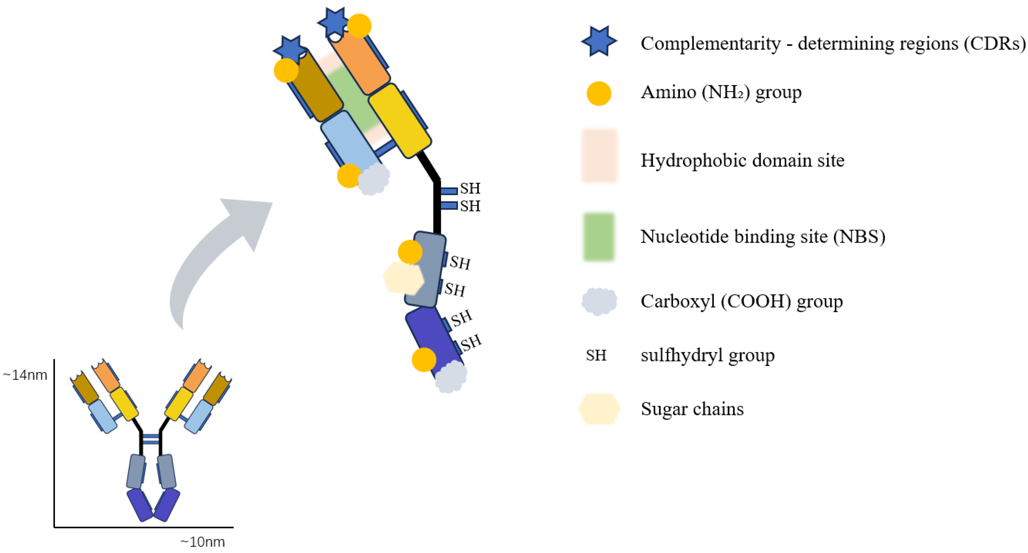
3.1. Nucleotide-Binding Site (NBS)

The nucleotide-binding site (NBS) of antibodies, a hitherto underexploited yet highly conserved region, resides between the light and heavy chains within the Fab domain of nearly all antibodies [33]. Rajagopalan and colleagues have furnished a relatively comprehensive characterization. Comprising amino acid residues with aromatic side chains in the VL domain, the NBS was later found by Meisenheimer, Koch, and others to generate active free radicals upon exposure to ultraviolet light (254 nm), permitting covalent photocrosslinking with small aromatic ring ligand molecules such as IBA. Directional immobilization via NBS can be effected under mild conditions, optimizing the retention of antibody 3D structure and antigen-binding affinities [34]. Subsequently, reactive thiols, click chemistry reagents, biotin, and other linking moieties can be coupled to the NBS through aromatic-ring ligands for antibody directional immobilization, as typified by (IBA-FITC) and (IBA-iRGD). Nathan J. Alves also validated the feasibility of the NBS-specific ultraviolet-photocrosslinking biotinylation method (UV-NBS-biotin) (Figure 7), manifesting a site-specific immobilization strategy [35]. Nevertheless, it is noteworthy that antibody denaturation may ensue from exposure to ultraviolet radiation of shorter wavelengths and higher energies [36,37].

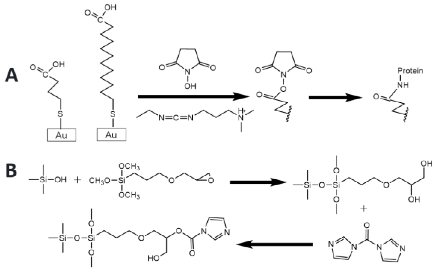
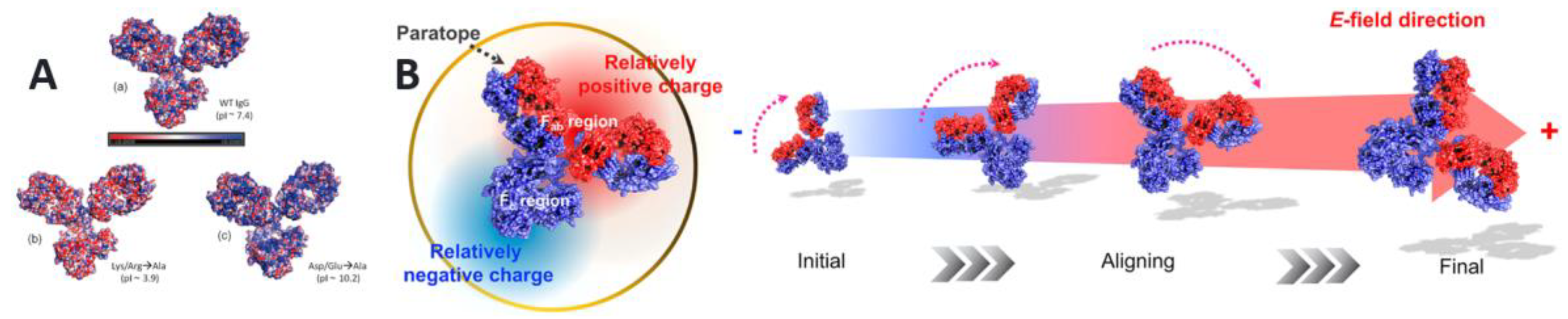
3.2. Amine, Carboxyl, and Hydroxyl Binding Sites

Active primary amine moieties, typified by lysine and arginine, along with active carboxyl groups, as represented by aspartic acid and glutamic acid, constitute crucial foci in the research of covalent and directional antibody immobilization. Substantial endeavors have been dedicated to employing a multitude of intermediate layers for the directional conjugation of antibodies to electrode or metal surfaces. The prevalently utilized 1-ethyl-3-(3-(dimethylamino) propyl) carbodiimide/succinimide ester (EDC/NHS) [29,38,39,40] facilitates the formation of covalent amide linkages via carbodiimide chemistry [41,42,43,44]. Moreover, carbonyl diimidazole (CDI) [45,46], glutaraldehyde [47,48,49,50], maleimide, and phenyl diisothiocyanate [51,52] have also been subjects of practical investigations. As shown in Figure 8.
Despite the relative paucity of hydroxyl groups in amino acids (confined to serine and threonine), the presence of glycan chains in antibodies endows hydroxyl moieties with the potential to function as active sites. Hyun-Ju Um exploited the interaction between the cyano group and the hydroxyl functionality within the antibody crystallizable region (Fc) to effectuate the immobilization of antibodies on the surface of a gold electrode modified with poly(2-cyanoethylpyrrole) (PCEPy) [53].
Nevertheless, given their ubiquitous distribution across the antibody surface, site-specific covalent immobilization targeting these groups presents a formidable challenge. It has been ascertained that:
1. For directional antibody immobilization, preference is accorded to the amino and carboxyl segments within the Fc region over the antigen-binding fragment (Fab) to preserve antigen-binding capacity.
2. The commonly occurring directional targets can be rendered specific through the application of particular external conditions.
In the intricate three-dimensional architecture of antibodies, the presence of charged amino acids engenders a characteristic variable charge distribution pattern. The polarity of antibodies predominantly stems from the existence of regions with opposing charges in disparate regions of the macromolecular space. Although the approximate surface spatial charge distribution of antibodies can be prognosticated from their sequences and spatial configurations, organic solvents or external conditions during the immobilization process can precipitate spontaneous charge alterations in antibodies [2,5,25,26] (Figure 9). In Sun’s study, EDC/NHS coupling was employed to introduce NHS reactive groups into random antibody loci. Subsequently, an electric field (E-field) was imposed during the antibody immobilization process to affect the directional translocation of antibodies, which then engaged in a reaction with the free amine on cysteine immobilized on a gold electrode, thereby realizing the orientation of antibodies. For instance, Shipeng Gao et al. capitalized on the differential activation extents of diverse amino active groups under varying pH conditions. By modulating the conjugated amino group during the covalent interaction, the orientation of IgG on the carrier surface could be regulated. Claudio Parolo harnessed the principle that when the pH of the solution is subjacent to the isoelectric point of the antibody, a high-density positive charge prevails on the principal plane of the antibody, permitting the formation of peptide bonds between the amino group of lysine residues in the antibody molecule and the carboxyl group on the surface of pegylated AuNP to ensure the directional fixation of the antibody [54]. Hye Jin Kim also corroborated the theory of aligning IgG orientation based on the E-field by applying an electric field (E-field) during antibody translocation [55], As illustrated in Figure 9, antibodies deflect in the electric field (as indicated by the arrow), leading to their directional movement.

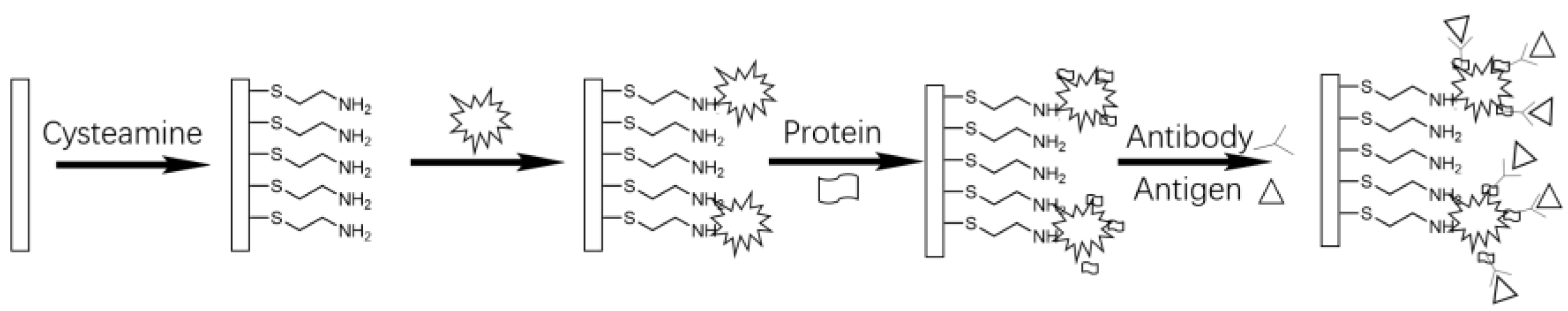
3.3. Thiol Binding Sites

In IgG antibodies, the light and heavy chains, as well as between the two heavy chains, are interconnected via disulfide bonds. Consequently, the application of disulfide bonds for site-specific antibody immobilization has drawn extensive scholarly attention. The cysteine-linked disulfide bonds can be severed by reducing agents, yielding active thiol groups that are capable of self-assembling onto gold nanoparticles or modified metal nanoparticles [56,57]. Moreover, a greater quantity of activated thiol groups engenders enhanced affinity interactions, facilitating a more robust covalent coupling than a single thiol moiety [58]. H. Sharma employed TCEP and 2-MEA to reductively cleave the disulfide bonds within the antibody hinge region, generating half-antibody molecules with free thiol groups. These can react with maleimide- or iodoacetyl-activated surfaces, thereby realizing a directional immobilization strategy. Julija Baniukevic further developed an immunosensor for bovine leukemia virus (BLV) antigen (gp51) using a similar approach and a plasmon resonance device, demonstrating a lower detection limit and favorable repeatability [59].
The disulfide bond between the two heavy chains is distal from the antibody active center and exerts no critical influence on the protein structure. To avert unnecessary steric hindrance and without compromising the antibody binding site, the disulfide bonds in the hinge region can be reduced, fragmenting the intact IgG into two half-IgG fragments. Post-cleavage, the protein activity alteration is minimal or accompanied by only a slight structural deformation [59].
The cleavage yields two monovalent antibody fragments F(ab′) with antigen-binding capabilities. The potent interaction between the thiol group and the gold surface represents the most direct strategy for thiolated antibody immobilization. Hua Wang successfully chemically conjugated the 2-MEA-reduced F(ab′) onto gold nanoparticles, accomplishing antibody immobilization [60]. Kathryn L. Brogan compared the direct fixation of F(ab′) and F(ab) fragments on the gold (Au) surface to form gold-sulfate bonds and established that the specifically fixed F(ab′) fragment exhibits a higher antigen-binding efficiency [9].
A. Kausaite-Minkstimiene contrasted the surface concentration and antigen-binding ability of immobilized intact anti-hgh and fragmented anti-hgh via surface plasmon resonance and ascertained that the antigen-binding signal was augmented by 50-fold [8,61] (Figure 10). However, it is crucial to note that the direct attachment of antibody fragments onto a metal platform may precipitate metal-induced denaturation or impact protein activity post-adsorption [62].
Arkady A. Karyakin adopted this method. Despite the exposure of only one antigen-binding epitope, compared to randomly fixed antibodies, the quantity of antigens attached on the semi-IgG-modified gold carrier was substantially higher than that of non-specific adsorption, attaining the directional fixation of antibodies. Additionally, the semi-IgG-modified electrode manifested considerable operational stability [11].
Nevertheless, reduction via a reducing agent may induce alterations in the antibody’s three-dimensional structure. R. Funari utilized 258 nm and 10 kHz ultraviolet pulses to photoreduce the disulfide bridges in the antibody hinge region, generating free thiols. Zhang also employed an ultraviolet light-induced approach to form half-antibody molecules with free thiol groups and self-assemble them onto a gold substrate, with the immunosensor exhibiting enhanced sensitivity [34]. Concurrently, it is essential to recognize that compared to other functional groups such as amines and carboxyl groups, the number of manipulable thiol groups on proteins is relatively restricted, potentially leading to a diminished capture efficiency of antibodies on solid particle carriers. Hermanson GT proposed that the system should be oxygen-free and chelating agents such as EDTA should be incorporated to preclude thiol oxidation in the solution and the formation of disulfide crosslinks.
Furthermore, digestion by pepsin into F(ab′)2 (a divalent fragment comprising two Fab fragments) is characterized by the retention of covalent bonds between antibody chains, modification of disulfide bonds at a location remote from the antibody binding site, and upward orientation of the antibody. Michelle K. Greene utilized F(ab′)2 to insert a pyridazine dione moiety into the disulfide bond via a reductive reconnection method. Through the other end of a novel heterobifunctional linker, the 1,3-dipolar cycloaddition between azide and alkyne was harnessed to achieve the directional fixation of Fab on nanoparticles, augmenting the coupling efficiency [63]. Harsh Sharma initially fixed the half-antibody fragment obtained by TCEP reduction through Protein G, and the resultant directional antibody exhibited a more pronounced specific antigen-binding ability [10].
Owing to the potent covalent binding affinity between thiols and gold nanoparticles and the well-regulated size, shape, and other properties of synthesized gold nanoparticles, extensive investigations have been conducted on diverse ligand exchange reactions on gold nanoparticles under thiol protection. These have been extended to water-dispersible gold nanoparticles with terminal functional groups, including carboxylic acids, amines, azides, alcohols, carbohydrates, amphiphilic polymers, amino acids, nucleic acids, polypeptides, and proteins, proffering a comprehensive perspective for antibody immobilization within the thiol covalent binding framework and the adsorption fixation of substrates in bioaffinity techniques [64].

3.4. Carbohydrate Binding Sites

Immunoglobulins harbor polysaccharide chains within the CH2 domain of the heavy chain, situated in the Fc region and distal from the critical active center or binding loci [65,66]. Thus, the directional manipulation of antibodies can be attained by furnishing fixation targets via carbohydrates. The Fc region per se is also exploited for directional immobilization. Through the binding of sugar chains to the Fc, antibodies are oriented upward, and their binding sites are rendered accessible to antigens. Generally, three strategies for fixation via sugar chain residues are recognized, which are as follows:
1. Conversion of multiple hydroxyls to active aldehydes.
Empirical investigations have revealed that periodate oxidation is among the simplest means to transform multiple hydroxyls in sugar residues into active aldehydes. The aldehyde moieties with fixation sites can react with amine or hydrazide groups under mild conditions to form relatively stable covalent bonds (hydrazone bonds), irreversibly tethering antibodies [67]. For instance, Hoffman and O’shannessy devised an antibody fixation protocol predicated on the hydrazine bond between the substrate and aldehyde groups [68,69,70]. Huang et al. oxidized the sugar chain of anti-alpha-fetoprotein to aldehyde and covalently conjugated it to an APTES-modified silicon substrate. In comparison to physical adsorption, the amount of immobilized antibody was augmented by 32%, and the antigen-binding capacity increased by 16%. Gering et al. effected a condensation reaction between the hydrazide group modified on the glass surface and the aldehyde group derived from the carbohydrate in the Fc moiety [71]. Beatriz Prieto-Simón established hydrazone bonds between the active aldehyde and the hydrazide group introduced by the electrografting of diazonium salts or the self-assembly of mono- and dithiohydrazide linkers, thereby realizing the directional fixation of antibodies on the functionalized gold electrode [72], as shown in Figure 11.
Although the directional immobilization based on sugar oxidation has been successfully accomplished in terms of antibody fixation on the carrier orientation, concerns regarding the accidental cross-linking of antibodies via Schiff base linkages persist [73]. Given the heterogeneous nature of proteins, which are composed of multiple functional groups, including amine, carboxyl, thiol, and hydroxyl, the aldehyde groups generated by oxidation may cross-link and aggregate with other groups in the molecule. The potential for significant oxidation side reactions, including protein denaturation, also restricts the immobilization efficiency and the biological activity of proteins to a certain extent [74]. The incorporation of boric acid may offer a viable solution [75].
2. Exploitation of boric acid derivatives for binding.
Boric acid (BA) and its derivatives can rapidly and reversibly form cyclic boronic esters with diols at room temperature, presenting the potential for directional antibody fixation [75,76,77,78]. In 2009, Lin et al. first reported the utilization of boric acid for directional Ab fixation. Moreno Guzmán et al. reported the construction of an electrochemical immunosensor, in which the adrenocorticotropic hormone (ACTH) antibody was directionally attached to a phenylboronic acid-modified screen-printed carbon electrode, enabling the ultrasensitive determination of adrenocorticotropic hormone [79]. Wei-Ching Liao facilitated the directional connection of sugar groups on the gold surface through a thiophene-3-boronic acid intermediate layer [80]. Jie Zhao employed the covalent binding of boric acid and GMA to directionally immobilize antibodies on the SBMA and GMA copolymer [81]. The directional fixation of antibodies using boric acid is expounded in more detail in Florine Duval’s review [75].
However, the reversible binding process of boric acid groups to form boronic esters entails the risk of feeble antibody binding, such as fluctuations in the concentration of diols and pH values in the system. Avijit K. Adak developed a directional Ab fixation method using boric acid [74,82]. By irradiating with ultraviolet light, (trifluoromethyl)phenyldiazine can irreversibly tether antibodies to the BA surface to enhance the sensitivity of antigen detection, thereby constructing a functional Ab microarray capable of detecting analytes such as RCA120, PSA, and IL-6 at low picomolar concentrations. In a subsequent study, an antibody microarray constructed by the BA−benzenesulfonate strategy was utilized. Through the SN2 reaction, Abs were directionally immobilized on glass slides through a boric acid ligand containing phenylsulfonate, and a photoaffinity group (photoactive phenyldiazine) was incorporated into the boric acid ligand. Through photoinitiated cross-linking, Abs were irreversibly attached to glass slides and nanoparticles, resulting in a heightened level of Ab fixation and antigen detection sensitivity. Nevertheless, in the method of fixing Abs based on boric acid, several significant challenges remain, such as the pretreatment of boric acid derivatives, the identification of antibodies with suitable sugar chains, the determination of binding conditions conducive to effective directional fixation of Abs, and the assurance that the immobilized antibodies are not dislodged from the surface by reverse reactions or other factors. Concurrently, Tacias-Pascacio noted that the sugar chain is a long and flexible spacer arm, and the fixed antibody may lose activity after collapsing on the solid carrier, and excessive exposure of the antibody to oxidizing chemicals may also lead to a diminution in antibody activity [83].
3. Connection of sugar chain residues with special structural functional groups.
In Anand K. Agrahari’s review [84], sugar chain residues can be relatively facilely converted into or conjugated with certain special structural functional groups, such as azide groups, double bonds, triple bonds, etc. These groups are directionally immobilized on the active substrate through the corresponding bifunctional linker intermediates. For example, in a typical Staudinger reaction, azide and phosphane react in an aqueous solution at room temperature to generate a phosphinane intermediate, which further reacts to form a permanent amide bond. Owing to the diminutive size of the azide, which engenders minimal steric hindrance, this reaction is an ideal candidate for the immobilization of biomolecules with high grafting density. For example, in the presence of a monovalent copper catalyst, Amblard, F. realized the directional fixation of antibodies and substrates through the [2 + 3] cycloaddition between azide and alkyne to form a five-membered triazole ring. However, the catalysis of metal ions can precipitate protein denaturation and a reduction in affinity efficiency. The success of Maximilian Brückner in generating nanocarrier conjugates through copper-free click chemistry also attests to the progress of metal-free click chemistry [85]. Since azide, conjugated diene, and other active sites scarcely exist in any naturally occurring biomolecules, such chemical fixation methods minimize the mislocation of reaction sites and exhibit favorable bio-orthogonality.
Figure 11. Antibody immobilization on residue glycan via (A) hydrazone [72], (B) boric acid derivative [74], and (C) click reaction [84]. Reproduced with permission from ACS.
Chemosensors 13 00050 g011

4. Bioaffinity Immobilization Technology

Covalent bond fixation is robust and irreversible. Nevertheless, challenges such as the high and heterogeneous distribution of specific antibody functional groups, potential alterations in protein conformation, shielding of active binding sites, partial antibody orientation, and additional losses in chemical environments [86] have spurred the exploration of milder alternatives. Bioaffinity immobilization technology has emerged as a viable solution. It capitalizes on selective, non-covalent linkages between proteins or nucleic acids and proteins, exemplified by the immobilization strategies involving Protein A and Protein G, biotin/(strept)avidin complexes, material-binding peptides orientation, and histidine-tagged protein immobilization on Ni2+-chelating surfaces, thereby enabling the bio-directional fixation of antibodies.

4.1. Specific Binding of Protein A and Protein G

Protein A, originating from Staphylococcus aureus, is capable of specifically binding to the Fc moiety of antibodies, thereby facilitating the establishment of a directional system [87,88]. Raffaele Caroselli constructed a protein A intermediate layer on the sensor surface via a ring-resonator sensor, attaining the directional immobilization of antibodies through a physical approach and augmenting the interaction with the target antigen under detection [89].
These proteins, serving as intermediate sublayers bridging nanoparticles and antibodies, possess conspicuous advantages (Figure 12), which are as follows:
1. The antibodies in linkage necessitate neither chemical environment modification nor optimization of decoration, thereby circumventing potential impacts on biological activity.
2. Their specific binding to the Fc segment enables directional anchoring on the substrate surface, rendering it facile for Fab to conjugate with the corresponding antigen. Xinyi Mao developed a lateral flow immunoassay (LFIA) through the directional fixation of time-resolved fluorescent microspheres by protein G, which exhibited high detection precision in the assay of aflatoxin B1 (AFB1) [90].
Nonetheless, their drawbacks are also evident:
1. Protein A selectively binds to specific IgG subclasses rather than all antibodies, thereby constraining its extensive application [91].
2. The binding between protein A and the Fc domain is reversible [92], mandating the presence of metal ions (such as Zn2+, Cu2+, Ni2+) or other adjuncts including chemical bonding to effectuate irreversible binding. With the aid of protein A or G, numerous immunosensors have been fabricated. In comparison to the scenario of random antibody attachment, the detection performance has been substantially enhanced. Xianbao Sun exploited the recombinant combination of protein A and cysteine residues (SPACys) to achieve the directional binding of antibodies to the AuNPs-PEI-MWCNTs nanocomposite via a one-pot protocol, demonstrating favorable linear response, selectivity, and anti-interference capacity in the recognition of kidney bean lectin (KBL) [93]. Jianguo Chang employed the co-coupling of phenylboronic acid and protein A to directionally tether antibodies on a polymethyl methacrylate (PMMA) polymer substrate and utilized MPC and BSA as blockers to attenuate background noise, endowing its detection with high sensitivity, specificity, and reliability [94]. Guangyu Shen indirectly attached protein A to the sensor through a hyperbranched polymer (HBP) synthesized from p-phenylenediamine and tricarboxylic acid via cysteamine treatment, accomplishing the directional control of immobilized protein A for the immunoadsorbent layer [95].
Figure 12. Schematic of protein A linking substrates to antibodies via bifunctional moieties. Reproduced with permission from [95] (Copyright 2011, Elsevier).
Chemosensors 13 00050 g012
3. Protein A can interact with other albumins, albeit to a considerably lesser extent, which may give rise to impurity signals or false positive outcomes. Nevertheless, such detections can be intervened by pliable external conditions such as pH, salt concentration, and additives.
A more comprehensive exposition can be retrieved from the reviews of Min Park and Liu [73,96]. The IgG-binding capabilities of these proteins vary across species. Protein G resembles protein A, yet generally, IgG exhibits a higher affinity for protein G than for protein A, and protein G can bind IgG from a broader spectrum of species [97]. Considerable attention has been directed toward protein G and the coordinated functionality of protein A and protein G.

4.2. Biotin Immobilization

Biotin, a water-soluble B vitamin found in all living cells, maintains its binding affinity with avidin and streptavidin even when attached to macromolecules. Streptavidin, a 60 kDa protein derived from Streptomyces affinis, consists of four identical subunits, each housing a biotin-binding site. The interaction between biotin and streptavidin is characterized by an extremely high affinity (Ka = 1015 mol−1), over one million times stronger than the binding strength of antigen-antibody interactions [98]. This powerful and nearly irreversible binding, along with its high tolerance to extreme pH and temperature conditions, as well as its remarkable structural stability, makes the biotin-streptavidin system an ideal choice for a variety of biosensor applications [99]. Cho explored the directed fixation of antibodies on solid surfaces through site-specific biotinylation in the hinge region of antibodies. By biotinizing the reduced intact antibody with maleimide-activated biotin, the reaction predominantly occurs on the sulfhydryl group between the CH1 and CL domains, and between the CH2 and CH2 domains at the hinge [100]. Based on these findings, the authors developed a method to bind biotin to the CH2 and CH2 domains under optimal conditions by introducing free maleimide to compete with activated biotin for priority sites between the CH1 and CL domains. The results demonstrated that the ability of a site-biotinylated intact antibody and F(ab′)2 fragment in sandwich immunoassay was 2.6 times and 20 times higher than that of a randomly biotinylated antibody, respectively. This suggests that site-specific biotinylation significantly enhances antibody fixation efficiency and detection sensitivity. Yang proposed a method for the Fc-specific biotinization of antibodies, which involves the engineered photoactivation of Z-Biotin (ZBpa-Biotin) [101]. This is achieved by incorporating ZBpa-Biotin into the target protein using the aaRS/tRNA system, followed by light activation. The process results in a specific binding to the Fc region of the antibody. The efficiency of this site-specific photobiotinized IgG in biosensing was assessed using the surface plasmonic resonance (SPR) technique, with the tumor biomarker carcinoembryonic antigen (CEA) serving as a model. The limit of detection (LOD) on the surface coated with photobiotinylated IgG was found to be 2 ng·mL−1, which is five times lower than that of randomly NHS-biotinylated IgG (10 ng·mL−1). The Human Mammary Tumor Virus (HMTV), also known as mouse mammary tumor virus, is an endogenous retrovirus belonging to the retrovirus family. It primarily infects human and mouse epithelial cells [102]. With advancements in nanobiosensors, an ideal interdigital electrode (IDE) is employed for the detection of HMTV. Cost-effective lithography techniques can be utilized to fabricate aluminum-rich IDEs (ALIDEs). In a 2019 study, Ghosh outlined a method for enhanced sensitivity using a modified aluminum finger electrode (ALIDE) and a streptavidin-biotin quadruvalent complex [103]. This approach enables the highly sensitive detection of human breast tumor virus (HMTV) DNA, achieving quantitative detection values as low as 100 aM.
MicroRNAs (miRNAs) serve a crucial regulatory function in cells [104,105], and their dysregulation has been associated with various diseases [106], thereby establishing them as potential biomarkers. In a study by Ding et al. (Figure 13), a novel biosensing strategy was introduced that leverages surface plasmonic resonance (SPR) in tandem with DNA super-sandwich and biotin-streptomycin amplification techniques to detect miRNAs without the need for labels [107]. The target miRNAs are selectively captured by surface-bound DNA probes. Following hybridization, streptavidin amplifies the signal on the extended DNA super-sandwich components by binding to biotin, resulting in a significant increase in the SPR signal. This method exhibits high sensitivity, capable of detecting miRNAs at concentrations as low as 9 pM within a span of 30 min. Additionally, Zhai et al. highlighted the application of DNA hybridization chain reaction (HCR) and biotin-streptomycin based amplification [108]. In the presence of the target miRNA, a hairpin DNA capture probe affixed to the gold electrode surface is activated. Subsequently, the initiating DNA strand hybridizes with the exposed capture probe, initiating the HCR process and culminating in the formation of an extended double-helix DNA structure. The proposed biosensor exhibits exceptional sensitivity, capable of detecting target miRNAs at concentrations as low as 0.56 fM, and maintains linearity over a range from 1 fM to 100 pM. Furthermore, it demonstrates high specificity, effectively distinguishing between target miRNAs and mismatched sequences.
Surface plasmon resonance (SPR) is a well-established optical biosensor technology that detects biomolecular interactions without the need for fluorescence or enzyme labeling. Yang et al. developed a sensitive SPR biosensor using ZnO@Au nanomaterial, which detected human IgG (hIgG) through the classic sandwich strategy of a biotin-streptavidin secondary signal amplification system [109]. The high selectivity of streptavidin (SA) in interacting with biotin led to the construction of GNRs-SA-biotin-Ab2 (GSAB-Ab2), which provided a secondary enhancement of the SPR signal. Under optimal experimental conditions, the GSAB-Ab2 conjugate sandwich format was introduced, resulting in an SPR biosensor that measured hIgG in a range of 0.0375–40 μg·mL−1. The minimum detection concentration of hIgG achieved by this method was approximately 67 times lower than that of conventional gold film-based SPR sensors. In light of these findings, Ozawa proposed a principle in 2017 to explain the enhancement [110]. The CH/pi interactions at biotin binding sites and subunit interfaces were explored and analyzed using the fragment molecular orbital method (FMO) and its extended applications: PIEDA and FMO4. The results indicated that the CH/pi interaction contributed to the high affinity of streptavidin binding sites to biotin. Furthermore, we demonstrated that the CH/pi networks involved in the CH/pi interaction and expansion at the subunit interface played a more critical role in determining high affinity, rather than participating in binding sites.

4.3. DNA Bio-Immobilization Technology

Jung developed a multifunctional biosensor for efficient antibody fixation by specifically binding protein G to DNA oligonucleotides (Figure 14) [111]. This protein G-DNA conjugate allows controlled fixation of the antibody at a predefined location on the bioassay chip or particle surface, while maintaining the activity and orientation of the bound antibody. A streptococcal protein G variant, with an n-terminal labeled with a cysteine residue, is covalently linked to amine-modified single-stranded DNA. Surface plasmon resonance (SPR) analysis revealed that protein G-DNA conjugates selectively bind to complementary, surface-fixed DNA probes. Notably, protein G hybridized to the DNA surface exhibits superior antibody/antigen binding ability compared to protein G chemically attached to the surface with the appropriate orientation. Lin uses tetrahedral DNA nanostructures (TDN) to construct a biosensing platform for sensitive detection of protein antigens by connecting antibodies (such as PSA monoclonal antibodies) at the vertices of the TDN [112]. Antibodies are bound to TDN using click chemistry by designing DNA probes with alkynyl modifications as linkers. This platform significantly enhances the efficiency and sensitivity of antigen-antibody binding by reducing the effect of disordered antibody fixation. Targeted detection can also be achieved through other targeted modifications. Konc proposes a novel platform for the preparation of DNA-antibody bioconjugates using a simple benzoylacrylate pentafluorophenyl ester (BA-PFP) reagent [113]. Benzoylacrylate-labeled oligonucleotides generated using this reagent can achieve site-specific conjugation of accessible cysteine residues on various proteins and antibodies. The method offers a site-specific, stable structure that addresses the limitations of maleimide bioconjugates, including the hydrolytic instability of reagents and challenges associated with anti-Michael addition reactions. It can also be stored as a solid for months or maintained in solution for weeks without degradation. Furthermore, it enables the modification of various amino-containing small molecules or biomolecules while maintaining high reactivity. The homogeneity of the obtained DNA-antibody bioconjugates was confirmed by a newly developed LC-MS protocol.
These bioconjugated probes have been successfully applied to fluorescence and super-resolution microscopy for cell-imaging experiments. This study underscores the versatility and robustness of the proposed bioconjugation method, which offers site-specific, well-defined, and plasma-stable DNA-antibody bioconjugations for diverse biological applications. Kazane introduces an innovative platform for the preparation of site-specific DNA-antibody bioconjugations using pentafluorophenyl benzoylacrylate reagents [114]. This approach employs benzoylacrylic acid-labeled oligonucleotides to achieve site-specific conjugation with accessible cysteine residues in antibodies. This method effectively addresses the drawbacks of conventional bioconjugation techniques, such as hydrolytic instability and non-specific labeling, producing homogeneous, plasma-stable structures suitable for biological applications. These structures demonstrate maintained structural integrity and functional specificity across various applications, indicating their vast potential in the realm of biosensors. Bano presents a novel platform that utilizes benzoylacrylic acid-labeled oligonucleotides for the creation of site-specific DNA-antibody biocouplings. Using AFM nanografting technology, DNA nanopatches of varying sizes were prepared [115]. The DNA-protein couplings were then nanografted onto the sensor surface through DNA directional immobilization (DDI), proving highly suitable for biosensing applications and facilitating the preparation of multi-feature protein nanoarrays. In 2013, Arrabito introduced a site-specific DNA-antibody biocoupling platform utilizing the benzoylacrylate pentafluorophenyl ester reagent [116]. This method achieves high homogeneity and plasma stability by coupling DNA oligonucleotides to accessible cysteine residues on antibodies.
The target DNA array is procured through DNA directional immobilization and can be further functionalized using a covalent DNA-streptavidin (DNA-STV) conjugate. The high homogeneity of the DNA-antibody conjugate was confirmed via liquid chromatography-mass spectrometry (LC-MS). Furthermore, these couplings have been successfully employed in fluorescence and super-resolution microscopy techniques, underscoring their efficacy in cell imaging. This method’s strength lies in its ability to generate chemically defined and site-specific conjugators, effectively minimizing non-specific interactions while preserving the functional integrity of the antibody. This robust platform has broad applications in diagnosis and treatment. DNA-directed enzyme immobilization technology offers an efficient and adaptable solution for developing multi-enzyme catalytic systems. By immobilizing glucose oxidase (GOx) onto functionalized nitrogen-doped graphene quantum dot-modified ferrite nanocomplexes (Fe3O4@N-GQDs), we significantly enhanced the system’s catalytic activity and stability using a DNA-directed immobilization strategy [117]. This method leverages the Watson–Crick base pairing properties of DNA to direct the enzyme to the nanocore under mild conditions, thereby addressing the instability and storage issues of natural enzymes and boosting the efficiency of the multi-enzyme cascade reaction. Fe3O4@N-GQDs not only function as a nanase mimic but also provide high peroxidase activity, optimizing substrate entry channels, and facilitating product formation. The technique under discussion offers a reversible fixation of the enzyme, allowing for the regeneration of the enzyme carrier through a gentle DNA-dehybridization process. This introduces a novel concept for the extended use of multi-catalyst systems. Furthermore, a new sensing system, based on electrochemical-luminescence nanosurface energy transfer (ECL-NSET), facilitates ultra-sensitive detection of procalcitonin (PCT) by incorporating a DNA-directed fixation strategy [118]. The researchers employed HWRGWVC heptapeptide to capture antibody Fc fragments, ensuring the exposure of the antibody active site, thereby significantly enhancing the sensor’s sensitivity and specificity. The multifunctional DNA-mediated spatially restricted assembly (SCA) strategy notably amplified electrochemical signal peaks compared to randomly arranged antibody fixation strategies, underscoring the benefits of DNA in antibody-directed fixation. In a separate study, the authors delve into various immobilized-technology microarrays for antibody generation, with a specific emphasis on a method that utilizes DNA-directed fixation of antibody-DNA couplings to establish a universal and robust protein array [119]. The DDI method, direct antibody spotting on chemically activated glass slides, and immobilization of biotinylated antibodies on streptavidin-coated slides were compared. In the model system studied, varying amounts of goat anti-rabbit IgG antibodies were immobilized as capture reagents to detect serial dilutions of rabbit IgG antigens. The detection limit achieved was 0.001 nM (150 pg/mL). Among the methods compared, both the DDI method and direct spotting yielded the highest fluorescence intensities. However, the DDI technique demonstrated superior spot uniformity and reproducibility. Notably, microarrays fabricated using DDI and direct lattice techniques displayed superior fluorescence intensity, facilitating the highly sensitive detection of antigens, even at reduced trapping reagent concentrations. Moreover, experiments utilizing the DDI-based microarray demonstrated optimal point uniformity and minimized internal and external standard deviations.
These outcomes unequivocally underscore the merits of DDI in crafting antibody microarrays. When contrasted with traditional approaches, DDI not only matches or surpasses them in signal strength, detection sensitivity, and repeatability but also offers marked benefits in flexibility and adaptability during the immunoassay configuration. This is attributed to the modular preparation of functional reagents, which optimizes the utilization of precious antibodies.
In addition, aptamers have garnered increasing attention in recent years. Ap-tamers are single-stranded DNA or RNA molecules selected through the SELEX (Systematic Evolution of Ligands by Exponential Enrichment) process to specifically bind to target molecules. Compared to antibodies, aptamers offer significant advantages in terms of stability, flexibility, and reproducibility, as they are selected in vitro and are less likely to trigger immune responses [45]. Their binding affinity is comparable to, or in some cases even better than, antibodies. Aptamers can be chemically modified to attach to various surfaces, such as metals [120], nanomaterials [121], or carbon nanotubes [122], enhancing their stability and enabling effective immobilization on sensor surfaces for signal transduction. They are also more chemically stable than antibodies, with higher resistance to heat and enzymatic degradation, and once the sequence is established, they can be reliably synthesized at scale [123]. Furthermore, aptamers can be regenerated under specific conditions, making them more cost-effective and efficient for repeated use in sensors [124]. In terms of sensitivity, aptamers exhibit high affinity for their targets, with detection limits in the micromolar to sub-nanomolar range, making them highly effective in biosensors [120]. Their application has shown remarkable progress in electrochemical, surface plasmon resonance (SPR), and piezoelectric sensors, demonstrating sensitivity comparable to antibody-based sensors [125]. These properties, including stability, regenerability, and high sensitivity, position aptamers as a competitive choice for use in biosensor development.

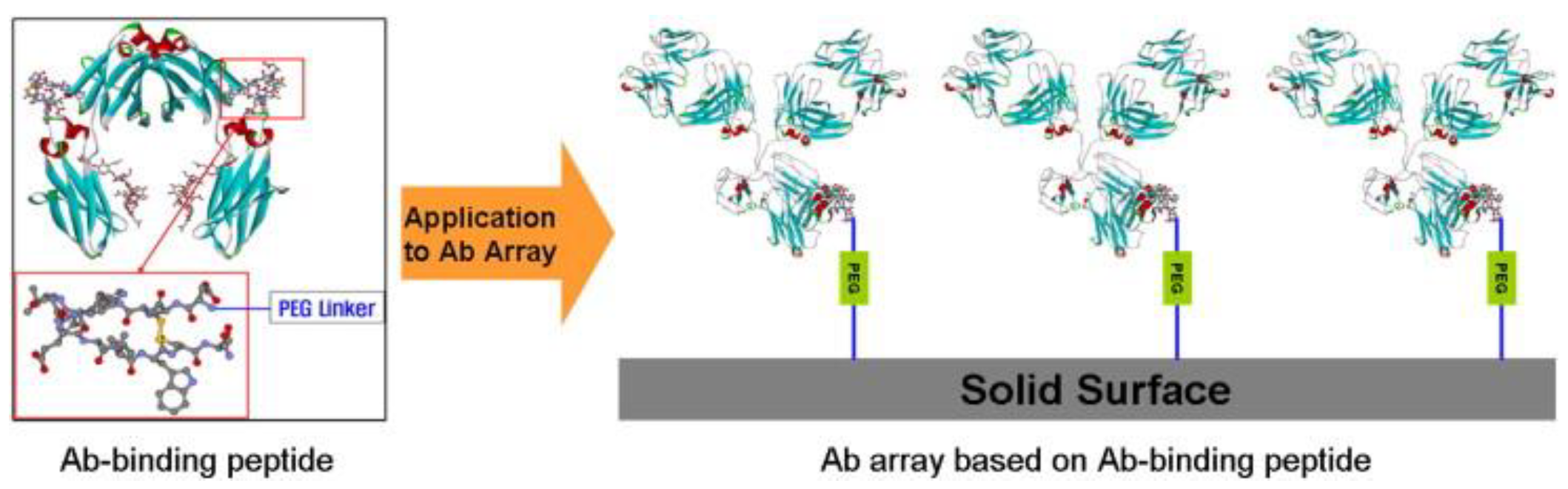
4.4. Material-Binding Peptide Orientation Technology

MBP, a specific short peptide, possesses the capability to recognize and bind to a substrate’s surface. In addition, compared to full-length antibodies, antibody fragments often exhibit lower specificity, stability, and affinity [126]. Previous research has identified numerous MBPs for various target substrates, spanning from inorganic to organic materials. The application of MBPs is extensive in the fields of biomineralization and crystallization, recombinant protein purification, and site-selective immobilization. A significant advantage of the MBP-mediated approach, compared to other affinity-based methods, is its ability to effectively affix the target protein to a specific substrate surface without necessitating any chemical modification. This is achieved by combining MBP with the target protein in the chosen region through genetic fusion or chemical coupling. The direct and site-specific adhesion of the target protein on the substrate surface is facilitated by the specific binding between MBPs and the substrate material. Fuchs and Raines demonstrated this by constructing a poly-Arg9 (R9)-labeled RNase a, which showed that the fused RNase a could be directly adsorbed onto glass slides or silicone resins without any loss of enzyme activity. But there are results on the surface adding additional cationic residues is likely to decrease the conformational stability of RNase a by increasing unfavorable Coulombic interactions within the protein [127]. Korodi presents a method for covalently immobilizing antibodies on a standard 96-well microplate [128]. This is achieved by optimizing the crosslinking procedure, enabling the reuse of the antibody-protein G complex on a minute scale. The process encompasses affinity binding of the antibody to the protein G surface fixation using various cross-linking agents (DMP and BS), termination of the cross-linking reaction, and subsequent binding of antibody-specific antigens. The research indicates that affixing peptides to a gold chip’s surface via a hydrophilic linker can effectively capture IgGs in both humans and rabbits. Furthermore, antibodies captured through peptides demonstrate enhanced antigen-binding capacity compared to randomly immobilized antibodies (Figure 15) [129]. Kishimoto introduces a chemical conjugation technique utilizing peptides with high affinity to human IgG-Fc-termed affinity peptide chemical conjugation (CCAP) [130]. This approach facilitates rapid, site-specific modification of a specific residue (Lys248 on Fc) under mild conditions, resulting in a stable peptide-Fc amide bond. Remarkably, this monovalent peptide-IgG conjugate retains its antigen-binding capability and also associates with Fc receptors (FcRn, FcγRI and FcγRIIIa). Tanwar delves into the design of inhibitor peptide sequences, leveraging insights from the interface of protein G-IgG crystal complexes [131]. Utilizing the binding interface knowledge of the IgG-protein G complex (PDB:1FCC), a series of peptide sequences were designed to inhibit these interactions. Both experimental and computational studies confirmed that all peptide sequences aligned closely with anticipated IgG binding sites, ensuring complex stability. Yoo’s research explored the identification of a peptide ligand using phage-displayed heptapeptide libraries, with the aim of immobilizing antibodies on biosensor surfaces [132]. The chosen peptide, KHRFNKD, exhibited not only high specificity and affinity for rabbit antibodies but also proved its efficacy in antibody capture and antigen detection within QCM biosensors. This offers a novel approach for the directional and stable integration of antibodies in biosensor applications.
Seo presents a comprehensive method for constructing antibody arrays, which facilitates regionally selective covalent fixation of antibodies on solid supports [133]. This is achieved by utilizing truncated forms of antibody binding proteins A, G, and L. These recombinant proteins possess C-terminal CVIX protein transferase recognition motifs, enabling the incorporation of biologically orthogonal azide or alkynyl functional groups. They are then affixed to suitably modified glass surfaces via a Cu(I)-catalyzed Huisgen cycloaddition reaction. Notably, this technique effectively allows for the regionally selective fixation of antibodies on solid surfaces without necessitating any modification of the antibodies themselves. Schroeder [134], on the other hand, introduces an innovative protein cross-linking approach for the directed irreversible fixation of antibodies on surfaces. This method ensures the directed and covalent fixation of the antibody in the Fc region by chemically pre-activating protein A or G and employing a chosen conventional cross-linking agent, followed by the introduction of an antibody for cross-linking. This pre-activated antibody orientation fixation approach circumvents issues associated with random orientation and the generation of inactive by-products inherent in traditional methods. Moreover, it eliminates the need for recombination and modification of binding partners and offers a superior signal output compared to conventional techniques. Consequently, it holds significant advantages in systems where minimizing antibody loss and maximizing binding capacity are paramount.
Aoyagi developed a reagent-free, renewable, and rapid immunosensing system for detecting immunoglobulin G based on the fluorescence enhancement of fluorescein isothiocyanate (FITC) induced by protein interactions. In hydrophobic environments, the fluorescence intensity of FITC generally increases, and thus the fluorescence intensity rises with the concentration of IgG bound to protein A. High reproducibility was achieved by washing the system with PBS at pH 4 after each measurement.

5. Other Binding and Fixation Technologies

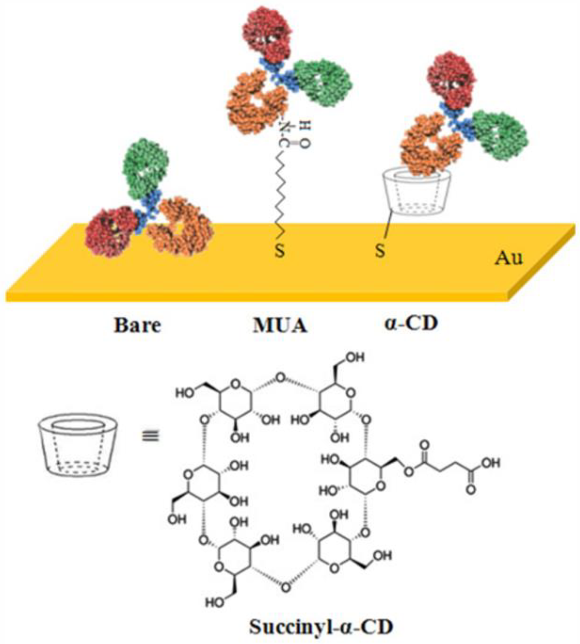
5.1. Hydrophobic Domain Binding Sites
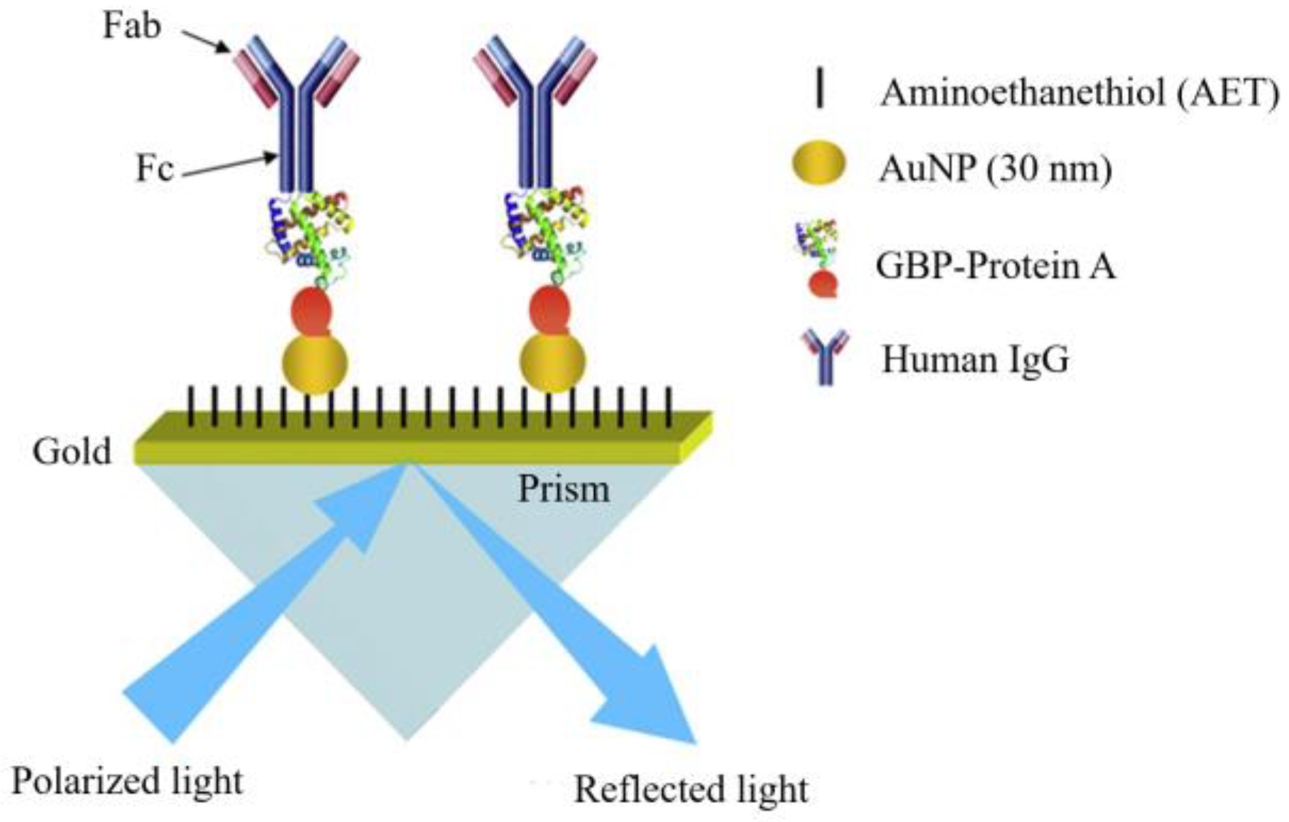
On the antibody surface, numerous hydrophilic domain binding sites are present, while internally, hydrophobic groups exist. The hydrophobic amino acid residues, such as leucine and isoleucine, contribute to the maintenance of the overall structural stability of the antibody. Through hydrophobic interactions, they stabilize the three-dimensional architecture of the antibody, ensuring that the antigen-binding sites retain an appropriate conformation for antigen recognition and binding. For instance, hydrophobic regions within the framework regions of the antibody variable regions furnish structural support for the antigen-binding sites; those in the Fc segment maintain its tertiary structure, permitting correct recognition and binding by Fc receptors and complement proteins. Additionally, hydrophobic portions are located between the two heavy chains and in the regions where the light and heavy chains connect. Via the synergistic effect of disulfide bonds and hydrophobic interactions, the overall structural integrity of the antibody molecule is conserved, guaranteeing the proper functioning of the antibody in antigen recognition and effector activities.
Cyclodextrin (CD), a cyclic oligosaccharide formed by multiple glucose units connected via α-1,4-glycosidic bonds, exhibits a distinct hollow cylindrical structure with a relatively hydrophobic cavity inside and a hydrophilic exterior. During antibody immobilization, it seizes hydrophobic side chain amino acids and forms a stable host–guest inclusion complex to mediate antibody interaction. As exemplified by Juyeon Jung (Figure 16), who utilized chiral α-cyclodextrin (α-CD) and SAM to effectively immobilize antibodies on an unmodified gold surface, enhancing the sensitivity of antigen detection [135]. Moreover, cyclodextrin offers several advantages, which are as follows:
1. Multiple modifiable binding sites are available on the cyclodextrin carrier surface. These can associate with antibodies through chemical bonds (e.g., covalent, coordination) or physical adsorption (e.g., hydrogen bonds, van der Waals forces), thereby augmenting the amount and efficiency of antibody immobilization on the carrier.
2. The size and shape of different cyclodextrin types can modulate the steric hindrance during antibody immobilization. Appropriately sized cyclodextrin can avert excessive aggregation of antibodies during fixation and preclude mutual collisions between antibodies and the shielding of active sites.
3. Functionalization of cyclodextrin can direct antibodies to be immobilized in a specific orientation on the carrier. For example, the introduction of specific functional groups on cyclodextrin that interact with the active groups of specific antibody regions enables directional fixation and improves the efficiency of antibody–antigen binding.

5.2. Metal Bonding

In the development of surface plasmonic resonance (SPR)-immune sensors, it is crucial to effectively immobilize antibodies onto the sensing platform to enhance sensitivity [136,137,138].
Studies have examined various antibody immobilization strategies, including random, covalent, directed, and their combinations, on the performance of SPR-based immunoassays. The covalent-directed fixation approach emerges as the most suitable for conducting SPR-based HFA immunoassays due to its superior HFA response [139]. Ko devised a sensor that integrates a gold-binding polypeptide (GBP) with a protein A (ProA) gene to create a unique fusion protein adept at antibody immobilization [140]. This GBP-ProA protein self-immobilizes directly onto the SPR chip surface, which is assembled with bare and AuNPs through the GBP segment (Figure 17). It then directionally binds human immunoglobulin G (hIgG) to the ProA domain, targeting both the antibody and anti-HIGG Fc region. Anti-salmonella antibodies were immobilized onto two BGP-ProA layered chips for Salmonella typhimurium detection. SPR analysis revealed that the hIgG and anti-hIgG consistently bound to the GBP-ProA layered AuNPs assembly chip, resulting in a signal increase of approximately 92%, significantly higher than the bare chip’s 30% under identical conditions. In chips equipped with AUNPs, this signal enhancement led to a tenfold increase in the detection sensitivity of Salmonella typhimurium compared to unmodified chips. Park employed a similar strategy for the detection of aflatoxin B1 (AFB1) by fusing the gold-binding protein (GBP), which has a strong affinity for gold surfaces, with the protein G (ProG) gene [141]. This gene interacts with the Fc portion of the antibody, resulting in the fusion protein. SPR chips prepared with the GBP-ProG crosslinker and an optimal density of anti-AFB1 antibodies (100 mg/mL) can detect AFB1 at concentrations as low as 1 mg/mL in both the buffer and corn extract. These chips also selectively detect AFB1 in the presence of control toxins such as zearalenone and ochratoxin A, while SPR reactions are negligible. This demonstrates excellent sensitivity and selectivity.
Dutta used a relatively mild method involving Fischer Carbene to modify the Au/glass surface, enabling rapid fixation of protein A and thus achieving targeted fixation of rabbit IgG [142]. Furthermore, a protein slide prepared using this protocol can purify rabbit IgG from rabbit serum three-times consecutively without any loss of efficiency. Studies have shown that the pH of the solution significantly influences the orientation of adsorbed proteins on Au, with protein G exhibiting the most suitable orientation in two different antibody-antigen systems (BSA and E. coli O157:H7) at pH = 7.2. The mechanism of protein adsorption was characterized by binding energy analysis, which revealed it to be a pseudo-chemisorption process driven by significant entropy enthalpy, rather than a classical physical adsorption phenomenon [143]. Qu developed a surface-enhanced Raman spectroscopy (SERS) immunoassay for the detection of human immunoglobulin G (HIgG) in serum [144]. Once the ordered AuA binds to the SERSIA substrate, the localized surface plasmon resonance (LSPR) effect is amplified by coupling with the SERSIA label, resulting in a robust and consistent Raman signal. The superior sensitivity of this method for HIgG detection is underscored by comparing its Raman characteristics with those of hemoglobin (Hgb) and human serum albumin (HSA). The broad linear range and low limit of detection (LOD) affirm its applicability in clinical settings. Juan-Franco introduces an innovative approach for targeting antibodies to gold surfaces using fusion proteins, specifically the protein A–gold binding domain (PAG) [145]. PAG comprises immunoglobulin binding domains that are strategically coupled to staphylococcal protein A via a gold-binding peptide (GBP). This fusion protein facilitates swift and directional immobilization of the antibody, maintaining its native conformation while ensuring the antigen-binding site (Fab) remains accessible. Consequently, the PAG method offers enhanced sensitivity.
Kausaite-Minkstimiene [8] undertook a comparative analysis of four distinct antibody fixation techniques, each suitable for the modification of surface plasmon resonance (SPR) chips. The study utilized an anti-human growth hormone antibody (anti-HGH) as a model system to evaluate these methods: the random immobilization of intact antibodies via self-assembly monolayer (SAM) based on 11-mercaptoundecanoic acid (MUA), the random immobilization of intact antibodies in carboxymethyl glucan (CMD) hydrogel using the direct covalent amine coupling technique, the directional coupling of Fc fragments to the protein G layer, and the “segmentation” of intact antibodies into two fragments followed by translocation via the chemical reduction method. These four SPR chip modification techniques were assessed for their ability to directly couple sulfhydryl groups to gold. The findings indicate that the immobilization method of fragment antibody, generated through chemical reduction, is the most suitable for designing SPR immunosensors to detect human growth hormone. This is due to its adequate antigen-binding capacity, and it offers a simpler and more cost-effective solution compared to other techniques.
The significance of Metal-Organic Frameworks (MOF) in biosensors is primarily attributed to their high specific surface area, excellent chemical and thermal stability, controllable pore size structure, and functionalization capabilities. These attributes render MOF a crucial material for constructing high-performance biosensors. Four types of layered copper metal-organic scaffolds (Cu-MOFs) were synthesized on the working electrode via electrodeposition [146], subsequently serving as electrochemical sensors for the sensitive detection of IgG in serum. The MOFs, synthesized under varying deposition potentials, exhibit distinct morphologies and properties. Subsequently, IgG serves as a template for the electropolymerization of pyrrole-imprinted films on glass carbon electrode surfaces. Following this, the template proteins are extracted, resulting in molecularly imprinted membranes with both qualitative and quantitative IgG signaling capabilities. Under optimal conditions, the IgG sensor’s detection range was established between 0.01 and 10 ng·mL−1, with a detection limit (LOD) of 3 pg·mL−1 (S/N =3). The relative standard deviation (RSD) for selectivity and reproducibility stood at 3.6%, while recovery rates ranged from 95.2% to 102.0%. Additionally, a novel one-pot synthesis method for the ZIF-8/IgG complex facilitates the directed synthesis of antibodies through IgG’s specific binding to the Fc region of the antibody [147] (Figure 18).
This process also ensures antibody protection by restricting structural alterations of the antibody within the ZIF-8 framework, thereby enhancing the antibody’s targeting ability and significantly improving its tolerance to organic solvents. Liao investigated a method to augment the sensitivity of an electrochemical luminescence (ECL) immune sensor by manipulating the direction of the antibody [148]. This approach employs Fe3O4 nanoparticles (FNs) and CdTe quantum dots (QDs) as nanocarriers and luminescent labels for modification, respectively. The ECL immunosensor’s sensitivity and selectivity are enhanced by encasing the FNs’ surface with SiO2 layers to immobilize the primary antibody (Ab1). Additionally, protein G is utilized to facilitate highly targeted immobilization of Ab1 while obstructing non-specific binding sites. Furthermore, protein G-coated CdTe QDs serve as signaling tags and are specifically targeted to secondary antibodies. The findings indicate that the sandwich-structure immunosensor demonstrates exceptional stability in cyclic voltammetry (CV) and electrochemical impedance spectroscopy (EIS) tests. Controlling the antibody direction significantly improves the immunosensor’s sensitivity in clinical bioanalysis. Yang presented an electrochemical biosensor, leveraging Argonaute assisted silver metallization, for detecting genomic fragments of the human pathogen Parvovirus B19 (B19V) [149]. The study introduced an innovative nanocomposite g-C3N4/DWCNT-COOH/PtNPs, synthesized via a one-step hydrothermal method, and applied it to the electrode surface to amplify the primary signal. By integrating nucleic acid detection with silver metallization methods, we developed a highly sensitive and specific electrochemical Argonaute biosensor. This biosensor is designed to detect the non-structural protein 1 (NS1) region of the viral genome.
A renewable, label-free electrochemical immunosensor was developed. In this study, pyrrole (Py) and pyrrolepropionic acid (Pa) were co-electropolymerized in the presence of gold nanoparticles to form a porous, conductive, stable, and hydrophilic nanocomposite. This composite was then covalently attached to protein G to immobilize capture antibodies, serving as probes for immunoassays. The sensor was regenerated by rinsing the electrode with 0.1 M glycine buffer (pH 2.7).

6. Conclusions and Outlook

This review outlines a diverse array of methods for the directional immobilization of antibodies (as shown in Table 1), signifying the multiplicity of fixation approaches yet also the absence of a universal one or an all-encompassing standard. Each method harbors its own limitations and fails to concurrently address the following concerns: (1) the antibody fixation strategy must avoid being deleterious and impinging on the biological activity of antibodies; (2) the linkage site should be situated within the Fc domain, distal from the antigen-binding site, and direct the antibody in an upward orientation; (3) antibodies ought to be immobilized at a high density with stable interspacing; (4) the stability of antibody conjugation should be substantial; (5) the overall antibody binding process should not be overly convoluted.
Moreover, the ongoing exploration and development of novel immobilization substrate materials is of critical significance. Take, for example, the silica-binding protein-assisted directional immobilization technique and the polystyrene-surface-based nanobody-directed immobilization method. It is expected that more materials with distinctive properties will find applications in this field in the future. Simultaneously, novel bioaffinity ligation technologies, like nucleic-acid-molecular-hybridization-based immobilization, ALFA-tag-based nanobody immobilization and molecular imprinting technology [150], are being developed. Hence, before novel bioaffinity ligation technologies reach full maturity, the integration of two or more binding techniques might be a more practical choice.
Table 1. Several common antibody immobilization methods.
Linking MethodActive Site RegionMechanismPath AdvantagePath Disadvantage
Nucleotide-binding site (NBS)Within the Fab domain, between the light and heavy chains [33].Exposed to 254 nm ultraviolet light, NBS forms active free radicals and covalently photo-crosslinks with small aromatic-ring ligand molecules like IBA.The directional immobilization of NBS proceeds under mild conditions, maximizing the retention of antibody 3D structure and antigen-binding affinity [33,34,35].Exposed to ultraviolet radiation of shorter wavelengths and higher energy, antibodies may denature [36,37].
Amine, carboxyl and hydroxyl binding sitesIn antibodies, active primary amine groups (exemplified by lysine and arginine), active carboxyl groups (represented by aspartic acid and glutamic acid), and a few hydroxyl groups (typified by serine and threonine) are present.Employ chemical binding between active sites and bases or double-active interlayers [40].Active sites are ubiquitous, extensively studied with mature methods.Owing to the high distribution of active sites, screening or external condition interference is necessary [2,5,25].
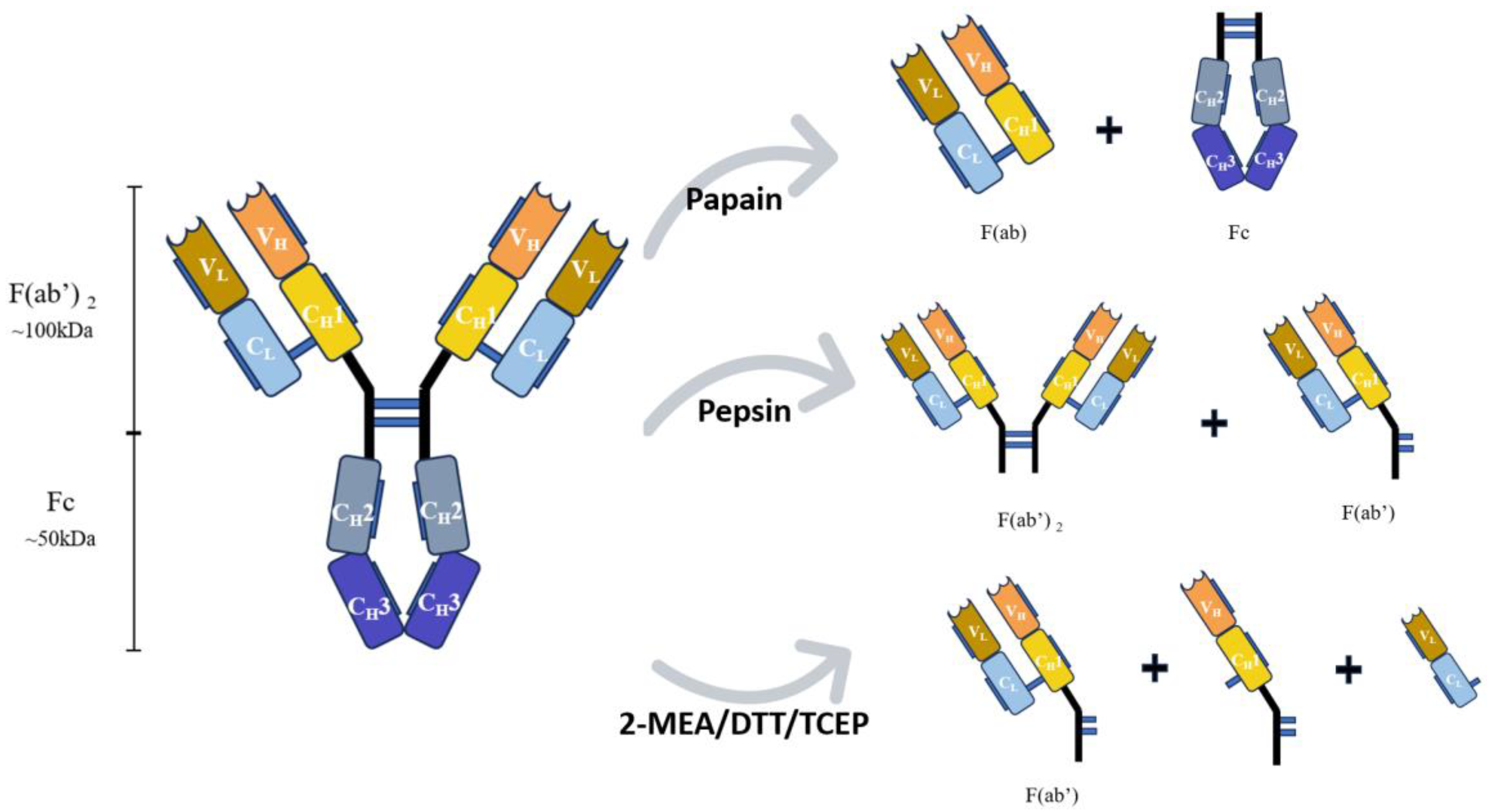
Thiol binding sitesDisulfide bonds are present between light and heavy chains and also between two heavy chains [56].Disulfide bonds, cleavable by reducing agents, yield active thiol groups with strong interaction on gold surface [57,58].The fixed locations of disulfide bonds allow antibody upward adsorption; reductive cleavage doubles thiol groups, improving antibody binding efficiency [9,60,62].Disulfide bond cleavage may impact protein activity; reduction by agents may alter antibody 3D structure and biological activity [86].
Carbohydrate binding sitesThe heavy chain’s CH2 domain, within the Fc region, harbors a polysaccharide chain [65,66].1. Transform multiple hydroxyls into active aldehydes [67]. 2. Employ boric acid derivatives for conjugation [75]. 3. Attach special structural functional groups to glycan residues [84].Glycan residues possess relatively fixed positions and have been intensively studied. Special-structured functional groups can be attached to enable click reactions.As a long spacer, glycan may lead to antibody collapse and inactivation post-immobilization. Excessive antibody exposure to oxidants may reduce its activity [73]. Certain reactions, requiring metal ion catalysis, might cause protein denaturation and lower affinity efficiency.
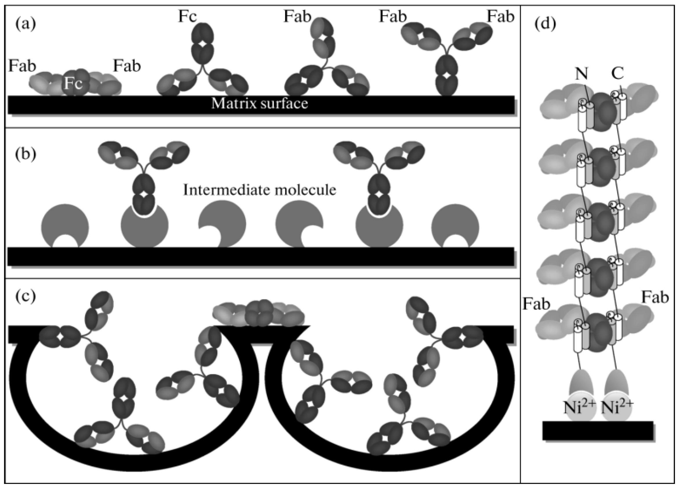
Specific binding of protein A and GThe Fc portion of an antibody [87,88].Proteins A and G, capable of specifically binding to the Fc portion of antibodies, enable an orientation system.1. The antibody requires no chemical environment modification and optimization, preventing the impact on its biological activity. 2. Specifically link the Fc part [90].1. Proteins A and G bind merely to specific IgG subclasses, restricting their application [91]. 2. The binding of Protein A to the Fc domain is reversible, necessitating metal ions or other aids like chemical bonding for irreversible binding [92].
Biotin immobilization and bindingStreptavidin comprises four identical subunits, each possessing a biotin-binding site [151].CH/pi interaction [110].The biotin-streptavidin interaction exhibits an extremely high affinity, over a million times stronger than that of antigen-antibody interaction, being irreversible and highly tolerant to extreme pH and temperature [98].The coupling system commonly employs light for activation. However, certain aromatic amino acids in antibodies are prone to photo-oxidation, leading to antibody inactivation [33].
DNA bio-immobilization technologyAmino acid sequences or domains with specific interaction to DNA [152].Enzymes are directionally immobilized on the carrier surface via DNA base complementary pairing to enhance multi-enzyme cascade activity [152].Efficient and controllable protein localization and immobilization. This technology averts protein denaturation common in traditional methods, with high operability and repeatability [152].The DDI coupling is essentially random. For a remarkable effect, antibodies usually bear multiple functional groups, causing non-specific binding and inactive conjugate formation. Moreover, it leads to elevated production costs and extended operation time [73].
Material-binding peptide orientation technologyThe Fc portion of an antibody [153].The short peptide sequence shows specific binding affinity to the antibody Fc domain [154].Requiring no antibody engineering and binding rapidly under mild conditions, short synthetic peptides facilitate screening numerous antibodies in a single experiment. Their size allows for enhanced control of surface molecule grafting, averts steric hindrance, and renders them more apt for antibody immobilization than proteins such as SpA or SpG [155].The Fc binding site on the peptide is pH-dependent, with its affinity affected by pH fluctuations of the system [155].
Hydrophobic domain binding siteHydrophobic groups such as leucine and isoleucine exist within the antibody. Hydrophobic regions are also present between the two heavy chains and at the junctions of light and heavy chains.In antibody immobilization, hydrophobic compounds (e.g., cyclodextrin) can capture hydrophobic side-chain amino acids via stable host-guest inclusion complex formation.1. Antibody requires no chemical environment modification and optimization. 2. Functional modification of hydrophobic compounds (e.g., cyclodcdextrin [135]) is feasible.The antibody stabilization and immobilization via hydrophobic interactions is unstable and reversible. Irreversible binding necessitates metal ions or other aids like chemical bonding.
Metal bonding modePolypeptides binding metals frequently contain peptide segments with specific bioactivities like immunologically and neuroactive peptides, while also exposing antigen-binding sites [156].Resonance angles vary with ligand-analyte binding on metal surface. Monitoring such changes reveals biomolecular interaction details like affinity, association constant and binding kinetics [157].Flexible design and abundant diversity can be attained by modulating particle size or dispersion state, while peptide chain editing offers numerous possibilities [156].Covalent binding has a long reaction time, may inactivate some biomolecules and impede other molecules’ functionalization. Non-covalent interaction has a weak binding force and is sensitive to the environment (e.g., temperature, pH), so the bound molecules tend to detach from the particle surface [156].

Author Contributions

Conceptualization, L.C. and H.A.A.; writing—original draft preparation, Y.Z. and M.M.; writing—review and editing, L.C. and H.A.A.; supervision, L.C. and H.A.A.; funding acquisition, H.A.A. All authors have read and agreed to the published version of the manuscript.

Funding

L.C. acknowledges support from the Xinjiang Uygur Autonomous Region’s Tianchi Talent Program.

Conflicts of Interest

The authors declare no conflicts of interest.

References

  1. Beaver, K.; Dantanarayana, A.; Minteer, S.D. Materials Approaches for Improving Electrochemical Sensor Performance. J. Phys. Chem. B 2021, 125, 11820–11834. [Google Scholar] [CrossRef]
  2. Boubeta, F.M.; Soler-Illia, G.; Tagliazucchi, M. Electrostatically Driven Protein Adsorption: Charge Patches versus Charge Regulation. Langmuir 2018, 34, 15727–15738. [Google Scholar] [CrossRef] [PubMed]
  3. Kubota, T.; Niwa, R.; Satoh, M.; Akinaga, S.; Shitara, K.; Hanai, N. Engineered therapeutic antibodies with improved effector functions. Cancer Sci. 2009, 100, 1566–1572. [Google Scholar] [CrossRef] [PubMed]
  4. Brezski, R.J.; Georgiou, G. Immunoglobulin isotype knowledge and application to Fc engineering. Curr. Opin. Immunol. 2016, 40, 62–69. [Google Scholar] [CrossRef] [PubMed]
  5. Boswell, C.A.; Tesar, D.B.; Mukhyala, K.; Theil, F.P.; Fielder, P.J.; Khawli, L.A. Effects of charge on antibody tissue distribution and pharmacokinetics. Bioconjugate Chem. 2010, 21, 2153–2163. [Google Scholar] [CrossRef]
  6. Trilling, A.K.; Beekwilder, J.; Zuilhof, H. Antibody orientation on biosensor surfaces: A minireview. Analyst 2013, 138, 1619–1627. [Google Scholar] [CrossRef]
  7. Collis, A.V.; Brouwer, A.P.; Martin, A.C. Analysis of the antigen combining site: Correlations between length and sequence composition of the hypervariable loops and the nature of the antigen. J. Mol. Biol. 2003, 325, 337–354. [Google Scholar] [CrossRef]
  8. Kausaite-Minkstimiene, A.; Ramanaviciene, A.; Kirlyte, J.; Ramanavicius, A. Comparative study of random and oriented antibody immobilization techniques on the binding capacity of immunosensor. Anal. Chem. 2010, 82, 6401–6408. [Google Scholar] [CrossRef]
  9. Brogan, K.L.; Wolfe, K.N.; Jones, P.A.; Schoenfisch, M.H. Direct oriented immobilization of F(ab′) antibody fragments on gold. Anal. Chim. Acta 2003, 496, 73–80. [Google Scholar] [CrossRef]
  10. Sharma, H.; Mutharasan, R. Half antibody fragments improve biosensor sensitivity without loss of selectivity. Anal. Chem. 2013, 85, 2472–2477. [Google Scholar] [CrossRef]
  11. Karyakin, A.A.; Presnova, G.V.; Rubtsova, M.Y.; Egorov, A.M. Oriented immobilization of antibodies onto the gold surfaces via their native thiol groups. Anal. Chem. 2000, 72, 3805–3811. [Google Scholar] [CrossRef]
  12. Trilling, A.K.; Hesselink, T.; van Houwelingen, A.; Cordewener, J.H.; Jongsma, M.A.; Schoffelen, S.; van Hest, J.C.; Zuilhof, H.; Beekwilder, J. Orientation of llama antibodies strongly increases sensitivity of biosensors. Biosens. Bioelectron. 2014, 60, 130–136. [Google Scholar] [CrossRef] [PubMed]
  13. Coluccio, M.L.; Grillo, F.; Onesto, V.; Garo, V.; Scala, C.; Cuzzola, P.; Calfa, M.; Candeloro, P.; Gentile, F.; Piletsky, S.; et al. Enhancing Antibodies’ Binding Capacity through Oriented Functionalization of Plasmonic Surfaces. Nanomaterials 2021, 11, 2620. [Google Scholar] [CrossRef] [PubMed]
  14. Welch, N.G.; Scoble, J.A.; Muir, B.W.; Pigram, P.J. Orientation and characterization of immobilized antibodies for improved immunoassays (Review). Biointerphases 2017, 12, 02d301. [Google Scholar] [CrossRef]
  15. Kosobrodova, E.; Jones, R.T.; Kondyurin, A.; Chrzanowski, W.; Pigram, P.J.; McKenzie, D.R.; Bilek, M.M. Orientation and conformation of anti-CD34 antibody immobilised on untreated and plasma treated polycarbonate. Acta Biomater. 2015, 19, 128–137. [Google Scholar] [CrossRef]
  16. Wang, C.; Feng, B. Research progress on site-oriented and three-dimensional immobilization of protein. J. Mol. Biol. 2015, 49, 1–20. [Google Scholar] [CrossRef]
  17. Fahmy, H.M.; Abu Serea, E.S.; Salah-Eldin, R.E.; Al-Hafiry, S.A.; Ali, M.K.; Shalan, A.E.; Lanceros-Méndez, S. Recent Progress in Graphene- and Related Carbon-Nanomaterial-based Electrochemical Biosensors for Early Disease Detection. ACS Biomater. Sci. Eng. 2022, 8, 964–1000. [Google Scholar] [CrossRef]
  18. Love, J.C.; Estroff, L.A.; Kriebel, J.K.; Nuzzo, R.G.; Whitesides, G.M. Self-assembled monolayers of thiolates on metals as a form of nanotechnology. Chem. Rev. 2005, 105, 1103–1169. [Google Scholar] [CrossRef]
  19. Chen, S.; Liu, L.; Zhou, J.; Jiang, S. Controlling Antibody Orientation on Charged Self-Assembled Monolayers. Langmuir 2003, 19, 2859–2864. [Google Scholar] [CrossRef]
  20. Sharafeldin, M.; McCaffrey, K.; Rusling, J.F. Influence of antibody immobilization strategy on carbon electrode immunoarrays. Analyst 2019, 144, 5108–5116. [Google Scholar] [CrossRef]
  21. Kumar, S.; Ch, R.; Rath, D.; Panda, S. Densities and orientations of antibodies on nano-textured silicon surfaces. Mater. Sci. Eng. C 2011, 31, 370–376. [Google Scholar] [CrossRef]
  22. Song, L.; Zhao, J.; Luan, S.; Ma, J.; Ming, W.; Yin, J. High-efficiency immunoassay platforms with controllable surface roughness and oriented antibody immobilization. J. Mat. Chem. B 2015, 3, 7499–7502. [Google Scholar] [CrossRef] [PubMed]
  23. Goyon, A.; Excoffier, M.; Janin-Bussat, M.C.; Bobaly, B.; Fekete, S.; Guillarme, D.; Beck, A. Determination of isoelectric points and relative charge variants of 23 therapeutic monoclonal antibodies. J. Chromatogr. B 2017, 1065–1066, 119–128. [Google Scholar] [CrossRef] [PubMed]
  24. Shi, Q.; Zhou, Y.; Sun, Y. Influence of pH and ionic strength on the steric mass-action model parameters around the isoelectric point of protein. Biotechnol. Prog. 2005, 21, 516–523. [Google Scholar] [CrossRef]
  25. Lautenbach, V.; Hosseinpour, S.; Peukert, W. Isoelectric Point of Proteins at Hydrophobic Interfaces. Front. Chem. 2021, 9, 712978. [Google Scholar] [CrossRef]
  26. Zhou, J.; Tsao, H.K.; Sheng, Y.J.; Jiang, S. Monte Carlo simulations of antibody adsorption and orientation on charged surfaces. J. Chem. Phys. 2004, 121, 1050–1057. [Google Scholar] [CrossRef]
  27. Gao, S.; Rojas-Vega, F.; Rocha-Martin, J.; Guisán, J.M. Oriented immobilization of antibodies through different surface regions containing amino groups: Selective immobilization through the bottom of the Fc region. Int. J. Biol. Macromol. 2021, 177, 19–28. [Google Scholar] [CrossRef]
  28. Ruiz, G.; Tripathi, K.; Okyem, S.; Driskell, J.D. pH Impacts the Orientation of Antibody Adsorbed onto Gold Nanoparticles. Bioconjugate Chem. 2019, 30, 1182–1191. [Google Scholar] [CrossRef]
  29. Goddard, J.M.; Hotchkiss, J.H. Polymer surface modification for the attachment of bioactive compounds. Prog. Polym. Sci. 2007, 32, 698–725. [Google Scholar] [CrossRef]
  30. Wiseman, M.E.; Frank, C.W. Antibody adsorption and orientation on hydrophobic surfaces. Langmuir 2012, 28, 1765–1774. [Google Scholar] [CrossRef]
  31. Kanthe, A.; Ilott, A.; Krause, M.; Zheng, S.; Li, J.; Bu, W.; Bera, M.K.; Lin, B.; Maldarelli, C.; Tu, R.S. No ordinary proteins: Adsorption and molecular orientation of monoclonal antibodies. Sci. Adv. 2021, 7, eabg2873. [Google Scholar] [CrossRef] [PubMed]
  32. Ahluwalia, A.; De Rossi, D.; Ristori, C.; Schirone, A.; Serra, G. A comparative study of protein immobilization techniques for optical immunosensors. Biosens. Bioelectron. 1992, 7, 207–214. [Google Scholar] [CrossRef] [PubMed]
  33. Gan, S.Y.; Tye, G.J.; Chew, A.L.; Ng, W.K.; Lai, N.S. Linker-mediated oriented antibody immobilisation strategies for a more efficient immunosensor and diagnostic applications: A review. Biosens. Bioelectron. 2023, 14, 100379. [Google Scholar] [CrossRef]
  34. Zhang, L.; Mazouzi, Y.; Salmain, M.; Liedberg, B.; Boujday, S. Antibody-Gold Nanoparticle Bioconjugates for Biosensors: Synthesis, Characterization and Selected Applications. Biosens. Bioelectron. 2020, 165, 112370. [Google Scholar] [CrossRef]
  35. Alves, N.J.; Mustafaoglu, N.; Bilgicer, B. Oriented antibody immobilization by site-specific UV photocrosslinking of biotin at the conserved nucleotide binding site for enhanced antigen detection. Biosens. Bioelectron. 2013, 49, 387–393. [Google Scholar] [CrossRef]
  36. Mustafaoglu, N.; Kiziltepe, T.; Bilgicer, B. Site-specific conjugation of an antibody on a gold nanoparticle surface for one-step diagnosis of prostate specific antigen with dynamic light scattering. Nanoscale 2017, 9, 8684–8694. [Google Scholar] [CrossRef]
  37. von Witting, E.; Hober, S.; Kanje, S. Affinity-Based Methods for Site-Specific Conjugation of Antibodies. Bioconjugate Chem. 2021, 32, 1515–1524. [Google Scholar] [CrossRef]
  38. Patel, N.; Davies, M.C.; Hartshorne, M.; Heaton, R.J.; Roberts, C.J.; Tendler, S.J.B.; Williams, P.M. Immobilization of Protein Molecules onto Homogeneous and Mixed Carboxylate-Terminated Self-Assembled Monolayers. Langmuir 1997, 13, 6485–6490. [Google Scholar] [CrossRef]
  39. Carrigan, S.D.; Scott, G.; Tabrizian, M. Real-time QCM-D immunoassay through oriented antibody immobilization using cross-linked hydrogel biointerfaces. Langmuir 2005, 21, 5966–5973. [Google Scholar] [CrossRef]
  40. Sun, Y.; Du, H.; Feng, C.; Lan, Y. Oriented immobilization of antibody through carbodiimide reaction and controlling electric field. J. Solid State Electrochem. 2015, 19, 3035–3043. [Google Scholar] [CrossRef]
  41. Subramanian, A.; Velander, W.H. Effect of antibody orientation on immunosorbent performance. J. Mol. Recognit. 1996, 9, 528–535. [Google Scholar] [CrossRef]
  42. Ferreira, N.S.; Sales, M.G. Disposable immunosensor using a simple method for oriented antibody immobilization for label-free real-time detection of an oxidative stress biomarker implicated in cancer diseases. Biosens. Bioelectron. 2014, 53, 193–199. [Google Scholar] [CrossRef] [PubMed]
  43. Vashist, S.K. Comparison of 1-Ethyl-3-(3-Dimethylaminopropyl) Carbodiimide Based Strategies to Crosslink Antibodies on Amine-Functionalized Platforms for Immunodiagnostic Applications. Diagnostics 2012, 2, 23–33. [Google Scholar] [CrossRef]
  44. Vashist, S.K.; Marion Schneider, E.; Lam, E.; Hrapovic, S.; Luong, J.H. One-step antibody immobilization-based rapid and highly-sensitive sandwich ELISA procedure for potential in vitro diagnostics. Sci. Rep. 2014, 4, 4407. [Google Scholar] [CrossRef]
  45. Potyrailo, R.A.; Conrad, R.C.; Ellington, A.D.; Hieftje, G.M. Adapting selected nucleic acid ligands (aptamers) to biosensors. Anal. Chem. 1998, 70, 3419–3425. [Google Scholar] [CrossRef]
  46. Jiang, H.; Xu, F.J. Biomolecule-functionalized polymer brushes. Chem. Soc. Rev. 2013, 42, 3394–3426. [Google Scholar] [CrossRef]
  47. Marco, M.-P.; Gee, S.; Hammock, B.D. Immunochemical techniques for environmental analysis II. Antibody production and immunoassay development. Trac-Trends Anal. Chem. 1995, 14, 415–425. [Google Scholar] [CrossRef]
  48. Gunda, N.S.K.; Singh, M.; Norman, L.; Kaur, K.; Mitra, S.K. Optimization and characterization of biomolecule immobilization on silicon substrates using (3-aminopropyl)triethoxysilane (APTES) and glutaraldehyde linker. Appl. Surf. Sci. 2014, 305, 522–530. [Google Scholar] [CrossRef]
  49. Yuan, Y.; Yin, M.; Qian, J.; Liu, C. Site-directed immobilization of antibodies onto blood contacting grafts for enhanced endothelial cell adhesion and proliferation. Soft Matter 2011, 7, 7207–7216. [Google Scholar] [CrossRef]
  50. Huy, T.Q.; Hanh, N.T.H.; Van Chung, P.; Anh, D.D.; Nga, P.T.; Tuan, M.A. Characterization of immobilization methods of antiviral antibodies in serum for electrochemical biosensors. Appl. Surf. Sci. 2011, 257, 7090–7095. [Google Scholar] [CrossRef]
  51. Walsh, M.K.; Wang, X.; Weimer, B.C. Optimizing the immobilization of single-stranded DNA onto glass beads. J. Biochem. Biophys. Methods 2001, 47, 221–231. [Google Scholar] [CrossRef] [PubMed]
  52. Zhu, H.; Suter, J.D.; White, I.M.; Fan, X. Aptamer Based Microsphere Biosensor for Thrombin Detection. Sensors 2006, 6, 785–795. [Google Scholar] [CrossRef]
  53. Um, H.J.; Kim, M.; Lee, S.H.; Min, J.; Kim, H.; Choi, Y.W.; Kim, Y.H. Electrochemically oriented immobilization of antibody on poly-(2-cyano-ethylpyrrole)-coated gold electrode using a cyclic voltammetry. Talanta 2011, 84, 330–334. [Google Scholar] [CrossRef]
  54. Parolo, C.; de la Escosura-Muñiz, A.; Polo, E.; Grazú, V.; de la Fuente, J.M.; Merkoçi, A. Design, preparation, and evaluation of a fixed-orientation antibody/gold-nanoparticle conjugate as an immunosensing label. ACS Appl. Mater. Interfaces 2013, 5, 10753–10759. [Google Scholar] [CrossRef]
  55. Kim, H.J.; Park, D.; Park, Y.; Kim, D.H.; Kim, J. Electric-Field-Mediated In-Sensor Alignment of Antibody’s Orientation to Enhance the Antibody-Antigen Binding for Ultrahigh Sensitivity Sensors. Nano Lett. 2022, 22, 6537–6544. [Google Scholar] [CrossRef]
  56. Shishido, M.; Kitagawa, D. Preparation of ordered mono-particulate film from colloidal solutions on the surface of water and continuous transcription of film to substrate. Colloids Surf. A-Physicochem. Eng. Asp. 2007, 311, 32–41. [Google Scholar] [CrossRef]
  57. Emaminejad, S.; Javanmard, M.; Gupta, C.; Chang, S.; Davis, R.W.; Howe, R.T. Tunable control of antibody immobilization using electric field. Proc. Natl. Acad. Sci. USA 2015, 112, 1995–1999. [Google Scholar] [CrossRef]
  58. Iarossi, M.; Schiattarella, C.; Rea, I.; De Stefano, L.; Fittipaldi, R.; Vecchione, A.; Velotta, R.; Ventura, B.D. Colorimetric Immunosensor by Aggregation of Photochemically Functionalized Gold Nanoparticles. ACS Omega 2018, 3, 3805–3812. [Google Scholar] [CrossRef]
  59. Baniukevic, J.; Kirlyte, J.; Ramanavicius, A.; Ramanaviciene, A. Application of oriented and random antibody immobilization methods in immunosensor design. Sens. Actuator B-Chem. 2013, 189, 217–223. [Google Scholar] [CrossRef]
  60. Wang, H.; Wu, J.; Li, J.; Ding, Y.; Shen, G.; Yu, R. Nanogold particle-enhanced oriented adsorption of antibody fragments for immunosensing platforms. Biosens. Bioelectron. 2005, 20, 2210–2217. [Google Scholar] [CrossRef]
  61. Bonroy, K.; Frederix, F.; Reekmans, G.; Dewolf, E.; De Palma, R.; Borghs, G.; Declerck, P.; Goddeeris, B. Comparison of random and oriented immobilisation of antibody fragments on mixed self-assembled monolayers. J. Immunol. Methods 2006, 312, 167–181. [Google Scholar] [CrossRef] [PubMed]
  62. Martin, C.R.; Mitchell, D.T. Peer reviewed: Nanomaterials in analytical chemistry. Anal. Chem. 1998, 70, 322a–327a. [Google Scholar] [CrossRef]
  63. Greene, M.K.; Richards, D.A.; Nogueira, J.C.F.; Campbell, K.; Smyth, P.; Fernández, M.; Scott, C.J.; Chudasama, V. Forming next-generation antibody-nanoparticle conjugates through the oriented installation of non-engineered antibody fragments. Chem. Sci. 2018, 9, 79–87. [Google Scholar] [CrossRef] [PubMed]
  64. Giersig, M.; Mulvaney, P. Preparation of ordered colloid monolayers by electrophoretic deposition. Langmuir 1993, 9, 3408–3413. [Google Scholar] [CrossRef]
  65. Suzuki, N.; Khoo, K.H.; Chen, C.M.; Chen, H.C.; Lee, Y.C. N-glycan structures of pigeon IgG: A major serum glycoprotein containing Galalpha1-4 Gal termini. J. Biol. Chem. 2003, 278, 46293–46306. [Google Scholar] [CrossRef] [PubMed]
  66. Yoo, E.M.; Chintalacharuvu, K.R.; Penichet, M.L.; Morrison, S.L. Myeloma expression systems. J. Immunol. Methods 2002, 261, 1–20. [Google Scholar] [CrossRef]
  67. Byeon, J.Y.; Limpoco, F.T.; Bailey, R.C. Efficient bioconjugation of protein capture agents to biosensor surfaces using aniline-catalyzed hydrazone ligation. Langmuir 2010, 26, 15430–15435. [Google Scholar] [CrossRef]
  68. Hoffman, W.L.; O’Shannessy, D.J. Site-specific immobilization of antibodies by their oligosaccharide moieties to new hydrazide derivatized solid supports. J. Immunol. Methods 1988, 112, 113–120. [Google Scholar] [CrossRef]
  69. Lou, D.; Fan, L.; Cui, Y.; Zhu, Y.; Gu, N.; Zhang, Y. Fluorescent Nanoprobes with Oriented Modified Antibodies to Improve Lateral Flow Immunoassay of Cardiac Troponin I. Anal. Chem. 2018, 90, 6502–6508. [Google Scholar] [CrossRef]
  70. Han, H.J.; Kannan, R.M.; Wang, S.; Mao, G.; Kusanovic, J.P.; Romero, R. Multifunctional Dendrimer-templated Antibody Presentation on Biosensor Surfaces for Improved Biomarker Detection. Adv. Funct. Mater. 2010, 20, 409–421. [Google Scholar] [CrossRef]
  71. Gering, J.P.; Quaroni, L.; Chumanov, G. Immobilization of antibodies on glass surfaces through sugar residues. J. Colloid Interface Sci. 2002, 252, 50–56. [Google Scholar] [CrossRef] [PubMed]
  72. Prieto-Simón, B.; Saint, C.; Voelcker, N.H. Electrochemical biosensors featuring oriented antibody immobilization via electrografted and self-assembled hydrazide chemistry. Anal. Chem. 2014, 86, 1422–1429. [Google Scholar] [CrossRef] [PubMed]
  73. Liu, Y.; Yu, J. Oriented immobilization of proteins on solid supports for use in biosensors and biochips: A review. Microchim. Acta 2016, 183, 1–19. [Google Scholar] [CrossRef]
  74. Adak, A.K.; Li, B.Y.; Huang, L.D.; Lin, T.W.; Chang, T.C.; Hwang, K.C.; Lin, C.C. Fabrication of antibody microarrays by light-induced covalent and oriented immobilization. ACS Appl. Mater. Interfaces 2014, 6, 10452–10460. [Google Scholar] [CrossRef]
  75. Duval, F.; van Beek, T.A.; Zuilhof, H. Key steps towards the oriented immobilization of antibodies using boronic acids. Analyst 2015, 140, 6467–6472. [Google Scholar] [CrossRef]
  76. Ho, J.A.; Hsu, W.L.; Liao, W.C.; Chiu, J.K.; Chen, M.L.; Chang, H.C.; Li, C.C. Ultrasensitive electrochemical detection of biotin using electrically addressable site-oriented antibody immobilization approach via aminophenyl boronic acid. Biosens. Bioelectron. 2010, 26, 1021–1027. [Google Scholar] [CrossRef]
  77. Springsteen, G.; Wang, B. A detailed examination of boronic acid–diol complexation. Tetrahedron 2002, 58, 5291–5300. [Google Scholar] [CrossRef]
  78. Lim, S.A.; Ahmed, M.U. A carbon nanofiber-based label free immunosensor for high sensitive detection of recombinant bovine somatotropin. Biosens. Bioelectron. 2015, 70, 48–53. [Google Scholar] [CrossRef]
  79. Moreno-Guzmán, M.; Ojeda, I.; Villalonga, R.; González-Cortés, A.; Yáñez-Sedeño, P.; Pingarrón, J.M. Ultrasensitive detection of adrenocorticotropin hormone (ACTH) using disposable phenylboronic-modified electrochemical immunosensors. Biosens. Bioelectron. 2012, 35, 82–86. [Google Scholar] [CrossRef]
  80. Liao, W.C.; Annie Ho, J.A. Improved activity of immobilized antibody by paratope orientation controller: Probing paratope orientation by electrochemical strategy and surface plasmon resonance spectroscopy. Biosens. Bioelectron. 2014, 55, 32–38. [Google Scholar] [CrossRef]
  81. Zhao, J.; Mo, R.; Tian, L.-M.; Song, L.-J.; Luan, S.-F.; Yin, J.-H.; Ren, L.-Q. Oriented Antibody Immobilization and Immunoassay Based on Boronic Acid-containing Polymer Brush. Chin. J. Polym. Sci. 2018, 36, 472–478. [Google Scholar] [CrossRef]
  82. Adak, A.K.; Huang, K.T.; Li, P.J.; Fan, C.Y.; Lin, P.C.; Hwang, K.C.; Lin, C.C. Regioselective S(N)2-Type Reaction for the Oriented and Irreversible Immobilization of Antibodies to a Glass Surface Assisted by Boronate Formation. ACS Appl. Bio Mater. 2020, 3, 6756–6767. [Google Scholar] [CrossRef] [PubMed]
  83. Tacias-Pascacio, V.G.; Ortiz, C.; Rueda, N.; Berenguer-Murcia, Á.; Acosta, N.; Aranaz, I.; Civera, C.; Fernandez-Lafuente, R.; Alcántara, A.R. Dextran Aldehyde in Biocatalysis: More Than a Mere Immobilization System. Catalysts 2019, 9, 622. [Google Scholar] [CrossRef]
  84. Agrahari, A.K.; Bose, P.; Jaiswal, M.K.; Rajkhowa, S.; Singh, A.S.; Hotha, S.; Mishra, N.; Tiwari, V.K. Cu(I)-Catalyzed Click Chemistry in Glycoscience and Their Diverse Applications. Chem. Rev. 2021, 121, 7638–7956. [Google Scholar] [CrossRef]
  85. Brückner, M.; Simon, J.; Landfester, K.; Mailänder, V. The conjugation strategy affects antibody orientation and targeting properties of nanocarriers. Nanoscale 2021, 13, 9816–9824. [Google Scholar] [CrossRef]
  86. Shen, M.; Rusling, J.; Dixit, C.K. Site-selective orientated immobilization of antibodies and conjugates for immunodiagnostics development. Methods 2017, 116, 95–111. [Google Scholar] [CrossRef]
  87. Palmer, D.A.; French, M.T.; Miller, J.N. Use of protein A as an immunological reagent and its application using flow injection. A review. Analyst 1994, 119, 2769–2776. [Google Scholar] [CrossRef]
  88. Saha, K.; Bender, F.; Gizeli, E. Comparative study of IgG binding to proteins G and A: Nonequilibrium kinetic and binding constant determination with the acoustic waveguide device. Anal. Chem. 2003, 75, 835–842. [Google Scholar] [CrossRef]
  89. Caroselli, R.; García Castelló, J.; Escorihuela, J.; Bañuls, M.J.; Maquieira, Á.; García-Rupérez, J. Experimental Study of the Oriented Immobilization of Antibodies on Photonic Sensing Structures by Using Protein A as an Intermediate Layer. Sensors 2018, 18, 1012. [Google Scholar] [CrossRef]
  90. Mao, X.; Yu, B.; Li, Z.; Li, Z.; Shi, G. Comparison of lateral flow immunoassays based on oriented and nonoriented immobilization of antibodies for the detection of aflatoxin B1. Anal. Chim. Acta 2022, 1221, 340135. [Google Scholar] [CrossRef]
  91. Björk, I.; Petersson, B.A.; Sjöquist, J. Some physiochemical properties of protein A from Staphylococcus aureus. Eur. J. Biochem. 1972, 29, 579–584. [Google Scholar] [CrossRef] [PubMed]
  92. Gao, S.; Guisán, J.M.; Rocha-Martin, J. Oriented immobilization of antibodies onto sensing platforms—A critical review. Anal. Chim. Acta 2022, 1189, 338907. [Google Scholar] [CrossRef]
  93. Sun, X.; Ye, Y.; He, S.; Wu, Z.; Yue, J.; Sun, H.; Cao, X. A novel oriented antibody immobilization based voltammetric immunosensor for allergenic activity detection of lectin in kidney bean by using AuNPs-PEI-MWCNTs modified electrode. Biosens. Bioelectron. 2019, 143, 111607. [Google Scholar] [CrossRef] [PubMed]
  94. Chang, J.; Gao, N.; Dai, P.; Zhu, Z.; You, H.; Han, W.; Li, L. Facile engineered polymeric microdevice via co-coupling of phenylboronic acid and Protein A for oriented antibody immobilization enables substantial signal enhancement for an enhanced fluorescence immunoassay. Sens. Actuator B-Chem. 2021, 346, 130444. [Google Scholar] [CrossRef]
  95. Shen, G.; Cai, C.; Wang, K.; Lu, J. Improvement of antibody immobilization using hyperbranched polymer and protein A. Anal. Biochem. 2011, 409, 22–27. [Google Scholar] [CrossRef]
  96. Park, M. Orientation Control of the Molecular Recognition Layer for Improved Sensitivity: A Review. BioChip J. 2019, 13, 82–94. [Google Scholar] [CrossRef]
  97. Kim, E.S.; Shim, C.K.; Lee, J.W.; Park, J.W.; Choi, K.Y. Synergistic effect of orientation and lateral spacing of protein G on an on-chip immunoassay. Analyst 2012, 137, 2421–2430. [Google Scholar] [CrossRef]
  98. Yang, H.; Zhang, Q.; Liu, X.; Yang, Y.; Yang, Y.; Liu, M.; Li, P.; Zhou, Y. Antibody-biotin-streptavidin-horseradish peroxidase (HRP) sensor for rapid and ultra-sensitive detection of fumonisins. Food Chem. 2020, 316, 126356. [Google Scholar] [CrossRef]
  99. Murillo, A.M.M.; Holgado, M.; Laguna, M. Reports on the sensitivity enhancement in interferometric based biosensors by biotin-streptavidin system. Heliyon 2023, 9, e23123. [Google Scholar] [CrossRef]
  100. Cho, I.H.; Paek, E.H.; Lee, H.; Kang, J.Y.; Kim, T.S.; Paek, S.H. Site-directed biotinylation of antibodies for controlled immobilization on solid surfaces. Anal. Biochem. 2007, 365, 14–23. [Google Scholar] [CrossRef]
  101. Yang, H.M.; Bao, R.M.; Yu, C.M.; Lv, Y.N.; Zhang, W.F.; Tang, J.B. Fc-specific biotinylation of antibody using an engineered photoactivatable Z-Biotin and its biosensing application. Anal. Chim. Acta 2017, 949, 76–82. [Google Scholar] [CrossRef] [PubMed]
  102. Lawson, J.S.; Salmons, B.; Glenn, W.K. Oncogenic Viruses and Breast Cancer: Mouse Mammary Tumor Virus (MMTV), Bovine Leukemia Virus (BLV), Human Papilloma Virus (HPV), and Epstein-Barr Virus (EBV). Front. Oncol. 2018, 8, 1. [Google Scholar] [CrossRef] [PubMed]
  103. Ghosh, A.; Gopinath, S.C.B.; Firdous, S.M.; Ramanathan, S. Early detection of viral DNA in breast cancer using fingered aluminium interdigitated electrode modified by Streptavidin-biotin tetravalent complex. J. Indian Chem. Soc. 2022, 99, 100604. [Google Scholar] [CrossRef]
  104. Ambros, V. The functions of animal microRNAs. Nature 2004, 431, 350–355. [Google Scholar] [CrossRef]
  105. Bartel, D.P. MicroRNAs: Genomics, biogenesis, mechanism, and function. Cell 2004, 116, 281–297. [Google Scholar] [CrossRef]
  106. Kosaka, N.; Iguchi, H.; Ochiya, T. Circulating microRNA in body fluid: A new potential biomarker for cancer diagnosis and prognosis. Cancer Sci. 2010, 101, 2087–2092. [Google Scholar] [CrossRef]
  107. Ding, X.; Yan, Y.; Li, S.; Zhang, Y.; Cheng, W.; Cheng, Q.; Ding, S. Surface plasmon resonance biosensor for highly sensitive detection of microRNA based on DNA super-sandwich assemblies and streptavidin signal amplification. Anal. Chim. Acta 2015, 874, 59–65. [Google Scholar] [CrossRef]
  108. Zhai, Q.; He, Y.; Li, X.; Guo, J.; Li, S.; Yi, G. A simple and ultrasensitive electrochemical biosensor for detection of microRNA based on hybridization chain reaction amplification. J. Electroanal. Chem. 2015, 758, 20–25. [Google Scholar] [CrossRef]
  109. Yang, H.; Zhao, X.; Zhang, Z.; Ma, P.; Wang, X.; Song, D.; Sun, Y. Biotin-streptavidin sandwich integrated PDA-ZnO@Au nanocomposite based SPR sensor for hIgG detection. Talanta 2022, 246, 123496. [Google Scholar] [CrossRef]
  110. Ozawa, M.; Ozawa, T.; Nishio, M.; Ueda, K. The role of CH/π interactions in the high affinity binding of streptavidin and biotin. J. Mol. Graph. Model. 2017, 75, 117–124. [Google Scholar] [CrossRef]
  111. Jung, Y.; Lee, J.M.; Jung, H.; Chung, B.H. Self-directed and self-oriented immobilization of antibody by protein G-DNA conjugate. Anal. Chem. 2007, 79, 6534–6541. [Google Scholar] [CrossRef] [PubMed]
  112. Lin, M.; Song, P.; Zhou, G.; Zuo, X.; Aldalbahi, A.; Lou, X.; Shi, J.; Fan, C. Electrochemical detection of nucleic acids, proteins, small molecules and cells using a DNA-nanostructure-based universal biosensing platform. Nat. Protoc. 2016, 11, 1244–1263. [Google Scholar] [CrossRef] [PubMed]
  113. Konč, J.; Brown, L.; Whiten, D.R.; Zuo, Y.; Ravn, P.; Klenerman, D.; Bernardes, G.J.L. A Platform for Site-Specific DNA-Antibody Bioconjugation by Using Benzoylacrylic-Labelled Oligonucleotides. Angew. Chem.-Int. Edit. 2021, 60, 25905–25913. [Google Scholar] [CrossRef]
  114. Kazane, S.A.; Sok, D.; Cho, E.H.; Uson, M.L.; Kuhn, P.; Schultz, P.G.; Smider, V.V. Site-specific DNA-antibody conjugates for specific and sensitive immuno-PCR. Proc. Natl. Acad. Sci. USA 2012, 109, 3731–3736. [Google Scholar] [CrossRef]
  115. Bano, F.; Fruk, L.; Sanavio, B.; Glettenberg, M.; Casalis, L.; Niemeyer, C.M.; Scoles, G. Toward multiprotein nanoarrays using nanografting and DNA directed immobilization of proteins. Nano Lett. 2009, 9, 2614–2618. [Google Scholar] [CrossRef]
  116. Arrabito, G.; Reisewitz, S.; Dehmelt, L.; Bastiaens, P.I.; Pignataro, B.; Schroeder, H.; Niemeyer, C.M. Biochips for cell biology by combined dip-pen nanolithography and DNA-directed protein immobilization. Small 2013, 9, 4243–4249. [Google Scholar] [CrossRef]
  117. Shen, H.; Song, J.; Yang, Y.; Su, P.; Yang, Y. DNA-directed enzyme immobilization on Fe3O4 modified with nitrogen-doped graphene quantum dots as a highly efficient and stable multi-catalyst system. J. Mater. Sci. 2019, 54, 2535–2551. [Google Scholar] [CrossRef]
  118. Yang, L.; Fan, D.; Zhang, Y.; Ding, C.; Wu, D.; Wei, Q.; Ju, H. Ferritin-Based Electrochemiluminescence Nanosurface Energy Transfer System for Procalcitonin Detection Using HWRGWVC Heptapeptide for Site-Oriented Antibody Immobilization. Anal. Chem. 2019, 91, 7145–7152. [Google Scholar] [CrossRef]
  119. Wacker, R.; Schröder, H.; Niemeyer, C.M. Performance of antibody microarrays fabricated by either DNA-directed immobilization, direct spotting, or streptavidin-biotin attachment: A comparative study. Anal. Biochem. 2004, 330, 281–287. [Google Scholar] [CrossRef]
  120. Li, Y.; Ren, J.; Nakajima, H.; Soh, N.; Nakano, K.; Imato, T. Surface plasmon resonance immunosensor for IgE analysis using two types of anti-IgE antibodies with different active recognition sites. JSAC 2007, 23, 31–38. [Google Scholar] [CrossRef]
  121. Lee, C.-Y.; Wu, K.-Y.; Su, H.-L.; Hung, H.-Y.; Hsieh, Y.-Z. Sensitive label-free electrochemical analysis of human IgE using an aptasensor with cDNA amplification. Biosens. Bioelectron. 2013, 39, 133–138. [Google Scholar] [CrossRef] [PubMed]
  122. Maehashi, K.; Matsumoto, K. Aptamer-based label-free immunosensors using carbon nanotube field-effect transistors. In Proceedings of the SENSORS, 2009 IEEE, Christchurch, New Zealand, 25–28 October 2009; pp. 1868–1871. [Google Scholar]
  123. Hianik, T. Affinity Biosensors for Detection Immunoglobulin E and Cellular Prions. Antibodies vs. DNA Aptamers. Electroanalysis 2016, 28, 1764–1776. [Google Scholar] [CrossRef]
  124. Yao, C.; Chen, Q.; Chen, M.; Zhang, B.; Luo, Y.; Huang, Q.; Huang, J.; Fu, W. A novel piezoelectric quartz micro-array immunosensor for detection of immunoglobulinE. J. Nanosci. Nanotechnol. 2006, 6, 3828–3834. [Google Scholar] [CrossRef]
  125. Kim, Y.H.; Kim, J.P.; Han, S.J.; Sim, S.J. Aptamer biosensor for lable-free detection of human immunoglobulin E based on surface plasmon resonance. Sens. Actuator B-Chem. 2009, 139, 471–475. [Google Scholar] [CrossRef]
  126. Röthlisberger, D.; Honegger, A.; Plückthun, A. Domain interactions in the Fab fragment: A comparative evaluation of the single-chain Fv and Fab format engineered with variable domains of different stability. J. Mol. Biol. 2005, 347, 773–789. [Google Scholar] [CrossRef]
  127. Fuchs, S.M.; Raines, R.T. Polyarginine as a multifunctional fusion tag. Protein Sci. 2005, 14, 1538–1544. [Google Scholar] [CrossRef]
  128. Korodi, M.; Rákosi, K.; Baibarac, M.; Fejer, S.N. Reusable on-plate immunoprecipitation method with covalently immobilized antibodies on a protein G covered microtiter plate. J. Immunol. Methods 2020, 483, 112812. [Google Scholar] [CrossRef]
  129. Jung, Y.; Kang, H.J.; Lee, J.M.; Jung, S.O.; Yun, W.S.; Chung, S.J.; Chung, B.H. Controlled antibody immobilization onto immunoanalytical platforms by synthetic peptide. Anal. Biochem. 2008, 374, 99–105. [Google Scholar] [CrossRef]
  130. Kishimoto, S.; Nakashimada, Y.; Yokota, R.; Hatanaka, T.; Adachi, M.; Ito, Y. Site-Specific Chemical Conjugation of Antibodies by Using Affinity Peptide for the Development of Therapeutic Antibody Format. Bioconjugate Chem. 2019, 30, 698–702. [Google Scholar] [CrossRef]
  131. Tanwar, N.; Ojha, R.; Aggarwal, S.; Prajapati, V.K.; Munde, M. Design of inhibitor peptide sequences based on the interfacial knowledge of the protein G-IgG crystallographic complex and their binding studies with IgG. Eur. Biophys. J. Biophys. Lett. 2024, 53, 159–170. [Google Scholar] [CrossRef]
  132. Yoo, R.-J.; Choi, S.-J. Identification of a peptide ligand for antibody immobilization on biosensor surfaces. BioChip J. 2016, 10, 88–94. [Google Scholar] [CrossRef]
  133. Seo, J.S.; Lee, S.; Poulter, C.D. Regioselective covalent immobilization of recombinant antibody-binding proteins A, G, and L for construction of antibody arrays. J. Am. Chem. Soc. 2013, 135, 8973–8980. [Google Scholar] [CrossRef] [PubMed]
  134. Schroeder, B.; Le Xuan, H.; Völzke, J.L.; Weller, M.G. Preactivation Crosslinking—An Efficient Method for the Oriented Immobilization of Antibodies. Methods Protoc. 2019, 2, 35. [Google Scholar] [CrossRef]
  135. Jung, J.; Yi, S.Y.; Jang, H.H.; Lee, C.-S.; Chung, B.H. Facile and oriented antibody immobilization on α-cyclodextrin-modified sensors surfaces. Macromol. Res. 2013, 21, 130–133. [Google Scholar] [CrossRef]
  136. Liedberg, B.; Nylander, C.; Lundström, I. Biosensing with surface plasmon resonance--how it all started. Biosens. Bioelectron. 1995, 10, i–ix. [Google Scholar] [CrossRef]
  137. Malmqvist, M. Biospecific interaction analysis using biosensor technology. Nature 1993, 361, 186–187. [Google Scholar] [CrossRef]
  138. Luppa, P.B.; Sokoll, L.J.; Chan, D.W. Immunosensors--principles and applications to clinical chemistry. Clin. Chim. Acta 2001, 314, 1–26. [Google Scholar] [CrossRef]
  139. Vashist, S.K.; Dixit, C.K.; MacCraith, B.D.; O’Kennedy, R. Effect of antibody immobilization strategies on the analytical performance of a surface plasmon resonance-based immunoassay. Analyst 2011, 136, 4431–4436. [Google Scholar] [CrossRef]
  140. Ko, S.; Park, T.J.; Kim, H.S.; Kim, J.H.; Cho, Y.J. Directed self-assembly of gold binding polypeptide-protein A fusion proteins for development of gold nanoparticle-based SPR immunosensors. Biosens. Bioelectron. 2009, 24, 2592–2597. [Google Scholar] [CrossRef]
  141. Park, J.H.; Kim, Y.-P.; Kim, I.-H.; Ko, S. Rapid detection of aflatoxin B1 by a bifunctional protein crosslinker-based surface plasmon resonance biosensor. Food Control 2014, 36, 183–190. [Google Scholar] [CrossRef]
  142. Dutta, P.; Sawoo, S.; Ray, N.; Bouloussa, O.; Sarkar, A. Engineering bioactive surfaces with Fischer carbene complex: Protein A on self-assembled monolayer for antibody sensing. Bioconjugate Chem. 2011, 22, 1202–1209. [Google Scholar] [CrossRef] [PubMed]
  143. Johnson, B.N.; Mutharasan, R. pH effect on protein G orientation on gold surfaces and characterization of adsorption thermodynamics. Langmuir 2012, 28, 6928–6934. [Google Scholar] [CrossRef] [PubMed]
  144. Qu, Q.; Wang, J.; Zeng, C.; Wang, M.; Qi, W.; He, Z. AuNP array coated substrate for sensitive and homogeneous SERS-immunoassay detection of human immunoglobulin G. RSC Adv. 2021, 11, 22744–22750. [Google Scholar] [CrossRef] [PubMed]
  145. de Juan-Franco, E.; Caruz, A.; Pedrajas, J.R.; Lechuga, L.M. Site-directed antibody immobilization using a protein A-gold binding domain fusion protein for enhanced SPR immunosensing. Analyst 2013, 138, 2023–2031. [Google Scholar] [CrossRef]
  146. Liang, A.; Tang, S.; Liu, M.; Yi, Y.; Xie, B.; Hou, H.; Luo, A. A molecularly imprinted electrochemical sensor with tunable electrosynthesized Cu-MOFs modification for ultrasensitive detection of human IgG. Bioelectrochemistry 2022, 146, 108154. [Google Scholar] [CrossRef]
  147. Xuan, Z.; Shen, W.; Liu, H.; Ni, B.; Lian, Z.; Li, L.; Chen, J.; Guo, B.; Wang, S.; Ye, J. One-pot green synthesis of ZIF-8/IgG composite for the precise orientation and protection of antibody and its application in purification and detection of aflatoxins in peanut oil. Food Chem. 2024, 449, 139272. [Google Scholar] [CrossRef]
  148. Liao, B.-Y.; Chang, C.-J.; Wang, C.-F.; Lu, C.-H.; Chen, J.-K. Controlled antibody orientation on Fe3O4 nanoparticles and CdTe quantum dots enhanced sensitivity of a sandwich-structured electrogenerated chemiluminescence immunosensor for the determination of human serum albumin. Sens. Actuator B-Chem. 2021, 336, 129710. [Google Scholar] [CrossRef]
  149. Yang, Z.; Chen, Z.; Zhang, Z.; Xu, H.; Jaffrezic-Renault, N.; Hu, J.; Guo, Z. Detection of parvovirus B19 genomic fragments using an electrochemical biosensor based on argonaute-assisted silver metallization. Microchem. J. 2025, 208, 112293. [Google Scholar] [CrossRef]
  150. Leibl, N.; Haupt, K.; Gonzato, C.; Duma, L. Molecularly Imprinted Polymers for Chemical Sensing: A Tutorial Review. Chemosensors 2021, 9, 123. [Google Scholar] [CrossRef]
  151. Weber, P.C.; Ohlendorf, D.H.; Wendoloski, J.J.; Salemme, F.R. Structural Origins of High-Affinity Biotin Binding to Streptavidin. Science 1989, 243, 85–88. [Google Scholar] [CrossRef]
  152. Niemeyer, C.M.; Sano, T.; Smith, C.L.; Cantor, C.R. Oligonucleotide-directed self-assembly of proteins: Semisynthetic DNA—Streptavidin hybrid molecules as connectors for the generation of macroscopic arrays and the construction of supramolecular bioconjugates. Nucleic Acids Res. 1994, 22, 5530–5539. [Google Scholar] [CrossRef] [PubMed]
  153. Hultschig, C.; Kreutzberger, J.; Seitz, H.; Konthur, Z.; Büssow, K.; Lehrach, H. Recent advances of protein microarrays. Curr. Opin. Chem. Biol. 2006, 10, 4–10. [Google Scholar] [CrossRef] [PubMed]
  154. Nelson, A.L.; Reichert, J.M. Development trends for therapeutic antibody fragments. Nat. Biotechnol. 2009, 27, 331–337. [Google Scholar] [CrossRef] [PubMed]
  155. Mori, S.; Abe, A.; Ishikawa, N.; Rafique, A.; Ito, Y. A novel site-specific chemical conjugation of IgG antibodies by affinity peptide for immunoassays. J. Biochem. 2021, 169, 35–42. [Google Scholar] [CrossRef]
  156. Neundorf, I. Metal Complex-Peptide Conjugates: How to Modulate Bioactivity of Metal-Containing Compounds by the Attachment to Peptides. Curr. Med. Chem. 2017, 24, 1853–1861. [Google Scholar] [CrossRef]
  157. Liedberg, B.; Nylander, C.; Lunström, I. Surface plasmon resonance for gas detection and biosensing. Sens. Actuators B Chem. 1983, 4, 299–304. [Google Scholar] [CrossRef]
Figure 1. Schematic diagram of IgG antibody structure and fragmentation pathway.
Chemosensors 13 00050 g001
Figure 2. (A) Rat anti-mouse CD34 antibody immobilized on untreated PC and (B) rat anti-mouse CD34 antibody immobilized on 360 s PIII treated PC. Reproduced with permission from [15] (Copyright 2015, Elsevier).
Chemosensors 13 00050 g002
Figure 3. Schematic presentation of immobilization models on the matrix surfaces. (a) Random immobilization, where the active sites of antibodies may be partially or totally blocked by the matrix surface. (b) Oriented immobilization, where all of the active sites are fully exposed, usually resulting in a lower surface density of immobilized (c) The capacity of antibody immobilization on a 3D matrix is much higher than on a conventional 2D surface. (d) The 3D antibody immobilization on a planar matrix surface based on the formation of an orderly organized aggregate of immunoglobulin G (IgG) and staphylococcal protein A (SPA). Reproduced with permission from [16] (Copyright 2015, Pleiades Publishing, Inc., Warrensburg, MI, USA).
Chemosensors 13 00050 g003
Figure 4. Schematic diagram of an ideal, single-crystalline SAM of alkanethiolates supported on a gold surface with a (111) texture. The anatomy and characteristics of the SAM are highlighted. Reproduced with permission from [18]. (Copyright 2005, Reproduced with permission from ACS).
Chemosensors 13 00050 g004
Figure 5. Studied antibody immobilization techniques on screen printed carbon [20]. (Copyright 2020, Reproduced with permission from RSC).
Chemosensors 13 00050 g005
Figure 6. Schematic of IgG antibody-related activity domains.
Chemosensors 13 00050 g006
Figure 7. Schematic representation of the UV-NBS-Biotin immobilization method. Reproduced with permission from [35] (Copyright 2013, Elsevier).
Chemosensors 13 00050 g007
Figure 8. Antibody immobilization through the (A) EDC/NHS [38] and (B) CDI [45] pathway. Reproduced with permission from ACS.
Chemosensors 13 00050 g008
Figure 9. Schematic diagram of antibody charges [5] (A) and their motion trajectories in an electric field [55] (B). Reproduced with permission from ACS.
Chemosensors 13 00050 g009
Figure 10. Schematic of connection to metal surface upon disulfide bond cleavage [8]. Reproduced with permission from ACS.
Chemosensors 13 00050 g010
Figure 13. Schematic representation of miRNA detection assay using SPR biosensor based on DNA super-sandwich assemblies and streptavidin amplification. Further signal enhancement can be achieved by using multiple probes to bind specifically to the target. Reproduced with permission from [107] (Copyright 2015, Elsevier).
Chemosensors 13 00050 g013
Figure 14. Schematic representations of DNA-directed antibody immobilization by the protein G−DNA conjugate [111]. Reproduced with permission from ACS.
Chemosensors 13 00050 g014
Figure 15. Schematic representation of antibody immobilization based on the antibody-binding cyclic peptide. Reproduced with permission from [129] (Copyright 2008, Elsevier).
Chemosensors 13 00050 g015
Figure 16. Schematic illustration of the antibody immobilization on bare gold, and on MUA SAMs and α-CD SAMs, both on gold surfaces. Reproduced with permission from [135] (Copyright 2012, Springer Nature).
Chemosensors 13 00050 g016
Figure 17. Schematic of the oriented immobilization of antibody onto the AuNPs-assembled SPR immunosensor chip using GBP-ProA fusion proteins. Reproduced with permission from [140] (Copyright 2009, Elsevier).
Chemosensors 13 00050 g017
Figure 18. IgG specifically binds to the antibody’s Fc region, making the target-binding Fab region protrude from the ZIF-8/IgG/anti-AFT composite surface. This may enhance target capture efficiency. Reproduced with permission from [147] (Copyright 2024, Elsevier).
Chemosensors 13 00050 g018
Disclaimer/Publisher’s Note: The statements, opinions and data contained in all publications are solely those of the individual author(s) and contributor(s) and not of MDPI and/or the editor(s). MDPI and/or the editor(s) disclaim responsibility for any injury to people or property resulting from any ideas, methods, instructions or products referred to in the content.

