Review

Immune Checkpoint Molecules and Glucose Metabolism in HIV-Induced T Cell Exhaustion

1
Department of Medical Microbiology, Faculty of Medicine, University of Malaya, Kuala Lumpur 50603, Malaysia
2
Department of Medicine, Faculty of Medicine, University of Malaya, Kuala Lumpur 50603, Malaysia
3
Centre of Excellence for Research in AIDS (CERiA), University of Malaya, Kuala Lumpur 50603, Malaysia
4
Innovation Centre in Agritechnology (ICA), Universiti Teknologi Malaysia, Pagoh 84600, Malaysia
5
School of Bioscience, Taylor’s University, Subang Jaya 47500, Malaysia
*
Author to whom correspondence should be addressed.
Biomedicines 2022, 10(11), 2809; https://doi.org/10.3390/biomedicines10112809
Submission received: 30 September 2022 / Revised: 24 October 2022 / Accepted: 2 November 2022 / Published: 4 November 2022
(This article belongs to the Section Gene and Cell Therapy)

Abstract

:
The progressive decline of CD8+ cytotoxic T cells in human immunodeficiency virus (HIV)-infected patients due to infection-triggered cell exhaustion and cell death is significantly correlated with disease severity and progression into the life-threatening acquired immunodeficiency syndrome (AIDS) stage. T cell exhaustion is a condition of cell dysfunction despite antigen engagement, characterized by augmented surface expression of immune checkpoint molecules such as programmed cell death protein 1 (PD-1), which suppress T cell receptor (TCR) signaling and negatively impact the proliferative and effector activities of T cells. T cell function is tightly modulated by cellular glucose metabolism, which produces adequate energy to support a robust reaction when battling pathogen infection. The transition of the T cells from an active to an exhausted state following pathogen persistence involves a drastic change in metabolic activity. This review highlights the interplay between immune checkpoint molecules and glucose metabolism that contributes to T cell exhaustion in the context of chronic HIV infection, which could deliver an insight into the rational design of a novel therapeutic strategy.

1. Introduction

The human immunodeficiency virus (HIV) is a human pathogen that attacks the immune system by targeting CD4+ T lymphocytes. HIV infection can result in acquired immunodeficiency syndrome (AIDS), a fatal stage at which the host immune system collapses and becomes vulnerable to many types of opportunistic infections. There is currently no effective cure or prophylactic vaccine for HIV. However, highly active antiretroviral therapy (HAART), which comprises a combination of at least three antiretroviral regimens of different classes such as nucleoside reverse transcriptase inhibitors (typically tenofovir), non-nucleoside reverse transcriptase inhibitors, or protease inhibitors, has significantly increased the lifespan of people living with HIV (PLWH). Nonetheless, low level of viral replication persists even in aviremic PLWH due to latently infected cells; hence, a lifelong treatment is necessary to prevent HIV rebound. Early HIV infection elicits robust T cell-mediated responses, but such antiviral responses wane as the infection persists. This is caused by a spectrum of functional defects in CD8+ T cells or exhaustion that arises following chronic antigen exposure [1,2].
T cells are the centerpiece in the field of immune checkpoint-based immunotherapy, which is widely applied in clinics nowadays. Immune checkpoint blockade inhibits the negative signal in T cells and thus results in active effector functions, i.e., secretion of cytokines and cytotoxic elimination of target cells. Immune checkpoint-based immunotherapy has been shown to be an effective therapeutic treatment for tumor malignancies, though less so in clinical trials involving infectious diseases such as HIV, hepatitis B virus (HBV), and hepatitis C virus (HCV) infections, particularly for reviving T cell activity. Studies have reported varying degrees of suboptimal T cell activity and poor durability in a simian immunodeficiency virus (SIV)-infected rhesus macaque model [3], as well as in case reports among PLWH [4]. Most PLWH could only regenerate a limited number of functional T cells below the immuno-threshold, hence the clinical use of immune checkpoint-based immunotherapy remains questionable in the infection setting [5]. This suggests that the immune checkpoint blockade alone might not be adequate to reprogram the chromatin landscape of exhausted T cells, and this necessitates the development of other potent interventions that could complement the treatment’s efficacy and efficiency in HIV eradication [6,7,8].
Immunometabolism is an emerging field that decrypts the mechanism of the cellular metabolic program to produce energy for robust activities when immune cells are triggered by internal or external stimuli such as pathogen infection [9]. In recent years, a substantial number of studies have attempted to uncover the complexity of the mechanism that underlies the immunometabolism process, and a new therapeutic perspective through manipulating immunometabolism has been proposed in the hope of reversing T cell exhaustion and boosting the host immune response to HIV. During chronic HIV infection, T cell exhaustion is accompanied by both elevated expression of immune checkpoint molecules and altered intrinsic metabolic programming, implying a close connection between these two events. A tilt in the balance between the two could disrupt T cell performance and provoke immune dysregulation. Hence, additional therapeutic options that take into consideration both immune checkpoint inhibitors and immunometabolism boosting drugs may be ideal to complement the current HIV treatment. This review discusses the interplay of checkpoint molecules and glucose metabolism that is associated with T cell exhaustion in HIV infection.

2. Elevated Expression of T Cell Immune Checkpoint Molecules in HIV

The concept of T cell exhaustion was first introduced in the 1990s by Moskophidis and colleagues as they observed the disappearance and dysfunction of the CD8+ T lymphocyte in a mouse model following infection with noncytopathic strains of chronic lymphocytic choriomeningitis virus (LCMV) [10]. Following this discovery, various studies have focused on elucidating the implication of T cell exhaustion in human diseases, particularly tumor progression and chronic infections by various pathogens such as HIV, HBV, and HCV. Cellular exhaustion of CD8+ T cells is characterized by impaired capability in cell proliferation, cytokine production, and effector activity, accompanied by increased expression of immune checkpoint molecules [11,12,13,14,15,16]. At present, the term “cellular exhaustion” is no longer restricted to CD8+ T cells, as similar observations have been reported in CD4+ T lymphocytes and other immune cells [17,18].
To tackle the immune dysfunction following T cell exhaustion in viral infection, many research attempts focus on reversing exhaustion and reinvigorating T cell function by administration of an immune checkpoint blockade. Immune checkpoint molecules that are directly associated with T cell exhaustion and have shown increased expression levels during chronic HIV infection include programmed cell death protein 1 (PD-1), cytotoxic T-lymphocyte antigen-4 (CTLA-4), T cell immunoglobulin and mucin domain-containing protein 3 (TIM-3), lymphocyte-activation gene 3 (LAG-3), and T cell immunoreceptor with immunoglobulin and ITIM domains (TIGIT). An association between these checkpoint inhibitors and HIV infection is discussed below.

2.1. PD-1

PD-1 receptor is the most extensively studied exhaustion factor that exerts a negative impact on T cells through binding to programmed death-ligand 1 (PD-L1) or PD-L2 [19]. The cytoplasmic domain of PD-1 possesses an immunotyrosine inhibitory motif (ITIM) and an immunotyrosine switch motif (ITSM) that recruit and dephosphorylate Src homology 2-domain-containing tyrosine phosphatase 2 (SHP-2), a key signal transducer of T cell receptor (TCR) signaling [20,21]. These negative regulatory motifs are important to the cell physiology as they transduce inhibitory signals that inactivate the effector T cells, which counteracts the TCR signaling. Although this negative signal is important to prevent cell hyperactivation, an elevated expression of PD-1 in effector cells during chronic viral infections results in an undesired outcome that dampens effector activity whilst viral antigens persist in the host [11,22].
Viruses exploit PD-1 mediated suppression to evade immune surveillance and sustain their replication in the host cells. In chronic LCMV infection, increased PD-1 mediates CD8+ T cell exhaustion, whereas therapy targeting PD-1 restores T cell function and proliferation [22,23,24]. In the case of HIV infection, the HIV Tat protein induces the expression of PD-L1 via tumor necrosis factor-alpha (TNF-α) and toll-like receptor 4 (TLR4), and this negatively affects the ability of dendritic cells to recruit T cells [25]. The HIV Nef protein drives the upregulation of PD-1 during in vitro infection through its proline-rich motif and the activation of p38 signaling pathway [26]. PD-1 overexpression on CD8+ T cells is correlated with HIV viral load and disease progression [27]. During the viremia stage, HIV-specific CD8+ T cells exhibit PD-1 upregulation and exhaustion phenotypes, which could be reverted by PD-1 blockade [11,28].
Inhibition of the PD-1 and PD-L1 axis using in vitro and in vivo experiments contributes to the recovery of T cell functions [29,30,31] and potentially reverses latency in PLWH receiving HAART [32]. Nonetheless, PD-1 and PD-L1 inhibitors are currently approved only for cancer immunotherapy owing to encouraging treatment results obtained from clinical trials, but not for chronic HIV infections [33,34,35]. To date, clinical trials have suggested treatment with PD-1 inhibitors enhances the HIV-specific CD8+ response, but none has shown promise in wiping out the viral reservoir, and there is concern over immune-mediated toxicities [36,37]. A recent study demonstrates no reduction in HIV reservoir among PLWH who received anti-PD-1 drugs [38]. This suggests that the blockade of the PD-1/PD-L1 axis alone might be insufficient to tackle the HIV reservoir. Perhaps a combination therapy strategy that includes other components such as cell metabolism enhancement should be assessed to achieve viral eradication without compromising the safety of PLWH.

2.2. CTLA-4

CTLA-4 is another important negative regulator of T cell function that is upregulated during chronic viral infections and tumor growth. Upon its interaction with CD80 and CD86 ligands, CTLA-4 transduces a signal through the serine-threonine protein phosphatase 2A to inhibit nuclear factor of activated T cell (NFAT) nuclear translocation, thereby inhibiting interleukin-2 (IL-2) production and cell proliferation [39]. CTLA-4 hinders the virus-specific CD4+ and CD8+ T cell activities in chronic HIV infections despite HAART prescription [14,40], resulting in weak immune responses and a poor disease prognosis.
CTLA-4 is upregulated in HIV-specific CD4+ T cells in untreated PLWH of viremic controllers with acute and chronic infections, excluding the elite controllers [14]. The level of CTLA-4 is negatively correlated with IL-2 production but positively correlated with HIV disease progression. HIV preferentially infects CTLA+ CD4+ T cells in vitro and negatively modulates CTLA-4 expression in the presence of Nef protein to allow productive viral replication [41]. A study using HAART-treated SIV-infected rhesus macaques uncovers that memory CD4+ T cells expressing CTLA-4 contain high levels of SIV DNA and infectious virus, suggesting CTLA-4 could be an additional target for eliminating the viral reservoir [42].
FoxP3+ CD8+ regulatory T (Treg) cells expressing a high level of CTLA-4 are found to have no cytolytic potential and are directly correlated with high viremia in SIV-infected rhesus macaques [43]. A similar observation of increased CTLA-4+ FoxP3+ CD8+ Treg cells in untreated PLWH and early initiation of HAART reduces this immunosuppressive population [44]. The same study also reports the elite controllers presenting a similar level of CTLA-4 on FoxP3+ CD8+ T cells as the HIV-negative individuals, which could be associated with the maintenance of T cell antiviral responses [44].
CTLA-4 blockade in vitro leads to an increase in HIV-specific CD4+ T cell function [14]. A SIV infection in a macaque model demonstrates an increase in T cell activation and latency reversal upon CTLA-4 blockade [45]. Anti-CTLA-4 treatment in PLWH on HAART results in increased CD4+ T cell activation and a decline in plasma HIV RNA [46]. Additionally, CD8+ T cells with HLA-B*35Px restriction display CTLA-4 upregulation and acquire a functionally impaired phenotype that is reversible by in vitro anti-CTLA-4 treatment [47].
Dual blockade of PD-1 and CTLA-4 in vitro demonstrates synergistic effects and latent reversal in the proliferating CD4+ T cells [48]. A recent clinical trial has shown a positive outcome of anti-PD-1 and anti-CTLA-4 antibodies in the clearance of the HIV reservoir in PLWH with advanced malignancies, highlighting their potential applications in HIV treatment [38].

2.3. TIM-3

TIM-3 was initially identified as a T helper 1 (Th1)-specific transmembrane protein that characterizes the differentiated Th1 CD4+ T cell [49]. TIM-3 regulates T cell proliferation, production of pro-inflammatory cytokines, interferon-gamma (IFN-γ), and peripheral tolerance [50], which are driven by the binding of galectin-9, a ligand of TIM-3 [51]. The TIM-3/galectin-9 pathway triggers inhibitory signaling, reduces IFN-γ producing Th1 cells, and induces cell death in activated Th1 cells [51].
During HIV infection, viral proteins such as Nef and Vpu exert opposing effects on the TIM-3 expression level in infected CD4+ T cells. As shown by a recent in vitro study, the HIV-1 Nef protein mediates the upregulation of TIM-3 through its dileucine motif and, contradictorily, activates the infected cells through TCR signaling [52]. Conversely, Vpu protein downregulates TIM-3 surface expression on infected CD4+ T cells via its transmembrane domain and alters its subcellular localization [53]. These effects might be due to the early expression of Nef, which favors viral replication, while Vpu is expressed late to facilitate viral release.
In progressive HIV infection, high expression of TIM-3 on HIV-1-specific CD8+ T cells is correlated with the frequency of a dysfunctional T cell population [15]. Treg cells utilize the TIM-3/galectin-9 pathway to suppress proliferation of HIV-specific CD8+ T cells; the protective allele HLA-B*27- and HLA-B*5-restricted CD8+ T cells present a low level of TIM-3 upregulation that can evade the Treg cell-mediated suppression [54]. This explains why HIV elite controllers possessing these HLA alleles are able to maintain functional HIV-specific CD8+ T cells, which are responsible for delayed HIV progression [54]. Nevertheless, blocking the TIM-3 signaling pathway could restore HIV-1-specific CD8+ T cell proliferation and cytotoxic capabilities [15,55].

2.4. LAG-3

LAG-3, also known as CD223, is an immunomodulatory molecule expressed on the surface of active T cells that interacts with the TCR/CD3 complex and transduces an inhibitory signal to suppress T cell responses [56]. Significant upregulation of LAG-3 has been observed in PLWH, particularly in the activated, effector memory, and fully differentiated memory subsets of CD4+ and CD8+ T cells, which can be reduced effectively by HAART [13,18]. It is also associated with the plasma HIV viral load and disease progression [57]. Overexpression of LAG-3 in vitro in Jurkat cells causes a severe reduction in pro-inflammatory IL-2 and IFN-γ production, and ex vivo administration of LAG-3–Fc fusion protein, which disrupts the interaction of LAG-3 with its MHC II ligands, augments IFN-γ production and proliferation of HIV-specific CD8+ and CD4+ T cells [13].
LAG-3 could act synergistically with PD-1 to regulate T cell-mediated immunity [58]. Among CD8+ T cell populations, the PD-1High (Hi) LAG-3+ subset has the lowest potential in producing cytokines and performing cytolytic activity, indicating severely impaired T cell function [59]. During chronic HBV and LCMV infection, the functionality of exhausted CD4+ and CD8+ T cells with high PD-1 and LAG-3 could be partially restored after blocking both inhibitory receptors [60,61,62]. Limited evidence of LAG-3 blockade has been shown in the case of HIV infection.

2.5. TIGIT

TIGIT is a co-inhibitory molecule that is expressed on different T cell subpopulations, including active, memory, exhausted, follicular helper, natural killer (NK), and Treg cells [63,64,65]. TIGIT attenuates the TCR signal and induces the release of anti-inflammatory IL-10 while inhibiting the production of pro-inflammatory IL-12; TIGIT-knockout mice generate high levels of pro-inflammatory cytokines and a reduced level of IL-10 compared to their wild-type counterparts [66,67].
Indeed, increased expression of TIGIT has been detected on HIV-specific CD8+ T cells with poor functionality in PLWH, suggesting this subset represents an exhausted phenotype [68]. In accordance with the finding, a recent study demonstrates that the TIGIT+ NK cells derived from PLWH lose the capability to produce TNF-α, IFN-γ, and CD107a, which are positively associated with viral load [69]. Co-expression of PD-1, TIGIT, and LAG-3 can be determined as markers of dysfunctional HIV-specific T cells during viral persistence [16,18]. In CD8+ T cells derived from cancer patients, TIGIT is upregulated along with PD-1, and combination therapy invariably results in a significant outcome of tumor growth inhibition [70,71]. A recent study demonstrates that the combination blockade of LAG-3, CTLA-4, or TIGIT increases the frequency of cells expressing CD107a and IL-2, which are associated with cytotoxicity and survival of HIV-specific CD4+ and CD8+ T cells in HAART-treated PLWH [72].

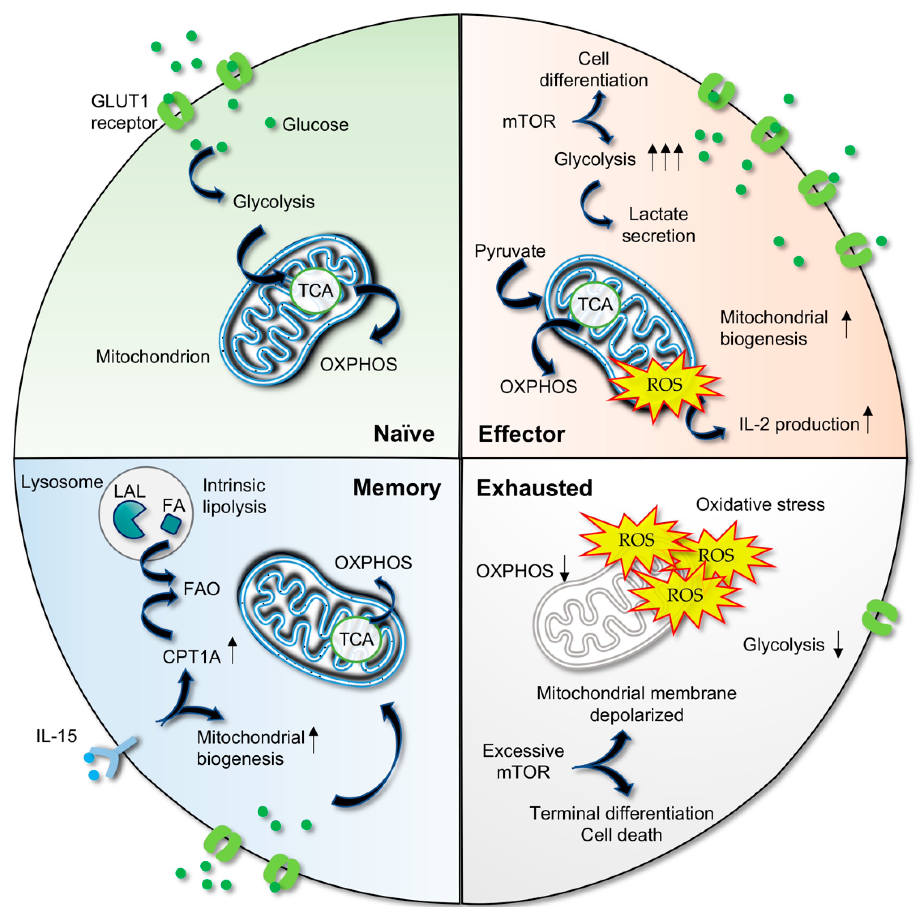
3. Metabolic Plasticity in T Cells

Cellular metabolism plays a fundamental role in balancing the nutrient consumption and energy production of a cell to suit the dynamic changes of an intracellular or extracellular environment. Glucose serves as an important energy source for T cells, and a lack of glucose compromises T cell functions [73,74]. The main routes of glucose metabolism comprise glycolysis, the tricarboxylic acid (TCA) cycle, and oxidative phosphorylation (OXPHOS) [75,76]. T cells adapt to different mechanisms to oxidize glucose to support the biosynthetic demands, which are integrally linked to T cell proliferation, differentiation, and cellular functions.

3.1. Activated T Cells Utilize Aerobic Glycolysis

Under normal circumstances, naïve T cells utilize OXPHOS, which is the most efficient energy production route (Figure 1). However, activation of T cells induces a switch from OXPHOS towards aerobic glycolysis in a phenomenon known as the “Warburg effect” to support rapid clonal expansion and effector function that require growth molecules such as amino acids, fatty acids, and nucleotides [77,78]. Studies demonstrate elevated glucose uptake, glycolysis, and lactate secretion [79], accompanied by high levels of expression of glycolysis-associated proteins, including glucose transporter 1 (GLUT1) and hexokinase 2 (HK2), a key enzyme engaged in glycolysis catalyzing the conversion of glucose to glucose-6-phosphate, in activated T cells [80]. The importance of glycolysis in the metabolic remodeling of T cells is further highlighted by diminished T cell viability and immunological function in the absence of glycolysis [81].
Both aerobic glycolysis and OXPHOS can be utilized by proliferating cells in an activation state [82]. Upon TCR stimulation, activated T cells upregulate complex IV (cytochrome c oxidase) in OXPHOS [83] and promote mitochondrial biogenesis to sustain cellular energy demands [84]. Sena et al. have shown that pyruvate, in the absence of glucose, increases CD69 and CD25 activation markers on CD4+ T cells after CD3/CD28 stimulation [85] and enters the mitochondria for the TCA cycle instead of being converted to lactate [85]. Moreover, reduced mitochondria-generated reactive oxygen species (ROS) negatively impacts IL-2 production, suggesting that ROS is one of the fundamental components of T cell activation following CD3-dependent calcium influx [85].
Current findings also indicate that OXPHOS is required to complement glycolysis because it is capable of fulfilling cytokine production demand, sustaining proliferation and antiviral function in CD8+ T cells [86,87]. These findings collectively denote that mitochondrial OXPHOS is still engaged in effector activity, although glycolysis is regarded as the predominant pathway. Indeed, HIV-1 preferentially infects CD4+ T cells with high OXPHOS and glycolysis, as indicated by positive correlations between the metabolic activities of activated cells and HIV infection levels [88]. The activated, proliferating primary CD4+ T cells are highly susceptible to HIV-1 infection ex vivo for productive virus replication [89].

3.2. Metabolic Shift in Memory T Cells

Following viral clearance, most of the effector cells die, whereas a small number of cells from the effector population will be converted to long-lived memory T cells that possess the ability to generate rapid effector functions in response to reactivation. Along with the cellular transition from effectors with high energy demands to memory CD8+ T cells with a lower demand, cell metabolism is shifted back to mitochondrial OXPHOS [90,91].
In addition, mitochondrial fatty acid oxidation (FAO) also acts as a main energy source for memory T cells [75,90,92,93]. Memory T cells use the intrinsic lysosomal hydrolase lysosomal acid lipase (LAL) to mobilize fatty acids for the FAO process and memory cell development [90]. IL-15, a critical cytokine for memory CD8+ T cells, is found to regulate mitochondrial spare respiratory capacity that allows extra capacity of cells to meet the need of increased stress and hence enhances cell survival [91]. By promoting mitochondrial biogenesis and the expression of carnitine palmitoyl transferase 1A (CPT1A), which regulates FAO, IL-15 controls the bioenergetic stability of memory T cells after infection [91].
Notably, a higher frequency of central memory CD8+ T cells has been reported in untreated PLWH with low viral load, implying memory cells mediate effective viral control [94]. Central memory CD4+ and CD8+ T cells in PLWH express high levels of GLUT1 and mitochondrial mass [95]. Consistently, memory CD4+ T cells in vitro exhibit increased OXPHOS relative to aerobic glycolysis, which acts as a major determinant of HIV infection [88,96].

3.3. Metabolic Deregulation Results in T Cell Exhaustion

Previous interrogations of exhausted T cells isolated from chronic LCMV murine models or chronic HBV patients have revealed that the glucose metabolism is shunted away from the mitochondria, resulting in less oxidative ATP production [97,98]. Functionality of CD8+ T cells strongly depends on the metabolite levels of the cells, and CD8+ T cells display a distinct metabolomic profile prior to and upon loss of cellular functionality [99].
Metabolic dysregulation activities occur at the early stage of T cell exhaustion; these include reduced glucose uptake, glycolysis, mitochondrial respiration, and a depolarized mitochondrial membrane [97]. Chronically stimulated T cells demonstrate increased OXPHOS utilization at an early stage but the absence of this activity at a later phase, as evidenced by a significant reduction in or little consumption of mitochondrial oxygen [100]. As OXPHOS activity regulates the ROS level in mitochondria, defective mitochondrial metabolism influences the redox environment. ROS accumulation directly inflicts damage to DNA and proteins, perturbs ATP production through the inactivation of complexes on the inner membrane of the mitochondria, and reduces OXPHOS efficiency [101]. Mitochondrial dysfunction not only limits glucose oxidation; it also alters other cellular pathways that are highly dependent on ATP supply [102], adversely affects the capacity to oxidize FAO and nucleotide biosynthesis, and promotes oxidative stress that exacerbates the exhaustion [103].
In addition, the mammalian target of rapamycin (mTOR) signaling pathway persists in the effector and early exhausted T cells, and its inhibition recovers the mitochondrial fitness of early exhausted T cells, signifying its involvement in metabolic remodeling in the T cell exhaustion process [97]. It is well established that the mTORC1 pathway favors T cell differentiation into effector cells and enhances aerobic glycolysis. However, excessive mTORC1 activation results in terminally differentiated effector cells [104], mitochondrial membrane depolarization, elevated ROS levels, and cell death [105].
In chronic viral infections such as HCV, CD8+ T cell exhaustion is characterized by a general suppression of a broad range of genes associated with metabolic functions; these include genes encoding for mitochondrial and OXPHOS components [106]. Impaired glucose and mitochondrial metabolism, as indicated by mitochondrial membrane depolarization and a markedly high level of ROS, are detected in exhausted HCV-specific T cells. It is interesting to note that the distinct metabolic nature of the exhaustion of CD8+ T cells in HIV infection is different from other viral diseases. In the HIV context, glycolysis is the main energy source for CD8+ T cells in early, untreated PLWH and viral controllers, which is crucial for T cell killing activity. Exhausted CD8+ T cells show a reduction in glycolysis but increased OXPHOS capacity and a dysregulated mTOR pathway that could not be manipulated to reverse exhaustion in T cells [107].
It can be concluded that the impairment of mitochondrial OXPHOS negatively impacts T cell expansion and effector function as well as enriches the terminal differentiation phenotype, followed by oxidative stress that exerts damaging effects on the surrounding cellular components. In line with this, enhancements of OXPHOS and T cell effector function have been documented after treating exhausted T cells with antioxidants [98,106]. Collectively, current findings corroborate the perception of metabolic alterations as a driver of T cell exhaustion.

4. Altered Glucose Metabolism in HIV Infection

4.1. Glucose Metabolism Enhances Cell Permissibility to HIV Entry

HIV infection results in substantial changes in the cellular metabolism of CD4+ T cells (Figure 2). Infection of primary CD4+ T cells with HIV-1 coincides with an increase in glycolysis, and upregulation of glucose uptake molecules, i.e., glucose transporters (GLUT1, GLUT3, GLUT4, and GLUT6) and hexokinase enzymes (HK1 and HK2) [108]. Studies have shown that GLUT1-mediated glucose transport is crucial in HIV infection, whereby a high glucose level significantly enhances HIV entry [109,110]. Increased GLUT1 expression in T cells following IL-7 stimulation results in augmented glucose uptake and increased permissibility to HIV-1 infection, whereas siRNA-mediated GLUT1 downregulation or signal inhibition abrogates the infection [110].
In HIV infection, ROS-mediated stabilization of hypoxia-inducible factor 1α (HIF-1α) results in enriched expression of GLUT1 and C-X-C chemokine receptor type 4 (CXCR4) and in CD4+ T cells, which thereby increases glucose uptake and viral entry [108,109,110]. On the contrary, silencing HIF-1α attenuates the expression of CXCR4 and decreases HIV entry into CD4+ T cells [109].

4.2. HIV Interferes T Cell Metabolism

Upon entry into the CD4+ T cell, HIV-1 replication further causes an increase in glycolytic flux whereby virion production can take place efficiently and eventually enhances virus-induced cell death in the infected cell [111]. Several key metabolites involved in glycolysis, such as hexose-P, fructose 1,6-bisphosphate (FBP), glyceraldehyde-3P (G3P), and 3-phosphoglycerate (3PG), are evidently elevated in infected CD4+ T cells [112]. Moreover, suboptimal inhibition of glycolysis eliminates HIV-infected cells and impairs viral amplification in CD4+ T cells isolated from HAART-treated PLWH [88].
Stimulation of HIV-encoded Vpr in human CD4+ T cells causes a dramatic reduction of mitochondrial membrane potential (MMP) and apoptotic cell death [113]. HIV Vpr protein interferes with mitochondrial outer membrane and MMP by promoting ubiquitin ligase degradation of mitochondrial fusion protein mitofusin 2, which leads to increased mitochondrial ROS levels [114]. Vpr also interacts with the adenine nucleotide translocator (ANT), a mitochondrial membrane protein that cooperate within the permeability of the transition pore complex (PTPC), to form large conductance channels that trigger mitochondrial membrane permeabilization and cell death [115].

4.3. Long-Term HAART Positively Influences T Cell Metabolism Fitness

In addition to HIV, HAART treatment causes T cell metabolic alteration in PLWH. The older generation of HAART treatment, in particular nucleoside reverse transcriptase inhibitors, triggers severe mitochondrial toxicity through inhibition of DNA polymerase gamma which is followed by the depletion of mitochondrial DNA [116]. While the newer treatment with different classes of HAART drugs is able to improve metabolic fitness in CD8+ T cells, it is unable to restore mitochondrial impairment [117]. Robustly functional mitochondria are coupled with a healthy bioenergetic phenotype and have a direct role in cellular exhaustion. Enforcement of metabolic fitness at an early phase of infection is important to counteract full scale cell exhaustion and is therefore crucial for repressing disease progression. Apart from that, early initiation of HAART allows rapid and close-to-complete normalization of T cell subsets and preserves T cell functions [118], as well as preventing metabolic alterations that drive T cell exhaustion.
HIV-1-specific CD8+ T cells from PLWH receiving long term HAART for more than 10 years demonstrate improved polyfunctionality and capacity to eliminate target cells in vitro, compared to the CD8+ T cells from PLWH on short term HAART [119]. This improvement is attributable to differences in the frequencies of central memory CD8+ T cells, reduced mitochondrial respiration, and glycolytic induction upon TCR activation. Further, CD4+ T cells derived from individuals receiving integrase strand transfer inhibitors such as dolutegravir or elvitegravir, exhibit a significant reduction in ex vivo proliferative capacity, supporting the notion that cellular metabolism and functionality are influenced by external factors, including drugs [117].

4.4. T Cell Metabolism in Elite Controllers

Elite controllers represent <1% of PLWH who can naturally suppress the viral infection in the absence of HAART therapy. Investigation of the plasma metabolites from the elite controller cohort without treatment intervention demonstrates that the transient controller subgroup, which eventually lost virological control within 12 months but not the persistent controller subgroup, is profiled by increased aerobic glycolysis, dysfunctional mitochondria, and a high level of oxidative stress [99]. Persistent immune activation and cytokine production are also detected in the transient controllers and strongly associated with the metabolite level [99], while the opposite trends are observed in the persistent controllers [120]. This suggests an intact mitochondrial function contributes to a greater control of the virus in PLWH.
Studies have shown that the mTOR pathway is upregulated in elite controllers and dysregulated between males and females in the elite controller cohort, indicating an imperative role in controlling latent HIV infection [121,122,123]. mTOR complex subunits knocked down by CRIPSR interference or pharmacological inhibition, as well as inhibition of Enolase 1 in CD4+ T cells, prevent viral reactivation and suppress latency reversal by repressing Tat-dependent and Tat-independent transcription of HIV [122].
The mTOR pathway regulates HIV-1 latency through its downstream pathways, such as HIF signaling. HIF signaling is a major regulator of cellular metabolism, which assists cellular adaptation to oxygen availability, growth factors, cytokines, and infections via mTOR. Hence, activation of HIF signaling results in the regulation of numerous downstream target genes mediating cell survival, proliferation, immune function, as well as metabolic reprogramming [124]. Interestingly, a recent study utilizing proteo-transcriptomic data integration suggests enriched HIF signaling, a high ratio of HIF-1α and HIF-1β nuclear localization, and glycolysis are unique features of the male elite controllers [121].
By comparing the immunometabolic profiles of HIV-specific CD8+ T cells between elite controllers and non-controllers (HAART-treated individuals), a greater metabolic plasticity of CD8+ T cells is observed in the HIV controllers [125]. Furthermore, while elite controllers possess HIV-specific CD8+ T cells that appear to be mitochondrial-dependent to fuel T cell responses in addition to glycolysis, HIV-specific CD8+ T cells from non-controllers show larger mitochondrial mass, which has been linked to dysfunctional mitochondria [125].

5. Achieving HIV Remission through Immune Checkpoint Inhibitors and Glucose Metabolic Enhancement

Immune checkpoint inhibitors are often defined as effective immunotherapies for various cancers and chronic infections. Emerging evidence supports the notion that metabolic reprogramming has a direct impact on T cell differentiation and exhaustion [78,85,126]. In HIV infection, chronically stimulated T cells display increased PD-1 expression accompanied by a significant reduction of cellular respiration [117]. It is interesting to note that metabolic activities vary between PD-1Intermediate (Int) and PD-1Hi exhausted T cell subsets; the PD-1Int subset displays a high rate of glucose uptake and OXPHOS and low mTOR activity and mitochondrial mass, whereas the PD-1Hi subset shows the opposite [97], which highlights a close relationship between cellular metabolism and immunoregulation. The effects of PD-1 on the metabolic functions and bioenergetics of activated CD4+ T cells have also been examined. Patsoukis et al. have revealed ex vivo PD-1+ T cells could not uptake and utilize glucose, as shown by the inhibition of GLUT1 expression, glucose transport, and HK2 mRNA levels in T cells upon receiving PD-1 signals [127].
Since upregulation of immune checkpoints in T cells derived from PLWH is positively correlated with metabolic dysregulation, immune checkpoint inhibitors such as anti-PD-1 have been suggested to rescue CD8+ T cells from exhaustion status. Nevertheless, there is still limited clinical data regarding the immune checkpoint antibodies and their impacts on T cell metabolic functions in an HIV context. Anti-PD-1 antibody treatment partially alleviates the progression of cellular exhaustion by enhancing OXPHOS, glycolysis, the IL-2 signaling pathway, and T cell effector function [97,98,106,128]. In addition, PD-1 suppresses peroxisome proliferator-activated receptor gamma coactivator 1-alpha (PGC-1α), a key metabolic regulator and transcriptional co-activator [97,129], which functions to maintain mitochondrial fitness in tumor-infiltrating lymphocytes [130], whereas blockade of the PD-1 signal enables PGC-1α activity to enhance glucose uptake and partially reverse the metabolic defects. These findings emphasize the efficiency of PD-1 inhibition on the restoration of metabolic fitness and, subsequently, T cell activation status.
Additionally, co-expression of PD-1 and CTLA-4 acts cooperatively to reduce cellular glucose uptake and causes glucose metabolic derangement, which is coherent with the progressive loss of T cell effector functions during viral antigen persistence [100]. Co-inhibitory receptor treatment targeting CTLA-4 and PD-1 induces a synergistic pattern of metabolic and effector T cell-specific functions, including enhancement of effector molecules such as IFN-γ, Granzyme A, Granzyme B, and Fas ligand production [128]. Treatment with a dual checkpoint blockade serves a more potent strategy by targeting different pathways that enhance antiviral responses and T cell metabolism.
Considering the effect of HAART on immune checkpoint expression, studies have combined multiple therapies to investigate T cell function and the reversal of HIV latency. PD-1 inhibition along with HAART reprofiles the CD8+ T cells with augmented antiviral responses, including perforin and granzyme B production, as well as viral reservoir depletion in the chronically SIV-infected rhesus macaque [131]. Treatment of CD8+ T cells from PLWH with pembrolizumab (anti-PD-1) and the latency reversing agent bryostatin reduces the latent reservoir in vivo without enhancing T cell activation [132]. Therefore, combined treatment, including immune checkpoint inhibitors, remains a possible solution to achieve HIV remission.
The study has also demonstrated a higher frequency of TIM3+ PD1- CD8+ T cells in long term HAART recipients as compared to higher PD1+ TIGIT+ cells in short-term HAART recipients. As such, high efficiency in restoring the cytotoxic capacities of CD8+ T cells has been reported upon combined treatment of pro-glycolytic drugs such as metformin with anti-TIGIT and anti-PD-1 antibodies in vitro [119].
Intriguingly, metformin (also an anti-diabetic drug) reduces the frequency of PD1+ TIM3+ TIGIT+-expressing CD4+ T cells in HAART-treated PLWH [133]. A recent study demonstrates that CD4+ T cells from women PLWH with diabetes mellitus receiving metformin show lower glucose metabolic activity as compared to those without medication [134]. Thus, it is suggested that metformin could be an adjunct therapy to reduce metabolic activation, partially correct glucose metabolism in CD4+ T cells, and well as reverse exhaustion [135].

6. Conclusions

T cell function and exhaustion are intimately linked to metabolic changes within cells. In chronic HIV infection, increased immune checkpoint molecules and an altered bioenergetic profile result in cellular exhaustion. The use of immune checkpoint inhibitors coupled with metabolism-enhancing drugs to restore T cell exhaustion serve as promising complementary therapeutic options for HIV. Therefore, further research is warranted to uncover the complex mechanism underlying metabolic changes in mitochondrial pathways that are intricately concomitant with immune cell exhaustion in chronic HIV infection.

Author Contributions

Conceptualization, K.-K.C., C.Y.L., and W.F.W.; writing—original draft preparation, Y.T.C.; writing—review and editing, H.C.C., T.F.T., and R.R.; supervision, A.K.; funding acquisition, W.F.W. and A.K. All authors have read and agreed to the published version of the manuscript.

Funding

This research was funded by the Malaysian Ministry of Higher Education Fundamental Research Grant Scheme, grant number FRGS/1/2018/SKK11/UM/01/1 to A.K. This work was also supported by grant number FRGS/1/2022/SKK12/UM/02/34 to W.F.W.

Acknowledgments

The authors thank the Malaysian Ministry of Higher Education and the University of Malaya for their support.

Conflicts of Interest

The authors declare no conflict of interest.

References

  1. Wherry, E.J.; Blattman, J.N.; Murali-Krishna, K.; Van Der Most, R.; Ahmed, R. Viral persistence alters CD8 T-cell immunodominance and tissue distribution and results in distinct stages of functional impairment. J. Virol. 2003, 77, 4911–4927. [Google Scholar] [CrossRef] [PubMed] [Green Version]
  2. Mueller, S.N.; Ahmed, R. High antigen levels are the cause of T cell exhaustion during chronic viral infection. Proc. Natl. Acad. Sci. USA 2009, 106, 8623–8628. [Google Scholar] [CrossRef] [Green Version]
  3. Harper, J.; Gordon, S.; Chan, C.N.; Wang, H.; Lindemuth, E.; Galardi, C.; Falcinelli, S.D.; Raines, S.L.; Read, J.L.; Nguyen, K. CTLA-4 and PD-1 dual blockade induces SIV reactivation without control of rebound after antiretroviral therapy interruption. Nat. Med. 2020, 26, 519–528. [Google Scholar] [CrossRef] [PubMed]
  4. Le Garff, G.; Samri, A.; Lambert-Niclot, S.; Even, S.; Lavolé, A.; Cadranel, J.; Spano, J.-P.; Autran, B.; Marcelin, A.-G.; Guihot, A. Transient HIV-specific T cells increase and inflammation in an HIV-infected patient treated with nivolumab. Aids 2017, 31, 1048–1051. [Google Scholar] [CrossRef] [PubMed] [Green Version]
  5. Bui, J.K.; Cyktor, J.C.; Fyne, E.; Campellone, S.; Mason, S.W.; Mellors, J.W. Blockade of the PD-1 axis alone is not sufficient to activate HIV-1 virion production from CD4+ T cells of individuals on suppressive ART. PLoS ONE 2019, 14, e0211112. [Google Scholar] [CrossRef] [PubMed]
  6. Philip, M.; Fairchild, L.; Sun, L.; Horste, E.L.; Camara, S.; Shakiba, M.; Scott, A.C.; Viale, A.; Lauer, P.; Merghoub, T. Chromatin states define tumour-specific T cell dysfunction and reprogramming. Nature 2017, 545, 452–456. [Google Scholar] [CrossRef] [Green Version]
  7. Pauken, K.E.; Sammons, M.A.; Odorizzi, P.M.; Manne, S.; Godec, J.; Khan, O.; Drake, A.M.; Chen, Z.; Sen, D.R.; Kurachi, M. Epigenetic stability of exhausted T cells limits durability of reinvigoration by PD-1 blockade. Science 2016, 354, 1160–1165. [Google Scholar] [CrossRef] [Green Version]
  8. Sen, D.R.; Kaminski, J.; Barnitz, R.A.; Kurachi, M.; Gerdemann, U.; Yates, K.B.; Tsao, H.-W.; Godec, J.; LaFleur, M.W.; Brown, F.D. The epigenetic landscape of T cell exhaustion. Science 2016, 354, 1165–1169. [Google Scholar] [CrossRef] [Green Version]
  9. O’Neill, L.A.; Kishton, R.J.; Rathmell, J. A guide to immunometabolism for immunologists. Nat. Rev. Immunol. 2016, 16, 553. [Google Scholar] [CrossRef] [Green Version]
  10. Moskophidis, D.; Lechner, F.; Pircher, H.; Zinkernagel, R.M. Virus persistence in acutely infected immunocompetent mice by exhaustion of antiviral cytotoxic effector T cells. Nature 1993, 362, 758. [Google Scholar] [CrossRef]
  11. Trautmann, L.; Janbazian, L.; Chomont, N.; Said, E.A.; Gimmig, S.; Bessette, B.; Boulassel, M.-R.; Delwart, E.; Sepulveda, H.; Balderas, R.S. Upregulation of PD-1 expression on HIV-specific CD8+ T cells leads to reversible immune dysfunction. Nat. Med. 2006, 12, 1198–1202. [Google Scholar] [CrossRef] [PubMed]
  12. Migueles, S.A.; Laborico, A.C.; Shupert, W.L.; Sabbaghian, M.S.; Rabin, R.; Hallahan, C.W.; Van Baarle, D.; Kostense, S.; Miedema, F.; McLaughlin, M. HIV-specific CD8+ T cell proliferation is coupled to perforin expression and is maintained in nonprogressors. Nat. Immunol. 2002, 3, 1061–1068. [Google Scholar] [CrossRef] [PubMed]
  13. Tian, X.; Zhang, A.; Qiu, C.; Wang, W.; Yang, Y.; Qiu, C.; Liu, A.; Zhu, L.; Yuan, S.; Hu, H. The upregulation of LAG-3 on T cells defines a subpopulation with functional exhaustion and correlates with disease progression in HIV-infected subjects. J. Immunol. 2015, 194, 3873–3882. [Google Scholar] [CrossRef] [Green Version]
  14. Kaufmann, D.E.; Kavanagh, D.G.; Pereyra, F.; Zaunders, J.J.; Mackey, E.W.; Miura, T.; Palmer, S.; Brockman, M.; Rathod, A.; Piechocka-Trocha, A. Upregulation of CTLA-4 by HIV-specific CD4+ T cells correlates with disease progression and defines a reversible immune dysfunction. Nat. Immunol. 2007, 8, 1246–1254. [Google Scholar] [CrossRef] [PubMed]
  15. Jones, R.B.; Ndhlovu, L.C.; Barbour, J.D.; Sheth, P.M.; Jha, A.R.; Long, B.R.; Wong, J.C.; Satkunarajah, M.; Schweneker, M.; Chapman, J.M. Tim-3 expression defines a novel population of dysfunctional T cells with highly elevated frequencies in progressive HIV-1 infection. J. Exp. Med. 2008, 205, 2763–2779. [Google Scholar] [CrossRef] [PubMed]
  16. Chew, G.M.; Fujita, T.; Webb, G.M.; Burwitz, B.J.; Wu, H.L.; Reed, J.S.; Hammond, K.B.; Clayton, K.L.; Ishii, N.; Abdel-Mohsen, M. TIGIT marks exhausted T cells, correlates with disease progression, and serves as a target for immune restoration in HIV and SIV infection. PLoS Pathog. 2016, 12, e1005349. [Google Scholar] [CrossRef] [PubMed] [Green Version]
  17. Ozkazanc, D.; Yoyen-Ermis, D.; Tavukcuoglu, E.; Buyukasik, Y.; Esendagli, G. Functional exhaustion of CD4+ T cells induced by co-stimulatory signals from myeloid leukaemia cells. Immunology 2016, 149, 460–471. [Google Scholar] [CrossRef] [Green Version]
  18. Fromentin, R.; Bakeman, W.; Lawani, M.B.; Khoury, G.; Hartogensis, W.; DaFonseca, S.; Killian, M.; Epling, L.; Hoh, R.; Sinclair, E. CD4+ T cells expressing PD-1, TIGIT and LAG-3 contribute to HIV persistence during ART. PLoS Pathog. 2016, 12, e1005761. [Google Scholar] [CrossRef] [Green Version]
  19. Okazaki, T.; Honjo, T. PD-1 and PD-1 ligands: From discovery to clinical application. Int. Immunol. 2007, 19, 813–824. [Google Scholar] [CrossRef] [Green Version]
  20. Okazaki, T.; Maeda, A.; Nishimura, H.; Kurosaki, T.; Honjo, T. PD-1 immunoreceptor inhibits B cell receptor-mediated signaling by recruiting src homology 2-domain-containing tyrosine phosphatase 2 to phosphotyrosine. Proc. Natl. Acad. Sci. USA 2001, 98, 13866–13871. [Google Scholar] [CrossRef]
  21. Sheppard, K.-A.; Fitz, L.J.; Lee, J.M.; Benander, C.; George, J.A.; Wooters, J.; Qiu, Y.; Jussif, J.M.; Carter, L.L.; Wood, C.R. PD-1 inhibits T-cell receptor induced phosphorylation of the ZAP70/CD3ζ signalosome and downstream signaling to PKCθ. FEBS Lett. 2004, 574, 37–41. [Google Scholar] [CrossRef] [PubMed] [Green Version]
  22. Barber, D.L.; Wherry, E.J.; Masopust, D.; Zhu, B.; Allison, J.P.; Sharpe, A.H.; Freeman, G.J.; Ahmed, R. Restoring function in exhausted CD8 T cells during chronic viral infection. Nature 2006, 439, 682–687. [Google Scholar] [CrossRef] [PubMed]
  23. Wherry, E.J.; Ha, S.-J.; Kaech, S.M.; Haining, W.N.; Sarkar, S.; Kalia, V.; Subramaniam, S.; Blattman, J.N.; Barber, D.L.; Ahmed, R. Molecular signature of CD8+ T cell exhaustion during chronic viral infection. Immunity 2007, 27, 670–684. [Google Scholar] [CrossRef] [PubMed] [Green Version]
  24. Im, S.J.; Hashimoto, M.; Gerner, M.Y.; Lee, J.; Kissick, H.T.; Burger, M.C.; Shan, Q.; Hale, J.S.; Lee, J.; Nasti, T.H. Defining CD8+ T cells that provide the proliferative burst after PD-1 therapy. Nature 2016, 537, 417–421. [Google Scholar] [CrossRef] [Green Version]
  25. Planès, R.; BenMohamed, L.; Leghmari, K.; Delobel, P.; Izopet, J.; Bahraoui, E. HIV-1 Tat protein induces PD-L1 (B7-H1) expression on dendritic cells through tumor necrosis factor alpha-and toll-like receptor 4-mediated mechanisms. J. Virol. 2014, 88, 6672–6689. [Google Scholar] [CrossRef] [Green Version]
  26. Muthumani, K.; Choo, A.Y.; Shedlock, D.J.; Laddy, D.J.; Sundaram, S.G.; Hirao, L.; Wu, L.; Thieu, K.P.; Chung, C.W.; Lankaraman, K.M. Human immunodeficiency virus type 1 Nef induces programmed death 1 expression through a p38 mitogen-activated protein kinase-dependent mechanism. J. Virol. 2008, 82, 11536–11544. [Google Scholar] [CrossRef] [Green Version]
  27. Day, C.L.; Kaufmann, D.E.; Kiepiela, P.; Brown, J.A.; Moodley, E.S.; Reddy, S.; Mackey, E.W.; Miller, J.D.; Leslie, A.J.; DePierres, C. PD-1 expression on HIV-specific T cells is associated with T-cell exhaustion and disease progression. Nature 2006, 443, 350. [Google Scholar] [CrossRef]
  28. Zhang, J.-Y.; Zhang, Z.; Wang, X.; Fu, J.-L.; Yao, J.; Jiao, Y.; Chen, L.; Zhang, H.; Wei, J.; Jin, L. PD-1 up-regulation is correlated with HIV-specific memory CD8+ T-cell exhaustion in typical progressors but not in long-term nonprogressors. Blood 2007, 109, 4671–4678. [Google Scholar] [CrossRef]
  29. Petrovas, C.; Casazza, J.P.; Brenchley, J.M.; Price, D.A.; Gostick, E.; Adams, W.C.; Precopio, M.L.; Schacker, T.; Roederer, M.; Douek, D.C. PD-1 is a regulator of virus-specific CD8+ T cell survival in HIV infection. J. Exp. Med. 2006, 203, 2281–2292. [Google Scholar] [CrossRef]
  30. Evans, V.A.; Van Der Sluis, R.M.; Solomon, A.; Dantanarayana, A.; McNeil, C.; Garsia, R.; Palmer, S.; Fromentin, R.; Chomont, N.; Sékaly, R.-P. PD-1 contributes to the establishment and maintenance of HIV-1 latency. AIDS 2018, 32, 1491. [Google Scholar] [CrossRef]
  31. Dafonseca, S.; Chomont, N.; El Far, M.; Boulassel, R.; Routy, J.; Sékaly, R. Purging the HIV-1 reservoir through the disruption of the PD-1 pathway. J. Int. AIDS Soc. 2010, 13, O15. [Google Scholar] [CrossRef]
  32. Guihot, A.; Marcelin, A.-G.; Massiani, M.-A.; Samri, A.; Soulié, C.; Autran, B.; Spano, J.-P. Drastic decrease of the HIV reservoir in a patient treated with nivolumab for lung cancer. Ann. Oncol. 2018, 29, 517–518. [Google Scholar] [CrossRef] [PubMed] [Green Version]
  33. Topalian, S.L.; Sznol, M.; McDermott, D.F.; Kluger, H.M.; Carvajal, R.D.; Sharfman, W.H.; Brahmer, J.R.; Lawrence, D.P.; Atkins, M.B.; Powderly, J.D. Survival, durable tumor remission, and long-term safety in patients with advanced melanoma receiving nivolumab. J. Clin. Oncol. 2014, 32, 1020. [Google Scholar] [CrossRef] [PubMed]
  34. Brahmer, J.R.; Tykodi, S.S.; Chow, L.Q.; Hwu, W.-J.; Topalian, S.L.; Hwu, P.; Drake, C.G.; Camacho, L.H.; Kauh, J.; Odunsi, K. Safety and activity of anti–PD-L1 antibody in patients with advanced cancer. N. Engl. J. Med. 2012, 366, 2455–2465. [Google Scholar] [CrossRef] [PubMed] [Green Version]
  35. Topalian, S.L.; Hodi, F.S.; Brahmer, J.R.; Gettinger, S.N.; Smith, D.C.; McDermott, D.F.; Powderly, J.D.; Carvajal, R.D.; Sosman, J.A.; Atkins, M.B. Safety, activity, and immune correlates of anti–PD-1 antibody in cancer. N. Engl. J. Med. 2012, 366, 2443–2454. [Google Scholar] [CrossRef] [Green Version]
  36. Gay, C.L.; Bosch, R.J.; Ritz, J.; Hataye, J.M.; Aga, E.; Tressler, R.L.; Mason, S.W.; Hwang, C.K.; Grasela, D.M.; Ray, N. Clinical trial of the anti-PD-L1 antibody BMS-936559 in HIV-1 infected participants on suppressive antiretroviral therapy. J. Infect. Dis. 2017, 215, 1725–1733. [Google Scholar] [CrossRef] [Green Version]
  37. Spano, J.-P.; Veyri, M.; Gobert, A.; Guihot, A.; Perré, P.; Kerjouan, M.; Brosseau, S.; Cloarec, N.; Montaudié, H.; Helissey, C. Immunotherapy for cancer in people living with HIV: Safety with an efficacy signal from the series in real life experience. Aids 2019, 33, F13–F19. [Google Scholar] [CrossRef]
  38. Rasmussen, T.A.; Rajdev, L.; Rhodes, A.; Dantanarayana, A.; Tennakoon, S.; Chea, S.; Spelman, T.; Lensing, S.; Rutishauser, R.; Bakkour, S. Impact of Anti–PD-1 and Anti–CTLA-4 on the Human Immunodeficiency Virus (HIV) Reservoir in People Living With HIV With Cancer on Antiretroviral Therapy: The AIDS Malignancy Consortium 095 Study. Clin. Infect. Dis. 2021, 73, e1973–e1981. [Google Scholar] [CrossRef]
  39. Rudd, C.E.; Taylor, A.; Schneider, H. CD28 and CTLA-4 coreceptor expression and signal transduction. Immunol. Rev. 2009, 229, 12–26. [Google Scholar] [CrossRef] [Green Version]
  40. Leng, Q.; Bentwich, Z.; Magen, E.; Kalinkovich, A.; Borkow, G. CTLA-4 upregulation during HIV infection: Association with anergy and possible target for therapeutic intervention. Aids 2002, 16, 519–529. [Google Scholar] [CrossRef]
  41. El-Far, M.; Ancuta, P.; Routy, J.-P.; Zhang, Y.; Bakeman, W.; Bordi, R.; DaFonseca, S.; Said, E.A.; Gosselin, A.; Tep, T.-S. Nef promotes evasion of human immunodeficiency virus type 1-infected cells from the CTLA-4-mediated inhibition of T-cell activation. J. Gen. Virol. 2015, 96, 1463. [Google Scholar] [CrossRef] [Green Version]
  42. McGary, C.S.; Deleage, C.; Harper, J.; Micci, L.; Ribeiro, S.P.; Paganini, S.; Kuri-Cervantes, L.; Benne, C.; Ryan, E.S.; Balderas, R. CTLA-4+ PD-1− memory CD4+ T cells critically contribute to viral persistence in antiretroviral therapy-suppressed, SIV-infected rhesus macaques. Immunity 2017, 47, 776–788. e775. [Google Scholar] [CrossRef] [PubMed] [Green Version]
  43. Nigam, P.; Velu, V.; Kannanganat, S.; Chennareddi, L.; Kwa, S.; Siddiqui, M.; Amara, R.R. Expansion of FOXP3+ CD8 T cells with suppressive potential in colorectal mucosa following a pathogenic simian immunodeficiency virus infection correlates with diminished antiviral T cell response and viral control. J. Immunol. 2010, 184, 1690–1701. [Google Scholar] [CrossRef] [PubMed] [Green Version]
  44. Yero, A.; Shi, T.; Routy, J.-P.; Tremblay, C.; Durand, M.; Costiniuk, C.T.; Jenabian, M.-A. FoxP3+ CD8 T-cells in acute HIV infection and following early antiretroviral therapy initiation. Front. Immunol. 2022, 13, 962912. [Google Scholar] [CrossRef] [PubMed]
  45. Cecchinato, V.; Tryniszewska, E.; Ma, Z.M.; Vaccari, M.; Boasso, A.; Tsai, W.-P.; Petrovas, C.; Fuchs, D.; Heraud, J.-M.; Venzon, D. Immune activation driven by CTLA-4 blockade augments viral replication at mucosal sites in simian immunodeficiency virus infection. J. Immunol. 2008, 180, 5439–5447. [Google Scholar] [CrossRef] [Green Version]
  46. Wightman, F.; Solomon, A.; Kumar, S.S.; Urriola, N.; Gallagher, K.; Hiener, B.; Palmer, S.; Mcneil, C.; Garsia, R.; Lewin, S.R. Effect of ipilimumab on the HIV reservoir in an HIV-infected individual with metastatic melanoma. AIDS 2015, 29, 504. [Google Scholar] [CrossRef] [Green Version]
  47. Elahi, S.; Shahbaz, S.; Houston, S. Selective upregulation of CTLA-4 on CD8+ T cells restricted by HLA-B* 35Px renders them to an exhausted phenotype in HIV-1 infection. PLoS Pathog. 2020, 16, e1008696. [Google Scholar] [CrossRef]
  48. Van der Sluis, R.M.; Kumar, N.A.; Pascoe, R.D.; Zerbato, J.M.; Evans, V.A.; Dantanarayana, A.I.; Anderson, J.L.; Sékaly, R.P.; Fromentin, R.; Chomont, N. Combination immune checkpoint blockade to reverse HIV latency. J. Immunol. 2020, 204, 1242–1254. [Google Scholar] [CrossRef]
  49. Monney, L.; Sabatos, C.A.; Gaglia, J.L.; Ryu, A.; Waldner, H.; Chernova, T.; Manning, S.; Greenfield, E.A.; Coyle, A.J.; Sobel, R.A. Th1-specific cell surface protein Tim-3 regulates macrophage activation and severity of an autoimmune disease. Nature 2002, 415, 536–541. [Google Scholar] [CrossRef]
  50. Sabatos, C.A.; Chakravarti, S.; Cha, E.; Schubart, A.; Sánchez-Fueyo, A.; Zheng, X.X.; Coyle, A.J.; Strom, T.B.; Freeman, G.J.; Kuchroo, V.K. Interaction of Tim-3 and Tim-3 ligand regulates T helper type 1 responses and induction of peripheral tolerance. Nat. Immunol. 2003, 4, 1102–1110. [Google Scholar] [CrossRef]
  51. Zhu, C.; Anderson, A.C.; Schubart, A.; Xiong, H.; Imitola, J.; Khoury, S.J.; Zheng, X.X.; Strom, T.B.; Kuchroo, V.K. The Tim-3 ligand galectin-9 negatively regulates T helper type 1 immunity. Nat. Immunol. 2005, 6, 1245–1252. [Google Scholar] [CrossRef] [PubMed]
  52. Jacob, R.A.; Edgar, C.R.; Prévost, J.; Trothen, S.M.; Lurie, A.; Mumby, M.J.; Galbraith, A.; Kirchhoff, F.; Haeryfar, S.M.; Finzi, A. The HIV-1 accessory protein Nef increases surface expression of the checkpoint receptor Tim-3 in infected CD4+ T cells. J. Biol. Chem. 2021, 297, 101042. [Google Scholar] [CrossRef] [PubMed]
  53. Prévost, J.; Edgar, C.R.; Richard, J.; Trothen, S.M.; Jacob, R.A.; Mumby, M.J.; Pickering, S.; Dubé, M.; Kaufmann, D.E.; Kirchhoff, F. HIV-1 Vpu downregulates Tim-3 from the surface of infected CD4+ T cells. J. Virol. 2020, 94, e01999-19. [Google Scholar] [CrossRef] [PubMed]
  54. Elahi, S.; Dinges, W.L.; Lejarcegui, N.; Laing, K.J.; Collier, A.C.; Koelle, D.M.; McElrath, M.J.; Horton, H. Protective HIV-specific CD8+ T cells evade Treg cell suppression. Nat. Med. 2011, 17, 989–995. [Google Scholar] [CrossRef]
  55. Sakhdari, A.; Mujib, S.; Vali, B.; Yue, F.Y.; MacParland, S.; Clayton, K.; Jones, R.B.; Liu, J.; Lee, E.Y.; Benko, E. Tim-3 negatively regulates cytotoxicity in exhausted CD8+ T cells in HIV infection. PLoS ONE 2012, 7, e40146. [Google Scholar] [CrossRef]
  56. Hannier, S.; Tournier, M.; Bismuth, G.; Triebel, F. CD3/TCR complex-associated lymphocyte activation gene-3 molecules inhibit CD3/TCR signaling. J. Immunol. 1998, 161, 4058–4065. [Google Scholar]
  57. Hoffmann, M.; Pantazis, N.; Martin, G.E.; Hickling, S.; Hurst, J.; Meyerowitz, J.; Willberg, C.B.; Robinson, N.; Brown, H.; Fisher, M.; et al. Exhaustion of Activated CD8 T Cells Predicts Disease Progression in Primary HIV-1 Infection. PLoS Pathog. 2016, 12, e1005661. [Google Scholar] [CrossRef] [Green Version]
  58. Woo, S.-R.; Turnis, M.E.; Goldberg, M.V.; Bankoti, J.; Selby, M.; Nirschl, C.J.; Bettini, M.L.; Gravano, D.M.; Vogel, P.; Liu, C.L. Immune inhibitory molecules LAG-3 and PD-1 synergistically regulate T-cell function to promote tumoral immune escape. Cancer Res. 2012, 72, 917–927. [Google Scholar] [CrossRef] [Green Version]
  59. Grosso, J.F.; Goldberg, M.V.; Getnet, D.; Bruno, T.C.; Yen, H.-R.; Pyle, K.J.; Hipkiss, E.; Vignali, D.A.; Pardoll, D.M.; Drake, C.G. Functionally distinct LAG-3 and PD-1 subsets on activated and chronically stimulated CD8 T cells. J. Immunol. 2009, 182, 6659–6669. [Google Scholar] [CrossRef] [Green Version]
  60. Dong, Y.; Li, X.; Zhang, L.; Zhu, Q.; Chen, C.; Bao, J.; Chen, Y. CD4+ T cell exhaustion revealed by high PD-1 and LAG-3 expression and the loss of helper T cell function in chronic hepatitis B. BMC Immunol. 2019, 20, 27. [Google Scholar] [CrossRef] [Green Version]
  61. Blackburn, S.D.; Shin, H.; Haining, W.N.; Zou, T.; Workman, C.J.; Polley, A.; Betts, M.R.; Freeman, G.J.; Vignali, D.A.; Wherry, E.J. Coregulation of CD8+ T cell exhaustion by multiple inhibitory receptors during chronic viral infection. Nat. Immunol. 2009, 10, 29–37. [Google Scholar] [CrossRef] [PubMed]
  62. Shin, H.; Blackburn, S.D.; Intlekofer, A.M.; Kao, C.; Angelosanto, J.M.; Reiner, S.L.; Wherry, E.J. A role for the transcriptional repressor Blimp-1 in CD8+ T cell exhaustion during chronic viral infection. Immunity 2009, 31, 309–320. [Google Scholar] [CrossRef] [Green Version]
  63. Levin, S.D.; Taft, D.W.; Brandt, C.S.; Bucher, C.; Howard, E.D.; Chadwick, E.M.; Johnston, J.; Hammond, A.; Bontadelli, K.; Ardourel, D. Vstm3 is a member of the CD28 family and an important modulator of T-cell function. Eur. J. Immunol. 2011, 41, 902–915. [Google Scholar] [CrossRef] [PubMed]
  64. Joller, N.; Lozano, E.; Burkett, P.R.; Patel, B.; Xiao, S.; Zhu, C.; Xia, J.; Tan, T.G.; Sefik, E.; Yajnik, V. Treg cells expressing the coinhibitory molecule TIGIT selectively inhibit proinflammatory Th1 and Th17 cell responses. Immunity 2014, 40, 569–581. [Google Scholar] [CrossRef] [Green Version]
  65. Yin, X.; Liu, T.; Wang, Z.; Ma, M.; Lei, J.; Zhang, Z.; Fu, S.; Fu, Y.; Hu, Q.; Ding, H. Expression of the inhibitory receptor TIGIT is up-regulated specifically on NK cells with CD226 activating receptor from HIV-infected individuals. Front. Immunol. 2018, 9, 2341. [Google Scholar] [CrossRef] [PubMed] [Green Version]
  66. Yu, X.; Harden, K.; Gonzalez, L.C.; Francesco, M.; Chiang, E.; Irving, B.; Tom, I.; Ivelja, S.; Refino, C.J.; Clark, H. The surface protein TIGIT suppresses T cell activation by promoting the generation of mature immunoregulatory dendritic cells. Nat. Immunol. 2009, 10, 48. [Google Scholar] [CrossRef]
  67. Joller, N.; Hafler, J.P.; Brynedal, B.; Kassam, N.; Spoerl, S.; Levin, S.D.; Sharpe, A.H.; Kuchroo, V.K. Cutting edge: TIGIT has T cell-intrinsic inhibitory functions. J. Immunol. 2011, 186, 1338–1342. [Google Scholar] [CrossRef] [Green Version]
  68. Tauriainen, J.; Scharf, L.; Frederiksen, J.; Naji, A.; Ljunggren, H.-G.; Sönnerborg, A.; Lund, O.; Reyes-Terán, G.; Hecht, F.M.; Deeks, S.G. Perturbed CD8+ T cell TIGIT/CD226/PVR axis despite early initiation of antiretroviral treatment in HIV infected individuals. Sci. Rep. 2017, 7, 40354. [Google Scholar] [CrossRef] [Green Version]
  69. Zhang, X.; Lu, X.; Cheung, A.K.L.; Zhang, Q.; Liu, Z.; Li, Z.; Yuan, L.; Wang, R.; Liu, Y.; Tang, B. Analysis of the Characteristics of TIGIT-Expressing CD3− CD56+ NK Cells in Controlling Different Stages of HIV-1 Infection. Front. Immunol. 2021, 12, 602492. [Google Scholar] [CrossRef]
  70. Hung, A.L.; Maxwell, R.; Theodros, D.; Belcaid, Z.; Mathios, D.; Luksik, A.S.; Kim, E.; Wu, A.; Xia, Y.; Garzon-Muvdi, T. TIGIT and PD-1 dual checkpoint blockade enhances antitumor immunity and survival in GBM. Oncoimmunology 2018, 7, e1466769. [Google Scholar] [CrossRef]
  71. Ostroumov, D.; Duong, S.; Wingerath, J.; Woller, N.; Manns, M.P.; Timrott, K.; Kleine, M.; Ramackers, W.; Roessler, S.; Nahnsen, S. Transcriptome profiling identifies TIGIT as a marker of T cell exhaustion in liver cancer. Hepatology 2020, 73, 1399–1418. [Google Scholar] [CrossRef] [PubMed]
  72. Chiu, C.Y.; Chang, J.J.; Dantanarayana, A.I.; Solomon, A.; Evans, V.A.; Pascoe, R.; Gubser, C.; Trautman, L.; Fromentin, R.; Chomont, N. Combination immune checkpoint blockade enhances IL-2 and CD107a production from HIV-specific T cells ex vivo in people living with HIV on antiretroviral therapy. J. Immunol. 2022, 208, 54–62. [Google Scholar] [CrossRef] [PubMed]
  73. Masson, J.J.R.; Palmer, C.S. Glucose metabolism in CD4 and CD8 T cells. In Molecular Nutrition: Carbohydrates; Elsevier: Amsterdam, The Netherlands, 2019; pp. 129–147. [Google Scholar]
  74. Ho, P.-C.; Bihuniak, J.D.; Macintyre, A.N.; Staron, M.; Liu, X.; Amezquita, R.; Tsui, Y.-C.; Cui, G.; Micevic, G.; Perales, J.C. Phosphoenolpyruvate is a metabolic checkpoint of anti-tumor T cell responses. Cell 2015, 162, 1217–1228. [Google Scholar] [CrossRef] [Green Version]
  75. Pearce, E.L.; Poffenberger, M.C.; Chang, C.-H.; Jones, R.G. Fueling immunity: Insights into metabolism and lymphocyte function. Science 2013, 342, 1242454. [Google Scholar] [CrossRef] [Green Version]
  76. Donnelly, R.P.; Finlay, D.K. Glucose, glycolysis and lymphocyte responses. Mol. Immunol. 2015, 68, 513–519. [Google Scholar] [CrossRef] [PubMed]
  77. Vander Heiden, M.G.; Cantley, L.C.; Thompson, C.B. Understanding the Warburg effect: The metabolic requirements of cell proliferation. Science 2009, 324, 1029–1033. [Google Scholar] [CrossRef] [Green Version]
  78. Lunt, S.Y.; Vander Heiden, M.G. Aerobic glycolysis: Meeting the metabolic requirements of cell proliferation. Annu. Rev. Cell Dev. Biol. 2011, 27, 441–464. [Google Scholar] [CrossRef] [PubMed] [Green Version]
  79. Frauwirth, K.A.; Riley, J.L.; Harris, M.H.; Parry, R.V.; Rathmell, J.C.; Plas, D.R.; Elstrom, R.L.; June, C.H.; Thompson, C.B. The CD28 signaling pathway regulates glucose metabolism. Immunity 2002, 16, 769–777. [Google Scholar] [CrossRef] [Green Version]
  80. Kishton, R.J.; Barnes, C.E.; Nichols, A.G.; Cohen, S.; Gerriets, V.A.; Siska, P.J.; Macintyre, A.N.; Goraksha-Hicks, P.; de Cubas, A.A.; Liu, T.; et al. AMPK Is Essential to Balance Glycolysis and Mitochondrial Metabolism to Control T-ALL Cell Stress and Survival. Cell Metab. 2016, 23, 649–662. [Google Scholar] [CrossRef] [Green Version]
  81. Macintyre, A.N.; Gerriets, V.A.; Nichols, A.G.; Michalek, R.D.; Rudolph, M.C.; Deoliveira, D.; Anderson, S.M.; Abel, E.D.; Chen, B.J.; Hale, L.P. The glucose transporter Glut1 is selectively essential for CD4 T cell activation and effector function. Cell Metab. 2014, 20, 61–72. [Google Scholar] [CrossRef] [Green Version]
  82. Chang, C.-H.; Curtis, J.D.; Maggi, L.B., Jr.; Faubert, B.; Villarino, A.V.; O’Sullivan, D.; Huang, S.C.-C.; Van Der Windt, G.J.; Blagih, J.; Qiu, J. Posttranscriptional control of T cell effector function by aerobic glycolysis. Cell 2013, 153, 1239–1251. [Google Scholar] [CrossRef] [PubMed]
  83. Tan, H.; Yang, K.; Li, Y.; Shaw, T.I.; Wang, Y.; Blanco, D.B.; Wang, X.; Cho, J.-H.; Wang, H.; Rankin, S. Integrative proteomics and phosphoproteomics profiling reveals dynamic signaling networks and bioenergetics pathways underlying T cell activation. Immunity 2017, 46, 488–503. [Google Scholar] [CrossRef] [PubMed] [Green Version]
  84. Ron-Harel, N.; Santos, D.; Ghergurovich, J.M.; Sage, P.T.; Reddy, A.; Lovitch, S.B.; Dephoure, N.; Satterstrom, F.K.; Sheffer, M.; Spinelli, J.B. Mitochondrial biogenesis and proteome remodeling promote one-carbon metabolism for T cell activation. Cell Metab. 2016, 24, 104–117. [Google Scholar] [CrossRef] [PubMed] [Green Version]
  85. Sena, L.A.; Li, S.; Jairaman, A.; Prakriya, M.; Ezponda, T.; Hildeman, D.A.; Wang, C.-R.; Schumacker, P.T.; Licht, J.D.; Perlman, H. Mitochondria are required for antigen-specific T cell activation through reactive oxygen species signaling. Immunity 2013, 38, 225–236. [Google Scholar] [CrossRef] [Green Version]
  86. Okoye, I.; Wang, L.; Pallmer, K.; Richter, K.; Ichimura, T.; Haas, R.; Crouse, J.; Choi, O.; Heathcote, D.; Lovo, E. The protein LEM promotes CD8+ T cell immunity through effects on mitochondrial respiration. Science 2015, 348, 995–1001. [Google Scholar] [CrossRef] [Green Version]
  87. Schurich, A.; Pallett, L.J.; Jajbhay, D.; Wijngaarden, J.; Otano, I.; Gill, U.S.; Hansi, N.; Kennedy, P.T.; Nastouli, E.; Gilson, R. Distinct metabolic requirements of exhausted and functional virus-specific CD8 T cells in the same host. Cell Rep. 2016, 16, 1243–1252. [Google Scholar] [CrossRef] [Green Version]
  88. Valle-Casuso, J.C.; Angin, M.; Volant, S.; Passaes, C.; Monceaux, V.; Mikhailova, A.; Bourdic, K.; Avettand-Fenoel, V.; Boufassa, F.; Sitbon, M. Cellular metabolism is a major determinant of HIV-1 reservoir seeding in CD4+ T cells and offers an opportunity to tackle infection. Cell Metab. 2019, 29, 611–626. e615. [Google Scholar] [CrossRef] [Green Version]
  89. Pan, X.; Baldauf, H.-M.; Keppler, O.T.; Fackler, O.T. Restrictions to HIV-1 replication in resting CD4+ T lymphocytes. Cell Res. 2013, 23, 876–885. [Google Scholar] [CrossRef] [Green Version]
  90. O’Sullivan, D.; van der Windt, G.J.; Huang, S.C.; Curtis, J.D.; Chang, C.H.; Buck, M.D.; Qiu, J.; Smith, A.M.; Lam, W.Y.; DiPlato, L.M.; et al. Memory CD8(+) T cells use cell-intrinsic lipolysis to support the metabolic programming necessary for development. Immunity 2014, 41, 75–88. [Google Scholar] [CrossRef] [Green Version]
  91. van der Windt, G.J.; Everts, B.; Chang, C.H.; Curtis, J.D.; Freitas, T.C.; Amiel, E.; Pearce, E.J.; Pearce, E.L. Mitochondrial respiratory capacity is a critical regulator of CD8+ T cell memory development. Immunity 2012, 36, 68–78. [Google Scholar] [CrossRef] [Green Version]
  92. van der Windt, G.J.; O’Sullivan, D.; Everts, B.; Huang, S.C.-C.; Buck, M.D.; Curtis, J.D.; Chang, C.-H.; Smith, A.M.; Ai, T.; Faubert, B. CD8 memory T cells have a bioenergetic advantage that underlies their rapid recall ability. Proc. Natl. Acad. Sci. USA 2013, 110, 14336–14341. [Google Scholar] [CrossRef]
  93. Pearce, E.L.; Walsh, M.C.; Cejas, P.J.; Harms, G.M.; Shen, H.; Wang, L.-S.; Jones, R.G.; Choi, Y. Enhancing CD8 T-cell memory by modulating fatty acid metabolism. Nature 2009, 460, 103–107. [Google Scholar] [CrossRef] [Green Version]
  94. Burgers, W.A.; Riou, C.; Mlotshwa, M.; Maenetje, P.; de Assis Rosa, D.; Brenchley, J.; Mlisana, K.; Douek, D.C.; Koup, R.; Roederer, M. Association of HIV-specific and total CD8+ T memory phenotypes in subtype C HIV-1 infection with viral set point. J. Immunol. 2009, 182, 4751–4761. [Google Scholar] [CrossRef] [Green Version]
  95. Masson, J.J.; Murphy, A.J.; Lee, M.K.; Ostrowski, M.; Crowe, S.M.; Palmer, C.S. Assessment of metabolic and mitochondrial dynamics in CD4+ and CD8+ T cells in virologically suppressed HIV-positive individuals on combination antiretroviral therapy. PLoS ONE 2017, 12, e0183931. [Google Scholar]
  96. Clerc, I.; Abba Moussa, D.; Vahlas, Z.; Tardito, S.; Oburoglu, L.; Hope, T.J.; Sitbon, M.; Dardalhon, V.; Mongellaz, C.; Taylor, N. Entry of glucose-and glutamine-derived carbons into the citric acid cycle supports early steps of HIV-1 infection in CD4 T cells. Nat. Metab. 2019, 1, 717–730. [Google Scholar]
  97. Bengsch, B.; Johnson, A.L.; Kurachi, M.; Odorizzi, P.M.; Pauken, K.E.; Attanasio, J.; Stelekati, E.; McLane, L.M.; Paley, M.A.; Delgoffe, G.M. Bioenergetic insufficiencies due to metabolic alterations regulated by the inhibitory receptor PD-1 are an early driver of CD8+ T cell exhaustion. Immunity 2016, 45, 358–373. [Google Scholar] [CrossRef] [Green Version]
  98. Fisicaro, P.; Barili, V.; Montanini, B.; Acerbi, G.; Ferracin, M.; Guerrieri, F.; Salerno, D.; Boni, C.; Massari, M.; Cavallo, M.C. Targeting mitochondrial dysfunction can restore antiviral activity of exhausted HBV-specific CD8 T cells in chronic hepatitis B. Nat. Med. 2017, 23, 327. [Google Scholar] [CrossRef]
  99. Tarancon-Diez, L.; Rodríguez-Gallego, E.; Rull, A.; Peraire, J.; Viladés, C.; Portilla, I.; Jimenez-Leon, M.R.; Alba, V.; Herrero, P.; Leal, M. Immunometabolism is a key factor for the persistent spontaneous elite control of HIV-1 infection. EBioMedicine 2019, 42, 86–96. [Google Scholar] [CrossRef] [Green Version]
  100. Vardhana, S.A.; Hwee, M.A.; Berisa, M.; Wells, D.K.; Yost, K.E.; King, B.; Smith, M.; Herrera, P.S.; Chang, H.Y.; Satpathy, A.T. Impaired mitochondrial oxidative phosphorylation limits the self-renewal of T cells exposed to persistent antigen. Nat. Immunol. 2020, 21, 1022–1033. [Google Scholar] [CrossRef]
  101. Flint, D.H.; Tuminello, J.; Emptage, M. The inactivation of Fe-S cluster containing hydro-lyases by superoxide. J. Biol. Chem. 1993, 268, 22369–22376. [Google Scholar] [CrossRef]
  102. Sullivan, L.B.; Gui, D.Y.; Hosios, A.M.; Bush, L.N.; Freinkman, E.; Vander Heiden, M.G. Supporting aspartate biosynthesis is an essential function of respiration in proliferating cells. Cell 2015, 162, 552–563. [Google Scholar] [CrossRef] [PubMed] [Green Version]
  103. Birsoy, K.; Wang, T.; Chen, W.W.; Freinkman, E.; Abu-Remaileh, M.; Sabatini, D.M. An essential role of the mitochondrial electron transport chain in cell proliferation is to enable aspartate synthesis. Cell 2015, 162, 540–551. [Google Scholar] [CrossRef] [PubMed] [Green Version]
  104. Pollizzi, K.N.; Patel, C.H.; Sun, I.-H.; Oh, M.-H.; Waickman, A.T.; Wen, J.; Delgoffe, G.M.; Powell, J.D. mTORC1 and mTORC2 selectively regulate CD8+ T cell differentiation. J. Clin. Investig. 2015, 125, 2090–2108. [Google Scholar] [CrossRef]
  105. O’Brien, T.F.; Gorentla, B.K.; Xie, D.; Srivatsan, S.; McLeod, I.X.; He, Y.W.; Zhong, X.P. Regulation of T-cell survival and mitochondrial homeostasis by TSC1. Eur. J. Immunol. 2011, 41, 3361–3370. [Google Scholar] [CrossRef] [PubMed]
  106. Barili, V.; Fisicaro, P.; Montanini, B.; Acerbi, G.; Filippi, A.; Forleo, G.; Romualdi, C.; Ferracin, M.; Guerrieri, F.; Pedrazzi, G. Targeting p53 and histone methyltransferases restores exhausted CD8+ T cells in HCV infection. Nat. Commun. 2020, 11, 604. [Google Scholar] [CrossRef] [PubMed] [Green Version]
  107. Rahman, A.N.-U.; Liu, J.; Mujib, S.; Kidane, S.; Ali, A.; Szep, S.; Han, C.; Bonner, P.; Parsons, M.; Benko, E. Elevated glycolysis imparts functional ability to CD8+ T cells in HIV infection. Life Sci. Alliance 2021, 4, e202101081. [Google Scholar] [CrossRef]
  108. Kavanagh Williamson, M.; Coombes, N.; Juszczak, F.; Athanasopoulos, M.; Khan, M.B.; Eykyn, T.R.; Srenathan, U.; Taams, L.S.; Dias Zeidler, J.; Da Poian, A.T. Upregulation of glucose uptake and hexokinase activity of primary human CD4+ T cells in response to infection with HIV-1. Viruses 2018, 10, 114. [Google Scholar] [CrossRef] [Green Version]
  109. Lan, X.; Cheng, K.; Chandel, N.; Lederman, R.; Jhaveri, A.; Husain, M.; Malhotra, A.; Singhal, P.C. High glucose enhances HIV entry into T cells through upregulation of CXCR4. J. Leukoc. Biol. 2013, 94, 769–777. [Google Scholar] [CrossRef] [Green Version]
  110. Loisel-Meyer, S.; Swainson, L.; Craveiro, M.; Oburoglu, L.; Mongellaz, C.; Costa, C.; Martinez, M.; Cosset, F.-L.; Battini, J.-L.; Herzenberg, L.A. Glut1-mediated glucose transport regulates HIV infection. Proc. Natl. Acad. Sci. USA 2012, 109, 2549–2554. [Google Scholar] [CrossRef] [Green Version]
  111. Hegedus, A.; Kavanagh Williamson, M.; Huthoff, H. HIV-1 pathogenicity and virion production are dependent on the metabolic phenotype of activated CD4+ T cells. Retrovirology 2014, 11, 98. [Google Scholar] [CrossRef] [Green Version]
  112. Hollenbaugh, J.A.; Munger, J.; Kim, B. Metabolite profiles of human immunodeficiency virus infected CD4+ T cells and macrophages using LC–MS/MS analysis. Virology 2011, 415, 153–159. [Google Scholar] [CrossRef] [PubMed] [Green Version]
  113. Arunagiri, C.; Macreadie, I.; Hewish, D.; Azad, A. A C-terminal domain of HIV-1 accessory protein Vpr is involved in penetration, mitochondrial dysfunction and apoptosis of human CD4+ lymphocytes. Apoptosis 1997, 2, 69–76. [Google Scholar] [CrossRef] [PubMed]
  114. Huang, C.-Y.; Chiang, S.-F.; Lin, T.-Y.; Chiou, S.-H.; Chow, K.-C. HIV-1 Vpr triggers mitochondrial destruction by impairing Mfn2-mediated ER-mitochondria interaction. PLoS ONE 2012, 7, e33657. [Google Scholar] [CrossRef] [PubMed]
  115. Deniaud, A.; Brenner, C.; Kroemer, G. Mitochondrial membrane permeabilization by HIV-1 Vpr. Mitochondrion 2004, 4, 223–233. [Google Scholar] [CrossRef]
  116. White, A.J. Mitochondrial toxicity and HIV therapy. Sex. Transm. Infect. 2001, 77, 158–173. [Google Scholar] [CrossRef] [Green Version]
  117. Korencak, M.; Byrne, M.; Richter, E.; Schultz, B.T.; Juszczak, P.; Ake, J.A.; Ganesan, A.; Okulicz, J.F.; Robb, M.L.; de Los Reyes, B. Effect of HIV infection and antiretroviral therapy on immune cellular functions. JCI Insight 2019, 4, e126675. [Google Scholar] [CrossRef] [Green Version]
  118. Plana, M.; García, F.; Gallart, T.; Tortajada, C.; Soriano, A.; Palou, E.; Maleno, M.J.; Barceló, J.J.; Vidal, C.; Cruceta, A. Immunological benefits of antiretroviral therapy in very early stages of asymptomatic chronic HIV-1 infection. Aids 2000, 14, 1921–1933. [Google Scholar] [CrossRef]
  119. Calvet-Mirabent, M.; Sánchez-Cerrillo, I.; Martín-Cófreces, N.; Martínez-Fleta, P.; de la Fuente, H.; Tsukalov, I.; Delgado-Arévalo, C.; Calzada, M.J.; Santos, I.; Sanz, J.; et al. Antiretroviral therapy duration and immunometabolic state determine efficacy of ex vivo dendritic cell-based treatment restoring functional HIV-specific CD8+ T cells in people living with HIV. EBioMedicine 2022, 81, 104090. [Google Scholar] [CrossRef]
  120. Pernas, M.; Tarancon-Diez, L.; Rodriguez-Gallego, E.; Gomez, J.; Prado, J.G.; Casado, C.; Dominguez-Molina, B.; Olivares, I.; Coiras, M.; Leon, A. Factors leading to the loss of natural elite control of HIV-1 infection. J. Virol. 2018, 92, e01805–e01817. [Google Scholar] [CrossRef] [Green Version]
  121. Akusjärvi, S.S.; Ambikan, A.T.; Krishnan, S.; Gupta, S.; Sperk, M.; Végvári, Á.; Mikaeloff, F.; Healy, K.; Vesterbacka, J.; Nowak, P. Integrative proteo-transcriptomic and immunophenotyping signatures of HIV-1 elite control phenotype: A cross-talk between glycolysis and HIF signaling. Iscience 2022, 25, 103607. [Google Scholar] [CrossRef]
  122. Besnard, E.; Hakre, S.; Kampmann, M.; Lim, H.W.; Hosmane, N.N.; Martin, A.; Bassik, M.C.; Verschueren, E.; Battivelli, E.; Chan, J. The mTOR complex controls HIV latency. Cell Host Microbe 2016, 20, 785–797. [Google Scholar] [CrossRef] [PubMed] [Green Version]
  123. Chowdhury, F.Z.; Ouyang, Z.; Buzon, M.; Walker, B.D.; Lichterfeld, M.; Yu, X.G. Metabolic pathway activation distinguishes transcriptional signatures of CD8+ T cells from HIV-1 elite controllers. AIDS 2018, 32, 2669–2677. [Google Scholar] [CrossRef] [PubMed]
  124. Palazon, A.; Goldrath, A.W.; Nizet, V.; Johnson, R.S. HIF transcription factors, inflammation, and immunity. Immunity 2014, 41, 518–528. [Google Scholar] [CrossRef] [PubMed]
  125. Angin, M.; Volant, S.; Passaes, C.; Lecuroux, C.; Monceaux, V.; Dillies, M.A.; Valle-Casuso, J.C.; Pancino, G.; Vaslin, B.; Le Grand, R.; et al. Metabolic plasticity of HIV-specific CD8(+) T cells is associated with enhanced antiviral potential and natural control of HIV-1 infection. Nat. Metab. 2019, 1, 704–716. [Google Scholar] [CrossRef]
  126. Taylor, H.E.; Calantone, N.A.; D’Aquila, R.T. mTOR signaling mediates effects of common gamma-chain cytokines on T cell proliferation and exhaustion: Implications for HIV-1 persistence and cure research. AIDS 2018, 32, 2847–2851. [Google Scholar] [CrossRef]
  127. Patsoukis, N.; Bardhan, K.; Chatterjee, P.; Sari, D.; Liu, B.; Bell, L.N.; Karoly, E.D.; Freeman, G.J.; Petkova, V.; Seth, P. PD-1 alters T-cell metabolic reprogramming by inhibiting glycolysis and promoting lipolysis and fatty acid oxidation. Nat. Commun. 2015, 6, 6692. [Google Scholar] [CrossRef] [Green Version]
  128. Gubin, M.M.; Zhang, X.; Schuster, H.; Caron, E.; Ward, J.P.; Noguchi, T.; Ivanova, Y.; Hundal, J.; Arthur, C.D.; Krebber, W.-J. Checkpoint blockade cancer immunotherapy targets tumour-specific mutant antigens. Nature 2014, 515, 577–581. [Google Scholar] [CrossRef] [Green Version]
  129. Fernandez-Marcos, P.J.; Auwerx, J. Regulation of PGC-1α, a nodal regulator of mitochondrial biogenesis. Am. J. Clin. Nutr. 2011, 93, 884S–890S. [Google Scholar] [CrossRef] [Green Version]
  130. Dumauthioz, N.; Tschumi, B.; Wenes, M.; Marti, B.; Wang, H.; Franco, F.; Li, W.; Lopez-Mejia, I.C.; Fajas, L.; Ho, P.-C. Enforced PGC-1α expression promotes CD8 T cell fitness, memory formation and antitumor immunity. Cell. Mol. Immunol. 2020, 18, 1761–1771. [Google Scholar] [CrossRef] [Green Version]
  131. Mylvaganam, G.H.; Chea, L.S.; Tharp, G.K.; Hicks, S.; Velu, V.; Iyer, S.S.; Deleage, C.; Estes, J.D.; Bosinger, S.E.; Freeman, G.J. Combination anti–PD-1 and antiretroviral therapy provides therapeutic benefit against SIV. JCI Insight 2018, 3, e122940. [Google Scholar] [CrossRef]
  132. Fromentin, R.; DaFonseca, S.; Costiniuk, C.T.; El-Far, M.; Procopio, F.A.; Hecht, F.M.; Hoh, R.; Deeks, S.G.; Hazuda, D.J.; Lewin, S.R. PD-1 blockade potentiates HIV latency reversal ex vivo in CD4+ T cells from ART-suppressed individuals. Nat. Commun. 2019, 10, 814. [Google Scholar] [CrossRef] [PubMed] [Green Version]
  133. Shikuma, C.M.; Chew, G.M.; Kohorn, L.; Souza, S.A.; Chow, D.; SahBandar, I.N.; Park, E.-Y.; Hanks, N.; Gangcuangco, L.M.A.; Gerschenson, M. metformin reduces CD4 T cell exhaustion in HIV-infected adults on suppressive antiretroviral therapy. AIDS Res. Hum. Retrovir. 2020, 36, 303–305. [Google Scholar] [CrossRef] [PubMed] [Green Version]
  134. Butterfield, T.R.; Hanna, D.B.; Kaplan, R.C.; Xue, X.; Kizer, J.R.; Durkin, H.G.; Kassaye, S.G.; Nowicki, M.; Tien, P.C.; Topper, E.T. Elevated CD4+ T-cell glucose metabolism in HIV+ women with diabetes mellitus. AIDS 2022, 36, 1327–1336. [Google Scholar] [CrossRef] [PubMed]
  135. Moyo, D.; Tanthuma, G.; Cary, M.; Mushisha, O.; Kwadiba, G.; Chikuse, F.; Steenhoff, A.; Reid, M. Cohort study of diabetes in HIV-infected adult patients: Evaluating the effect of diabetes mellitus on immune reconstitution. Diabetes Res. Clin. Pract. 2014, 103, e34–e36. [Google Scholar] [CrossRef]
Figure 1. Cellular metabolism in different states of cell-mediated responses. Naive T cells utilize glucose and generate adenosine triphosphate (ATP) using oxidative phosphorylation (OXPHOS). During the transition from naïve to effector cells, glucose transporter 1 (GLUT1) receptors are increased, followed by elevated glucose uptake. The mammalian target of rapamycin (mTOR) signaling pathway assists in the increased glycolytic and lactate pathway activities to sustain cell proliferation, differentiation, and effector functions. Pyruvate, in the absence of glucose, enters the tricarboxylic acid (TCA) cycle and mediates OXPHOS. These result in increased mitochondrial biogenesis and reactive oxygen species (ROS) levels, which are important for interleukin-2 (IL-2) production. In an exhausted T cell, glucose metabolism is decreased, as shown by the reduction of the GLUT1 receptor, glycolysis, OXPHOS, and ATP production. Excessive mTOR signaling in exhausted T cells results in terminal differentiation of T cells, depolarized mitochondrial membrane, and cell death. Accumulation of a high ROS level leads to oxidative stress and negatively impacts OXPHOS efficiency as well as other metabolisms that depend on ATP. In memory T cells, metabolism is shifted back to OXPHOS and fatty acid oxidation (FAO). IL-15 plays a role in enhancing the mitochondrial biogenesis and the carnitine palmitoyl transferase IA (CPT1A) enzyme to facilitate FAO. The supply of fatty acids (FA) for FAO is acquired from intrinsic lipolysis by lysosomal acid lipase (LAL).
Biomedicines 10 02809 g001
Figure 2. HIV infection causes metabolic alteration in the infected CD4+ T cell. During HIV infection, upregulation of the glucose transporter 1 (GLUT1) receptor and interleukin-7 (IL-7) signaling in T cells enhances glucose uptake and the glycolytic process, which promote viral entry through the CD4 receptor and C-X-C chemokine receptor type 4 (CXCR4). High glucose levels increase the generation of reactive oxygen species (ROS), which leads to the stabilization of the hypoxia-inducible factor 1-alpha (HIF-1α) molecule and increased expression of both GLUT1 and CXCR4, which are crucial elements in HIV infection. HIV-encoded Vpr protein promotes ubiquitin ligase degradation of the mitochondrial fusion protein mitofusin 2, causing disruption of the mitochondrial outer membrane and mitochondrial membrane potential (MMP) that leads to an increased level of ROS. Vpr also migrates to mitochondria, interacts with permeability transition pore complex (PTPC) molecules such as adenine nucleotide translocator (ANT) to form large conductance channels that trigger mitochondrial membrane permeabilization, thus resulting in a reduction of MMP and apoptotic cell death.
Biomedicines 10 02809 g002
Publisher’s Note: MDPI stays neutral with regard to jurisdictional claims in published maps and institutional affiliations.

