Article

The Calcium Channel Subunit Gamma-4 as a Novel Regulator of MafA in Pancreatic Beta-Cell Controls Glucose Homeostasis

Department of Clinical Sciences Malmö, Lund University Diabetes Centre, Lund University, 20502 Malmö, Sweden
*
Authors to whom correspondence should be addressed.
These authors contributed equally to this work.
Biomedicines 2022, 10(4), 770; https://doi.org/10.3390/biomedicines10040770
Submission received: 23 February 2022 / Revised: 21 March 2022 / Accepted: 23 March 2022 / Published: 25 March 2022
(This article belongs to the Special Issue Molecular Research of Genes Involved in Metabolic Diseases)

Abstract

:
Impaired fasting glucose (IFG) and impaired glucose tolerance (IGT) are high-risk factors of diabetes development and may be caused by defective insulin secretion in pancreatic beta-cells. Glucose-stimulated insulin secretion is mediated by voltage-gated Ca2+ (CaV) channels in which the gamma-4 subunit (CaVγ4) is required for the beta-cell to maintain its differentiated state. We here aim to explore the involvement of CaVγ4 in controlling glucose homeostasis by employing the CaVγ4−/− mice to study in vivo glucose-metabolism-related phenotypes and glucose-stimulated insulin secretion, and to investigate the underlying mechanisms. We show that CaVγ4−/− mice exhibit perturbed glucose homeostasis, including IFG and IGT. Glucose-stimulated insulin secretion is blunted in CaVγ4−/− mouse islets. Remarkably, CaVγ4 deletion results in reduced expression of the transcription factor essential for beta-cell maturation, MafA, on both mRNA and protein levels in islets from human donors and CaVγ4−/− mice, as well as in INS-1 832/13 cells. Moreover, we prove that CaMKII is responsible for mediating this regulatory pathway linked between CaVγ4 and MafA, which is further confirmed by human islet RNA-seq data. We demonstrate that CaVγ4 is a key player in preserving normal blood glucose homeostasis, which sheds light on CaVγ4 as a novel target for the treatment of prediabetes through correcting the impaired metabolic status.

1. Introduction

Type 2 diabetes (T2D) is progressive pathological hyperglycemia caused by insulin resistance and pancreatic beta-cell failure. Dysfunction of the beta-cells rapidly presents as damaged glucose-stimulated insulin secretion [1]. Development of T2D is inevitable when the deteriorated beta-cell function no longer copes with compensating for insulin resistance [2]. High risk for developing diabetes often relates to obesity, impaired fasting glucose (IFG) and impaired glucose tolerance (IGT) [3].
Maintaining the beta-cell identity is fundamental for preventing the development of diabetes [4]. The islet-enriched transcription factors control pancreatic beta-cell lineage determination, differentiation, and specification, e.g., MafA, Pdx1, MafB and Nkx6.1 [5,6]. MafA is exclusively expressed in adult beta-cells of man and rodents and plays an imperative role in promoting beta-cell maturation and function [5,7]. The unconditional presence of MafA for preserving the beta-cell identity and function has been broadly explored in different MafA-deleted animal models [8,9,10]. Such studies have demonstrated that MafA binds the insulin enhancer element, thus boosting insulin gene expression [11]. The importance of MafA is also supported by the fact that several vital islet transcription factors, such as Pdx1, Nkx6.1 and MafB, act in concert to stimulate MafA transcription [12,13]. In addition, Ca2+ signaling molecules, such as calcium/calmodulin-dependent protein kinase (CaMK) II, have been shown to control MafA expression in beta-cells as well [14,15]. However, whether extracellular stimuli such as Ca2+ can regulate MafA activity and further affect the beta-cell identity has not been explored.
The influx of extracellular Ca2+ in pancreatic beta-cells is mainly triggered by voltage-gated calcium (CaV) channels. This process is initiated by glucose-evoked membrane depolarization that results in the discharge of docked insulin granules [16]. The beta-cell typically expresses several types of CaV channels. They are hetero-multimeric complexes composed of the transmembrane pore-forming alpha1 subunit and three auxiliary subunits: beta, alpha2delta, and gamma [17]. The predominant CaV alpha1 are well investigated and control beta-cell function through mediating Ca2+ entry [18,19,20], while the accessory beta and alpha2delta subunits have been identified as important regulators of CaV alpha1 trafficking [17,21]. However, the role of CaV gamma subunits (CaVγ) in pancreatic beta-cells is only sketchily clarified. We have previously shown that CaV subunit gamma-4 (CaVγ4) is necessary for preserving normal glucose-stimulated insulin secretion by permitting L-type Ca2+ currents/entry in both human and rodent pancreatic islets [22].
CaVγ4 belongs to the transmembrane α-amino-3-hydroxy-5-methyl-4-isoxazolepropionic acid (AMPA) receptor regulatory protein (TARP) family, which modulates channel desensitization, deactivation, and trafficking, thereby affecting the channel decay kinetics and total charge transfer [23,24,25]. These findings were corroborated in CaVγ4 (Cacng4) knockout mice that displayed disrupted excitatory postsynaptic currents in brain [26]. Furthermore, CaVγ4 was shown to regulate both activation and inactivation of CaV channels via a direct association with the CaV alpha1 subunits [27,28]. In the beta-cell, it is noteworthy that among the eight CaVγ variants, CaVγ4 exhibits the highest expression in both man and mice [29], as well as in human beta-cell lines [30]. Intriguingly, CaVγ4 levels are decreased in islets from T2D human donors, diabetic rodent models (Goto-Kakizaki (GK) rats and db/db mice), as well as in islets challenged with gluco-/lipotoxic environment [22]. Importantly, CaVγ4 has been demonstrated as a direct downstream target of the beta-cell unique transcription factor MafA [22]. Nonetheless, the effect of CaVγ4 on blood glucose levels and metabolic homeostasis in vivo remains unknown.
Here, we have extended our initial findings by employing the Cacng4 overall knockout (Cacng4tm1Ran) mouse model with the aim of addressing the involvement of CaVγ4 in glucose metabolism in vivo and underlying mechanism to provide a comprehensive view of the role of CaVγ4 in the beta-cell. We found that genetic ablation of Cacng4 in vivo confers IFG and IGT to the mice. Glucose-stimulated insulin secretion ex vivo is blunted in pancreatic islets from Cacng4 knockout mice. This study provides novel insights into the causal signaling cascade from CaVγ4 via CaMKII to MafA.

2. Materials and Methods

2.1. Human Islets

Human pancreatic islets were provided by EXODIAB Human Tissue Laboratory at Lund University through the Nordic Network for Clinical Islet Transplantation (Uppsala University, Uppsala, Sweden). All procedures were approved by the ethics committees at Uppsala and Lund Universities. The human islets were cultured in CMRL 1066 (ICN Biomedicals, Costa Mesa, CA, USA) supplemented with 10 mM HEPES, 2 mM L-glutamine, 50 μg/mL gentamicin, 0.25 μg/mL fungizone (Gibco, BRL, Gaithersburg, MD, USA), 20 μg/mL ciprofloxacin (Bayer Healthcare, Leverkusen, Germany) and 10 mM nicotinamide at 37 °C (5% CO2) for 1 to 5 days before arrival. The islets were then handpicked under the stereomicroscope. Islets from 6 non-diabetic donors were used in the experiment. Detailed information regarding human islet donors was listed in Supplementary Table S1.

2.2. Human Pancreatic Islet RNA-seq Data

Human islet RNA-seq data was acquired from a cohort of 188 human donors [31]. Pearson correlations between the expression levels of the genes of interest were performed on their log-transformed expression values.

2.3. Animal Studies

All animal experimentation was conducted in accord with accepted standards of humane animal care and approved by the local ethics committee.

2.3.1. Animal Model

Cacng4 knockout (Cacng4tm1Ran) mice were directly purchased from the Jackson Laboratory (JAX stock #028445), originally donated by Dr R. A. Nicoll, University of California, USA [26]. The live colony was maintained by mating the heterozygous (CaVγ4+/−) mice together. Wild type (CaVγ4+/+) mice inbred from the colony were used as control mice. Mice were housed in cages of 2–6, separated by sex. Some cages contained a mix of all three genotypes. Transgenic mice were identified by PCR of tail DNA using the following primers: Mutant Forward primer 5′-TAC CCG GTA GAA TTG ACC TGC-3′, Common primer 5′-ATC TGG TGA TGG CGT TCA GT-3′ and Wild type Forward primer 5′-GGA TCT ACA GCC GCA AGA AC-3′ with dreamtaq hot start DNA polymerase (#EP1702, Thermo Fisher Scientific, Waltham, MA, USA) under the following cycling conditions: 95 °C, 4 min; 95 °C, 30 s, 55 °C, 30 s, 72 °C, 30 s (35 cycles); 72 °C, 5 min. Reactions were separated on 2% agarose gels. The following band sizes were observed for Mutant = ~250 bp, Heterozygote = ~250 bp and 319 bp, Wild type = ~319 bp. Confirmation of Cacng4 deletion was performed by qPCR (see Figure 1A).

2.3.2. Blood Glucose Measurements

The mice underwent a 4-h fast each week from the time of weaning (3 weeks of age) for a period of 27 weeks (to 30 weeks of age) to examine the fasting blood glucose levels by using a OneTouch Lifescan (LifeScan Inc., Malvern, PA, USA) blood glucometer.

2.3.3. IPGTT

Intraperitoneal glucose tolerance test (IPGTT) was performed in all 3-genotype mice without a fasting period from 11 to 15 weeks old. Blood glucose levels were measured with a handheld glucometer (OneTouch; Lifescan) prior to an intraperitoneal injection of 2 g glucose/kg body weight. Blood glucose values were quantified at 0, 5, 15, 30, 60, and 120 min post glucose injection.

2.4. Cell Culture

INS-1 832/13 cells (kindly donated by Dr C. B. Newgaard, Duke University, USA) were cultured in RPMI-1640 containing 11.1 mM D-glucose, and supplemented with 10% fetal bovine serum, 100 U/mL penicillin (Gibco, Gaithersburg, MD, USA), 100 μg/mL streptomycin (Gibco), 10 mM N-2 hydroxyethylpiperazine-N′-2-ethanesulfonic acid (HEPES), 2 mM glutamine, 1 mM sodium pyruvate, and 50 μM β-mercaptoethanol (Sigma), at 37 °C in a humidified atmosphere containing 95% air and 5% CO2.

2.5. Mouse Islet Isolation and Preparation

Intact primary mouse pancreatic islets were isolated by retrograde injection of a collagenase solution via the pancreatic duct from wild type and mutant mice and were handpicked under a stereomicroscope at room temperature. The isolated islets were kept in RPMI 1640 medium substituted with 10 mM D-glucose but lack of β-mercaptoethanol for culture. All indicated experiments were conducted on freshly isolated islets. No islet size and number alterations were observed in Cacng4 knockout mice compared to wild type mice.

2.6. siRNA Transfection

INS-1 832/13 cells were seeded in a 24-well plate 1 day before transfection. 30 nM RNA interference oligonucleotides (Ambion, Austin, TX, USA) or Negative Control (Ambion, USA) were applied to silence target genes. Transfection reagent (Lipofectamine RNAiMAX, Invitrogen, Waltham, MA, USA) was used. For primary human islets, reverse transfection was performed to reach a faster and high-throughput transfection. Islets and siRNA-lipid complexes were prepared on the same day with Lipofectamine RNAiMAX (Invitrogen, USA). Transfection efficiency was measured by real-time PCR, western blotting, and visualized by BLOCK-iT Alexa Fluor Red Fluorescent Control (Invitrogen, USA) as described in the previous study [22].

2.7. Lentiviral Transfection

Cacng4 or control (lentiviral particles without targeting any specific region) plasmids cloned in Lentiviral based shuttle vectors with mGFP tagged (ORIGENE, Rockville, MA, USA) were transformed into E. coli on LB-agar plates supplemented with 34 μg/mL chloramphenicol. Amplified plasmids were purified with QIAGEN Plasmid Midi Kit (QIAGEN, Hilden, Germany). Lentiviral vectors were amplified through transfection into HEK293T cells and harvested, followed by concentration and titration by service from Lund University Stem Cell Center Vector Unit. INS-1 832/13 cells were transfected with the Cacng4 or control lentiviral vectors by directly adding into the culture medium under the calculation of 1 MOI (Multiplicity of Infection) for 72 h (change with fresh medium after 48 h transfection, and incubate another 24 h), followed by certain experiments as indicated in the Results session. Successful transfection was validated by qPCR, western blotting and visualized through UV light under microscopy as described in the previous study [22].

2.8. Insulin Secretion and Insulin Content Measurement

Freshly isolated mouse islets were preincubated with Krebs Ringer bicarbonate buffer (KRB), pH 7.4 (120 mM NaCl, 4.7 mM KCl, 2.5 mM CaCl2, 1.2 mM KH2PO4, 1.2 mM MgSO4, 25 mM NaHCO3, 10 mM HEPES and 1 mg/mL BSA) containing 2.8 mM glucose for 30 min at 37 °C with 95% O2 and 5% CO2 to obtain constant pH and oxygenation. The islets were then transferred to a 24-well plate with 8 size-matched islets in 0.5 mL KRB buffer supplemented with either 2.8 mM or 16.7 mM glucose per well and were incubated for 1 h at 37 °C with 95% O2 and 5% CO2. Immediately after incubation, an aliquot of the supernatant was collected for analysis of secreted insulin by mouse insulin ELISA kit (Mercodia, Uppsala, Sweden). The islets were then homogenized by RIPA buffer for 30 min at 4 °C followed by centrifugation (10,000× g, 15 min, 4 °C). The supernatant was collected for analysis of insulin content by mouse insulin ELISA kit (Mercodia, Sweden). Measurements were parallelly performed in 3–5 wells per condition.
For measuring insulin content in INS-1 823/13 cells, the cells were seeded in a 24-well plate followed by Cacng4 siRNA or lentiviral transfection for 72 h. Then the insulin content measurements were performed under the same procedure as for mouse islets but detected by rat insulin ELISA kit (Mercodia, Sweden).

2.9. Real-Time Quantitative PCR

After transfection, total RNA from primary islets or INS-1 823/13 cells were extracted using the RNAeasy Kit (QIAGEN, Germany). 0.5–1 μg of RNA was used for cDNA synthesis. Primers of housekeeping genes HPRT1, B2M and POLR2A (Thermo Fisher Scientific, Waltham, MA, USA) and genes of interest (Thermo Fisher Scientific) which tagged FAM dyes, were used for amplification detection. The real-time PCR was carried out as follows: 50 °C for 2 min, 95 °C for 10 min, 40 cycles of 95 °C for 15 s, and 60 °C for 1 min by running on a ViiA 7 Real-Time System (Applied Biosystems, Waltham, MA, USA) with total reaction mixture (10 μL) consisting of 5 μL TaqMan Universal PCR Master Mix (Applied Biosystems), 2.5 μL 4× primer and 2.5 μL cDNA.

2.10. Western Blotting

INS-1 832/13 cells or mouse islets were homogenized in ice-cold RIPA buffer containing protease inhibitor (Roche, Basel, Switzerland) by shaking on ice for 30 min. The supernatant was collected after centrifugation (10,000× g, 15 min, 4 °C). Extracted total protein content was measured by Pierce BCA Protein Assay Kit (Thermo Fisher Scientific), and 10–20 μg of protein was electrophoresed on 4–15% SDS-PAGE (BIO-RAD, Hercules, CA, USA). The separated proteins were then transferred onto a PVDF membrane (BIO-RAD), followed by blocking with 5.0% fat-free dry milk in TBST (Tris-buffered saline with Tween 20) (pH 7.4; 0.15M NaCl, 10 mM Tris-HCl, and 0.1% Tween 20) for 1 h at room temperature. Afterwards, the membrane was incubated overnight at 4 °C with anti-Pdx1 (1:1000, Cell Signaling, Danvers, MA, USA), MafA (1:400, Abcam, Cambridge, United Kingdom), Nkx6.1 (1:1000, R&D Systems, Minneapolis, MN, USA), CaMKII (1:1000, Cell Signaling) and p-CaMKII (1:1000, Cell Signaling) antibodies followed by incubation with anti-rabbit IgG (1:2000, Cell Signaling) or goat anti-mouse IgG (1:2000, Dako, Glostrup, Denmark) at least 1 h at room temperature. Normalization was carried out by incubating the membrane with anti-PPIB (1:2000, Abcam) or Gapdh (1:1000, Abcam) antibodies. Immunoreactivity was detected using an enhanced chemiluminescence reaction (Pierce, Rockford, IL, USA) by SuperSignal West Femto Maximum Sensitivity Substrate (Thermo Fisher Scientific).

2.11. Statistics

The data are presented as means ± S.E.M. for the indicated number of observations or different experiments. The significance of random differences was analyzed by Student’s t-test. Pearson correlation coefficient (R) was used and tested (t-test) to measure the linear correlation between the expression of genes. One-way ANOVA with Tukey’s test and two-way ANOVA with Dunnett’s or Tukey’s or Two-stage linear step-up procedure of Benjamini, Krieger and Yekutieli’s test were used for multiple group comparisons. p < 0.05 was considered as significant. * or #, p < 0.05; **, p < 0.01; ***, p < 0.001.

3. Results

3.1. Cacng4 Knockout Mice Exhibit Impaired Fasting Glucose (IFG) and Impaired Glucose Tolerance (IGT)
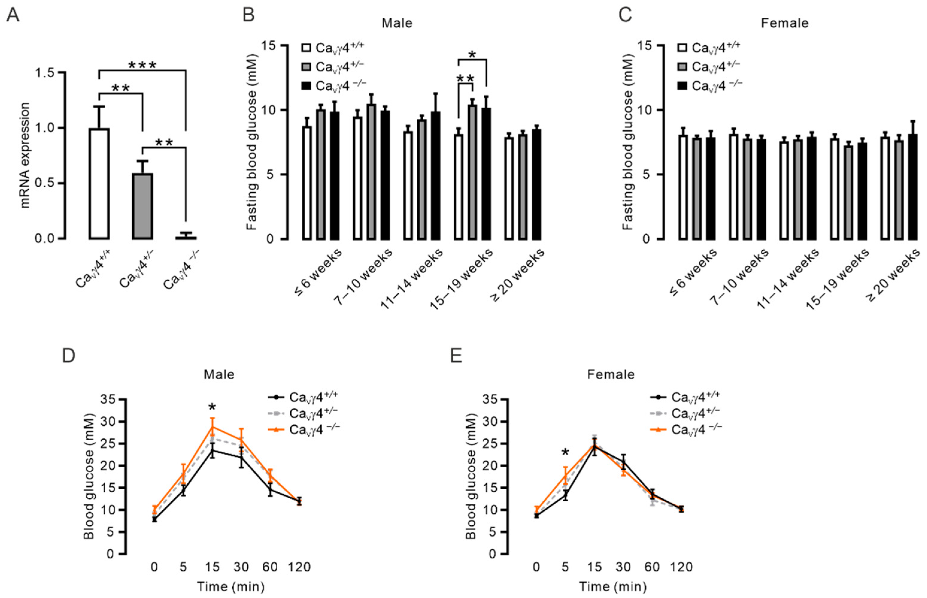
We previously reported that CaVγ4 is required for maintaining a functional differentiated beta-cell phenotype [22]. To investigate the more comprehensive role of CaVγ4 in metabolism in vivo, the constitutive Cacng4 knockout mouse (Cacng4tm1Ran, Jackson Laboratory), in which exon 3 is replaced with a neomycin resistance cassette, was used in the study [26]. First, successful deletion of Cacng4 was confirmed by real-time qPCR using primers targeting exon 3 in isolated pancreatic islets from wild type (CaVγ4+/+), heterozygous (CaVγ4+/−), and homozygous (CaVγ4−/−) Cacng4 knockout mice. CaVγ4 mRNA expression was downregulated by almost 50% in CaVγ4+/− mice, and a further complete abrogation of CaVγ4 was verified in CaVγ4−/− mice (Figure 1A).
We then examined the effect of CaVγ4 ablation on blood glucose levels. Interestingly, an elevated 4-h fasting blood glucose level was observed in both male CaVγ4+/− and male CaVγ4−/− mice at the age of 15–19 weeks compared to the male CaVγ4+/+ mice (Figure 1B), but not in female transgenic mice (Figure 1C). To corroborate further the impact of CaVγ4 on glucose metabolism in vivo, we performed the intraperitoneal glucose tolerance test (IPGTT). Notably, in contrast to CaVγ4+/+ mice, administration of glucose without fasting led to a significantly increased blood glucose level at 15 min post-injection in male CaVγ4−/− mice at 11–15 weeks of age (Figure 1D). The raised blood glucose level was also observed at 5 min post-injection in female CaVγ4−/− mice at the same age (Figure 1E). These results show that CaVγ4 is involved in the regulation of blood glucose metabolism in an age- and gender-specific manner.
Figure 1. Impaired fasting blood glucose and glucose tolerance in Cacng4 knockout mice: (A) CaVγ4 (Cacng4) mRNA levels in isolated islets from wild type (CaVγ4+/+), heterozygous (CaVγ4+/−), and homozygous (CaVγ4−/−) Cacng4 knockout mice, n = 3, 4 and 3 mice, respectively. *** p < 0.001 (CaVγ4+/+ vs. CaVγ4−/−) and ** p < 0.01 (CaVγ4+/+ vs. CaVγ4+/− and CaVγ4+/− vs. CaVγ4−/−). (B) Blood glucose levels in 4-h fasted male CaVγ4+/+, CaVγ4+/− and CaVγ4−/− mice from 3 weeks to 30 weeks. n = 5, 21 and 3 mice (≤ 6 weeks); 9, 9 and 5 mice (7–10 weeks); 8, 19 and 5 mice (11–14 weeks); 7, 10 and 4 mice (15–19 weeks, ** p = 0.003 (CaVγ4+/+ vs. CaVγ4+/−), * p = 0.034 (CaVγ4+/+ vs. CaVγ4−/−)); 4, 27 and 4 mice (≥20 weeks), respectively. (C) As in (B) but in female CaVγ4+/+, CaVγ4+/− and CaVγ4−/− mice. n = 8, 20 and 4 mice (≤ 6 weeks); 6, 19 and 15 mice (7–10 weeks); 6, 21 and 13 mice (11–14 weeks); 6, 14 and 14 mice (15–19 weeks); 8, 14 and 3 mice (≥20 weeks), respectively. (D) Plasma glucose concentrations during IPGTT (2 g/kg) in male CaVγ4+/+, CaVγ4+/− and CaVγ4−/− mice at 11–15 weeks of age. n = 7, 9 and 5 mice, respectively. * p = 0.025, CaVγ4+/+ vs. CaVγ4−/− at 15 min. (E) As in (D) but in female mice. n = 10, (CaVγ4+/+), 14 (CaVγ4+/−) and 14 (CaVγ4−/−) mice, respectively. * p = 0.024, CaVγ4+/+ vs. CaVγ4−/− at 5 min. Data is presented as Mean ± SEM and were analyzed with one-way ANOVA with Tukey’s multiple comparisons test (A); and two-way ANOVA with Tukey’s, Dunnett’s or Two-stage linear step-up procedure of Benjamini, Krieger and Yekutieli’s multiple comparisons tests (BE).
Biomedicines 10 00770 g001

3.2. CaVγ4 Is Required for Glucose-Stimulated Insulin Secretion in Beta-Cells

The observation of CaVγ4-related effects on glucose homeostasis in vivo may result from damaged beta-cell function [22]. To explore this possibility, ex vivo glucose-stimulated insulin secretion assay was conducted in pancreatic islets from Cacng4 knockout mice at 11–15 weeks of age. As expected, in freshly isolated CaVγ4−/− mouse islets, glucose-stimulated insulin secretion was markedly reduced in comparison to that in CaVγ4+/+ mouse islets (Figure 2A). A tendency of attenuated glucose-stimulated insulin secretion was seen in CaVγ4 heterozygotes islets (Figure 2A). This data is in line with our previous findings [22], demonstrating that CaVγ4 is of importance for glucose-stimulated insulin secretion in pancreatic beta-cells, and in turn, involved in the regulation of global glucose metabolism.
Given the decisive role of CaVγ4 in mediating pancreatic beta-cell function, the underlying molecular mechanism is of great interest. It has been documented that loss of CaVγ4 induces beta-cell dedifferentiation [22], which is attributed to the decreased expression of critical beta-cell identity transcription factors (e.g., MafA, Pdx1, Nkx6.1) during glucotoxicity [4,32]. Accordingly, we assessed mRNA levels of key islet-enriched genes as well as the beta-cell dedifferentiation marker aldehyde dehydrogenase1a3 (Aldh1a3) [33] in Cacng4 knockout mouse islets. Real-time qPCR revealed that MafA mRNA was significantly low in islets from both CaVγ4+/− and CaVγ4−/− mice compared with islets from wild type mice (Figure 2B). Remarkably, Aldh1a3 mRNA expression was ~35% higher in CaVγ4−/− islets than that in wild type islets, which agrees with our earlier finding that CaVγ4 deletion leads to loss of beta-cell identity by dedifferentiation [22] (Figure 2B). Collectively, these data support the hypothesis that CaVγ4-ablation-induced disturbed insulin secretion results from downregulation of beta-cell identity genes (i.e., MafA), leading to beta-cell dedifferentiation.

3.3. CaVγ4 Regulates MafA via CaMKII

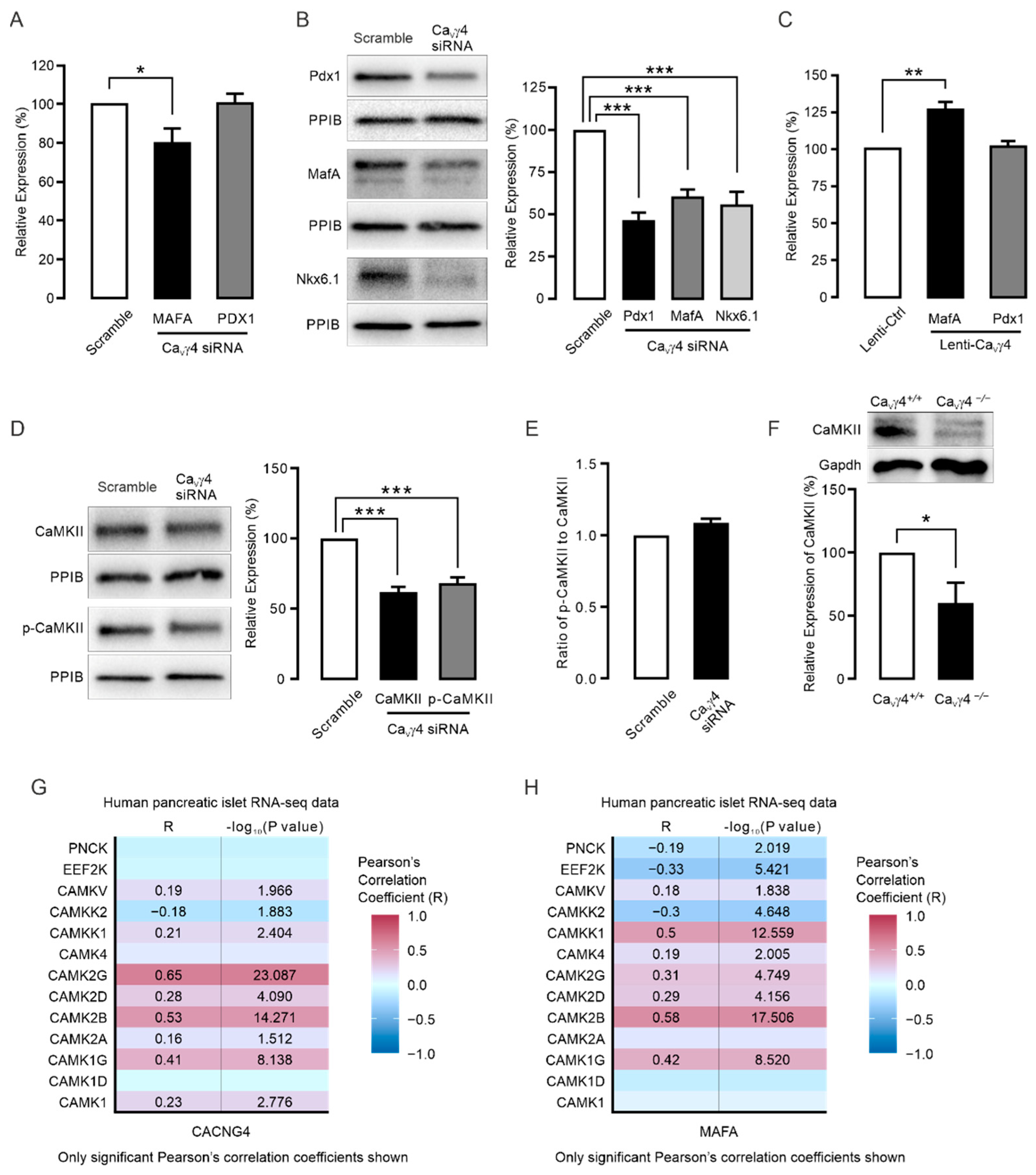
The gene expression data in transgenic mouse islets (Figure 2B), were next complemented by assaying mRNA and protein levels of MafA and Pdx1 in human islets and INS-1 832/13 cells when silencing or overexpressing CaVγ4. Notably, MAFA mRNA expression, but not PDX1, was significantly decreased in CaVγ4-silenced non-diabetic human islets (Figure 3A). This was supported by data obtained from western blotting (Figure 3B). Confirmation in INS-1 832/13 cells by lentiviral-based overexpressing CaVγ4 also showed that MafA mRNA was substantially enhanced, whereas Pdx1 mRNA remained unaltered (Figure 3C). These data together provide evidence for MafA being one of the downstream targets of CaVγ4 in regulating beta-cell function and differentiation status. Subsequently, we focused on how CaVγ4 exerts its effects on MafA in beta-cells.
It is well established that CaMKII inhibition causes MafA downregulation in pancreatic beta-cells [14,15]. Moreover, as CaVγ4 affects intracellular calcium homeostasis [22], it is likely that CaVγ4 may regulate CaMKII expression/activation. This leads to the exciting possibility that CaMKII is the missing link in the chain of events from CaVγ4 ablation to reduced MafA levels. Indeed, silencing CaVγ4 strongly suppressed the expression of both CaMKII and phosphorylated-CaMKII in INS-1 832/13 cells (Figure 3D). The decreased extent of total CaMKII in CaVγ4-silenced beta-cells was similar to that of phosphorylated-CaMKII, suggesting no additional regulation of CaVγ4 on CaMKII phosphorylation (Figure 3E). This reduced CaMKII protein expression was ideally confirmed in CaVγ4−/− mouse islets in comparison to wild type islets (Figure 3F). To corroborate the relationship between CaVγ4 and CaMK gene family, RNA-seq data of pancreatic islets from 188 human donors was analyzed. In agreement with our hypothesis, CaVγ4 (CACNG4) expression robustly exhibited a positive correlation with all 4 CaMKII subunits in human islets (Figure 3G). In addition, a similar correlation was also observed between CaMKII variants and MAFA (Figure 3H).
We finally studied the outcome of the chain of events that CaVγ4 through mediating CaMKII levels controls MafA expression, which in turn affects insulin production. As expected, removal of CaVγ4 resulted in a decline in insulin content in INS-1 832/13 cells (Figure 4A). Conversely, overexpression of CaVγ4 increased the total intracellular insulin content in INS-1 832/13 cells (Figure 4B). These findings were strongly supported by the positive correlation of mRNA expressions between CaVγ4 (CACNG4) and insulin (INS) in human pancreatic islets (Figure 4C).
Taken together, these results demonstrate that reduction of CaVγ4 in the beta-cell decreases MafA expression by suppressing CaMKII expression and activation, which reduces insulin gene expression and secretion and loss of the mature beta-cell identity (Figure 4D).

4. Discussion

Studies in human and rodent pancreatic islets in vitro have established that CaVγ4 determines beta-cell function by enhancing Ca2+ entry and the subsequent glucose-stimulated insulin secretion [22]. Here, we further demonstrate that CaVγ4 is essential for in vivo glucose homeostasis in mice. Our results disclose that CaVγ4 is pivotal for maintaining normal fasting blood glucose levels and glucose tolerance in vivo by regulating the beta-cell determining transcription factor MafA expression. When this signaling pathway is disrupted in CaVγ4-ablated beta-cells, glucose metabolism is impaired and insulin-producing and -releasing properties of beta-cells are deteriorated (Figure 4D).

4.1. CaVγ4 and Glucose Metabolism

CaVγ subunits have been reported to be expressed in both fetal and adult human brains and are widespread in different tissues [34,35]. All eight variants are detected in human pancreatic islets with, however, only CaVγ4 being differentially expressed in islets from T2D human donors and diabetic animal modes, GK rats and db/db mice [22], rendering the rationale for aiming at CaVγ4. In fact, in vitro data in human islets identify that the changes in CaVγ4 expression, with resultant effects on beta-cell stimulus-secretion coupling, can provide a novel interpretation for beta-cell failure [22]. Accordingly, CaVγ4 plays key roles in the brain. There is evidence that CaVγ4 rescues the defective excitatory synaptic transmission in cerebellar granule cells from the Cacng2 mutant mice (stargazer mice) [23]. This is confirmed by the discovery that CaVγ4 silencing is seizure inducible [36]. Importantly, the total charge transfer through postsynaptic AMPA receptors at central synapses is diminished in Cacng4 knockout mice [26]. These studies together strongly imply that CaVγ4 may exert an indispensable effect in modulating metabolic process and glucose homeostasis in vivo. Indeed, the ability to control fasting blood glucose levels was lost in male Cacng4 knockout mice (Figure 1B). Interestingly, this was unaffected in female Cacng4 transgenic mice (Figure 1C). We speculate that this gender-specific phenotype may associate with estrogens signal networks in brain. The physiology of estrogen action on managing glucose transport into cells and glycolysis primarily through its receptors have been intensively studied. Withdrawal of estrogens or estrogen receptors causes dysregulation of glucose metabolism and body weight and is associated with development of diabetes [37]. Given the significant role of CaVγ4 in neurons, it would not be surprising that the estrogen physiology is disturbed by CaVγ4 deletion in brain. Future investigations focused on CaVγ4-derived estrogen signal regulation is warranted. Secondly, this elevated 4-h fasting blood glucose levels and IGT seen in Cacng4 knockout mice, jointly with ex vivo downregulated insulin secretion in CaVγ4−/− mouse islets (Figure 1B,D,E and Figure 2A), perfectly confirmed our hypothesis that CaVγ4 governs glucose homeostasis in vivo. Be that as it may, the effects of CaVγ4 on plasma insulin levels and insulin responsive peripheral tissues are unclear, since insulin resistance is highly related to prediabetes [2] which is marked by IFG and IGT. Additionally, both IFG and IGT became manifest in Cacng4 knockout mice (Figure 1B,D,E). Therefore, the plasma insulin concentration and insulin action may also be lowered in these mice that partly contributes to the impaired glucose homeostasis in vivo. The insulin levels in plasma and insulin signaling transduction via insulin receptors in fat, muscle, and liver cells from Cacng4 knockout mice will be examined in our next paper. In agreement with our previous findings [22], CaVγ4−/− mouse islets exhibited blunted glucose-stimulated insulin secretion and upregulated expression of Aldh1a3, a beta-cell dedifferentiation marker [33] (Figure 2A,B), suggesting the vital role of CaVγ4 in determining beta-cell functionality. In summary, CaVγ4 is more important than anticipated in controlling metabolism in vivo and appears a key character in the regulation of blood glucose levels and insulin secretion.

4.2. CaVγ4 and MafA Signaling

Apart from allowing Ca2+ entry to activate Ca2+-dependent signaling pathways, CaV channels have been increasingly documented that are substantial for modification of gene expression and beta-cell differentiation [18,22,38]. In this respect, they behave directly as transcription factors (by CaV subunits or fragments) or indirectly by Ca2+-binding/-activated proteins to regulate a plethora of downstream transcription factors, for instance, CREB, NFAT, DREAM and MafA [14,15,38]. Being the principal beta-cell-enriched transcription factor, MafA is crucial for beta-cell maturation and mediating insulin transcription and other genes in a glucose-responsive manner [5,39]. Our previous study revealed that CaVγ4 expression is under the control of MafA [22]. On the other hand, as part of CaV channels, CaVγ4 is also responsible for Ca2+ entry and transients in the cytosol [22], raising the likelihood of a negative feedback from CaVγ4 to MafA. Indeed, we here add new knowledge to the effect that CaVγ4 has a profound impact on MafA expression, and most importantly, this negative feedback is conducted via the Ca2+-binding enzyme CaMKII. First, MafA expression was reduced in Cacng4 knockout mouse islets, and its levels in human islets and INS-1 832/13 cells can be manipulated by silencing or overexpressing CaVγ4 (Figure 2B and Figure 3A–C). Then, CaMKII activation has been shown to clearly modulate MafA expression and insulin secretion in pancreatic beta-cells [14,15]. This conclusion is extended by our western blotting data that both CaMKII and phosphorylated-CaMKII expressions were weakened in CaVγ4-silenced beta-cells (Figure 3D), and the decreased CaMKII protein levels were also perfectly observed in islets from CaVγ4−/− mice (Figure 3F). Conspicuously, the positive associations between CaMKII subunits and CaVγ4 (CACNG4) as well as MAFA were firmly echoed by the analysis of human pancreatic islet RNA-seq data (Figure 3G,H). However, the mechanism behind that bridges CaVγ4 and CaMKII is unexplored and is of interest to study in the future. We would speculate this regulatory interaction is either resulted from the intracellular Ca2+ concentrations determined by CaVγ4 expression [22], or simply through a specific binding [40]. Finally, insulin content was significantly associated with CaVγ4 levels in both human islets and INS-1 832/13 cells (Figure 4A–C), which fully agrees with the fact that MafA is a dedicated activator of the insulin transcription [7].

4.3. Limitations of the Study

The principal limitations of the present study are the inadequate experiments in the Cacng4 knockout mice. Given the observed prediabetes associated phenotypes (IFG and IGT), first and foremost, in-depth in vivo exploration of the involvement of CaVγ4 in insulin resistance need to be conducted. It would be essential to examine the influences of CaVγ4 on plasma insulin levels and insulin signaling pathways in insulin target peripheral tissues, for example liver, fat, and muscle. The reciprocal regulation between CaVγ4 and estrogen, which might be responsible for the gender-specific phenotype, requires further in vivo study, and the employment of beta-cell conditional knockout mice would be necessary. In addition, it would be important to investigate the regulatory machinery of CaVγ4 on CaMKII.

5. Conclusions

We identify a comprehensive role of the CaVγ4 subunit for regulating glucose homeostasis in mice in vivo. Our results suggest new beneficial effects of CaVγ4 with the potential to preclude the development of prediabetes-related IFG and IGT through its influence on insulin secretion capability in islets. This fairly resonates with the existing view that CaVγ4 contributes the key impingement to sustaining a functional beta-cell phenotype [22]. Strikingly, we clarify a novel negative feedback pathway from CaVγ4 to MafA by the involvement of CaMKII. These results together shed light on prediabetes treatment in the future by aiming at a novel target, the CaVγ4 subunit, to prevent hyperglycemia.

Supplementary Materials

The following supporting information can be downloaded at: https://www.mdpi.com/article/10.3390/biomedicines10040770/s1, Table S1: Information list of human islet donors used for the experiment.

Author Contributions

C.L., E.Z. and E.R. designed and supervised the study, finalized manuscript writing with feedback from all authors. C.L. performed experiments on transgenic mice, human islets, isolated mouse islets and INS-1 832/13 cells, conducted data analysis and drafted the manuscript. R.W. performed experiments. A.K. conducted Human islet RNA-seq data analysis. L.E. gave expert input to the study and revised manuscript. All authors have read and agreed to the published version of the manuscript.

Funding

This research was funded by the Swedish Research Council, Strategic Research Area Exodiab (Dnr 2009-1039), Swedish Foundation for Strategic Research (Dnr IRC 15-0067), Swedish Research Council, Linnaeus grant (Dnr 349-2006-237) and the Diabetes Wellness foundation (E.R. and E.Z., Dnr 1904-PG). Salaries and infrastructure were supported by the Swedish Research Council (E.R., Dnr 2018-03258; L.E., Dnr 2019-01406; E.Z., Dnr 2019-01567), Diabetesfonden (E.R., L.E.) and the Crafoord Foundation (E.Z., Dnr 20190514).

Institutional Review Board Statement

The study was conducted according to the guidelines of the Declaration of Helsinki, and approved by the Malmö/Lund Ethical committee (#5.2.18-10992/18, 23 February 2019). All experiments were performed in accordance with relevant guidelines and regulations.

Informed Consent Statement

Informed consent was obtained from all subjects involved in the study.

Data Availability Statement

All data generated and/or analyzed during the current study are available from the corresponding authors on reasonable request.

Acknowledgments

We thank B-M. Nilsson and A-M. Veljonskaja-Ramsay (Lund University Diabetes Center, Sweden) for expert technical assistance. We are grateful to M. Magnusson for mouse strain maintenance.

Conflicts of Interest

The authors declare no conflict of interest.

References

  1. Zhang, E.; Mohammed Al-Amily, I.; Mohammed, S.; Luan, C.; Asplund, O.; Ahmed, M.; Ye, Y.; Ben-Hail, D.; Soni, A.; Vishnu, N.; et al. Preserving Insulin Secretion in Diabetes by Inhibiting VDAC1 Overexpression and Surface Translocation in beta Cells. Cell Metab. 2019, 29, 64–77.e66. [Google Scholar] [CrossRef] [PubMed]
  2. Kahn, B.B. Type 2 diabetes: When insulin secretion fails to compensate for insulin resistance. Cell 1998, 92, 593–596. [Google Scholar] [CrossRef] [Green Version]
  3. Tabak, A.G.; Herder, C.; Rathmann, W.; Brunner, E.J.; Kivimaki, M. Prediabetes: A high-risk state for diabetes development. Lancet 2012, 379, 2279–2290. [Google Scholar] [CrossRef] [Green Version]
  4. Talchai, C.; Xuan, S.; Lin, H.V.; Sussel, L.; Accili, D. Pancreatic beta cell dedifferentiation as a mechanism of diabetic beta cell failure. Cell 2012, 150, 1223–1234. [Google Scholar] [CrossRef] [Green Version]
  5. Hang, Y.; Stein, R. MafA and MafB activity in pancreatic beta cells. Trends Endocrinol. Metab. 2011, 22, 364–373. [Google Scholar] [CrossRef] [Green Version]
  6. Wilson, M.E.; Scheel, D.; German, M.S. Gene expression cascades in pancreatic development. Mech. Dev. 2003, 120, 65–80. [Google Scholar] [CrossRef]
  7. Artner, I.; Hang, Y.; Guo, M.; Gu, G.; Stein, R. MafA is a dedicated activator of the insulin gene in vivo. J. Endocrinol. 2008, 198, 271–279. [Google Scholar] [CrossRef] [Green Version]
  8. Artner, I.; Hang, Y.; Mazur, M.; Yamamoto, T.; Guo, M.; Lindner, J.; Magnuson, M.A.; Stein, R. MafA and MafB regulate genes critical to beta-cells in a unique temporal manner. Diabetes 2010, 59, 2530–2539. [Google Scholar] [CrossRef] [Green Version]
  9. Ganic, E.; Singh, T.; Luan, C.; Fadista, J.; Johansson, J.K.; Cyphert, H.A.; Bennet, H.; Storm, P.; Prost, G.; Ahlenius, H.; et al. MafA-Controlled Nicotinic Receptor Expression Is Essential for Insulin Secretion and Is Impaired in Patients with Type 2 Diabetes. Cell Rep. 2016, 14, 1991–2002. [Google Scholar] [CrossRef] [Green Version]
  10. Zhang, C.; Moriguchi, T.; Kajihara, M.; Esaki, R.; Harada, A.; Shimohata, H.; Oishi, H.; Hamada, M.; Morito, N.; Hasegawa, K.; et al. MafA is a key regulator of glucose-stimulated insulin secretion. Mol. Cell. Biol. 2005, 25, 4969–4976. [Google Scholar] [CrossRef] [Green Version]
  11. Matsuoka, T.A.; Artner, I.; Henderson, E.; Means, A.; Sander, M.; Stein, R. The MafA transcription factor appears to be responsible for tissue-specific expression of insulin. Proc. Natl. Acad. Sci. USA 2004, 101, 2930–2933. [Google Scholar] [CrossRef] [PubMed] [Green Version]
  12. Artner, I.; Blanchi, B.; Raum, J.C.; Guo, M.; Kaneko, T.; Cordes, S.; Sieweke, M.; Stein, R. MafB is required for islet beta cell maturation. Proc. Natl. Acad. Sci. USA 2007, 104, 3853–3858. [Google Scholar] [CrossRef] [PubMed] [Green Version]
  13. Raum, J.C.; Hunter, C.S.; Artner, I.; Henderson, E.; Guo, M.; Elghazi, L.; Sosa-Pineda, B.; Ogihara, T.; Mirmira, R.G.; Sussel, L.; et al. Islet beta-cell-specific MafA transcription requires the 5′-flanking conserved region 3 control domain. Mol. Cell. Biol. 2010, 30, 4234–4244. [Google Scholar] [CrossRef] [Green Version]
  14. Santos, G.J.; Ferreira, S.M.; Ortis, F.; Rezende, L.F.; Li, C.; Naji, A.; Carneiro, E.M.; Kaestner, K.H.; Boschero, A.C. Metabolic memory of ss-cells controls insulin secretion and is mediated by CaMKII. Mol. Metab. 2014, 3, 484–489. [Google Scholar] [CrossRef] [PubMed]
  15. Vanderford, N.L.; Cantrell, J.E.; Popa, G.J.; Ozcan, S. Multiple kinases regulate mafA expression in the pancreatic beta cell line MIN6. Arch. Biochem. Biophys. 2008, 480, 138–142. [Google Scholar] [CrossRef] [PubMed] [Green Version]
  16. Rorsman, P.; Renstrom, E. Insulin granule dynamics in pancreatic beta cells. Diabetologia 2003, 46, 1029–1045. [Google Scholar] [CrossRef]
  17. Yang, S.N.; Berggren, P.O. The role of voltage-gated calcium channels in pancreatic beta-cell physiology and pathophysiology. Endocr. Rev. 2006, 27, 621–676. [Google Scholar] [CrossRef] [Green Version]
  18. Jing, X.; Li, D.Q.; Olofsson, C.S.; Salehi, A.; Surve, V.V.; Caballero, J.; Ivarsson, R.; Lundquist, I.; Pereverzev, A.; Schneider, T.; et al. CaV2.3 calcium channels control second-phase insulin release. J. Clin. Investig. 2005, 115, 146–154. [Google Scholar] [CrossRef] [Green Version]
  19. Schulla, V.; Renstrom, E.; Feil, R.; Feil, S.; Franklin, I.; Gjinovci, A.; Jing, X.J.; Laux, D.; Lundquist, I.; Magnuson, M.A.; et al. Impaired insulin secretion and glucose tolerance in beta cell-selective Ca(v)1.2 Ca2+ channel null mice. EMBO J. 2003, 22, 3844–3854. [Google Scholar] [CrossRef] [Green Version]
  20. Scholze, A.; Plant, T.D.; Dolphin, A.C.; Nurnberg, B. Functional expression and characterization of a voltage-gated CaV1.3 (alpha1D) calcium channel subunit from an insulin-secreting cell line. Mol. Endocrinol. 2001, 15, 1211–1221. [Google Scholar] [CrossRef] [Green Version]
  21. Arikkath, J.; Campbell, K.P. Auxiliary subunits: Essential components of the voltage-gated calcium channel complex. Curr. Opin. Neurobiol. 2003, 13, 298–307. [Google Scholar] [CrossRef]
  22. Luan, C.; Ye, Y.; Singh, T.; Barghouth, M.; Eliasson, L.; Artner, I.; Zhang, E.; Renstrom, E. The calcium channel subunit gamma-4 is regulated by MafA and necessary for pancreatic beta-cell specification. Commun. Biol. 2019, 2, 106. [Google Scholar] [CrossRef] [PubMed] [Green Version]
  23. Cho, C.H.; St-Gelais, F.; Zhang, W.; Tomita, S.; Howe, J.R. Two families of TARP isoforms that have distinct effects on the kinetic properties of AMPA receptors and synaptic currents. Neuron 2007, 55, 890–904. [Google Scholar] [CrossRef] [PubMed] [Green Version]
  24. Korber, C.; Werner, M.; Kott, S.; Ma, Z.L.; Hollmann, M. The transmembrane AMPA receptor regulatory protein gamma 4 is a more effective modulator of AMPA receptor function than stargazin (gamma 2). J. Neurosci. 2007, 27, 8442–8447. [Google Scholar] [CrossRef] [Green Version]
  25. Tomita, S. Regulation of ionotropic glutamate receptors by their auxiliary subunits. Physiology 2010, 25, 41–49. [Google Scholar] [CrossRef] [Green Version]
  26. Milstein, A.D.; Zhou, W.; Karimzadegan, S.; Bredt, D.S.; Nicoll, R.A. TARP subtypes differentially and dose-dependently control synaptic AMPA receptor gating. Neuron 2007, 55, 905–918. [Google Scholar] [CrossRef] [Green Version]
  27. Klugbauer, N.; Dai, S.; Specht, V.; Lacinova, L.; Marais, E.; Bohn, G.; Hofmann, F. A family of gamma-like calcium channel subunits. FEBS Lett. 2000, 470, 189–197. [Google Scholar] [CrossRef] [Green Version]
  28. Yang, L.; Katchman, A.; Morrow, J.P.; Doshi, D.; Marx, S.O. Cardiac L-type calcium channel (Cav1.2) associates with gamma subunits. FASEB J. 2011, 25, 928–936. [Google Scholar] [CrossRef] [Green Version]
  29. Rorsman, P.; Ashcroft, F.M. Pancreatic beta-Cell Electrical Activity and Insulin Secretion: Of Mice and Men. Physiol. Rev. 2018, 98, 117–214. [Google Scholar] [CrossRef]
  30. Hastoy, B.; Godazgar, M.; Clark, A.; Nylander, V.; Spiliotis, I.; van de Bunt, M.; Chibalina, M.V.; Barrett, A.; Burrows, C.; Tarasov, A.I.; et al. Electrophysiological properties of human beta-cell lines EndoC-betaH1 and -betaH2 conform with human beta-cells. Sci. Rep. 2018, 8, 16994. [Google Scholar] [CrossRef]
  31. Asplund, O.; Storm, P.; Chandra, V.; Ottosson-Laakso, E.; Hatem, G.; Mansour-Aly, D.; Krus, U.; Ibrahim, H.; Ahlqvist, E.; Tuomi, T.; et al. Islet Gene View—A tool to facilitate islet research. BioRxiv 2020, 435743. [Google Scholar] [CrossRef]
  32. Bensellam, M.; Jonas, J.C.; Laybutt, D.R. Mechanisms of beta-cell dedifferentiation in diabetes: Recent findings and future research directions. J. Endocrinol. 2018, 236, R109–R143. [Google Scholar] [CrossRef] [PubMed] [Green Version]
  33. Cinti, F.; Bouchi, R.; Kim-Muller, J.Y.; Ohmura, Y.; Sandoval, P.R.; Masini, M.; Marselli, L.; Suleiman, M.; Ratner, L.E.; Marchetti, P.; et al. Evidence of beta-Cell Dedifferentiation in Human Type 2 Diabetes. J. Clin. Endocrinol. Metab. 2016, 101, 1044–1054. [Google Scholar] [CrossRef] [PubMed] [Green Version]
  34. Black, J.L., 3rd. The voltage-gated calcium channel gamma subunits: A review of the literature. J. Bioenerg. Biomembr. 2003, 35, 649–660. [Google Scholar] [CrossRef]
  35. Burgess, D.L.; Gefrides, L.A.; Foreman, P.J.; Noebels, J.L. A cluster of three novel Ca2+ channel gamma subunit genes on chromosome 19q13.4: Evolution and expression profile of the gamma subunit gene family. Genomics 2001, 71, 339–350. [Google Scholar] [CrossRef]
  36. Letts, V.A.; Mahaffey, C.L.; Beyer, B.; Frankel, W.N. A targeted mutation in Cacng4 exacerbates spike-wave seizures in stargazer (Cacng2) mice. Proc. Natl. Acad. Sci. USA 2005, 102, 2123–2128. [Google Scholar] [CrossRef] [Green Version]
  37. Rettberg, J.R.; Yao, J.; Brinton, R.D. Estrogen: A master regulator of bioenergetic systems in the brain and body. Front. Neuroendocrinol. 2014, 35, 8–30. [Google Scholar] [CrossRef] [Green Version]
  38. Barbado, M.; Fablet, K.; Ronjat, M.; De Waard, M. Gene regulation by voltage-dependent calcium channels. Biochim. Biophys. Acta 2009, 1793, 1096–1104. [Google Scholar] [CrossRef] [Green Version]
  39. Cataldo, L.R.; Singh, T.; Achanta, K.; Bsharat, S.; Prasad, R.B.; Luan, C.; Renström, E.; Eliasson, L.; Artner, I. MAFA and MAFB regulate exocytosis-related genes in human β-cells. Acta Physiol. 2022, 234, e13761. [Google Scholar] [CrossRef]
  40. Pinto, T.M.; Schilstra, M.J.; Roque, A.C.; Steuber, V. Binding of Filamentous Actin to CaMKII as Potential Regulation Mechanism of Bidirectional Synaptic Plasticity by beta CaMKII in Cerebellar Purkinje Cells. Sci. Rep. 2020, 10, 9019. [Google Scholar] [CrossRef]
Figure 2. Insulin secretion and gene expression in Cacng4 knockout mouse islets: (A) Glucose-stimulated insulin secretion in CaVγ4+/+, CaVγ4+/− and CaVγ4−/− mouse islets. n = 6, 7 and 6 mice, respectively. * p = 0.016. (B) MafA, Pdx1 and Aldh1a3 mRNA expression in CaVγ4+/+, CaVγ4+/− and CaVγ4−/− mouse islets. n = 4, 4 and 4 mice, respectively. ** p < 0.01, * p < 0.05 and # p < 0.05. Data are presented as Mean ± SEM and were analyzed with two-way ANOVA with Dunnett’s multiple comparisons test.
Biomedicines 10 00770 g002
Figure 3. CaMKII expression is downregulated in CaVγ4-silenced beta-cells: (A) PDX1 and MAFA mRNA expression in CaVγ4-silenced human islets. n = 4 (PDX1) and 6 (MAFA) donors. * p = 0.031. (B) Pdx1, MafA and Nkx6.1 immunoblotting and means of expression in CaVγ4-silenced INS-1 832/13 cells. n = 4, *** p < 0.001. (C) As in (A) but in CaVγ4-overexpressed INS-1 832/13 cells. n = 4, ** p = 0.002. (D) Decreased CaMKII and phosphorylated-CaMKII expressions in CaVγ4-silenced INS-1 832/13 cells. n = 4, *** p < 0.001 for both. (E) Ratio of protein expressions in (D) of phosphorylated-CaMKII to CaMKII. n = 4, p > 0.05. (F) CaMKII protein levels in CaVγ4+/+ and CaVγ4−/− mouse islets. n = 4 mice each. * p < 0.05. (G) Pearson’s correlation coefficients (R) calculated by mRNA expression (RNA-seq, human islets) between the CAMK family genes and CACNG4. n = 188 human donors. (H) As in (G) but between CAMK family genes and MAFA. n = 188 human donors. Data are presented as Mean ± SEM and were analyzed with one-way ANOVA with Dunnett’s multiple comparisons test (AD); and two-tailed Student’s t-test (EH). See Supplementary Table S1 for the details of the human donors utilized for the experiment in this study.
Biomedicines 10 00770 g003
Figure 4. CaVγ4 regulates insulin production in beta-cells: (A) Reduced insulin content in CaVγ4-silenced INS-1 832/13 cells. n = 4, * p = 0.014. (B) Increased insulin content in CaVγ4-overexpressed INS-1 832/13 cells. n = 4, * p = 0.035. (C) Pearson correlation performed between the log2-transformed normalized expression values of CACNG4 and INS in Counts Per Million (CPM). n = 188 human donors. (D) Schematic model of cascading effects from CaVγ4 deletion through CaMKII expression to insulin secretion in CaVγ4−/− beta-cell. See main text for details. Data are presented as Mean ± SEM and were analyzed with two-tailed unpaired Student’s t-test.
Biomedicines 10 00770 g004
Publisher’s Note: MDPI stays neutral with regard to jurisdictional claims in published maps and institutional affiliations.