
Prognostic Role of Systemic Inflammatory Markers in Patients Undergoing Surgical Resection for Oral Squamous Cell Carcinoma


Abstract
:1. Introduction
2. Materials and Methods
2.1. Study Population
2.2. Data Collection
2.3. Statistics
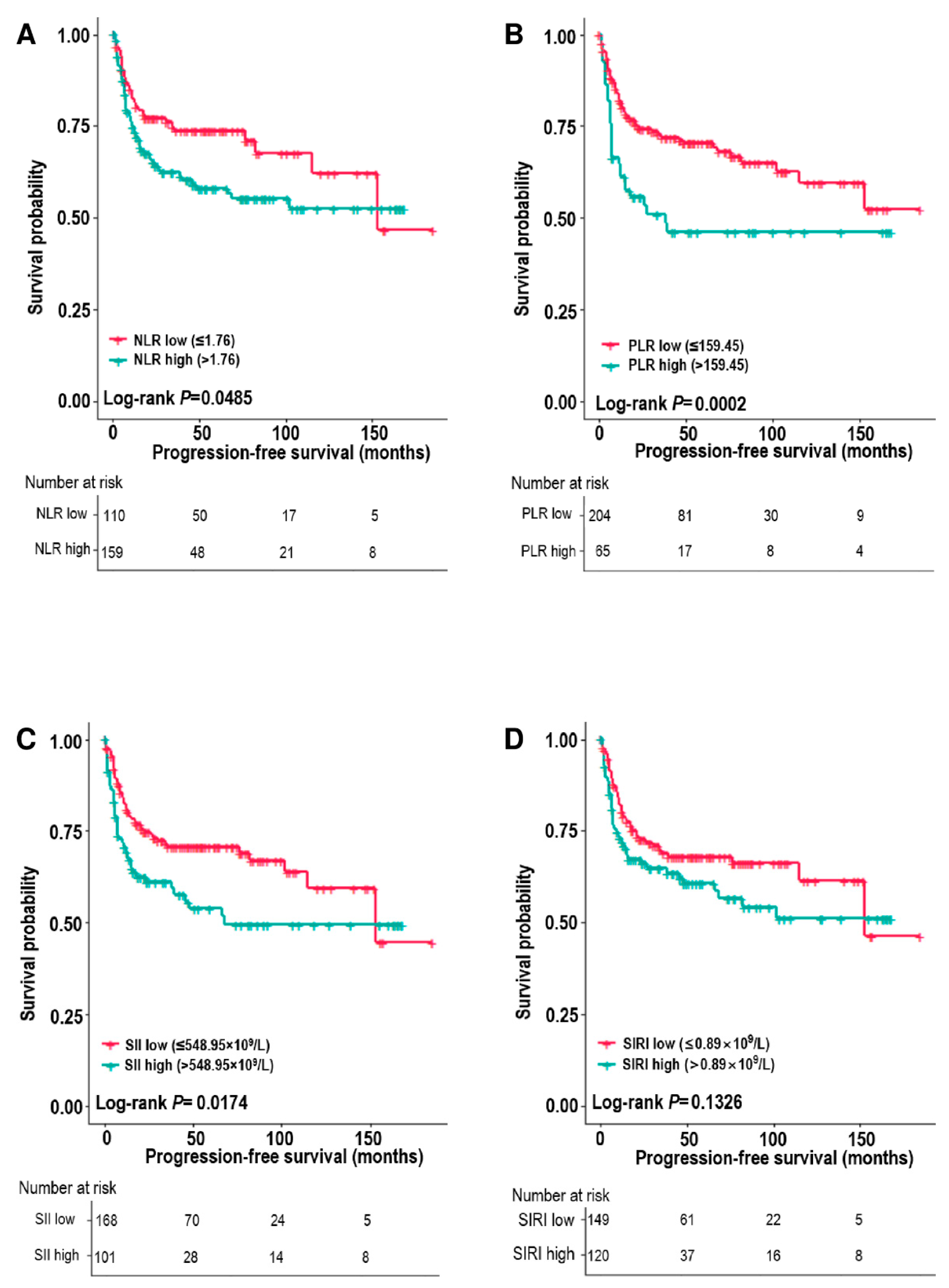
3. Results
3.1. Patient Characteristics
3.2. Correlation between Inflammatory Markers and Clinical Factors
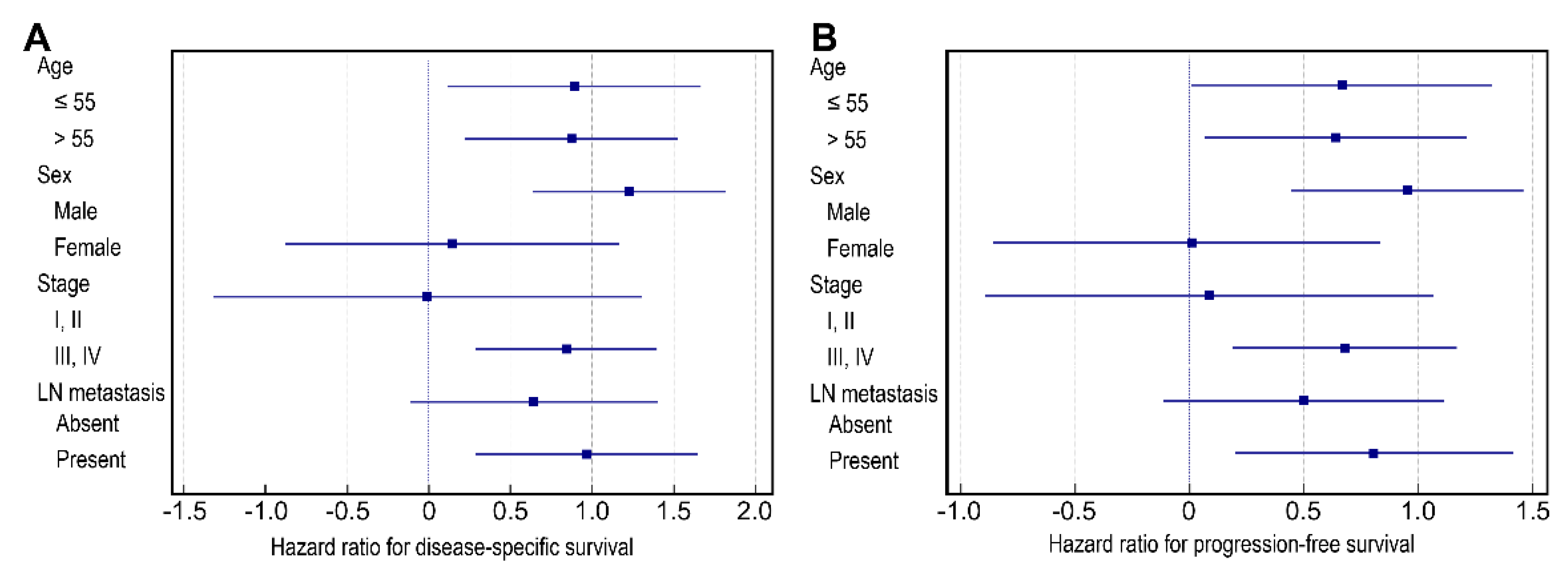
3.3. Analysis of the Relationship between PLR and Survival According to Clinical Factors
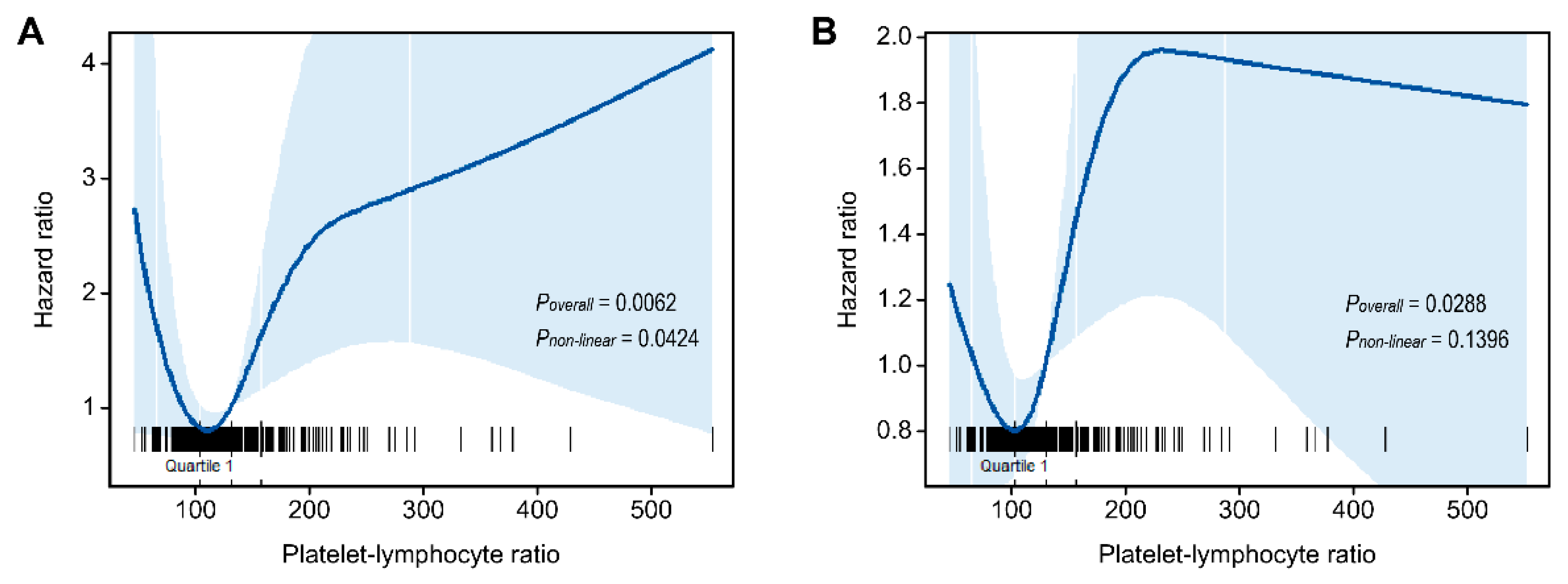
3.4. Nonlinear Association between PLR and Survival
4. Discussion
Supplementary Materials
Author Contributions
Funding
Institutional Review Board Statement
Informed Consent Statement
Data Availability Statement
Conflicts of Interest
References
- Bray, F.; Ferlay, J.; Soerjomataram, I.; Siegel, R.L.; Torre, L.A.; Jemal, A. Global cancer statistics 2018: GLOBOCAN estimates of incidence and mortality worldwide for 36 cancers in 185 countries. CA Cancer J. Clin. 2018, 68, 394–424. [Google Scholar] [CrossRef] [PubMed] [Green Version]
- Suh, J.D.; Cho, J.H. Trends in head and neck cancer in South Korea between 1999 and 2012. Clin. Exp. Otorhinolaryngol. 2016, 9, 263–269. [Google Scholar] [CrossRef] [PubMed] [Green Version]
- Hong, S.; Won, Y.J.; Park, Y.R.; Jung, K.W.; Kong, H.J.; Lee, E.S. Cancer statistics in korea: Incidence, mortality, survival, and prevalence in 2017. Cancer Res. Treat. 2020, 52, 335–350. [Google Scholar] [CrossRef] [PubMed]
- Jung, Y.S.; Seok, J.; Hong, S.; Ryu, C.H.; Ryu, J.; Jung, K.W. The emergence of oral cavity cancer and the stabilization of oropharyngeal cancer: Recent contrasting epidemics in the South Korean population. Cancer 2021, 127, 1638–1647. [Google Scholar] [CrossRef]
- Sun, J.R.; Kim, S.M.; Seo, M.H.; Kim, M.J.; Lee, J.H.; Myoung, H. Oral cancer incidence based on annual cancer statistics in Korea. J. Korean Assoc. Oral Maxillofac. Surg. 2012, 38, 20. [Google Scholar] [CrossRef]
- El-Naggar, A.K.; Chan, J.K.C.; Grandis, J.R.; Takata, T.; Slootweg, P.J. WHO Classification of Head and Neck Tumours; IARC: Lyon, France, 2017. [Google Scholar]
- Botta, L.; Gatta, G.; Trama, A.; Bernasconi, A.; Sharon, E.; Capocaccia, R.; Mariotto, A.B.; RARECAREnet Working Group. Incidence and survival of rare cancers in the US and Europe. Cancer Med. 2020, 9, 5632–5642. [Google Scholar] [CrossRef]
- Almangush, A.; Heikkinen, I.; Mäkitie, A.A.; Coletta, R.D.; Läärä, E.; Leivo, I.; Salo, T. Prognostic biomarkers for oral tongue squamous cell carcinoma: A systematic review and meta-analysis. Br. J. Cancer 2017, 117, 856–866. [Google Scholar] [CrossRef] [Green Version]
- Lenouvel, D.; Gonzalez-Moles, M.A.; Ruiz-Avila, I.; Gonzalez-Ruiz, L.; Gonzalez-Ruiz, I.; Ramos-Garcia, P. Prognostic and clinicopathological significance of PD-L1 overexpression in oral squamous cell carcinoma: A systematic review and comprehensive meta-analysis. Oral Oncol. 2020, 106, 104722. [Google Scholar] [CrossRef]
- Kujan, O.; van Schaijik, B.; Farah, C.S. Immune Checkpoint Inhibitors in Oral Cavity Squamous Cell Carcinoma and Oral Potentially Malignant Disorders: A Systematic Review. Cancers 2020, 12, 1937. [Google Scholar] [CrossRef]
- Hiam-Galvez, K.J.; Allen, B.M.; Spitzer, M.H. Systemic immunity in cancer. Nat. Rev. Cancer 2021, 21, 345–359. [Google Scholar] [CrossRef]
- Selders, G.S.; Fetz, A.E.; Radic, M.Z.; Bowlin, G.L. An overview of the role of neutrophils in innate immunity, inflammation and host-biomaterial integration. Regen. Biomater. 2017, 4, 55–68. [Google Scholar] [CrossRef] [PubMed]
- Kersten, K.; Coffelt, S.B.; Hoogstraat, M.; Verstegen, N.J.; Vrijland, K.; Ciampricotti, M.; Doornebal, C.W.; Hau, C.S.; Wellenstein, M.D.; Salvagno, C. Mammary tumor-derived CCL2 enhances pro-metastatic systemic inflammation through upregulation of IL1β in tumor-associated macrophages. Oncoimmunology 2017, 6, e1334744. [Google Scholar] [CrossRef] [PubMed]
- Cupp, M.A.; Cariolou, M.; Tzoulaki, I.; Aune, D.; Evangelou, E.; Berlanga-Taylor, A.J. Neutrophil to lymphocyte ratio and cancer prognosis: An umbrella review of systematic reviews and meta-analyses of observational studies. BMC Med. 2020, 18, 360. [Google Scholar] [CrossRef] [PubMed]
- Patel, A.; Ravaud, A.; Motzer, R.J.; Pantuck, A.J.; Staehler, M.; Escudier, B.; Martini, J.F.; Lechuga, M.; Lin, X.; George, D.J. Neutrophil-to-lymphocyte ratio as a prognostic factor of disease-free survival in postnephrectomy high-risk locoregional renal cell carcinoma: Analysis of the S-TRAC trial. Clin. Cancer Res. 2020, 26, 4863–4868. [Google Scholar] [CrossRef]
- Chrom, P.; Stec, R.; Bodnar, L.; Szczylik, C. Incorporating neutrophil-to-lymphocyte ratio and platelet-to-lymphocyte ratio in place of neutrophil count and platelet count improves prognostic accuracy of the international metastatic renal cell carcinoma database consortium model. Cancer Res. Treat. 2018, 50, 103–110. [Google Scholar] [CrossRef] [Green Version]
- Mleko, M.; Pitynski, K.; Pluta, E.; Czerw, A.; Sygit, K.; Karakiewicz, B.; Banas, T. Role of systemic inflammatory reaction in female genital organ malignancies–state of the art. Cancer Manag. Res. 2021, 13, 5491–5508. [Google Scholar] [CrossRef]
- Schneider, M.; Schäfer, N.; Bode, C.; Borger, V.; Eichhorn, L.; Giordano, F.A.; Güresir, E.; Heimann, M.; Ko, Y.D.; Lehmann, F.; et al. Prognostic value of preoperative inflammatory markers in melanoma patients with brain metastases. J. Clin. Med. 2021, 10, 634. [Google Scholar] [CrossRef]
- Dolan, R.D.; McSorley, S.T.; Horgan, P.G.; Laird, B.; McMillan, D.C. The role of the systemic inflammatory response in predicting outcomes in patients with advanced inoperable cancer: Systematic review and meta-analysis. Crit. Rev. Oncol./Hematol. 2017, 116, 134–146. [Google Scholar] [CrossRef] [Green Version]
- Guthrie, G.J.K.; Charles, K.A.; Roxburgh, C.S.D.; Horgan, P.G.; McMillan, D.C.; Clarke, S.J. The systemic inflammation-based neutrophil–lymphocyte ratio: Experience in patients with cancer. Crit. Rev. Oncol./Hematol. 2013, 88, 218–230. [Google Scholar] [CrossRef]
- Cho, U.; Park, H.S.; Im, S.Y.; Yoo, C.Y.; Jung, J.H.; Suh, Y.J.; Choi, H.J. Prognostic value of systemic inflammatory markers and development of a nomogram in breast cancer. PLoS ONE 2018, 13, e0200936. [Google Scholar] [CrossRef] [Green Version]
- Chon, S.; Lee, S.; Jeong, D.; Lim, S.; Lee, K.; Shin, J. Elevated platelet lymphocyte ratio is a poor prognostic factor in advanced epithelial ovarian cancer. J. Gynecol. Obstet. Hum. Reprod. 2021, 50, 101849. [Google Scholar] [CrossRef] [PubMed]
- Golder, A.M.; McMillan, D.C.; Park, J.H.; Mansouri, D.; Horgan, P.G.; Roxburgh, C.S. The prognostic value of combined measures of the systemic inflammatory response in patients with colon cancer: An analysis of 1700 patients. Br. J. Cancer 2021, 124, 1828–1835. [Google Scholar] [CrossRef] [PubMed]
- Zhang, Y.; Chen, B.; Wang, L.; Wang, R.; Yang, X. Systemic immune-inflammation index is a promising noninvasive marker to predict survival of lung cancer: A meta-analysis. Medicine 2019, 98, e13788. [Google Scholar] [CrossRef] [PubMed]
- Wang, L.; Wang, C.; Wang, J.; Huang, X.; Cheng, Y. A novel systemic immune-inflammation index predicts survival and quality of life of patients after curative resection for esophageal squamous cell carcinoma. J. Cancer Res. Clin. Oncol. 2017, 143, 2077–2086. [Google Scholar] [CrossRef] [PubMed]
- Wang, L.; Zhou, Y.; Xia, S.; Lu, L.; Dai, T.; Li, A.; Chen, Y.; Gao, E. Prognostic value of the systemic inflammation response index (SIRI) before and after surgery in operable breast cancer patients. Cancer Biomark. 2020, 28, 537–547. [Google Scholar] [CrossRef] [PubMed]
- Li, S.; Yang, Z.; Du, H.; Zhang, W.; Che, G.; Liu, L. Novel systemic inflammation response index to predict prognosis after thoracoscopic lung cancer surgery: A propensity score-matching study. ANZ J. Surg. 2019, 89, E507–E513. [Google Scholar] [CrossRef] [PubMed]
- Huang, Y.; Gao, Y.; Wu, Y.; Lin, H. Prognostic value of systemic immune-inflammation index in patients with urologic cancers: A meta-analysis. Cancer Cell Int. 2020, 20, 499. [Google Scholar] [CrossRef]
- Diao, P.; Wu, Y.; Li, J.; Zhang, W.; Huang, R.; Zhou, C.; Wang, Y.; Cheng, J. Preoperative systemic immune-inflammation index predicts prognosis of patients with oral squamous cell carcinoma after curative resection. J. Transl. Med. 2018, 16, 1–11. [Google Scholar] [CrossRef]
- Lu, Z.; Yan, W.; Liang, J.; Yu, M.; Liu, J.; Hao, J.; Wan, Q.; Liu, J.; Luo, C.; Chen, Y. Nomogram based on systemic immune-inflammation index to predict survival of tongue cancer patients who underwent cervical dissection. Front. Oncol. 2020, 10, 341. [Google Scholar] [CrossRef] [Green Version]
- Valero, C.; Zanoni, D.K.; McGill, M.R.; Ganly, I.; Morris, L.G.T.; Quer, M.; Shah, J.P.; Wong, R.J.; León, X.; Patel, S.G. Pretreatment peripheral blood leukocytes are independent predictors of survival in oral cavity cancer. Cancer 2020, 126, 994–1003. [Google Scholar] [CrossRef]
- Nie, Z.; Zhao, P.; Shang, Y.; Sun, B. Nomograms to predict the prognosis in locally advanced oral squamous cell carcinoma after curative resection. BMC Cancer 2021, 21, 372. [Google Scholar] [CrossRef] [PubMed]
- Wang, Y.; Wang, P.; Andrukhov, O.; Wang, T.; Song, S.; Yan, C.; Zhang, F. Meta-analysis of the prognostic value of the neutrophil-to-lymphocyte ratio in oral squamous cell carcinoma. J. Oral Pathol. Med. 2018, 47, 353–358. [Google Scholar] [CrossRef] [PubMed]
- Hasegawa, T.; Iga, T.; Takeda, D.; Amano, R.; Saito, I.; Kakei, Y.; Kusumoto, J.; Kimoto, A.; Sakakibara, A.; Akashi, M. Neutrophil-lymphocyte ratio associated with poor prognosis in oral cancer: A retrospective study. BMC Cancer 2020, 20, 568. [Google Scholar] [CrossRef] [PubMed]
- Edition, S.; Edge, S.B.; Byrd, D.R. AJCC Cancer Staging Manual; Springer: Cham, Switzerland, 2017. [Google Scholar]
- Durrleman, S.; Simon, R. Flexible regression models with cubic splines. Stat. Med. 1989, 8, 551–561. [Google Scholar] [CrossRef]
- Diakos, C.I.; Charles, K.A.; McMillan, D.C.; Clarke, S.J. Cancer-related inflammation and treatment effectiveness. Lancet Oncol. 2014, 15, e493–e503. [Google Scholar] [CrossRef]
- Mantovani, A.; Allavena, P.; Sica, A.; Balkwill, F. Cancer-related inflammation. Nature 2008, 454, 436–444. [Google Scholar] [CrossRef]
- Janssen, L.M.E.; Ramsay, E.E.; Logsdon, C.D.; Overwijk, W.W. The immune system in cancer metastasis: Friend or foe? J. Immunother. Cancer 2017, 5, 79. [Google Scholar] [CrossRef]
- Park, G.; Song, S.Y.; Ahn, J.H.; Kim, W.L.; Lee, J.S.; Jeong, S.Y.; Park, J.W.; Choi, E.K.; Choi, W.; Jung, I.H. The pretreatment erythrocyte sedimentation rate predicts survival outcomes after surgery and adjuvant radiotherapy for extremity soft tissue sarcoma. Radiat. Oncol. 2019, 14, 116. [Google Scholar] [CrossRef] [Green Version]
- Alexandrakis, M.G.; Passam, F.H.; Ganotakis, E.S.; Sfiridaki, K.; Xilouri, I.; Perisinakis, K.; Kyriakou, D.S. The clinical and prognostic significance of erythrocyte sedimentation rate (ESR), serum interleukin-6 (IL-6) and acute phase protein levels in multiple myeloma. Clin. Lab. Haematol. 2003, 25, 41–46. [Google Scholar] [CrossRef]
- Dupré, A.; Malik, H.Z. Inflammation and cancer: What a surgical oncologist should know. Eur. J. Surg. Oncol. 2018, 44, 566–570. [Google Scholar] [CrossRef]
- Templeton, A.J.; Ace, O.; McNamara, M.G.; Al-Mubarak, M.; Vera-Badillo, F.E.; Hermanns, T.; Šeruga, B.; Ocana, A.; Tannock, I.F.; Amir, E. Prognostic role of platelet to lymphocyte ratio in solid tumors: A systematic review and meta-analysis. Cancer Epidemiol. Prev. Biomark. 2014, 23, 1204–1212. [Google Scholar] [CrossRef] [PubMed] [Green Version]
- McAllister, S.S.; Weinberg, R.A. The tumour-induced systemic environment as a critical regulator of cancer progression and metastasis. Nat. Cell Biol. 2014, 16, 717–727. [Google Scholar] [CrossRef] [PubMed]
- McSorley, S.T.; Lau, H.Y.N.; McIntosh, D.; Forshaw, M.J.; McMillan, D.C.; Crumley, A.B. Staging the tumor and staging the host: Pretreatment combined neutrophil lymphocyte ratio and modified glasgow prognostic score is associated with overall survival in patients with esophagogastric cancers undergoing treatment with curative intent. Ann. Surg. Oncol. 2021, 28, 722–731. [Google Scholar] [CrossRef] [PubMed]
- Tazeen, S.; Prasad, K.; Harish, K.; Sagar, P.; Kapali, A.S.; Chandramouli, S. Assessment of pretreatment neutrophil/lymphocyte ratio and platelet/lymphocyte ratio in prognosis of oral squamous cell carcinoma. J. Oral Maxillofac. Surg. 2020, 78, 949–960. [Google Scholar] [CrossRef]
- Ong, H.S.; Gokavarapu, S.; Wang, L.Z.; Tian, Z.; Zhang, C.P. Low pretreatment lymphocyte-monocyte ratio and high platelet-lymphocyte ratio indicate poor cancer outcome in early tongue cancer. J. Oral Maxillofac. Surg. 2017, 75, 1762–1774. [Google Scholar] [CrossRef]
- Rosculet, N.; Zhou, X.C.; Ha, P.; Tang, M.; Levine, M.A.; Neuner, G.; Califano, J. Neutrophil-to-lymphocyte ratio: Prognostic indicator for head and neck squamous cell carcinoma. Head Neck 2017, 39, 662–667. [Google Scholar] [CrossRef]
- Gonzalez, H.; Hagerling, C.; Werb, Z. Roles of the immune system in cancer: From tumor initiation to metastatic progression. Genes Dev. 2018, 32, 1267–1284. [Google Scholar] [CrossRef] [Green Version]
- Chokshi, D.A.; El-Sayed, A.M.; Stine, N.W. J-shaped curves and public health. JAMA 2015, 314, 1339–1340. [Google Scholar] [CrossRef]
- Urabe, M.; Yamashita, H.; Uemura, Y.; Tanabe, A.; Yagi, K.; Aikou, S.; Seto, Y. Non-linear association between long-term outcome and preoperative neutrophil-to-lymphocyte ratio in patients undergoing curative resection for gastric cancer: A retrospective analysis of 1335 cases in a tetrachotomous manner. Jpn. J. Clin. Oncol. 2018, 48, 343–349. [Google Scholar] [CrossRef]
- Shimada, H.; Takiguchi, N.; Kainuma, O.; Soda, H.; Ikeda, A.; Cho, A.; Miyazaki, A.; Gunji, H.; Yamamoto, H.; Nagata, M. High preoperative neutrophil-lymphocyte ratio predicts poor survival in patients with gastric cancer. Gastric Cancer 2010, 13, 170–176. [Google Scholar] [CrossRef] [Green Version]
- Koh, C.H.; Bhoo-Pathy, N.; Ng, K.L.; Jabir, R.S.; Tan, G.H.; See, M.H.; Jamaris, S.; Taib, N.A. Utility of pre-treatment neutrophil-lymphocyte ratio and platelet-lymphocyte ratio as prognostic factors in breast cancer. Br. J. Cancer 2015, 113, 150–158. [Google Scholar] [CrossRef] [PubMed]
- Mattavelli, D.; Lombardi, D.; Missale, F.; Calza, S.; Battocchio, S.; Paderno, A.; Bozzola, A.; Bossi, P.; Vermi, W.; Piazza, C.; et al. Prognostic nomograms in oral squamous cell carcinoma: The negative impact of Low Neutrophil to Lymphocyte Ratio. Front. Oncol. 2019, 9, 339. [Google Scholar] [CrossRef] [Green Version]
- Song, M.; Graubard, B.I.; Rabkin, C.S.; Engels, E.A. Neutrophil-to-lymphocyte ratio and mortality in the United States general population. Sci. Rep. 2021, 11, 464. [Google Scholar] [CrossRef] [PubMed]
- Nash, G.F.; Turner, L.F.; Scully, M.F.; Kakkar, A.K. Platelets and cancer. Lancet Oncol. 2002, 3, 425–430. [Google Scholar] [CrossRef]
- Gay, L.J.; Felding-Habermann, B. Contribution of platelets to tumour metastasis. Nat. Rev. Cancer 2011, 11, 123–134. [Google Scholar] [CrossRef]
- Yu, L.; Guo, Y.; Chang, Z.; Zhang, D.; Zhang, S.; Pei, H.; Pang, J.; Zhao, Z.J.; Chen, Y. Bidirectional interaction between cancer cells and platelets provides potential strategies for cancer therapies. Front. Oncol. 2021, 11, 764119. [Google Scholar] [CrossRef]
- Sierko, E.; Wojtukiewicz, M.Z. Platelets and angiogenesis in malignancy. Semin Thromb Hemost 2004, 30, 95–108. [Google Scholar] [CrossRef]
- Egan, K.; Crowley, D.; Smyth, P.; O’Toole, S.; Spillane, C.; Martin, C.; Gallagher, M.; Canney, A.; Norris, L.; Conlon, N.; et al. Platelet adhesion and degranulation induce pro-survival and pro-angiogenic signalling in ovarian cancer cells. PLoS ONE 2011, 6, e26125. [Google Scholar] [CrossRef]
- Palumbo, J.S.; Talmage, K.E.; Massari, J.V.; La Jeunesse, C.M.; Flick, M.J.; Kombrinck, K.W.; Jirouskova, M.; Degen, J.L. Platelets and fibrin(ogen) increase metastatic potential by impeding natural killer cell-mediated elimination of tumor cells. Blood 2005, 105, 178–185. [Google Scholar] [CrossRef] [Green Version]
- Suzuki, K.; Aiura, K.; Ueda, M.; Kitajima, M. The influence of platelets on the promotion of invasion by tumor cells and inhibition by antiplatelet agents. Pancreas 2004, 29, 132–140. [Google Scholar] [CrossRef] [Green Version]
- Klinger, M.H.; Jelkmann, W. Role of blood platelets in infection and inflammation. J. Interferon Cytokine Res. 2002, 22, 913–922. [Google Scholar] [CrossRef] [PubMed]
- Nieswandt, B.; Hafner, M.; Echtenacher, B.; Mannel, D.N. Lysis of tumor cells by natural killer cells in mice is impeded by platelets. Cancer Res. 1999, 59, 1295–1300. [Google Scholar] [PubMed]
- Roweth, H.G.; Battinelli, E.M. Lessons to learn from tumor-educated platelets. Blood 2021, 137, 3174–3180. [Google Scholar] [CrossRef] [PubMed]
- Li, N. Platelets in cancer metastasis: To help the “villain” to do evil. Int. J. Cancer 2016, 138, 2078–2087. [Google Scholar] [CrossRef] [PubMed]
- Saito, R.; Shoda, K.; Maruyama, S.; Yamamoto, A.; Takiguchi, K.; Furuya, S.; Hosomura, N.; Akaike, H.; Kawaguchi, Y.; Amemiya, H. Platelets enhance malignant behaviours of gastric cancer cells via direct contacts. Br. J. Cancer 2021, 124, 570–573. [Google Scholar] [CrossRef] [PubMed]
- Skog, J.; Würdinger, T.; Van Rijn, S.; Meijer, D.H.; Gainche, L.; Curry, W.T.; Carter, B.S.; Krichevsky, A.M.; Breakefield, X.O. Glioblastoma microvesicles transport RNA and proteins that promote tumour growth and provide diagnostic biomarkers. Nat. Cell Biol. 2008, 10, 1470–1476. [Google Scholar] [CrossRef]
- Liu, C.; Yu, S.; Zinn, K.; Wang, J.; Zhang, L.; Jia, Y.; Kappes, J.C.; Barnes, S.; Kimberly, R.P.; Grizzle, W.E. Murine mammary carcinoma exosomes promote tumor growth by suppression of NK cell function. J. Immunol. 2006, 176, 1375–1385. [Google Scholar] [CrossRef] [Green Version]


| Cutoff Value | AUC | Sensitivity | Specificity | Accuracy | |
|---|---|---|---|---|---|
| Platelet | 296.5 | 0.5667 | 0.2923 | 0.8676 | 0.7286 |
| NLR | 1.7584 | 0.5407 | 0.6769 | 0.4363 | 0.4944 |
| PLR | 159.4521 | 0.5983 | 0.4 | 0.8088 | 0.71 |
| SII, 109/L | 548.9451 | 0.5615 | 0.5077 | 0.6667 | 0.6283 |
| SIRI, 109/L | 0.8938 | 0.5422 | 0.5385 | 0.5833 | 0.5725 |
| Characteristic | Number (%) |
|---|---|
| Total | 269 |
| Age (mean, years) | 55.1 ± 15.2 |
| Sex | |
| Male | 173 (64.3%) |
| Female | 96 (35.7%) |
| Location | |
| Mobile tongue | 200 (74.3%) |
| Other (palate, lip, retromolar area, etc.) | 69 (25.7%) |
| Tumor size (cm) | 2.7 ± 1.7 |
| Depth of invasion (cm) | 1.0 ± 0.9 |
| Differentiation | |
| Well | 133 (49.4%) |
| Moderate | 120 (44.6%) |
| Poor | 16 (6.0%) |
| T stage | |
| T1 | 82 (30.5%) |
| T2 | 73 (27.1%) |
| T3 | 87 (32.3%) |
| T4 | 27 (10.0%) |
| N stage | |
| N0 | 146 (61.1%) |
| N1 | 22 (9.2%) |
| N2 | 23 (9.6%) |
| N3 | 46 (19.3%) |
| N4 | 2 (0.8%) |
| Stage | |
| I | 79 (29.4%) |
| II | 49 (18.2%) |
| III | 55 (20.5%) |
| IV | 86 (32.0%) |
| Adverse pathologic features | |
| Lymphatic invasion | 73 (27.1%) |
| Vascular invasion | 8 (3.0%) |
| Perineural invasion | 77 (28.6%) |
| Adjuvant therapy | |
| Radiation therapy alone | 56 (20.8%) |
| Chemotherapy and radiation therapy | 60 (22.3%) |
| None | 153 (56.9%) |
| Parameter | Mean ± SD | Median (Range) | Cutoff Value | Population (n = 269) with Given Cutoff, Number (%) |
|---|---|---|---|---|
| Differential white blood cell count | ||||
| Neutrophil count, 109/L | 4.23 ± 2.14 | 3.64 (0.87–12.90) | NA | NA |
| Lymphocyte count, 109/L | 1.89 ± 0.67 | 1.82 (0.52–0.44) | NA | NA |
| Monocyte count, 109/L | 0.47 ± 0.19 | 0.42 (0.00–1.42) | NA | NA |
| Platelet count, 109/L | 241.45 ± 69.75 | 234.0. (37.20–652.00) | >296.5 | 46 (17.10%) |
| Calculated ratio and index | ||||
| NLR | 2.58 ± 2.02 | 1.94 (0.37–16.00) | >1.76 | 159 (59.11%) |
| PLR | 140.70 ± 61.26 | 130.99 (22.17–551.81) | >159.45 | 65 (24.16%) |
| SII, 109/L | 619.53 ± 494.38 | 452.42 (66.78–3515.33) | >548.95 | 101 (37.55%) |
| SIRI, 109/L | 1.33 ± 1.64 | 0.83 (0–16.04) | >0.89 | 120 (44.61%) |
| Parameter | No. | NLR High (>1.76) | PLR High (>159.45) | SII High (>548.95 × 10⁹/L) | SIRI High (>0.89 × 10⁹/L) | ||||
|---|---|---|---|---|---|---|---|---|---|
| (n = 159) | p | (n = 65) | p | (n = 101) | p | (n = 120) | p | ||
| Age | |||||||||
| ≤55 | 134 | 84 (52.8%) | 0.2970 | 33 (50.8%) | 0.9140 | 53 (52.5%) | 0.5604 | 67 (55.8%) | 0.0964 |
| >55 | 135 | 75 (47.2%) | 32 (49.2%) | 48 (47.5%) | 53 (44.2%) | ||||
| Sex | |||||||||
| Male | 173 | 52 (32.7%) | 0.2195 | 22 (33.9%) | 0.7219 | 32 (31.7%) | 0.2878 | 34 (28.3%) | 0.0239 |
| Female | 96 | 107 (67.3%) | 43 (66.2%) | 69 (68.3%) | 86 (71.7%) | ||||
| Location | |||||||||
| Mobile Tongue | 200 | 116 (73.0%) | 0.5292 | 45 (69.2%) | 0.2779 | 73 (72.3%) | 0.5462 | 84 (70.0%) | 0.1427 |
| Other | 69 | 43 (27.0%) | 20 (30.8%) | 28 (27.7%) | 36 (30.0%) | ||||
| Depth of invasion | |||||||||
| ≤1 cm | 165 | 93 (58.5%) | 0.2489 | 30 (46.2%) | 0.0039 | 55 (54.5%) | 0.0723 | 66 (55.0%) | 0.0554 |
| >1 cm | 104 | 66 (41.5%) | 35 (53.9%) | 46 (45.5%) | 54 (45.0%) | ||||
| Lymphatic invasion | |||||||||
| Absent | 196 | 112 (70.4%) | 0.2828 | 45 (69.2%) | 0.4496 | 65 (64.4%) | 0.015 | 80 (66.7%) | 0.0403 |
| Present | 73 | 47 (29.6%) | 20 (30.8%) | 36 (35.6%) | 40 (33.3%) | ||||
| Vascular invasion | |||||||||
| Absent | 261 | 105 (95.5%) | 0.2783 | 198 (97.1%) | 0.9553 | 99 (98.0%) | 0.7141 | 119 (99.2%) | 0.0789 |
| Present | 8 | 5 (4.6%) | 6 (2.9%) | 2 (2.0%) | 1 (0.8%) | ||||
| Perineural invasion | |||||||||
| Absent | 192 | 85 (77.3%) | 0.0751 | 150 (73.5%) | 0.1662 | 66 (65.3%) | 0.0904 | 74 (63.3%) | 0.0090 |
| Present | 77 | 25 (22.7%) | 54 (26.5%) | 35 (34.7%) | 44 (36.7%) | ||||
| T stage | |||||||||
| T1 and T2 | 155 | 88 (55.4%) | 0.3640 | 27 (41.5%) | 0.0026 | 52 (51.5%) | 0.1143 | 62 (51.7%) | 0.0761 |
| T3 and T4 | 114 | 71 (44.7%) | 38 (58.5%) | 49 (48.5%) | 58 (48.3%) | ||||
| Lymph node metastasis | |||||||||
| Absent | 176 | 75 (68.2%) | 0.4295 | 40 (61.5%) | 0.4490 | 60 (59.4%) | 0.1074 | 72 (60.0%) | 0.0930 |
| Present | 93 | 35 (31.8%) | 25 (38.5%) | 41 (40.6%) | 47 (40.0%) | ||||
| Stage | |||||||||
| I, II | 128 | 69 (43.4%) | 0.0983 | 22 (33.9%) | 0.0109 | 37 (36.6%) | 0.0053 | 47 (39.2%) | 0.0131 |
| III, IV | 141 | 90 (56.0%) | 43 (66.2%) | 64 (63.4%) | 73 (60.8%) | ||||
| Distant metastasis | |||||||||
| Absent | 254 | 152 (95.6%) | 0.3132 | 60 (92.3%) | 0.3932 | 95 (94.1%) | 0.8400 | 115 (95.8%) | 0.3659 |
| Present | 15 | 7 (4.4%) | 5 (7.7%) | 6 (5.9%) | 5 (4.2%) | ||||
| Variables | Disease-Specific Survival | Progression-Free Survival | ||||
|---|---|---|---|---|---|---|
| Univariate Analysis | Multivariate Analysis | Univariate Analysis | Multivariate Analysis | |||
| p | p | Hazard Ratio (95% CI) | p | p | Hazard Ratio (95% CI) | |
| Age (>55 years) | 0.1293 | 0.2252 | 1.38 (0.82–2.34) | 0.1329 | 0.3500 | 1.23 (0.79–1.92) |
| Gender (Male) | 0.2874 | 0.9221 | 0.97 (0.54–1.73) | 0.3462 | 0.7593 | 0.93 (0.58–1.48) |
| T stage (reference 1) | <0.0001 | 0.8286 | <0.0001 | 0.6317 | ||
| 2 | 0.6694 | 0.62 (0.07–5.63) | 0.9388 | 0.92 (0.11–7.81) | ||
| 3 | 0.4558 | 0.39 (0.03–4.70) | 0.8899 | 1.18 (0.12–11.61) | ||
| 4 | 0.6402 | 0.54 (0.04–7.15) | 0.5508 | 2.04 (0.20–21.25) | ||
| N stage (reference 0) | <0.0001 | 0.6277 | <0.0001 | 0.9787 | ||
| 1 | 0.1091 | 0.32 (0.08–1.29) | 0.6247 | 0.77 (0.27–2.17) | ||
| 2 | 0.6264 | 0.69 (0.15–3.13) | 0.6875 | 0.77 (0.21–2.78) | ||
| 3 | 0.6979 | 0.76 (0.18–3.12) | 0.8974 | 0.92 (0.27–3.10) | ||
| 4 | 0.9726 | 0.00 (0–1000) | 0.9609 | 0.00 (0–1000) | ||
| DOI (>1 cm) | <0.0001 | 0.4120 | 1.75 (0.46–6.60) | <0.0001 | 0.7644 | 0.86 (0.32–2.33) |
| Stage (reference I) | <0.0001 | 0.3739 | <0.0001 | 0.7194 | ||
| II | 0.4611 | 2.53 (0.22–29.72) | 0.7179 | 1.52 (0.16–14.96) | ||
| III | 0.2311 | 4.22 (0.40–44.57) | 0.5945 | 1.83 (0.20–16.92) | ||
| IV | 0.1079 | 8.59 (0.62–118.32) | 0.3427 | 3.19 (0.29–34.96) | ||
| Lymphatic invasion | 0.00086 | 0.1813 | 1.56 (0.81–2.98) | 0.0010 | 0.0757 | 1.64 (0.95–2.84) |
| Vascular invasion | 0.7050 | - | - | 0.6535 | - | - |
| Perineural invasion | <0.0001 | 0.1651 | 1.51 (0.84–2.71) | 0.0030 | 0.9179 | 1.03 (0.62–1.70) |
| Differentiation (reference well) | 0.0022 | 0.0663 | 0.0025 | 0.1059 | ||
| Moderate | 0.4403 | 0.80 (0.46–1.41) | 0.8445 | 1.05 (0.66–1.65) | ||
| Poor | 0.0622 | 2.33 (0.96–5.67) | 0.0380 | 2.28 (1.05–4.97) | ||
| Platelet high | 0.0013 | 0.1474 | 1.60 (0.84–3.00) | 0.0093 | 0.5314 | 1.20 (0.68–2.13) |
| NLR high | 0.1221 | - | - | 0.0485 | 0.4553 | 1.25 (0.69–2.27) |
| PLR high | 0.0004 | 0.0064 | 2.33 (1.27–4.28) | 0.0020 | 0.0300 | 1.80 (1.06–3.06) |
| SII high | 0.0119 | 0.6822 | 0.88 (0.46–1.65) | 0.0174 | 0.5836 | 0.83 (0.44–1.59) |
| SIRI high | 0.0949 | - | - | 0.1326 | - | - |
Publisher’s Note: MDPI stays neutral with regard to jurisdictional claims in published maps and institutional affiliations. |
© 2022 by the authors. Licensee MDPI, Basel, Switzerland. This article is an open access article distributed under the terms and conditions of the Creative Commons Attribution (CC BY) license (https://creativecommons.org/licenses/by/4.0/).
Share and Cite
Cho, U.; Sung, Y.-E.; Kim, M.-S.; Lee, Y.-S. Prognostic Role of Systemic Inflammatory Markers in Patients Undergoing Surgical Resection for Oral Squamous Cell Carcinoma. Biomedicines 2022, 10, 1268. https://doi.org/10.3390/biomedicines10061268
Cho U, Sung Y-E, Kim M-S, Lee Y-S. Prognostic Role of Systemic Inflammatory Markers in Patients Undergoing Surgical Resection for Oral Squamous Cell Carcinoma. Biomedicines. 2022; 10(6):1268. https://doi.org/10.3390/biomedicines10061268
Chicago/Turabian StyleCho, Uiju, Yeoun-Eun Sung, Min-Sik Kim, and Youn-Soo Lee. 2022. "Prognostic Role of Systemic Inflammatory Markers in Patients Undergoing Surgical Resection for Oral Squamous Cell Carcinoma" Biomedicines 10, no. 6: 1268. https://doi.org/10.3390/biomedicines10061268
APA StyleCho, U., Sung, Y.-E., Kim, M.-S., & Lee, Y.-S. (2022). Prognostic Role of Systemic Inflammatory Markers in Patients Undergoing Surgical Resection for Oral Squamous Cell Carcinoma. Biomedicines, 10(6), 1268. https://doi.org/10.3390/biomedicines10061268