
Therapy Effect of the Stable Gastric Pentadecapeptide BPC 157 on Acute Pancreatitis as Vascular Failure-Induced Severe Peripheral and Central Syndrome in Rats

 ,
,  , , , ,
Abstract
:1. Introduction
2. Materials and Methods
2.1. Animals
2.2. Drugs
2.3. Experimental Protocol
2.4. Superior Sagittal Sinus, Portal, Superior Mesenteric and Caval Vein and Abdominal Aorta Pressure Recording
2.5. ECG Recording
2.6. Thrombus Assessment
2.7. Brain Volume and Vessel Volume Presentation
2.8. Gross Assessment of Gastrointestinal and Pancreas Lesions
2.9. Microscopy
2.9.1. Brain Histology
2.9.2. Lung Histology
2.9.3. Renal, Liver, and Heart Histology
2.9.4. Gastrointestinal Histology
2.9.5. Pancreas Histology
2.10. Analytical Method
2.11. Immunohistochemistry
Quantitative Analysis of CD20, CD3, CD68 (PG-M1) and CD163 Protein Expression
2.12. Statistical Analysis
3. Results
3.1. A Perilous Syndrome Occurred Peripherally and Centrally
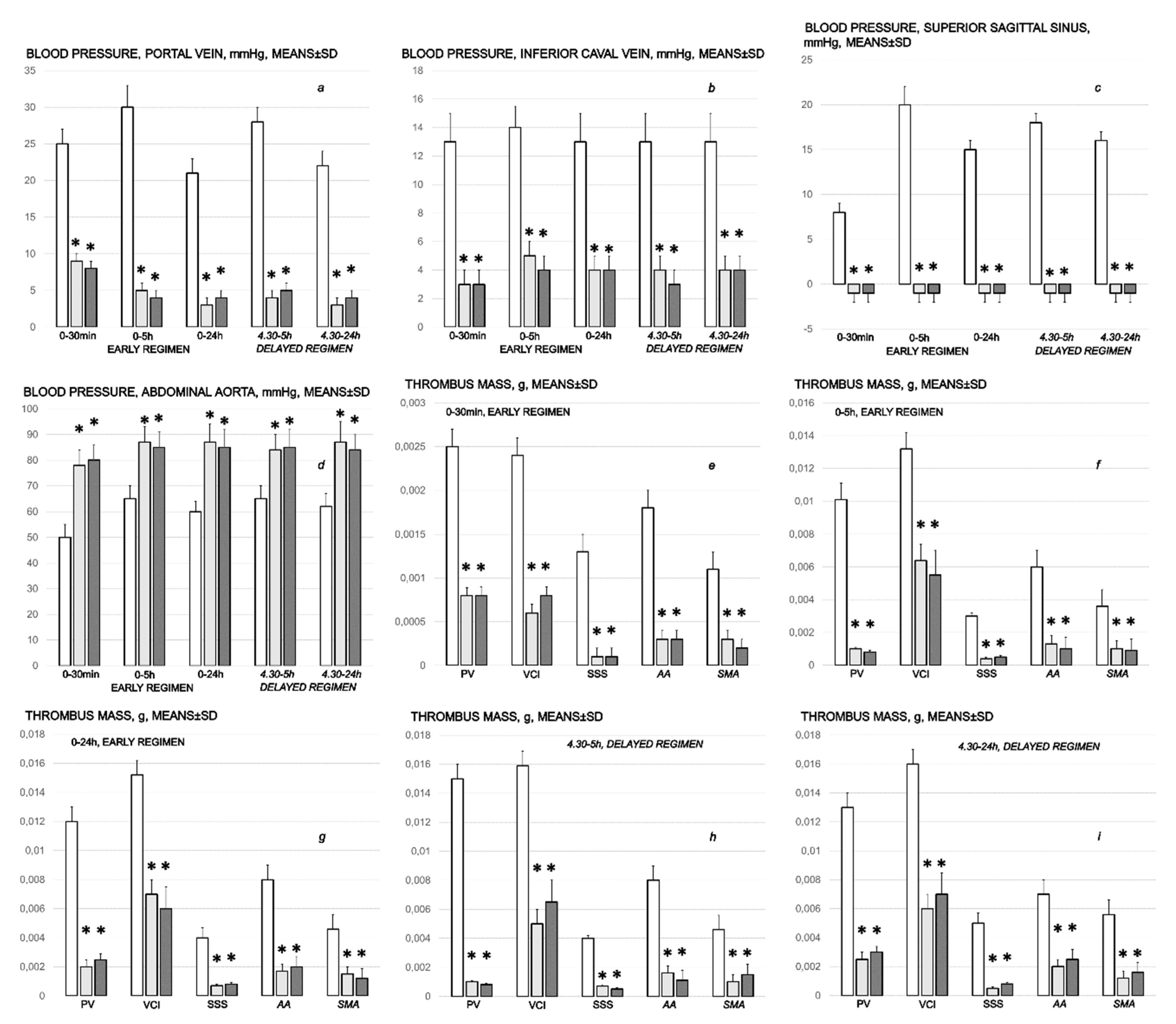
3.1.1. Blood Pressure Disturbances
3.1.2. Thrombosis
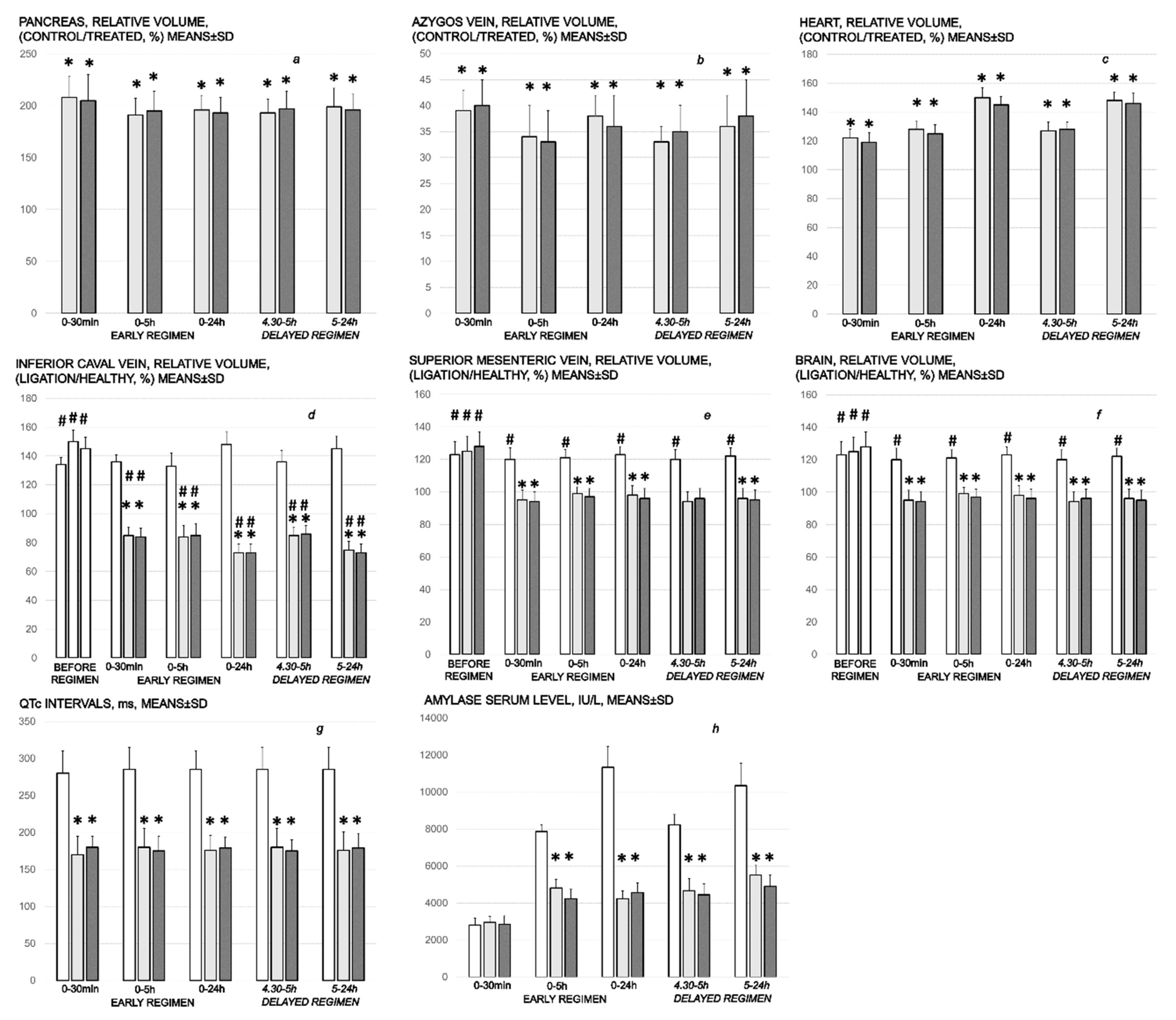
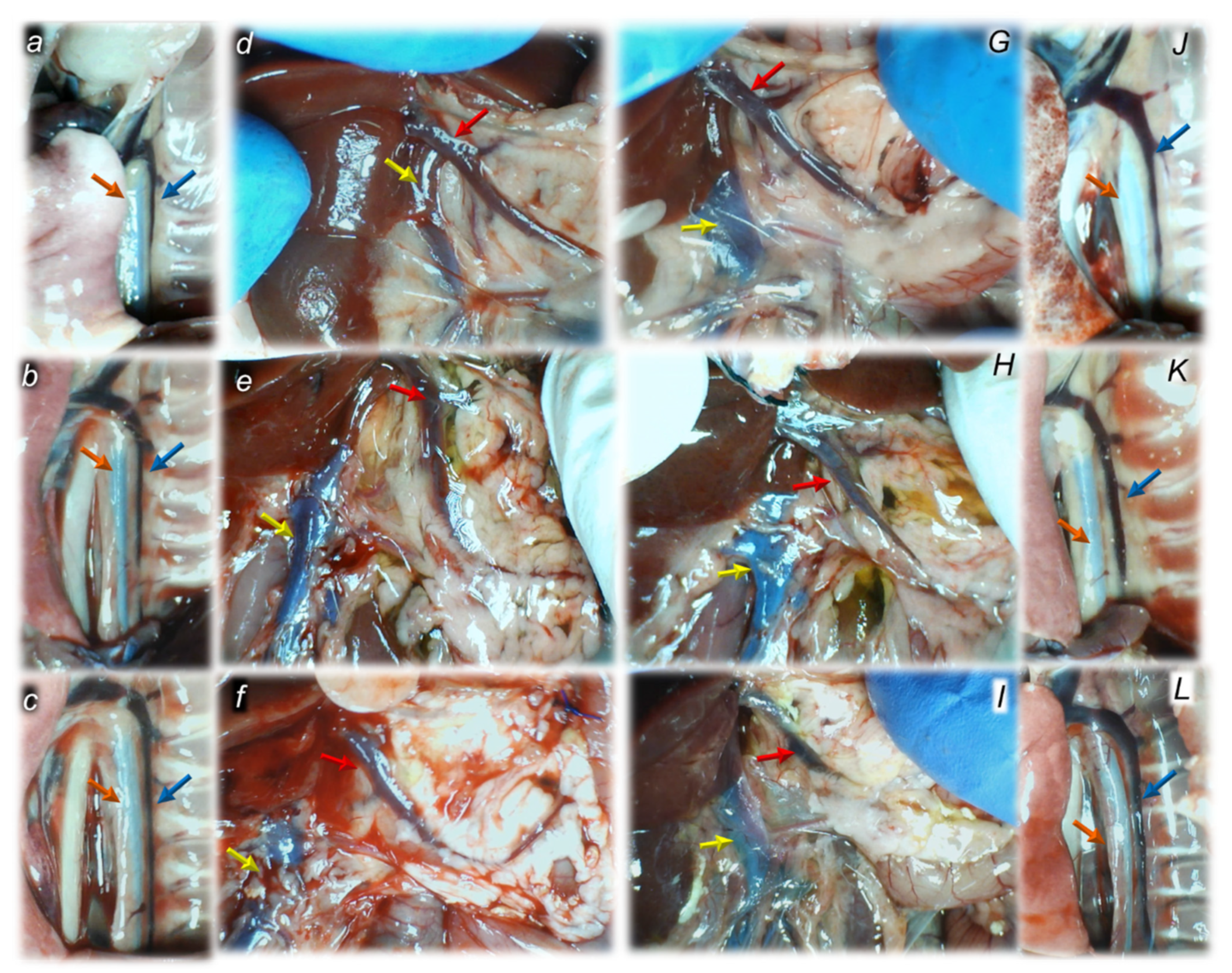
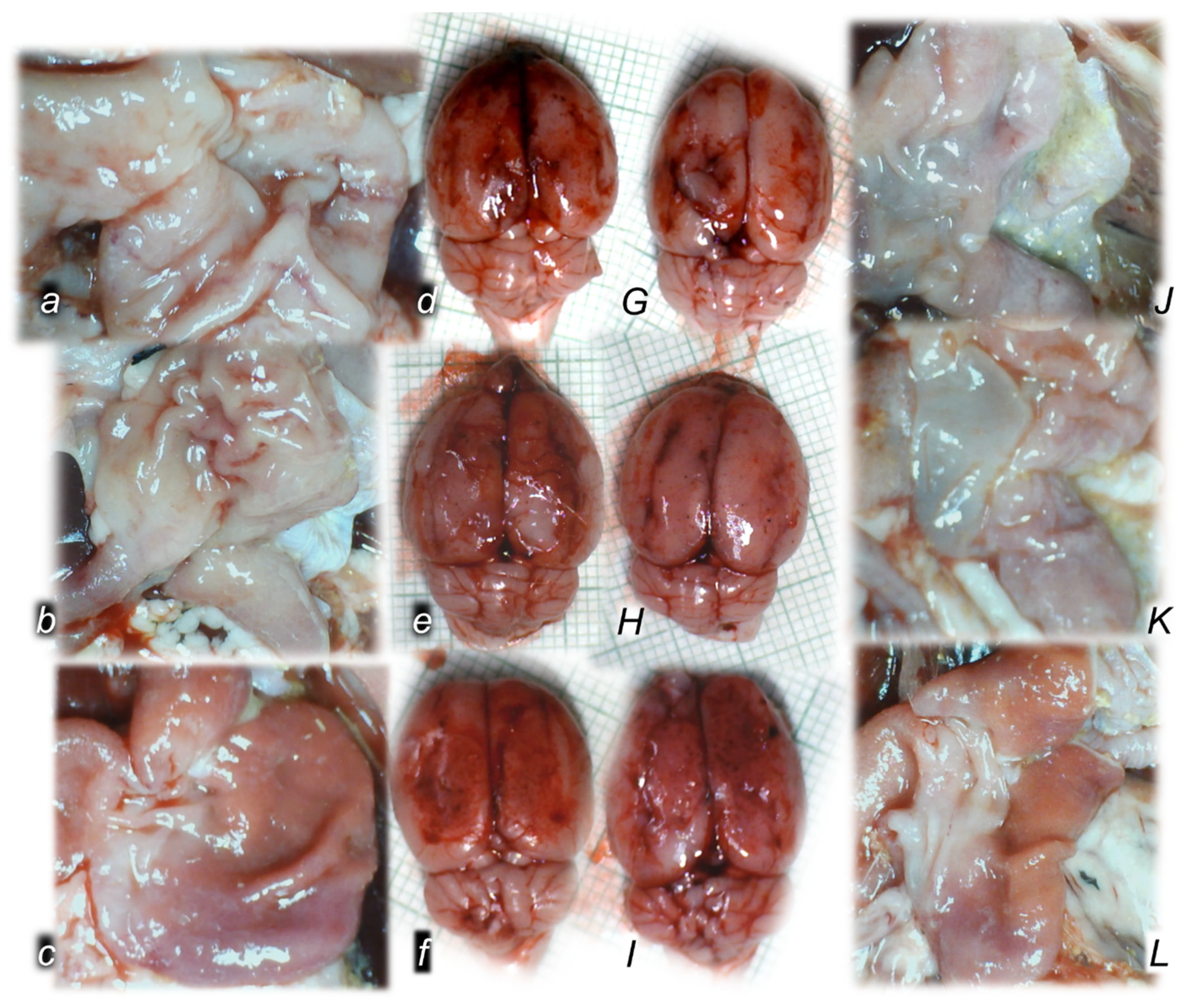
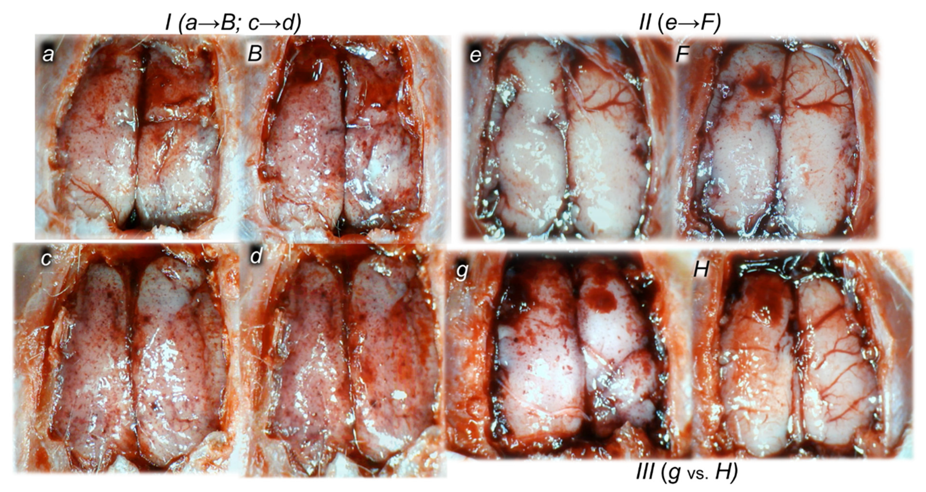
3.1.3. Collateral Pathways, Blood Vessels, and Brain Gross Presentation
3.1.4. Heart and ECG Disturbances
3.1.5. Serum Amylase Level
3.2. A Perilous Syndrome Occurred Peripherally
3.2.1. Pancreas, Gastrointestinal, Lung, Liver, Kidney, and Heart Lesions
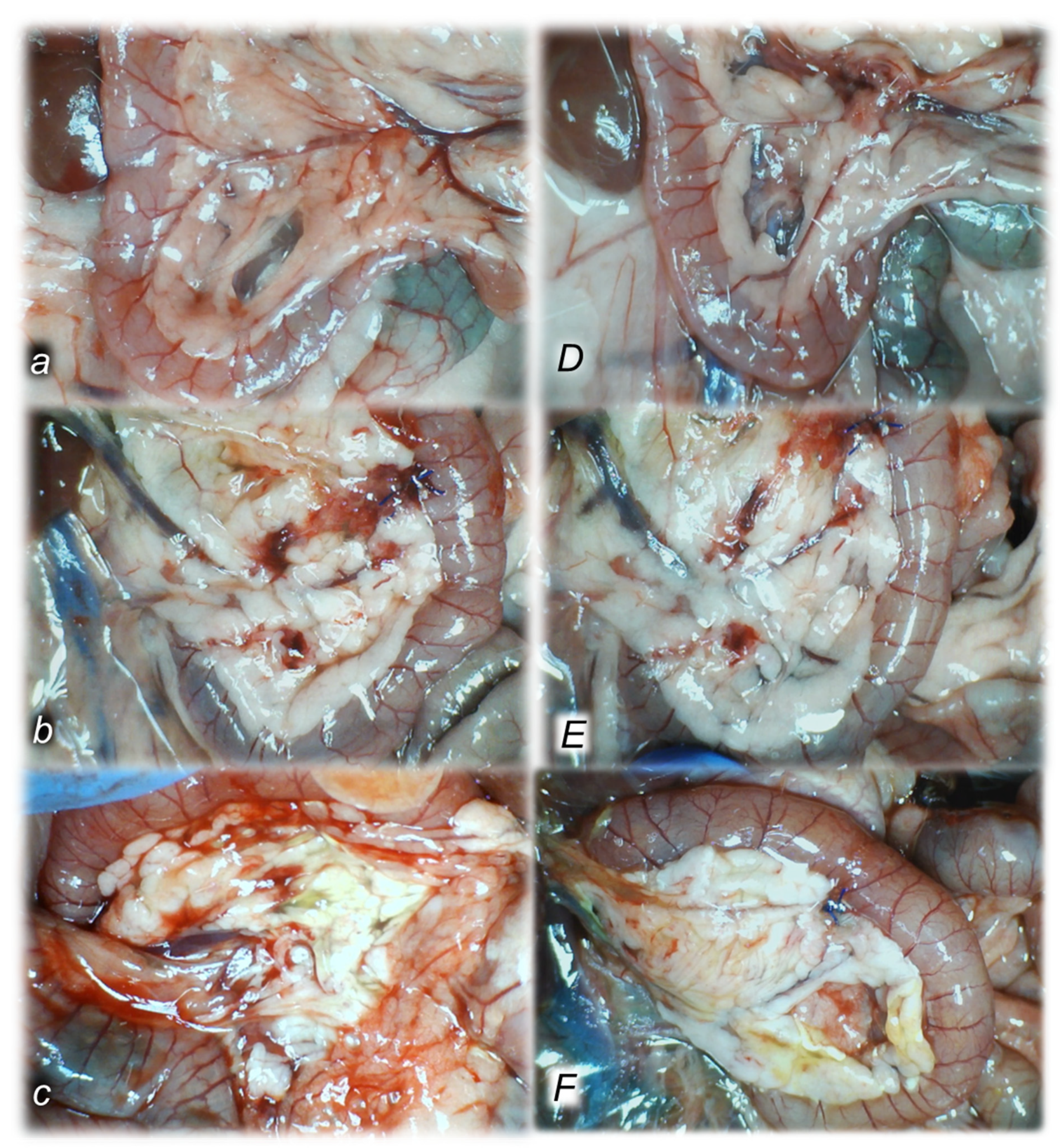
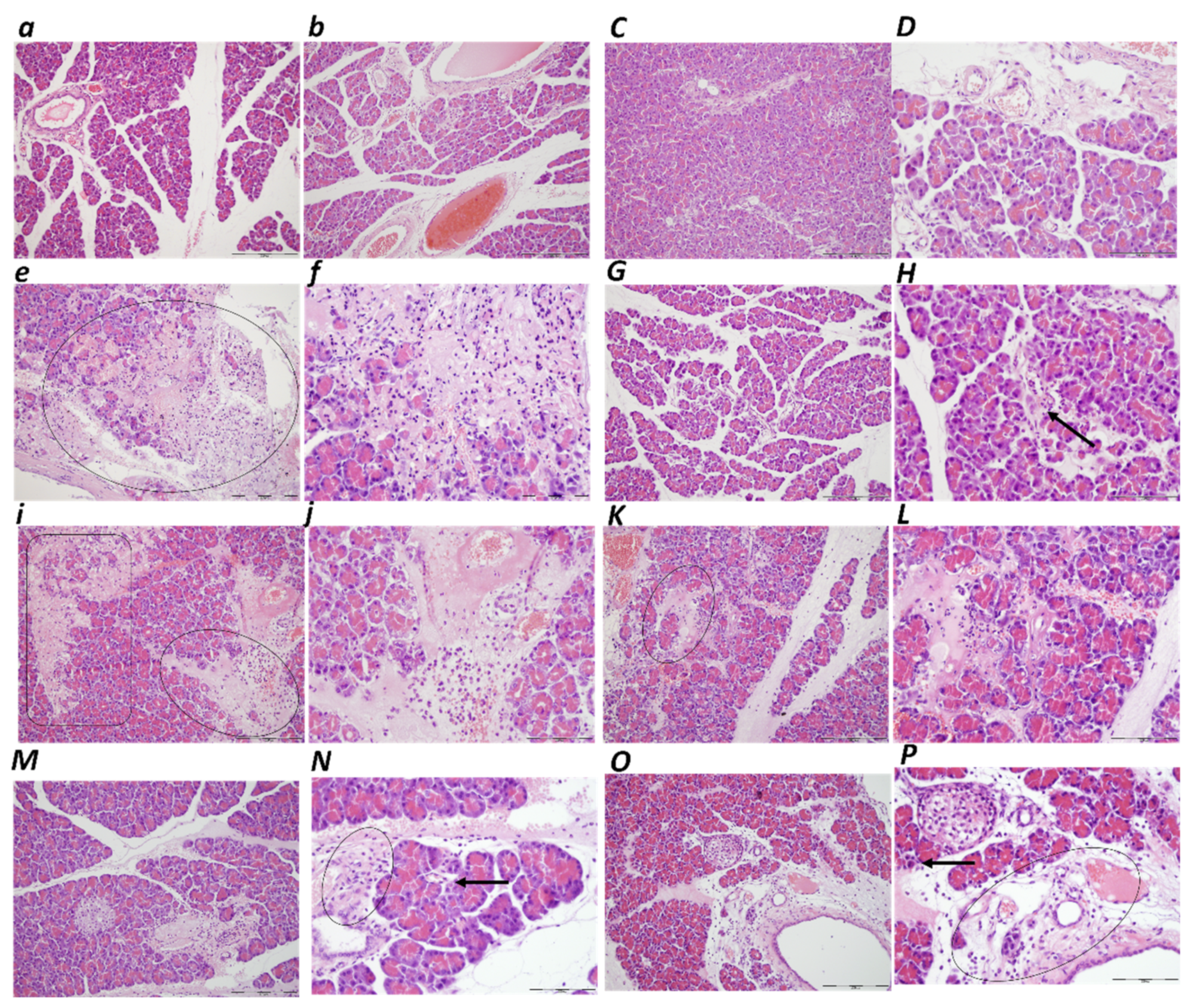
Pancreas
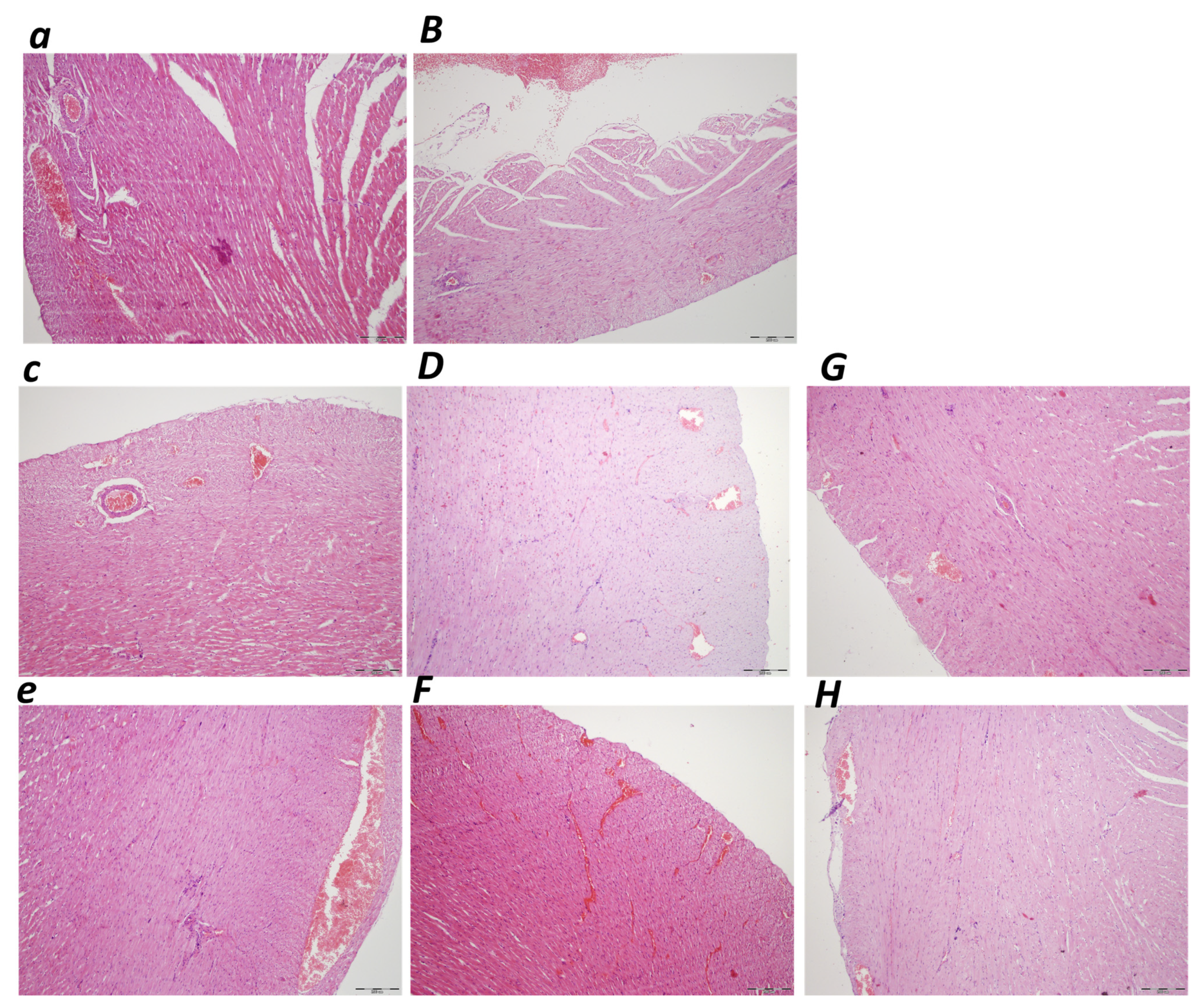
Heart
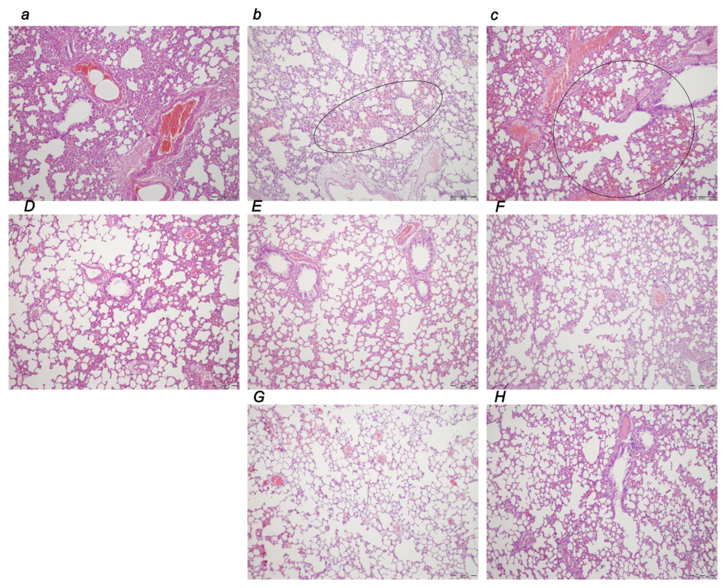
Lung
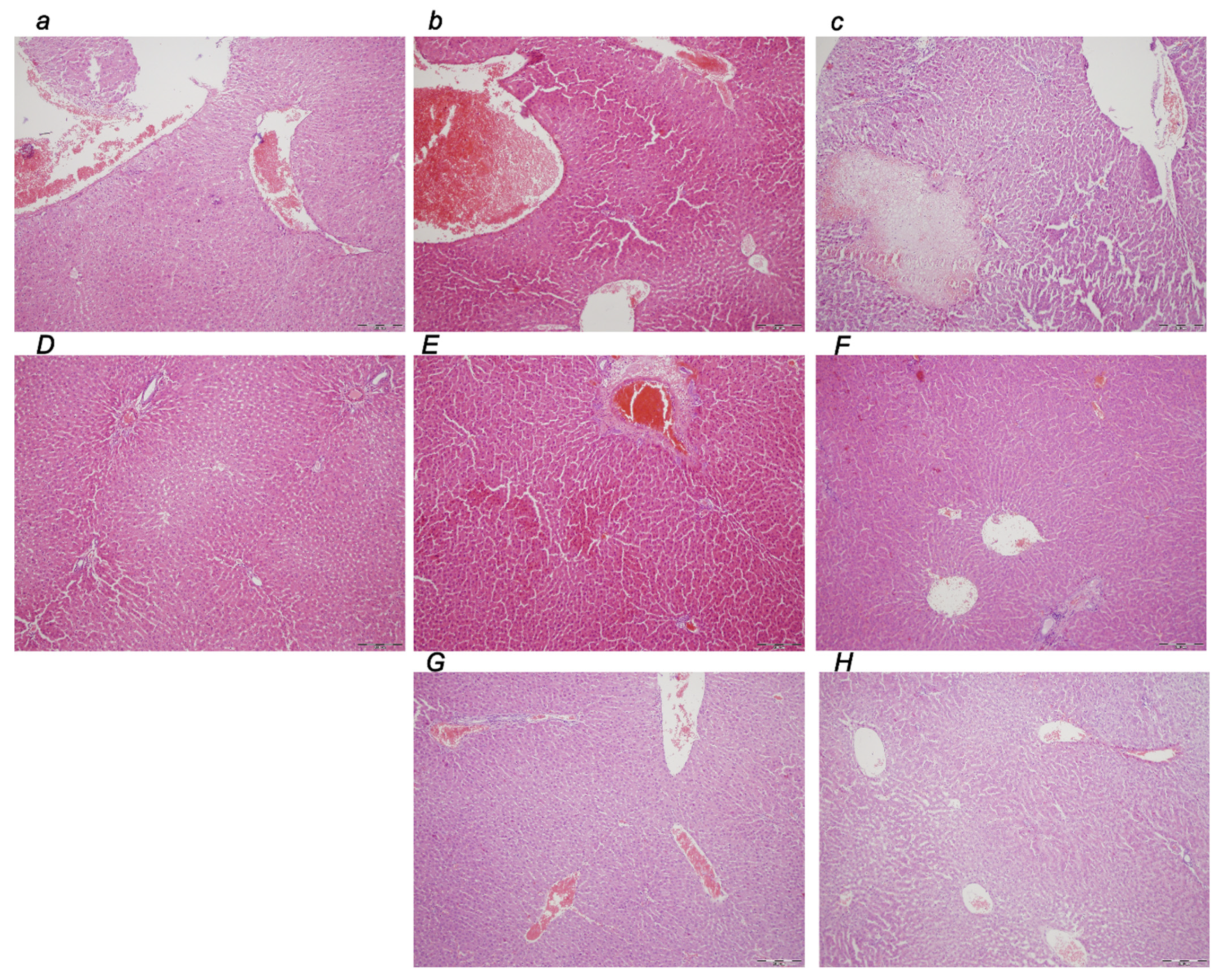
Liver
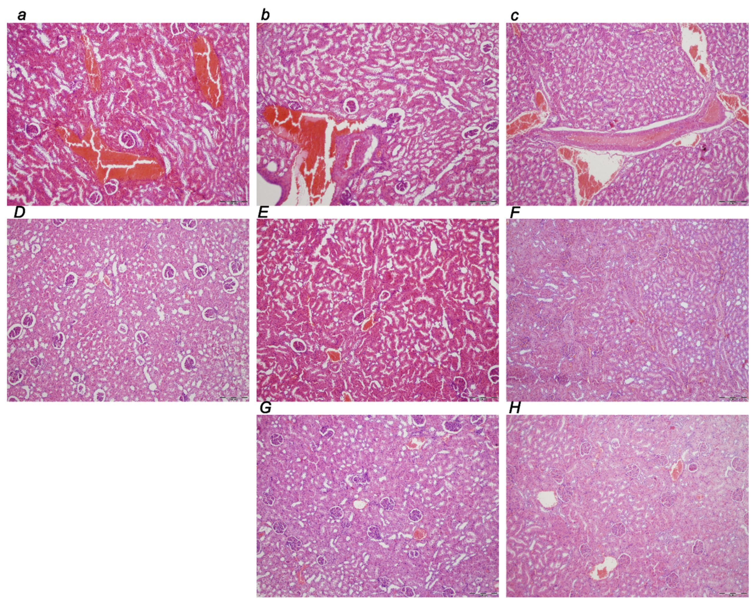
Kidney
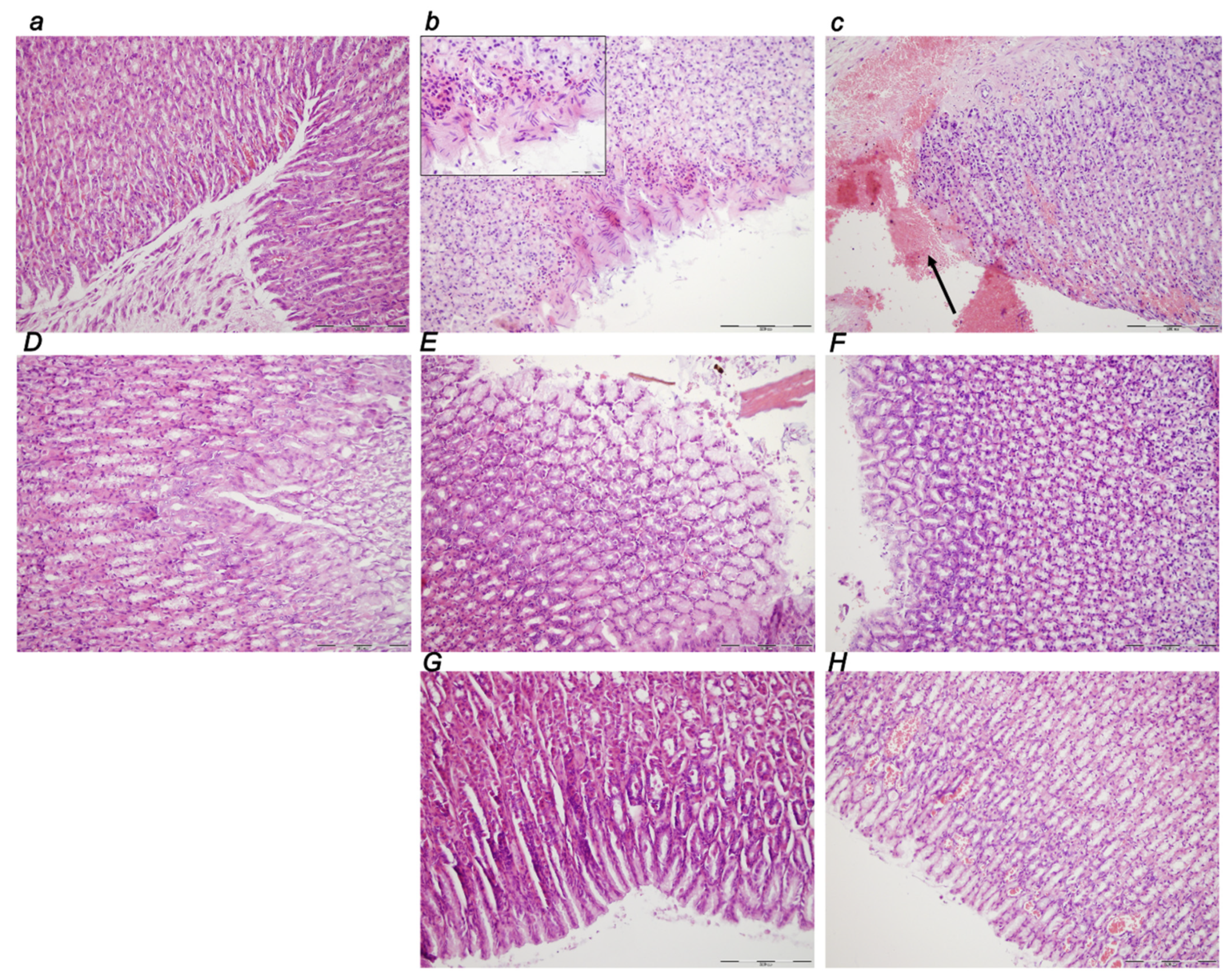
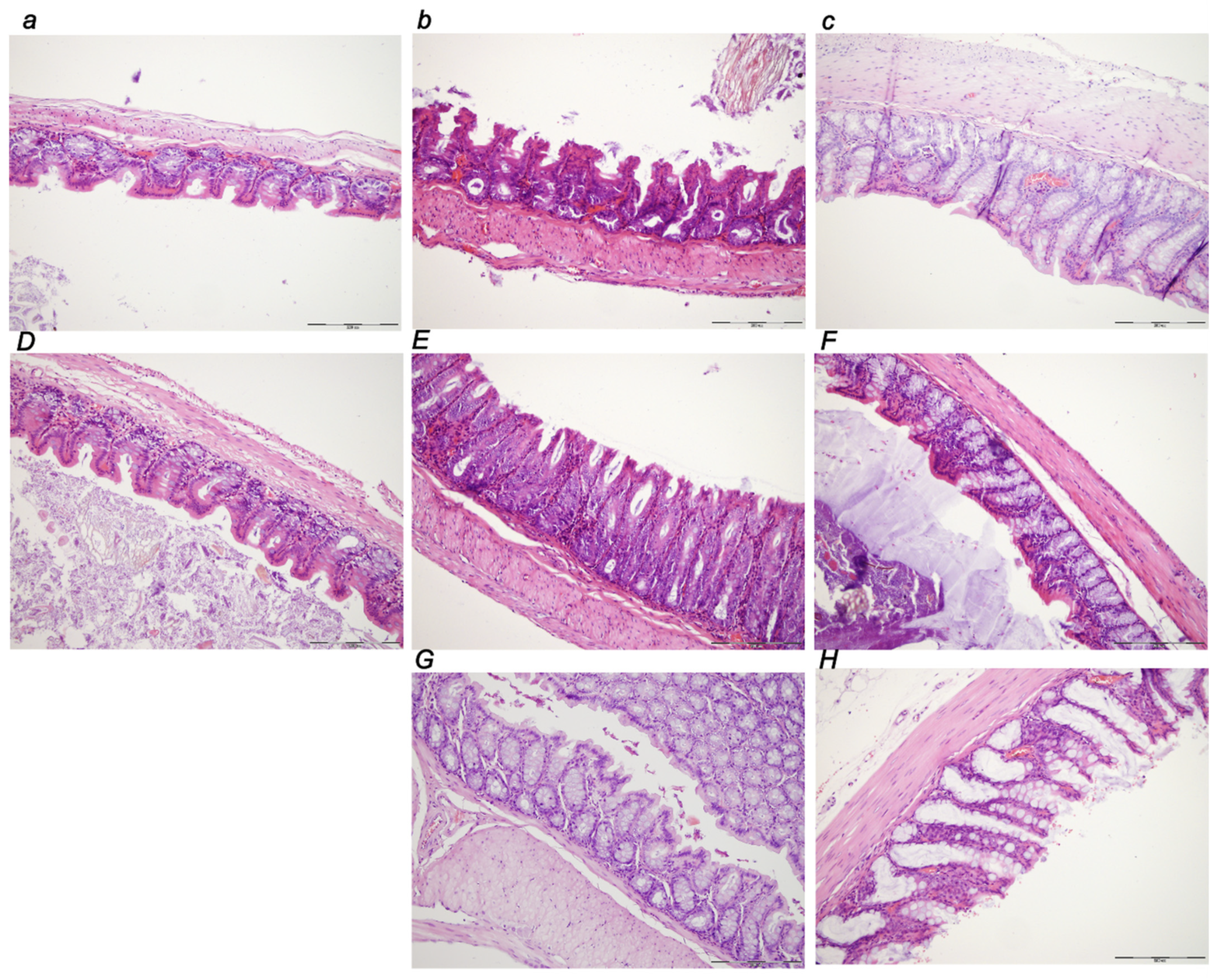
Stomach
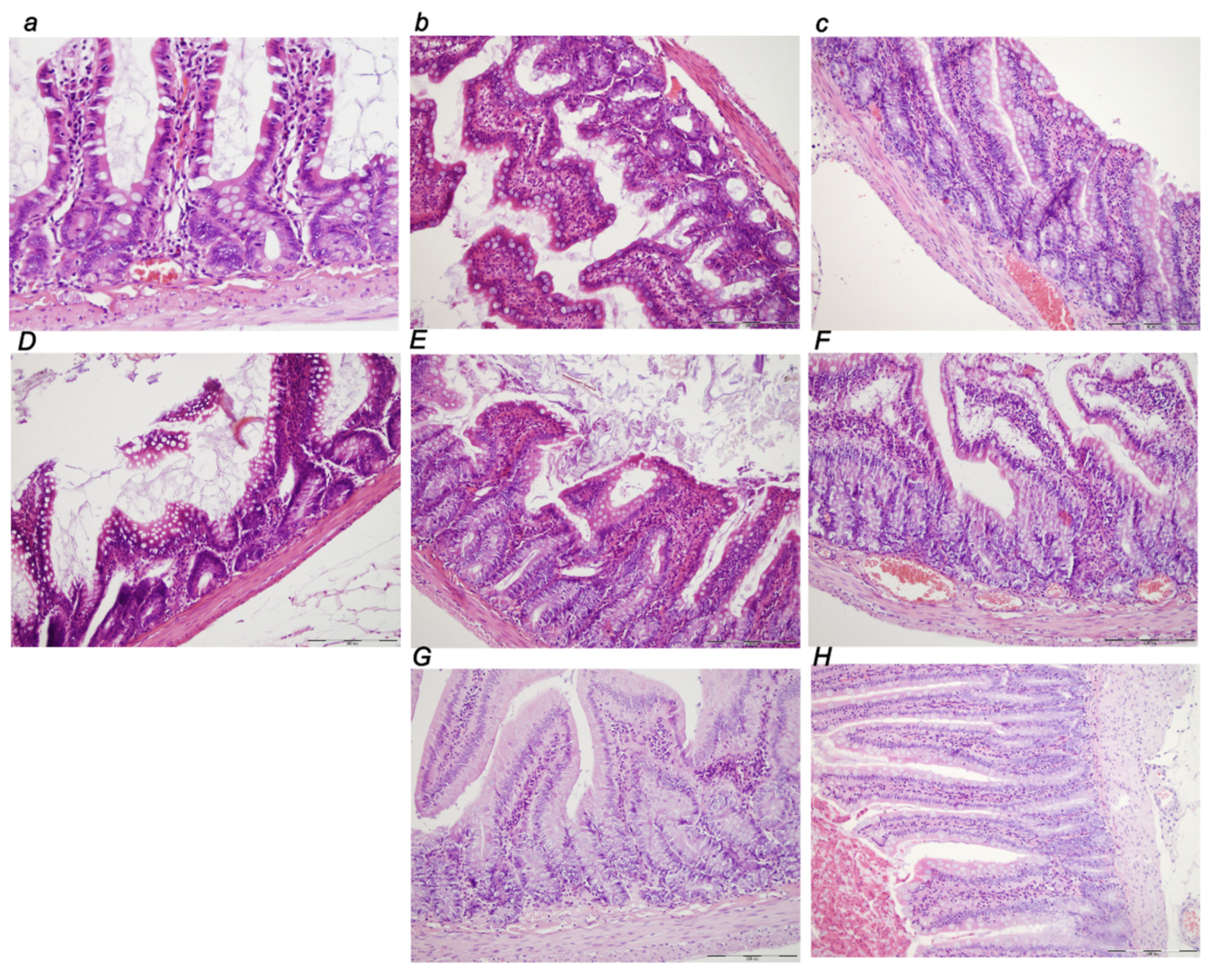
Small Intestine
Colon
3.3. A Perilous Syndrome Occurred Centrally
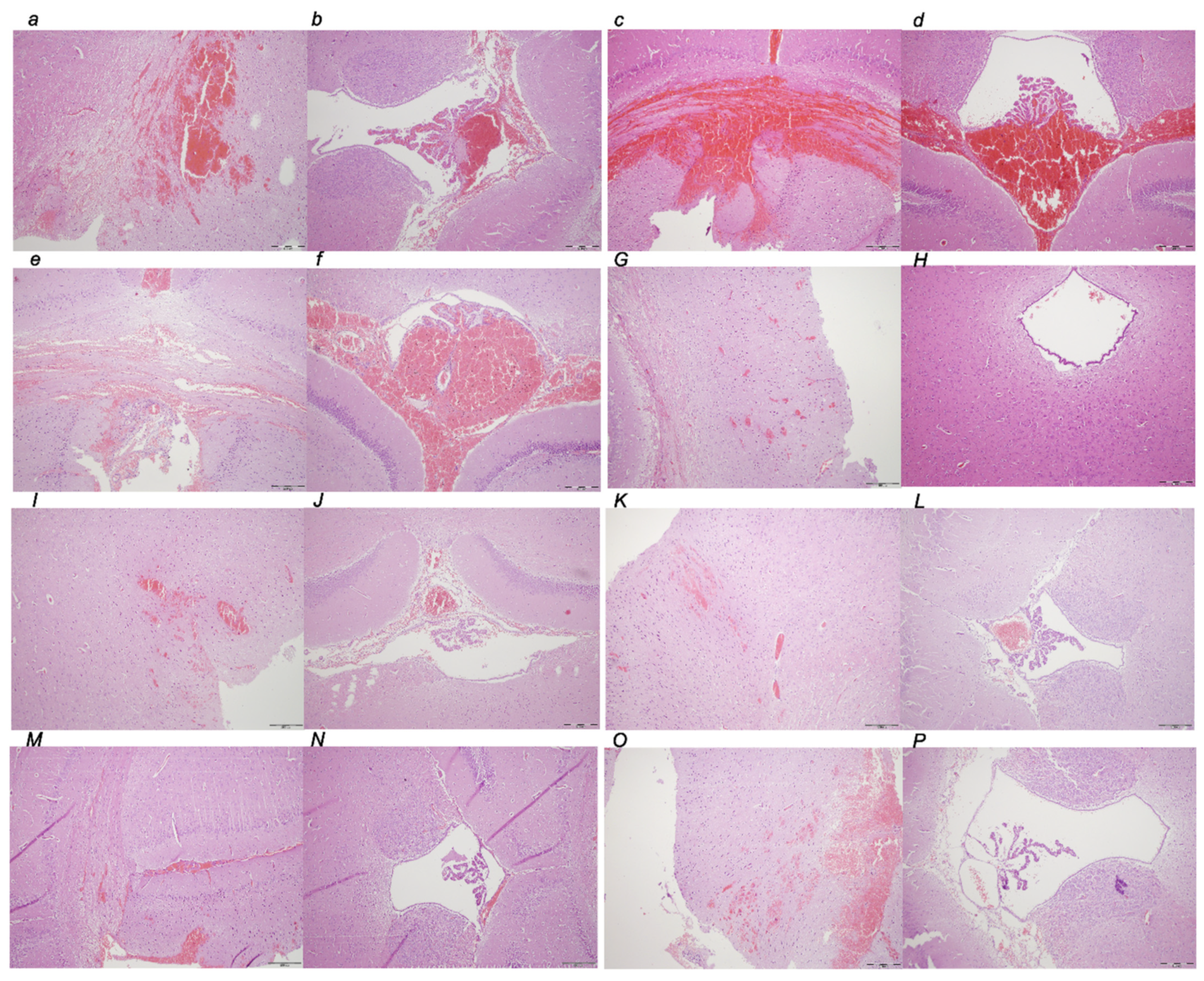
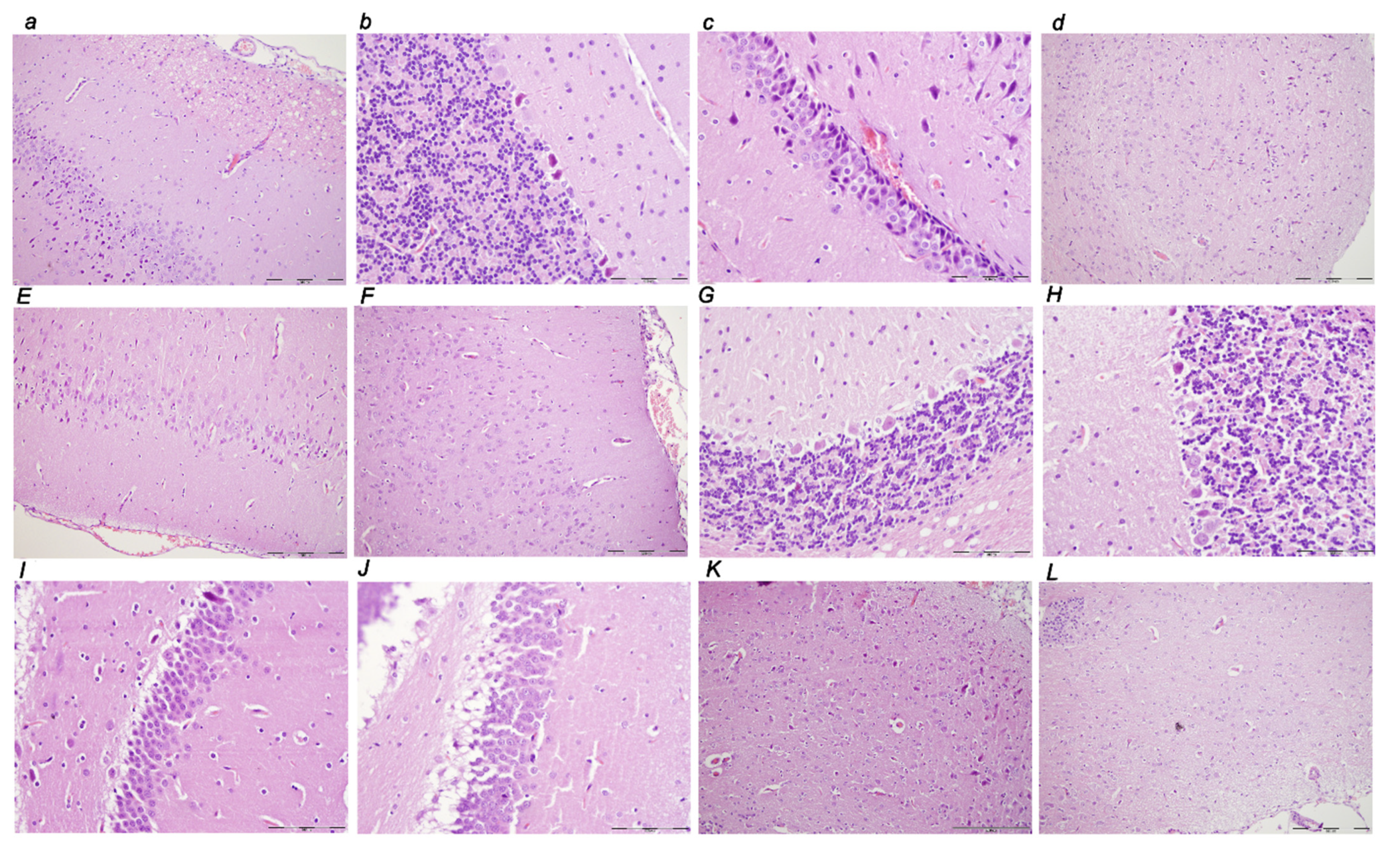
3.3.1. Brain Lesions, Cerebral and Cerebellar Cortex, Hypothalamus/Thalamus, and Hippocampus
3.3.2. Brain Damage
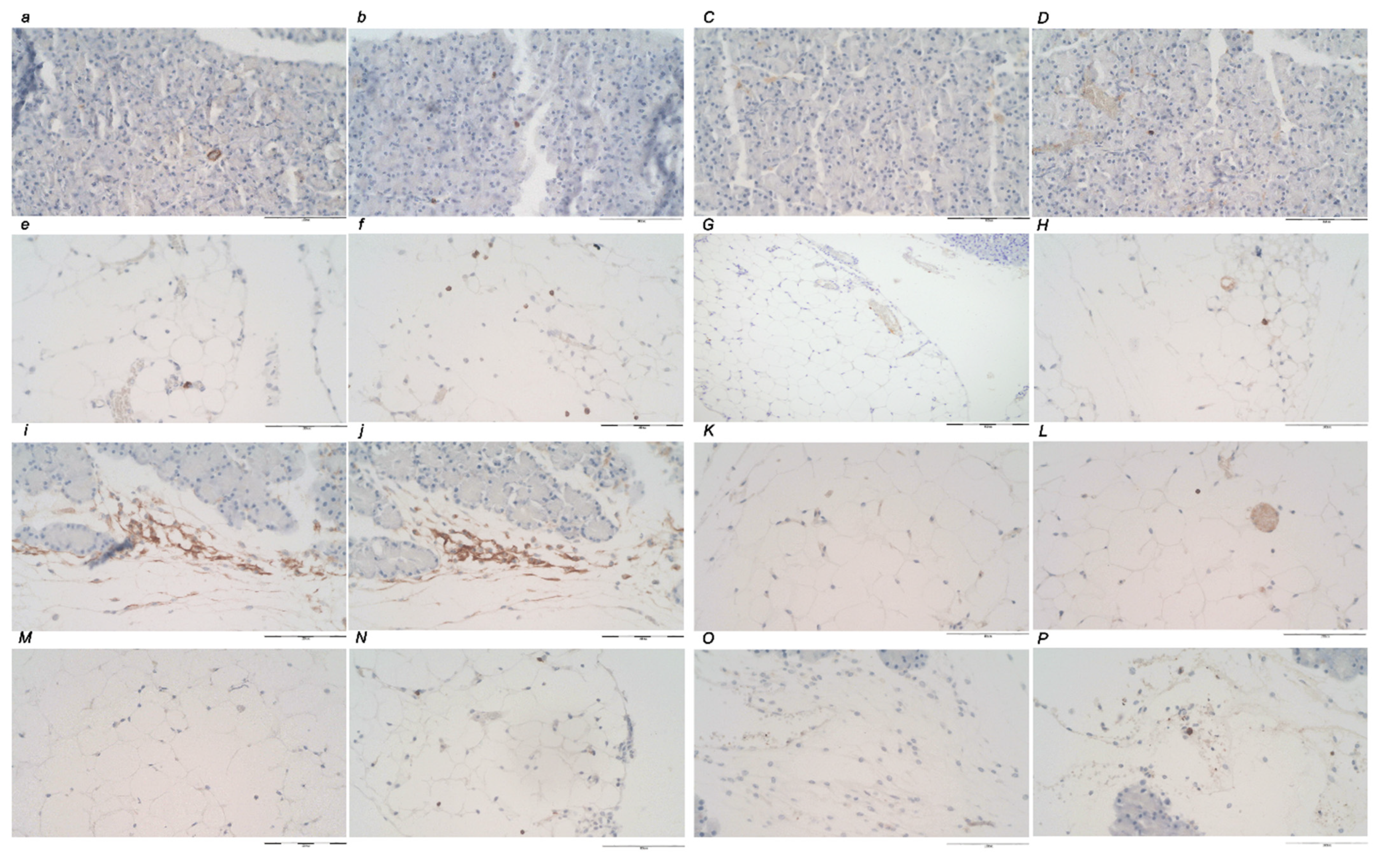
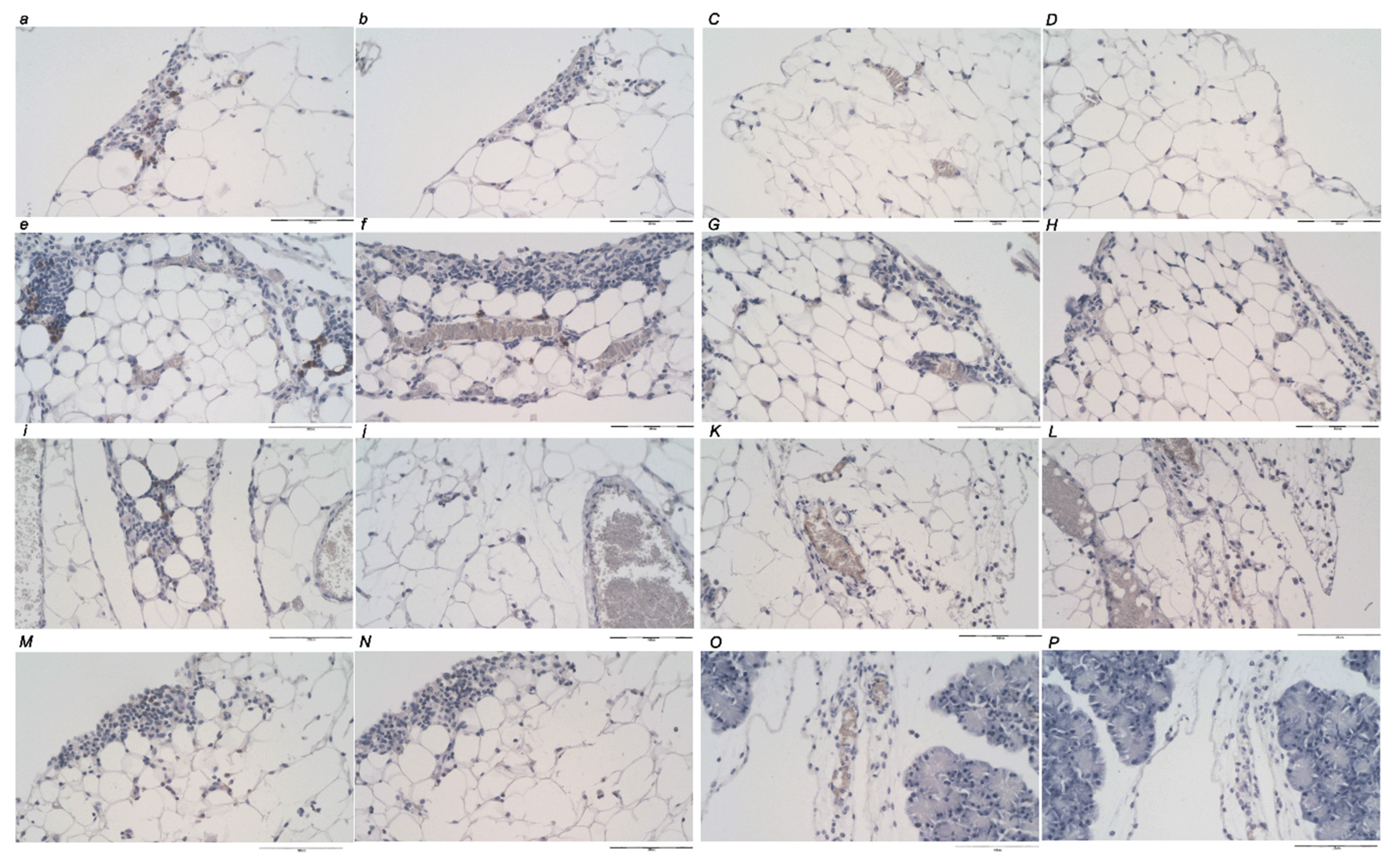
3.4. Immunochemistry
4. Discussion
Author Contributions
Funding
Institutional Review Board Statement
Data Availability Statement
Conflicts of Interest
References
- Vukojevic, J.; Milavic, M.; Perovic, D.; Ilic, S.; Zemba Cilic, A.; Duran, N.; Strbe, S.; Zoricic, Z.; Filipcic, I.; Brecic, P.; et al. Pentadecapeptide BPC 157 and the central nervous system. Neural Regen. Res. 2022, 17, 482–487. [Google Scholar] [CrossRef] [PubMed]
- Seiwerth, S.; Milavic, M.; Vukojevic, J.; Gojkovic, S.; Krezic, I.; Vuletic, L.B.; Pavlov, K.H.; Petrovic, A.; Sikiric, S.; Vranes, H.; et al. Stable gastric pentadecapeptide BPC 157 and wound healing. Front. Pharmacol. 2021, 12, 627533. [Google Scholar] [CrossRef] [PubMed]
- Sikiric, P.; Hahm, K.B.; Blagaic, A.B.; Tvrdeic, A.; Pavlov, K.H.; Petrovic, A.; Kokot, A.; Gojkovic, S.; Krezic, I.; Drmic, D.; et al. Stable gastric pentadecapeptide BPC 157, Robert’s stomach cytoprotection/adaptive cytoprotection/organoprotection, and Selye’s stress coping response: Progress, achievements, and the future. Gut Liver 2020, 14, 153–167. [Google Scholar] [CrossRef] [PubMed]
- Park, J.M.; Lee, H.J.; Sikiric, P.; Hahm, K.B. BPC 157 rescued NSAID-cytotoxicity via stabilizing intestinal permeability and enhancing cytoprotection. Curr. Pharm. Des. 2020, 26, 2971–2981. [Google Scholar] [CrossRef]
- Sikiric, P.; Rucman, R.; Turkovic, B.; Sever, M.; Klicek, R.; Radic, B.; Drmic, D.; Stupnisek, M.; Misic, M.; Vuletic, L.B.; et al. Novel cytoprotective mediator, stable gastric pentadecapeptide BPC 157. Vascular recruitment and gastrointestinal tract healing. Curr. Pharm. Des. 2018, 24, 1990–2001. [Google Scholar] [CrossRef]
- Sikiric, P.; Skrtic, A.; Gojkovic, S.; Krezic, I.; Zizek, H.; Lovric, E.; Sikiric, S.; Knezevic, M.; Strbe, S.; Milavic, M.; et al. Cytoprotective gastric pentadecapeptide BPC 157 resolves major vessel occlusion disturbances, ischemia-reperfusion injury following Pringle maneuver, and Budd-Chiari syndrome. World J. Gastroenterol. 2022, 28, 23–46. [Google Scholar] [CrossRef]
- Deek, S.A. BPC 157 as potential treatment for COVID-19. Med. Hypotheses 2021, 158, 110736. [Google Scholar] [CrossRef]
- Komara, N.L.; Paragomi, P.; Greer, P.J.; Wilson, A.S.; Breze, C.; Papachristou, G.I.; Whitcomb, D.C. Severe acute pancreatitis: Capillary permeability model linking systemic inflammation to multiorgan failure. Am. J. Physiol. Gastrointest. Liver Physiol. 2020, 319, 573–583. [Google Scholar] [CrossRef]
- Yegneswaran, B.; Kostis, J.B.; Pitchumoni, C.S. Cardiovascular manifestations of acute pancreatitis. J. Crit. Care 2011, 26, 225.e11–225.e18. [Google Scholar] [CrossRef]
- Kong, L.; Santiago, N.; Han, T.Q.; Zhang, S.D. Clinical characteristics and prognostic factors of severe acute pancreatitis. World J. Gastroenterol. 2004, 10, 3336–3338. [Google Scholar] [CrossRef]
- Vukojevic, J.; Siroglavic, M.; Kasnik, K.; Kralj, T.; Stancic, D.; Kokot, A.; Kolaric, D.; Drmic, D.; Sever, A.Z.; Barisic, I.; et al. Rat inferior caval vein (ICV) ligature and particular new insights with the stable gastric pentadecapeptide BPC 157. Vascul. Pharmacol. 2018, 106, 54–66. [Google Scholar] [CrossRef] [PubMed]
- Kolovrat, M.; Gojkovic, S.; Krezic, I.; Malekinusic, D.; Vrdoljak, B.; Kasnik Kovac, K.; Kralj, T.; Drmic, D.; Barisic, I.; Horvat Pavlov, K.; et al. Pentadecapeptide BPC 157 resolves Pringle maneuver in rats, both ischemia and reperfusion. World J. Hepatol. 2020, 12, 184–206. [Google Scholar] [CrossRef] [PubMed]
- Gojkovic, S.; Krezic, I.; Vrdoljak, B.; Malekinusic, D.; Barisic, I.; Petrovic, A.; Horvat Pavlov, K.; Kolovrat, M.; Duzel, A.; Knezevic, M.; et al. Pentadecapeptide BPC 157 resolves suprahepatic occlusion of the inferior caval vein, Budd-Chiari syndrome model in rats. World J. Gastrointest. Pathophysiol. 2020, 11, 1–19. [Google Scholar] [CrossRef]
- Knezevic, M.; Gojkovic, S.; Krezic, I.; Zizek, H.; Vranes, H.; Malekinusic, D.; Vrdoljak, B.; Knezevic, T.; Pavlov, K.H.; Drmic, D.; et al. Complex syndrome of the complete occlusion of the end of the superior mesenteric vein, opposed with the stable gastric pentadecapeptide BPC 157 in rats. Biomedicines 2021, 9, 1029. [Google Scholar] [CrossRef] [PubMed]
- Knezevic, M.; Gojkovic, S.; Krezic, I.; Zizek, H.; Malekinusic, D.; Vrdoljak, B.; Knezevic, T.; Vranes, H.; Drmic, D.; Staroveski, M.; et al. Occluded superior mesenteric artery and vein. Therapy with the stable gastric pentadecapeptide BPC 157. Biomedicines 2021, 9, 792. [Google Scholar] [CrossRef] [PubMed]
- Knezevic, M.; Gojkovic, S.; Krezic, I.; Zizek, H.; Malekinusic, D.; Vrdoljak, B.; Vranes, H.; Knezevic, T.; Barisic, I.; Horvat Pavlov, K.; et al. Occlusion of the superior mesenteric artery in rats reversed by collateral pathways activation: Gastric pentadecapeptide BPC 157 therapy counteracts multiple organ dysfunction syndrome; intracranial, portal and caval hypertension; and aortal hypotension. Biomedicines 2021, 9, 609. [Google Scholar] [CrossRef]
- Amic, F.; Drmic, D.; Bilic, Z.; Krezic, I.; Zizek, H.; Peklic, M.; Klicek, R.; Pajtak, A.; Amic, E.; Vidovic, T.; et al. Bypassing major venous occlusion and duodenal lesions in rats, and therapy with the stable gastric pentadecapeptide BPC 157, L-NAME and L-arginine. World J. Gastroenterol. 2018, 24, 5366–5378. [Google Scholar] [CrossRef]
- Duzel, A.; Vlainic, J.; Antunovic, M.; Malekinusic, D.; Vrdoljak, B.; Samara, M.; Gojkovic, S.; Krezic, I.; Vidovic, T.; Bilic, Z.; et al. Stable gastric pentadecapeptide BPC 157 in the treatment of colitis and ischemia and reperfusion in rats: New insights. World J. Gastroenterol. 2017, 23, 8465–8488. [Google Scholar] [CrossRef]
- Kralj, T.; Kokot, A.; Zlatar, M.; Masnec, S.; Kasnik Kovac, K.; Milkovic Perisa, M.; Batelja Vuletic, L.; Giljanovic, A.; Strbe, S.; Sikiric, S.; et al. Stable gastric pentadecapeptide BPC 157 therapy of rat glaucoma. Biomedicines 2021, 10, 89. [Google Scholar] [CrossRef]
- Gojkovic, S.; Krezic, I.; Vranes, H.; Zizek, H.; Drmic, D.; Pavlov, K.H.; Petrovic, A.; Batelja, L.; Milavic, M.; Sikiric, S.; et al. BPC 157 therapy and the permanent occlusion of the superior sagittal sinus in rat. Vascular recruitment. Biomedicines 2021, 9, 744. [Google Scholar] [CrossRef]
- Vukojevic, J.; Vrdoljak, B.; Malekinusic, D.; Siroglavic, M.; Milavic, M.; Kolenc, D.; Boban Blagaic, A.; Bateljam, L.; Drmic, D.; Seiwerth, S.; et al. The effect of pentadecapeptide BPC 157 on hippocampal ischemia/reperfusion injuries in rats. Brain Behav. 2020, 10, e01726. [Google Scholar] [CrossRef] [PubMed]
- Tepes, M.; Gojkovic, S.; Krezic, I.; Zizek, H.; Madzar, Z.; Santak, G.; Batelja, L.; Milavic, M.; Sikiric, S.; Kocman, I.; et al. Stable gastric pentadecapeptide BPC 157 therapy for primary abdominal compartment syndrome in rats. Front. Pharmacol. 2021, 12, 718147. [Google Scholar] [CrossRef] [PubMed]
- Barisic, I.; Balenovic, D.; Udovicic, M.; Bardak, D.; Strinic, D.; Vlainic, J.; Vranes, H.; Smoday, I.M.; Krezic, I.; Milavic, M.; et al. Stable gastric pentadecapeptide BPC 157 may counteract myocardial infarction induced by isoprenaline in rats. Biomedicines 2022, 10, 265. [Google Scholar] [CrossRef] [PubMed]
- Strbe, S.; Gojkovic, S.; Krezic, I.; Zizek, H.; Vranes, H.; Barisic, I.; Strinic, D.; Orct, T.; Vukojevic, J.; Ilic, S.; et al. Over-dose lithium toxicity as an occlusive-like syndrome in rats and gastric pentadecapeptide BPC 157. Biomedicines 2021, 9, 1506. [Google Scholar] [CrossRef]
- Gojkovic, S.; Krezic, I.; Vranes, H.; Zizek, H.; Drmic, D.; Batelja Vuletic, L.; Milavic, M.; Sikiric, S.; Stilinovic, I.; Simeon, P.; et al. Robert’s intragastric alcohol-induced gastric lesion model as an escalated general peripheral and central syndrome, counteracted by the stable gastric pentadecapeptide BPC 157. Biomedicines 2021, 9, 1300. [Google Scholar] [CrossRef]
- Drmic, D.; Samara, M.; Vidovic, T.; Malekinusic, D.; Antunovic, M.; Vrdoljak, B.; Ruzman, J.; Milkovic, P.M.; Horvat, K.P.; Jeyakumar, J.; et al. Counteraction of perforated cecum lesions in rats: Effects of pentadecapeptide BPC 157, L-NAME and L-arginine. World J. Gastroenterol. 2018, 24, 5462–5476. [Google Scholar] [CrossRef]
- Bilic, Z.; Gojkovic, S.; Kalogjera, L.; Krezic, I.; Malekinusic, D.; Knezevic, M.; Sever, M.; Lojo, N.; Kokot, A.; Kasnik, K.; et al. Novel insight into Robert’s cytoprotection: Complex therapeutic effect of cytoprotective pentadecapeptide pentadecapeptide BPC 157 in rats with perforated stomach throughout modulation of nitric oxide-system. Comparison with L-arginine, ranitidine and pantoprazole therapy and L-NG-nitro-L-arginine methyl ester worsening. J. Physiol. Pharmacol. 2021, 72, 940–955. [Google Scholar]
- Cesar, L.B.; Gojkovic, S.; Krezic, I.; Malekinusic, D.; Zizek, H.; Vuletic, L.B.; Petrovic, A.; Pavlov, K.H.; Drmic, D.; Kokot, A.; et al. Bowel adhesion and therapy with the stable gastric pentadecapeptide BPC 157, L-NAME and L-arginine in rats. World J. Gastrointest. Pharmacol. Ther. 2020, 11, 93–109. [Google Scholar] [CrossRef]
- Sikirić, P.; Seiwerth, S.; Grabarević, Z.; Rucman, R.; Petek, M.; Jagić, V.; Turković, B.; Rotkvić, I.; Mise, S.; Zoricić, I.; et al. Salutary and prophylactic effect of pentadecapeptide BPC 157 on acute pancreatitis and concomitant gastroduodenal lesions in rats. Dig. Dis. Sci. 1996, 41, 1518–1526. [Google Scholar] [CrossRef]
- Petrovic, I.; Dobric, I.; Drmic, D.; Sever, M.; Klicek, R.; Radic, B.; Brcic, L.; Kolenc, D.; Zlatar, M.; Kunjko, K.; et al. BPC 157 therapy to detriment sphincters failure-esophagitis-pancreatitis in rat and acute pancreatitis patients low sphincters pressure. J. Physiol. Pharmacol. 2011, 62, 527–534. [Google Scholar]
- Sever, A.Z.; Sever, M.; Vidovic, T.; Lojo, N.; Kolenc, D.; Vuletic, L.B.; Drmic, D.; Kokot, A.; Zoricic, I.; Coric, M.; et al. Stable gastric pentadecapeptide BPC 157 in the therapy of the rats with bile duct ligation. Eur. J. Pharmacol. 2019, 847, 130–142. [Google Scholar] [CrossRef] [PubMed]
- Robert, A. Cytoprotection by prostaglandins. Gastroenterology 1979, 77, 761–767. [Google Scholar] [CrossRef]
- Szabo, S. Mechanism of mucosal protection. In Gastric Cytoprotection: A Clinician’s Guide; Hollander, D., Tarnawski, A., Eds.; Plenum Medical Book Co.: New York, NY, USA, 1989; pp. 49–90. [Google Scholar]
- Szabo, S.; Trier, J.S.; Brown, A.; Schnoor, J. Early vascular injury and increased vascular permeability in gastric mucosal injury caused by ethanol in the rat. Gastroenterology 1985, 88, 228–236. [Google Scholar] [CrossRef]
- Sikiric, P.; Seiwerth, S.; Rucman, R.; Turkovic, B.; Rokotov, D.S.; Brcic, L.; Sever, M.; Klicek, R.; Radic, B.; Drmic, D.; et al. Toxicity by NSAIDs: Counteraction by stable gastric pentadecapeptide BPC 157. Curr. Pharm. Des. 2013, 19, 76–83. [Google Scholar] [PubMed]
- Luetic, K.; Sucic, M.; Vlainic, J.; Halle, Z.B.; Strinic, D.; Vidovic, T.; Luetic, F.; Marusic, M.; Gulic, S.; Pavelic, T.T.; et al. Cyclophosphamide induced stomach and duodenal lesions as a NO-system disturbance in rats: L-NAME, L-arginine, stable gastric pentadecapeptide BPC 157. Inflammopharmacology 2017, 25, 255–264. [Google Scholar] [CrossRef]
- Sucic, M.; Luetic, K.; Jandric, I.; Drmic, D.; Sever, A.Z.; Vuletic, L.B.; Halle, Z.B.; Strinic, D.; Kokot, A.; Seiwerth, R.S.; et al. Therapy of the rat hemorrhagic cystitis induced by cyclophosphamide. Stable gastric pentadecapeptide BPC 157, L-arginine, L-NAME. Eur. J. Pharmacol. 2019, 861, 172593. [Google Scholar] [CrossRef]
- Belosic Halle, Z.; Vlainic, J.; Drmic, D.; Strinic, D.; Luetic, K.; Sucic, M.; Medvidovic-Grubisic, M.; Pavelic Turudic, T.; Petrovic, I.; Seiwerth, S.; et al. Class side effects: Decreased pressure in the lower oesophageal and the pyloric sphincters after the administration of dopamine antagonists, neuroleptics, anti-emetics, L-NAME, pentadecapeptide BPC 157 and L-arginine. Inflammopharmacology 2017, 25, 511–522. [Google Scholar] [CrossRef]
- Sikiric, P.; Seiwerth, S.; Rucman, R.; Turkovic, B.; Rokotov, D.S.; Brcic, L.; Sever, M.; Klicek, R.; Radic, B.; Drmic, D.; et al. Stable gastric pentadecapeptide BPC 157-NO-system relation. Curr. Pharm. Des. 2014, 20, 1126–1135. [Google Scholar] [CrossRef]
- Sikiric, P.; Seiwerth, S.; Grabarevic, Z.; Rucman, R.; Petek, M.; Jagic, V.; Turkovic, B.; Rotkvic, I.; Mise, S.; Zoricic, I.; et al. The influence of a novel pentadecapeptide, BPC 157, on N(G)-nitro-L-arginine methylester and L-arginine effects on stomach mucosa integrity and blood pressure. Eur. J. Pharmacol. 1997, 332, 23–33. [Google Scholar] [CrossRef]
- Turkovic, B.; Sikiric, P.; Seiwerth, S.; Mise, S.; Anic, T.; Petek, M. Stable gastric pentadecapeptide BPC 157 studied for inflammatory bowel disease (PLD-116, PL14736, Pliva) induces nitric oxide synthesis. Gastroenterology 2004, 126, 287. [Google Scholar]
- Stupnisek, M.; Kokot, A.; Drmic, D.; Hrelec Patrlj, M.; Zenko Sever, A.; Kolenc, D.; Radic, B.; Suran, J.; Bojic, D.; Vcev, A.; et al. Pentadecapeptide BPC 157 reduces bleeding and thrombocytopenia after amputation in rats treated with heparin, warfarin, L-NAME and L-arginine. PLoS ONE 2015, 10, e0123454. [Google Scholar] [CrossRef] [PubMed]
- Stupnisek, M.; Franjic, S.; Drmic, D.; Hrelec, M.; Kolenc, D.; Radic, B.; Bojic, D.; Vcev, A.; Seiwerth, S.; Sikiric, P. Pentadecapeptide BPC 157 reduces bleeding time and thrombocytopenia after amputation in rats treated with heparin, warfarin or aspirin. Thromb. Res. 2012, 129, 652–659. [Google Scholar] [CrossRef] [PubMed]
- Konosic, S.; Petricevic, M.; Ivancan, V.; Konosic, L.; Goluza, E.; Krtalic, B.; Drmic, D.; Stupnisek, M.; Seiwerth, S.; Sikiric, P. Intragastric application of aspirin, clopidogrel, cilostazol, and BPC 157 in rats: Platelet aggregation and blood clot. Oxid. Med. Cell. Longev. 2019, 2019, 9084643. [Google Scholar] [CrossRef] [PubMed]
- Chang, C.H.; Tsai, W.C.; Lin, M.S.; Hsu, Y.H.; Pang, J.H.S. The promoting effect of pentadecapeptide BPC 157 on tendon healing involves tendon outgrowth, cell survival, and cell migration. J. Appl. Physiol. 2011, 110, 774–780. [Google Scholar] [CrossRef] [Green Version]
- Chang, C.H.; Tsai, W.C.; Hsu, Y.H.; Pang, J.H.S. Pentadecapeptide BPC 157 enhances the growth hormone receptor expression in tendon fibroblasts. Molecules 2014, 19, 19066–19077. [Google Scholar] [CrossRef] [Green Version]
- Huang, T.; Zhang, K.; Sun, L.; Xue, X.; Zhang, C.; Shu, Z.; Mu, N.; Gu, J.; Zhang, W.; Wang, Y.; et al. Body protective compound-157 enhances alkali-burn wound healing in vivo and promotes proliferation, migration, and angiogenesis in vitro. Drug Des. Dev. Ther. 2015, 9, 2485–2499. [Google Scholar] [CrossRef] [Green Version]
- Hsieh, M.J.; Lee, C.H.; Chueh, H.Y.; Chang, G.J.; Huang, H.Y.; Lin, Y.; Pang, J.S. Modulatory effects of BPC 157 on vasomotor tone and the activation of Src-Caveolin-1-endothelial nitric oxide synthase pathway. Sci. Rep. 2020, 10, 17078. [Google Scholar] [CrossRef]
- Hsieh, M.J.; Liu, H.T.; Wang, C.N.; Huang, H.Y.; Lin, Y.; Ko, Y.S.; Wang, J.S.; Chang, V.H.; Pang, J.S. Therapeutic potential of pro-angiogenic BPC157 is associated with VEGFR2 activation and up-regulation. J. Mol. Med. 2017, 95, 323–333. [Google Scholar] [CrossRef]
- Kang, E.A.; Han, Y.M.; An, J.M.; Park, Y.J.; Sikiric, P.; Kim, D.H.; Kwon, K.A.; Kim, Y.J.; Yang, D.; Tchah, H.; et al. BPC157 as potential agent rescuing from cancer cachexia. Curr. Pharm. Des. 2018, 24, 1947–1956. [Google Scholar] [CrossRef]
- Wang, X.Y.; Qu, M.; Duan, R.; Shi, D.; Jin, L.; Gao, J.; Wood, J.D.; Li, J.; Wang, G.D. Cytoprotective mechanism of the novel gastric peptide BPC157 in gastrointestinal tract and cultured enteric neurons and glial cells. Neurosci. Bull. 2019, 35, 167–170. [Google Scholar] [CrossRef]
- Chui, C.J.; McArdle, A.H.; Brown, R.; Scott, H.; Gurd, F. Intestinal mucosal lesion in low-flow states. Arch. Surg. 1970, 101, 478–483. [Google Scholar] [CrossRef]
- Lane, J.S.; Todd, K.E.; Lewis, M.P.; Gloor, B.; Ashley, S.W.; Reber, H.A.; McFadden, D.W.; Chandler, C.F. Interleukin-10 reduces the systemic inflammatory response in a murine model of intestinal ischemia/reperfusion. Surgery 1997, 122, 288–294. [Google Scholar] [CrossRef]
- Schmidt, J.; Lewandrowski, K.; Fernandez-del Castillo, C.; Mandavilli, U.; Compton, C.C.; Warshaw, A.L.; Rattner, D.W. Histopathologic correlates of serum amylase activity in acute experimental pancreatitis. Dig. Dis. Sci. 1992, 37, 1426–1433. [Google Scholar] [CrossRef] [PubMed]
- Schmidt, J.; Ebeling, D.; Ryschich, E.; Werner, J.; Gebhard, M.M.; Klar, E. Pancreatic capillary blood flow in an improved model of necrotizing pancreatitis in the rat. J. Surg. Res. 2002, 106, 335–341. [Google Scholar] [CrossRef] [PubMed]
- Perovic, D.; Kolenc, D.; Bilic, V.; Somun, N.; Drmic, D.; Elabjer, E.; Buljat, G.; Seiwerth, S.; Sikiric, P. Stable gastric pentadecapeptide BPC 157 can improve the healing course of spinal cord injury and lead to functional recovery in rats. J. Orthop. Surg. Res. 2019, 14, 199. [Google Scholar] [CrossRef] [PubMed]
- Perovic, D.; Milavic, M.; Dokuzovic, S.; Krezic, I.; Gojkovic, S.; Vranes, H.; Bebek, I.; Bilic, V.; Somun, N.; Brizic, I.; et al. Novel therapeutic effects in rat spinal cord injuries: Recovery of the definitive and early spinal cord injury by the administration of pentadecapeptide BPC 157 therapy. Curr. Issues Mol. Biol. 2022, 44, 1901–1927. [Google Scholar] [CrossRef]
- Li, G.; Zeng, X.; Ji, T.; Fredrickson, V.; Wang, T.; Hussain, M.; Ren, C.; Chen, J.; Sikhram, C.; Ding, Y.; et al. A new thrombosis model of the superior sagittal sinus involving cortical veins. World Neurosurg. 2014, 82, 169–174. [Google Scholar] [CrossRef]
- Rahal, J.P.; Malek, A.M.; Heilman, C.B. Toward a better model of cerebral venous sinus thrombosis. World Neurosurg. 2014, 82, 50–53. [Google Scholar] [CrossRef]
- Röttger, C.; Bachmann, G.; Gerriets, T.; Kaps, M.; Kuchelmeister, K.; Schachenmayr, W.; Walberer, M.; Wessels, T.; Stolz, E. A new model of reversible sinus sagittalis superior thrombosis in the rat: Magnetic resonance imaging changes. Neurosurgery 2005, 57, 573–580. [Google Scholar] [CrossRef]
- Röttger, C.; Madlener, K.; Heil, M.; Gerriets, T.; Walberer, M.; Wessels, T.; Bachmann, G.; Kaps, M.; Stolz, E. Is heparin treatment the optimal management for cerebral venous thrombosis? Effect of abciximab, recombinant tissue plasminogen activator, and enoxaparin in experimentally induced superior sagittal sinus thrombosis. Stroke 2005, 36, 841–846. [Google Scholar] [CrossRef] [Green Version]
- Miller, E.B.; Zhang, P.; Ching, K.; Pugh, E.N., Jr.; Burns, M.E. In vivo imaging reveals transient microglia recruitment and functional recovery of photoreceptor signaling after injury. Proc. Natl. Acad. Sci. USA 2019, 116, 16603–16612. [Google Scholar] [CrossRef] [PubMed] [Green Version]
- Lozic, M.; Stambolija, V.; Krezic, I.; Dugandzic, A.; Zivanovic-Posilovic, G.; Gojkovic, S.; Kovacevic, J.; Vrdoljak, L.; Mirkovic, I.; Kokot, A.; et al. In relation to NO-system, stable pentadecapeptide BPC 157 counteracts lidocaine-Induced adverse effects in rats and depolarisation in vitro. Emerg. Med. Int. 2020, 2020, 6805354. [Google Scholar] [CrossRef] [PubMed]
- Zivanovic-Posilovic, G.; Balenovic, D.; Barisic, I.; Strinic, D.; Stambolija, V.; Udovicic, M.; Uzun, S.; Drmic, D.; Vlainic, J.; Bencic, M.L.; et al. Stable gastric pentadecapeptide BPC 157 and bupivacaine. Eur. J. Pharmacol. 2016, 793, 56–65. [Google Scholar] [CrossRef] [PubMed]
- Barisic, I.; Balenovic, D.; Klicek, R.; Radic, B.; Nikitovic, B.; Drmic, D.; Udovicic, M.; Strinic, D.; Bardak, D.; Berkopic, L.; et al. Mortal hyperkalemia disturbances in rats are NO-system related. The life saving effect of pentadecapeptide BPC 157. Regul. Pept. 2013, 181, 50–66. [Google Scholar] [CrossRef] [PubMed]
- Balenovic, D.; Bencic, M.L.; Udovicic, M.; Simonji, K.; Hanzevacki, J.S.; Barisic, I.; Kranjcevic, S.; Prkacin, I.; Coric, V.; Brcic, L.; et al. Inhibition of methyldigoxin-induced arrhythmias by pentadecapeptide BPC 157: A relation with NO-system. Regul. Pept. 2009, 156, 83–89. [Google Scholar] [CrossRef]
- Strinic, D.; Belosic Halle, Z.; Luetic, K.; Nedic, A.; Petrovic, I.; Sucic, M.; Zivanovic Posilovic, G.; Balenovic, D.; Strbe, S.; Udovicic, M.; et al. BPC 157 counteracts QTc prolongation induced by haloperidol, fluphenazine, clozapine, olanzapine, quetiapine, sulpiride, and metoclopramide in rats. Life Sci. 2017, 186, 66–79. [Google Scholar] [CrossRef]
- Lovric-Bencic, M.; Sikiric, P.; Hanzevacki, J.S.; Seiwerth, S.; Rogic, D.; Kusec, V.; Aralica, G.; Konjevoda, P.; Batelja, L.; Blagaic, A.B. Doxorubicine-congestive heart failure-increased big endothelin-1 plasma concentration: Reversal by amlodipine, losartan, and gastric pentadecapeptide BPC157 in rat and mouse. J. Pharmacol. Sci. 2004, 95, 19–26. [Google Scholar] [CrossRef] [Green Version]
- Stancic-Rokotov, D.; Slobodnjak, Z.; Aralica, J.; Aralica, G.; Perovic, D.; Staresinic, M.; Gjurasin, M.; Anic, T.; Zoricic, I.; Buljat, G.; et al. Lung lesions and anti-ulcer agents beneficial effect: Anti-ulcer agents pentadecapeptide BPC 157, ranitidine, omeprazole and atropine ameliorate lung lesion in rats. J. Physiol. Paris 2001, 95, 303–308. [Google Scholar] [CrossRef]
- Udovicic, M.; Sever, M.; Kavur, L.; Loncaric, K.; Barisic, I.; Balenovic, D.; Zivanovic Posilovic, G.; Strinic, D.; Uzun, S.; Batelja Vuletic, L.; et al. Stable gastric pentadecapeptide BPC 157 therapy for monocrotaline-induced pulmonary hypertension in rats leads to prevention and reversal. Biomedicines 2021, 9, 822. [Google Scholar] [CrossRef]
- Grabarevic, Z.; Tisljar, M.; Artukovic, B.; Bratulic, M.; Dzaja, P.; Seiwerth, S.; Sikiric, P.; Peric, J.; Geres, D.; Kos, J. The influence of BPC 157 on nitric oxide agonist and antagonist induced lesions in broiler chicken. J. Physiol. Paris 1997, 91, 139–149. [Google Scholar] [CrossRef]
- Stancic-Rokotov, D.; Sikiric, P.; Seiwerth, S.; Slobodnjak, Z.; Aralica, J.; Aralica, G.; Perovic, D.; Anic, T.; Zoricic, I.; Buljat, G.; et al. Ethanol gastric lesion aggravated by lung injury in rat. Therapy effect of antiulcer agents. J. Physiol. Paris 2001, 95, 289–293. [Google Scholar] [CrossRef]
- Sikiric, P.; Seiwerth, S.; Grabarevic, Z.; Rucman, R.; Petek, M.; Rotkvic, I.; Turkovic, B.; Jagic, V.; Mildner, B.; Duvnjak, M.; et al. Hepatoprotective effect of BPC 157, a 15-amino acid peptide, on liver lesions induced by either restraint stress or bile duct and hepatic artery ligation or CCl4 administration. A comparative study with dopamine agonists and somatostatin. Life Sci. 1993, 53, PL291–PL296. [Google Scholar] [CrossRef]
- Prkacin, I.; Separovic, J.; Aralica, G.; Perovic, D.; Gjurasin, M.; Lovric-Bencic, M.; Stancic-Rokotov, D.; Staresinic, M.; Anic, T.; Mikus, D.; et al. Portal hypertension and liver lesions in chronically alcohol drinking rats prevented and reversed by stable gastric pentadecapeptide BPC 157 (PL-10, PLD-116), and propranolol, but not ranitidine. J. Physiol. Paris 2001, 95, 315–324. [Google Scholar] [CrossRef]
- Ilic, S.; Drmic, D.; Zarkovic, K.; Kolenc, D.; Coric, M.; Brcic, L.; Klicek, R.; Radic, B.; Sever, M.; Djuzel, V.; et al. High hepatotoxic dose of paracetamol produces generalized convulsions and brain damage in rats. A counteraction with the stable gastric pentadecapeptide BPC 157 (PL 14736). J. Physiol. Pharmacol. 2010, 61, 241–250. [Google Scholar]
- Drmic, D.; Kolenc, D.; Ilic, S.; Bauk, L.; Sever, M.; Zenko Sever, A.; Luetic, K.; Suran, J.; Seiwerth, S.; Sikiric, P. Celecoxib-induced gastrointestinal, liver and brain lesions in rats, counteraction by BPC 157 or L-arginine, aggravation by L-NAME. World J. Gastroenterol. 2017, 23, 5304–5312. [Google Scholar] [CrossRef]
- Lojo, N.; Rasic, Z.; Sever, A.Z.; Kolenc, D.; Vukusic, D.; Drmic, D.; Zoricic, I.; Sever, M.; Seiwerth, S.; Sikiric, P. Effects of diclofenac, L-NAME, L-arginine, and pentadecapeptide BPC157 on gastrointestinal, liver, and brain lesions, failed anastomosis, and intestinal adaptation deterioration in 24 h-short-bowel rats. PLoS ONE 2016, 11, e0162590. [Google Scholar] [CrossRef] [Green Version]
- Ilic, S.; Drmic, D.; Zarkovic, K.; Kolenc, D.; Brcic, L.; Radic, B.; Djuzel, V.; Blagaic, A.B.; Romic, Z.; Dzidic, S.; et al. Ibuprofen hepatic encephalopathy, hepatomegaly, gastric lesion and gastric pentadecapeptide BPC 157 in rats. Eur. J. Pharmacol. 2011, 667, 322–329. [Google Scholar] [CrossRef]
- Ilic, S.; Drmic, D.; Franjic, S.; Kolenc, D.; Coric, M.; Brcic, L.; Klicek, R.; Radic, B.; Sever, M.; Djuzel, V.; et al. Pentadecapeptide BPC 157 and its effects on a NSAID toxicity model: Diclofenac-induced gastrointestinal, liver, and encephalopathy lesions. Life Sci. 2011, 88, 535–542. [Google Scholar] [CrossRef]
- Ilic, S.; Brcic, I.; Mester, M.; Filipovicm, M.; Sever, M.; Klicek, R.; Barisic, I.; Radic, B.; Zoricic, Z.; Bilic, V.; et al. Over-dose insulin and stable gastric pentadecapeptide BPC 157: Attenuated gastric ulcers, seizures, brain lesions, hepatomegaly, fatty liver, breakdown of liver glycogen, profound hypoglycemia and calcification in rats. J. Physiol. Pharmacol. 2009, 60, 107–114. [Google Scholar]
- Robert, A.; Lum, J.T.; Lancaster, C.; Olafsson, A.S.; Kolbasa, K.P.; Nezamis, J.E. Prevention by prostaglandins of caerulein-induced pancreatitis in rats. Lab. Investig. 1989, 60, 677–691. [Google Scholar]
- Ivanova, A.D.; Kuzmin, V.S. Electrophysiological characteristics of the rat azygos vein under electrical pacing and adrenergic stimulation. J. Physiol. Sci. 2018, 68, 617–628. [Google Scholar] [CrossRef] [PubMed]
- Japjec, M.; Horvat Pavlov, K.; Petrovic, A.; Staresinic, M.; Sebecic, B.; Buljan, M.; Vranes, H.; Giljanovic, A.; Drmic, D.; Japjec, M.; et al. Stable gastric pentadecapeptide BPC 157 as a therapy for the disable myotendinous junctions in rats. Biomedicines 2021, 9, 1547. [Google Scholar] [CrossRef] [PubMed]
- Seiwerth, S.; Rucman, R.; Turkovic, B.; Sever, M.; Klicek, R.; Radic, B.; Drmic, D.; Stupnisek, M.; Misic, M.; Vuletic, L.B.; et al. BPC 157 and standard angiogenic growth factors. Gastrointestinal tract healing, lessons from tendon, ligament, muscle and bone healing. Curr. Pharm. Des. 2018, 24, 1972–1989. [Google Scholar] [CrossRef]
- Xu, C.; Sun, L.; Ren, F.; Huang, P.; Tian, Z.; Cui, J.; Zhang, W.; Wang, S.; Zhang, K.; He, L.; et al. Preclinical safety evaluation of body protective compound-157, a potential drug for treating various wounds. Regul. Toxicol. Pharmacol. 2020, 114, 104665. [Google Scholar] [CrossRef] [PubMed]
- de-Madaria, E.; Capurso, G. COVID-19 and acute pancreatitis: Examining the causality. Nat. Rev. Gastroenterol. Hepatol. 2021, 18, 3–4. [Google Scholar] [CrossRef]


| Regimens | Early Regimen | Delayed Regimen | |||
|---|---|---|---|---|---|
| Periods | 0–30 min | 0–5 h | 0–24 h | 4.30 h–5 h | 5 h–24 h |
| Treatment | Pancreatitis (gross presentation scored 0–5, Min/Med/Max) | ||||
| Control | 1/2/2 | 2/2/3 | 4/4/5 | 2/2/3 | 4/4/5 |
| BPC 157 10 μg/kg | 0/1/1 * | 1/1/2 * | 2/2/3 * | 1/1/2 * | 2/2/3 * |
| BPC 157 10 ng/kg | 0/1/1 * | 1/1/2 * | 2/2/3 * | 1/1/2 * | 2/2/3 * |
| Pancreatitis (microscopy scored 0–20, Min/Med/Max) | |||||
| Control | 4/4/5 | 12/13/13 | 19/20/20 | 12/13/13 | 19/20/20 |
| BPC 157 10 μg/kg | 0/0/1 * | 1/1/2 * | 4/4/4 * | 7/7/8 * | 11/12/12 * |
| BPC 157 10 ng/kg | 0/0/1 * | 1/1/2 * | 4/4/4 * | 7/7/8 * | 11/12/12 * |
| Heart (scored 0–3, Min/Med/Max) | |||||
| Control | 2/2/2 | 2/2/2 | 3/3/3 | 2/2/2 | 3/3/3 |
| BPC 157 10 μg/kg | 0/0/0 * | 1/1/1 * | 1/1/1 * | 1/1/1 * | 1/1/1 * |
| BPC 157 10 ng/kg | 0/0/0 * | 1/1/1 * | 1/1/1 * | 1/1/1 * | 1/1/1 * |
| Lung (scored 0–3, Min/Med/Max) | |||||
| Control | 2/2/2 | 3/3/3 | 3/3/3 | 3/3/3 | 3/3/3 |
| BPC 157 10 μg/kg | 0/0/0 * | 0/0/0 * | 1/1/1 * | 1/1/1 * | 1/1/1 * |
| BPC 157 10 ng/kg | 0/0/0 * | 0/0/0 * | 1/1/1 * | 1/1/1 * | 1/1/1 * |
| Liver (scored 0–3, Min/Med/Max) | |||||
| Control | 2/2/2 | 2/2/2 | 3/3/3 | 2/2/2 | 3/3/3 |
| BPC 157 10 μg/kg | 0/0/0 * | 1/1/1 * | 1/1/1 * | 1/1/1 * | 1/1/1 * |
| BPC 157 10 ng/kg | 0/0/0 * | 1/1/1 * | 1/1/1 * | 1/1/1 * | 1/1/1 * |
| Kidney (scored 0–3, Min/Med/Max) | |||||
| Control | 2/2/2 | 2/2/2 | 2/2/2 | 2/2/2 | 2/2/2 |
| BPC 157 10 μg/kg | 0/0/0 * | 0/0/0 * | 1/1/1 * | 0/0/0 * | 1/1/1 * |
| BPC 157 10 ng/kg | 0/0/0 * | 0/0/0 * | 1/1/1 * | 0/0/0 * | 1/1/1 * |
| Stomach (sum of longest diameters, mm, means ± SD) | |||||
| Control | 5 ± 1 | 5 ± 1 | 10 ± 1 | 5 ± 1 | 10 ± 1 |
| BPC 157 10 μg/kg | 0 ± 0 * | 0 ± 0 * | 0 ± 0 * | 0 ± 0 * | 0 ± 0 * |
| BPC 157 10 ng/kg | 0 ± 0 * | 0 ± 0 * | 0 ± 0 * | 0 ± 0 * | 0 ± 0 * |
| Stomach (scored 0–15, Min/Med/Max) | |||||
| Control | 5/5/5 | 8/8/8 | 5/5/5 | 8/8/8 | 5/5/5 |
| BPC 157 10 μg/kg | 0/0/0 * | 0/0/0 * | 2/2/2 * | 0/0/0 * | 0/0/0 * |
| BPC 157 10 ng/kg | 0/0/0 * | 0/0/0 * | 2/2/2 * | 0/0/0 * | 0/0/0 * |
| Small intestine (scored 0–15, Min/Med/Max) | |||||
| Control | 4/4/4 | 4/4/4 | 5/5/5 | 4/4/4 | 5/5/5 |
| BPC 157 10 μg/kg | 0/0/0 * | 0/0/0 * | 2/2/2 * | 0/0/0 * | 2/2/2 * |
| BPC 157 10 ng/kg | 0/0/0 * | 0/0/0 * | 2/2/2 * | 0/0/0 * | 2/2/2 * |
| Large intestine (scored 0–15, Min/Med/Max) | |||||
| Control | 2/2/2 | 2/2/2 | 4/4/4 | 2/2/2 | 4/4/4 |
| BPC 157 10 μg/kg | 0/0/0 * | 0/0/0 * | 2/2/2 * | 0/0/0 * | 1/1/1 * |
| BPC 157 10 ng/kg | 0/0/0 * | 0/0/0 * | 2/2/2 * | 0/0/0 * | 1/1/1 * |
| Regimens | Early Regimen | Delayed Regimen | |||
|---|---|---|---|---|---|
| Periods | 0–30 min | 0–5 h | 0–24 h | 4.30 h–5 h | 5 h–24 h |
| Treatment | Cerebrum (scored 0–8, Min/Med/Ma) # | ||||
| Control | 0/0/0 | 0/0/0 | 2/2/3 | 0/0/0 | 2/2/3 |
| BPC 157 10 μg/kg | 0/0/0 | 0/0/0 | 0/1/1 * | 0/0/0 | 0/1/1 * |
| BPC 157 10 ng/kg | 0/0/0 | 0/0/0 | 0/1/1 * | 0/0/0 | 0/1/1 * |
| Neuronal damage in the karyopyknotic areas, %, Means ± SD (10 HPF, 400×) | |||||
| Control | 0 ± 0 | 0 ± 0 | 50 ± 15 | 0 ± 0 | 50 ± 15 |
| BPC 157 10 μg/kg | 0 ± 0 | 0 ± 0 | 5 ± 5 * | 0 ± 0 | 5 ± 5 * |
| BPC 157 10 ng/kg | 0 ± 0 | 0 ± 0 | 5 ± 5 * | 0 ± 0 | 5 ± 5 * |
| Hemorrhage (% of total area) | |||||
| Control | 25 ± 5 | 30 ± 5 | 25 ± 5 | 30 ± 5 | 25 ± 5 |
| BPC 157 10 μg/kg | 2 ± 2 * | 5 ± 1 * | 10 ± 5 * | 10 ± 5 * | 10 ± 5 * |
| BPC 157 10 ng/kg | 2 ± 2 * | 7 ± 1 * | 12 ± 5 * | 13 ± 5 * | 11 ± 5 * |
| Edema (scored 0–3, Min/Med/Max) | |||||
| Control | 3/3/3 | 3/3/3 | 3/3/3 | 3/3/3 | 3/3/3 |
| BPC 157 10 μg/kg | 1/1/1 * | 1/1/1 * | 1/1/1 * | 1/1/1 * | 1/1/1 * |
| BPC 157 10 ng/kg | 1/1/1 * | 1/1/1 * | 1/1/1 * | 1/1/1 * | 1/1/1 * |
| Treatment | Cerebellum (scored 0–8, Min/Med/Max) | ||||
| Control | 0/0/0 | 0/0/0 | 2/2/3 | 0/0/0 | 2/2/3 |
| BPC 157 10 μg/kg | 0/0/0 | 0/0/0 | 0/1/1 * | 0/0/0 | 0/1/1 * |
| BPC 157 10 ng/kg | 0/0/0 | 0/0/0 | 0/1/1 * | 0/0/0 | 0/1/1 * |
| Neuronal damage in the karyopyknotic areas, %, Means ± SD (10 HPF, 400×) | |||||
| Control | 0 ± 0 | 0 ± 0 | 40 ± 10 | 0 ± 0 | 40 ± 10 |
| BPC 157 10 μg/kg | 0 ± 0 | 0 ± 0 | 5 ± 5 * | 0 ± 0 | 5 ± 5 * |
| BPC 157 10 ng/kg | 0 ± 0 | 0 ± 0 | 5 ± 5 * | 0 ± 0 | 5 ± 5 * |
| Hemorrhage (% of total area) | |||||
| Control | 0 ± 0 | 0 ± 0 | 0 ± 0 | 0 ± 0 | 0 ± 0 |
| BPC 157 10 μg/kg | 0 ± 0 | 0 ± 0 | 0 ± 0 | 0 ± 0 | 0 ± 0 |
| BPC 157 10 ng/kg | 0 ± 0 | 0 ± 0 | 0 ± 0 | 0 ± 0 | 0 ± 0 |
| Edema (scored 0–3, Min/Med/Max) | |||||
| Control | 3/3/3 | 3/3/3 | 3/3/3 | 3/3/3 | 3/3/3 |
| BPC 157 10 μg/kg | 1/1/1 * | 1/1/1 * | 1/1/1 * | 1/1/1 * | 1/1/1 * |
| BPC 157 10 ng/kg | 1/1/1 * | 1/1/1 * | 1/1/1 * | 1/1/1 * | 1/1/1 * |
| Treatment | Hippocampus (scored 0–8, Min/Med/Max) | ||||
| Control | 0/0/0 | 0/0/0 | 2/2/2 | 0/0/0 | 2/2/2 |
| BPC 157 10 μg/kg | 0/0/0 | 0/0/0 | 0/1/1 * | 0/0/0 | 0/1/1 * |
| BPC 157 10 ng/kg | 0/0/0 | 0/0/0 | 0/1/1 * | 0/0/0 | 0/1/1 * |
| Neuronal damage in the karyopyknotic areas, %, Means ± SD | |||||
| Control | 0 ± 0 | 0 ± 0 | 30 ± 10 | 0 ± 0 | 30 ± 10 |
| BPC 157 10 μg/kg | 0 ± 0 | 0 ± 0 | 5 ± 5 * | 0 ± 0 | 5 ± 5 * |
| BPC 157 10 ng/kg | 0 ± 0 | 0 ± 0 | 5 ± 5 * | 0 ± 0 | 5 ± 5 * |
| Hemorrhage (% of total area) | |||||
| Control | 0 ± 0 | 10 ± 5 | 10 ± 5 | 10 ± 5 | 10 ± 5 |
| BPC 157 10 μg/kg | 0 ± 0 | 0 ± 0 * | 0 ± 0 * | 0 ± 0 * | 0 ± 0 * |
| BPC 157 10 ng/kg | 0 ± 0 | 0 ± 0 * | 0 ± 0 * | 0 ± 0 * | 0 ± 0 * |
| Edema (scored 0–3, Min/Med/Max) | |||||
| Control | 2/2/2 | 3/3/3 | 3/3/3 | 3/3/3 | 3/3/3 |
| BPC 157 10 μg/kg | 1/1/1 * | 1/1/1 * | 1/1/1 * | 1/1/1 * | 1/1/1 * |
| BPC 157 10 ng/kg | 1/1/1 * | 1/1/1 * | 1/1/1 * | 1/1/1 * | 1/1/1 * |
| Treatment | Hypothalamus (scored 0–8, Min/Med/Max) | ||||
| Control | 0/0/0 | 0/0/0 | 2/2/2 | 0/0/0 | 2/2/2 |
| BPC 157 10 μg/kg | 0/0/0 | 0/0/0 | 0/1/1 * | 0/0/0 | 0/1/1 * |
| BPC 157 10 ng/kg | 0/0/0 | 0/0/0 | 0/1/1 * | 0/0/0 | 0/1/1 * |
| Neuronal damage in the karyopyknotic areas, %, Means ± SD | |||||
| Control | 0 ± 0 | 0 ± 0 | 20 ± 5 | 0 ± 0 | 20 ± 5 |
| BPC 157 10 μg/kg | 0 ± 0 | 0 ± 0 | 5 ± 5 * | 0 ± 0 | 5 ± 5 * |
| BPC 157 10 ng/kg | 0 ± 0 | 0 ± 0 | 5 ± 5 * | 0 ± 0 | 5 ± 5 * |
| Hemorrhage (% of total area) | |||||
| Control | 0 ± 0 | 10 ± 5 | 10 ± 5 | 10 ± 5 | 10 ± 5 |
| BPC 157 10 μg/kg | 0 ± 0 | 0 ± 0 * | 0 ± 0 * | 0 ± 0 * | 0 ± 0 * |
| BPC 157 10 ng/kg | 0 ± 0 | 0 ± 0 * | 0 ± 0 * | 0 ± 0 * | 0 ± 0 * |
| Edema (scored 0–3, Min/Med/Max) | |||||
| Control | 2/2/2 | 3/3/3 | 3/3/3 | 3/3/3 | 3/3/3 |
| BPC 157 10 μg/kg | 1/1/1 * | 1/1/1 * | 1/1/1 * | 1/1/1 * | 1/1/1 * |
| BPC 157 10 ng/kg | 1/1/1 * | 1/1/1 * | 1/1/1 * | 1/1/1 * | 1/1/1 * |
| Regimens | Early Regimen | Delayed Regimen | |||
|---|---|---|---|---|---|
| Periods | 0–30 min | 0–5 h | 0–24 h | 4.30 h–5 h | 5 h–24 h |
| Treatment | Protein CD20 (Means ± SD) | ||||
| Control | 1 ± 1 | 1 ± 0 | 36 ± 3 | 10 ± 2 | 34 ± 3 |
| BPC 157 10 μg/kg | 0 ± 0 * | 1 ± 0 | 0 ± 0 * | 1 ± 1 * | 0 ± 0 * |
| BPC 157 10 ng/kg | 0 ± 0 * | 1 ± 0 | 0 ± 0 * | 1 ± 1 * | 0 ± 0 * |
| Treatment | Protein CD3 (Means ± SD) | ||||
| Control | 2 ± 0 | 8 ± 1 | 21 ± 4 | 1 ± 0 | 23 ± 3 |
| BPC 157 10 μg/kg | 1 ± 0 * | 0 ± 0 * | 1 ± 0 * | 0 ± 0 * | 1 ± 0 * |
| BPC 157 10 ng/kg | 1 ± 0 * | 0 ± 0 * | 1 ± 0 * | 0 ± 0 * | 1 ± 0 * |
| Treatment | Protein CD68 (PG-M1) (Means ± SD) | ||||
| Control | 16 ± 2 | 24 ± 2 | 18 ± 2 | 20 ± 3 | 20 ± 2 |
| BPC 157 10 μg/kg | 0 ± 0 * | 0 ± 0 * | 0 ± 0 * | 0 ± 0 * | 0 ± 0 * |
| BPC 157 10 ng/kg | 0 ± 0 * | 0 ± 0 * | 0 ± 0 * | 0 ± 0 * | 0 ± 0 * |
| Protein CD163 (Means ± SD) | |||||
| Control | 0 ± 0 | 8 ± 0 | 1 ± 0 | 7 ± 0 | 1 ± 0 |
| BPC 157 10 μg/kg | 0 ± 0 | 1 ± 0 * | 0 ± 0 * | 1 ± 0 * | 0 ± 0 * |
| BPC 157 10 ng/kg | 0 ± 0 | 1 ± 0 * | 0 ± 0 * | 1 ± 0 * | 0 ± 0 * |
Publisher’s Note: MDPI stays neutral with regard to jurisdictional claims in published maps and institutional affiliations. |
© 2022 by the authors. Licensee MDPI, Basel, Switzerland. This article is an open access article distributed under the terms and conditions of the Creative Commons Attribution (CC BY) license (https://creativecommons.org/licenses/by/4.0/).
Share and Cite
Smoday, I.M.; Petrovic, I.; Kalogjera, L.; Vranes, H.; Zizek, H.; Krezic, I.; Gojkovic, S.; Skorak, I.; Hriberski, K.; Brizic, I.; et al. Therapy Effect of the Stable Gastric Pentadecapeptide BPC 157 on Acute Pancreatitis as Vascular Failure-Induced Severe Peripheral and Central Syndrome in Rats. Biomedicines 2022, 10, 1299. https://doi.org/10.3390/biomedicines10061299
Smoday IM, Petrovic I, Kalogjera L, Vranes H, Zizek H, Krezic I, Gojkovic S, Skorak I, Hriberski K, Brizic I, et al. Therapy Effect of the Stable Gastric Pentadecapeptide BPC 157 on Acute Pancreatitis as Vascular Failure-Induced Severe Peripheral and Central Syndrome in Rats. Biomedicines. 2022; 10(6):1299. https://doi.org/10.3390/biomedicines10061299
Chicago/Turabian StyleSmoday, Ivan Maria, Igor Petrovic, Luka Kalogjera, Hrvoje Vranes, Helena Zizek, Ivan Krezic, Slaven Gojkovic, Ivan Skorak, Klaudija Hriberski, Ivan Brizic, and et al. 2022. "Therapy Effect of the Stable Gastric Pentadecapeptide BPC 157 on Acute Pancreatitis as Vascular Failure-Induced Severe Peripheral and Central Syndrome in Rats" Biomedicines 10, no. 6: 1299. https://doi.org/10.3390/biomedicines10061299