

Comparing the Diagnostic Accuracy of Ultrasonography, CT, MRI, and PET/CT in Cervical Lymph Node Metastasis of Oral Squamous Cell Carcinoma

 , , , and
Abstract
:1. Introduction
2. Materials and Methods
2.1. Patients
2.2. Imaging Protocol
2.3. Evaluation of Lymph Nodes
2.4. Statistical Analysis
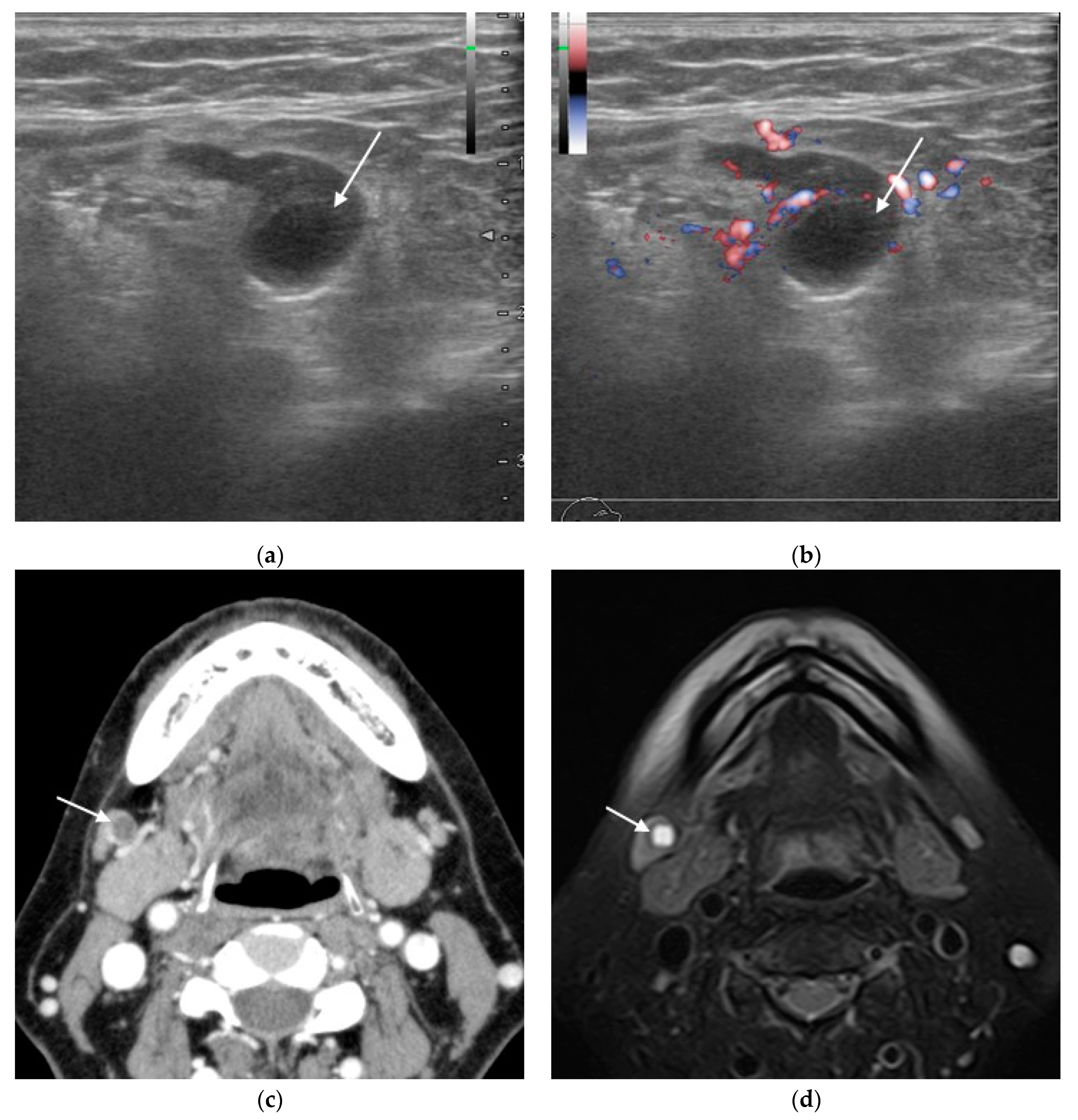
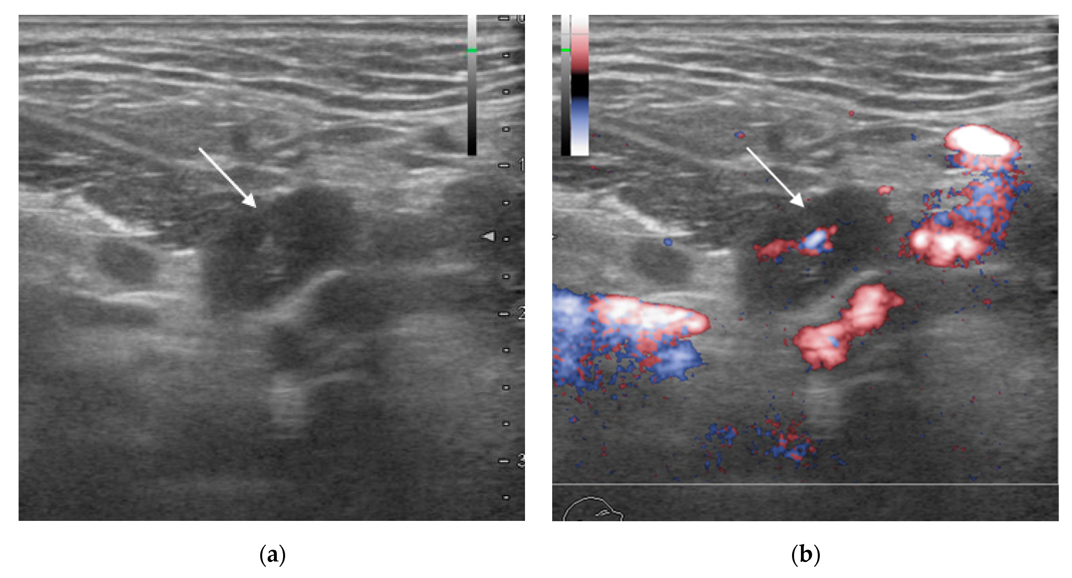
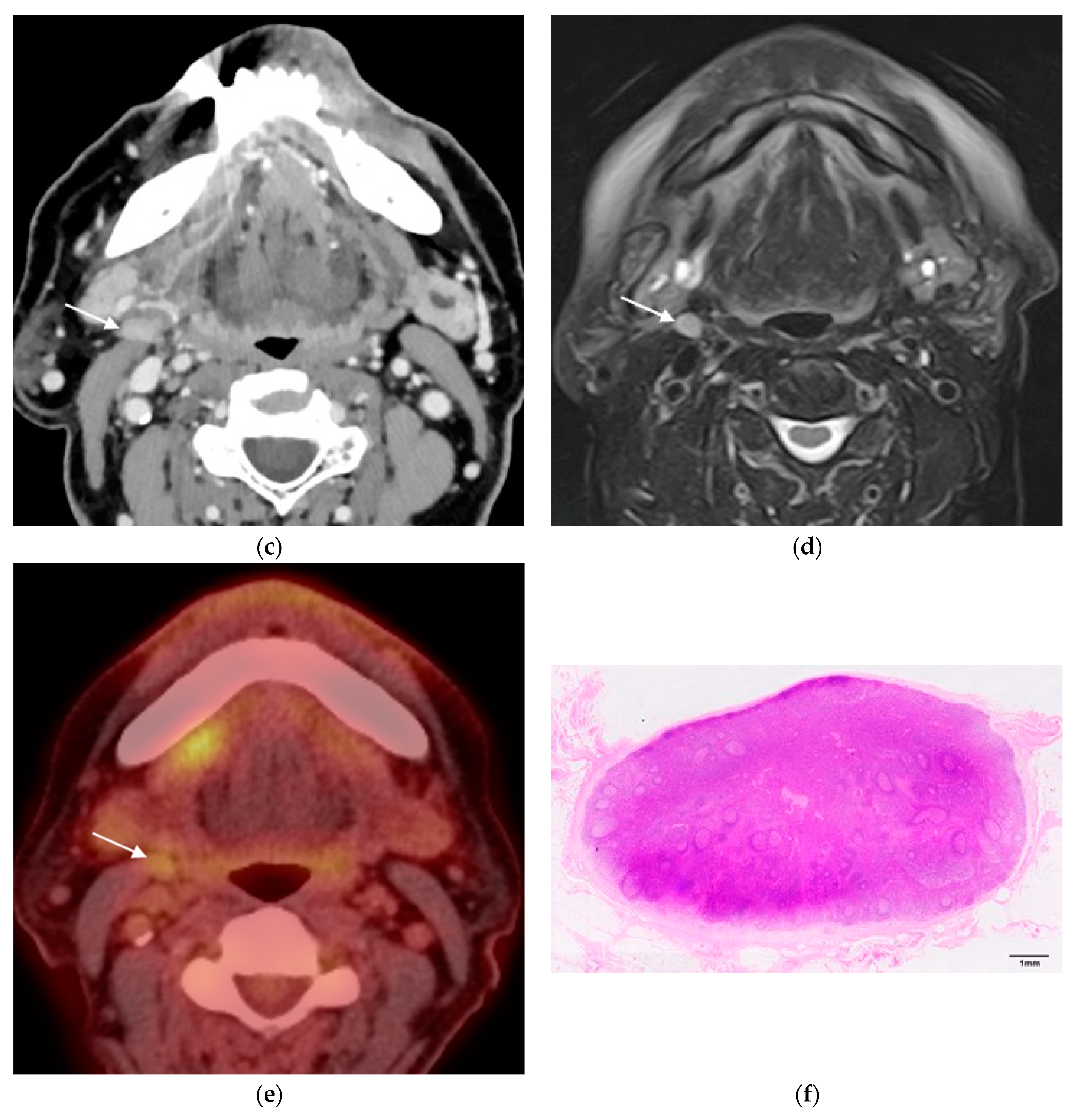
3. Results
4. Discussion
5. Conclusions
Author Contributions
Funding
Institutional Review Board Statement
Informed Consent Statement
Data Availability Statement
Acknowledgments
Conflicts of Interest
References
- Castelijns, J.A.; van den Brekel, M.W. Imaging of lymphadenopathy in the neck. Eur. Radiol. 2002, 12, 727–738. [Google Scholar] [CrossRef] [PubMed]
- Massey, C.; Dharmarajan, A.; Bannuru, R.R.; Rebeiz, E. Management of N0 neck in early oral squamous cell carcinoma: A systematic review and meta-analysis. Laryngoscope 2019, 129, E284–E298. [Google Scholar] [CrossRef] [PubMed]
- Weiss, M.H.; Harrison, L.B.; Isaacs, R.S. Use of decision analysis in planning a management strategy for the stage N0 neck. Arch. Otolaryngol. Head Neck Surg. 1994, 120, 699–702. [Google Scholar] [CrossRef] [PubMed]
- Kumar, A.; Ghai, S.; Mhaske, S.; Singh, R. Elective Neck Dissection Versus Therapeutic Neck Dissection in Clinically Node-Negative Early Stage Oral Cancer: A Meta-Analysis of Randomized Controlled Trials. J. Maxillofac. Oral Surg. 2022, 21, 340–349. [Google Scholar] [CrossRef]
- Oh, L.J.; Phan, K.; Kim, S.W.; Low, T.H.; Gupta, R.; Clark, J.R. Elective neck dissection versus observation for early-stage oral squamous cell carcinoma: Systematic review and meta-analysis. Oral Oncol. 2020, 105, 104661. [Google Scholar] [CrossRef] [PubMed]
- Cai, H.; Zhu, Y.; Wang, C.; Zhang, Y.; Hou, J. Neck nodal recurrence and survival of clinical T1-2 N0 oral squamous cell carcinoma in comparison of elective neck dissection versus observation: A meta-analysis. Oral Surg. Oral Med. Oral Pathol. Oral Radiol. 2020, 129, 296–310. [Google Scholar] [CrossRef]
- Kim, S.J.; Pak, K.; Kim, K. Diagnostic accuracy of F-18 FDG PET or PET/CT for detection of lymph node metastasis in clinically node negative head and neck cancer patients; A systematic review and meta-analysis. Am. J. Otolaryngol. 2019, 40, 297–305. [Google Scholar] [CrossRef]
- Sun, R.; Tang, X.; Yang, Y.; Zhang, C. 18FDG-PET/CT for the detection of regional nodal metastasis in patients with head and neck cancer: A meta-analysis. Oral Oncol. 2015, 51, 314–320. [Google Scholar] [CrossRef]
- Roh, J.-L.; Park, J.P.; Kim, J.S.; Lee, J.H.; Cho, K.-J.; Choi, S.-H.; Nam, S.Y.; Kim, S.Y. 18F fluorodeoxyglucose PET/CT in head and neck squamous cell carcinoma with negative neck palpation findings: A prospective study. Radiology 2014, 271, 153–161. [Google Scholar] [CrossRef]
- Liao, L.J.; Lo, W.C.; Hsu, W.L.; Wang, C.T.; Lai, M.S. Detection of cervical lymph node metastasis in head and neck cancer patients with clinically N0 neck-a meta-analysis comparing different imaging modalities. BMC Cancer 2012, 12, 236. [Google Scholar] [CrossRef]
- Matsubara, R.; Kawano, S.; Chikui, T.; Kiyosue, T.; Goto, Y.; Hirano, M.; Jinno, T.; Nagata, T.; Oobu, K.; Abe, K.; et al. Clinical significance of combined assessment of the maximum standardized uptake value of F-18 FDG PET with nodal size in the diagnosis of cervical lymph node metastasis of oral squamous cell carcinoma. Acad. Radiol. 2012, 19, 708–717. [Google Scholar] [CrossRef] [PubMed]
- Seitz, O.; Chambron-Pinho, N.; Middendorp, M.; Sader, R.; Mack, M.; Vogl, T.J.; Bisdas, S. 18F-Fluorodeoxyglucose-PET/CT to evaluate tumor, nodal disease, and gross tumor volume of oropharyngeal and oral cavity cancer: Comparison with MR imaging and validation with surgical specimen. Neuroradiology 2009, 51, 677–686. [Google Scholar] [CrossRef] [PubMed]
- Yoon, D.Y.; Hwang, H.S.; Chang, S.K.; Rho, Y.-S.; Ahn, H.Y.; Kim, J.H.; Lee, I.J. CT, MR, US, 18F-FDG PET/CT, and their combined use for the assessment of cervical lymph node metastases in squamous cell carcinoma of the head and neck. Eur. Radiol. 2009, 19, 634–642. [Google Scholar] [CrossRef] [PubMed]
- Sumi, M.; Kimura, Y.; Sumi, T.; Nakamura, T. Diagnostic performance of MRI relative to CT for metastatic nodes of head and neck squamous cell carcinomas. J. Magn. Reson. Imaging 2007, 26, 1626–1633. [Google Scholar] [CrossRef] [PubMed]
- Murakami, R.; Uozumi, H.; Hirai, T.; Nishimura, R.; Shiraishi, S.; Ota, K.; Murakami, D.; Tomiguchi, S.; Oya, N.; Katsuragawa, S.; et al. Impact of FDG-PET/CT imaging on nodal staging for head-and-neck squamous cell carcinoma. Int. J. Radiat. Oncol. Biol. Phys. 2007, 68, 377–382. [Google Scholar] [CrossRef]
- Ng, S.-H.; Yen, T.-C.; Chang, J.T.-C.; Chan, S.-C.; Ko, S.-F.; Wang, H.-M.; Lee, L.-Y.; Kang, C.-J.; Wong, A.M.-C.; Liao, C.-T. Prospective study of [18F]fluorodeoxyglucose positron emission tomography and computed tomography and magnetic resonance imaging in oral cavity squamous cell carcinoma with palpably negative neck. J. Clin. Oncol. 2006, 24, 4371–4376. [Google Scholar] [CrossRef]
- Schöder, H.; Carlson, D.L.; Kraus, D.H.; Stambuk, H.E.; Gönen, M.; Erdi, Y.E.; Yeung, H.W.D.; Huvos, A.G.; Shah, J.P.; Larson, S.M.; et al. 18F-FDG PET/CT for detecting nodal metastases in patients with oral cancer staged N0 by clinical examination and CT/MRI. J. Nucl. Med. 2006, 47, 755–762. [Google Scholar]
- Hayashi, T.; Ito, J.; Taira, S.; Katsura, K.; Shingaki, S.; Hoshina, H. The clinical significance of follow-up sonography in the detection of cervical lymph node metastases in patients with stage I or II squamous cell carcinoma of the tongue. Oral Surg. Oral Med. Oral Pathol. Oral Radiol. Endodontol. 2003, 96, 112–117. [Google Scholar] [CrossRef]
- Sumi, M.; Ohki, M.; Nakamura, T. Comparison of sonography and CT for differentiating benign from malignant cervical lymph nodes in patients with squamous cell carcinoma of the head and neck. AJR Am. J. Roentgenol. 2001, 176, 1019–1024. [Google Scholar] [CrossRef]
- Stuckensen, T.; Kovács, A.F.; Adams, S.; Baum, R.P. Staging of the neck in patients with oral cavity squamous cell carcinomas: A prospective comparison of PET, ultrasound, CT and MRI. J. Craniomaxillofac. Surg. 2000, 28, 319–324. [Google Scholar] [CrossRef]
- Ridge, J.A.; Lydiatt, W.M.; Patel, S.G.; Glastonbury, C.M.; Brandwein-Gensler, M.; Ghossein, R.A.; Shah, J.P. AJCC Cancer Staging Manual, 8th ed.; Springer: New York, NY, USA, 2017; pp. 79–94. [Google Scholar]
- Piao, Y.; Bold, B.; Tayier, A.; Ishida, R.; Omura, K.; Okada, N.; Shibuya, H. Evaluation of 18F-FDG PET/CT for diagnosing cervical nodal metastases in patients with oral cavity or oropharynx carcinoma. Oral Surg. Oral Med. Oral Pathol. Oral Radiol. Endodontol. 2009, 108, 933–938. [Google Scholar] [CrossRef] [PubMed]
- van den Brekel, M.W.; Castelijns, J.A.; Snow, G.B. The size of lymph nodes in the neck on sonograms as a radiologic criterion for metastasis: How reliable is it? AJNR Am. J. Neuroradiol. 1998, 19, 695–700. [Google Scholar] [PubMed]
- Niu, L.; Zheng, D.; Wang, D.; Zhang, J.; Fei, J.; Guo, C. Accuracy of 18 F-FDG PET/CT in detection of neck metastases of oral squamous cell carcinoma in patients without large palpable lymph nodes. Oral Surg. Oral Med. Oral Pathol. Oral Radiol. 2020, 129, 418–426. [Google Scholar] [CrossRef] [PubMed]
- Zhang, H.; Seikaly, H.; Biron, V.L.; Jeffery, C.C. Utility of PET-CT in detecting nodal metastasis in cN0 early stage oral cavity squamous cell carcinoma. Oral Oncol. 2018, 80, 89–92. [Google Scholar] [CrossRef]
- Yonetsu, K.; Sumi, M.; Izumi, M.; Ohki, M.; Eida, S.; Nakamura, T. Contribution of doppler sonography blood flow information to the diagnosis of metastatic cervical nodes in patients with head and neck cancer: Assessment in relation to anatomic levels of the neck. AJNR Am. J. Neuroradiol. 2001, 22, 163–169. [Google Scholar]
- Steinkamp, H.J.; Mueffelmann, M.; Böck, J.C.; Thiel, T.; Kenzel, P.; Felix, R. Differential diagnosis of lymph node lesions: A semiquantitative approach with colour Doppler ultrasound. Br. J. Radiol. 1998, 71, 828–833. [Google Scholar] [CrossRef]
- Gray, B.R.; Koontz, N.A. Normal patterns and pitfalls of FDG uptake in the head and neck. Semin. Ultrasound CT MRI 2019, 40, 367–375. [Google Scholar] [CrossRef]
- Payabvash, S.; Meric, K.; Cayci, Z. Differentiation of benign from malignant cervical lymph nodes in patients with head and neck cancer using PET/CT imaging. Clin. Imaging 2016, 40, 101–105. [Google Scholar] [CrossRef]
- Nakagawa, T.; Yamada, M.; Suzuki, Y. 18F-FDG uptake in reactive neck lymph nodes of oral cancer: Relationship to lymphoid follicles. J. Nucl. Med. 2008, 49, 1053–1059. [Google Scholar] [CrossRef]
- Van den Brekel, M.W.M.; Stel, H.V.; Castelijns, J.A.; Nauta, J.J.; Van der Waal, I.; Valk, J.; Meyer, C.J.; Snow, G.B. Cervical lymph node metastasis: Assessment of radiologic criteria. Radiology 1990, 177, 379–384. [Google Scholar] [CrossRef]
- Kojima, I.; Takanami, K.; Ogawa, T.; Ishikawa, K.; Morishita, Y.; Ishii, R.; Ohkoshi, A.; Nakanome, A.; Odagiri, H.; Iikubo, M. High diagnostic accuracy for lymph node metastasis of oral squamous cell carcinoma using PET/CT with a silicon photomultiplier. Oral Radiol. 2022, 38, 540–549. [Google Scholar] [CrossRef] [PubMed]
- Wang, Y.; Mao, M.; Li, J.; Feng, Z.; Qin, L.; Han, Z. Diagnostic value of magnetic resonance imaging in cervical lymph node metastasis of oral squamous cell carcinoma. Oral Surg. Oral Med. Oral Pathol. Oral Radiol. 2022, 133, 582–592. [Google Scholar] [CrossRef] [PubMed]
- Horváth, A.; Prekopp, P.; Polony, G.; Székely, E.; Tamás, L.; Dános, K. Accuracy of the preoperative diagnostic workup in patients with head and neck cancers undergoing neck dissection in terms of nodal metastases. Eur. Arch. Otorhinolaryngol. 2021, 278, 2041–2046. [Google Scholar] [CrossRef]
- Souren, C.; Kloss-Brandstätter, A.; Stadler, A.; Kross, K.; Yamauchi, K.; Ketelsen, D.; Kessler, P.; Lethaus, B. Ultrasound-guided fine-needle aspiration cytology as a diagnostic tool in comparison to ultrasound and MRI for staging in oral- and oropharyngeal squamous cell tumors. J. Craniomaxillofac. Surg. 2016, 44, 197–201. [Google Scholar] [CrossRef] [PubMed]
- Stoeckli, S.J.; Haerle, S.K.; Strobel, K.; Haile, S.R.; Hany, T.F.; Schuknecht, B. Initial staging of the neck in head and neck squamous cell carcinoma: A comparison of CT, PET/CT, and ultrasound-guided fine-needle aspiration cytology. Head Neck 2012, 34, 469–476. [Google Scholar] [CrossRef]
- Majumdar, K.S.; Rao, V.U.S.; Prasad, R.; Ramaswamy, V.; Sinha, P.; Subash, A. Incidence of micrometastasis and isolated tumour cells in clinicopathologically node-negative head and neck squamous cell carcinoma. J. Maxillofac. Oral Surg. 2020, 19, 131–135. [Google Scholar] [CrossRef]
- Yamazaki, Y.; Chiba, I.; Hirai, A.; Satoh, C.; Sakakibara, N.; Notani, K.-I.; Iizuka, T.; Totsuka, Y. Clinical value of genetically diagnosed lymph node micrometastasis for patients with oral squamous cell carcinoma. Head Neck 2005, 27, 676–681. [Google Scholar] [CrossRef]
- Hamakawa, H.; Takemura, K.; Sumida, T.; Kayahara, H.; Tanioka, H.; Sogawa, K. Histological study on pN upgrading of oral cancer. Virchows Arch. 2000, 437, 116–121. [Google Scholar] [CrossRef]
- van den Brekel, M.W.; van der Waal, I.; Meijer, C.J.; Freeman, J.L.; Castelijns, J.A.; Snow, G.B. The incidence of micrometastases in neck dissection specimens obtained from elective neck dissections. Laryngoscope 1996, 106, 987–991. [Google Scholar] [CrossRef]
- Loeffelbein, D.; Eiber, M.; Mayr, P.; Souvatzoglou, M.; Mücke, T.; von Bomhard, A.; Kesting, M.; Wolff, K.-D. Loco-regional recurrence after surgical treatment of oral squamous cell carcinoma: Proposals for follow-up imaging based on literature, national guidelines and institutional experience. J. Craniomaxillofac. Surg. 2015, 43, 1546–1552. [Google Scholar] [CrossRef]
- Zhou, Y.; Yu, T.; Rui, X.; Jin, T.; Huang, Z.; Huang, Z. Effectiveness of diffusion-weighted imaging in predicting cervical lymph node metastasis in head and neck malignancies. Oral Surg. Oral Med. Oral Pathol. Oral Radiol. 2021, 131, 122–129.e2. [Google Scholar] [CrossRef]
- Jiang, S.; Locatello, L.G.; Maggiore, G.; Gallo, O. Radiomics-Based Analysis in the Prediction of Occult Lymph Node Metastases in Patients with Oral Cancer: A Systematic Review. J. Clin. Med. 2023, 12, 4958. [Google Scholar] [CrossRef]
- Xu, X.; Xi, L.; Wei, L.; Wu, L.; Xu, Y.; Liu, B.; Li, B.; Liu, K.; Hou, G.; Lin, H.; et al. Deep learning assisted contrast-enhanced CT-based diagnosis of cervical lymph node metastasis of oral cancer: A retrospective study of 1466 cases. Eur. Radiol. 2023, 33, 4303–4312. [Google Scholar] [CrossRef]


| One-to-One Matched Lymph Nodes | Unmatched Lymph Nodes | |||||||
|---|---|---|---|---|---|---|---|---|
| Imaging Modality | TP | FP | FN | TN | TP | FP | FN | TN |
| US | 11 | 2 | 4 | 58 | 0 | 0 | 7 | 342 |
| CT | 11 | 3 | 5 | 72 | 0 | 0 | 6 | 327 |
| MRI | 9 | 3 | 4 | 46 | 0 | 0 | 9 | 353 |
| PET/CT | 14 | 16 | 1 | 45 | 0 | 3 | 7 | 338 |
| Imaging Modality | TP | FP | FN | TN | Sensitivity | Specificity | Accuracy | PPV | NPV |
|---|---|---|---|---|---|---|---|---|---|
| US | 9 | 2 | 9 | 62 | 50.0 (39.4–60.6) | 96.9 (90.5–99.0) | 86.6 (77.6–92.3) | 81.8 (72.1–88.7) | 87.3 (78.4–92.9) |
| CT | 10 | 3 | 8 | 61 | 55.6 (44.8–65.8) | 95.3 (88.4–98.2) | 86.6 (77.6–92.3) | 76.9 (66.7–84.7) | 88.4 (79.7–93.7) |
| MRI | 8 | 3 | 10 | 61 | 44.4 (34.2–55.2) | 95.3 (88.4–98.2) | 84.1 (74.7–90.5) | 72.7 (62.2–81.2) | 85.9 (76.8–91.8) |
| PET/CT | 14 | 13 | 4 | 51 | 77.8 (67.7–85.4) | 79.7 (69.7–87.0) | 79.3 (69.3–86.6) | 51.9 (41.2–62.3) | 92.7 (85.0–96.6) |
| Imaging Modality | TP | FP | FN | TN | Sensitivity | Specificity | Accuracy | PPV | NPV |
|---|---|---|---|---|---|---|---|---|---|
| US | 11 | 2 | 11 | 400 | 50.0 (45.3–54.7) | 99.5 (98.3–99.9) | 96.9 (94.8–98.2) | 84.6 (80.9–87.7) | 97.3 (95.3–98.5) |
| CT | 11 | 3 | 11 | 399 | 50.0 (45.3–54.7) | 99.3 (97.9–99.7) | 96.7 (94.5–98.0) | 78.6 (74.4–82.2) | 97.3 (95.3–98.5) |
| MRI | 9 | 3 | 13 | 399 | 40.9 (36.3–45.7) | 99.3 (97.9–99.7) | 96.2 (94.0–97.7) | 75.0 (70.7–78.9) | 96.8 (94.7–98.1) |
| PET/CT | 14 | 19 | 8 | 383 | 63.6 (59.0–68.1) | 95.3 (92.8–96.9) | 93.6 (90.9–95.6) | 42.4 (37.8–47.2) | 98.0 (96.1–98.9) |
Disclaimer/Publisher’s Note: The statements, opinions and data contained in all publications are solely those of the individual author(s) and contributor(s) and not of MDPI and/or the editor(s). MDPI and/or the editor(s) disclaim responsibility for any injury to people or property resulting from any ideas, methods, instructions or products referred to in the content. |
© 2023 by the authors. Licensee MDPI, Basel, Switzerland. This article is an open access article distributed under the terms and conditions of the Creative Commons Attribution (CC BY) license (https://creativecommons.org/licenses/by/4.0/).
Share and Cite
Takamura, M.; Nikkuni, Y.; Hayashi, T.; Katsura, K.; Nishiyama, H.; Yamazaki, M.; Maruyama, S.; Tanuma, J.-i. Comparing the Diagnostic Accuracy of Ultrasonography, CT, MRI, and PET/CT in Cervical Lymph Node Metastasis of Oral Squamous Cell Carcinoma. Biomedicines 2023, 11, 3119. https://doi.org/10.3390/biomedicines11123119
Takamura M, Nikkuni Y, Hayashi T, Katsura K, Nishiyama H, Yamazaki M, Maruyama S, Tanuma J-i. Comparing the Diagnostic Accuracy of Ultrasonography, CT, MRI, and PET/CT in Cervical Lymph Node Metastasis of Oral Squamous Cell Carcinoma. Biomedicines. 2023; 11(12):3119. https://doi.org/10.3390/biomedicines11123119
Chicago/Turabian StyleTakamura, Masaki, Yutaka Nikkuni, Takafumi Hayashi, Kouji Katsura, Hideyoshi Nishiyama, Manabu Yamazaki, Satoshi Maruyama, and Jun-ichi Tanuma. 2023. "Comparing the Diagnostic Accuracy of Ultrasonography, CT, MRI, and PET/CT in Cervical Lymph Node Metastasis of Oral Squamous Cell Carcinoma" Biomedicines 11, no. 12: 3119. https://doi.org/10.3390/biomedicines11123119
APA StyleTakamura, M., Nikkuni, Y., Hayashi, T., Katsura, K., Nishiyama, H., Yamazaki, M., Maruyama, S., & Tanuma, J.-i. (2023). Comparing the Diagnostic Accuracy of Ultrasonography, CT, MRI, and PET/CT in Cervical Lymph Node Metastasis of Oral Squamous Cell Carcinoma. Biomedicines, 11(12), 3119. https://doi.org/10.3390/biomedicines11123119