

T and Small Protrusion (TAP) Technique in Bifurcations: Coronary Artery Disease in Acute Myocardial Infarction Patients after COVID-19 Pneumonia


Abstract
:
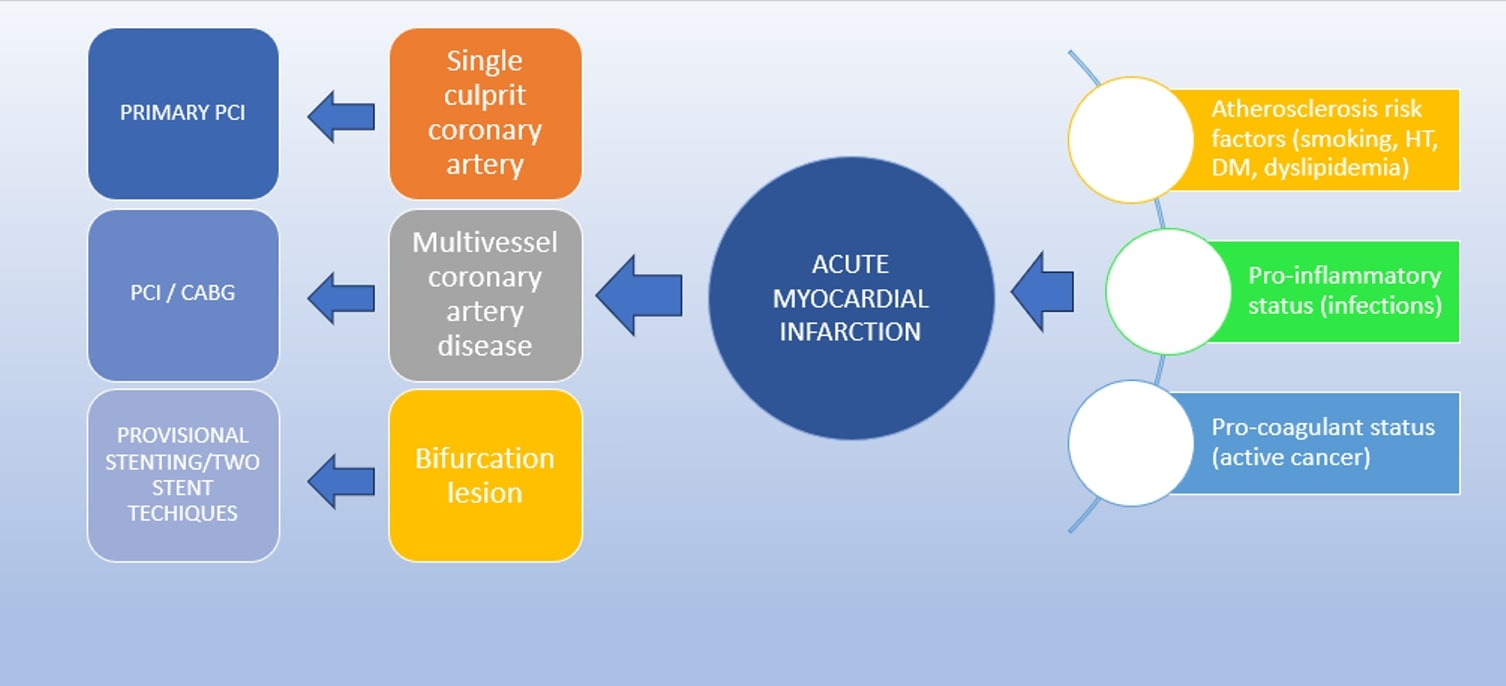
1. Introduction
1.1. Anatomy Changes of Bifurcation Lesions
1.2. Bifurcation Two-Stent Techniques
1.2.1. T-Stenting and TAP (T and Small Protrusion) Technique
1.2.2. Culotte Technique
1.2.3. Double-Kissing Crush (DK Crush)
1.2.4. Simultaneous Kissing Stents (SKS)/V-Stenting
2. Case Presentation
3. Discussions
4. Conclusions
Author Contributions
Funding
Institutional Review Board Statement
Informed Consent Statement
Data Availability Statement
Conflicts of Interest
References
- Jebari-Benslaiman, S.; Galicia-García, U.; Larrea-Sebal, A.; Olaetxea, J.R.; Alloza, I.; Vandenbroeck, K.; Benito-Vicente, A.; Martín, C. Pathophysiology of atherosclerosis. Int. J. Mol. Sci. 2022, 23, 3346. [Google Scholar] [CrossRef] [PubMed]
- Moscucci, M. Grossman & Baim’s Cardiac Catheterization, Angiography and Intervention; Wolter Kluwer: Alphen aan den Rijn, The Netherlands, 2021; pp. 757–759. [Google Scholar]
- Topol, E.J.; Teirstein, P.S. Textbook of Interventional Cardiology; Elsevier: Amsterdam, The Netherlands, 2016; pp. 375–392. [Google Scholar]
- Brilakis, E. Manual of Percutaneous Coronary Intervention—A Step-by-Step Approach; Elsevier: Amsterdam, The Netherlands; Academic Press: Cambridge, MA, USA, 2021; pp. 267–300. [Google Scholar]
- Louvard, Y.; Lefèvre, T.; Morice, M.C. Percutaneous coronary intervention for bifurcation coronary disease. Heart 2004, 90, 713–722. [Google Scholar] [CrossRef] [PubMed] [Green Version]
- Mohamed, M.O.; Polad, J.; Hildick-Smith, D.; Bizeau, O.; Baisebenov, R.K.; Roffi, M.; Íñiguez-Romo, A.; Chevalier, B.; von Birgelen, C.; Roguin, A.; et al. Impact of coronary lesion complexity in percutaneous coronary intervention: One-year outcomes from the large, multicentre e-Ultimaster registry. EuroIntervention 2020, 16, 603–612. [Google Scholar] [CrossRef] [PubMed]
- de Lezo, J.S.; Medina, A.; Martín, P.; Novoa, J.; de Lezo, J.S.; Pan, M.; Caballero, E.; Melián, F.; Mazuelos, F.; Quevedo, V. Predictors of ostial side branch damage during provisional stenting of coronary bifurcation lesions not involving the side branch origin: An ultrasonographic study. EuroIntervention 2012, 7, 1147–1154. [Google Scholar] [CrossRef] [PubMed]
- Kang, S.J.; Mintz, G.S.; Kim, W.J.; Lee, J.Y.; Oh, J.H.; Park, D.W.; Lee, S.W.; Kim, Y.H.; Lee, C.W.; Park, S.W.; et al. Changes in left main bifurcation geometry after a single-stent crossover technique: An intravascular ultrasound study using direct imaging of both the left anterior descending and the left circumflex coronary arteries before and after intervention. Circ. Cardiovasc. Interv. 2011, 4, 355–361. [Google Scholar] [CrossRef] [PubMed] [Green Version]
- Tu, S.; Xu, L.; Ligthart, J.; Xu, B.; Witberg, K.; Sun, Z.; Koning, G.; Reiber, J.H.C.; Regar, E. In vivo comparison of arterial lumen dimensions assessed by co-registered three-dimensional (3D) quantitative coronary angiography, intravascular ultrasound and optical coherence tomography. Int. J. Cardiovasc. Imaging 2012, 28, 1315–1327. [Google Scholar] [CrossRef] [Green Version]
- Abdel-Wahab, M.; Richardt, G.; Joachim Büttner, H.; Toelg, R.; Geist, V.; Meinertz, T.; Schofer, J.; King, L.; Neumann, F.J.; Khattab, A.A. High speed rotational atherectomy before paclitaxel-eluting stent implantation in complex calcified coronary lesions: The randomized ROTAXUS trial. JACC Cardiovasc. Interv. 2013, 6, 10–19. [Google Scholar] [CrossRef] [PubMed] [Green Version]
- Burzotta, F.; Džavík, V.; Ferenc, M.; Trani, C.; Stankovic, G. Technical aspects of the T and small protrusion (TAP) technique. EuroIntervention 2015, 11 (Suppl. V), V91–V95. [Google Scholar] [CrossRef]
- Galassi, A.R.; Colombo, A.; Buchbinder, M.; Grasso, C.; Tomasello, S.D.; Ussia, G.P.; Tamburino, C. Long-term outcomes of bifurcation lesions after implantation of drug-eluting stents with the “mini-crush” technique. Catheter. Cardiovasc. Interv. 2007, 69, 976–983. [Google Scholar] [CrossRef]
- Chen, X.; Li, X.; Zhang, J.-J.; Han, Y.; Kan, J.; Chen, L.; Qiu, C.; Santoso, T.; Paiboon, C.; Kwan, T.W.; et al. 3-Years outcomes of the DKCRUSH—V trial comparing DK crush with provisional stenting for left main bifurcation lesions. JACC Cardiovasc. Interv. 2019, 12, 1927–1937. [Google Scholar] [CrossRef]
- Banning, A.P.; Lassen, J.F.; Burzotta, F.; Lefèvre, T.; Darremont, O.; Hildick-Smith, D.; Louvard, Y.; Stankovic, G. Percutaneous coronary intervention for obstructive bifurcation lesions: The 14th consensus document from the European bifurcation club. EuroIntervention 2019, 15, 90–98. [Google Scholar] [CrossRef] [PubMed]
- Lunardi, M.; Louvard, Y.; Lefèvre, T.; Stankovic, G.; Burzotta, F.; Kassab, G.S.; Lassen, J.F.; Darremont, O.; Garg, S.; Koo, B.-K.; et al. Definitions and standardized endpoints for treatment of coronary bifurcations. J. Am. Coll. Cardiol. 2022, 80, 63–88. [Google Scholar] [CrossRef]
- Raphael, C.E.; O’Kane, P.D. Contemporary approaches to bifurcation stenting. JRSM Cardiovasc. Dis. 2021, 10, 2048004021992190. [Google Scholar] [CrossRef] [PubMed]
- Chen, S.L.; Zhang, J.J.; Ye, F.; Chen, Y.D.; Patel, T.; Kawajiri, K.; Lee, M.; Kwan, T.W.; Mintz, G.; Tan, H.C. Study comparing the double kissing (DK) crush with classical crush for the treatment of coronary bifurcation lesions: The DKCRUSH-1 bifurcation study with drug-eluting stents. Eur. J. Clin. Investig. 2008, 38, 361–371. [Google Scholar] [CrossRef] [PubMed]
- Chen, S.-L.; Xu, B.; Han, Y.-L.; Sheiban, I.; Zhang, J.-J.; Ye, F.; Kwan, T.W.; Paiboon, C.; Zhou, Y.-J.; Lv, S.-Z.; et al. Clinical outcome after DK crush versus culotte stenting of distal left main bifurcation lesions. JACC Cardiovasc. Interv. 2015, 8, 1335–1342. [Google Scholar] [CrossRef] [Green Version]
- Chen, S.-L.; Zhang, J.-J.; Han, Y.; Kan, J.; Chen, L.; Qiu, C.; Jiang, T.; Tao, L.; Zeng, H.; Li, L.; et al. Double kissing crush versus provisional stenting for left main distal bifurcation lesions. J. Am. Coll. Cardiol. 2017, 70, 2605–2617. [Google Scholar] [CrossRef] [PubMed]
- Mrdovic, I.; Savic, L.; Krljanac, G.; Asanin, M.; Perunicic, J.; Lasica, R.; Marinkovic, J.; Kocev, N.; Vasiljevic, Z.; Ostojic, M. Predicting 30-day major adverse cardiovascular events after primary percutaneous coronary intervention. The RISK-PCI score. Int. J. Cardiol. 2013, 162, 220–227. [Google Scholar] [CrossRef] [PubMed]
- De Lemos, J.A.; Omland, T. Chronic Coronary Artery Disease; Elsevier: Amsterdam, The Netherlands, 2018; pp. 489–498. [Google Scholar]
- Oikonomou, E.; Mourouzis, K.; Vogiatzi, G.; Siasos, G.; Deftereos, S.; Papaioannou, S.; Latsios, G.; Tsalamandris, S.; Tousoulis, D. Coronary Microcirculation and the No-reflow Phenomenon. Curr. Pharm. Des. 2018, 24, 2934–2942. [Google Scholar] [CrossRef]
- Marc, M.C.; Iancu, A.C.; Balanescu, S.; Dregoesc, M.I. Microvascular obstruction in acute myocardial infarction: An old and unsolved mystery. Med. Pharm. Rep. 2019, 92, 216–219. [Google Scholar] [CrossRef] [Green Version]
- Pothineni, N.V.; Shah, N.N.; Rochlani, Y.; Saad, M.; Kovelamudi, S.; Marmagkiolis, K.; Bhatti, S.; Cilingiroglu, M.; Aronow, W.S.; Hakeem, A. Temporal trends and outcomes of acute myocardial infarction in patients with cancer. Ann. Transl. Med. 2017, 5, 482. [Google Scholar] [CrossRef] [Green Version]
- Iliescu, C.A.; Grines, C.L.; Herrmann, J.; Yang, E.H.; Cilingiroglu, M.; Charitakis, K.; Hakeem, A.; Toutouzas, K.P.; Leesar, M.A.; Marmagkiolis, K. SCAI expert consensus statement: Evaluation, management, and special considerations of cardio-oncology patients in the cardiac catheterization laboratory (endorsed by the cardiological society of India, and sociedad Latino Americana de Cardiologıa intervencionista). Catheter. Cardiovasc. Interv. 2016, 87, E202–E223. [Google Scholar] [PubMed]
- Ziun, M.; Rigatelli, G.; Battisti, V.; Costola, G.; Roncon, L.; Bilato, C. Increased risk of acute myocardial infarction after COVID-19 recovery: A systematic review and meta-analysis. Int. J. Cardiol. 2023, 372, 138–143. [Google Scholar] [CrossRef] [PubMed]
- Corrales-Medina, V.F.; Madjid, M.; Musher, D.M. Role of acute infection in triggering acute coronary syndromes. Lancet Infect. Dis. 2010, 10, 83–92. [Google Scholar] [CrossRef] [PubMed]
- Marfella, R.; Paolisso, P.; Sardu, C.; Palomba, L.; D’onofrio, N.; Cesaro, A.; Barbieri, M.; Rizzo, M.R.; Sasso, F.C.; Scisciola, L.; et al. SARS-COV-2 colonizes coronary thrombus and impairs heart microcirculation bed in asymptomatic SARS-CoV-2 positive subjects with acute myocardial infarction. Crit. Care 2021, 25, 217. [Google Scholar] [CrossRef] [PubMed]
- Zuin, M.; Mugnai, G.; Zamboni, A.; Zakja, E.; Valle, R.; Turiano, G.; Themistoclakis, S.; Scarpa, D.; Saccà, S.; Roncon, L.; et al. Decline of admission for acute coronary syndromes and acute cardiovascular conditions during COVID-19 pandemic in Veneto region. Viruses 2022, 14, 1925. [Google Scholar] [CrossRef] [PubMed]
- Sutherland, N.; Dayawansa, N.H.; Filipopoulos, B.; Vasanthakumar, S.; Narayan, O.; Ponnuthurai, F.A.; van Gaal, W. Acute coronary syndrome in the COVID-19 pandemic: Reduced cases and increased ischaemic time. Heart Lung Circ. 2022, 31, 69–76. [Google Scholar] [CrossRef] [PubMed]
- Alnima, T.; Mulder, M.M.G.; van Bussel, B.C.T.; Ten Cate, H. COVID-19 coagulopathy: From pathogenesis to treatment. Acta Haematol. 2022, 145, 282–296. [Google Scholar] [CrossRef]
- Sardu, C.; Marfella, R.; Maggi, P.; Messina, V.; Cirillo, P.; Codella, V.; Gambardella, J.; Sardu, A.; Gatta, G.; Santulli, G.; et al. Implications of AB0 blood group in hypertensive patients with COVID-19. BMC Cardiovasc. Disord. 2020, 20, 373. [Google Scholar] [CrossRef]


| Assay | Value | Normal Rage |
|---|---|---|
| WBC | 14.2 × 10³ | 4.00–10.00 × 10³ |
| GLU | 131 mg/dL | 74–100 mg/dL |
| GGT | 38 U/L | 9–36 U/L |
| CREA | 0.78 mg/dL | 0.57–1.11 mg/dL |
| CK-MB | 592 U/L | 0.00–24.0 U/L |
| HS-TROP | 10,003.1 pg/mL | 0.00–15.6 pg/mL |
Disclaimer/Publisher’s Note: The statements, opinions and data contained in all publications are solely those of the individual author(s) and contributor(s) and not of MDPI and/or the editor(s). MDPI and/or the editor(s) disclaim responsibility for any injury to people or property resulting from any ideas, methods, instructions or products referred to in the content. |
© 2023 by the authors. Licensee MDPI, Basel, Switzerland. This article is an open access article distributed under the terms and conditions of the Creative Commons Attribution (CC BY) license (https://creativecommons.org/licenses/by/4.0/).
Share and Cite
Rus, M.; Filimon, G.C.; Ardelean, A.I. T and Small Protrusion (TAP) Technique in Bifurcations: Coronary Artery Disease in Acute Myocardial Infarction Patients after COVID-19 Pneumonia. Biomedicines 2023, 11, 2255. https://doi.org/10.3390/biomedicines11082255
Rus M, Filimon GC, Ardelean AI. T and Small Protrusion (TAP) Technique in Bifurcations: Coronary Artery Disease in Acute Myocardial Infarction Patients after COVID-19 Pneumonia. Biomedicines. 2023; 11(8):2255. https://doi.org/10.3390/biomedicines11082255
Chicago/Turabian StyleRus, Marius, Georgiana Carmen Filimon, and Adriana Ioana Ardelean. 2023. "T and Small Protrusion (TAP) Technique in Bifurcations: Coronary Artery Disease in Acute Myocardial Infarction Patients after COVID-19 Pneumonia" Biomedicines 11, no. 8: 2255. https://doi.org/10.3390/biomedicines11082255