Review

Towards Improved Human In Vitro Models for Cardiac Arrhythmia: Disease Mechanisms, Treatment, and Models of Atrial Fibrillation

by
Carla Cofiño-Fabres
1,
Robert Passier
1,2,* and
Verena Schwach
1,*
1
Department of Applied Stem Cell Technologies, TechMed Centre, University of Twente, Drienerlolaan 5, 7500 AE Enschede, The Netherlands
2
Department of Anatomy and Embryology, Leiden University Medical Centre, 2300 RC Leiden, The Netherlands
*
Authors to whom correspondence should be addressed.
Biomedicines 2023, 11(9), 2355; https://doi.org/10.3390/biomedicines11092355
Submission received: 25 July 2023 / Revised: 18 August 2023 / Accepted: 19 August 2023 / Published: 23 August 2023
(This article belongs to the Special Issue Mechanisms and Novel Therapeutic Approaches for Cardiac Arrhythmia)

Abstract

:
Heart rhythm disorders, arrhythmias, place a huge economic burden on society and have a large impact on the quality of life of a vast number of people. Arrhythmias can have genetic causes but primarily arise from heart tissue remodeling during aging or heart disease. As current therapies do not address the causes of arrhythmias but only manage the symptoms, it is of paramount importance to generate innovative test models and platforms for gaining knowledge about the underlying disease mechanisms which are compatible with drug screening. In this review, we outline the most important features of atrial fibrillation (AFib), the most common cardiac arrhythmia. We will discuss the epidemiology, risk factors, underlying causes, and present therapies of AFib, as well as the shortcomings and opportunities of current models for cardiac arrhythmia, including animal models, in silico and in vitro models utilizing human pluripotent stem cell (hPSC)-derived cardiomyocytes.

1. Introduction

Cardiovascular diseases cause 32% (about 18 million) of global deaths yearly, according to the World Health Organization [1], and are the leading cause of death. Cardiovascular disease can be classified into diseases of the heart muscle, the valves, and vessels. Heart rhythm disorders, arrhythmias, are irregular heartbeats and form one very common type of heart disease. Arrhythmias can be subdivided into those with slower (bradyarrhythmias) or faster (tachyarrhythmias) heart rates than a physiological resting heart rate (60–100 beats per minute (BPM)). Atrial fibrillation (AFib), the most common arrhythmia, is characterized by uncoordinated electrical activity and contraction of the atria, which causes a rapid and irregular heartbeat higher than 100 BPM.
Over time, AFib is associated with a doubling in all-cause mortality, a five-fold increase in stroke, an accelerated development of heart failure, thromboembolic events, and a substantially poorer quality of life [2]. Since the duration of a specific AFib episode can drastically vary between patients, AFib is usually divided into three main categories: (1) paroxysmal AFib, when episodes last less than 7 days and spontaneously convert to normal sinus rhythm, (2) persistent AFib when episodes last more than 7 days, or (3) permanent AFib, a chronic arrhythmia [3].
According to a 2019 global burden of disease (GBD) study, AFib affects an estimated 59.7 million people (30.28 million men and 29.41 million women) worldwide, with a rising incidence and precedence, leading to almost five million new cases of AFib annually. Incidence, prevalence, and mortality of AFib are strongly dependent on age and gender, as well as ethnicity and race (Figure 1). Incidence is significantly higher in developed regions compared to developing countries. The highest prevalence estimated was for North America, while the lowest was found in the Sub-Saharian Africa region (2364.9 vs. 134.31 cases per 100,000 people). However, the prevalence of AFib may be underestimated because of the large number of asymptomatic individuals. Over the next 2 decades, AFib is projected to increase in an exponential manner, perchance doubling in prevalence, which is essentially due to the aging of the population, but also to the improved ability to treat other diseases. In the particular case of Europe, the number of patients with AFib is estimated to be between 14 and 17 million in 2030. Thus, AFib is currently identified as one of the biggest public health problems and even a growing epidemic, placing a large burden on both social and economic levels [4,5,6,7,8,9].
Many studies have reported several risk factors associated with AFib, for example, increasing age, ethnicity, and gender (Figure 2). Common cardiovascular risk factors are thought to promote AFib by increasing atrial pressure and/or by causing atrial dilation [2]. Although the majority of AFib subjects develop arrhythmic behavior in the context of preexisting cardiovascular disease, genetic factors may play an important role in the onset and maintenance of the disease [10]. Many ion channel genes have been re-sequenced, and a cascade of new mutations associated with AFib has been identified (Figure 2). For example, gain-of-function mutations are predicted to promote repolarization, shorten the atrial action potential and facilitate re-entry [11], while loss-of-function mutations seem to lead to prolongation of the atrial action potential, characteristic of atrial remodeling [12]. Genome-wide association studies have also provided evidence implicating common genetic variants in the pathogenesis of AFib. Therefore, understanding the mechanisms underlying the complex relationship between AFib and its associated multiple risk factors is crucial for the development of AFib models predictive for the human condition.
Arrhythmias, including AFib, are leading causes of morbidity and mortality, predominantly through increased risk of stroke, heart failure, and other heart-related complications [29]. Modeling AFib has primarily focused on different in vivo animal models to understand the underlying causes of the disease and the development of new treatments [30]. However, animals do not recapitulate human physiology because of species differences in cardiac physiology, such as expression, distribution, and function of contractile proteins or ion channels [31,32]. Hence, there is an urgent need for predictive modeling of AFib, capable of dynamically mimicking and incorporating various known cardiac disease features and risk factors. In recent years, the realization of efficient protocols for the differentiation of cardiovascular cellular subtypes from human pluripotent stem cells (hPSCs) has made a significant contribution to the development of personalized cardiac disease models. HPSC-derived progeny, such as ventricular or atrial cardiomyocytes, have been established in preclinical disease modeling, drug testing, as well as advancing new therapy [33]. In this review, we will describe the current knowledge about the underlying disease mechanisms and therapy of AFib. After an overview of animal models to study arrhythmia, we will dive into an extended discussion on the state-of-the-art in vitro models utilizing hPSC-derived cardiomyocytes for AFib modeling. Finally, we will provide our future perspectives on the field.

2. Underlying Disease Mechanisms of AFib

2.1. Brief Overview of Normal Electrophysiology of Cardiomyocytes

In a healthy heart, cardiomyocytes are electrically coupled together, forming a functional syncytium that allows the rapid propagation of the electrical impulse throughout the cardiac muscle, causing continuous and highly coordinated contractions. Each action potential is initiated by the sinoatrial (SA) node, a specialized group of myocardial cells located in the posterior wall of the right atrium, close to the orifice of the superior vena cava (Figure 3A). Pacemaker cells exhibit automaticity, meaning they spontaneously depolarize to initiate the action potential. Even though SA node cells are not the only conductive cells, these are the ones with the highest rate of depolarization and thus are the primary pacemaker of the heart responsible for establishing the normal sinus rhythm. After being generated in the SA node, the impulse spreads to the adjacent atrial cardiomyocytes through cell-to-cell gap junctions, as well as radially via a specialized internodal pathway called Bachmann’s bundle or the interatrial band. This structure conducts the impulse directly from the right atrium to the left atrium. Ultimately, the wave of depolarization reaches a second group of specialized cells at the bottom of the right atrium, the atrioventricular (AV) node. Because the atria and ventricles are electrically isolated from one another by a circumferential band of fibrous tissue, the only path for impulse propagation is via the AV node. After a brief, intrinsic delay at the AV node, the action potential is propagated quickly down the bundle of His and Purkinje fibers within the ventricular myocardium [34] (Figure 3A). At rest, the cardiomyocyte membrane is more permeable to potassium (K+) than sodium (Na+) or calcium (Ca2+), resulting in a negative electrical gradient with respect to the extracellular environment since potassium has an equilibrium potential of around −80 Mv, the resting membrane potential of cardiomyocytes. With sufficient stimulus, alterations in the permeability to Na+ result in a rapid increase in the membrane potential to values of about 40 Mv, creating a net positive electrical gradient (depolarization). Further, changes in the ion permeability to K+, Cl, and Ca2+ results in the eventual restoration of the negative intracellular environment (Figure 3B). The action potential is linked to the cardiac contraction via excitation-contraction coupling. In the healthy condition, contraction is induced by an influx of Ca2+ ions through voltage-dependent L-type channels [35,36,37] after these have been opened by depolarization through the influx of Na+. This initial increase in Ca2+ in the cytosol triggers the release of Ca2+ from the sarcoplasmic reticulum (SR) through Ryanodine receptor 2 (RyR2). The high levels of cytosolic Ca2+ trigger contraction. Relaxation is achieved by moving cytosolic Ca2+ into the SR via sarco/endoplasmic reticulum Ca2+-ATPase (SERCA2a) or through the plasma membrane via the rheogenic Na+/Ca2+-exchanger (NCX), which transports 1 Ca2+ ion outward while moving 3 Na+ ions inward.

2.2. Trigger and Substrate Cause AFib

The pathophysiology of AFib contains three major components, namely onset, maintenance, and progression toward longer-lasting AFib forms. AFib is initiated as a response to a trigger rapidly exciting the atria in combination with a substrate able to maintain fibrillations. Generally accepted trigger phenomena are rapidly firing ectopic foci, re-entrant waves, and rotors, while substrates include electrical, structural, and autonomic remodeling [38,39].

2.3. Triggers: AFib Is Induced by Ectopic Foci, Re-Entry and Rotors

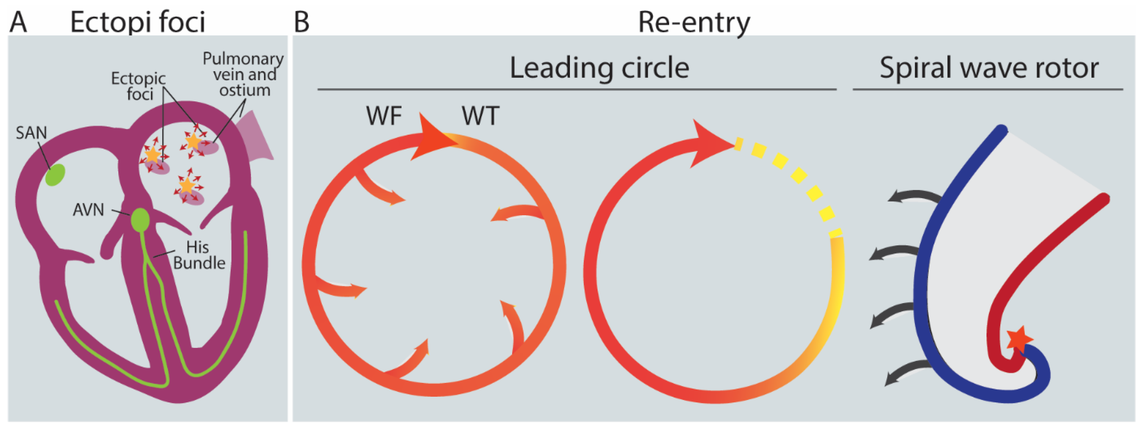
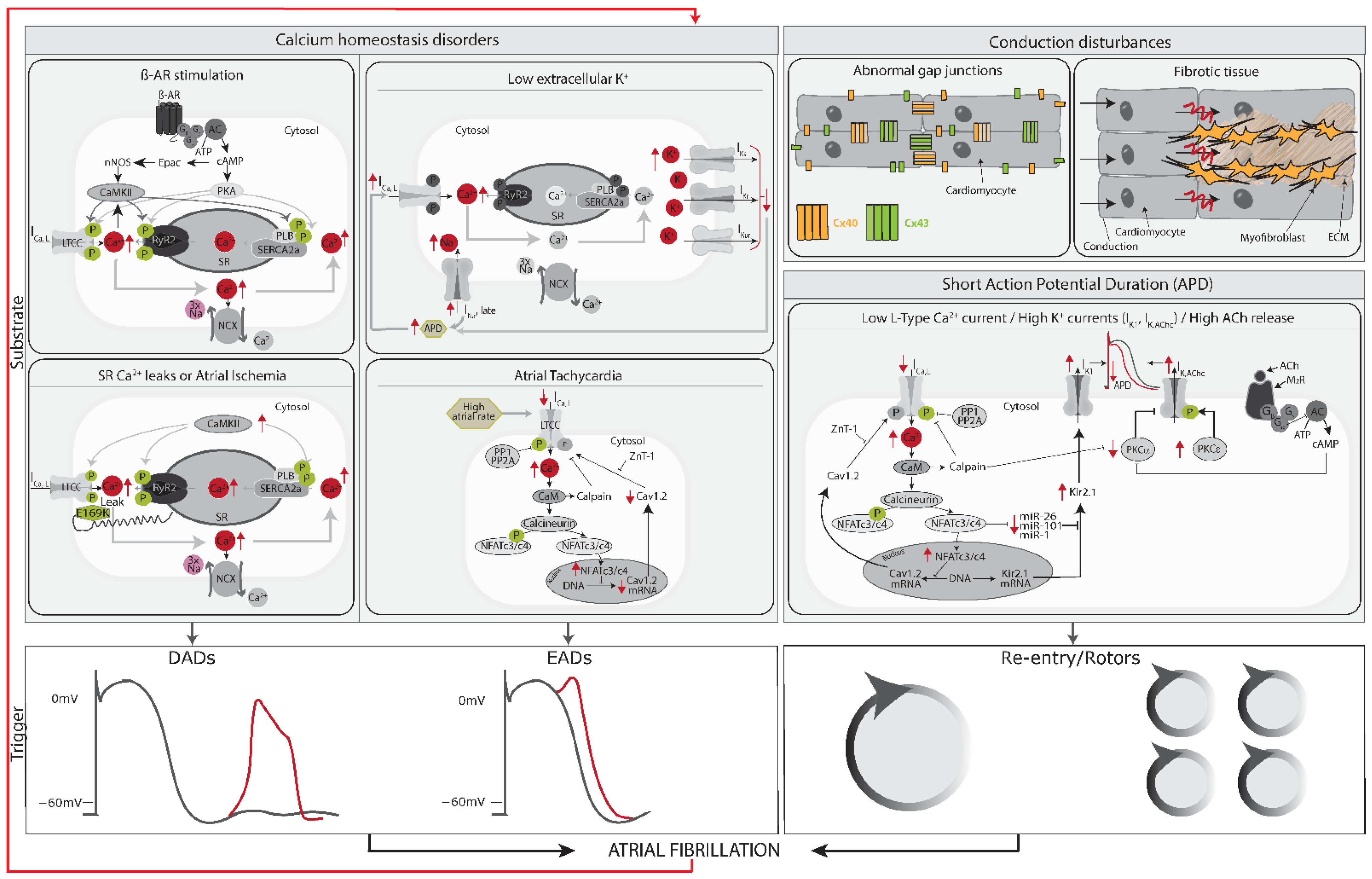
To be initiated, AFib generally requires an initiating electrical stimulus, a ‘trigger.’ Three main sources as triggers for the rapid and uncoordinated atrial activity generated in AFib have been established: (1) rapidly firing ectopic foci with irregular conduction and fibrillatory activity; (2) re-entrant activity, which can be associated with complex multiple re-entrant circuits, or (3) rotors which are formed by functional re-entry spiral waves with irregular patterns and no consistent activation pattern [37] (Figure 4). Triggers often arise from the pulmonary veins [40] (Figure 4A). Atrial ectopic activity arises from triggered activity as a result of diastolic delayed afterdepolarizations (DADs) [3,41,42] or spontaneous extra-systolic activity by early afterdepolarizations (EADs) [43]. DADs are spontaneous depolarizations occurring in cardiomyocytes after the repolarization phase of the action potential, while EADs occur during the repolarization phase [35,44]. EADs or DADs arise because of disturbed calcium homeostasis. If the DAD is large enough, it depolarizes the cell and causes a premature atrial ectopic beat. A series of DADs can cause atrial tachycardia, which can trigger atrial re-entry or, if rapid enough can maintain AFib. Direct evidence for DAD-mediated ectopic activity related to abnormal Ca2+ handling has been provided in animal models of AFib [45], as well as in patients with AFib [46]. A high Ca2 load in the cytoplasm at the repolarization phase causes depolarization through the NCX and consequently aberrant excitation and impulse formation [41,42]. The high amount of Ca2+ in the cytosol can be caused by tachycardia, ischemia, β-adrenergic receptor stimulation, low extracellular potassium concentration [47], or SR Ca2+ leaks [41,42]. SR Ca2+ leaks can be caused by increased SERCA2a activity [46] and dysregulated RyR2 or L-type channels that cause an increase in SR Ca2+ load [3,48] (Figure 5).
Re-entry is the phenomenon of activation that can travel and re-excite the tissue it originated from [38]. As a trigger, re-entry is heavily dependent on a supporting substrate, as cardiac tissue cannot be activated again during the refractory period. The likelihood of re-entry in a re-entrant circuit is defined by the distance traveled by the wavefront during the refractory period (wavelength of the circuit). If the wavelength of the circuit is equal to its circumference due to either a short refractory period or low conduction velocity, there will be no excitation gap. Thus, the leading edge of the impulse will trespass the excitable and recovered tissue, establishing the smallest circuit (leading circle) with a continuously depolarized center around a conduction barrier, which can be the pulmonary vein or scar tissue [39,49] (Figure 4B middle).
Rotors are another type of re-entry, where the wave moves in a spiral or curved form instead of in a circle (Figure 4B right). Where the wavefront and wave tail meet, the singularity point is formed. Due to a current-to-load mismatch (see below), the wavefront curvature influences the wavefront conduction velocity. Near the singularity point, the curvature of the wavefront is so strong, and the conduction velocity is consequently so slow that an excitable core of tissue cannot be excited, forming a functional but not refractory block [39,49].

2.4. Current-to-Load (Source-Sink) Mismatches Affect Conduction Velocity

One of the mechanisms that maintain re-entry is current source-sink mismatches, where the action potential propagates from a source tissue with depolarizing current towards the sink tissue that will be depolarized (Figure 6A). Current-to-load mismatches at boundaries between inhomogeneous tissue volumes change the conduction velocity and may lead to the onset or maintenance of arrhythmia. Conduction velocity in a 2D isotropic medium can be described as: θ = θ 0 + D ρ [56]. Where theta is the conduction velocity, theta0 is the conduction velocity of a non-curved wavefront, and ρ denotes wavefront curvature. D is a coefficient equal to 1/CmSvRi where Cm is the specific membrane capacitance, Sv is the cell surface-to-volume ratio, and Ri is the intracellular resistivity. Thus, if the source and sink are the same size (ρ remains equal), the signal will propagate without a change in conduction velocity. When the volume of the sink tissue is smaller compared to the source tissue (positive ρ, resulting in a concave wavefront), the electrical propagation will continue with increased velocity as there is sufficient electrical current available to depolarize the sink tissue. However, when the source tissue is smaller than the sink (negative ρ, convex wavefront), the conduction velocity will slow or even be blocked [57,58,59] (Figure 6B). This decreased conduction velocity predisposes to arrhythmia.

2.5. Substrate: AFib Is Maintained via Electrical, Structural, or Autonomic Remodeling of Atrial Tissue

A vulnerable atrial substrate will enhance susceptibility for AFib induction, maintenance, and progression. Atrial remodeling, regarding electrical and structural properties as well as autonomic innervation, constitutes a substrate for AFib (Figure 5).
Electrical remodeling in AFib consists of several components; those identified so far are (1) decreased L-type Ca2+ channel current, (2) increased K+ currents, and (3) abnormalities in gap junction ion channel functions [61]. In the first case, the overload of Ca2+ often present in AFib causes restoration of homeostasis by activation of the NFAT system, which inhibits, at the same time, transcription of Cav1.2, a subunit of L-type Ca2+ channels. Consequently, the duration of the atrial action potential shortens, leaving the tissue more vulnerable to re-entry [45,62]. Secondly, upregulation of K+ currents in AFib, such as the inward rectifying K+ current (IK1) channel protein KIR2.1 or the atrial-specific acetylcholine-activated inward rectifier potassium current (IKACh), is associated with shortened action potential duration [37,63]. Moreover, atrial tachypacing has been shown to increase the expression of Ca2+ dependent K+ (SK) channels and enhance the trafficking of SK channels to the cell membrane, contributing to the reduction in the action potential duration [64,65]. Finally, the electrical coupling between cardiomyocytes is mediated by gap junction ion channels [61]. Overexpression of atrial-specific connexin 40 or ventricular-enriched connexin 43 has been shown to increase conduction velocity, thus preventing the development of persistent AFib in pigs [66]. Therefore, alteration of connexins could potentially lead to conduction velocity abnormalities, contributing to the development of AFib.
Structural remodeling of the atria in the context of AFib mainly involves fibrosis, which makes AFib more persistent and resistant to therapy over time [61]. Cardiac fibrosis involves increased deposition of extracellular matrix components (ECM), including collagen I and fibronectin, mainly produced by myofibroblasts [67]. This fibrotic tissue can form a conduction barrier, where an unexcitable zone at the center of the circuit is produced, being an anchor for re-entry. The border between the fibrotic and non-fibrotic tissue might help stabilize the re-entry. A recent optical mapping study in explanted human hearts showed evidence that re-entry underlies AFib and found that transmural re-entry was stabilized by anatomical structures with local fibrosis [68,69,70]. Additionally, to fibrosis, oxidative stress increases the levels of Reactive Oxygen Species (ROS). High ROS levels can lead to structural remodeling by inducing inflammatory reactions or direct oxidative damage to myofibrils. Oxidative stress damage to mitochondrial DNA can also drive to modulation of Ca2+ handling proteins and/or channels, causing a Ca2+ overload, leading to electrical remodeling [71,72].
Finally, the autonomous nervous system consists of the sympathetic (fight or flight) and parasympathetic (rest and digest) nervous systems, which mostly act as antagonists to each other. Both subsystems innervate the heart, with the sympathetic (vagal) system increasing and the parasympathetic system decreasing heart rate. Increased firing from both the sympathetic and the parasympathetic systems contribute to AFib [73]. Parasympathetic activation leads to the release of acetylcholine, which increases IKACh, thus shortening the action potential duration and supporting re-entry. Sympathetic innervation has been shown to increase in response to rapid atrial pacing and leads to β-adrenergic receptor stimulation. This leads to protein kinase A phosphorylation, which in turn phosphorylates several proteins involved in SR Ca2+ loading [74]. Together these effects raise the risk of EAD, DAD, and re-entry [75,76].

2.6. AFib Begets AFib

As AFib progresses, it may remodel the substrate and reinforce the vulnerability of the substrate to maintain AFib. This self-reinforcement is known as “AFib begets AFib” and limits the effectiveness of treatments after prolonged AFib [61] (Figure 5). AFib results in rapid shortening of the refractory period as a result of disturbed ion channel activity. The shorter refractory period stabilizes AFib, as indicated by spontaneous AFib maintenance [43,49]. Another contributor to this self-reinforcement is atrial fibrosis. Rapidly firing atrial cardiomyocytes generate substances that make fibroblasts differentiate into collagen-producing myofibroblasts, hence reducing the cardiac impulse propagation homogeneity and inducing irregular rhythm. AFib becomes increasingly persistent and resistant to therapy over time.

3. Treatment of AFib

Despite the availability of various therapies, treatments for AFib (such as long-term pharmacology, cardioversion, or surgical intervention by ablation) primarily focus on alleviating the symptoms and have significant limitations, making AFib still a clinical challenge. Cardiac arrhythmias have been traditionally treated by rate or rhythm control.
Rate control therapies focus on the reduction in the rapid ventricular rate (often found in patients with AFib) by the use of negative chronotropic drugs (beta-blockers, calcium channel blockers, cardiac glycosides, or combination therapies) or electrophysiological/surgical interventions. This treatment effectively reduces the symptoms of AFib [77].
Rhythm control therapies aim to restore proper sinus rhythm through drugs (pharmacological cardioversion) or electric currents (electrical cardioversion). After successful cardioversion, patients are generally treated with long-term anti-arrhythmic drug therapy to prevent the recurrence of AFib by blocking the function of ion channels, reducing the excitation of the cardiomyocytes, and prolonging action potential duration [71,77]. Anti-arrhythmic drugs in clinical use (and their target current) are flecainide (INa), propafenone (INa), ibutilide (IKr), vernakalant (multiple ion channel blocker), amiodarone (multiple ion channel blocker), dronedarone (multiple ion channel blocker), quinidine (IKr), disopyramide (INa), solatol (IKr) and dofetilide (IKr) (not available in Europe). Of these anti-arrhythmic agents, potassium ion channel blockers are preferred in the treatment of AFib. The choice of agent is dependent on the persistency of AFib and preexisting heart conditions. The success rates of these drugs in restoring and maintaining the sinus rhythm range from 30 to 50% [78]. Rhythm control, together with rate control, does not improve the clinical outcome of AFib when compared to just rate control. However, rhythm control is still employed for symptom relief [71,79,80].
Rate and rhythm control strategies require a co-treatment with anti-thrombotic drugs to lower the risk of stroke and thromboembolic events [77].
The main drawback to most anti-arrhythmic drugs for the treatment of AFib is non-specificity to the atria that paradoxically leads to life-threatening ventricular ‘proarrhythmias’ [81,82]. Current research efforts, therefore, focus on drugs that mainly target atrial-specific ion channels, IKur and IKAch, and rate-dependent inhibition of INa [83]. For example, vernakalant inhibits atrial-specific IKur and IKAch, but also affects IKr, Ito, and INa current. Nevertheless, inhibition of IKr is significantly lower than that of other drugs, such as flecainide, propafenone, ibutilide, and dofetilide. Ito is not exclusive to atria, but its contribution to the atrial action potential is larger compared to the ventricular action potential. Na+ channels have a higher affinity for blocking drugs during the action potential. Consequently, inhibition of INa is more pronounced in fast-beating atria when compared to the ventricles during AFib [82,84]. The progression from paroxysmal to persistent and permanent forms of AFib has pronounced therapeutic implications, with paroxysmal AFib being more amenable to rhythm control therapy [3]. However, most drug treatments have profound side effects and are expensive to use over long periods [85]. Consequently, there has been a shift towards non-pharmacological therapies for cardiac arrhythmias, including electrical cardioversion and controlled destruction of arrhythmia-generating substrate via ablation of tissue close to pulmonary veins, left atrial roof and posterior wall and right interatrial septum [71,79]. A significant weak point in the current treatment guidelines for AFib is that treatments are prescribed based on the persistency of AFib, not on the underlying mechanism of an individual patient [3]. More advanced human models would allow the development of powerful personalized treatments of AFib.

4. Models of AFib

Despite all the advantages provided by different types of modeling systems, there are still important limitations that halt their use for direct clinical purposes. The limited possibility of constructing a de novo human model of cardiomyocyte electrophysiology and the need for a better understanding of the underlying mechanisms that lead to the onset and maintenance of atrial arrhythmias demand the development of improved and distinct disease models. Nattel, Bourne, and Talajic [86] succinctly outlined the main features of a model system of atrial arrhythmias: (1) The models should be relevant to clinically observed atrial arrhythmias to include structural and functional abnormalities; (2) The atrial fibrillation/flutter should be inducible and able to be maintained chronically to ensure that a putative intervention has indeed terminated the arrhythmia, and it did not occur spontaneously; and (3) it should be possible to restore and maintain normal sinus rhythm.

4.1. In Vivo Modeling: Animal Models

AFib has been studied in both large and small animal models with rate-related electrical or atrial-structural remodeling following acute atrial insults and in the presence of autonomic nervous system modulation. Some of the most relevant animal models used for AFib are summarized in Table 1.
Generally, dogs have been extensively used to study AFib. Although the spontaneous occurrence of AFib in dogs, dog models of AFib are experimentally induced, commonly with atrial tachypacing (at a rate of 400–600 bpm), to study their electrophysiology and electrical remodeling. Nevertheless, AFib episodes in dogs are relatively short due to the difficulty to induce and sustain atrial tachycardias [87,88]. Dogs have also been used to study AFib in the context of inflammation (sterile pericarditis), congestive heart failure, atrial ischemia, and the autonomic nervous system [88]. Goats constitute a more robust sustained model for AFib since they are able to better tolerate AFib. Therefore, long-term studies with atrial tachypacing are possible, facilitating the study of structural and electrical remodeling that is associated with persistent AFib [88]. Interestingly, the persistent AFib maintained by the tachycardia-induced electrical remodeling led to the concept of ‘AFib begets AFib’ [87]. Pigs are interesting models to study AFib due to their similarities to human heart size and electrophysiology. Induction of AFib with atrial tachypacing is commonly used, although it also often triggers systolic dysfunction [88]. To prevent the occurrence of systolic dysfunction, atrioventricular node blockade has been used and facilitated the study of self-sustained and persistent AFib for >4 months [89,90].
Structural remodeling has been observed in dogs, sheep, pigs, goats, and humans alike, but differences exist in the nature of remodeling [53]. Despite the fact that different animal models show similar structural remodeling, achieving atrial fibrosis as commonly observed in human AFib is still limiting since tissue fibrosis and degenerative changes are usually related to aging and/or associated heart diseases [37]. Nevertheless, some investigations in AFib dog models have shown that 4 or 6-week rapid atrial pacing promotes ECM synthesis in the atrial tissue and interstitial fibrosis [91,92]. Similarly, chronic atrial dilatation in goat AFib models might create atrial conduction disturbances suggesting the formation of tissue fibrosis [93].
The study of AFib in small animals, such as mice and rabbits, is more challenging due to the large differences in scale and electrophysiology compared to humans. Since mice do not commonly develop AFib, models of AFib require genetic manipulation, after which programmed electrical stimulation is necessary to assess their ability to generate AFib. These models can be taken as interrogations about the functional effects of particular proteins or systems on fibrillatory potential. Interestingly, an investigation with a murine model using optogenetics as a tool to detect and terminate rapid arrhythmia in a safe, repetitive, and shock-free manner holds promise in how these models can be useful to develop potential shock-free therapies for the treatment of AFib [94].
Despite the fact that experimental animal models have been designed to study the pathophysiology of AFib, including molecular basis, ion-current determinants, anatomical features, and macroscopic mechanisms, there are obvious drawbacks when considering their use. First, animal models are not exactly representative of the ionic and molecular basis of AFib in humans as there are significant interspecies differences in cardiac function and also in the type and density of ion channels, pumps, exchangers, and gap junction proteins. Thus, the mechanistically important parameters can be quite different between species, which often complicates the translation to the clinic. As a result, the mechanisms of AFib in animals are not necessarily analogous to those in humans and have not been sufficiently predictive for drug development.
Table 1. Overview of in vivo animal models for AFib.
Animal ModelAFib ModelAFib PromotionClinical Causes of AFib
Dog
[50,53,87,88]
Paroxysmal and Persistent
AFib models
Electrical, structural and
autonomic remodeling
Sterile pericarditis, atrial tachycardia remodeling, CHF-related AFib,
acute atrial ischemia, atrial volume overload, mitral regurgitation,
cesium infusion
Goat
[53,88,93,95]
Paroxysmal and Persistent
AFib models
Electrical and structural
remodeling
Atrial tachycardia remodeling
Pig
[53,88,89]
Paroxysmal and Persistent
AFib models
Structural remodelingAtrial tachycardia remodeling
Sheep
[88,96,97]
Paroxysmal and Persistent
AFib models
Structural and autonomic
remodeling
Atrial volume overload,
aortopulmonary shunt,
atrial tachycardia remodeling
Rabbit
[50,88]
Paroxysmal AFib modelElectrical, structural and
autonomic remodeling
Atrial volume overload
Transgenic mice
[88,98,99]
Atrial conduction
abnormalities models
-Dilated cardiomyopathy,
hypertrophic cardiomyopathy,
atrial pathology in CHF,
atrial tachycardia remodeling
Paroxysmal: AFib of up to 7 days of duration spontaneously terminates; Persistent: AFib lasts >7 days without spontaneous termination; CHF, Congestive Heart Failure; AT, Atrial tachyarrhythmia; HF, Heart failure.

4.2. Alternatives to Animal Models

4.2.1. In Silico Models

By using in silico models, a multitude of models describing atrial cell electrophysiology have been developed over the last few decades for different mammalian species, e.g., rabbit and canine. For human atrial cells, there have been two principal, longstanding models that reconstruct the action potential (AP) using ordinary differential equations (ODEs) based on overlapping experimental data [100,101]. In the absence of human data, both models rely partially on data obtained from other mammals and have slightly different formulations of ionic currents, pumps, exchangers, and others, resulting in divergent behaviors. Nevertheless, outstanding progress with in silico modeling has enabled the confirmation of the multiple wavelet hypothesis as a mechanism underlaying AFib [102], as well as the study of the contribution of different ionic currents to AFib. Moreover, 3D realistic human atrial computer models have proved to be a complementary tool for the study of atrial arrhythmias and for the advancement of improved therapies by offering the possibility of testing the effects of different anti-arrhythmic strategies in a human model without irreversible damage to the patients. A clear example is the integration of computer modeling to design a biological integrated cardiac defibrillator and proof in an in vitro system its validity to detect and correct arrhythmia [103]. Additionally, these models are also a great platform for studying the contributions of patient-specific structural remodeling to AFib initiation (i.e., fibrosis distribution, fibrotic architecture, and atrial dilatation), and there is even a possibility for in silico-guided ablation therapy [104]. Despite these advances, several challenges remain in order to fully exploit the potential of in silico modeling. The main challenges include the lack of personalized details, limited availability of experimental data for model validation, limited patient-specific electrophysiological information, and unresolved issues to simulate complex aspects of AFib progression. Future advancements in experimental methodologies, clinical imaging modalities, computational performance, standardization, and the need for clinical evidence to support the routine use of computational models in AFib management will help move this technology forward [105]. For that, the generation of hPSC-derived atrial cardiomyocytes [106,107,108] and the development of atrial tissues [109,110,111] have indicated the potential to model AFib in vitro and open new possibilities to generate robust datasets that facilitate in silico modeling.

4.2.2. In Vitro Modeling Using hPSC-Derived CMs

Due to the clear interspecies variability found in in vivo models and the necessity of biological validation in in silico models, in vitro models with human cardiomyocytes represent a promising alternative to study mechanisms and processes involved in AFib. However, while it is possible to isolate adult heart cells from patients after heart surgery, the difficulty of obtaining such tissue is further compounded by the fact that human cardiomyocytes are non-dividing cells and do not survive long in culture, severely limiting their application for larger-scale studies. Some studies using conditionally immortalized rat or human atrial myocyte (iAM) lines [112,113] are a valuable source of cardiomyocytes in vitro. However, the generation of patient-specific disease models is more challenging, and the associated high investment to produce the lines make them less attractive for disease modeling [113,114]. In addition, the use of living myocardial slices has been an important approach for studying several aspects of AFib over the last years [115,116,117,118]. Since they retain the cellular architecture and connections found in the heart, mechanisms related to electrophysiology, fibrosis, or calcium handling can be studied. In this context, myocardial slices have been utilized to understand the underlying mechanisms of cardiac arrhythmias by studying ectopic foci throughout the atria [16,119], as well as to examine the role of cardiac re-entry involved in tachyarrhythmias [120,121]. Although numerous efforts have been made to maintain them as unaltered as possible once isolated and preserve them for long-term cultivation [115], they are kept in a 2D environment prone to trigger changes in cellular behavior and functionality and potentially affecting the physiological relevance of the observed responses. Therefore it is also necessary to develop other complementary models to this system.
For the last decades, the major source of human cardiomyocytes has come from hPSCs, which have been successfully established to model disease at a single cell level, reliably reproducing the human cardiac electrical phenotype in health and disease. HPSCs can form atrial, ventricular, and pacemaker-like cardiomyocytes in vitro by differentiating them in the presence of specific growth factors [122] or small-molecules. For example, atrial- and ventricular-like cardiomyocytes were derived from hPSCs via differential modulation of retinoic acid signaling during differentiation [106,107,108,123]. Initially, cardiomyocyte differentiation protocols generally did not yield well-delineated populations of a single terminal cell type and instead produced mixed populations of atrial, nodal, ventricular, and non-cardiac cells. Therefore, it was crucial to purify cell populations in order to develop cell-type specific disease models in order to avoid any potential contaminating factors. Recently, the use of cell type-specific reporter lines that facilitate the isolation of the subtype of interest by flow cytometry [124] or the use of metabolic selection protocols that enrich the CM population [108] facilitates the retrieval of pure populations of the desired cell type.
Although the cardiomyocytes derived from hPSCs show many typical features of their in vivo equivalents, overt symptoms of many diseases at the organ level develop only in the late stages of adulthood. Hence, one important problem with developing physiological and disease models based on hPSCs-derived cardiomyocytes is that these differentiated derivatives present features typical of immature or fetal cardiomyocytes, as evidenced by their spontaneous electrical activity, action potentials with low upstroke velocities, irregular shape, less-developed sarcomeres, metabolism and gene expression [125]. Nevertheless, hPSC-derived atrial cardiomyocytes do possess several specific currents and ion channels, in particular the ultra-rapid delayed-rectifier K+ current (IKur), the acetylcholine-activated inward-rectifying K+ current (IKACh), all of which are largely absent in the ventricles. Therefore, hPSC-derived cardiomyocyte models may represent a valuable platform for the screening of preclinical anti-arrhythmic drugs and cardiotoxicity and modeling cardiac disease. In fact, when the IKur blocker 4-aminopyridine was tested in hPSC-derived atrial cardiomyocytes, reduced atrial repolarization was observed, while an IKACh blocker (carbachol) reversed carbachol-induced action potential shortening. Since equivalent effects were not demonstrated in ventricular-like cardiomyocytes, atrial cardiomyocytes emerged as a powerful preclinical model for assessing the efficacy and safety of atrial-selective agents [106]. Additionally, one of the first models concerning the induction of re-entrant activity in atrial cells has recently been developed in cell sheets of hPSC-derived atrial cardiomyocytes [109] or ring-like tissues [111]. The resulting rotors induced in the cell sheets were comparable to those observed in previous computer simulations and animal studies, validating the use of such preparations as an AFib disease model. Nakanishi et al. cultured hPSC-derived atrial cardiomyocytes as monolayers in a geometrical narrow-to-wide pattern together with cardiac fibroblasts to mimic the abrupt anatomical transition at the junction of pulmonary veins (PVs) and left atrium (LA) [126]. Under high-frequency field stimulation, the narrow isthmus model and cell heterogeneity provoked impaired electrical conduction [126]. One of the major limitations presented is the absence of spiral waves/rotors (non-induction of AFib). It can be argued that monolayer models may not be applicable to high-throughput screening strategies, given the spatial requirements to produce re-entrant activity and the need for more sophisticated measurements of wave behavior. Other constraints associated with this 2D model are the lack of cell organization, fiber thickness, extracellular matrix content, and electrophysiological differences between PV and atrial cardiomyocytes when compared to in vivo. Moreover, the high non-physiological pacing frequency required to induce conduction disturbances and the electrophysiology of the atrial cardiomyocytes, elucidate their immature phenotype [126].
Approaches to improve the maturation of the hPSC-derived cardiomyocytes include advanced 3D models, such as Engineered Heart Tissues (EHTs) [127] or atrial-ventricular co-cultures systems, the Biowire II [128]. Atrial or ventricular hPSC-cardiomyocytes cultured in an EHT format show a higher gene expression for chamber-specific markers compared to monolayers, suggesting an improvement in maturation [110]. Atrial-like EHTs from hPSC have been successfully formed and exhibit different types of rhythm disorders, being suitable candidates to study AFib in vitro [110,111]. Lemme et al. presented atrial-EHTs as a model for AFib, considering that the tissues displayed short action potentials prone to tachyarrhythmias and with a shape similar to APs from patients with persistent AFib. Nevertheless, no further studies were followed to modulate the disorder.
In another model, atrial and ventricular-hPSC-derived CMs were embedded in a collagen-hydrogel to form chamber-specific ring-shaped EHTs, and the resultant tissues recapitulated the expected atrial-ventricular physiological differences in their conduction properties. This was further evidenced by optical mapping analysis, where ventricular-EHTs presented an activation pattern initiated at a primary pacemaker region and activating the rest of the tissue synchronously, and the majority of the atrial-EHTs presented arrhythmias with re-entrant nature and different levels of complexity. Interestingly, these arrhythmias could be converted into a normal rhythm by applying electrical field stimulation or exposing the tissue to anti-arrhythmic agents (Vernakalant and Flecainide). In this study, the utility of an in vitro model for AFib is evidenced by showing how a drug-testing platform can be used to study pharmacological cardioversion and rhythm-control strategy of arrhythmias [111]. Chronic intermittent tachypacing by optogenetics in hPSC-derived atrial-EHTs has recently been described as a strategy to induce cardiac dysfunction and study the mechanisms of heart failure and arrhythmogenesis [129]. Interestingly, no contractile dysfunction was observed as a result of the optical tachypacing, but some tissues exhibited an arrhythmic spontaneous beating pattern, and various aspects of AFib remodeling were partly recapitulated, such as a trend to a more negative take-off potential and to an increased action potential amplitude. Further research using different stimulation patterns may be valuable to reveal some insights into the mechanisms of the remodeling process during AFib. Using the Biowire II platform, Zhao et al. reported a co-culture tissue formed of hPSC-derived atrial and ventricular cardiomyocytes cultured between two wires. While AFib was not the goal of the study, it shows its potential, by selectively modulating the tissue response to atrial-specific drugs causing tachycardia, such as serotonin [128].
Successful termination of tachycardia has been achieved by burst pacing (20 Hz), light administration, or drug treatment (Flecainide, JTV-519, and E-4031) [130]. Despite the fact that in this study, the model has been described as a tool for ventricular tachycardia, it sheds some light on how to use chronic interval pacing in models with hPSC-atrial-cardiomyocytes to induce other types of arrhythmia, such as AFib. The atrial-ventricular Biowire II enables the growth of thin and cylindrical tissues with two distinct regions: an atrial and a ventricular end. Two parallel wires allow simultaneous measurement of force and Ca2+ transients [128]. Tissue conditioning using chronic electrical stimulation with progressively increasing stimulation rates (up to 6 Hz) promotes cardiac maturation, suggesting that it may be a promising model for selective pharmacology and understanding arrhythmia.
A substantial proportion of AFib patients display no history of AFib-associated disorders, which suggests a hereditary component. Indeed, a familial history of AFib has been linked to a 40% risk increase. Over the last years, a number of AFib-related gene loci and mutations have been identified [10,131]. These comprise several developmental regulators, as well as ion channel genes involved in cardiac action potential generation, suggesting a direct causative role of the latter group. Genome editing technologies, such as CRISPR/Cas9, allow the development of isogenic hPSC disease lines of AFib harboring different genetic variants, including rare mutations observed in familial AFib, as well as common risk alleles. This enables the study of patient-specific disease mechanisms and sets the stage for a pharmaco-genomic screen. Combined targeted genetic engineering with cardiac subtype-specific differentiation of hPSCs was conducted by Marczenke et al. to explore the role of Kv1.5 channels (encoded by the KCNA5 gene and responsible for IKur) in atrial hPSCs-cardiomyocytes [132]. In this model, CRISPR/Cas9-mediated mutagenesis of integration-fee hPSCs was employed to generate a functional KCNA5 knockout. Interestingly, both loss and gain-of-function mutations in KCNA5 have been shown to cause familial AFib [12]. This seeming paradox is explained by the fact that AFib may be promoted via distinct mechanisms, namely, triggered activity and electrical re-entry. While the latter tends to be favored by action potential shortening, the likelihood of the former becomes increased by AP prolongation. Therefore, in addition to their contribution to drug development and discovery, gene-edited stem cell models can serve as a valuable tool for identifying the variables, such as specific SNPs and drugs under comparison, that can optimize the design of clinical trials. By introducing disease-associated genetic variants into hPSCs, it is possible to assess the impact of these variants on cellular responses to drug treatment, thereby increasing the likelihood of designing a clinical trial “in a dish” that is more likely to yield positive results. For example, a pair of anti-arrhythmic drugs (AADs) demonstrating highly variable efficacy in gene-edited hPSC models harboring a risk allele at 4q25 (i.e., a locus associated with PITX2) could inspire a more cost-effective clinical trial for this relatively large AFib sub-population with a high likelihood of producing statistically significant outcomes.

5. Conclusions and Future Perspectives

AFib remains the most common sustained arrhythmia linked to several serious comorbidities, such as heart failure and stroke, which makes it one of the biggest public health problems. Additionally, currently, available treatments for AFib, namely long-term treatment with anti-arrhythmic drugs, cardioversion, or surgical intervention by ablation, solely rely on treating symptoms, including control of the heartbeat, prevention of blood clotting and rhythm control, thus presenting noticeable limitations and making AFib still a clinical challenge. As a result, there is a high unmet need for groundbreaking predictive models of human AFib, which would allow for the exploration of the disease’s underlying mechanisms and streamline the process of high-throughput drug screening to discover new therapeutic approaches. Despite all advantages provided by different types of modeling systems, there are still constrains that halt their use for direct clinical purposes. Even though animal models have provided an initial understanding of the mechanisms that trigger and maintain AFib, these display significant morphological and physiological features which differ from those found in humans, thus hindering their use. On the other hand, in silico, and in vitro modeling approaches have proven to be pivotal tools in the progress of AFib treatment. Nonetheless, these models still present restrictions regarding development and subsequent use as a preclinical model. HPSC-derived cardiomyocytes usually exhibit an immature phenotype, a major limitation for disease modeling of adult-stage conditions. Several aspects regarding cardiomyocyte maturation, such as the necessary time to develop the disease phenotype, need to be addressed in order to obtain robust in vitro models for AFib. Different approaches have been suggested to boost the maturation of hPSC-derived cardiomyocytes by identifying the missing factors characteristic of adult cardiomyocytes. Mainly, cardiomyocytes should recapitulate the expression of specific genes that are representative of the adult stage, the exhibition of an organized cytoskeleton and specialized organelles, an oxidative metabolism, and an adult electrophysiological behavior. Kolanowski et al. reported strategies to stimulate maturation considering these features, such as biochemical stimulation (by activation or repression of signaling pathways), physical stimulation (through electrical or mechanical stimuli) as well as the development of 3D models able to reinforce the mechanical properties of cardiomyocytes [133]. Moreover, cardiomyocytes in vivo strongly interact with non-cardiomyocytes, influencing cardiac development and specification. For instance, cardiac fibroblasts play an important role by providing the extracellular matrix of the heart, mechanical support, and paracrine factors. Cardiac endothelial cells contribute to myocardial microvasculature, regulating oxygen and fatty acids supply to cardiomyocytes. Therefore, the combination of these different cardiac populations at controlled rates might improve the maturity of hPSCs-cardiomyocytes [134]. Nevertheless, there is no current protocol that combines all different approaches representing the in vivo cardiac physiology (macro- and microenvironments) exploring the full potential of hPSC-cardiomyocytes to be used for high-throughput screening and therapeutic applications for AFib. It is important to highlight the progress provided by the advent of CRISPR/Cas9 technology in the development of disease model lines of AFib harboring different genetic variants known to have AFib-related effects. Incorporation of these disease lines and genetic tools can provide a great advantage for a drug screening platform. In addition, although genetically modified disease lines replicate the genetic component of AFib pathophysiology, non-genetic factors also need to be taken into account when modeling arrhythmia. For that, more advanced 3D-defined multicellular models combining different AFib-leading features are required. Illustrative of that would be a geometrically-constrained culture of cardiomyocytes with cardiac fibroblasts, endothelial and smooth muscle cells as a way of developing engineered atrial heart tissue capable of modeling the electrophysiological mechanisms characteristics of AFib through environmental elements that contribute to the initiation, maintenance and progression of AFib. In this context, 3D (bio)printing and advanced bioengineering techniques can serve as a promising strategy for further developing such models, by enabling precise spatial arrangement of each cell type.
In conclusion, taking into account most available models and the drawbacks associated with each one (Table 2), it is clear that the complex multifactorial phenotype of AFib can only be fully appreciated in a 3D anatomically comprehensive system, and perhaps a simple monolayer stem cell model will not be able to capture structural aspects of the substrate that are important in AFib pathogenesis. Hence, it is unrealistic to expect a single model to fully capture all aspects of AFib. Instead, the integration of diverse models, including in vivo, in vitro, and computational approaches, can enhance our understanding of the molecular mechanisms driving the onset, progression, and persistence of the disease. This comprehensive approach ultimately paves the way for novel personalized therapeutic interventions tailored to individual patients.

Funding

This work was supported by the Dutch Heart Foundation (Hartstichting Innovation grant: grant number 2019T104 and CVON PREDICT2: grant number 2018-30).

Institutional Review Board Statement

Not applicable.

Informed Consent Statement

Not applicable.

Data Availability Statement

Not applicable.

Conflicts of Interest

R.P. is a cofounder of Pluriomics (Ncardia) and River BioMedics BV. V.S. and C.C.F. declare no conflict of interest.

Abbreviations

AFibAtrial fibrillation
HpscHuman pluripotent stem cells
NCXRheogenic Na+/Ca2+-exchanger
SERCA2aSarco/endoplasmic reticulum Ca2+-ATPase
RyR2Ryanodine receptor 2
SRSarcoplasmic reticulum
EADEarly afterdepolarizations
DADDiastolic delayed afterdepolarizations
APsAction potentials
AADsAnti-arrhythmic drugs
EHTsEngineered Heart Tissues
ECMextracellular matrix
IKURultrarapid outward current I.
Kv1.5Channel responsible for IKUR.
KCNA5Gene coding for Kv1.5
IKAChAcetylcholine-activated inward rectifier potassium current
Kir3.1Inward-rectifier potassium ion channel mediating IKACh. Also called GIRK1.
Kir3.4Inward-rectifier potassium ion channel mediating IKACh. Also called GIRK4.
KCNJ3Gene coding for Kir3.1
KCNJ5Gene coding for Kir3.4
IKrDelayed rectifier K+ current
ItoTransient outward K+ current
INaLate sodium current
Cav1.2subunit of L-type Ca2+ channels, mediating L-type calcium current (ICa,L)
IK1inward rectifying K+ current
KIR2channelprotein of IK1 current

References

  1. WHO. Cardiovascular Diseases WHO, 2017. Available online: https://www.who.int/health-topics/cardiovascular-diseases/#tab=tab_1 (accessed on 11 June 2020).
  2. Lip, G.Y.H.; Fauchier, L.; Freedman, S.B.; Van Gelder, I.; Natale, A.; Gianni, C.; Nattel, S.; Potpara, T.; Rienstra, M.; Tse, H.-F.; et al. Atrial fibrillation. Nat. Rev. Dis. Prim. 2016, 2, 16016. [Google Scholar] [CrossRef] [PubMed]
  3. Heijman, J.; Voigt, N.; Nattel, S.; Dobrev, D. Cellular and molecular electrophysiology of atrial fibrillation initiation, maintenance, and progression. Circ. Res. 2014, 114, 1483–1499. [Google Scholar] [CrossRef] [PubMed]
  4. Flaker, G.C.; Belew, K.; Beckman, K.; Vidaillet, H.; Kron, J.; Safford, R.; Mickel, M.; Barrell, P. Asymptomatic atrial fibrillation: Demographic features and prognostic information from the Atrial Fibrillation Follow-up Investigation of Rhythm Management (AFFIRM) study. Am. Heart J. 2005, 149, 657–663. [Google Scholar] [CrossRef] [PubMed]
  5. Chugh, S.S.; Havmoeller, R.; Narayanan, K.; Singh, D.; Rienstra, M.; Benjamin, E.J.; Gillum, R.F.; Kim, Y.H.; McAnulty, J.H.; Zheng, Z.J.; et al. Worldwide Epidemiology of Atrial Fibrillation: A Global Burden of Disease 2010 Study. Circulation 2014, 129, 837–847. [Google Scholar] [CrossRef] [PubMed]
  6. Padanilam, B.J.; Prystowsky, E.N. Epidemiology of Atrial Fibrillation The Rising Prevalence. In Atrial Fibrillation; Humana: Totowa, NJ, USA, 2008; pp. 3–11. [Google Scholar] [CrossRef]
  7. Rahman, F.; Kwan, G.F.; Benjamin, E.J. Global epidemiology of atrial fibrillation. Nat. Rev. Cardiol. 2014, 11, 639–654. [Google Scholar] [CrossRef]
  8. Global Burden of Disease 2019. Available online: https://vizhub.healthdata.org/gbd-results/ (accessed on 4 July 2023).
  9. Li, H.; Song, X.; Liang, Y.; Bai, X.; Liu-Huo, W.S.; Tang, C.; Chen, W.; Zhao, L. Global, regional, and national burden of disease study of atrial fibrillation/flutter, 1990–2019: Results from a global burden of disease study, 2019. BMC Public Health 2022, 22, 2015. [Google Scholar] [CrossRef]
  10. Christophersen, I.E.; Ellinor, P.T. Genetics of atrial fibrillation: From families to genomes. J. Hum. Genet. 2016, 61, 61–70. [Google Scholar] [CrossRef]
  11. Chen, Y.; Xu, S.-J.; Bendahhou, S.; Wang, X.-L.; Wang, Y.; Xu, W.-Y.; Jin, H.; Sun, H.; Su, X.; Zhuang, Q.; et al. KCNQ1 Gain-of-Function Mutation in Familial Atrial Fibrillation. Science 2003, 299, 251–254. [Google Scholar] [CrossRef] [PubMed]
  12. Olson, T.M.; Alekseev, A.E.; Liu, X.K.; Park, S.; Zingman, L.V.; Bienengraeber, M.; Sattiraju, S.; Ballew, J.D.; Jahangir, A.; Terzic, A. Kv1.5 channelopathy due to KCNA5 loss-of-function mutation causes human atrial fibrillation. Hum. Mol. Genet. 2006, 15, 2185–2191. [Google Scholar] [CrossRef]
  13. Oldgren, J.; Healey, J.S.; Ezekowitz, M.; Commerford, P.; Avezum, A.; Pais, P.; Zhu, J.; Jansky, P.; Sigamani, A.; Morillo, C.A.; et al. Variations in cause and management of atrial fibrillation in a prospective registry of 15 400 emergency department patients in 46 countries: The RE-LY atrial fibrillation registry. Circulation 2014, 129, 1568–1576. [Google Scholar] [CrossRef]
  14. Go, A.S.; Hylek, E.M.; Phillips, K.A.; Chang, Y.; Henault, L.E.; Selby, J.V.; Singer, D.E. Prevalence of Diagnosed Atrial Fibrillation in Adults. JAMA 2001, 285, 2370–2375. [Google Scholar] [CrossRef]
  15. Ko, D.; Rahman, F.; Schnabel, R.B.; Yin, X.; Benjamin, E.J.; Christophersen, I.E. Atrial fibrillation in women: Epidemiology, pathophysiology, presentation, and prognosis. Nat. Rev. Cardiol. 2016, 13, 321–332. [Google Scholar] [CrossRef]
  16. Li, G.; Brumback, B.D.; Huang, L.; Zhang, D.M.; Yin, T.; Lipovsky, C.E.; Hicks, S.C.; Jimenez, J.; Boyle, P.M.; Rentschler, S.L. Acute Glycogen Synthase Kinase-3 Inhibition Modulates Human Cardiac Conduction. JACC Basic Transl. Sci. 2022, 7, 1001–1017. [Google Scholar] [CrossRef]
  17. Veldhuis, M.G. A Little Too Much: Cardiac Electrophysiological Effects of Elevated Inward Rectifying Current Carried by the K_(IR)2.1 Ion Channel Protein. Adapt. Med. 2015, 7, 1–8. [Google Scholar] [CrossRef]
  18. Makiyama, T.; Akao, M.; Shizuta, S.; Doi, T.; Nishiyama, K.; Oka, Y.; Ohno, S.; Nishio, Y.; Tsuji, K.; Itoh, H.; et al. A Novel SCN5A Gain-of-Function Mutation M1875T Associated With Familial Atrial Fibrillation. J. Am. Coll. Cardiol. 2008, 52, 1326–1334. [Google Scholar] [CrossRef]
  19. Roberts, J.D.; Gollob, M.H. A Contemporary Review on the Genetic Basis of Atrial Fibrillation. Methodist Debakey Cardiovasc. J. 2014, 10, 18–24. [Google Scholar] [CrossRef]
  20. Fatkin, D.; Santiago, C.F.; Huttner, I.G.; Lubitz, S.A.; Ellinor, P.T. Genetics of Atrial Fibrillation: State of the Art in 2017. Heart Lung Circ. 2017, 26, 894–901. [Google Scholar] [CrossRef] [PubMed]
  21. Du, X.; Dong, J.; Ma, C. Is Atrial Fibrillation a Preventable Disease? J. Am. Coll. Cardiol. 2017, 69, 1968–1982. [Google Scholar] [CrossRef]
  22. Staerk, L.; Sherer, J.A.; Ko, D.; Benjamin, E.J.; Helm, R.H. Atrial Fibrillation: Epidemiology, Pathophysiology, and Clinical Outcomes. Circ. Res. 2017, 120, 1501–1517. [Google Scholar] [CrossRef] [PubMed]
  23. Odutayo, A.; Wong, C.X.; Hsiao, A.J.; Hopewell, S.; Altman, D.G.; Emdin, C.A. Atrial fibrillation and risks of cardiovascular disease, renal disease, and death: Systematic review and meta-analysis. BMJ 2016, 354, i4482. [Google Scholar] [CrossRef]
  24. Imazio, M.; Lazaros, G.; Picardi, E.; Vasileiou, P.; Orlando, F.; Carraro, M.; Tsiachris, D.; Vlachopoulos, C.; Georgiopoulos, G.; Tousoulis, D.; et al. Incidence and prognostic significance of new onset atrial fibrillation/flutter in acute pericarditis. Heart 2015, 101, 1463–1467. [Google Scholar] [CrossRef] [PubMed]
  25. Darby, A.E.; DiMarco, J.P. Management of atrial fibrillation in patients with structural heart disease. Circulation 2012, 125, 945–957. [Google Scholar] [CrossRef] [PubMed]
  26. Hu, W.-S.; Lin, C.-L. Risk of Atrial Fibrillation in Patients with Congenital Heart Disease: Results of a Propensity Score-Matched, Nationwide Cohort Study. J. Atheroscler. Thromb. 2019, 26, 670–677. [Google Scholar] [CrossRef] [PubMed]
  27. Heist, E.K.; Ruskin, J.N. Atrial Fibrillation and Congestive Heart Failure: Risk Factors, Mechanisms, and Treatment. Prog. Cardiovasc. Dis. 2006, 48, 256–269. [Google Scholar] [CrossRef] [PubMed]
  28. Barbisan, J.N.; Fuchs, F.D.; Schaan, B.D. Prevalence of thyroid dysfunction in patients with acute atrial fibrillation attended at a cardiology emergency room. Sao Paulo Med. J. 2003, 121, 159–162. [Google Scholar] [CrossRef]
  29. Knollmann, B.C.; Roden, D.M. A genetic framework for improving arrhythmia therapy. Nature 2008, 451, 929–936. [Google Scholar] [CrossRef]
  30. Wilhelms, M.; Hettmann, H.; Maleckar, M.M.; Koivumäki, J.T.; Dössel, O.; Seemann, G. Benchmarking electrophysiological models of human atrial myocytes. Front. Physiol. 2013, 3, 487. [Google Scholar] [CrossRef]
  31. Kaese, S.; Verheule, S. Cardiac electrophysiology in mice: A matter of size. Front. Physiol. 2012, 3, 345. [Google Scholar] [CrossRef]
  32. De Sousa Lopes, S.M.C.; Hassink, R.J.; Feijen, A.; Van Rooijen, M.A.; Doevendans, P.A.; Tertoolen, L.; De La Rivière, A.B.; Mummery, C.L. Patterning the heart, a template for human cardiomyocyte development. Dev. Dyn. 2006, 235, 1994–2002. [Google Scholar] [CrossRef]
  33. Zanella, F.; Lyon, R.C.; Sheikh, F. Modeling heart disease in a dish: From somatic cells to disease-relevant cardiomyocytes. Trends Cardiovasc. Med. 2014, 24, 32–44. [Google Scholar] [CrossRef]
  34. Anderson, R. The Gross Physiology of the Cardiovascular System; Racquet Press: Tucson, AZ, USA, 2008; ISBN 9780961752811. [Google Scholar]
  35. Dobrev, D.; Wehrens, X.H.T. Calcium-mediated cellular triggered activity in atrial fibrillation. J. Physiol. 2017, 595, 4001–4008. [Google Scholar] [CrossRef] [PubMed]
  36. Priest, B.T.; McDermott, J.S. Cardiac ion channels. Channels 2015, 9, 352–359. [Google Scholar] [CrossRef] [PubMed]
  37. Schotten, U.; Verheule, S.; Kirchhof, P.; Goette, A. Pathophysiological mechanisms of atrial fibrillation: A translational appraisal. Physiol. Rev. 2011, 91, 265–325. [Google Scholar] [CrossRef] [PubMed]
  38. Nattel, S. New ideas about atrial fibrillation 50 years on. Nature 2002, 415, 219–226. [Google Scholar] [CrossRef] [PubMed]
  39. Krummen, D.E.; Hebsur, S.; Salcedo, J.; Narayan, S.M.; Lalani, G.G.; Schricker, A.A. Mechanisms Underlying AF: Triggers, Rotors, Other? Curr. Treat. Options Cardiovasc. Med. 2015, 17, 14. [Google Scholar] [CrossRef]
  40. Chen, Y.J.; Chen, S.A.; Chen, Y.C.; Yeh, H.I.; Chan, P.; Chang, M.S.; Lin, C.I. Effects of rapid atrial pacing on the arrhythmogenic activity of single cardiomyocytes from pulmonary veins: Implication in initiation of atrial fibrillation. Circulation 2001, 104, 2849–2854. [Google Scholar] [CrossRef]
  41. Voigt, N.; Li, N.; Wang, Q.; Wang, W.; Trafford, A.W.; Abu-Taha, I.; Sun, Q.; Wieland, T.; Ravens, U.; Nattel, S.; et al. Enhanced Sarcoplasmic Reticulum Ca2+ Leak and Increased Na+-Ca2+ Exchanger Function Underlie Delayed Afterdepolarizations in Patients With Chronic Atrial Fibrillation. Circulation 2012, 125, 2059–2070. [Google Scholar] [CrossRef]
  42. Voigt, N.; Heijman, J.; Trausch, A.; Mintert-Jancke, E.; Pott, L.; Ravens, U.; Dobrev, D. Impaired Na+-dependent regulation of acetylcholine-activated inward-rectifier K+ current modulates action potential rate dependence in patients with chronic atrial fibrillation. J. Mol. Cell. Cardiol. 2013, 61, 142–152. [Google Scholar] [CrossRef]
  43. Burashnikov, A.; Antzelevitch, C. Reinduction of Atrial Fibrillation Immediately After Termination of the Arrhythmia Is Mediated by Late Phase 3 Early Afterdepolarization–Induced Triggered Activity. Circulation 2003, 107, 2355–2360. [Google Scholar] [CrossRef]
  44. Weiss, J.N.; Garfinkel, A.; Karagueuzian, H.S.; Chen, P.-S.; Qu, Z. Early afterdepolarizations and cardiac arrhythmias. Heart Rhythm 2010, 7, 1891–1899. [Google Scholar] [CrossRef]
  45. Yeh, Y.H.; Wakili, R.; Qi, X.Y.; Chartier, D.; Boknik, P.; Kääb, S.; Ravens, U.; Coutu, P.; Dobrev, D.; Nattel, S. Calcium-handling abnormalities underlying atrial arrhythmogenesis and contractile dysfunction in dogs with congestive heart failure. Circ. Arrhythm. Electrophysiol. 2008, 1, 93–102. [Google Scholar] [CrossRef] [PubMed]
  46. Voigt, N.; Heijman, J.; Wang, Q.; Chiang, D.Y.; Li, N.; Karck, M.; Wehrens, X.H.T.; Nattel, S.; Dobrev, D. Cellular and Molecular Mechanisms of Atrial Arrhythmogenesis in Patients With Paroxysmal Atrial Fibrillation. Circulation 2014, 129, 145–156. [Google Scholar] [CrossRef] [PubMed]
  47. Buckingham, M.; Meilhac, S.; Zaffran, S. Building the mammalian heart from two sources of myocardial cells. Nat. Rev. Genet. 2005, 6, 826–835. [Google Scholar] [CrossRef] [PubMed]
  48. Beavers, D.L.; Wang, W.; Ather, S.; Voigt, N.; Garbino, A.; Dixit, S.S.; Landstrom, A.P.; Li, N.; Wang, Q.; Olivotto, I.; et al. Mutation E169K in Junctophilin-2 Causes Atrial Fibrillation Due to Impaired RyR2 Stabilization. J. Am. Coll. Cardiol. 2013, 62, 2010–2019. [Google Scholar] [CrossRef]
  49. Waks, J.W.; Josephson, M.E. Mechanisms of Atrial Fibrillation—Reentry, Rotors and Reality. Arrhythm. Electrophysiol. Rev. 2014, 3, 90. [Google Scholar] [CrossRef]
  50. Nattel, S.; Shiroshita-Takeshita, A.; Brundel, B.J.J.M.; Rivard, L. Mechanisms of Atrial Fibrillation: Lessons From Animal Models. Prog. Cardiovasc. Dis. 2005, 48, 9–28. [Google Scholar] [CrossRef]
  51. Cheniti, G.; Vlachos, K.; Pambrun, T.; Hooks, D.; Frontera, A.; Takigawa, M.; Bourier, F.; Kitamura, T.; Lam, A.; Martin, C.; et al. Atrial fibrillation mechanisms and implications for catheter ablation. Front. Physiol. 2018, 9, 1458. [Google Scholar] [CrossRef]
  52. Antoons, G.; Sipido, K.R. Targeting calcium handling in arrhythmias. Europace 2008, 10, 1364–1369. [Google Scholar] [CrossRef]
  53. Nishida, K.; Michael, G.; Dobrev, D.; Nattel, S. Animal models for atrial fibrillation: Clinical insights and scientific opportunities. EP Eur. 2010, 12, 160–172. [Google Scholar] [CrossRef]
  54. Nattel, S.; Burstein, B.; Dobrev, D. Atrial remodeling and atrial fibrillation: Mechanisms and implications. Circ. Arrhythm. Electrophysiol. 2008, 1, 62–73. [Google Scholar] [CrossRef]
  55. Kato, T.; Iwasaki, Y.K.; Nattel, S. Connexins and atrial fibrillation: Filling in the gaps. Circulation 2012, 125, 203–206. [Google Scholar] [CrossRef] [PubMed]
  56. Davidson, D.M. An Introduction to Cardiovascular Disease. In Social Support and Cardiovascular Disease; Springer: Boston, MA, USA, 1994. [Google Scholar]
  57. Fast, V.G.; Kléber, A.G. Role of wavefront curvature in propagation of cardiac impulse. Cardiovasc. Res. 1997, 33, 258–271. [Google Scholar] [CrossRef] [PubMed]
  58. Ciaccio, E.J.; Coromilas, J.; Wit, A.L.; Peters, N.S.; Garan, H. Source-Sink Mismatch Causing Functional Conduction Block in Re-Entrant Ventricular Tachycardia. JACC Clin. Electrophysiol. 2018, 4, 1–16. [Google Scholar] [CrossRef] [PubMed]
  59. Spector, P. Principles of cardiac electric propagation and their implications for re-entrant arrhythmias. Circ. Arrhythm. Electrophysiol. 2013, 6, 655–661. [Google Scholar] [CrossRef]
  60. Rohr, S.; Kucera, J.P.; Fast, V.G.; Kleber, A.G. Paradoxical Improvement of Impulse Conduction in Cardiac Tissue by Partial Cellular Uncoupling. Science 1997, 275, 841–844. [Google Scholar] [CrossRef]
  61. Nattel, S.; Harada, M. Atrial Remodeling and Atrial Fibrillation: Recent Advances and Translational Perspectives. J. Am. Coll. Cardiol. 2014, 63, 2335–2345. [Google Scholar] [CrossRef]
  62. Qi, X.Y.; Yeh, Y.H.; Xiao, L.; Burstein, B.; Maguy, A.; Chartier, D.; Villeneuve, L.R.; Brundel, B.J.J.M.; Dobrev, D.; Nattel, S. Cellular signaling underlying atrial tachycardia remodeling of L-type calcium current. Circ. Res. 2008, 103, 845–854. [Google Scholar] [CrossRef]
  63. Chi, X.; Chatterjee, P.K.; Wilson, W.; Zhang, S.-X.; Demayo, F.J.; Schwartz, R.J. Complex cardiac Nkx2-5 gene expression activated by noggin-sensitive enhancers followed by chamber-specific modules. Proc. Natl. Acad. Sci. USA 2005, 102, 13490–13495. [Google Scholar] [CrossRef]
  64. Qi, X.Y.; Diness, J.G.; Brundel, B.J.J.M.; Zhou, X.B.; Naud, P.; Wu, C.T.; Huang, H.; Harada, M.; Aflaki, M.; Dobrev, D.; et al. Role of small-conductance calcium-activated potassium channels in atrial electrophysiology and fibrillation in the dog. Circulation 2014, 129, 430–440. [Google Scholar] [CrossRef]
  65. Özgen, N.; Dun, W.; Sosunov, E.A.; Anyukhovsky, E.P.; Hirose, M.; Duffy, H.S.; Boyden, P.A.; Rosen, M.R. Early electrical remodeling in rabbit pulmonary vein results from trafficking of intracellular SK2 channels to membrane sites. Cardiovasc. Res. 2007, 75, 758–769. [Google Scholar] [CrossRef]
  66. Igarashi, T.; Finet, J.E.; Takeuchi, A.; Fujino, Y.; Strom, M.; Greener, I.D.; Rosenbaum, D.S.; Donahue, J.K. Connexin gene transfer preserves conduction velocity and prevents atrial fibrillation. Circulation 2012, 125, 216–225. [Google Scholar] [CrossRef] [PubMed]
  67. Pellman, J.; Lyon, R.C.; Sheikh, F. Extracellular matrix remodeling in atrial fibrosis: Mechanisms and implications in atrial fibrillation. J. Mol. Cell. Cardiol. 2010, 48, 461–467. [Google Scholar] [CrossRef] [PubMed]
  68. Hansen, B.J.; Zhao, J.; Csepe, T.A.; Moore, B.T.; Li, N.; Jayne, L.A.; Kalyanasundaram, A.; Lim, P.; Bratasz, A.; Powell, K.A.; et al. Atrial fibrillation driven by micro-anatomic intramural re-entry revealed by simultaneous sub-epicardial and sub-endocardial optical mapping in explanted human hearts. Eur. Heart J. 2015, 36, 2390–2401. [Google Scholar] [CrossRef] [PubMed]
  69. Van der Does, L.J.M.E.; Starreveld, R.; Kharbanda, R.K.; Knops, P.; Kik, C.; Bogers, A.J.J.C.; de Groot, N.M.S. Detection of Endo-epicardial Asynchrony in the Atrial Wall Using One-Sided Unipolar and Bipolar Electrograms. J. Cardiovasc. Transl. Res. 2021, 14, 902–911. [Google Scholar] [CrossRef]
  70. Van der Does, L.J.M.E.; Kik, C.; Bogers, A.J.J.C.; Allessie, M.A.; de Groot, N.M.S. Dynamics of Endo- and Epicardial Focal Fibrillation Waves at the Right Atrium in a Patient With Advanced Atrial Remodelling. Can. J. Cardiol. 2016, 32, 1260.e19–1260.e21. [Google Scholar] [CrossRef]
  71. Pellman, J.; Sheikh, F. Atrial fibrillation: Mechanisms, therapeutics, and future directions. Compr. Physiol. 2015, 5, 649–665. [Google Scholar] [CrossRef]
  72. Youn, J.Y.; Zhang, J.; Zhang, Y.; Chen, H.; Liu, D.; Ping, P.; Weiss, J.N.; Cai, H. Oxidative stress in atrial fibrillation: An emerging role of NADPH oxidase. J. Mol. Cell. Cardiol. 2013, 62, 72–79. [Google Scholar] [CrossRef]
  73. Shen, M.J.; Zipes, D.P. Role of the autonomic nervous system in modulating cardiac arrhythmias. Circ. Res. 2014, 114, 1004–1021. [Google Scholar] [CrossRef]
  74. Bers, D.M. Cardiac excitation–contraction coupling. Nature 2002, 415, 198–205. [Google Scholar] [CrossRef]
  75. Burashnikov, A.; Antzelevitch, C. Late-phase 3 EAD. A unique mechanism contributing to initiation of atrial fibrillation. PACE—Pacing Clin. Electrophysiol. 2006, 29, 290–295. [Google Scholar] [CrossRef]
  76. Andrade, J.; Khairy, P.; Dobrev, D.; Nattel, S. The Clinical Profile and Pathophysiology of Atrial Fibrillation. Circ. Res. 2014, 114, 1453–1468. [Google Scholar] [CrossRef] [PubMed]
  77. Camm, A.J.; Savelieva, I.; Lip, G.Y.H. Rate control in the medical management of atrial fibrillation. BMJ 2007, 93, 35–38. [Google Scholar] [CrossRef] [PubMed]
  78. Morin, D.P.; Bernard, M.L.; Madias, C.; Rogers, P.A.; Thihalolipavan, S.; Estes, N.A.M. The State of the Art: Atrial Fibrillation Epidemiology, Prevention, and Treatment. Mayo Clin. Proc. 2016, 91, 1778–1810. [Google Scholar] [CrossRef] [PubMed]
  79. Kirchhof, P.; Benussi, S.; Kotecha, D.; Ahlsson, A.; Atar, D.; Casadei, B.; Castella, M.; Diener, H.C.; Heidbuchel, H.; Hendriks, J.; et al. 2016 ESC Guidelines for the management of atrial fibrillation developed in collaboration with EACTS. Eur. Heart J. 2016, 37, 2893–2962. [Google Scholar] [CrossRef] [PubMed]
  80. January, C.T.; Wann, L.S.; Alpert, J.S.; Calkins, H.; Cigarroa, J.E.; Cleveland, J.C.; Conti, J.B.; Ellinor, P.T.; Ezekowitz, M.D.; Field, M.E.; et al. 2014 AHA/ACC/HRS guideline for the management of patients with atrial fibrillation: Executive summary: A report of the American College of cardiology/American heart association task force on practice guidelines and the heart rhythm society. Circulation 2014, 130, 2071–2104. [Google Scholar] [CrossRef]
  81. Nattel, S. Experimental evidence for proarrhythmic mechanisms of antiarrhythmic drugs. Cardiovasc. Res. 1998, 37, 567–577. [Google Scholar] [CrossRef]
  82. Ravens, U.; Poulet, C.; Wettwer, E.; Knaut, M. Atrial selectivity of antiarrhythmic drugs. J. Physiol. 2013, 591, 4087–4097. [Google Scholar] [CrossRef]
  83. Milnes, J.T.; Madge, D.J.; Ford, J.W. New pharmacological approaches to atrial fibrillation. Drug Discov. Today 2012, 17, 654–659. [Google Scholar] [CrossRef]
  84. Savelieva, I.; Graydon, R.; Camm, A.J. Pharmacological cardioversion of atrial fibrillation with vernakalant: Evidence in support of the ESC Guidelines. Europace 2014, 16, 162–173. [Google Scholar] [CrossRef]
  85. Woods, C.E.; Olgin, J. AF Therapy Now and in the Future: Drugs, Biologicals, and Ablation. Circ. Res. 2014, 114, 1532–1546. [Google Scholar] [CrossRef]
  86. Nattel, S.; Bourne, G.; Talajic, M. Insights into mechanisms of antiarrhythmic drug action from experimental models of atrial fibrillation. J. Cardiovasc. Electrophysiol. 1997, 8, 469–480. [Google Scholar] [CrossRef] [PubMed]
  87. Yue, L.; Melnyk, P.; Gaspo, R.; Wang, Z.; Nattel, S. Molecular mechanisms underlying ionic remodeling in a dog model of atrial fibrillation. Circ. Res. 1999, 84, 776–784. [Google Scholar] [CrossRef] [PubMed]
  88. Schüttler, D.; Bapat, A.; Kääb, S.; Lee, K.; Tomsits, P.; Clauss, S.; Hucker, W.J. Animal Models of Atrial Fibrillation. Circ. Res. 2020, 127, 91–110. [Google Scholar] [CrossRef] [PubMed]
  89. Bauer, A.; McDonald, A.D.; Donahue, J.K. Pathophysiological findings in a model of persistent atrial fibrillation and severe congestive heart failure. Cardiovasc. Res. 2004, 61, 764–770. [Google Scholar] [CrossRef] [PubMed]
  90. Quintanilla, J.G.; Alfonso-Almazán, J.M.; Pérez-Castellano, N.; Pandit, S.V.; Jalife, J.; Pérez-Villacastín, J.; Filgueiras-Rama, D. Instantaneous Amplitude and Frequency Modulations Detect the Footprint of Rotational Activity and Reveal Stable Driver Regions as Targets for Persistent Atrial Fibrillation Ablation. Circ. Res. 2020, 125, 609–627. [Google Scholar] [CrossRef]
  91. Kishihara, J.; Niwano, S.; Niwano, H.; Aoyama, Y.; Satoh, A.; Oikawa, J.; Kiryu, M.; Fukaya, H.; Masaki, Y.; Tamaki, H.; et al. Effect of carvedilol on atrial remodeling in canine model of atrial fibrillation. Cardiovasc. Diagn. Ther. 2014, 4, 28–35. [Google Scholar] [CrossRef]
  92. Nakatani, Y.; Nishida, K.; Sakabe, M.; Kataoka, N.; Sakamoto, T.; Yamaguchi, Y.; Iwamoto, J.; Mizumaki, K.; Fujiki, A.; Inoue, H. Tranilast Prevents Atrial Remodeling and Development of Atrial Fibrillation in a Canine Model of Atrial Tachycardia and Left Ventricular Dysfunction. J. Am. Coll. Cardiol. 2013, 61, 582–588. [Google Scholar] [CrossRef]
  93. Remes, J.; van Brakel, T.J.; Bolotin, G.; Garber, C.; de Jong, M.M.; van der Veen, F.H.; Maessen, J.G. Persistent atrial fibrillation in a goat model of chronic left atrial overload. J. Thorac. Cardiovasc. Surg. 2008, 136, 1005–1011. [Google Scholar] [CrossRef]
  94. Nyns, E.C.A.; Poelma, R.H.; Volkers, L.; Plomp, J.J.; Bart, C.I.; Kip, A.M.; van Brakel, T.J.; Zeppenfeld, K.; Schalij, M.J.; Zhang, G.Q.; et al. An automated hybrid bioelectronic system for autogenous restoration of sinus rhythm in atrial fibrillation. Sci. Transl. Med. 2019, 11, eaau6447. [Google Scholar] [CrossRef]
  95. Ausma, J.; Van der Velden, H.M.W.; Lenders, M.H.; Van Ankeren, E.P.; Jongsma, H.J.; Ramaekers, F.C.S.; Borgers, M.; Allessie, M.A. Reverse Structural and Gap-Junctional Remodeling After Prolonged Atrial Fibrillation in the Goat. Circulation 2003, 107, 2051–2058. [Google Scholar] [CrossRef]
  96. Anné, W.; Willems, R.; Holemans, P.; Beckers, F.; Roskams, T.; Lenaerts, I.; Ector, H.; Heidbüchel, H. Self-terminating AF depends on electrical remodeling while persistent AF depends on additional structural changes in a rapid atrially paced sheep model. J. Mol. Cell. Cardiol. 2007, 43, 148–158. [Google Scholar] [CrossRef] [PubMed]
  97. Haemers, P.; Hamdi, H.; Guedj, K.; Suffee, N.; Farahmand, P.; Popovic, N.; Claus, P.; LePrince, P.; Nicoletti, A.; Jalife, J.; et al. Atrial fibrillation is associated with the fibrotic remodelling of adipose tissue in the subepicardium of human and sheep atria. Eur. Heart J. 2017, 38, 53–61. [Google Scholar] [CrossRef] [PubMed]
  98. Ozcan, C.; Battaglia, E.; Young, R.; Suzuki, G. Lkb1 knockout mouse develops spontaneous atrial fibrillation and provides mechanistic insights into human disease process. J. Am. Heart Assoc. 2015, 4, 19–25. [Google Scholar] [CrossRef] [PubMed]
  99. Müller, F.U.; Lewin, G.; Baba, H.A.; Bokník, P.; Fabritz, L.; Kirchhefer, U.; Kirchhof, P.; Loser, K.; Matus, M.; Neumann, J.; et al. Heart-directed expression of a human cardiac isoform of cAMP-response element modulator in transgenic mice. J. Biol. Chem. 2005, 280, 6906–6914. [Google Scholar] [CrossRef]
  100. Courtemanche, M.; Ramirez, R.J.; Nattel, S. Ionic mechanisms underlying human atrial action potential properties: Insights from a mathematical model. Am. J. Physiol.-Heart Circ. Physiol. 1998, 275, H301–H321. [Google Scholar] [CrossRef]
  101. Nygren, A.; Fiset, C.; Firek, L.; Clark, J.W.; Lindblad, D.S.; Clark, R.B.; Giles, W.R. Mathematical model of an adult human atrial cell: The role of K+ currents in repolarization. Circ. Res. 1998, 82, 63–81. [Google Scholar] [CrossRef]
  102. Moe, G.K.; Rheinboldt, W.C.; Abildskov, J. A computer model of atrial fibrillation. Am. Heart J. 1964, 67, 200–220. [Google Scholar] [CrossRef]
  103. Majumder, R.; De Coster, T.; Kudryashova, N.; Verkerk, A.O.; Kazbanov, I.V.; Ördög, B.; Harlaar, N.; Wilders, R.; de Vries, A.A.F.; Ypey, D.L.; et al. Self-restoration of cardiac excitation rhythm by anti-arrhythmic ion channel gating. eLife 2020, 9, e55921. [Google Scholar] [CrossRef]
  104. Boyle, P.M.; Zahid, S.; Trayanova, N.A. Using personalized computer models to custom-tailor ablation procedures for atrial fibrillation patients: Are we there yet? Expert Rev. Cardiovasc. Ther. 2017, 15, 339–341. [Google Scholar] [CrossRef]
  105. Heijman, J.; Sutanto, H.; Crijns, H.J.G.M.; Nattel, S.; Trayanova, N.A. Computational models of atrial fibrillation: Achievements, challenges, and perspectives for improving clinical care. Cardiovasc. Res. 2021, 117, 1682–1699. [Google Scholar] [CrossRef]
  106. Devalla, H.D.; Schwach, V.; Ford, J.W.; Milnes, J.T.; El-Haou, S.; Jackson, C.; Gkatzis, K.; Elliott, D.A.; Chuva de Sousa Lopes, S.M.; Mummery, C.L.; et al. Atrial-like cardiomyocytes from human pluripotent stem cells are a robust preclinical model for assessing atrial-selective pharmacology. EMBO Mol. Med. 2015, 7, 394–410. [Google Scholar] [CrossRef] [PubMed]
  107. Lee, J.H.; Protze, S.I.; Laksman, Z.; Backx, P.H.; Keller, G.M. Human Pluripotent Stem Cell-Derived Atrial and Ventricular Cardiomyocytes Develop from Distinct Mesoderm Populations. Cell Stem Cell 2017, 21, 179–194.e4. [Google Scholar] [CrossRef] [PubMed]
  108. Schwach, V.; Cofiño-Fabres, C.; ten Den, S.A.; Passier, R. Improved Atrial Differentiation of Human Pluripotent Stem Cells by Activation of Retinoic Acid Receptor Alpha (RARα). J. Pers. Med. 2022, 12, 628. [Google Scholar] [CrossRef] [PubMed]
  109. Laksman, Z.; Wauchop, M.; Lin, E.; Protze, S.; Lee, J.; Yang, W.; Izaddoustdar, F.; Shafaattalab, S.; Gepstein, L.; Tibbits, G.F.; et al. Modeling Atrial Fibrillation using Human Embryonic Stem Cell-Derived Atrial Tissue. Sci. Rep. 2017, 7, 5268. [Google Scholar] [CrossRef]
  110. Lemme, M.; Ulmer, B.M.; Lemoine, M.D.; Zech, A.T.L.; Flenner, F.; Ravens, U.; Reichenspurner, H.; Rol-Garcia, M.; Smith, G.; Hansen, A.; et al. Atrial-like Engineered Heart Tissue: An In Vitro Model of the Human Atrium. Stem Cell Rep. 2018, 11, 1378–1390. [Google Scholar] [CrossRef]
  111. Goldfracht, I.; Protze, S.; Shiti, A.; Setter, N.; Gruber, A.; Shaheen, N.; Nartiss, Y.; Keller, G.; Gepstein, L. Generating ring-shaped engineered heart tissues from ventricular and atrial human pluripotent stem cell-derived cardiomyocytes. Nat. Commun. 2020, 11, 75. [Google Scholar] [CrossRef]
  112. Liu, J.; Volkers, L.; Jangsangthong, W.; Bart, C.I.; Engels, M.C.; Zhou, G.; Schalij, M.J.; Ypey, D.L.; Pijnappels, D.A.; de Vries, A.A.F. Generation and primary characterization of iAM-1, a versatile new line of conditionally immortalized atrial myocytes with preserved cardiomyogenic differentiation capacity. Cardiovasc. Res. 2018, 114, 1848–1859. [Google Scholar] [CrossRef]
  113. Harlaar, N.; Dekker, S.O.; Zhang, J.; Snabel, R.R.; Veldkamp, M.W.; Verkerk, A.O.; Fabres, C.C.; Schwach, V.; Lerink, L.J.S.; Rivaud, M.R.; et al. Conditional immortalization of human atrial myocytes for the generation of in vitro models of atrial fibrillation. Nat. Biomed. Eng. 2022, 6, 389–402. [Google Scholar] [CrossRef]
  114. Van Gorp, P.R.R.; Trines, S.A.; Pijnappels, D.A.; de Vries, A.A.F. Multicellular In vitro Models of Cardiac Arrhythmias: Focus on Atrial Fibrillation. Front. Cardiovasc. Med. 2020, 7, 43. [Google Scholar] [CrossRef]
  115. Hamers, J.; Sen, P.; Merkus, D.; Seidel, T.; Lu, K.; Dendorfer, A. Preparation of Human Myocardial Tissue for Long-Term Cultivation. J. Vis. Exp. 2022, 184, e63964. [Google Scholar] [CrossRef]
  116. Amesz, J.H.; Zhang, L.; Everts, B.R.; De Groot, N.M.S.; Taverne, Y.J.H.J. Living myocardial slices: Advancing arrhythmia research. Front. Physiol. 2023, 14, 17. [Google Scholar] [CrossRef]
  117. Meki, M.H.; Miller, J.M.; Mohamed, T.M.A. Heart Slices to Model Cardiac Physiology. Front. Pharmacol. 2021, 12, 617922. [Google Scholar] [CrossRef]
  118. Amesz, J.H.; de Groot, N.M.S.; Langmuur, S.J.J.; el Azzouzi, H.; Tiggeloven, V.P.C.; van Rooij, M.M.M.M.; Knops, P.; Bogers, A.J.J.C.; Taverne, Y.J.H.J. Biomimetic cultivation of atrial tissue slices as novel platform for in-vitro atrial arrhythmia studies. Sci. Rep. 2023, 13, 3648. [Google Scholar] [CrossRef]
  119. Kang, C.; Qiao, Y.; Li, G.; Baechle, K.; Camelliti, P.; Rentschler, S.; Efimov, I.R. Human Organotypic Cultured Cardiac Slices: New Platform For High Throughput Preclinical Human Trials. Sci. Rep. 2016, 6, 28798. [Google Scholar] [CrossRef]
  120. Pertsov, A.M.; Davidenko, J.M.; Salomonsz, R.; Baxter, W.T.; Jalife, J. Spiral waves of excitation underlie reentrant activity in isolated cardiac muscle. Circ. Res. 1993, 72, 631–650. [Google Scholar] [CrossRef]
  121. Watanabe, M.; Feola, I.; Majumder, R.; Jangsangthong, W.; Teplenin, A.S.; Ypey, D.L.; Schalij, M.J.; Zeppenfeld, K.; de Vries, A.A.F.; Pijnappels, D.A. Optogenetic manipulation of anatomical re-entry by light-guided generation of a reversible local conduction block. Cardiovasc. Res. 2017, 113, 354–366. [Google Scholar] [CrossRef]
  122. Devalla, H.D.; Passier, R. Cardiac differentiation of pluripotent stem cells and implications for modeling the heart in health and disease. Sci. Transl. Med. 2018, 10, eaah5457. [Google Scholar] [CrossRef]
  123. Zhang, Q.; Jiang, J.; Han, P.; Yuan, Q.; Zhang, J.; Zhang, X.; Xu, Y.; Cao, H.; Meng, Q.; Chen, L.; et al. Direct differentiation of atrial and ventricular myocytes from human embryonic stem cells by alternating retinoid signals. Cell Res. 2011, 21, 579–587. [Google Scholar] [CrossRef]
  124. Schwach, V.; Verkerk, A.O.; Mol, M.; Monshouwer-Kloots, J.J.; Devalla, H.D.; Orlova, V.V.; Anastassiadis, K.; Mummery, C.L.; Davis, R.P.; Passier, R. A COUP-TFII Human Embryonic Stem Cell Reporter Line to Identify and Select Atrial Cardiomyocytes. Stem Cell Rep. 2017, 9, 1765–1779. [Google Scholar] [CrossRef]
  125. Veerman, C.C.; Kosmidis, G.; Mummery, C.L.; Casini, S.; Verkerk, A.O.; Bellin, M. Immaturity of human stem-cell-derived cardiomyocytes in culture: Fatal flaw or soluble problem? Stem Cells Dev. 2015, 24, 1035–1052. [Google Scholar] [CrossRef]
  126. Nakanishi, H.; Lee, J.K.; Miwa, K.; Masuyama, K.; Yasutake, H.; Li, J.; Tomoyama, S.; Honda, Y.; Deguchi, J.; Tsujimoto, S.; et al. Geometrical patterning and constituent cell heterogeneity facilitate electrical conduction disturbances in a human induced pluripotent stem cell-based platform: An in vitro disease model of atrial arrhythmias. Front. Physiol. 2019, 10, 818. [Google Scholar] [CrossRef] [PubMed]
  127. Ribeiro, M.C.; Rivera-Arbeláez, J.M.; Cofiño-Fabres, C.; Schwach, V.; Slaats, R.H.; ten Den, S.A.; Vermeul, K.; van den Berg, A.; Pérez-Pomares, J.M.; Segerink, L.I.; et al. A New Versatile Platform for Assessment of Improved Cardiac Performance in Human-Engineered Heart Tissues. J. Pers. Med. 2022, 12, 214. [Google Scholar] [CrossRef] [PubMed]
  128. Zhao, Y.; Rafatian, N.; Feric, N.T.; Cox, B.J.; Aschar-Sobbi, R.; Wang, E.Y.; Aggarwal, P.; Zhang, B.; Conant, G.; Ronaldson-Bouchard, K.; et al. A Platform for Generation of Chamber-Specific Cardiac Tissues and Disease Modeling. Cell 2019, 176, 913–927.e18. [Google Scholar] [CrossRef]
  129. Lemoine, M.D.; Lemme, M.; Ulmer, B.M.; Braren, I.; Krasemann, S.; Hansen, A.; Kirchhof, P.; Meyer, C.; Eschenhagen, T.; Christ, T. Intermittent Optogenetic Tachypacing of Atrial Engineered Heart Tissue Induces Only Limited Electrical Remodelling. J. Cardiovasc. Pharmacol. 2020, 77, 291–299. [Google Scholar] [CrossRef]
  130. Lemme, M.; Braren, I.; Prondzynski, M.; Aksehirlioglu, B.; Ulmer, B.M.; Schulze, M.L.; Ismaili, D.; Meyer, C.; Hansen, A.; Christ, T.; et al. Chronic intermittent tachypacing by an optogenetic approach induces arrhythmia vulnerability in human engineered heart tissue. Cardiovasc. Res. 2019, 116, 1487–1499. [Google Scholar] [CrossRef] [PubMed]
  131. Lubitz, S.A.; Yi, B.A.; Ellinor, P.T. Genetics of Atrial Fibrillation. Heart Fail. Clin. 2010, 6, 239–247. [Google Scholar] [CrossRef] [PubMed]
  132. Marczenke, M.; Piccini, I.; Mengarelli, I.; Fell, J.; Röpke, A.; Seebohm, G.; Verkerk, A.O.; Greber, B. Cardiac Subtype-Specific Modeling of Kv1.5 Ion Channel Deficiency Using Human Pluripotent Stem Cells. Front. Physiol. 2017, 8, 469. [Google Scholar] [CrossRef]
  133. Kolanowski, T.J.; Antos, C.L.; Guan, K. Making human cardiomyocytes up to date: Derivation, maturation state and perspectives. Int. J. Cardiol. 2017, 241, 379–386. [Google Scholar] [CrossRef]
  134. Ravenscroft, S.M.; Pointon, A.; Williams, A.W.; Cross, M.J.; Sidaway, J.E. Cardiac Non-myocyte Cells Show Enhanced Pharmacological Function Suggestive of Contractile Maturity in Stem Cell Derived Cardiomyocyte Microtissues. Toxicol. Sci. 2016, 152, 99–112. [Google Scholar] [CrossRef]
  135. Vunjak Novakovic, G.; Eschenhagen, T.; Mummery, C. Myocardial tissue engineering: In vitro models. Cold Spring Harb. Perspect. Med. 2014, 4, a014076. [Google Scholar] [CrossRef]
  136. Heijman, J.; Algalarrondo, V.; Voigt, N.; Melka, J.; Wehrens, X.H.T.; Dobrev, D.; Nattel, S. The value of basic research insights into atrial fibrillation mechanisms as a guide to therapeutic innovation: A critical analysis. Cardiovasc. Res. 2016, 109, 467–479. [Google Scholar] [CrossRef] [PubMed]
Figure 1. Epidemiology of AFib. Strong increase in prevalence, incidence, mortality, and disease burden from 1990 to 2019.
Biomedicines 11 02355 g001
Figure 2. Risk factors for AFib. AFib, atrial fibrillation; GOF, gain-of-function mutation; LOF, loss-of-function mutation; KCNH2, gene encoding a subunit of the voltage-gated potassium channel Kv11; KCNK3, gene encoding TASK-1, a two-pore domain K+ channel with atrial-specific expression shown to be a major determinant of resting membrane potential in human atrial cardiomyocytes; GJA5, gene encoding for connexin 40; NPPA, gene encoding the precursor protein for atrial natriuretic peptide (ANP), a circulating hormone produced in the cardiac atria; BMI, body mass index; NA, not applicable; RR, relative risk; HR, hazard ratio; OR, odds ratio; CI, Confidence interval; RR, Relative Risk; HR, Hazard Ratio [2,5,11,12,13,14,15,16,17,18,19,20,21,22,23,24,25,26,27,28].
Biomedicines 11 02355 g002
Figure 3. (A) Propagation of electrical signals within the heart. (B) Atrial and ventricular action potential in healthy cardiomyocytes. SAN = Sinoatrial node; AVN = Atrioventricular node; RA = Right atrium; LA = Left atrium; RV = Right ventricle; LV = Left ventricle; RMP = Resting membrane potential. The dashed line in A indicates electrical propagation.
Biomedicines 11 02355 g003
Figure 4. Types of triggers for AFib. (A) Rapid firing ectopic foci, for example arise in the pulmonary vein. (B) Types of re-entry. Left: re-entrant circuit around an anatomical barrier. Depending on the size of the excitable gap (space between WF and WT), re-entry will be sustained or not. Middle: a leading circle with no excitable gap (WF anchors WT) forming a refractory area in its center due to continuous centripetal activation. The smallest circuit that can sustain re-entry. Right: spiral wave re-entrant rotor. At the singularity point (star), an excitable but not excited core is formed. (Depending on the curvature of the WF blue), the rotor will be maintained. Arrows indicate velocity. SAN = Sinoatrial node; AVN = Atrioventricular node; WF = Wavefront; WT = Wavetail; WL = Wavelength [3,49,50,51].
Biomedicines 11 02355 g004
Figure 5. Overview of underlying disease mechanisms of AFib. Calcium disorders resultant from β-adrenergic (β-AR) stimulation, sarcoplasmic reticulum (SR) Ca2+ leaks, or atrial ischemia typically result in diastolic Ca2+ release from the SR, increasing cytosolic Ca2+ and activating the NCX exchanger which produces a transient-inward current causing a membrane depolarization (Delayed AP Duration, DAD—red line) [3,41,52]. In contrast, calcium disorders, such as low extracellular potassium (K+), allow L-type Ca2+ (ICa,L) channels to recover from the inactivation that occurs during the AP, generating an inward current and resulting in Early APDs (EADs—red line) [3]. Atrial tachycardia can also act as a substrate for EADs, caused by a reduced L-type Ca2+ channel (ICa,L) and the increase in inward-rectifier currents (IK1 and IKACh,C), overall reducing the APD [3,53,54]. Conduction disturbances, such as abnormal gap junctions or tissue fibrosis, and short APDs act as substrates for re-entry and rotors [2,55]. The triggers ultimately lead to AFib. With AFib progression, substrates might change, reinforcing the vulnerability of the substrate to maintain AFib (“AFib begets AFib,” looping red line from trigger to substrates). Red arrows up: increase; red arrows down: decrease. Black line in DADs/EADs represents normal reference action potential and the red line the DAD or EAD tracing. APD = Action potential duration. SR = Sarcoplasmic reticulum. E169K = Junctophilin-2 (JPH2) missense mutation. Cx = connexin. Ach = acetylcholine. M2R = M2 muscarinic receptor.
Biomedicines 11 02355 g005
Figure 6. (A) Effect of wavefront curvature on conduction velocity (CV) [57,60]. (B) Current-to-load mismatch where the source volume is smaller than the sink volume, leading to a block of the activation wavefront (purple lines). Arrows indicate direction of electrical propagation.
Biomedicines 11 02355 g006
Table 2. Models of AFib with their advantages (pros) and disadvantages (cons). AADs = Anti-arrhythmic drugs.
Biomedicines 11 02355 i001
In vivo models
Biomedicines 11 02355 i002
In silico models
Biomedicines 11 02355 i003
In vitro models
PROS
  • Full access to tissues and cells [53]
  • Careful control of variables [50]
  • Study of single cause-effect relationship [50]
  • Multiscaled integrated models [105]
  • Controlled ionic and molecular inputs [105]
  • Evaluation of therapy efficacy and patient-specific structural remodeling [105]
  • Mimic human physiology [135]
  • Single-cell or mini-tissue level [33]
  • Capture health and disease phenotypes [33]
  • Screen for AADs [106]
  • High throughput [135]
  • Disease lines of AFib by CRISPR/Cas9 [132]
CONS
  • Interspecies differences and variability [114]
  • Ethical issues [114]
  • Limited availability of spontaneous-AFib models [136]
  • Reliability on data from other animal models [88]
  • Lack of information of arrythmias maintenance [105]
  • Lack of systemic influences [135]
  • Need for 3D anatomically comprehensive system [135]
  • Isolation of specific cell subtypes [114]
  • Immature phenotype [114]
  • Lack of systemic influences [135]
Disclaimer/Publisher’s Note: The statements, opinions and data contained in all publications are solely those of the individual author(s) and contributor(s) and not of MDPI and/or the editor(s). MDPI and/or the editor(s) disclaim responsibility for any injury to people or property resulting from any ideas, methods, instructions or products referred to in the content.