Article

Exploring the Relationship between Gut Microbiome Composition and Blood Indole-3-acetic Acid in Hemodialysis Patients

1
Division of Nephrology, Department of Internal Medicine, Kaohsiung Medical University Hospital, Kaohsiung Medical University, Kaohsiung 807, Taiwan
2
Faculty of Medicine, College of Medicine, Kaohsiung Medical University, Kaohsiung 807, Taiwan
3
Center for Big Data Research, Kaohsiung Medical University, Kaohsiung 807, Taiwan
4
Research Center for Precision Environmental Medicine, Kaohsiung Medical University, Kaohsiung 807, Taiwan
5
Department of Family Medicine, Kaohsiung Municipal Hsiao-Kang Hospital, Kaohsiung Medical University Hospital, Kaohsiung Medical University, Kaohsiung 807, Taiwan
6
Department of Biotechnology, Kaohsiung Medical University, Kaohsiung 807, Taiwan
7
Department of Family Medicine, Kaohsiung Medical University Hospital, Kaohsiung 807, Taiwan
8
Institute of Biotechnology and Pharmaceutical Research, National Health Research Institutes, Miaoli 350, Taiwan
9
Graduate Institute of Data Science, College of Management, Taipei Medical University, Taipei 110, Taiwan
10
Post Baccalaureate Medicine, Kaohsiung Medical University, Kaohsiung 807, Taiwan
11
Department of Microbiology and Immunology, College of Medicine, Kaohsiung Medical University, Kaohsiung 807, Taiwan
*
Authors to whom correspondence should be addressed.
These authors contributed equally to this work.
Biomedicines 2024, 12(1), 148; https://doi.org/10.3390/biomedicines12010148
Submission received: 5 December 2023 / Revised: 29 December 2023 / Accepted: 4 January 2024 / Published: 10 January 2024
(This article belongs to the Section Microbiology in Human Health and Disease)

Abstract

:
Indole-3-acetic acid (IAA), a protein-bound uremic toxin resulting from gut microbiota-driven tryptophan metabolism, increases in hemodialysis (HD) patients. IAA may induce endothelial dysfunction, inflammation, and oxidative stress, elevating cardiovascular and cognitive risk in HD patients. However, research on the microbiome–IAA association is limited. This study aimed to explore the gut microbiome’s relationship with plasma IAA levels in 72 chronic HD patients aged over 18 (August 2016–January 2017). IAA levels were measured using tandem mass spectrometry, and gut microbiome analysis utilized 16s rRNA next-generation sequencing. Linear discriminative analysis effect size and random forest analysis distinguished microbial species linked to IAA levels. Patients with higher IAA levels had reduced microbial diversity. Six microbial species significantly associated with IAA levels were identified; Bacteroides clarus, Bacteroides coprocola, Bacteroides massiliensi, and Alisteps shahii were enriched in low-IAA individuals, while Bacteroides thetaiotaomicron and Fusobacterium varium were enriched in high-IAA individuals. This study sheds light on specific gut microbiota species influencing IAA levels, enhancing our understanding of the intricate interactions between the gut microbiota and IAA metabolism.

1. Introduction

Indole-3-acetic acid (IAA) belongs to the family of indolic uremic solutes and is a protein-bound uremic solute resulting from tryptophan metabolism [1]. Its levels have been observed to increase in patients with chronic kidney disease (CKD), with higher serum concentrations found in individuals with stage 3–5 CKD and undergoing hemodialysis compared to healthy controls [1]. Recent studies have implicated IAA in inducing endothelial dysfunction, inflammation, and oxidative stress, thereby raising cardiovascular risk in CKD patients [1,2]. However, IAA has shown promise as a potential therapeutic agent in certain contexts as it mitigates hepatotoxicity in mice by reducing hepatic lipogenesis, as well as ameliorating oxidative and inflammatory stress [3]. Notably, IAA has been linked to anxiety and depressive symptoms in CKD patients [2] and cognitive function decline in hemodialysis patients [4], highlighting the complex interplay between this compound and systemic health. Additionally, IAA induced an elevation in tissue factor expression in human endothelial cells, peripheral blood mononuclear cells, and vascular smooth muscle cells through the activation of the aryl hydrocarbon receptor (AHR) [5].
The human gut microbiome is a complex community of microorganisms that live in the gastrointestinal system [1]. There have been observed changes in the gut microbiome in patients with CKD and those undergoing hemodialysis [6,7,8,9,10,11]. CKD patients tend to have decreased microbiota diversity compared to healthy individuals, suggesting an altered gut microbial ecosystem [6]. Studies have also investigated the gut microbiota composition in patients undergoing hemodialysis or peritoneal dialysis [6,7,8], showing that gut dysbiosis or an imbalance in the gut microbiota caused by dialysis is associated with inflammation and adverse outcomes in patients with CKD [6,7,9]. However, more research is needed to fully understand the complex relationship between the gut microbiome and CKD or hemodialysis patients. Larger clinical studies are required to establish more comprehensive patterns and implications [6,7,9].
IAA, as a microbial tryptophan metabolite, plays a crucial role in influencing the intestinal microbiota and has the potential to impact various systems in patients with CKD [12,13,14]. Several Bacteroides species, including Bacteroides thetaiotaomicron, Bacteroides eggerthii, and Bacteroides ovatus, as well as Clostridium sp., such as Clostridium difficile and Clostridium perfringens, have been documented to produce IAA in healthy populations [15,16,17]. Despite its significance, there remains a notable lack of research concerning the specific association between IAA and the gut microbiome in CKD patients. Addressing this research gap, the current study sought to explore the connections between the gut microbiome and blood IAA in hemodialysis patients, shedding light on the intricate interplay between IAA, the gut microbiome, and kidney disease.

2. Materials and Methods

2.1. Study Participants

This research was carried out at the dialysis units of Kaohsiung Medical University and Kaohsiung Municipal Hsiao-Kang Hospital spanning from August 2016 to January 2017. A total of 72 participants, aged over 18 years and undergoing hemodialysis for more than 90 days, were defined as long-term dialysis and enrolled. All participants underwent regular hemodialysis using high-efficiency dialyzers three times weekly, with blood and dialysate flow rates set at 250–300 mL/min and 500 mL/min, respectively, during each 3.5 to 4 h session. The study protocol (KMU-HIRB-E(I)-20160095 and KMUHIRB-E(I)-20190189) was approved by the Institutional Review Board of Kaohsiung Medical University, and written informed consent was obtained from all participants.

2.2. Comorbidity, Laboratory, and Clinical Variables

Patient data, encompassing anthropometric details, gender, age, medical history, and biochemical information, were extracted from the electronic healthcare system within each hemodialysis unit. Blood samples were collected from the arteriovenous fistula or graft before the scheduled hemodialysis session, following an overnight fasting period. Biochemical analyses comprised assessments of albumin, low-density lipoprotein (LDL) cholesterol, fasting glucose, blood urea nitrogen (BUN), creatinine, uric acid, sodium, potassium, total calcium, phosphate, and normalized protein catabolic rate (nPCR). Additionally, fecal and blood samples were collected.

2.3. IAA Measurement by Tandem Mass Spectrometry Laboratory

IAA levels were evaluated utilizing an Agilent 1200 High-Performance Liquid Chromatography (HPLC) system (Agilent Technologies, Palo Alto, CA, USA) connected to an API 4000Q triple-quadrupole mass spectrometer (API 4000QTrap, Applied Biosystems/MDS SCIEX, Concord, ON, Canada) with an electrospray ionization (ESI) source in positive ion mode. A Kinetex Phenomenex C8 column (250 mm × 4.6 mm × 5 m) was used for chromatographic separation at room temperature using a mobile phase of 5% acetonitrile with 0.1% formic acid (solvent A) and 95% acetonitrile with 0.1% formic acid (solvent B). Standard IAA (Sigma-Aldrich, Catalogue #15148, St. Louis, MO, USA) solutions were prepared in acetonitrile and diluted to 10,000 g/mL as stock solutions. For each analytical batch, calibration standards were created using a confined mixture of serial concentrations ranging from 0.012 to 25 g/mL in IAA. These standards were refrigerated at 4 °C throughout the study, while human serum samples were stored at −20 °C.
The samples were prepared by precipitating the proteins with acetonitrile for 8 min, then centrifuging them at 13,400× g and 4 °C before transferring the supernatants to clean 1.5 mL Eppendorf tubes. The sample was diluted ten times in Solvent A, vortexed for ten seconds, and then filtered through a 0.22 m PVDF filter into the injection vial before analysis on the LC–MS/MS apparatus. Typical fragmentation transitions were employed together with the multiple reaction monitoring (MRM) mode for measurements of the IAA standard, the MRM transitions, the de-clustering potential, the collision energy, and the collision cell exit potential. Applied Biosystems Analyst (1.4.2) software was used for data collection and quantitative analysis.

2.4. Bacterial 16S rRNA Amplicon Sequencing

All fecal samples were promptly frozen upon collection and transported in cooler bags to the laboratory (Germark Biotechnology, Taichung, Taiwan) within 24 h. DNA extraction was performed using the QIAamp DNA Stool Mini Kit (Qiagen, Germantown, MD, USA), and an amplicon library was generated by amplifying the variable regions 3 and 4 (V3–V4) of the 16S rRNA gene with barcode-indexed PCR primers (341F and 805R) [18]. To prevent batch effects, the amplicons were sequenced (300 bp paired-end) using an Illumina MiSeq sequencer (Germark Biotechnology, Taichung, Taiwan). The data were preprocessed before importing the raw sequence data into Quantitative Insights Into Microbial Ecology 2 (QIIME2) and involving adapter removal to ensure the cleanliness of the data. Then, the paired-end reads were merged and denoised using Divisive Amplicon Denoising Algorithm 2 (DADA2), which yielded precise amplicon sequence variants (ASVs). Taxonomy classification of ASVs was conducted utilizing the SciKit Learn-based approach [19], employing a search within the SILVA reference database (release v138, trimming to V3–V4 region) [20]. The bases were filtered out with a Phred quality score below 30 to preserve data quality.

2.5. Statistical Analysis

Percentages or mean ± SD were used to present the demographic characteristics. A chi-square test or independent t-test was used to compare the differences between categorical or continuous variables of individuals with different IAA levels, respectively. Patients were divided into two groups based on the median value of IAA, which was 0.5 µg/mg. The linear discriminant analysis of effect size (LEfSe) analysis [21] and the random forest method [22] were applied to explore the bacterial community differences between individuals with varying levels of IAA. The logarithmic linear discriminant analysis (LDA) scores for discriminative IAA groups and the significance level for the non-parametric factorial Kruskal–Wallis test were set to 2 and 0.05, respectively. All statistical analyses were conducted using R statistical software (version 3.5.1) with a p-value less than 0.05 considered statistically significant.

3. Results

3.1. The Baseline Characteristics

A total of 72 chronic long-term hemodialysis participants were enrolled in this study and categorized into two groups based on their serum IAA levels: the low IAA group (IAA < 0.5 μg/mL) consisting of 35 participants with an average age of 57.6 years, and the high IAA group (IAA ≥ 0.5 μg/mL) comprising 37 participants with an average age of 60.9 years. No significant differences were observed between the two groups in age, sex, hemodialysis vintage, clinical biochemistry data, or proton pump inhibitor use (Table 1). The distribution of IAA levels is shown in Supplementary Figure S1.

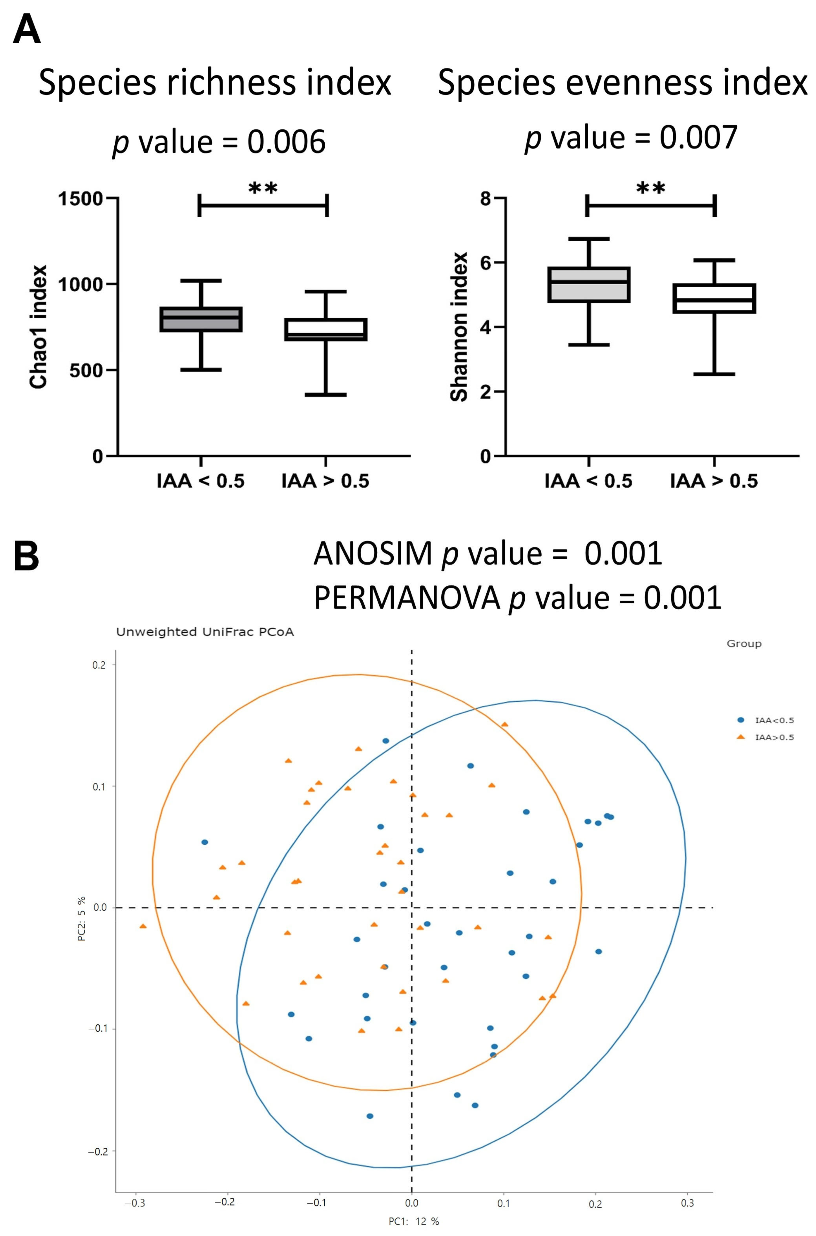
3.2. The Gut Microbiota Diversity Index Was Associated with IAA Levels

Patients exhibiting high IAA levels demonstrated lower α-diversity in their gut microbiome, as evidenced by lower values of both the Chao 1 diversity index and the Shannon diversity index, compared to patients with low IAA levels (Figure 1A). Moreover, in terms of β-diversity, patients with high IAA levels exhibited a significant distinction in the Bray–Curtis index compared to those with low IAA levels, as determined by the similarity-based analysis of similarities (ANOSIM) test and permutational multivariate analysis of variance (PERMANOVA) test (Figure 1B).

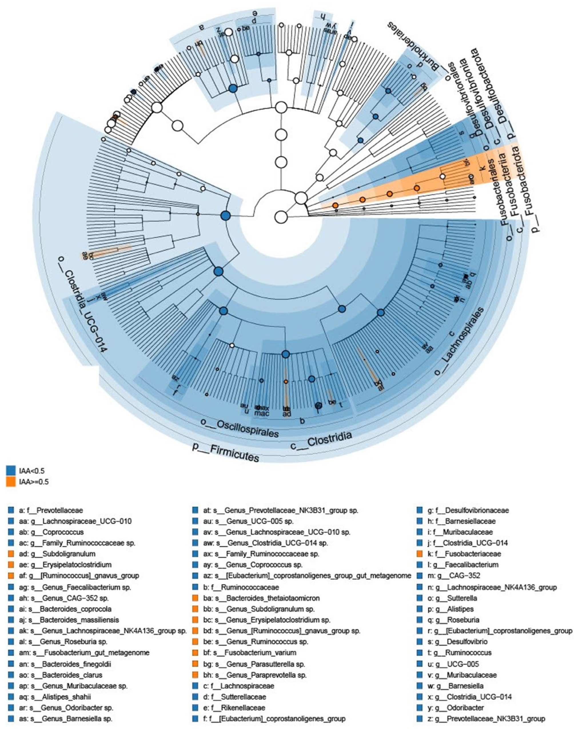
3.3. The Microbial Features Associated with IAA Levels

The gut microbiome features associated with patients exhibiting low and high IAA levels were analyzed by LefSe analysis to investigate bacterial taxa significantly enriched in the high- or low-IAA groups. The cladogram illustrating the tree associations between taxonomy and IAA levels is shown in Figure 2, and the detailed microbial taxa are depicted in Supplementary Figure S2. The high-IAA group was enriched in the following taxa: family Fusobacteriaceae; genera Subdoligranulum, Erysipelatoclostridium, Ruminococcus, and Parasutterella; and species Fusobacterium varium and Bacteroides thetaiotaomicron. Random forest cross-validation analyses at the genus (Figure 3A) and species (Figure 3B) levels revealed the top 20 taxa predicting IAA levels. Among these, the Bacteroides genus emerged as the most associated with IAA levels. Notably, within the Bacteroides genus, four species were enriched in individuals with low IAA levels: Bacteroides clarus, Bacteroides coprocola, Bacteroides massiliensis, and Bacteroides finegoldii. Conversely, one species, B. thetaiotaomicron, was enriched in individuals with higher IAA levels (Figure 2 and Figure 3). Furthermore, the heatmap demonstrated the correlation between these five species and sex, age, albumin, BUN, creatinine, glucose, potassium, LDL, sodium, nPCR, phosphate, total calcium, and uric acid (Figure 4). Notably, B. thetaiotaomicron showed a significant positive correlation with BUN, creatinine, sodium, phosphate, and uric acid, while B. clarus exhibited a significant negative association with BUN and nPCR. Additionally, B. coprocola demonstrated a negative correlation with uric acid (Figure 4).

4. Discussion

Within the cohort of 72 patients undergoing long-term hemodialysis, those in the higher-IAA group (IAA ≥ 0.5 μg/mL) displayed reduced α-diversity in the gut microbiome, as indicated by lower values in the Chao 1 diversity index and the Shannon diversity index. A statistically significant difference in β-diversity was also noted between the two patient groups with different IAA levels. The combination of LEfSe and random forest analysis revealed that the genus Bacteroides was most associated with IAA levels. Four of the species analyzed were enriched in individuals with low IAA levels, including B. clarus, B. coprocola, B. massiliensis, and B. finegoldii, whereas B. thetaiotaomicron was enriched in individuals with higher IAA levels. These findings offer valuable insights into the potential roles of the gut microbiota in modulating IAA levels and contribute to a deeper understanding of the intricate interplay between gut microbiota and IAA metabolism.
IAA is a tryptophan metabolite processed by the gut microbiota and is a protein-bound uremic toxin that cannot be adequately eliminated in patients undergoing hemodialysis. Consequently, this leads to the accumulation of toxins and functional alterations in the gut microbiota, contributing to the development of chronic systemic diseases. IAA is a specific ligand of AhR that can activate the AhR signaling pathway [23], thereby helping to regulate intestinal immunity and inflammation and maintain intestinal homeostasis [24]. A previous study reported that IAA is potentially involved in regulating cell proliferation, hematopoietic cells, and the Janus kinase (JAK)–signal transducer and activator of transcription (STAT) signaling pathway [5]. Understanding the intricate interplay between IAA and the gut microbiota is paramount to gaining insights into potential therapeutic targets for regulating IAA levels in patients with kidney disease. To the best of our knowledge, this study represents the first attempt to investigate and evaluate the link between gut microbiota composition, especially several specific Bacteroides species, and IAA levels in hemodialysis patients, offering novel perspectives for comprehending the role of the gut microbiota in IAA metabolism.
In previous studies, alterations in the gut microbiome have been observed in individuals undergoing hemodialysis [6], with hemodialysis patients often exhibiting decreased diversity of the gut microbiota [6]. Furthermore, in the present study, a higher level of IAA was associated with reduced microbiota diversity, potentially attributed to gut dysbiosis. This condition directly relates to increased intestinal permeability due to the deterioration of the epithelial barrier, leading to chronic systemic inflammation [25]. Furthermore, gut dysbiosis can result in elevated production of uremic toxins, such as trimethylamine (TMA), and other harmful metabolites while decreasing beneficial metabolites like short-chain fatty acids (SCFAs) [26]. Consequently, these metabolic disturbances caused by gut dysbiosis may contribute to the progression of kidney disease. Hence, the relationship between gut dysbiosis and kidney disease is bidirectional, as kidney disease can lead to changes in gut microecology.
Numerous studies have investigated the association between specific species and kidney disease but few have focused on the species associated with IAA levels in kidney disease. Tryptophan conversion into indole is primarily controlled by the enzyme tryptophanase [27], which has been detected in approximately 27 genera associated with the gut [28]. Indeed, several gut bacteria, including Clostridia, Bacteroides, and Escherichia produce IAA through tryptophan metabolism [29]. The present study revealed that Bacteroides was most associated with IAA levels, with four species (B. clarus, B. coprocola, B. massiliensi, and B. finegoldii) associated with a lower level of IAA and one species (B. thetaiotaomicron) with a higher level of IAA. B. thetaiotaomicron is a human symbiont that stabilizes the colon ecosystem [30] and synthesizes large amounts of spermidine and putrescine in the cecum of pectin-fed gnotobiotic rats [31]. Pectin is a soluble indigestible polysaccharide that stimulates cecal polyamine formation in rats [31]. It has been shown that the cecal contents of mono-associated rats fed a fiber-free diet contained large amounts of spermidine, the major polyamine [31]. IAA is the precursor of indole-3-aldehyde and 3-methylindole, and 3-methylindole is formed by the decarboxylation of IAA and produced by B. thetaiotaomicron [32]. Furthermore, a tryptophanase gene has been identified in B. thetaiotaomicron which is absent in other Bacteroides species. The gene BT1492 is prevalent among the gut metagenomes of healthy humans and correlated with Bacteroides abundance. B. thetaiotaomicron, unlike other Bacteroides species, can produce indole when cultured in tryptophan-rich conditions and a B. thetaiotaomicron mutant lacking the BT1492 gene showed no indole production, affirming its role in Bacteroides indole production [33]. Therefore, our investigation yielded a noteworthy revelation, as we observed a positive association between B. thetaiotaomicron and IAA levels but also a negative correlation between circulating IAA levels and four specific microbial species, namely B. clarus, B. coprocola, B. massiliensis, and B. finegoldii. This intriguing observation gains additional support from the Gusty atlas database [34], highlighting B. clarus as being negatively associated with blood IAA levels in healthy subjects. Our findings suggest that individual Bacteroides species may exert distinct effects on IAA levels at the species or sub-species level but further research is warranted to explore the underlying mechanisms and implications of these interactions.
The findings demonstrating associations between specific Bacteroides species and IAA levels suggest that the microbiome composition may impact circulating levels of this uremic toxin, which may in turn influence clinical outcomes. For example, B. thetaiotaomicron was positively correlated with uremic toxins, creatinine, sodium, phosphate, and uric acid, while other Bacteroides species showed negative correlations with some markers. These correlations hint that B. thetaiotaomicron enrichment and lower levels of other Bacteroides species may promote increased production and reduced clearance of uremic toxins and metabolites linked to worse cardiovascular outcomes. In contrast, higher abundances of B. clarus, B. coprocola, and B. massiliensis appear protective, correlating with reduced uremic toxin burden. Further mechanistic studies in animal models or clinical interventions modulating the gut microbiota are needed to determine if promoting the growth of beneficial Bacteroides species and limiting B. thetaiotaomicron could improve IAA metabolism, reduce systemic uremic toxin load, and translate to better clinical outcomes for CKD and dialysis patients. Ultimately, targeted manipulation of the gut microbiota and IAA pathways may offer therapeutic promise.
The strength of this study lies in its focused examination of the gut microbiome in hemodialysis patients and its impact on IAA levels. It used multiple algorithms to identify associated features; employing both LefSe and random forest approaches provides greater confidence in determining influential taxa. Thus, exploring correlations between individual bacteroid species and clinical variables can further support the clinical relevance of the microbiome findings, in particular the key species like B. thetaiotaomicron. Nevertheless, some limitations inherent in this study should be acknowledged. Firstly, the sample size was relatively small, concentrating solely on the hemodialysis population. Nonetheless, it is noteworthy that our study stands as the inaugural exploration into the interplay between IAA and the gut microbiota within hemodialysis patients. Amplifying the cohort with a larger sample size would undoubtedly offer a more comprehensive portrayal of microbiome alterations. Secondly, the utilization of a cross-sectional approach inherently restricts the establishment of causality. Nevertheless, we successfully identified robust associations between IAA levels and gut microbiota composition. To substantiate these findings and comprehend the migratory dynamics of the microbiome, future endeavors must encompass longitudinal investigations and follow-up assessments, encompassing both CKD and hemodialysis populations. Thirdly, we postulated an inverse relationship between IAA levels and clinical prognosis with a direct influence of shifts within the gut microbiota. It is essential to acknowledge that our analysis was conducted without continuous sampling of fecal specimens, limiting our capacity to discern the dynamic fluctuations therein. Notably, the dietary guidelines for CKD (pre-dialysis) patients diverge from those of dialysis patients, transitioning from low-protein to high-protein regimens as the condition progresses. This dietary evolution contributes to a uremic milieu within the gut lumen, triggering intricate changes that are still inadequately comprehended. Fourthly, the absence of a control group (such as patients with kidney disease not undergoing hemodialysis or healthy individuals) could limit the ability to draw definitive conclusions about the specific impacts of hemodialysis on gut microbiota and IAA levels. Lastly, a follow-up study is needed to enhance our understanding of these interlocking modifications. It is crucial to periodically assess fecal samples, capturing the microbial composition at various stages and facilitating a comprehensive, long-term assessment.

5. Conclusions

Our study demonstrated that a higher level of IAA was linked to reduced gut microbiota diversity, with five Bacteroides species being associated with circulating IAA levels.

Supplementary Materials

The following supporting information can be downloaded at: https://www.mdpi.com/article/10.3390/biomedicines12010148/s1, Figure S1: The distribution of indole-3-acetic acid (IAA) levels. Figure S2: Linear discriminative analysis (LDA) effect size (LEfSe) analysis between patients with low (blue) and high (orange) levels of indole-3-acetic acid.

Author Contributions

Conceptualization, P.-H.W., Y.-T.L. and W.-C.H.; Data curation, P.-H.W., Y.-T.L. and W.-C.H.; Study design and analysis plan, P.-H.W., Y.-T.L. and W.-C.H.; Statistical analysis, P.-H.W., Y.-T.L. and W.-C.H.; Funding acquisition, P.-H.W., Y.-F.T., W.L., C.-W.T., M.-C.K., S.-J.H. and Y.-T.L.; Investigation, P.-H.W., Y.-F.T., W.L., C.-W.T. and Y.-T.L.; Writing—first draft, P.-H.W. and Y.-T.L.; Writing—review and editing, Y.-F.T., W.L., C.-W.T., M.-C.K., Y.-W.C., S.-J.H., Y.-S.C., C.-J.T., K.-Y.L. and W.-C.H. All authors have read and agreed to the published version of the manuscript.

Funding

The research received financial support through grants from the Ministry of Science and Technology, Taiwan (MOST 111-2314-B-037-032-MY3, MOST 111-2314-B-037-083-MY3, and MOST 109-2314-B-037-088), Kaohsiung Medical University Hospital, Taiwan (KMUH111-1M60, KMUH111-1R73, KMUH111-1M09, KMUH110-0M13, KMUH110-0M73, and KMUH110-0M12), and Kaohsiung Medical University, Taiwan (KT112P012, NHRIKMU-111-I003, NHRIKMU-111-I003-2, NHRIKMU-111-I003-4, NHRIKMU-111-I001-3, NHRIKMU-113-I005, NPUST-KMU-111-P001, KMU-DK(B)110003, KMUH-DK(B)110003-1, KMU-DK(B)110003-2, KMU-DK(B)110003-3, KMU-DK(B)110003-4, KMU-DK(B)110003-5).

Institutional Review Board Statement

The research adhered to the principles of the Declaration of Helsinki and received approval from the Institutional Review Board of Kaohsiung Medical University for the study protocol (KMUHIRB-E(I)-20160095 and KMUHIRB-E(I)-20190189).

Informed Consent Statement

Informed consent was obtained from all participants involved in this study.

Data Availability Statement

The data will be provided upon request to the corresponding author.

Conflicts of Interest

The authors declare no conflicts of interest.

References

  1. Dou, L.; Sallee, M.; Cerini, C.; Poitevin, S.; Gondouin, B.; Jourde-Chiche, N.; Fallague, K.; Brunet, P.; Calaf, R.; Dussol, B.; et al. The cardiovascular effect of the uremic solute indole-3 acetic acid. J. Am. Soc. Nephrol. 2015, 26, 876–887. [Google Scholar] [CrossRef] [PubMed]
  2. Karu, N.; McKercher, C.; Nichols, D.S.; Davies, N.; Shellie, R.A.; Hilder, E.F.; Jose, M.D. Tryptophan metabolism, its relation to inflammation and stress markers and association with psychological and cognitive functioning: Tasmanian chronic kidney disease pilot study. BMC Nephrol. 2016, 17, 171. [Google Scholar] [CrossRef]
  3. Ji, Y.; Gao, Y.; Chen, H.; Yin, Y.; Zhang, W. Indole-3-acetic acid alleviates nonalcoholic fatty liver disease in mice via attenuation of hepatic lipogenesis, and oxidative and inflammatory stress. Nutrients 2019, 11, 2062. [Google Scholar] [CrossRef]
  4. Lin, Y.T.; Wu, P.H.; Lee, H.H.; Mubanga, M.; Chen, C.S.; Kuo, M.C.; Chiu, Y.W.; Kuo, P.L.; Hwang, S.J. Indole-3 acetic acid increased risk of impaired cognitive function in patients receiving hemodialysis. Neurotoxicology 2019, 73, 85–91. [Google Scholar] [CrossRef] [PubMed]
  5. Wu, P.-H.; Lin, Y.-T.; Wu, P.-Y.; Lee, H.-H.; Lee, S.-C.; Hung, S.-C.; Chen, S.-C.; Kuo, M.-C.; Chiu, Y.-W. Association between circulation indole-3-acetic acid levels and stem cell factor in maintenance hemodialysis patients: A cross-sectional study. J. Clin. Med. 2020, 9, 124. [Google Scholar] [CrossRef]
  6. Martin-Del-Campo, F.; Avesani, C.M.; Stenvinkel, P.; Lindholm, B.; Cueto-Manzano, A.M.; Cortes-Sanabria, L. Gut microbiota disturbances and protein-energy wasting in chronic kidney disease: A narrative review. J. Nephrol. 2023, 36, 873–883. [Google Scholar] [CrossRef] [PubMed]
  7. Liu, W.; Huang, J.; Liu, T.; Hu, Y.; Shi, K.; Zhou, Y.; Zhang, N. Changes in gut microbial community upon chronic kidney disease. PLoS ONE 2023, 18, e0283389. [Google Scholar] [CrossRef]
  8. Rysz, J.; Franczyk, B.; Lawinski, J.; Olszewski, R.; Cialkowska-Rysz, A.; Gluba-Brzozka, A. The impact of ckd on uremic toxins and gut microbiota. Toxins 2021, 13, 252. [Google Scholar] [CrossRef]
  9. Lin, T.Y.; Wu, P.H.; Lin, Y.T.; Hung, S.C. Gut dysbiosis and mortality in hemodialysis patients. NPJ Biofilms Microbiomes 2021, 7, 20. [Google Scholar] [CrossRef]
  10. Liu, F.; Xu, X.; Chao, L.; Chen, K.; Shao, A.; Sun, D.; Hong, Y.; Hu, R.; Jiang, P.; Zhang, N.; et al. Alteration of the gut microbiome in chronic kidney disease patients and its association with serum free immunoglobulin light chains. Front. Immunol. 2021, 12, 609700. [Google Scholar] [CrossRef]
  11. Hou, K.; Wu, Z.X.; Chen, X.Y.; Wang, J.Q.; Zhang, D.; Xiao, C.; Zhu, D.; Koya, J.B.; Wei, L.; Li, J.; et al. Microbiota in health and diseases. Signal Transduct. Target. Ther. 2022, 7, 135. [Google Scholar] [CrossRef]
  12. Gao, K.; Mu, C.L.; Farzi, A.; Zhu, W.Y. Tryptophan metabolism: A link between the gut microbiota and brain. Adv. Nutr. 2020, 11, 709–723. [Google Scholar] [CrossRef] [PubMed]
  13. Li, S. Modulation of immunity by tryptophan microbial metabolites. Front. Nutr. 2023, 10, 1209613. [Google Scholar] [CrossRef] [PubMed]
  14. Madella, A.M.; Van Bergenhenegouwen, J.; Garssen, J.; Masereeuw, R.; Overbeek, S.A. Microbial-derived tryptophan catabolites, kidney disease and gut inflammation. Toxins 2022, 14, 645. [Google Scholar] [CrossRef]
  15. Smith, E.A.; Macfarlane, G.T. Enumeration of human colonic bacteria producing phenolic and indolic compounds: Effects of ph, carbohydrate availability and retention time on dissimilatory aromatic amino acid metabolism. J. Appl. Bacteriol. 1996, 81, 288–302. [Google Scholar] [CrossRef] [PubMed]
  16. Elsden, S.R.; Hilton, M.G.; Waller, J.M. The end products of the metabolism of aromatic amino acids by clostridia. Arch. Microbiol. 1976, 107, 283–288. [Google Scholar] [CrossRef] [PubMed]
  17. Russell, W.R.; Duncan, S.H.; Scobbie, L.; Duncan, G.; Cantlay, L.; Calder, A.G.; Anderson, S.E.; Flint, H.J. Major phenylpropanoid-derived metabolites in the human gut can arise from microbial fermentation of protein. Mol. Nutr. Food Res. 2013, 57, 523–535. [Google Scholar] [CrossRef] [PubMed]
  18. Herlemann, D.P.; Labrenz, M.; Jurgens, K.; Bertilsson, S.; Waniek, J.J.; Andersson, A.F. Transitions in bacterial communities along the 2000 km salinity gradient of the baltic sea. ISME J. 2011, 5, 1571–1579. [Google Scholar] [CrossRef]
  19. Pedregosa, F.; Varoquaux, G.; Gramfort, A.; Michel, V.; Thirion, B.; Grisel, O.; Blondel, M.; Prettenhofer, P.; Weiss, R.; Dubourg, V.; et al. Scikit-learn: Machine learning in python. J. Mach. Learn. Res. 2011, 12, 2825–2830. [Google Scholar]
  20. Quast, C.; Pruesse, E.; Yilmaz, P.; Gerken, J.; Schweer, T.; Yarza, P.; Peplies, J.; Glockner, F.O. The silva ribosomal rna gene database project: Improved data processing and web-based tools. Nucleic Acids Res. 2013, 41, D590–D596. [Google Scholar] [CrossRef]
  21. Segata, N.; Izard, J.; Waldron, L.; Gevers, D.; Miropolsky, L.; Garrett, W.S.; Huttenhower, C. Metagenomic biomarker discovery and explanation. Genome Biol. 2011, 12, R60. [Google Scholar] [CrossRef]
  22. Svetnik, V.; Liaw, A.; Tong, C.; Culberson, J.C.; Sheridan, R.P.; Feuston, B.P. Random forest: A classification and regression tool for compound classification and qsar modeling. J. Chem. Inf. Comput. Sci. 2003, 43, 1947–1958. [Google Scholar] [CrossRef] [PubMed]
  23. Shen, J.; Yang, L.; You, K.; Chen, T.; Su, Z.; Cui, Z.; Wang, M.; Zhang, W.; Liu, B.; Zhou, K.; et al. Indole-3-acetic acid alters intestinal microbiota and alleviates ankylosing spondylitis in mice. Front. Immunol. 2022, 13, 762580. [Google Scholar] [CrossRef] [PubMed]
  24. Li, X.; Zhang, B.; Hu, Y.; Zhao, Y. New insights into gut-bacteria-derived indole and its derivatives in intestinal and liver diseases. Front. Pharmacol. 2021, 12, 769501. [Google Scholar] [CrossRef] [PubMed]
  25. Vijay, A.; Valdes, A.M. Role of the gut microbiome in chronic diseases: A narrative review. Eur. J. Clin. Nutr. 2022, 76, 489–501. [Google Scholar] [CrossRef]
  26. Cao, C.; Zhu, H.; Yao, Y.; Zeng, R. Gut dysbiosis and kidney diseases. Front. Med. 2022, 9, 829349. [Google Scholar] [CrossRef]
  27. Roager, H.M.; Licht, T.R. Microbial tryptophan catabolites in health and disease. Nat. Commun. 2018, 9, 3294. [Google Scholar] [CrossRef]
  28. Kaur, H.; Bose, C.; Mande, S.S. Tryptophan metabolism by gut microbiome and gut-brain-axis: An in silico analysis. Front. Neurosci. 2019, 13, 1365. [Google Scholar] [CrossRef]
  29. Gao, J.; Xu, K.; Liu, H.; Liu, G.; Bai, M.; Peng, C.; Li, T.; Yin, Y. Impact of the gut microbiota on intestinal immunity mediated by tryptophan metabolism. Front. Cell. Infect. Microbiol. 2018, 8, 13. [Google Scholar] [CrossRef]
  30. Rios-Covian, D.; Arboleya, S.; Hernandez-Barranco, A.M.; Alvarez-Buylla, J.R.; Ruas-Madiedo, P.; Gueimonde, M.; de los Reyes-Gavilan, C.G. Interactions between bifidobacterium and bacteroides species in cofermentations are affected by carbon sources, including exopolysaccharides produced by bifidobacteria. Appl. Environ. Microbiol. 2013, 79, 7518–7524. [Google Scholar] [CrossRef]
  31. Noack, J.; Dongowski, G.; Hartmann, L.; Blaut, M. The human gut bacteria bacteroides thetaiotaomicron and fusobacterium varium produce putrescine and spermidine in cecum of pectin-fed gnotobiotic rats. J. Nutr. 2000, 130, 1225–1231. [Google Scholar] [CrossRef] [PubMed]
  32. Ye, X.; Li, H.; Anjum, K.; Zhong, X.; Miao, S.; Zheng, G.; Liu, W.; Li, L. Dual role of indoles derived from intestinal microbiota on human health. Front. Immunol. 2022, 13, 903526. [Google Scholar] [CrossRef] [PubMed]
  33. Devlin, A.S.; Marcobal, A.; Dodd, D.; Nayfach, S.; Plummer, N.; Meyer, T.; Pollard, K.S.; Sonnenburg, J.L.; Fischbach, M.A. Modulation of a circulating uremic solute via rational genetic manipulation of the gut microbiota. Cell Host Microbe 2016, 20, 709–715. [Google Scholar] [CrossRef] [PubMed]
  34. Dekkers, K.F.; Sayols-Baixeras, S.; Baldanzi, G.; Nowak, C.; Hammar, U.; Nguyen, D.; Varotsis, G.; Brunkwall, L.; Nielsen, N.; Eklund, A.C.; et al. An online atlas of human plasma metabolite signatures of gut microbiome composition. Nat. Commun. 2022, 13, 5370. [Google Scholar] [CrossRef]
Figure 1. The α-diversity and β-diversity in hemodialysis patients with high and low levels of indole-3-acetic acid (IAA). (A) Patients with high IAA levels presented a higher α-diversity (species richness index and species evenness index) than patients with low IAA levels. The star mark ** referred to p-value <0.05 (B) Patients with high IAA levels had a different β-diversity (Bray–Curtis index) compared to patients with low IAA levels. The β-diversity p-value was calculated using homogeneity of group dispersions by analysis of similarities (ANOSIM) test and permutational multivariate analysis of variance (PERMANOVA) test.
Biomedicines 12 00148 g001
Figure 2. Taxonomic differences were detected between patients with low and high levels of indole-3-acetic acid. Cladogram showing differentially abundant taxonomic clades with an LDA score > 3.0 among patients with low and high levels of indole-3-acetic acid.
Biomedicines 12 00148 g002
Figure 3. Determination of bacteria specific for discrimination across low and high levels of indole-3-acetic acid in hemodialysis patients by applying random forest analysis at the (A) genus-level abundance and (B) species-level abundance. The star sign *** referred to the significant difference in abundance with p-value < 0.05.
Biomedicines 12 00148 g003
Figure 4. The correlation between IAA-associated Bacteroides species and sex, age, albumin, BUN, creatinine, glucose, potassium, LDL, sodium, nPCR, phosphate, total calcium, and uric acid in hemodialysis patients. * indicated p < 0.05.
Biomedicines 12 00148 g004
Table 1. Baseline characteristics of hemodialysis participants with high and low levels of indole-3-acetic acid.
IAA < 0.5 μg/mL
(n = 35)
IAA ≥ 0.5 μg/mL
(n = 37)
p-Value
Age, Mean (SD)57.6 (10.8)60.9 (9.7)0.171
Male, n (%)20.0 (57.1%)20.0 (54.1%)0.792
Hemodialysis vintage, months, Mean (SD)83.0 (66.8)95.2 (83.0)0.497
Proton pump inhibitor used, n (%)5.0 (14.3%)5.0 (13.5%)0.925
Clinical biochemistry, Mean (SD)
   Albumin (g/dL)4.0 (0.4)3.9 (0.3)0.482
   LDL cholesterol (mg/dL)107.7 (30.1)101.4 (26.3)0.343
   Fasting glucose (mg/dL)113.4 (38.0)105.8 (32.5)0.361
   Blood urea nitrogen (mg/dL)63.9 (11.5)65.8 (14.2)0.539
   Creatinine (mg/dL)10.9 (2.1)10.7 (1.8)0.655
   Uric acid (mg/dL)7.1 (1.1)6.8 (1.7)0.386
   Sodium (mmol/L)137.4 (2.3)137.2 (2.6)0.703
   Potassium (mmol/L)4.6 (0.7)4.4 (0.6)0.180
   Total calcium (mg/dL)9.2 (1.0)9.3 (0.9)0.574
   Phosphate (mg/dL)4.8 (0.9)4.6 (1.1)0.480
   nPCR (g/kg/day)1.1 (0.2)1.2 (0.2)0.079
LDL, low-density lipoprotein cholesterol; nPCR, normalized protein catabolic rate.
Disclaimer/Publisher’s Note: The statements, opinions and data contained in all publications are solely those of the individual author(s) and contributor(s) and not of MDPI and/or the editor(s). MDPI and/or the editor(s) disclaim responsibility for any injury to people or property resulting from any ideas, methods, instructions or products referred to in the content.

