Article

Differential Post-Translational Modifications of Proteins in Bladder Ischemia

1
Proteomics Laboratory, VA Boston Healthcare System, Boston, MA 02130, USA
2
Proteomics Laboratory, Department of Surgery, VA Boston Healthcare System, Boston University School of Medicine, Boston, MA 02130, USA
3
Departments of Urology and Pathology, VA Boston Healthcare System, Boston University School of Medicine, Boston, MA 02130, USA
*
Author to whom correspondence should be addressed.
Biomedicines 2024, 12(1), 81; https://doi.org/10.3390/biomedicines12010081
Submission received: 7 November 2023 / Revised: 23 December 2023 / Accepted: 27 December 2023 / Published: 28 December 2023
(This article belongs to the Special Issue Bench to Bedside in Neuro-Urology)

Abstract

:
Clinical and basic research suggests that bladder ischemia may be an independent variable in the development of lower urinary tract symptoms (LUTS). We have reported that ischemic changes in the bladder involve differential expression and post-translational modifications (PTMs) of the protein’s functional domains. In the present study, we performed in-depth analysis of a previously reported proteomic dataset to further characterize proteins PTMs in bladder ischemia. Our proteomic analysis of proteins in bladder ischemia detected differential formation of non-coded amino acids (ncAAs) that might have resulted from PTMs. In-depth analysis revealed that three groups of proteins in the bladder proteome, including contractile proteins and their associated proteins, stress response proteins, and cell signaling-related proteins, are conspicuously impacted by ischemia. Differential PTMs of proteins by ischemia seemed to affect important signaling pathways in the bladder and provoke critical changes in the post-translational structural integrity of the stress response, contractile, and cell signaling-related proteins. Our data suggest that differential PTMs of proteins may play a role in the development of cellular stress, sensitization of smooth muscle cells to contractile stimuli, and deferential cell signaling in bladder ischemia. These observations may provide the foundation for future research to validate and define clinical translation of the modified biomarkers for precise diagnosis of bladder dysfunction and the development of new therapeutic targets against LUTS.

1. Introduction

Proteins are produced in different shapes, sizes, and forms. Proteins play a fundamental role in the regulation of major processes of cell function and dysfunction, with an enigmatic position between human genetics and the varying phenotypes of both health and disease [1]. Systematic studies quantifying transcripts and proteins have revealed that transcript levels do not necessarily predict protein levels in many circumstances [2]. The spatial and temporal variations in the availability of precursors for protein biosynthesis and post-translational conformational changes in protein structure strongly influence their expression patterns and bioavailability [3]. There are multiple processes beyond the transcript level that modulate the expression level of a protein, including translation rates, modulation of protein half-life, protein synthesis delay, protein transport, and post-translational modifications (PTMs) [4]. Many proteins undergo PTMs that could immensely influence their structural integrity and function. PTMs of proteins activate important signaling pathways and regulate many biological processes [1,2,3,4]. Analyzing the diversity of PTMs is crucial for understanding downstream mechanisms of cell regulation. Mass spectrometry-based proteomic technologies are fundamental tools for identifying and mapping covalent modifications, providing an exceptional opportunity to detect and analyze a large number of differentially expressed protein variants in disease conditions [5]. Modern proteomic approaches have made large-scale assessments possible, enabling the mapping of modification sites and the quantification of the modified sites of proteins.
Despite tremendous advances in our understanding of the pathophysiology of systemic disorders, there are still substantial gaps in defining the precise molecular basis for early diagnosis and effective treatment of most disease conditions. Further knowledge toward a better understanding of differentially expressed proteins and protein modifications in tissues, cells, subcellular elements, and biological fluids may help the development of novel biomarkers for early diagnosis of systemic disorders and could lead to new therapeutic targets with the potential of new drug development through novel strategies. Proteomic approaches in these areas involve the comparative assessment of protein expression and modifications in disease conditions to detect aberrantly expressed proteins with the potential of illuminating new biomarkers. Thus, mass spectrometry-based proteomic studies of altered and modified proteins may have great potential to yield comprehensive profiles of proteins in tissues and cells for the potential identification of diagnostic and therapeutic markers.
Lower urinary tract symptoms (LUTS) increase with aging in both men and women [6,7,8,9]. Prostatic enlargement and bladder outlet obstruction (BOO) are the leading causes of LUTS in men. However, growing evidence suggests that all cases of LUTS in men are not necessarily due to prostatic enlargement or BOO, suggesting other aging-associated causes [10,11,12,13,14]. It is widely accepted that aging-associated LUTS involve multiple possible etiologies, including primary detrusor overactivity, compromised bladder smooth muscle contractility, or a combination of the two. A possible mechanism of non-obstructed bladder dysfunction and LUTS receiving increasing attention is the involvement of pelvic arterial atherosclerosis and bladder exposure to ischemia [6,7,8,9,10,11,12,13,14,15,16,17,18,19,20,21,22,23,24,25,26]. Ischemia was shown to interfere with lower urinary tract function and is believed to be an independent variable in the development of non-obstructed non-neurogenic detrusor overactivity and LUTS [6,7,8,9,10,11,12,13,14,15,16,17,18,19,20,21,22,23,24,25,26]. Ischemia alters the proteomic profiles of many organs, including the heart [27], brain [28,29], and intestine [30], but its impact on the bladder proteome remains largely elusive. Recent proteomic profiling using liquid chromatography tandem mass spectrometry (LC-MS/MS) has revealed critical information on alterations and modifications of proteins in bladder ischemia [31,32]. Differential expression and post-translational modifications (PTMs) of proteins appear to play important roles in ischemia-provoked bladder dysfunction [31,32]. We have previously reported that a total of 359 proteins are differentially expressed in bladder ischemia with twofold changes [32]. Gene ontology analysis suggested that differentially expressed proteins with fivefold changes are involved in molecular mechanisms underlying proteolysis and degenerative processes [32]. In the bladder proteome, a large number of nonzero delta masses (11,056) were detected, spanning over 1295 protein residues and grouped into 23 delta mass clusters, where each cluster represented a specific chemical reaction occurring on the side chain of the modified proteins. Among them, 12 delta mass clusters were significantly dysregulated in bladder ischemia (R2 > 0.5, ratio > twofold, p < 0.05), which were considered potential ischemia-regulated protein modifications [31]. Pathway analysis suggested the involvement of altered proteins in heat shock transcription factor 1-dependent transactivation, smooth muscle contraction, attenuation phase, and tetrahydrobiopterin synthesis, recycling, salvage, and regulation pathways [31]. These observations suggest that ischemia initiates a series of complex molecular responses through differential protein expression and massive PTMs in the bladder. It was shown that alterations of the bladder proteome by ischemia trigger a cascade of downstream signaling pathways with critical structural and functional consequences [31,32]. Differential expression and PTMs of proteins in bladder ischemia were associated with increased contractile activity consistent with detrusor overactivity, loss of smooth muscle cells, diffuse fibrosis, non-compliance, neural structural damage, and marked changes in the ultrastructure of the subcellular elements [31,32]. In this study, we focused on the proteomic assessment of bladder ischemia to define comprehensive differential (post-translational) modifications of the stress response, contractile, and cell signaling-related proteins and determine activation of downstream pathways in an animal model.

2. Materials and Methods

2.1. Bladder Ischemia Model

Animal care and experimental procedures followed protocol number 396-J-0522 and guidelines approved by the Institutional Animal Care and Use Committee (IACUC) at VA Boston Healthcare System. Our IACUC is accredited by the Association for Assessment and Accreditation of Laboratory Animal Care (AAALAC). A total of six male Sprague-Dawley rats, 10–12 weeks old, weighing 200–400 g, were used. Control rats were obtained from Charles River Laboratories in Boston, Massachusetts, USA. Bladder ischemia was produced in apolipoprotein E knockout rats using balloon endothelial denudation of the iliac arteries with a 2F Fogarty arterial embolectomy catheter to produce atherosclerotic occlusive disease, as we have previously reported [31,32,33]. Sham control rats underwent similar surgical procedures without arterial endothelial denudation. After 8 weeks, bladder ischemia was verified by measuring microcirculatory bladder perfusion using the Moor FLPI-2 Laser Speckle Contrast Imaging system (Moor Instruments, Wilmington, DE, USA). This system provides blood flow imaging in a real-time and contact-free manner and is capable of measuring tissue microcirculation blood flow up to a tissue depth of 3 mm. To obtain blood flow measurements, the animals were anesthetized, and then a lower abdominal incision was made to expose the bladder. The speckle laser was positioned 25 cm above the bladder, and blood perfusion was recorded for 30 s at five different sites of the bladder. Mean bladder blood perfusion was analyzed using the moorFLPI-2 Review Software, v. 4.0 (Moor Instruments, Wilmington, DE, USA). After blood flow measurement, the animals were euthanized, and bladder tissues were prepared and processed for liquid chromatography-tandem mass spectrometry (LC-MS/MS) proteomic analysis as described below.

2.2. Sample Preparation for Proteomic Analysis

Bladder tissue samples were collected from three animals with bladder ischemia and three sham control bladders and processed for proteomic analysis. Bladder tissue samples were homogenized in 1 mL of 1X RIPA lysis buffer (Millipore 20–188, MilliporeSigma, Burlington, MA, USA) supplemented with 10 µL of 500 mM DTT and 10 µL of protease inhibitor cocktail (MilliporeSigma, Burlington, MA, USA), and sonicated three times for 5 s on ice. The tissue lysates were centrifuged, the supernatants were collected, and protein concentrations were determined with the bicinchoninic acid (BCA) assay (MilliporeSigma, Burlington, MA, USA).

2.3. Sodium Dodecyl Sulfate Polyacrylamide Gel Electrophoresis (SDS-PAGE) Fractionation and Trypsin Digestion

The proteins were fractionated using one-dimensional SDS-PAGE. One hundred micrograms of each bladder tissue lysate protein were run into each well on the SDS-PAGE gel. The gel was then stained with Coomassie Brilliant Blue, and each gel lane was subsequently divided into 10 fractions based on molecular weight. Gel slices underwent the following steps: washing with 50 mM ammonium bicarbonate, dehydration with acetonitrile, and lyophilization in a SpeedVac (Thermo Scientific, Waltham, MA, USA). Subsequently, proteins were in-gel digested with sequencing-grade modified trypsin (Promega, Madison, WI, USA) at a protein-to-trypsin ratio of 25:1 in 50 mM ammonium bicarbonate (pH 8) overnight at 37 °C. The extracted peptides from each gel slice were subjected to desalting using ZipTip C18 columns (MilliporeSigma, Burlington, MA, USA) and then dried in a SpeedVac before LC-MS/MS analysis. The raw data for the 10 fractions from each tissue sample were searched against the UniProt database as a group.

2.4. Liquid Chromatography-Tandem Mass Spectrometry (LC-MS/MS) Analysis

The goal was to explore proteomic modifications in bladder ischemia. Therefore, the calculated statistical differences described in this report are not hypothetical. The desalted peptides were subjected to peptide fractionation using an EASY-nLC 1000 system (Thermo Scientific, Waltham, MA, USA). Samples were initially trapped on a C18 pre-column and subsequently fractionated using a long C18 column (300 × Ø 0.075 mm, 3 μm; Reprosil, Germany) employing a 180-min linear gradient of 5–35% acetonitrile/0.1% formic acid at a flow rate of 250 nL/min. Solvent A consisted of 0.1% formic acid in water, while solvent B comprised 0.1% formic acid in acetonitrile. MS and MS/MS spectra were acquired using an LTQ-Orbitrap Elite mass spectrometer (Thermo Scientific, Waltham, MA, USA) in a data-dependent mode. Each biological sample was replicated three times in the LC-MS/MS analysis, and the delta masses were individually subjected to an open search. Subsequently, the delta masses from all samples were clustered, and the average of the three replicates was given. The spray voltage was set at 2.1 kV, and the capillary temperature was maintained at 275 °C. MS spectra were acquired in profile mode within the m/z range of 350 to 1800 at a resolution of 60,000 at 400 m/z. The automatic gain control was set at 1 × 10⁶. MS/MS fragmentation was applied to the 30 most intense peaks for every full MS scan in the collision-induced dissociation mode. Normalized collision energies were fixed at 35%, with an activation time of 10 ms for the MS/MS method. The maximum precursor ion injection time for MS/MS was set at 100 ms. A repeat count of 2 and a dynamic exclusion duration of 90 s were applied. The minimal signal required for MS/MS was set at 1000.

2.5. The Clustered Delta Masses

Typically, MS/MS spectra undergo a database search against the rat protein database using wildcard search functionality within the Byonic software (Version 3.6, Protein Metrics Inc., Cupertino, CA, USA) [34]. Results were then filtered based on criteria including Score ≥ 300, DeltaModsScore ≥ 10, FDR2D ≤ 0.01, and FDR_uniq.2D ≤ 0.01. Amino acid residues differing from coded genomic sequences were identified to calculate nonzero delta masses (delta mass > 0.01), representing the mass disparities between actual and theoretical (coded) amino acids. These amino acids with nonzero delta masses were referred to as “non-coded amino acids (ncAAs)”. For occurrence analysis, delta masses were rounded to four decimal places, and the number of redundancies was considered as the frequency of the delta mass. In clustering, delta masses were grouped into subcategories with 1-Da intervals, defined by n − 0.5 and n + 0.5 Da (where n ranged from −200 to 1000). The delta masses within each mass window were subjected to multivariate clustering using Gaussian mixture components [35]. Specific constraints were applied, including (1) a peak half-width > 1 ppm, (2) a peak distance > 2 peak widths, and (3) a cluster size > 20. The determination of clusters within each window was based on the Bayesian information criterion (BIC) [36], where larger BIC values indicated a stronger model and greater confidence in the number of clusters. Clusters within each window were individually fitted with Gaussian regression to calculate parameters such as the peak value (clustered delta mass), standard deviation (SD), and goodness-of-fit (R2). Clustered delta masses were annotated with known PTMs and amino acid substitutions found in databases like UniMod, RESID, ExPASy, and ABRF. Matched clusters were regarded as true delta masses present in the analyzed protein samples. Unmatched clusters were assigned with confidence only when the goodness-of-fit R2 > 0.5 and/or when the cluster was predominantly associated with a single amino acid (>50%). The significance of delta masses (modifications) was determined using parameters from a Gaussian distribution, assuming random errors. Thus, a significant delta mass was defined with R2 > 0.5, which represents a very high standard for non-linear regression. When comparing 3 ischemia samples to 3 controls, only ratios > 2-fold with p < 0.05 between the ischemia and control group were considered “significant”. Top of Form.

2.6. Statistical Analysis
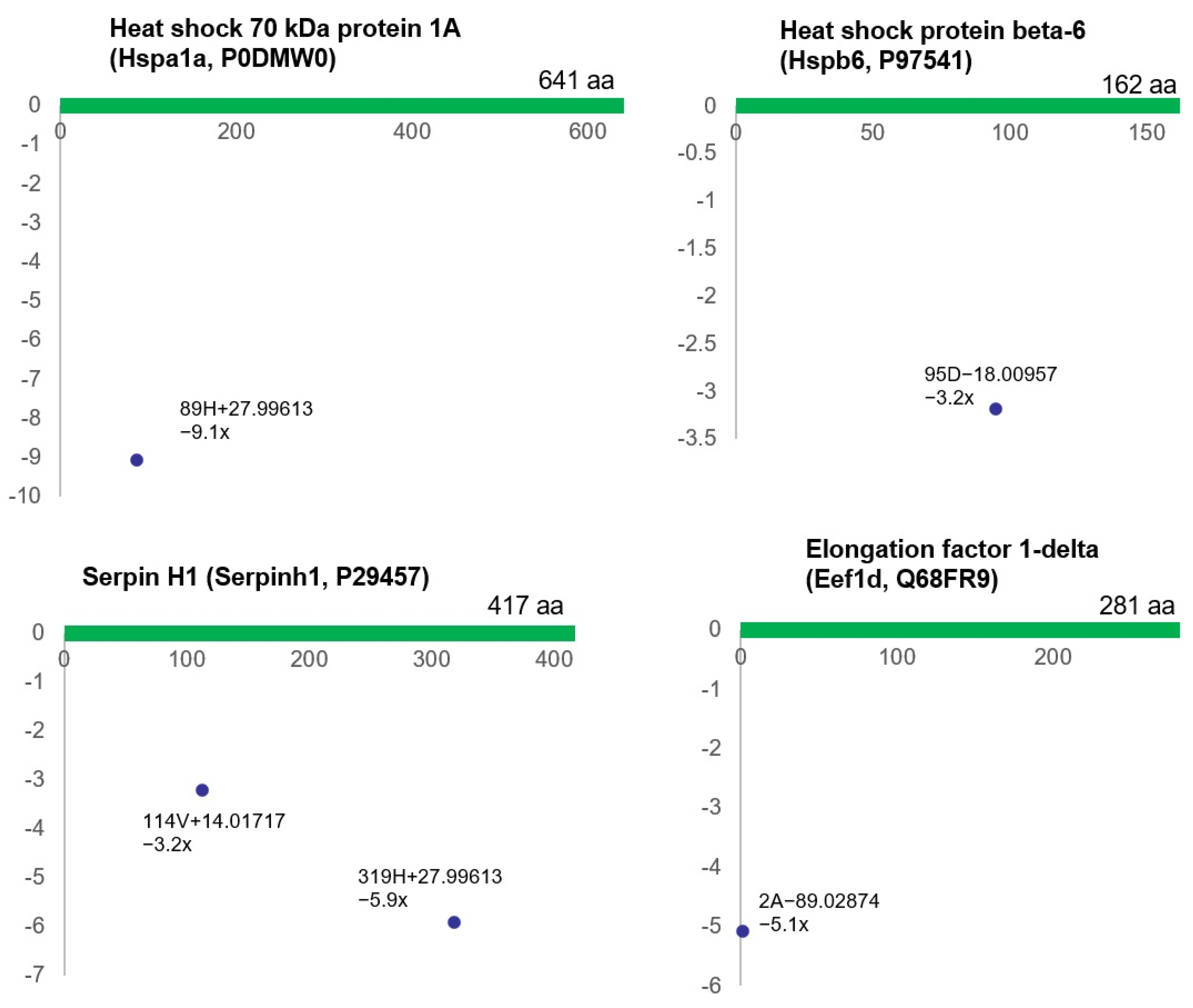
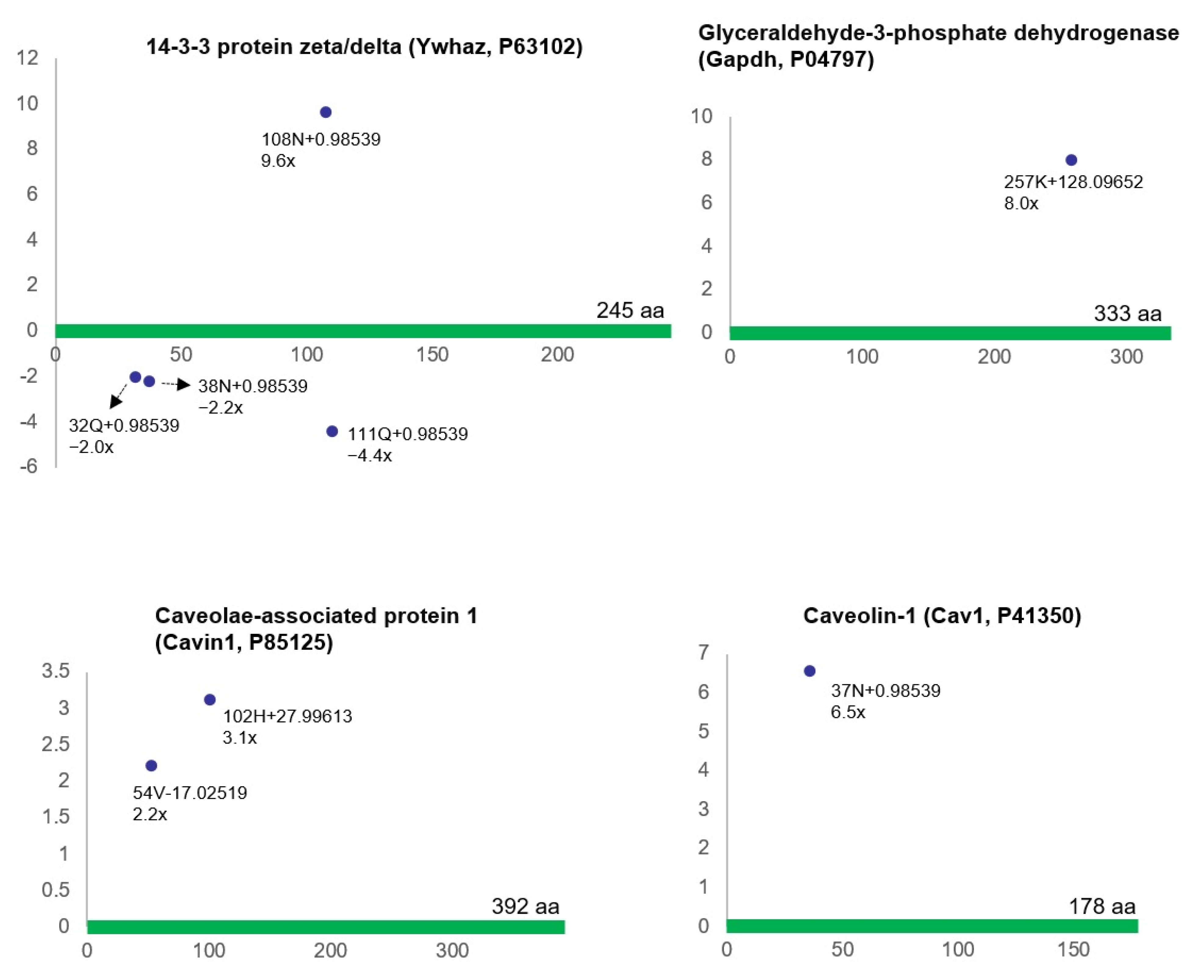
Statistical analysis was performed using IBM SPSS 19.0 software (Armonk, NY, USA). Group comparisons were conducted using two-tailed Student’s t-tests, and an ANOVA with Tukey’s Honest Significant Difference (HSD) post-hoc test was utilized to assess variation between the groups. Significant differences were determined at the p < 0.05 level. p-values in proteomic data are based on a comparison of samples from three rats with bladder ischemia versus samples from three control rats. The term “significant” refers to changes in the bladder ischemia group in comparison with controls with a p value of <0.05.

3. Results

3.1. Validation of Bladder Ischemia in the Rat Model

Measurements of bladder microcirculatory blood flow with the Moor FLPI-2 Laser Speckle Contrast Imaging system provided full-field laser recording and detected mean microcirculatory blood flow up to a tissue depth of 3 mm. Using this measurement system, we compared the bladder blood perfusion of rats with iliac artery atherosclerosis versus sham control rats. Statistical analysis of blood flow images suggested significant bladder ischemia in animals with arterial atherosclerosis. Ischemia was characterized by a statistically significant decrease in bladder blood perfusion in animals with atherosclerotic occlusive disease in comparison with sham controls, as shown in Figure 1.

3.2. Differential Protein Modifications in Bladder Ischemia

In-depth analysis of a previously reported proteomic dataset [31] suggested that ischemia compromises the bladder proteome. These findings, along with our previous reports [31,32], imply that ischemia may play a key role in differential expression and post-translational modifications of proteins. The LC-MS/MS analysis of rat bladder tissues in our previous proteomic study showed that 172 proteins are upregulated and 527 proteins are downregulated in the ischemic bladder in comparison with control bladder samples (p ≤ 0.05) [32]. Proteomic analysis with consideration of at least a twofold increase or decrease revealed 97 up-regulated and 262 down-regulated proteins in the ischemic bladder in comparison with control samples [32].
Further analysis of the ischemic bladder proteome compared to controls revealed that 12 of the 23 amino acid variations were significantly dysregulated (R2 > 0.5, ratio > twofold, p < 0.05), suggesting potential ischemia-regulated protein modifications [31]. Interestingly, some of the amino acid variations provoked by ischemia that matched the molecular weights of the amino acid substitution did not corroborate the point mutations at the DNA and/or RNA levels, suggesting the production of non-coded amino acids (ncAAs) that might have resulted from post-translational modifications in the ischemic bladder. It is thought that ncAAs are produced in the process of amino acid substitutions or post-translational modifications of proteins. However, the precise nature of ncAAs and the underlying mechanisms regulating their formation remain elusive and warrant further investigation. Up-regulated and/or down-regulated ischemia-associated ncAA-containing proteins in bladder ischemia are summarized in Table 1 below.
Proteomic analysis in our present study suggested that three groups of proteins in the bladder proteome, including contractile proteins and their associated proteins, stress response proteins, and cell signaling-related proteins, are conspicuously impacted by ischemia, as shown in Table 1 and Figure 2, Figure 3 and Figure 4. Widespread differential (post-translational) modifications were detected in the contractile proteins and their associated proteins in the ischemic bladder smooth muscle. This included actin (Actg1, P63259), myosin light polypeptide 6 (Myl6, Q64119), calponin-1 (Cnn1, Q08290), protein phosphatase 1 regulatory subunit 12A (Ppp1r12a, Q10728), alpha-actinin-1 (Actn1, Q9Z1P2), gelsolin (Gsn, Q68FP1), and transgelin (Tagln, P31232) (Figure 2).
Stress response proteins were also markedly affected by bladder ischemia, showing differential (post-translational) modifications of the proteins, including heat shock 70 kDa protein 1A (Hspa1a, P0DMW0), heat shock protein beta-6 (Hspb6, P97541), serpin H1 (Serpinh1, P29457), and elongation factor 1-delta (Eef1d, Q68FR9) (Figure 3).
Bladder ischemia also provoked differential (post-translational) modifications of cell signaling-related proteins, including 14-3-3 protein zeta/delta (Ywhaz, P63102), glyceraldehyde-3-phosphate dehydrogenase (Gapdh, P04797), caveolae-associated protein 1 (Cavin1, P85125), and caveolin-1 (Cav1, P41350) (Figure 4).

4. Discussion

The role of ischemia in overactive bladder and lower urinary tract symptoms (LUTS) has been documented by clinical measurement of bladder blood flow, which revealed a close link between decreased bladder perfusion and LUTS in elderly men and women [6,7,8,9,10,11,12,13,14,15,16,17,18,19,20,21,22,23,24,25,26]. It was shown that the severity of LUTS closely correlates with the degree of bladder ischemia [6,7,8,9,10,11,12,13,14,15,16,17,18,19,20,21,22,23,24,25,26]. An epidemiological study involving 36,042 patients reported a significant correlation between LUTS and peripheral arterial occlusive disease [24]. Several other studies have shown that human bladder blood flow decreases with aging in both men and women, and decreased bladder blood flow significantly correlates with reduced bladder compliance and the development of LUTS [6,7,8,9,10,11,12,13,14,15,16,17,18,19,20,21,22,23,24,25,26]. Studies of animal models have revealed that ischemia compromises the bladder proteomic profiles and instigates a cascade of downstream signaling pathways with critical structural and functional consequences [37,38,39,40,41]. These observations, cumulatively, suggest that therapeutic strategies to improve bladder perfusion could prevent or reverse detrusor overactivity and LUTS in patients with pelvic ischemia. Downstream mechanisms mediating overactivity and structural damage in bladder ischemia appear to involve disruption of cellular energy homeostasis, cellular stress, and cell survival signaling [37,38,39,40,41]. It is thought that the upregulation of cellular stress response molecules by ischemia activates homeostatic mechanisms to signal nutrient deficiency, hypoxic stressors, and cell danger [42,43,44,45,46,47,48,49,50]. Our present study suggests differential (post-translational) modifications of contractile proteins and their associated proteins, cellular stress response proteins, and cell signaling-related proteins in bladder ischemia. The contractile proteins and their associated proteins modified by ischemia included actin (Actg1, P63259), myosin light polypeptide 6 (Myl6, Q64119), calponin-1 (Cnn1, Q08290), protein phosphatase 1 regulatory subunit 12A (Ppp1r12a, Q10728), alpha-actinin-1 (Actn1, Q9Z1P2), gelsolin (Gsn, Q68FP1), and transgelin (Tagln, P31232). The stress response proteins modified by ischemia included heat shock 70 kDa protein 1A (Hspa1a, P0DMW0), heat shock protein beta-6 (Hspb6, P97541), serpin H1 (Serpinh1, P29457), and elongation factor 1-delta (Eef1d, Q68FR9). The cell signaling-related proteins modified by ischemia included 14-3-3 protein zeta/delta (Ywhaz, P63102), glyceraldehyde-3-phosphate dehydrogenase (Gapdh, P04797), caveolae-associated protein 1 (Cavin1, P85125), and caveolin-1 (Cav1, P41350).
Post-translational modifications (PTMs) involve the loss of structural integrity of one or more amino acid residues resulting from the addition or removal of modifying groups on a translated protein for covalent processing modification [51,52,53,54,55]. Functional consequences of PTMs depend on the domain and motif that regulate cell function and changes that could provoke cellular structural damage, cell dysfunction, and the development of pathological conditions. The process of PTMs involves numerous episodes, including phosphorylation, acetylation, methylation, ubiquitination, glycosylation, and succinylation [51,52,53,54,55]. These processes play key roles in various aspects of cellular function and structural modifications [51,52,53,54,55]. For example, the intracellular concentration of sodium and potassium ions is regulated by the phosphorylation of the Na+/K+-ATPase pump on the cell membrane [52]. Acetylation may compromise protein conformation and alter interactions with substrates and cofactors [53]. Methylation of proteins regulates cell function by altering the original structural sequence of peptide chains, or could provoke dysfunction through the activation of downstream mechanistic pathways [54]. Ubiquitination can regulate cell function or mediate signaling pathways instigated by pathological conditions involving protein degradation or blockade of protein interactions [55].
It has been shown that ischemia may be a key player in provoking PTMs in a variety of cells [56,57,58,59,60,61,62,63]. The process of PTMs in ischemia appears to involve oxidative insult resulting from excessive production of free radicals in the ischemic cells [56,57,58,59,60,61,62]. The accumulation of free radicals instigates modification of the translated protein by inserting or detaching modified groups from the amino acid residues [56,57,58,59,60,61,62]. Oxidative insults mediated by free radicals in the ischemic tissues were shown to modify the fibrinogen molecule and provoke inflammatory responses [59]. DNA damage associated with histone PTMs was shown to play a key role in retinal neurovascular degeneration following ischemia/reperfusion injury [61]. Transient cerebral ischemia was shown to activate various protein PTMs involved in key neuronal functions and signaling pathways [62]. These observations suggest that the pathogenesis of chronic ischemia and ischemia/reperfusion injury is closely related to PTMs of proteins at functional domains [56,57,58,59,60,61,62,63]. It is thought that ischemia and oxidative stress compromise the physical and chemical properties of proteins that synchronize cell function in living organisms or elicit pathophysiological development in disease conditions [56,57,58,59,60,61,62,63]. However, certain limitations in protein sequencing technologies prevent the precise determination of global protein modifications throughout the proteome [63]. Therefore, it is difficult to precisely determine how many protein modifications are involved in tissue ischemia and how many underlying mechanisms are activated by PTMs under ischemic conditions. As each modification is likely to alter cellular structure and lead to functional consequences, systematic assessment of protein PTMs may be a convenient approach to examine molecular mechanisms and determine downstream pathways in the ischemic bladder.
Our previous LC-MS/MS-based proteomic approach detected 4277 proteins in the ischemic bladder and 4602 proteins in the control bladder [32]. Spectral count analysis revealed 172 up-regulated proteins and 527 down-regulated proteins in the ischemic bladders in comparison with controls [32]. Gene ontology analysis of the altered proteins in bladder ischemia based on their biological properties suggested the involvement of 66 ischemia-regulated proteins with a greater than five-fold change in functional categories regulating protein metabolic processes, proteolysis, and hydrolase activity [32]. Gene ontology analysis based on the molecular function of the 66 altered proteins implied their involvement in regulating peptidase and other enzymatic activities in bladder ischemia [32]. Canonical pathway analysis suggested a close link between altered proteins with a greater than two-fold change in bladder ischemia and several signaling pathways, including the ubiquitination pathway, the nuclear factor-erythroid 2-related factor 2 (NRF2)-mediated oxidative stress response pathway, and the extracellular-signal-regulated kinase (ERK)/mitogen-activated protein kinase (MAPK) pathway [32]. Network analysis revealed a link between altered proteins and cytoskeleton organization, cell death, and glucose metabolism disorders in bladder ischemia [32]. In another proteomic study, we found that ischemia provokes the differential formation of non-coded amino acids (ncAAs) in the bladder through mechanisms involving post-translational protein modifications and amino acid substitution [31]. Gene ontology analysis using Protein ANalysis THrough Evolutionary Relationships (PANTHER) revealed that the largest group of ischemia-associated ncAA-containing proteins in bladder ischemia relates to the cytoskeletal proteins [31]. Search Tool for Recurring Instances of Neighboring Genes (STRING) analysis of the protein-protein interaction network among the up-regulated ischemia-associated ncAA-containing proteins suggested that the protein-protein interaction networks were broadly grouped into three clusters: cytoskeleton-associated proteins, cell signaling-related proteins, and binding/transport-related proteins [31]. STRING analysis also suggested that interaction networks between the down-regulated ischemia-associated ncAA-containing proteins were broadly grouped into three clusters: cytoskeleton-associated proteins, metabolic enzymes, and molecular chaperones [31]. In our present study, a comprehensive analysis of the ischemia-associated ncAA-containing proteins revealed that three groups of proteins in the bladder proteome, including contractile proteins and their associated proteins, stress response proteins, and cell signaling-related proteins, are conspicuously impacted by ischemia. Widespread differential (post-translational) modifications were detected in the contractile proteins and their associated proteins in the ischemic bladder smooth muscle, including actin (Actg1, P63259), myosin light polypeptide 6 (Myl6, Q64119), calponin-1 (Cnn1, Q08290), protein phosphatase 1 regulatory subunit 12A (Ppp1r12a, Q10728), alpha-actinin-1 (Actn1, Q9Z1P2), gelsolin (Gsn, Q68FP1), and transgelin (Tagln, P31232). The actin cytoskeleton plays a key role in the configuration of subcellular structures and the preservation of cell function [64]. We have previously reported that bladder ischemia compromises actin cytoskeleton signaling and provokes aberrant smooth muscle contractile activity characterized by smooth muscle hypersensitivity to contractile stimuli [38,39,41]. Our findings are consistent with studies in the heart showing that oxidative modification of actin plays a key role in the pathophysiology of ischemic disorders [64,65,66]. It was shown that the oxidation of cysteines in actin interferes with the polymerization/elongation of G-actin and enhances F-actin fragility [67,68,69]. In cardiomyocytes, S-nitrosylation of α-actin promotes relaxation while impairing contraction [70]. These observations suggest that oxidative modification of actin may contribute to overactive smooth muscle contractions and detrusor overactivity in bladder ischemia. Ischemia-induced PTMs may compromise the dynamic properties of actin, provoke actin depolymerization, and sensitize actin to contractile stimuli.
Proteomic analysis of ischemia-associated ncAA-containing proteins also revealed that bladder ischemia elicits differential (post-translational) modifications of the proteins involved in cellular stress response, including heat shock 70 kDa protein 1A (Hspa1a, P0DMW0), heat shock protein beta-6 (Hspb6, P97541), serpin H1 (Serpinh1, P29457), and elongation factor 1-delta (Eef1d, Q68FR9). Such reactions of the stress response proteins may suggest cellular responses to ischemia under environmental changes with complex homeostatic disturbances in the ischemic cells that could compromise subcellular elements and provoke macromolecular damage. Our study suggests a close link between the interruption of nutrients and oxygen delivery to the ischemic cells, the accumulation of free radicals and metabolic waste, and differential (post-translational) modifications of the stress response proteins in bladder ischemia. The cell’s fate after exposure to stressful conditions such as ischemia depends on the severity and duration of ischemia and the cell’s defensive capacity to confine stress, eliciting molecular responses including PTMs [37,38,39]. Cellular stress sensors may play a key role in regulating molecular responses and protein modifications in the ischemic bladder [37,38,39].
In addition, proteomic analysis of the ischemia-associated ncAA-containing proteins revealed differential (post-translational) modifications of the cell signaling-related proteins, including 14-3-3 protein zeta/delta (Ywhaz, P63102), glyceraldehyde-3-phosphate dehydrogenase (Gapdh, P04797), caveolae-associated protein 1 (Cavin1, P85125), and caveolin-1 (Cav1, P41350). Cell signaling-related proteins regulate cellular responses to a diverse, multifaceted, and alternating mix of signals involved in protein expression, protein localization, protein endeavor, and protein–protein interactions [56,71,72,73]. These proteins are involved in critical aspects of signal transduction, modulating critical cellular responses to a nearly unlimited set of stimuli [56,71,72,73]. Our data suggest that the differential (post-translational) modifications of cell signaling-related proteins in the ischemic bladder may vary depending on the sensitivity, duration, and dynamics of the cellular stress response to ischemia. Signal-dependent modifications in the expression levels and PTMs of cell signaling-related proteins may play an important role in the regulation of downstream processes including protein degradation, localization, and protein-protein interactions [56,71,72,73]. The activity of cell signaling-related proteins is tightly regulated so that the products of the responses that they control are only elicited at the appropriate time when cells are stressed and signal danger [56,71,72,73]. It seems that some of the regulatory mechanisms involve protein expression or protein localization, but crucial functional consequences result from the regulation of protein activity by PTMs of the signaling molecule, intramolecular crosstalk between different domains within the protein, and/or activation of crosstalk mechanisms with other proteins [56,71,72,73]. A limitation of our proteomic analysis may relate to the sample size of 3 animals per group. However, LC-MS/MS analysis yielded an extensive proteomic dataset with a very large amount of data to be analyzed and interpreted. In LC-MS/MS analysis, each sample was run three times. We extracted proteins from each animal, ran SDS-PAGE, and divided them into 10 gel fragments. After sample preparations (in-gel digestions) from each 10 fragments, each sample of 10 was analyzed three times in LC-MS/MS. Therefore, each animal generated 30 sets of MS/MS data, allowing proper data interpretation with exploratory data analysis.

5. Conclusions

The role of bladder ischemia as a contributing factor in detrusor overactivity and LUTS has been documented by epidemiological studies and clinical measurements of bladder blood flow that suggest a close link between vascular risk factors, pelvic ischemia, and the development of lower urinary tract dysfunction. Ischemia appears to provoke substantial changes in the proteomic profiles of the bladder. The proteomic approach in our present study detected a widespread differential occurrence of ncAAs in bladder ischemia that may result from post-translational protein modifications and amino acid substitution mechanisms. These modifications may contribute to the dysregulation of downstream pathways, with important structural and functional consequences. The profiles of differential (post-translational) modifications and subsequent dysregulation of corresponding pathways in bladder ischemia may suggest a high sensitivity of the bladder to reduced blood perfusion and hypoxia. Differential (post-translational) modifications of proteins provoked by ischemia may play a vital role in increased smooth muscle contractions, structural damage, and the development of an overactive bladder. Proteomic responses, including differential post-translational modifications of proteins in bladder ischemia, may provide the foundation for future research towards clinical translation to identify diagnostic targets and develop effective therapeutic strategies against bladder dysfunction and LUTS.

Author Contributions

Conceptualization, J.-H.Y. and K.M.A.; Methodology, J.-H.Y. and K.M.A.; Formal analysis, H.-P.C. and J.-H.Y.; Investigation, H.-P.C., J.-H.Y. and K.M.A.; Resources, K.M.A.; Writing—original draft, K.M.A.; Writing—review & editing, H.-P.C. and K.M.A.; Visualization, H.-P.C. and K.M.A.; Supervision, J.-H.Y. and K.M.A.; Funding acquisition, K.M.A. All authors have read and agreed to the published version of the manuscript.

Funding

This work was supported by Merit Review Award Number I01 BX004372 from the United States (U.S.) Department of Veterans Affairs Biomedical Laboratory R&D (BLRD) Service.

Institutional Review Board Statement

Animal care and experimental procedures were following protocol number 396-J-0522 and guidelines approved by the Institutional Animal Care and Use Committee (IACUC) at VA Boston Healthcare System. Our IACUC is accredited by the Association for Assessment and Accreditation of Laboratory Animal Care (AAALAC).

Informed Consent Statement

Not applicable.

Data Availability Statement

Our research data will be shared with the public through the National Library of Medicine (NML) PubMed Central website and presentations at scientific meetings.

Conflicts of Interest

The authors declare no conflict of interest.

References

  1. Liu, Y.; Beyer, A.; Aebersold, R. On the Dependency of Cellular Protein Levels on mRNA Abundance. Cell 2016, 165, 535–550. [Google Scholar] [CrossRef]
  2. Maier, T.; Güell, M.; Serrano, L. Correlation of mRNA and protein in complex biological samples. FEBS Lett. 2009, 583, 3966–3973. [Google Scholar] [CrossRef]
  3. Lu, P.; Vogel, C.; Wang, R.; Yao, X.; Marcotte, E.M. Absolute protein expression profiling estimates the relative contributions of transcriptional and translational regulation. Nat. Biotechnol. 2007, 25, 117–124. [Google Scholar] [CrossRef]
  4. Battle, A.; Khan, Z.; Wang, S.H.; Mitrano, A.; Ford, M.J.; Pritchard, J.K.; Gilad, Y. Genomic variation. Impact of regulatory variation from RNA to protein. Science 2015, 347, 664–667. [Google Scholar] [CrossRef]
  5. Schweppe, R.E.; Haydon, C.E.; Lewis, T.S.; Resing, K.A.; Ahn, N.G. The characterization of protein post-translational modifications by mass spectrometry. Acc. Chem. Res. 2003, 36, 453–461. [Google Scholar] [CrossRef]
  6. Pinggera, G.M.; Mitterberger, M.; Steiner, E.; Pallwein, L.; Frauscher, F.; Aigner, F.; Bartsch, G.; Strasser, H. Association of lower urinary tract symptoms and chronic ischaemia of the lower urinary tract in elderly women and men: Assessment using colour Doppler ultrasonography. BJU Int. 2008, 102, 470–474. [Google Scholar] [CrossRef]
  7. De, E.J.; Hou, P.; Estrera, A.L.; Sdringola, S.; Kramer, L.A.; Graves, D.E.; Westney, O.L. Pelvic ischemia is measurable and symptomatic in patients with coronary artery disease: A novel application of dynamic contrast-enhanced magnetic resonance imaging. J. Sex. Med. 2008, 5, 2635–2645. [Google Scholar] [CrossRef] [PubMed]
  8. Antunes-Lopes, T.; Vasconcelos, A.; Costa, D.; Charrua, A.; Neves, J.; Silva, J.; Cruz, F.; Silva, C. The Impact of Chronic Pelvic Ischemia on LUTS and Urinary Levels of Neuroinflammatory, Inflammatory, and Oxidative Stress Markers in Elderly Men: A Case-control Study. Urology 2019, 123, 230–234. [Google Scholar] [CrossRef] [PubMed]
  9. Wang, W.; Zhang, N.; Zhu, X.H.; He, Z.S.; Wahafu, W.; Xu, Z.Q.; Yang, Y. Involvement of TL1A and DR3 in induction of ischaemia and inflammation in urinary bladder dysfunction in the elderly. Mol. Med. Rep. 2012, 6, 434–438. [Google Scholar] [CrossRef] [PubMed]
  10. Mitterberger, M.; Pallwein, L.; Gradl, J.; Frauscher, F.; Neuwirt, H.; Leunhartsberger, N.; Strasser, H.; Bartsch, G.; Pinggera, G.M. Persistent detrusor overactivity after transurethral resection of the prostate is associated with reduced perfusion of the urinary bladder. BJU Int. 2007, 99, 831–835. [Google Scholar] [CrossRef]
  11. Pinggera, G.M.; Mitterberger, M.; Pallwein, L.; Schuster, A.; Herwig, R.; Frauscher, F.; Bartsch, G.; Strasser, H. Alpha-Blockers improve chronic ischaemia of the lower urinary tract in patients with lower urinary tract symptoms. BJU Int. 2008, 101, 319–324. [Google Scholar] [CrossRef] [PubMed]
  12. Collado, A.; Batista, J.E.; Garcia-Penit, J.; Gelabert, A.; Arañó, P.; Villavicencio, H. Bladder blood flow and de-obstructive open prostatectomy: Correlation with clinical and urodynamic parameters. Int. Urol. Nephrol. 2005, 37, 79–87. [Google Scholar] [CrossRef] [PubMed]
  13. Batista, J.E.; Wagner, J.R.; Azadzoi, K.M.; Siroky, M.B. Direct measurement of blood flow in the human bladder. J. Urol. 1996, 155, 630–633. [Google Scholar] [CrossRef] [PubMed]
  14. Kershen, R.T.; Azadzoi, K.M.; Siroky, M.B. Blood flow, pressure and compliance in the male human bladder. J. Urol. 2002, 168, 121–125. [Google Scholar] [CrossRef] [PubMed]
  15. Takahashi, N.; Shishido, K.; Sato, Y.; Ogawa, S.; Oguro, T.; Kataoka, M.; Shiomi, H.; Uchida, H.; Haga, N.; Hosoi, T.; et al. The Association between Severity of Atherosclerosis and Lower Urinary Tract Function in Male Patients with Lower Urinary Tract Symptoms. Low. Urin. Tract Symptoms 2012, 4, 9–13. [Google Scholar] [CrossRef] [PubMed]
  16. Lee, D.; Koo, K.C.; Yoo, J.W.; Lee, K.S. Effect of systemic atherosclerosis on overactive bladder symptoms in men with benign prostatic hyperplasia. Low. Urin. Tract Symptoms 2022, 14, 35–40. [Google Scholar] [CrossRef] [PubMed]
  17. Yabe, M.; Haga, N.; Ogawa, S.; Kataoka, M.; Akaihata, H.; Sato, Y.; Hata, J.; Ishibashi, K.; Kojima, Y. Atherosclerosis as a predictor of delayed recovery from lower urinary tract dysfunction after robot-assisted laparoscopic radical prostatectomy. Neurourol. Urodyn. 2016, 35, 920–925. [Google Scholar] [CrossRef] [PubMed]
  18. Camões, J.; Coelho, A.; Castro-Diaz, D.; Cruz, F. Lower Urinary Tract Symptoms and Aging: The Impact of Chronic Bladder Ischemia on Overactive Bladder Syndrome. Urol. Int. 2015, 95, 373–379. [Google Scholar] [CrossRef]
  19. Irwin, P.; Galloway, N.T. Impaired bladder perfusion in interstitial cystitis: A study of blood supply using laser Doppler flowmetry. J. Urol. 1993, 149, 890–892. [Google Scholar] [CrossRef]
  20. Tsai, E.; Yang, C.; Chen, H.; Wu, C.; Lee, J. Bladder neck circulation by Doppler ultrasonography in postmenopausal women with urinary stress incontinence. Obstet. Gynecol. 2001, 98, 52–56. [Google Scholar] [CrossRef]
  21. Speich, J.E.; Tarcan, T.; Hashitani, H.; Vahabi, B.; McCloskey, K.D.; Andersson, K.E.; Wein, A.J.; Birder, L.A. Are oxidative stress and ischemia significant causes of bladder damage leading to lower urinary tract dysfunction? Report from the ICI-RS 2019. Neurourol. Urodyn. 2020, 39 (Suppl. S3), S16–S22. [Google Scholar] [CrossRef] [PubMed]
  22. Saito, K.; Hisasue, S.; Ide, H.; Aoki, H.; Muto, S.; Yamaguchi, R.; Tsujimura, A.; Horie, S. The Impact of increased bladder blood flow on storage symptoms after holmium laser enucleation of the prostate. PLoS ONE 2015, 10, e0129111. [Google Scholar] [CrossRef] [PubMed]
  23. Andersson, K.E.; Boedtkjer, D.B.; Forman, A. The link between vascular dysfunction, bladder ischemia, and aging bladder dysfunction. Ther. Adv. Urol. 2017, 9, 11–27. [Google Scholar] [CrossRef] [PubMed]
  24. Lin, W.Y.; Andersson, K.E.; Lin, C.L.; Kao, C.H.; Wu, H.C. Association of lower urinary tract syndrome with peripheral arterial occlusive disease. PLoS ONE 2017, 12, e0170288. [Google Scholar] [CrossRef] [PubMed]
  25. Wada, N.; Matsumoto, S.; Kita, M.; Hashizume, K.; Kakizaki, H. Improvement of overactive bladder symptoms and bladder ischemia with dutasteride in patients with benign prostatic enlargement. Low. Urin. Tract Symptoms 2015, 7, 37–41. [Google Scholar] [CrossRef] [PubMed]
  26. Wada, N.; Watanabe, M.; Kita, M.; Matsumoto, S.; Kakizaki, H. Analysis of bladder vascular resistance before and after prostatic surgery in patients with lower urinary tract symptoms suggestive of benign prostatic obstruction. Neurourol. Urodyn. 2012, 31, 659–663. [Google Scholar] [CrossRef] [PubMed]
  27. Emechebe, U.; Giraud, D.; Ammi, A.Y.; Scott, K.L.; Jacobs, J.M.; McDermott, J.E.; Dykan, I.V.; Alkayed, N.J.; Barnes, A.P.; Kaul, S.; et al. Phosphoproteomic response of cardiac endothelial cells to ischemia and ultrasound. Biochim. Biophys. Acta Proteins Proteom. 2021, 1869, 140683. [Google Scholar] [CrossRef] [PubMed]
  28. Fiskum, G.; Danilov, C.A.; Mehrabian, Z.; Bambrick, L.L.; Kristian, T.; McKenna, M.C.; Hopkins, I.; Richards, E.M.; Rosenthal, R.E. Postischemic oxidative stress promotes mitochondrial metabolic failure in neurons and astrocytes. Ann. N. Y. Acad. Sci. 2008, 1147, 129–138. [Google Scholar] [CrossRef]
  29. Noll, J.M.; Augello, C.J.; Kürüm, E.; Pan, L.; Pavenko, A.; Nam, A.; Ford, B.D. Spatial Analysis of Neural Cell Proteomic Profiles Following Ischemic Stroke in Mice Using High-Plex Digital Spatial Profiling. Mol. Neurobiol. 2022, 59, 7236–7252. [Google Scholar] [CrossRef]
  30. Kip, A.M.; Valverde, J.M.; Altelaar, M.; Heeren, R.M.A.; Hundscheid, I.H.R.; Dejong, C.H.C.; Olde Damink, S.W.M.; Balluff, B.; Lenaerts, K. Combined Quantitative (Phospho)proteomics and Mass Spectrometry Imaging Reveal Temporal and Spatial Protein Changes in Human Intestinal Ischemia-Reperfusion. J. Proteome Res. 2022, 21, 49–66. [Google Scholar] [CrossRef]
  31. Yang, J.H.; Choi, H.P.; Yang, A.; Azad, R.; Chen, F.; Liu, Z.; Azadzoi, K.M. Post-Translational Modification Networks of Contractile and Cellular Stress Response Proteins in Bladder Ischemia. Cells 2021, 10, 1031. [Google Scholar] [CrossRef] [PubMed]
  32. Su, N.; Choi, H.P.; Wang, F.; Su, H.; Fei, Z.; Yang, J.H.; Azadzoi, K.M. Quantitative Proteomic Analysis of Differentially Expressed Proteins and Downstream Signaling Pathways in Chronic Bladder Ischemia. J. Urol. 2016, 195, 515–523. [Google Scholar] [CrossRef] [PubMed]
  33. Yang, J.H.; Niu, W.; Li, Y.; Azadzoi, K.M. Impairment of AMPK-α2 augments detrusor contractions in bladder ischemia. Investig. Clin. Urol. 2021, 62, 600–609. [Google Scholar] [CrossRef]
  34. Bern, M.; Kil, Y.J.; Becker, C. Byonic: Advanced peptide and protein identification software. Curr. Protoc. Bioinform. 2012, 40, 13–20. [Google Scholar] [CrossRef] [PubMed]
  35. Fraley, C.; Raftery, A. mclust Version 4 for R: Normal Mixture Modeling for Model-Based Clustering, Classification, and Density Estimation. Available online: https://mclust-org.github.io/mclust (accessed on 23 February 2021).
  36. Fraley, C.; Raftery, A.E. How Many Clusters? Which Clustering Method? Answers Via Model-Based Cluster Analysis. Comput. J. 1998, 41, 578–588. [Google Scholar] [CrossRef]
  37. Yang, J.H.; Zhao, Z.; Niu, W.; Choi, H.P.; Azadzoi, K.M. Formation of Double Stranded RNA Provokes Smooth Muscle Contractions and Structural Modifications in Bladder Ischemia. Res. Rep. Urol. 2022, 14, 399–414. [Google Scholar] [CrossRef]
  38. Azadzoi, K.M.; Chen, B.G.; Radisavljevic, Z.M.; Siroky, M.B. Molecular reactions and ultrastructural damage in the chronically ischemic bladder. J. Urol. 2011, 186, 2115–2122. [Google Scholar] [CrossRef]
  39. Azadzoi, K.M.; Tarcan, T.; Kozlowski, R.; Krane, R.J.; Siroky, M.B. Overactivity and structural changes in the chronically ischemic bladder. J. Urol. 1999, 162, 1768–1778. [Google Scholar] [CrossRef]
  40. Nomiya, M.; Yamaguchi, O.; Andersson, K.E.; Sagawa, K.; Aikawa, K.; Shishido, K.; Yanagida, T.; Kushida, N.; Yazaki, J.; Takahashi, N. The effect of atherosclerosis-induced chronic bladder ischemia on bladder function in the rat. Neurourol. Urodyn. 2012, 31, 195–200. [Google Scholar] [CrossRef]
  41. Azadzoi, K.M.; Yalla, S.V.; Siroky, M.B. Oxidative stress and neurodegeneration in the ischemic overactive bladder. J. Urol. 2007, 178, 710–715. [Google Scholar] [CrossRef]
  42. Minhas, G.; Sharma, J.; Khan, N. Cellular Stress Response and Immune Signaling in Retinal Ischemia-Reperfusion Injury. Front. Immunol. 2016, 7, 444. [Google Scholar] [CrossRef] [PubMed]
  43. Martindale, J.J.; Metzger, J.M. Uncoupling of increased cellular oxidative stress and myocardial ischemia reperfusion injury by directed sarcolemma stabilization. J. Mol. Cell Cardiol. 2014, 67, 26–37. [Google Scholar] [CrossRef] [PubMed]
  44. Swan, C.L.; Sistonen, L. Cellular stress response cross talk maintains protein and energy homeostasis. EMBO J. 2015, 34, 267–269. [Google Scholar] [CrossRef] [PubMed]
  45. Mo, J.S.; Meng, Z.; Kim, Y.C.; Park, H.W.; Hansen, C.G.; Kim, S.; Lim, D.S.; Guan, K.L. Cellular energy stress induces AMPK-mediated regulation of YAP and the Hippo pathway. Nat. Cell Biol. 2015, 17, 500–510. [Google Scholar] [CrossRef] [PubMed]
  46. Leavy, O. Signalling: New roles for cell stress sensor. Nat. Rev. Immunol. 2014, 14, 135. [Google Scholar] [CrossRef] [PubMed]
  47. Soboloff, J.; Madesh, M.; Gill, D.L. Sensing cellular stress through STIM proteins. Nat. Chem. Biol. 2011, 7, 488–492. [Google Scholar] [CrossRef] [PubMed]
  48. Galluzzi, L.; Yamazaki, T.; Kroemer, G. Linking cellular stress responses to systemic homeostasis. Nat. Rev. Mol. Cell Biol. 2018, 19, 731–745. [Google Scholar] [CrossRef]
  49. Kultz, D. Molecular and evolutionary basis of the cellular stress response. Annu. Rev. Physiol. 2005, 67, 225–257. [Google Scholar] [CrossRef]
  50. Yang, J.H.; Siroky, M.B.; Yalla, S.V.; Azadzoi, K.M. Mitochondrial stress and activation of PI3K and Akt survival pathway in bladder ischemia. Res. Rep. Urol. 2017, 9, 93–100. [Google Scholar] [CrossRef]
  51. Lothrop, A.P.; Torres, M.P.; Fuchs, S.M. Deciphering post-translational modification codes. FEBS Lett. 2013, 587, 1247–1257. [Google Scholar] [CrossRef]
  52. Holmgren, M.; Wagg, J.; Bezanilla, F.; Rakowski, R.F.; De Weer, P.; Gadsby, D.C. Three distinct and sequential steps in the release of sodium ions by the Na+/K+-ATPase. Nature 2000, 403, 898–901. [Google Scholar] [CrossRef] [PubMed]
  53. Zentner, G.E.; Henikoff, S. Regulation of nucleosome dynamics by histone modifications. Nat. Struct. Mol. Biol. 2013, 20, 259–266. [Google Scholar] [CrossRef] [PubMed]
  54. He, Y.; Korboukh, I.; Jin, J.; Huang, J. Targeting protein lysine methylation and demethylation in cancers. Acta Biochim. Biophys. Sin. 2012, 44, 70–79. [Google Scholar] [CrossRef] [PubMed]
  55. Popovic, D.; Vucic, D.; Dikic, I. Ubiquitination in disease pathogenesis and treatment. Nat. Med. 2014, 20, 1242–1253. [Google Scholar] [CrossRef] [PubMed]
  56. Smith, L.E.; White, M.Y. The role of post-translational modifications in acute and chronic cardiovascular disease. Proteomics Clin. Appl. 2014, 8, 506–521. [Google Scholar] [CrossRef]
  57. Kristian, T.; Hu, B. The Protein Modification and Degradation Pathways after Brain Ischemia. Transl. Stroke Res. 2018, 9, 199–200. [Google Scholar] [CrossRef]
  58. Yan, L.J. Protein redox modification as a cellular defense mechanism against tissue ischemic injury. Oxid. Med. Cell Longev. 2014, 2014, 343154. [Google Scholar] [CrossRef]
  59. Tadeusiewicz, J.; Nowak, P. The role of post-translational modification of fibrinogen in the pathogenesis of thrombosis. Pol. Merkur. Lekarski 2015, 38, 107–112. [Google Scholar]
  60. Hwang, N.R.; Yim, S.H.; Kim, Y.M.; Jeong, J.; Song, E.J.; Lee, Y.; Lee, J.H.; Choi, S.; Lee, K.J. Oxidative modifications of glyceraldehyde-3-phosphate dehydrogenase play a key role in its multiple cellular functions. Biochem. J. 2009, 423, 253–264. [Google Scholar] [CrossRef]
  61. Zhao, X.; Sidoli, S.; Wang, L.; Wang, W.; Guo, L.; Jensen, O.N.; Zheng, L. Comparative proteomic analysis of histone post-translational modifications upon ischemia/reperfusion-induced retinal injury. J. Proteome Res. 2014, 13, 2175–2186. [Google Scholar] [CrossRef]
  62. Iwabuchi, M.; Sheng, H.; Thompson, J.W.; Wang, L.; Dubois, L.G.; Gooden, D.; Moseley, M.; Paschen, W.; Yang, W. Characterization of the ubiquitin-modified proteome regulated by transient forebrain ischemia. J. Cereb. Blood Flow. Metab. 2014, 34, 425–432. [Google Scholar] [CrossRef] [PubMed]
  63. Shinmura, K. Post-translational modification of mitochondrial proteins by caloric restriction: Possible involvement in caloric restriction-induced cardioprotection. Trends Cardiovasc. Med. 2013, 23, 18–25. [Google Scholar] [CrossRef]
  64. Powell, S.R.; Gurzenda, E.M.; Wahezi, S.E. Actin is oxidized during myocardial ischemia. Free Radic. Biol. Med. 2001, 30, 1171–1176. [Google Scholar] [CrossRef] [PubMed]
  65. Eaton, P.; Byers, H.L.; Leeds, N.; Ward, M.A.; Shattock, M.J. Detection, quantitation, purification, and identification of cardiac proteins S-thiolated during ischemia and reperfusion. J. Biol. Chem. 2002, 277, 9806–9811. [Google Scholar] [CrossRef] [PubMed]
  66. DalleDonne, I.; Milzani, A.; Colombo, R. The tert-butyl hydroperoxide-induced oxidation of actin Cys-374 is coupled with structural changes in distant regions of the protein. Biochemistry 1999, 38, 12471–12480. [Google Scholar] [CrossRef] [PubMed]
  67. Wang, J.; Boja, E.S.; Tan, W.; Tekle, E.; Fales, H.M.; English, S.; Mieyal, J.J.; Chock, P.B. Reversible glutathionylation regulates actin polymerization in A431 cells. J. Biol. Chem. 2001, 276, 47763–47766. [Google Scholar] [CrossRef] [PubMed]
  68. Stournaras, C.; Drewes, G.; Blackholm, H.; Merkler, I.; Faulstich, H. Glutathionyl (cysteine-374) actin forms filaments of low mechanical stability. Biochim. Biophys. Acta (BBA)/Protein Struct. Mol. 1990, 1037, 86–91. [Google Scholar] [CrossRef]
  69. Xu, Q.; Huff, L.P.; Fujii, M.; Griendling, K.K. Redox regulation of the actin cytoskeleton and its role in the vascular system. Free Radic. Biol. Med. 2017, 109, 84–107. [Google Scholar] [CrossRef]
  70. Figueiredo-Freitas, C.; Dulce, R.A.; Foster, M.W.; Liang, J.; Yamashita, A.M.S.; Lima-Rosa, F.L.; Thompson, J.W.; Moseley, M.A.; Hare, J.M.; Nogueira, L.; et al. S-nitrosylation of sarcomeric proteins depresses myofilament Ca2+ sensitivity in intact cardiomyocytes. Antioxid. Redox Signal 2015, 23, 1017–1034. [Google Scholar] [CrossRef]
  71. Cardaci, S.; Filomeni, G.; Ciriolo, M.R. Redox implications of AMPK-mediated signal transduction beyond energetic clues. J. Cell Sci. 2012, 125, 2115–2125. [Google Scholar] [CrossRef]
  72. Athéa, Y.; Viollet, B.; Mateo, P.; Rousseau, D.; Novotova, M.; Garnier, A.; Vaulont, S.; Wilding, J.R.; Grynberg, A.; Veksler, V.; et al. AMP-activated protein kinase alpha2 deficiency affects cardiac cardiolipin homeostasis and mitochondrial function. Diabetes 2007, 56, 786–794. [Google Scholar] [CrossRef] [PubMed]
  73. Zhang, P.; Hu, X.; Xu, X.; Fassett, J.; Zhu, G.; Viollet, B.; Xu, W.; Wiczer, B.; Bernlohr, D.A.; Bache, R.J.; et al. AMP activated protein kinase-alpha2 deficiency exacerbates pressure-overload-induced left ventricular hypertrophy and dysfunction in mice. Hypertension 2008, 52, 918–924. [Google Scholar] [CrossRef] [PubMed]
Figure 1. This figure shows samples of bladder microcirculatory blood perfusion measurement with the Speckle Contrast Imaging system in an animal with iliac artery atherosclerosis in comparison with blood perfusion measurement in a sham control animal. Statistical analysis of the blood flow data revealed that the average bladder blood flow of 770 ± 120.31 perfusion units in animals with arterial atherosclerosis was significantly lower in comparison with 1127 ± 219.01 perfusion units in sham controls (p = 0.013), suggesting bladder ischemia in animals with arterial atherosclerosis. Analyzed blood flow values are presented as mean ± standard deviation.
Biomedicines 12 00081 g001
Figure 2. This figure shows the modified residues of contractile proteins and their associated proteins (R2 > 0.5, ratio > 2, p < 0.05). Each dot represents the ncAA of a specific protein. The annotation information for each dot is as follows: modified (protein) position, modified amino acid residue, delta mass, and ratio of modification. The horizontal axis represents the amino acid position of each protein; the vertical axis represents the ratio of modification (up or down). aa = amino acid.
Biomedicines 12 00081 g002
Figure 3. This figure shows the modified residues of stress response proteins (R2 > 0.5, ratio > 2, p < 0.05). Each dot represents the ncAA of each protein. The annotation information for each dot is as follows: modified (protein) position, modified amino acid residue, delta mass, and ratio of modification. The horizontal axis represents the amino acid position of each protein; the vertical axis represents the ratio of modification (up or down). aa = amino acid.
Biomedicines 12 00081 g003
Figure 4. This figure shows the modified residues of cell signaling-related proteins (R2 > 0.5, ratio > 2, p < 0.05). Each dot represents the ncAA of each protein. The annotation information for each dot is as follows: modified (protein) position, modified amino acid residue, delta mass, and ratio of modification. The horizontal axis represents the amino acid position of each protein; the vertical axis represents the ratio of modification (up or down). aa = amino acid.
Biomedicines 12 00081 g004
Table 1. Proteins containing ischemia-associated ncAAs (R2 > 0.5, ratio > twofold, p < 0.05).
Proteins Containing Up-Regulated ncAAsProteins Containing Down-Regulated ncAAs
Protein NameGene NameProtein IDa FreqProtein NameGene NameProtein IDa Freq
Hemoglobin subunit beta-1HbbP020915ATP synthase subunit beta, mitochondrialAtp5bP107192
14-3-3 protein zeta/deltaYwhazP631021AlbuminAlbP027707
Non-muscle caldesmonCald1Q62736240S ribosomal protein S4, X isoformRps4xP627031
Glyceraldehyde-3-phosphate dehydrogenaseGapdhP047971Heat shock 70 kDa protein 1AHspa1aP0DMW01
Caveolin-1Cav1P413501Serpin H1Serpinh1P294572
Alpha-actinin-1Actn1Q9Z1P22Alpha-crystallin B chainCryabP239282
TransgelinTaglnP312326L-lactate dehydrogenase A chainLdhaP046422
AlbuminAlbP0277021Creatine kinase B-typeCkbP073351
Polymerase I and transcript release factorPtrfP851252Collagen alpha-2(I) chainCol1a2P024662
Protein phosphatase 1 regulatory subunit 12APpp1r12aQ107283Serine protease inhibitor A3KSerpina3kP055453
Major urinary proteinP027611Oligoribonuclease, mitochondrialRexo2Q5U1X11
Calmodulin-1Calm1P0DP292ATP synthase subunit alpha, mitochondrialAtp5a1P159991
Histone H1.0 H1f0P432781Ig gamma-2B chain C regionIgh-1aP207611
Glutathione S-transferase PGstp1P049061Elongation factor 1-deltaEef1dQ68FR91
Cytochrome b5Cyb5aP001731DesminDesP486754
Histone H1.4H1-4P158651Sepiapterin reductaseSprP182971
DesminDesP486755Alpha-1-antiproteinaseSerpina1P174754
Hemoglobin subunit alpha-1/2Hba1P019464Myosin light polypeptide 6Myl6Q641191
T-kininogen 2P089321Lactoylglutathione lyaseGlo1Q6P7Q41
Complement component C9C9Q629301Selenium-binding protein 1Selenbp1Q8VIF71
Actin, cytoplasmic 2Actg1P632591Ubiquitin carboxyl-terminal hydrolase isozyme L1Uchl1Q009811
Actin, aortic smooth muscleActa2P627387Myosin regulatory light polypeptide 9Myl9Q641223
Heat shock protein HSP 90-betaHsp90ab1P340581GelsolinGsnQ68FP12
Cysteine and glycine-rich protein 1Csrp1P478753Phosphoglycerate mutase 1Pgam1P251131
Alpha-1-inhibitor 3A1i3P140464Hemoglobin subunit beta-2P115171
Calponin-1Cnn1Q082901Heat shock protein beta-6Hspb6P975411
Tropomyosin beta chainTpm2P587753Cofilin-1Cfl1P455921
Alpha-enolaseEno1P04764114-3-3 protein zeta/deltaYwhazP631023
Tubulin alpha-1B chainTuba1bQ6P9V91Four and a half LIM domains protein 1Fhl1Q9WUH42
Prothymosin alphaPtmaP063021Heat shock cognate 71 kDa proteinHspa8P630181
TransgelinTaglnP312327
Non-muscle caldesmonCald1Q627361
Heat shock protein HSP 90-alphaHsp90aa1P829951
a Freq (frequency) is the number of unique ncAAs for each protein.
Disclaimer/Publisher’s Note: The statements, opinions and data contained in all publications are solely those of the individual author(s) and contributor(s) and not of MDPI and/or the editor(s). MDPI and/or the editor(s) disclaim responsibility for any injury to people or property resulting from any ideas, methods, instructions or products referred to in the content.

