Bacterial Meningoencephalitis in Newborns
Abstract
1. Introduction
2. Epidemiology
3. Pathogenesis
4. Role of Neuroimaging and Neuroradiological Techniques
4.1. US Technique
4.2. MRI Technique and Protocol
4.3. The Limited Role of CT and Low-Dose Protocols
5. Bacterial Meningitis and Complications: Neuroradiological Findings (Table S1)
5.1. Choroid Plexitis and Ventriculitis
5.2. Meninges Involvement
5.3. Ventriculomegaly and Hydrocephalus
5.4. Effusions and Empyema
6. Meningoencephalitis Stage: Neuroradiological Findings
6.1. Cerebritis and Abscesses
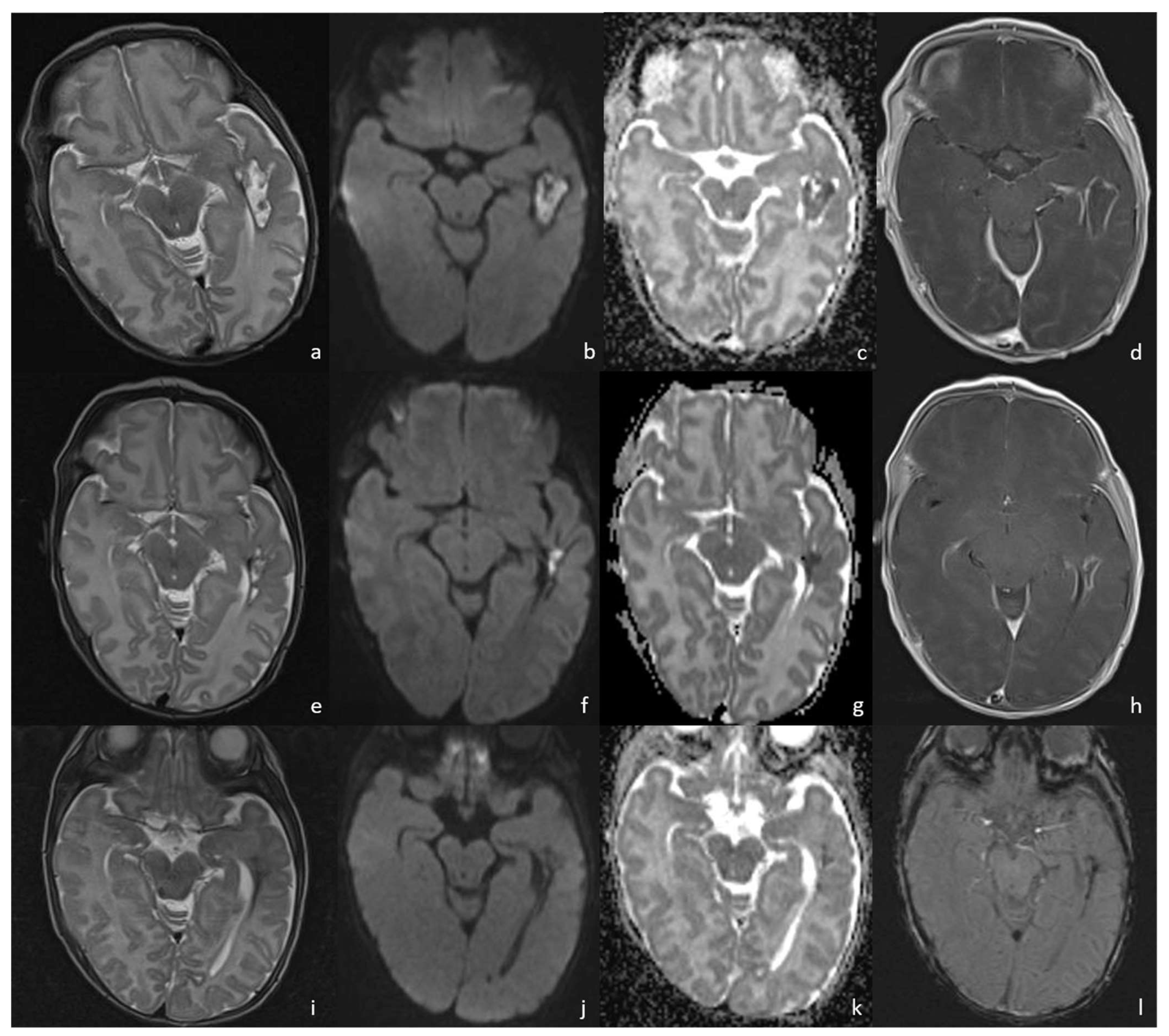
- Early cerebritis stage (3–5 days): Bacteria infiltrate the vessels causing vessel wall inflammation and vessel necrosis, which lead to blood barrier disruption and parenchymal invasion. The resulting cerebral infection, namely the early cerebritis, is limited to a focal portion of the brain, does not present a capsule, and presents a coexisting edema. In MRI, early cerebritis is seen as an inhomogeneous and ill-defined area of hyperintensity on T2WI and hypointensity on T1WI, surrounded by edema appearing hypointense on T1WI and hyperintense on T2WI. It presents a diffusion restriction on DWI/ADC in relation to cytotoxic edema and inflammatory hypercellularity. Hemorrhagic foci present as T1WI hyperintense areas within the lesion. After contrast administration, a patchy enhancement is observed, yet no capsule may be identified. On the US, early cerebritis appears as an ill-defined area of inhomogeneous echogenicity presenting increased vascularity on Transcranial Doppler, pairing CT findings, with an ill-defined area of inhomogeneous hypodensity with inhomogeneous and patchy enhancement.
- Late cerebritis (5–14 days): Cerebritis progressively evolves to show a necrotic core and an initial encapsulation. This stage flows into and partly overlaps with the early capsule stage since this last represents a progression with similar, yet more advanced features of the late cerebritis stage. In MRI, the late cerebritis results in a focal formation characterized by a necrotic core, appearing inhomogeneous on both T1 and T2WI, without a complete and regular contrast peripheral enhancement, yet with a defined diffusion restriction on DWI/ADC. On the US, the appearance is similar to the early cerebritis, yet the lesion appears more focal and the core starts becoming hypoechogenic, similar to CT showing a significantly hypodense core in the lesion with irregular and incomplete peripheral enhancement. Early capsule formation (14–30 days): The cerebritis is becoming an abscess since the capsule is evident, yet it is incomplete and thin and appears as a hyperintense rim on T1WI and a hypointense rim on T2WI with contrast enhancement on T1WI.
- Early capsule formation (2 weeks to 2 months): the lesion presents diffusion restriction on DWI/ADC, mainly in relation to hypercellularity. Sonographically, the lesion presents a well-defined hypoechoic core and an incomplete hyperechoic rim. CT shows a well-defined hypodense core and an incomplete peripheral enhancement.
- Late capsule formation (weeks to months): The parenchymal abscess presents a necrotic core, appearing hypointense on T1WI and hyperintense on T2WI with diffusion restriction on DWI/ADC. The capsule is inhomogeneously thick, appearing thicker towards the cortex and thinner towards the ventricles, appears isointense on T1WI and hypointense on T2WI, and presents an intense enhancement. On the US, the abscess presents a well-defined hypoechoic core and a complete hyperechoic rim, pairing CT that shows a well-defined hypodense core and a complete peripheral enhancement.
6.2. Infarcts
7. Peculiar MRI Patterns
7.1. Group B Streptococcus
7.2. Listeria Monocytogenes
7.3. Gram-Negative Bacteria and Abscesses
7.4. Gas-Producing Bacteria and Pneumocephalus
8. Conclusions
Supplementary Materials
Author Contributions
Funding
Institutional Review Board Statement
Informed Consent Statement
Data Availability Statement
Conflicts of Interest
Abbreviations
References
- Di Mauro, A.; Cortese, F.; Laforgia, N.; Pantaleo, B.; Giuliani, R.; Bonifazi, D.; Ciccone, M.M.; Giordano, P. Neonatal Bacterial Meningitis: A Systematic Review of European Available Data. Minerva Pediatr. 2019, 71, 201–208. [Google Scholar] [CrossRef] [PubMed]
- Schneider, J.F.; Hanquinet, S.; Severino, M.; Rossi, A. MR Imaging of Neonatal Brain Infections. Magn. Reson. Imaging Clin. N. Am. 2011, 19, 761–775. [Google Scholar] [CrossRef] [PubMed]
- Bonfield, C.M.; Sharma, J.; Dobson, S. Pediatric Intracranial Abscesses. J. Infect. 2015, 71 (Suppl. 1), S42–S46. [Google Scholar] [CrossRef] [PubMed]
- Cortese, F.; Scicchitano, P.; Gesualdo, M.; Filaninno, A.; De Giorgi, E.; Schettini, F.; Laforgia, N.; Ciccone, M.M. Early and Late Infections in Newborns: Where Do We Stand? A Review. Pediatr. Neonatol. 2016, 57, 265–273. [Google Scholar] [CrossRef] [PubMed]
- Moltoni, G.; Lucignani, G.; Sgrò, S.; Guarnera, A.; Rossi Espagnet, M.C.; Dellepiane, F.; Carducci, C.; Liberi, S.; Iacoella, E.; Evangelisti, G.; et al. MRI Scan with the “Feed and Wrap” Technique and with an Optimized Anesthesia Protocol: A Retrospective Analysis of a Single-Center Experience. Front. Pediatr. 2024, 12, 1415603. [Google Scholar] [CrossRef]
- Gupta, N.; Grover, H.; Bansal, I.; Hooda, K.; Sapire, J.M.; Anand, R.; Kumar, Y. Neonatal Cranial Sonography: Ultrasound Findings in Neonatal Meningitis-a Pictorial Review. Quant. Imaging Med. Surg. 2017, 7, 123–131. [Google Scholar] [CrossRef]
- Nakai, Y.; Miyazaki, O.; Kitamura, M.; Imai, R.; Okamoto, R.; Tsutsumi, Y.; Miyasaka, M.; Ogiwara, H.; Miura, H.; Yamada, K.; et al. Evaluation of Radiation Dose Reduction in Head CT Using the Half-Dose Method. Jpn. J. Radiol. 2023, 41, 872–881. [Google Scholar] [CrossRef]
- Parmar, H.; Ibrahim, M. Pediatric Intracranial Infections. Neuroimaging Clin. N. Am. 2012, 22, 707–725. [Google Scholar] [CrossRef]
- Berardi, A.; Lugli, L.; Rossi, C.; China, M.C.; Vellani, G.; Contiero, R.; Calanca, F.; Camerlo, F.; Casula, F.; Di Carlo, C.; et al. Neonatal Bacterial Meningitis. Minerva Pediatr. 2010, 62, 51–54. [Google Scholar]
- Brouwer, M.C.; Tunkel, A.R.; van de Beek, D. Epidemiology, Diagnosis, and Antimicrobial Treatment of Acute Bacterial Meningitis. Clin. Microbiol. Rev. 2010, 23, 467–492. [Google Scholar] [CrossRef]
- Ku, L.C.; Boggess, K.A.; Cohen-Wolkowiez, M. Bacterial Meningitis in Infants. Clin. Perinatol. 2015, 42, 29–45. [Google Scholar] [CrossRef] [PubMed]
- Bundy, L.M.; Rajnik, M.; Noor, A. Neonatal Meningitis. In StatPearls; StatPearls Publishing: Treasure Island, FL, USA, 2023. [Google Scholar]
- Anderson, D.C.; Hughes, B.J.; Edwards, M.S.; Buffone, G.J.; Baker, C.J. Impaired Chemotaxigenesis by Type III Group B Streptococci in Neonatal Sera: Relationship to Diminished Concentration of Specific Anticapsular Antibody and Abnormalities of Serum Complement. Pediatr. Res. 1983, 17, 496–502. [Google Scholar] [CrossRef] [PubMed]
- Baker, C.J.; Kasper, D.L. Correlation of Maternal Antibody Deficiency with Susceptibility to Neonatal Group B Streptococcal Infection. N. Engl. J. Med. 1976, 294, 753–756. [Google Scholar] [CrossRef] [PubMed]
- Vaughn, J.A.; Goncalves, L.F.; Cornejo, P. Intrauterine and Perinatal Infections. Clin. Perinatol. 2022, 49, 751–770. [Google Scholar] [CrossRef]
- Coggins, S.A.; Glaser, K. Updates in Late-Onset Sepsis: Risk Assessment, Therapy, and Outcomes. Neoreviews 2022, 23, 738–755. [Google Scholar] [CrossRef]
- Miyairi, I.; Causey, K.T.; DeVincenzo, J.P.; Buckingham, S.C. Group B Streptococcal Ventriculitis: A Report of Three Cases and Literature Review. Pediatr. Neurol. 2006, 34, 395–399. [Google Scholar] [CrossRef] [PubMed]
- Berman, P.H.; Banker, B.Q. Neonatal Meningitis. A Clinical and Pathological Study of 29 Cases. Pediatrics 1966, 38, 6–24. [Google Scholar] [CrossRef]
- Hristeva, L.; Booy, R.; Bowler, I.; Wilkinson, A.R. Prospective Surveillance of Neonatal Meningitis. Arch. Dis. Child. 1993, 69, 14–18. [Google Scholar] [CrossRef]
- Fitz, C.R. Inflammatory Diseases of the Brain in Childhood. AJNR Am. J. Neuroradiol. 1992, 13, 551–567. [Google Scholar]
- Renier, D.; Flandin, C.; Hirsch, E.; Hirsch, J.F. Brain Abscesses in Neonates. A Study of 30 Cases. J. Neurosurg. 1988, 69, 877–882. [Google Scholar] [CrossRef]
- Goodkin, H.P.; Harper, M.B.; Pomeroy, S.L. Intracerebral Abscess in Children: Historical Trends at Children’s Hospital Boston. Pediatrics 2004, 113, 1765–1770. [Google Scholar] [CrossRef] [PubMed]
- Parmar, H.; Sitoh, Y.-Y.; Anand, P.; Chua, V.; Hui, F. Contrast-Enhanced Flair Imaging in the Evaluation of Infectious Leptomeningeal Diseases. Eur. J. Radiol. 2006, 58, 89–95. [Google Scholar] [CrossRef]
- Yikilmaz, A.; Taylor, G.A. Sonographic Findings in Bacterial Meningitis in Neonates and Young Infants. Pediatr. Radiol. 2008, 38, 129–137. [Google Scholar] [CrossRef]
- Nagayama, Y.; Oda, S.; Nakaura, T.; Tsuji, A.; Urata, J.; Furusawa, M.; Utsunomiya, D.; Funama, Y.; Kidoh, M.; Yamashita, Y. Radiation Dose Reduction at Pediatric CT: Use of Low Tube Voltage and Iterative Reconstruction. Radiographics 2018, 38, 1421–1440. [Google Scholar] [CrossRef]
- Nagy, E.; Tschauner, S.; Schramek, C.; Sorantin, E. Paediatric CT Made Easy. Pediatr. Radiol. 2023, 53, 581–588. [Google Scholar] [CrossRef] [PubMed]
- Mactier, H.; Galea, P.; McWilliam, R. Acute Obstructive Hydrocephalus Complicating Bacterial Meningitis in Childhood. BMJ 1998, 316, 1887–1889. [Google Scholar] [CrossRef]
- Nzeh, D.; Oyinloye, O.I.; Odebode, O.T.; Akande, H.; Braimoh, K. Ultrasound Evaluation of Brain Infections and Its Complications in Nigerian Infants. Trop. Doct. 2010, 40, 178–180. [Google Scholar] [CrossRef]
- James Barkovich, A.; Raybaud, C. Pediatric Neuroimaging; Wolters Kluwer: Alphen aan den Rijn, The Netherlands, 2018; ISBN 9781496337207. [Google Scholar]
- Fujikawa, A.; Tsuchiya, K.; Honya, K.; Nitatori, T. Comparison of MRI Sequences to Detect Ventriculitis. AJR Am. J. Roentgenol. 2006, 187, 1048–1053. [Google Scholar] [CrossRef] [PubMed]
- Fukui, M.B.; Williams, R.L.; Mudigonda, S. CT and MR Imaging Features of Pyogenic Ventriculitis. AJNR Am. J. Neuroradiol. 2001, 22, 1510–1516. [Google Scholar]
- Jéquier, S.; Jéquier, J.C. Sonographic Nomogram of the Leptomeninges (pia-Glial Plate) and Its Usefulness for Evaluating Bacterial Meningitis in Infants. AJNR Am. J. Neuroradiol. 1999, 20, 1359–1364. [Google Scholar]
- Han, K.-T.; Choi, D.S.; Ryoo, J.W.; Cho, J.M.; Jeon, K.N.; Bae, K.-S.; You, J.J.; Chung, S.H.; Koh, E.H.; Park, K.-J. Diffusion-Weighted MR Imaging of Pyogenic Intraventricular Empyema. Neuroradiology 2007, 49, 813–818. [Google Scholar] [CrossRef] [PubMed]
- Korbecki, A.; Zimny, A.; Podgórski, P.; Sąsiadek, M.; Bladowska, J. Imaging of Cerebrospinal Fluid Flow: Fundamentals, Techniques, and Clinical Applications of Phase-Contrast Magnetic Resonance Imaging. Pol. J. Radiol. 2019, 84, e240–e250. [Google Scholar] [CrossRef] [PubMed]
- Bekiesińska-Figatowska, M.; Duczkowska, A.; Duczkowski, M.; Brągoszewska, H.; Mądzik, J.; Iwanowska, B.; Romaniuk-Doroszewska, A.; Antczak-Marach, D. Pneumococcal Meningitis and Its Sequelae—A Devastating CNS Disease. J Mother Child 2020, 24, 13–18. [Google Scholar] [PubMed]
- Chen, B.; Zhai, Q.; Ooi, K.; Cao, Y.; Qiao, Z. Risk Factors for Hydrocephalus in Neonatal Purulent Meningitis: A Single-Center Retrospective Analysis. J. Child Neurol. 2021, 36, 491–497. [Google Scholar] [CrossRef]
- Wong, A.M.; Zimmerman, R.A.; Simon, E.M.; Pollock, A.N.; Bilaniuk, L.T. Diffusion-Weighted MR Imaging of Subdural Empyemas in Children. AJNR Am. J. Neuroradiol. 2004, 25, 1016–1021. [Google Scholar]
- Foerster, B.R.; Thurnher, M.M.; Malani, P.N.; Petrou, M.; Carets-Zumelzu, F.; Sundgren, P.C. Intracranial Infections: Clinical and Imaging Characteristics. Acta Radiol. 2007, 48, 875–893. [Google Scholar] [CrossRef] [PubMed]
- Banerjee, A.D.; Pandey, P.; Devi, B.I.; Sampath, S.; Chandramouli, B.A. Pediatric Supratentorial Subdural Empyemas: A Retrospective Analysis of 65 Cases. Pediatr. Neurosurg. 2009, 45, 11–18. [Google Scholar] [CrossRef]
- Lipton, J.D.; Schafermeyer, R.W. Evolving Concepts in Pediatric Bacterial Meningitis--Part I: Pathophysiology and Diagnosis. Ann. Emerg. Med. 1993, 22, 1602–1615. [Google Scholar] [CrossRef] [PubMed]
- Tsai, Y.-D.; Chang, W.-N.; Shen, C.-C.; Lin, Y.-C.; Lu, C.-H.; Liliang, P.-C.; Su, T.-M.; Rau, C.-S.; Lu, K.; Liang, C.-L. Intracranial Suppuration: A Clinical Comparison of Subdural Empyemas and Epidural Abscesses. Surg. Neurol. 2003, 59, 191–196; discussion 196. [Google Scholar] [CrossRef]
- Osborn, A.G.; Hedlund, G.L.; Salzman, K.L. Osborn’s Brain: Imaging, Pathology, and Anatomy; Elsevier: Amsterdam, The Netherlands, 2017; ISBN 9780323477765. [Google Scholar]
- Foreman, S.D.; Smith, E.E.; Ryan, N.J.; Hogan, G.R. Neonatal Citrobacter Meningitis: Pathogenesis of Cerebral Abscess Formation. Ann. Neurol. 1984, 16, 655–659. [Google Scholar] [CrossRef]
- Chowdhary, V.; Gulati, P.; Sachdev, A.; Mittal, S.K. Pyogenic Meningitis: Sonographic Evaluation. Indian Pediatr. 1991, 28, 749–755. [Google Scholar] [PubMed]
- Krajewski, R.; Stelmasiak, Z. Brain Abscess in Infants. Childs. Nerv. Syst. 1992, 8, 279–280. [Google Scholar] [CrossRef] [PubMed]
- Falcone, S.; Post, M.J. Encephalitis, Cerebritis, and Brain Abscess: Pathophysiology and Imaging Findings. Neuroimaging Clin. N. Am. 2000, 10, 333–353. [Google Scholar] [PubMed]
- Kim, Y.J.; Chang, K.H.; Song, I.C.; Kim, H.D.; Seong, S.O.; Kim, Y.H.; Han, M.H. Brain Abscess and Necrotic or Cystic Brain Tumor: Discrimination with Signal Intensity on Diffusion-Weighted MR Imaging. AJR Am. J. Roentgenol. 1998, 171, 1487–1490. [Google Scholar] [CrossRef]
- Desprechins, B.; Stadnik, T.; Koerts, G.; Shabana, W.; Breucq, C.; Osteaux, M. Use of Diffusion-Weighted MR Imaging in Differential Diagnosis between Intracerebral Necrotic Tumors and Cerebral Abscesses. AJNR Am. J. Neuroradiol. 1999, 20, 1252–1257. [Google Scholar]
- Muzumdar, D.; Jhawar, S.; Goel, A. Brain Abscess: An Overview. Int. J. Surg. 2011, 9, 136–144. [Google Scholar] [CrossRef]
- Riederer, I.; Sollmann, N.; Mühlau, M.; Zimmer, C.; Kirschke, J.S. Gadolinium-Enhanced 3D T1-Weighted Black-Blood MR Imaging for the Detection of Acute Optic Neuritis. AJNR Am. J. Neuroradiol. 2020, 41, 2333–2338. [Google Scholar] [CrossRef]
- Moon, C.-J.; Kwon, T.H.; Lee, K.S.; Lee, H.-S. Recurrent Neonatal Sepsis and Progressive White Matter Injury in a Premature Newborn Culture-Positive for Group B Streptococcus: A Case Report. Medicine 2021, 100, e26387. [Google Scholar] [CrossRef]
- Sherman, G.; Lamb, G.S.; Platt, C.D.; Wessels, M.R.; Chochua, S.; Nakamura, M.M. Simultaneous Late, Late-Onset Group B Streptococcal Meningitis in Identical Twins. Clin. Pediatr. 2023, 62, 96–99. [Google Scholar] [CrossRef]
- Jaremko, J.L.; Moon, A.S.; Kumbla, S. Patterns of Complications of Neonatal and Infant Meningitis on MRI by Organism: A 10 Year Review. Eur. J. Radiol. 2011, 80, 821–827. [Google Scholar] [CrossRef]
- Magnus, J.; Parizel, P.M.; Ceulemans, B.; Cras, P.; Luijks, M.; Jorens, P.G. Streptococcus pneumoniae Meningoencephalitis with Bilateral Basal Ganglia Necrosis: An Unusual Complication due to Vasculitis. J. Child Neurol. 2011, 26, 1438–1443. [Google Scholar] [CrossRef] [PubMed]
- Choi, S.Y.; Kim, J.-W.; Ko, J.W.; Lee, Y.S.; Chang, Y.P. Patterns of Ischemic Injury on Brain Images in Neonatal Group B Streptococcal Meningitis. Korean J. Pediatr. 2018, 61, 245–252. [Google Scholar] [CrossRef] [PubMed]
- Rutherford, M.A. MRI of the Neonatal Brain; Saunders Limited: London, UK, 2002. [Google Scholar]
- Brisca, G.; La Valle, A.; Campanello, C.; Pacetti, M.; Severino, M.; Losurdo, G.; Palmieri, A.; Buffoni, I.; Renna, S. Listeria Meningitis Complicated by Hydrocephalus in an Immunocompetent Child: Case Report and Review of the Literature. Ital. J. Pediatr. 2020, 46, 111. [Google Scholar] [CrossRef] [PubMed]
- Ganau, M.; Mankad, K.; Srirambhatla, U.R.; Tahir, Z.; D’Arco, F. Ring-Enhancing Lesions in Neonatal Meningitis: An Analysis of Neuroradiology Pitfalls through Exemplificative Cases and a Review of the Literature. Quant. Imaging Med. Surg. 2018, 8, 333–341. [Google Scholar] [CrossRef]
- Lai, P.H.; Chang, H.C.; Chuang, T.C.; Chung, H.W.; Li, J.Y.; Weng, M.J.; Fu, J.H.; Wang, P.C.; Li, S.C.; Pan, H.B. Susceptibility-Weighted Imaging in Patients with Pyogenic Brain Abscesses at 1.5T: Characteristics of the Abscess Capsule. AJNR Am. J. Neuroradiol. 2012, 33, 910–914. [Google Scholar] [CrossRef]
- Alviedo, J.N.; Sood, B.G.; Aranda, J.V.; Becker, C. Diffuse Pneumocephalus in Neonatal Citrobacter Meningitis. Pediatrics 2006, 118, e1576–e1579. [Google Scholar] [CrossRef]
- Palma, J.A.; Zubieta, J.L.; Dominguez, P.D.; Garcia-Eulate, R. Pneumocephalus Mimicking Cerebral Cavernous Malformations in MR Susceptibility-Weighted Imaging. AJNR Am. J. Neuroradiol. 2009, 30, e83, author reply e84. [Google Scholar] [CrossRef]
- Bucci, S.; Coltella, L.; Martini, L.; Santisi, A.; De Rose, D.U.; Piccioni, L.; Campi, F.; Ronchetti, M.P.; Longo, D.; Lucignani, G.; et al. Clinical and Neurodevelopmental Characteristics of and Meningitis in Neonates. Front. Pediatr. 2022, 10, 881516. [Google Scholar] [CrossRef]
- Pietrasanta, C.; Ronchi, A.; Bassi, L.; De Carli, A.; Caschera, L.; Lo Russo, F.M.; Crippa, B.L.; Pisoni, S.; Crimi, R.; Artieri, G.; et al. Enterovirus and Parechovirus Meningoencephalitis in Infants: A Ten-Year Prospective Observational Study in a Neonatal Intensive Care Unit. J. Clin. Virol. 2024, 173, 105664. [Google Scholar] [CrossRef]
- Heckmann, J.G.; Ganslandt, O. Images in Clinical Medicine. The Mount Fuji Sign. N. Engl. J. Med. 2004, 350, 1881. [Google Scholar] [CrossRef]
- Michel, S.J. The Mount Fuji Sign. Radiology 2004, 232, 449–450. [Google Scholar] [CrossRef] [PubMed]





| MRI PROTOCOL | |
|---|---|
| SEQUENCES | PARAMETERS |
| Axial TSE T2 | RT 4730 ms, ET 109 ms, ST 2.5 mm |
| Coronal TSE T2 | RT 5620 ms, ET 95 ms, ST 2.5 mm |
| Axial DWI | RT 7930 ms, ET 59 ms, ST 2.5 mm |
| 3D T1 MPRAGE | RT 2200 ms, ET 3.29 ms, TI 900 ms, ST 0.8 mm |
| Axial SWI | RT 27 ms, ET 20 ms, ST 1.5 mm |
| ADDITIONAL SEQUENCES | PARAMETERS |
| Axial FLAIR | RT 9000 ms, ET 81 ms, ST 2.5 mm |
| Axial T2 SPACE | RT 2900 ms, ET 413 ms, ST 2.5 mm |
Disclaimer/Publisher’s Note: The statements, opinions and data contained in all publications are solely those of the individual author(s) and contributor(s) and not of MDPI and/or the editor(s). MDPI and/or the editor(s) disclaim responsibility for any injury to people or property resulting from any ideas, methods, instructions or products referred to in the content. |
© 2024 by the authors. Licensee MDPI, Basel, Switzerland. This article is an open access article distributed under the terms and conditions of the Creative Commons Attribution (CC BY) license (https://creativecommons.org/licenses/by/4.0/).
Share and Cite
Guarnera, A.; Moltoni, G.; Dellepiane, F.; Lucignani, G.; Rossi-Espagnet, M.C.; Campi, F.; Auriti, C.; Longo, D. Bacterial Meningoencephalitis in Newborns. Biomedicines 2024, 12, 2490. https://doi.org/10.3390/biomedicines12112490
Guarnera A, Moltoni G, Dellepiane F, Lucignani G, Rossi-Espagnet MC, Campi F, Auriti C, Longo D. Bacterial Meningoencephalitis in Newborns. Biomedicines. 2024; 12(11):2490. https://doi.org/10.3390/biomedicines12112490
Chicago/Turabian StyleGuarnera, Alessia, Giulia Moltoni, Francesco Dellepiane, Giulia Lucignani, Maria Camilla Rossi-Espagnet, Francesca Campi, Cinzia Auriti, and Daniela Longo. 2024. "Bacterial Meningoencephalitis in Newborns" Biomedicines 12, no. 11: 2490. https://doi.org/10.3390/biomedicines12112490
APA StyleGuarnera, A., Moltoni, G., Dellepiane, F., Lucignani, G., Rossi-Espagnet, M. C., Campi, F., Auriti, C., & Longo, D. (2024). Bacterial Meningoencephalitis in Newborns. Biomedicines, 12(11), 2490. https://doi.org/10.3390/biomedicines12112490


