Review

Role of Microbiota-Derived Hydrogen Sulfide (H2S) in Modulating the Gut–Brain Axis: Implications for Alzheimer’s and Parkinson’s Disease Pathogenesis

1
Department of Biomedical Sciences, Faculty of Medical Bioengineering, University of Medicine and Pharmacy “Grigore T. Popa”, 700454 Iasi, Romania
2
Neuromuscular Rehabilitation Clinic Division, Clinical Emergency Hospital “Bagdasar-Arseni”, 041915 Bucharest, Romania
3
Faculty of Medicine, University of Medicine and Pharmacy “Carol Davila”, 020022 Bucharest, Romania
*
Authors to whom correspondence should be addressed.
Biomedicines 2024, 12(12), 2670; https://doi.org/10.3390/biomedicines12122670 (registering DOI)
Submission received: 27 October 2024 / Revised: 11 November 2024 / Accepted: 20 November 2024 / Published: 23 November 2024
(This article belongs to the Special Issue Cellular and Molecular Biology of Neurodegenerative Disorders)

Abstract

:
Microbiota-derived hydrogen sulfide (H2S) plays a crucial role in modulating the gut–brain axis, with significant implications for neurodegenerative diseases such as Alzheimer’s and Parkinson’s. H2S is produced by sulfate-reducing bacteria in the gut and acts as a critical signaling molecule influencing brain health via various pathways, including regulating inflammation, oxidative stress, and immune responses. H2S maintains gut barrier integrity at physiological levels and prevents systemic inflammation, which could impact neuroinflammation. However, as H2S has a dual role or a Janus face, excessive H2S production, often resulting from gut dysbiosis, can compromise the intestinal barrier and exacerbate neurodegenerative processes by promoting neuroinflammation and glial cell dysfunction. This imbalance is linked to the early pathogenesis of Alzheimer’s and Parkinson’s diseases, where the overproduction of H2S exacerbates beta-amyloid deposition, tau hyperphosphorylation, and alpha-synuclein aggregation, driving neuroinflammatory responses and neuronal damage. Targeting gut microbiota to restore H2S homeostasis through dietary interventions, probiotics, prebiotics, and fecal microbiota transplantation presents a promising therapeutic approach. By rebalancing the microbiota-derived H2S, these strategies may mitigate neurodegeneration and offer novel treatments for Alzheimer’s and Parkinson’s diseases, underscoring the critical role of the gut–brain axis in maintaining central nervous system health.

1. Introduction

The human gastrointestinal (GI) tract harbors a complex community of trillions of microorganisms, including bacteria, archaea, fungi, and viruses, collectively referred to as the gut microbiota [1]. These microorganisms are crucial for maintaining homeostasis and performing essential functions such as digestion, nutrient absorption, immune system regulation, and the maintenance of the intestinal barrier [2]. In recent years, a growing body of evidence has highlighted the role of gut microbiota beyond the GI tract in modulating brain functions and contributing to the onset and progression of neurodegenerative diseases [3]. The gut and brain interaction, called the gut–brain axis, is a bidirectional communication system involving neural, immune, and endocrine pathways [4]. This system ensures proper gut and brain functioning and is significantly influenced by microbial metabolites such as short-chain fatty acids (SCFAs), tryptophan metabolites, and hydrogen sulfide (H2S).
H2S is a gaseous signaling molecule that plays a dual role in human physiology [5]. Endogenously produced by host cells and gut bacteria, H2S has been implicated in various processes, including regulating inflammation, maintaining vascular tone, and the modulation of the immune system [6]. Its concentration-dependent effects render H2S a double-edged sword: while low concentrations of H2S are associated with cytoprotective and anti-inflammatory effects, high levels of H2S have been linked to cellular toxicity, tissue damage, and inflammatory processes [7]. This dichotomy is particularly relevant in the context of neurodegenerative diseases such as Alzheimer’s disease (AD) and Parkinson’s disease (PD), where disruptions in gut microbiota, known as dysbiosis, can lead to abnormal H2S production and the subsequent worsening of neuroinflammatory processes (Figure 1) [8].
Our manuscript provides a novel perspective by exploring microbiota-derived H2S as a critical modulator of the gut–brain axis, with implications for AD and PD. Unlike previous studies that focused on the individual roles of H2S, our manuscript integrates its dual effects on gut and blood–brain barrier (BBB) integrity, glial cell dysfunction, and therapeutic strategies involving probiotics, prebiotics, and dietary interventions. This holistic approach addresses key knowledge gaps and lays the foundation for novel therapeutic paradigms targeting H2S dysbiosis.
Dysbiosis refers to the imbalance in the composition and function of the gut microbiota, which can arise due to various factors such as diet, aging, infection, and antibiotics [9]. In neurodegenerative diseases, dysbiosis has been shown to affect the gut–brain axis, leading to impaired communication between the gut and the central nervous system (CNS) [10]. Alterations in gut microbial composition can result in changes in the production of microbial metabolites, including H2S, that influence glial cell function, neuroinflammation, and neuronal survival. In particular, the excessive production of H2S by sulfate-reducing bacteria (SRB) in the gut has been implicated in gut barrier dysfunction, systemic inflammation, and the increased permeability of the BBB, contributing to AD and PD [11].
SRBs are a significant subset of the gut microbiota known for their ability to utilize sulfate as a terminal electron acceptor, producing H2S as a metabolic byproduct. Commonly found SRBs in the human gut include Desulfovibrio desulfuricans, Desulfovibrio vulgaris, and Desulfovibrio indonesiensis. Elevated SRB populations are observed in patients with inflammatory bowel diseases (IBDs), such as ulcerative colitis (UC), where they have been correlated with active disease states. The presence of SRB in the gut becomes clinically relevant due to the production of H2S, with cytotoxic properties, by inhibiting butyrate oxidation in colonocytes, disrupting intestinal epithelial cell metabolism. This disruption of cellular functions, compounded with H2S’s ability to impair the protective mucus barrier of the intestines, allows for closer bacterial contact with the epithelial layer, thereby exacerbating inflammatory processes. Studies indicate that H2S influences immune responses and tissue architecture within the colon. For instance, SRB colonization in experimental models has led to the increased infiltration of immune cells in the gut lining and activation of T helper (Th17) and regulatory T (Treg) cells, both critical in managing mucosal immunity and inflammation. Additionally, SRB-related H2S may potentiate neutrophil recruitment and migration, driving further tissue damage and inflammation. SRB populations like Desulfovibrio spp. contribute to this pathogenic cycle by enhancing inflammation and perpetuating immune dysregulation within the gut, highlighting the importance of microbial composition and H2S modulation in managing gut health and diseases [12].
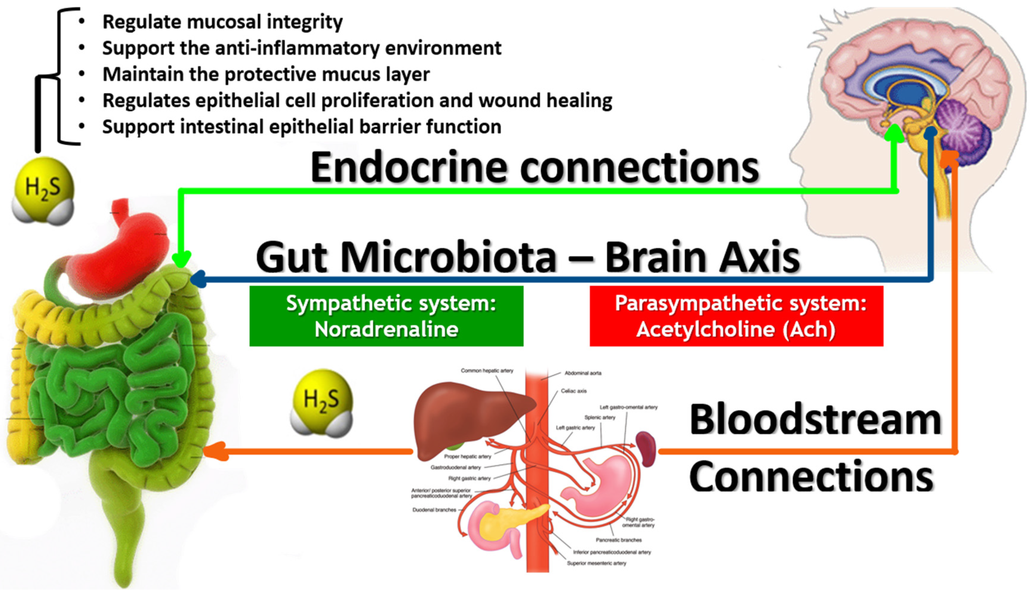
The gut–brain axis encompasses multiple communication pathways (Figure 2), including the vagus nerve, the immune system, and the circulatory system [13]. Microbial metabolites such as H2S, SCFAs, and neurotransmitter precursors can traverse the gut barrier and directly influence CNS function [14]. The vagus nerve, which provides a direct neural link between the gut and the brain, plays a crucial role in communicating signals from the gut to brain regions involved in mood regulation, cognition, and motor control [15]. Dysregulation of the gut–brain axis, mainly due to alterations in microbial metabolite production, can disrupt this communication, leading to neuroinflammatory responses and glial cell activation, hallmarks of neurodegenerative diseases [16].
Neuroinflammation, a critical process in developing neurodegenerative diseases, involves activating glial cells, including astrocytes and microglia, which are responsible for maintaining CNS homeostasis and responding to injury [17]. In AD and PD, glial cells become activated in response to the accumulation of misfolded proteins, such as amyloid-beta in AD and alpha-synuclein in PD [18]. This activation releases pro-inflammatory cytokines, reactive oxygen species (ROS), and nitric oxide (NO), contributing to neuronal damage and synaptic dysfunction [19]. Microbial metabolites, including H2S, have been shown to modulate glial cell activity, with excessive H2S production exacerbating glial activation and promoting neuroinflammation [20].
Dysbiosis and altered H2S production in AD are associated with increased neuroinflammation, amyloid plaque formation, and synaptic dysfunction [21]. Studies have demonstrated that changes in gut microbiota composition, particularly the overgrowth of SRB, lead to elevated H2S levels in the gut, which can compromise the integrity of the gut barrier [22,23]. This action allows pro-inflammatory molecules and microbial products to enter the systemic circulation and reach the brain, triggering microglial activation and promoting amyloid-beta deposition. The resulting neuroinflammatory environment accelerates the progression of AD and contributes to cognitive decline [24].
Similarly, in PD, gut dysbiosis has been linked to gastrointestinal dysfunction, one of the earliest non-motor symptoms of the disease [25]. The gut microbiota of PD patients is often characterized by decreased beneficial bacteria and increased SRB, leading to abnormal H2S production [26]. Excessive H2S in the gut has been shown to impair gut barrier function, leading to the translocation of bacterial endotoxins and inflammatory mediators into the bloodstream [27]. These factors can cross the BBB and induce neuroinflammation, contributing to the accumulation of alpha-synuclein aggregates and the degeneration of dopaminergic neurons in the substantia nigra, a hallmark of PD [28].
The role of glial cells in neurodegenerative diseases has garnered considerable attention, particularly in their interaction with gut-derived metabolites [29]. Microglia, the resident immune cells of the CNS, are highly sensitive to changes in the gut microbiota and its metabolic products. Under normal conditions, microglia maintain CNS homeostasis by clearing debris, regulating synaptic plasticity, and modulating the immune response. However, in dysbiosis and excessive microbial H2S production, the microglial function becomes dysregulated, leading to chronic activation and the release of neurotoxic molecules. This sustained activation contributes to neurodegeneration by promoting neuronal death and synaptic dysfunction [30].
Astrocytes, another type of glial cell, also play a crucial role in maintaining CNS homeostasis and supporting neuronal function. Astrocytes regulate the extracellular environment, modulate synaptic transmission, and provide metabolic support to neurons. Astrocytes become reactive in response to neuroinflammation in neurodegenerative diseases and produce pro-inflammatory cytokines and ROS, further contributing to neuronal damage [31]. Microbial H2S has been shown to influence astrocyte function, with high levels of H2S exacerbating astrocyte reactivity and promoting neuroinflammation. The interaction between astrocytes and microglia in the context of dysbiosis and altered H2S production is a key factor in the progression of neurodegenerative diseases [32].
One central mechanism by which H2S contributes to neurodegeneration is through its impact on the BBB [33]. The BBB is a critical barrier that protects the brain from harmful substances in the bloodstream while allowing the selective transport of essential nutrients and molecules. Dysbiosis-induced changes in H2S production can compromise BBB integrity, allowing inflammatory mediators, microbial products, and other neurotoxic molecules to enter the brain. Once in the CNS, these factors contribute to neuroinflammation, glial activation, and neuronal dysfunction, all characteristic features of AD and PD [34].
The therapeutic potential of targeting the gut microbiota to modulate H2S production and restore the gut–brain axis function has become an area of intense research. Probiotics, live microorganisms that confer health benefits to the host, and prebiotics, non-digestible compounds that promote the growth of beneficial bacteria, have been proposed as potential interventions to mitigate dysbiosis and reduce neuroinflammation in neurodegenerative diseases [35]. Studies have shown that probiotic and prebiotic supplementation can restore microbial balance, reduce H2S production, and improve gut barrier integrity, attenuating neuroinflammatory responses and promoting neuroprotection [36,37].
In addition to probiotics and prebiotics, other therapeutic strategies targeting the gut–brain axis have been explored. Fecal microbiota transplantation (FMT), which involves the transfer of gut microbiota from a healthy donor to a recipient with dysbiosis, has shown promise in restoring microbial diversity and reducing neuroinflammation in preclinical models of AD and PD. By reintroducing beneficial microbial species and reducing the abundance of SRB, FMT can modulate H2S production and improve the gut–brain axis function, offering a novel therapeutic approach for neurodegenerative diseases [38].
Dietary interventions that modulate the gut microbiota and reduce H2S production have also been investigated as potential therapeutic strategies for neurodegenerative diseases. Diets rich in fiber and polyphenols, which promote the growth of beneficial bacteria and reduce the abundance of SRB, have been shown to improve gut barrier function and reduce neuroinflammation in animal models of AD and PD. These findings suggest that dietary modifications could effectively manage dysbiosis and its associated neuroinflammatory consequences in neurodegenerative diseases [39,40,41].
This study aims to investigate the dual role of microbiota-derived H2S as both a protective and harmful metabolite in the gastrointestinal tract and its broader systemic implications and to explore how H2S influences gut homeostasis, neuroinflammation, and neurodegeneration, particularly in Alzheimer’s and Parkinson’s diseases. It will also assess the impact of dysbiosis-driven H2S production on gut and blood–brain barrier integrity and examine H2S interactions with glial cells in modulating neuroinflammation and neuronal survival. Additionally, this study seeks to identify therapeutic interventions, such as probiotics and prebiotics, to modulate H2S production, restore homeostasis, and mitigate disease progression while expanding the understanding of the gut–brain axis in neurodegenerative pathogenesis.

2. Methodology

This narrative review aims to collect and synthesize evidence on how microbiota-derived H2S modulates the gut–brain axis and its implications for the pathogenesis of AD and PD. The review included studies focusing on human or animal models related to AD, PD, or general neurodegenerative mechanisms involving the gut–brain axis. Eligible studies explored microbiota-derived H2S production and its roles in gut homeostasis, neuroinflammation, gut–brain axis function, and neurodegenerative diseases. Outcomes of interest include the modulation of the gut–brain axis by H2S, its effects on neurodegeneration, neuroinflammation, the integrity of the gut and blood–brain barriers, and glial cell activity in AD and PD. Only reviews, original research, randomized controlled trials, observational studies, cohort studies, in vivo and in vitro experiments, and mechanistic studies were considered. At the same time, editorials, case reports, and conference abstracts were excluded.
A comprehensive search was conducted to identify relevant studies across multiple databases, including PubMed, Web of Science, Scopus, Nature, and Google Scholar. The search strategy included a combination of keywords and Medical Subject Headings (MeSH) terms such as “Microbiota”, “Hydrogen Sulfide” OR “H2S”, “Gut-Brain Axis”, “Alzheimer’s Disease”, “Parkinson’s Disease”, “Neuroinflammation”, “Dysbiosis”, “Glial cells”, and “Probiotics” OR “Prebiotics”. Boolean operators (AND, OR) were applied to refine the search, and truncation (*) was used to capture all relevant term variations.
A qualitative synthesis was conducted, providing a detailed narrative description of the findings, focusing on key themes such as the role of microbiota-derived H2S in modulating the gut–brain axis, the impact of dysbiosis on H2S production and its effects on gut and blood–brain barrier integrity, and the influence of H2S on neuroinflammation, glial cell activation, and neurodegeneration. As this study involves a narrative review of published data, no ethical approval was required.

3. Results

3.1. H2S as a Protective Agent in Gut Homeostasis

Numerous studies highlighted the protective role of H2S at physiological levels, contributing to gut homeostasis and overall gastrointestinal health (Figure 3). At low concentrations, H2S was found to regulate mucosal integrity, support the anti-inflammatory environment of the gut, and maintain the protective mucus layer [42]. It also regulates epithelial cell proliferation and wound healing, promotes intestinal epithelial barrier function, and inhibits pathogenic bacteria’s adhesion to the gut lining [43].
Moreover, H2S protects gut barrier integrity by preserving tight junction proteins (e.g., claudin, occludin, and zonula occludens-1) and preventing microbial toxins’ translocation into the systemic circulation. These findings underscore the importance of H2S in regulating immune responses, preventing microbial invasion, and maintaining gut health overall [44].

3.2. Dichotomous Nature of H2S in Inflammation and Gut Permeability

Despite its protective role at physiological concentrations, studies demonstrated that elevated H2S levels, often associated with dysbiosis, harm gut health. Excessive production of H2S, particularly by SRB such as Desulfovibrio species, was linked to increased gut permeability, often referred to as “leaky gut” [45]. Elevated H2S levels disrupt the epithelial barrier, translocating pro-inflammatory molecules (e.g., lipopolysaccharides) into the bloodstream, triggering systemic inflammation [46].
In animal models of dysbiosis, high concentrations of microbiota-derived H2S were associated with the degradation of gut mucins, weakening the mucus layer and further compromising gut integrity. These findings suggest that H2S plays a dual role: it supports gut health at low concentrations but contributes to inflammatory responses and gut barrier dysfunction when produced in excess [47].

3.3. Impact of H2S on Neuroinflammation and the Gut–Brain Axis

Neuroinflammation is a key mechanism through which H2S influences the gut–brain axis. Chronic neuroinflammation is a hallmark of disease progression in both AD and PD, often driven by the activation of glial cells and the accumulation of misfolded proteins (e.g., amyloid-beta in AD and alpha-synuclein in PD). In studies examining neurodegenerative models, elevated H2S levels were associated with the increased expression of neuroinflammatory markers, including IL-1β, TNF-α, and IL-6, known to exacerbate neuronal damage. The inhibition of H2S production or the modulation of gut microbiota to lower H2S levels has been shown to reduce these markers, highlighting the potential therapeutic value of targeting H2S in neuroinflammatory pathways [48].
The review identified several key studies linking dysregulated H2S production to alterations in the gut–brain axis, mainly through promoting neuroinflammation. Excessive H2S production affected both peripheral and central immune responses, leading to the chronic activation of glial cells, such as microglia and astrocytes, in the central nervous system (CNS). This chronic activation contributes to the neurodegenerative processes. In both human and animal studies, elevated H2S levels were correlated with increased markers of neuroinflammation, including elevated levels of pro-inflammatory cytokines (e.g., IL-1β, IL-6, TNF-α) in cerebrospinal fluid and brain tissue. These cytokines are known to exacerbate neuronal damage and are central to the neuroinflammatory cascades observed in neurodegenerative diseases [49,50,51,52,53].

3.4. H2S and Blood–Brain Barrier Dysfunction

The blood–brain barrier plays a crucial role in maintaining CNS homeostasis by selectively regulating the passage of substances from the bloodstream into the brain. Similar to its effects on the gut barrier, H2S can influence the permeability of the BBB. Under normal conditions, H2S may help preserve the BBB’s integrity by regulating oxidative stress and inflammatory processes. However, elevated levels of microbiota-derived H2S, particularly in the presence of systemic inflammation, can compromise the BBB [54].
When the BBB is disrupted, harmful substances such as pro-inflammatory cytokines, microbial toxins, and activated immune cells can infiltrate the CNS. This breach in the BBB is a critical factor in neurodegenerative diseases like AD and PD, where neuroinflammation accelerates the degeneration of neurons. In animal models, high levels of microbiota-derived H2S were associated with increased expressions of endothelial adhesion molecules, which facilitate the infiltration of immune cells into the brain. This infiltration is a critical factor in neuroinflammation and further supports the detrimental role of excessive H2S production in neurodegeneration [55,56,57].

3.5. H2S and Glial Cell Dysfunction in Neurodegeneration

At physiological levels, H2S exerts anti-inflammatory effects on glial cells, limiting excessive microglial activation and promoting the resolution of inflammation. This neuroprotective role of H2S helps prevent chronic neuroinflammatory conditions that could damage neurons [58,59]. This review highlights the role of microbiota-derived H2S in modulating glial cell activity, with studies demonstrating that dysregulated H2S production exacerbates glial cell dysfunction [60]. In AD and PD, glial cells (microglia and astrocytes) play a central role in maintaining CNS homeostasis and responding to neuronal injury. However, the excessive activation of these cells, driven in part by elevated H2S levels, contributes to a sustained inflammatory environment in the brain, leading to progressive neuronal loss [61].
In vitro studies showed that H2S can directly modulate glial cell activation. At physiological concentrations, H2S exerts neuroprotective effects by limiting microglial activation and promoting the resolution of inflammation. However, at higher concentrations, it promotes a pro-inflammatory phenotype in glial cells. It is characterized by releasing neurotoxic molecules such as ROS and nitric oxide (NO), further accelerating neurodegeneration [62].

3.6. Interaction with Neural Signaling via the Vagus Nerve

The vagus nerve is a significant component of the parasympathetic nervous system and provides a direct communication pathway between the gut and the brain. H2S can modulate vagal signaling by affecting the release of neuroactive substances from the gut. For example, H2S may influence the production of neurotransmitters such as serotonin, dopamine, and gamma-aminobutyric acid (GABA), all of which play crucial roles in CNS function and mood regulation [63,64,65].
In the context of neurodegenerative diseases, alterations in H2S levels may lead to the dysregulation of vagal signaling, contributing to symptoms such as mood disturbances, cognitive decline, and motor dysfunction. The gut–brain signaling mediated by the vagus nerve may be particularly important in the early stages of PD, where gastrointestinal symptoms often precede the onset of motor symptoms by several years. Dysregulated H2S production in the gut may be an early contributor to this process, suggesting that the therapeutic modulation of H2S levels could have far-reaching effects on gut and brain health [66,67].

3.7. Modulation of Mitochondrial Function and Oxidative Stress

H2S is also involved in modulating mitochondrial function, essential for maintaining cellular energy balance and managing oxidative stress. Within the gut and CNS, H2S regulates mitochondrial biogenesis and functions by influencing electron transport and ATP production. At low levels, H2S enhances mitochondrial function and reduces oxidative stress, thereby protecting neurons from damage [68].
However, excessive H2S can inhibit mitochondrial respiratory enzymes and increase the production of ROS, leading to oxidative stress, mitochondrial dysfunction, and neuronal injury. This effect is particularly relevant in neurodegenerative diseases, where mitochondrial dysfunction is a known contributor to disease progression. H2S-induced oxidative stress further exacerbates the inflammatory environment in the brain, amplifying neurodegeneration [69].

3.8. Microbial Metabolite Interactions and Neurotransmitter Regulation

Microbiota-derived H2S interacts with other microbial metabolites, such as short-chain fatty acids (SCFAs) and tryptophan metabolites, which play critical roles in gut–brain communication [70]. These metabolites can regulate neurotransmitter production and modulate brain function. Dysbiosis-associated increases in H2S may alter the balance of these metabolites, leading to disruptions in neurotransmitter synthesis and signaling [71,72,73].
For example, SCFAs have anti-inflammatory effects and can protect the integrity of the gut and blood–brain barriers. High levels of H2S may interfere with the beneficial effects of SCFAs by promoting inflammatory responses and barrier dysfunction [74]. Additionally, tryptophan metabolism by gut bacteria is a key pathway for producing serotonin, a neurotransmitter implicated in mood regulation and cognitive function. Dysregulated H2S production may affect tryptophan metabolism, contributing to neurotransmitter imbalances commonly seen in AD and PD [75].

4. Age, Gender, and Comorbidities in Gut Microbiota Composition and H2S Production

4.1. Age-Related Changes in Gut Microbiota Composition and H2S Production

The aging process is a critical factor influencing the composition and function of the gut microbiota and the production of microbiota-derived metabolites such as H2S. As individuals age, profound changes occur within the gut–brain axis, impacting immune responses, barrier integrity, and neurological health. These alterations have significant implications for the development and progression of neurodegenerative diseases such as AD and PD. Understanding how age-related changes in microbiota-derived H2S influence the gut–brain axis provides crucial insights into the mechanisms underlying neurodegeneration [76].
Aging is associated with a decline in the diversity and stability of the gut microbiota, often called “microbiota aging”. In elderly individuals, beneficial bacterial populations such as Lactobacillus and Bifidobacterium species tend to decline. In contrast, opportunistic and pathogenic bacteria, including SRB such as Desulfovibrio, may increase in abundance. This shift in microbial composition can lead to the increased production of microbial metabolites like H2S, which has protective and harmful effects depending on its concentration [77]. The rise in SRB populations during aging is particularly concerning, as these bacteria produce higher levels of H2S by reducing sulfur compounds. Increased H2S production in the aging gut can exacerbate inflammation and impair gut barrier integrity, contributing to the translocation of pro-inflammatory molecules into the bloodstream. This systemic inflammation, in turn, influences the gut–brain axis, playing a role in neurodegenerative processes [78].
With age, the integrity of the gut barrier often deteriorates due to several factors, including reduced tight junction protein expression, immune dysregulation, and increased oxidative stress. These factors contribute to the “leaky gut”, where the barrier becomes more permeable to harmful substances, including microbial endotoxins and inflammatory cytokines [79]. Excessive H2S production by age-associated dysbiotic microbiota further compromises gut barrier function. While at physiological levels, H2S helps maintain barrier integrity by promoting mucin secretion and tight junction stabilization, elevated levels lead to the degradation of the mucus layer and the disruption of tight junctions, allowing inflammatory molecules to enter the systemic circulation. The increased permeability and systemic inflammation associated with this gut barrier dysfunction have been linked to the development of neuroinflammation and neurodegenerative diseases [80,81].

4.2. Gender Differences in Gut Microbiota Composition and H2S Production

Gut microbiota composition differs between men and women, influenced by factors such as sex hormones (e.g., estrogen, progesterone, testosterone), immune responses, and lifestyle behaviors. Gender plays a significant role in modulating the production and effects of microbiota-derived H2S, influencing the gut–brain axis and contributing to the pathogenesis of neurodegenerative diseases like AD and PD. Differences in gut microbiota composition, hormone regulation, immune responses, and barrier integrity between men and women lead to distinct patterns of H2S dysregulation [82,83].
Estrogen has protective effects on gut barrier integrity, enhancing the expression of tight junction proteins and reducing gut permeability. These protective effects may help mitigate the harmful effects of excessive H2S production in women by maintaining gut barrier function and preventing the translocation of harmful substances like lipopolysaccharides (LPSs) and pro-inflammatory cytokines into the bloodstream. As a result, women may be somewhat protected from the gut barrier dysfunction associated with elevated H2S levels, at least until hormonal changes occur during menopause [84,85].
Estrogen has been shown to enhance BBB integrity, reduce permeability, and protect the brain from inflammatory mediators. This protective effect may limit the impact of excessive H2S on the BBB in women, reducing the translocation of inflammatory molecules from the gut into the brain. Consequently, women may experience less H2S-driven neuroinflammation and BBB disruption, especially during their reproductive years [86]. However, after menopause, the decline in estrogen levels may reduce this protective effect, leading to increased BBB permeability and heightened susceptibility to H2S-induced neuroinflammation. This phenomenon may partly explain why postmenopausal women experience a higher risk of developing AD, as the combination of gut dysbiosis, increased H2S production, and BBB disruption may accelerate neurodegenerative processes [87,88].
In women, higher levels of Bifidobacterium and Lactobacillus species are commonly observed, associated with the lower production of harmful metabolites, including H2S. However, fluctuations in hormonal levels, such as during the menstrual cycle, pregnancy, and menopause, can influence gut microbial composition and metabolism. For example, estrogen has been shown to promote the growth of beneficial bacterial species less likely to produce excessive H2S. During menopause, however, the decline in estrogen levels may lead to shifts in the gut microbiota, including an increased abundance of SRB like Desulfovibrio, which produces higher levels of H2S [89].
In men, higher testosterone levels have been associated with a less protective gut barrier, potentially increasing susceptibility to H2S-induced gut barrier dysfunction. The higher H2S production from SRBs and reduced gut barrier integrity may lead to greater gut permeability (“leaky gut”) in men, contributing to systemic inflammation and promoting neuroinflammatory processes linked to neurodegenerative diseases like PD [90,91].
Men tend to have a higher abundance of SRBs than women, potentially resulting in more outstanding productions of H2S. Higher testosterone levels may partly influence this gender-specific microbial composition in men, which has been linked to alterations in gut microbiota that favor H2S production. These differences in microbial H2S production may contribute to the heightened risk of certain gastrointestinal and neuroinflammatory conditions observed in men. Moreover, men may experience more pronounced H2S-related disruptions in the gut–brain axis, influencing the progression of neurodegenerative diseases such as PD, which has a higher prevalence in males [92,93].
Men, with their typically lower estrogen levels, may have less protection against BBB disruption, especially in the presence of elevated H2S. The combination of higher gut-derived H2S production reduced gut and BBB integrity, and more pronounced neuroinflammatory responses may contribute to men’s greater vulnerability to PD, where neuroinflammation plays a central role in dopaminergic neuron loss [94].
Given the gender differences in microbiota composition, immune responses, and hormone regulation, gender-specific therapeutic approaches may be necessary for effectively modulating H2S and mitigating its impact on neurodegenerative diseases [95]. Hormone replacement therapy (HRT) could also help maintain gut and BBB integrity by compensating for the loss of estrogen. Probiotics, prebiotics, and dietary interventions designed to reduce SRB abundance and promote beneficial bacteria may mainly effectively reduce H2S production and neuroinflammation in women [96].
In men, interventions targeting the higher baseline levels of SRBs and H2S production may be critical. Anti-inflammatory therapies and probiotics or dietary approaches that specifically inhibit SRBs could help reduce the harmful effects of excessive H2S on the gut–brain axis. Additionally, therapies aimed at enhancing gut barrier integrity and protecting the BBB may be particularly beneficial for men, who are more susceptible to H2S-driven barrier dysfunction and neuroinflammation [97,98,99].

4.3. Other Comorbidities, Gut Microbiota Composition and H2S Production

Comorbidities, particularly those related to metabolic, cardiovascular, gastrointestinal, and immune systems, significantly influence the production and effects of microbiota-derived H2S on the gut–brain axis. These comorbidities often exacerbate the pathogenesis of neurodegenerative diseases like AD and PD by disrupting gut homeostasis, increasing systemic inflammation, and promoting neuroinflammation [100].
Metabolic disorders, including obesity, type 2 diabetes (T2D), and metabolic syndrome, are strongly associated with alterations in gut microbiota composition and function, leading to dysbiosis and changes in H2S production. These conditions exacerbate systemic inflammation and increase the risk of neurodegenerative diseases by disrupting the gut–brain axis. Type 2 diabetes (T2D) is characterized by chronic hyperglycemia, insulin resistance, and systemic inflammation, all linked to changes in gut microbiota composition. T2D is associated with increased H2S production due to dysbiosis, particularly overrepresenting SRBs in the gut. Elevated H2S exacerbates insulin resistance by disrupting gut barrier integrity and promoting systemic inflammation. The gut–brain axis plays a critical role in linking T2D and neurodegeneration. Insulin resistance in the CNS is associated with cognitive decline and increased amyloid-beta deposition, key features of AD. Furthermore, increased H2S production in T2D contributes to neuroinflammation by promoting the activation of microglia and astrocytes, which release pro-inflammatory mediators that damage neurons. This inflammation is particularly harmful in the hippocampus, a brain region essential for memory and cognition severely affected by AD [101].
Obesity is linked to a pro-inflammatory state and changes in the gut microbiota that promote the overgrowth of sulfate-reducing bacteria (SRB), such as Desulfovibrio species, which produce H2S. Excessive H2S production in obese individuals contributes to gut barrier dysfunction, allowing the translocation of lipopolysaccharides (LPSs) and other bacterial endotoxins into the bloodstream. This systemic inflammation affects metabolic tissues and the CNS, promoting neuroinflammation and contributing to the development of AD and PD. In obesity, the elevated levels of pro-inflammatory cytokines, such as IL-6 and TNF-α, further exacerbate the disruption of the gut–brain axis. This chronic low-grade inflammation increases oxidative stress and accelerates neuronal damage, creating a conducive environment for neurodegeneration. Moreover, adipose tissue dysfunction in obesity releases free fatty acids and inflammatory molecules that further amplify neuroinflammatory responses, particularly in brain regions affected by AD and PD [102,103,104,105].
Cardiovascular diseases, including hypertension, atherosclerosis, and heart failure, significantly impact gut microbiota composition and H2S production, contributing to neuroinflammation and neurodegeneration through multiple pathways. Hypertension is closely associated with gut dysbiosis, where an imbalance in microbial populations leads to increased H2S production by SRBs. This dysregulation contributes to endothelial dysfunction and inflammation, both exacerbating hypertension and increasing the BBB permeability. A compromised BBB allows the translocation of inflammatory molecules and microbial metabolites, such as H2S, into the CNS, leading to neuroinflammation. In hypertensive individuals, the interaction between dysregulated H2S and vascular endothelial cells contributes to oxidative stress and ROS formation. This oxidative damage not only affects the cardiovascular system but also promotes neuroinflammation, increasing the risk of neurodegenerative diseases like AD and PD. Elevated H2S levels in the context of hypertension may also impair cerebral blood flow, further contributing to neuronal dysfunction and cognitive decline [101,106].
Atherosclerosis, characterized by chronic vascular inflammation and plaque formation in arteries, is linked to gut microbiota dysbiosis and altered microbial metabolite production, including H2S. Increased H2S levels contribute to systemic inflammation and endothelial dysfunction, promoting plaque instability and increasing the risk of cardiovascular events. The systemic inflammation associated with atherosclerosis extends to the CNS, where H2S modulates the activation of glial cells, particularly microglia, leading to neuroinflammation. This chronic inflammatory state accelerates neuronal damage and promotes the aggregation of misfolded proteins, such as amyloid-beta in AD and alpha-synuclein in PD. The vascular dysfunction in atherosclerosis also affects cerebral blood flow, reducing oxygen and nutrient delivery to the brain and exacerbating neurodegenerative processes [107,108,109].
Gastrointestinal (GI) disorders such as inflammatory bowel disease (IBD), irritable bowel syndrome (IBS), and small intestinal bacterial overgrowth (SIBO) are associated with significant alterations in gut microbiota and H2S production [110]. These conditions disrupt the gut–brain axis and increase the risk of neuroinflammation and neurodegeneration. IBD, including Crohn’s disease and ulcerative colitis, is characterized by chronic inflammation of the gastrointestinal tract. Dysbiosis in IBD is associated with increased populations of SRBs, leading to elevated H2S production. Excessive H2S exacerbates intestinal inflammation and contributes to gut barrier dysfunction, allowing microbial products to enter the bloodstream and reach the brain. The systemic inflammation and increased permeability associated with IBD extend to the CNS, where the inflammatory mediators and microbial metabolites such as H2S contribute to neuroinflammation. In patients with IBD, neuroinflammatory processes are linked to an increased risk of cognitive decline and neurodegenerative diseases like AD and PD. Chronic gut inflammation in IBD may also prime microglia in the CNS, making them more responsive to inflammatory signals and increasing the likelihood of neurodegeneration. IBS is a functional gastrointestinal disorder characterized by altered gut motility, visceral hypersensitivity, and dysbiosis. In IBS, the gut microbiota often shows an increased abundance of SRBs, resulting in elevated H2S levels. This increase in H2S can disrupt gut barrier integrity, leading to a heightened immune response and systemic inflammation [111,112].
Patients with IBS frequently report anxiety, depression, and cognitive symptoms, which are linked to gut–brain axis dysregulation. Elevated H2S levels may play a role in this dysregulation by modulating neural and immune signaling pathways. Although the exact mechanisms are still under investigation, the relationship between elevated H2S, gut inflammation, and CNS dysfunction suggests that IBS may increase the risk of neuroinflammatory conditions that contribute to AD and PD [113].
Immune-mediated disorders, including autoimmune diseases such as rheumatoid arthritis (RA) and systemic lupus erythematosus (SLE), are characterized by chronic inflammation and altered immune responses. These conditions affect the gut microbiota and H2S production, contributing to systemic and neuroinflammation [114,115].
RA is associated with gut dysbiosis, leading to increased H2S production and altered immune responses. Chronic inflammation in RA affects the joints and extends to the gut and CNS. Elevated H2S levels exacerbate gut barrier dysfunction, promoting the translocation of inflammatory mediators to the brain and increasing the risk of neuroinflammation. In RA, microglial activation in response to systemic inflammation contributes to neurodegenerative processes, particularly AD. The chronic immune activation and increased H2S production in RA may accelerate neuronal damage by promoting oxidative stress and mitochondrial dysfunction in neurons [116,117,118].
SLE, an autoimmune disorder characterized by widespread inflammation and tissue damage, is linked to gut dysbiosis and increased H2S production. Similar to RA, SLE involves chronic inflammation that disrupts the gut–brain axis. Elevated H2S levels contribute to gut barrier dysfunction and promote systemic inflammation, which can extend to the CNS. In SLE, the neuroinflammatory processes linked to increased H2S production may accelerate neurodegeneration by promoting glial cell activation and the release of pro-inflammatory cytokines. Patients with SLE are at increased risk of developing cognitive impairments and neurodegenerative diseases, likely due to the combined effects of chronic inflammation, dysbiosis, and H2S dysregulation [119,120,121].
Neuropsychiatric conditions, such as depression and anxiety, are associated with altered gut microbiota and the dysregulated production of microbial metabolites, including H2S. These conditions influence mood and cognition, affect the gut–brain axis, and may increase the risk of neurodegenerative diseases. Patients with depression and anxiety often exhibit dysbiosis, including increased H2S production. This dysregulation may exacerbate gut barrier dysfunction and promote systemic inflammation, contributing to neuroinflammatory processes in the brain. The chronic low-grade inflammation associated with depression and anxiety has been linked to the development of AD and PD, where neuroinflammation plays a central role in disease progression [122,123,124].

5. Therapeutic Targets and Interventions Related to H2S Modulation

Modulating microbiota-derived H2S offers promising therapeutic potential, particularly for neurodegenerative diseases, where dysregulated H2S levels contribute to gut–brain axis disruption, neuroinflammation, and neuronal damage. Effective interventions focus on regulating microbial H2S production and mitigating its harmful effects while harnessing its beneficial roles. The following sections outline various therapeutic strategies and targets aimed at H2S modulation.

5.1. Probiotics and Prebiotics

Probiotics and prebiotics are well-established interventions for modulating gut microbiota and have demonstrated potential in regulating H2S production, particularly by reducing the abundance of sulfate-reducing bacteria (SRB), such as Desulfovibrio species, responsible for excessive H2S production [125,126,127].
Probiotics are live microorganisms that confer health benefits when administered in adequate amounts. Certain probiotic strains, such as Lactobacillus and Bifidobacterium species, have been shown to restore gut microbial balance by inhibiting the growth of SRBs and promoting beneficial bacteria. By outcompeting SRBs for resources in the gut, probiotics can reduce H2S production and its associated toxic effects on the gut barrier [128,129].
Additionally, probiotics modulate gut permeability by enhancing the expression of tight junction proteins, thereby reducing gut barrier dysfunction (“leaky gut”) and limiting the translocation of harmful microbial products into the bloodstream. In neurodegenerative diseases such as AD and PD, probiotics have shown promise in reducing systemic inflammation, improving cognitive and motor functions, and protecting against neuroinflammation [130,131,132].
Prebiotics are non-digestible food components that promote the growth and activity of beneficial gut bacteria. Prebiotics such as inulin, fructooligosaccharides (FOSs), and resistant starch can increase the production of short-chain fatty acids (SCFAs), particularly butyrate, which helps maintain gut integrity and inhibit SRB proliferation [133,134].
Prebiotics promote a favorable gut environment and reduce microbial dysbiosis and the overproduction of H2S. The increase in SCFA levels also has anti-inflammatory effects in the gut and the CNS, as SCFAs are known to modulate immune responses and reduce neuroinflammation. These effects make prebiotics a valuable therapeutic option for managing neuroinflammation in AD and PD [135,136].

5.2. Dietary Modifications

Diet plays a significant role in shaping the gut microbiota composition and, consequently, the levels of microbial-derived metabolites, including H2S. Dietary interventions can modulate H2S production by influencing the substrates available for SRBs and promoting the growth of beneficial bacteria [137,138].
Sulfate-reducing bacteria derive H2S from sulfur-containing compounds such as sulfates, sulfites, and sulfur-containing amino acids (e.g., cysteine and methionine). A low-sulfur diet, which reduces the intake of these compounds, can limit the substrate availability for SRBs and consequently decrease H2S production. This dietary intervention may help reduce the harmful effects of excessive H2S on gut and BBB integrity, thereby alleviating systemic inflammation and neuroinflammation [139].
A diet rich in dietary fiber promotes the growth of beneficial gut bacteria that produce SCFAs while reducing the growth of SRBs. High-fiber foods, such as whole grains, legumes, fruits, and vegetables, support a healthy gut environment that reduces H2S production. Fiber fermentation by beneficial bacteria increases SCFA production, which helps to preserve gut barrier integrity, reduce inflammation, and enhance the anti-inflammatory effects on the CNS [140,141]. High-fiber diets have been linked to improved gut health and reduced risks of neuroinflammatory conditions. In neurodegenerative disease models, they have mitigated cognitive decline and motor dysfunction by promoting neuroprotective microbial metabolites and reducing harmful H2S levels [142].

5.3. Fecal Microbiota Transplantation (FMT)

Fecal microbiota transplantation (FMT) involves transferring gut microbiota from a healthy donor to a recipient with dysbiosis. FMT has shown promise in restoring microbial balance, reducing dysbiosis-associated H2S production and improving gut barrier function. In preclinical models of neurodegenerative diseases, FMT has demonstrated the potential to alleviate symptoms by reducing systemic and neuroinflammation. By restoring microbial diversity and reducing the abundance of SRBs, FMT can help modulate H2S levels, thereby protecting the gut–brain axis and mitigating the progression of AD and PD. However, further research and clinical trials are necessary to fully understand the long-term effects of FMT on H2S modulation and neurodegenerative diseases [143,144,145].

5.4. Pharmacological Inhibitors of H2S Production

Pharmacological interventions targeting H2S-producing enzymes or SRBs offer a more direct approach to modulating H2S levels. Enzymatic inhibitors or compounds that selectively target H2S-producing bacteria have been explored as potential therapeutic strategies for reducing the harmful effects of excessive H2S [146].
Host-derived H2S is produced by enzymes such as cystathionine γ-lyase (CSE) and cystathionine β-synthase (CBS). Inhibitors of these enzymes can reduce endogenous H2S production, which may be beneficial in conditions where excessive H2S contributes to neuroinflammation. Selective inhibitors of CSE and CBS are being explored for their potential to modulate H2S levels in inflammatory and neurodegenerative diseases [147]. However, care must be taken with enzyme inhibitors, as H2S plays protective roles at physiological levels, particularly in maintaining vascular homeostasis and modulating immune responses. Targeted inhibition should aim to reduce excessive H2S without compromising its beneficial effects [148]. Developing antibacterial agents that selectively inhibit SRBs could help reduce microbial H2S production. Such agents would ideally target SRB-specific metabolic pathways without disrupting the overall balance of the gut microbiota. Although these targeted approaches are still under investigation, the potential of the selective inhibition of SRBs represents a promising therapeutic strategy for reducing the harmful effects of microbiota-derived H2S in neurodegenerative diseases, offering optimism for future treatments [149].

5.5. Antioxidants and Anti-Inflammatory Agents

Given that excessive H2S promotes oxidative stress and inflammation, therapeutic strategies to counteract these effects have gained attention. Antioxidants and anti-inflammatory agents may mitigate the damaging effects of H2S on neurons and glial cells, particularly in neuroinflammation. Antioxidants such as N-acetylcysteine (NAC), glutathione, and vitamin C can scavenge ROS generated by excessive H2S. These compounds help protect neurons from oxidative damage and may reduce the pro-inflammatory effects of H2S in the CNS. In preclinical models, antioxidant therapy has been shown to alleviate neuroinflammation and improve cognitive function in neurodegenerative diseases [150,151].
Pharmacological agents inhibiting pro-inflammatory signaling pathways, such as NF-κB inhibitors, can reduce the inflammatory response of excessive H2S. By limiting the activation of glial cells and reducing the production of pro-inflammatory cytokines, these agents may help counteract the neuroinflammatory effects of dysregulated H2S levels. Non-steroidal anti-inflammatory drugs (NSAIDs) and other more selective anti-inflammatory agents are being explored for their potential in modulating neuroinflammation in AD and PD [152,153].

5.6. Combination Therapies

Given the multifactorial nature of H2S’s role in neurodegenerative diseases, combination therapies that target multiple aspects of H2S modulation may offer the most effective outcomes. For example, combining probiotics or prebiotics with dietary interventions, antioxidant therapy, or pharmacological inhibitors of H2S production may synergistically restore gut and brain homeostasis [154,155].
The modulation of microbiota-derived H2S represents a promising therapeutic strategy for addressing neuroinflammation and neurodegeneration, particularly in diseases such as Alzheimer’s and Parkinson’s. Probiotics, prebiotics, dietary modifications, fecal microbiota transplantation, and pharmacological inhibitors offer potential avenues for reducing excessive H2S production and mitigating its harmful effects. Additionally, antioxidants and anti-inflammatory agents provide complementary approaches to counteract the oxidative stress and inflammation associated with dysregulated H2S levels. Future research and clinical trials will be essential in optimizing these therapeutic interventions and developing effective treatments for neurodegenerative diseases by targeting H2S modulation [156].

6. Limitations and Gaps in Current Research

While the review identified a substantial body of evidence supporting the role of microbiota-derived H2S in modulating the gut–brain axis and contributing to neurodegeneration, several gaps remain. First, H2S levels or the exact mechanisms through which H2S modulates glial cell activity and promotes neuroinflammation require further elucidation. Second, human studies investigating the direct impact of H2S modulation on neurodegenerative outcomes are limited, necessitating more clinical trials in this area. Lastly, neurodegeneration needs further exploration of the interaction between H2S and other microbial metabolites, such as SCFAs and tryptophan metabolites [157,158].

7. Conclusions

Microbiota-derived H2S plays a critical dual role in modulating the gut–brain axis, with protective and harmful effects depending on its concentration. At physiological levels, H2S supports gut barrier integrity, regulates immune responses, and maintains homeostasis within the gut–brain axis. However, dysbiosis and comorbidities like metabolic disorders, cardiovascular diseases, and gastrointestinal conditions often lead to excessive H2S production, exacerbating gut permeability, BBB disruption, and systemic inflammation. This dysregulation promotes chronic neuroinflammation, primarily through activating glial cells, including microglia and astrocytes, which release pro-inflammatory cytokines and ROS, accelerating neuronal damage in neurodegenerative diseases such as AD and PD. Gender differences further influence H2S production, with estrogen offering protective effects in women by maintaining barrier integrity and limiting H2S-induced inflammation. At the same time, men with higher levels of sulfate-reducing bacteria may be more vulnerable to the harmful effects of H2S. Therapeutic interventions that modulate H2S production, including probiotics, prebiotics, dietary changes, and pharmacological inhibitors, offer promising strategies to restore microbial balance, improve gut–brain axis function, and mitigate neuroinflammation. Given the central role of H2S in disrupting gut and BBB integrity, targeting microbial H2S and restoring gut homeostasis are critical for preventing or slowing the progression of neurodegenerative diseases. These approaches must consider individual factors such as gender, age, and comorbidities to optimize therapeutic efficacy.

Author Contributions

Conceptualization, C.M. and G.O.; methodology, M.R. and A.I.G.; software, M.T.; validation, M.P.; formal analysis, M.R. and M.T.; investigation, M.P.; resources, A.I.G.; data curation, M.T.; writing—original draft preparation, C.M.; writing—review and editing, C.M., G.O., M.R., M.P. and A.I.G.; visualization, C.M. and G.O.; project administration, C.M.; funding acquisition, A.I.G. All authors have read and agreed to the published version of the manuscript.

Funding

Supporting the institutional capacity for research and innovation through transdisciplinary biotechnologies (InovBiotech) CNFIS—FDI—2024—F—0099.

Conflicts of Interest

The authors declare no conflicts of interest.

References

  1. Thursby, E.; Juge, N. Introduction to the human gut microbiota. Biochem. J. 2017, 474, 1823–1836. [Google Scholar] [CrossRef] [PubMed]
  2. Pessemier, B.; De Grine, L.; Debaere, M.; Maes, A.; Paetzold, B.; Callewaert, C. Gut–skin axis: Current knowledge of the interrelationship between microbial dysbiosis and skin conditions. Microorganisms 2021, 9, 353. [Google Scholar] [CrossRef] [PubMed]
  3. Jandhyala, S.M.; Talukdar, R.; Subramanyam, C.; Vuyyuru, H.; Sasikala, M.; Reddy, D.N. Role of the normal gut microbiota. World J. Gastroenterol. 2015, 21, 8836–8847. [Google Scholar] [CrossRef] [PubMed]
  4. Carabotti, M.; Scirocco, A.; Antonietta Maselli, M.; Severi, C. The gut-brain axis: Interactions between enteric microbiota, central and enteric nervous systems. Ann. Gastroenterol. 2015, 28, 203–209. [Google Scholar] [PubMed]
  5. Andrés, C.M.C.; Pérez de la Lastra, J.M.; Andrés Juan, C.; Plou, F.J.; Pérez-Lebeña, E. Chemistry of Hydrogen Sulfide—Pathological and Physiological Functions in Mammalian Cells. Cells 2023, 12, 2684. [Google Scholar] [CrossRef]
  6. Georgakopoulou, V.; Lempesis, I.; Sklapani, P.; Trakas, N.; Spandidos, D. Exploring the pathogenetic mechanisms of Mycoplasma pneumoniae (Review). Exp. Ther. Med. 2024, 28, 424. [Google Scholar] [CrossRef]
  7. El-Saadony, M.T.; Yang, T.; Korma, S.A.; Sitohy, M.; El-Mageed, T.A.A.; Selim, S.; Al Jaouni, S.K.; Salem, H.M.; Mahmmod, Y.; Soliman, S.M.; et al. Impacts of turmeric and its principal bioactive curcumin on human health: Pharmaceutical, medicinal, and food applications: A comprehensive review. Front. Nutr. 2023, 9, 1040259. [Google Scholar] [CrossRef]
  8. Breijyeh, Z.; Karaman, R. Comprehensive Review on Alzheimer’s Disease: Causes and Treatment. Molecules 2020, 25, 5789. [Google Scholar] [CrossRef]
  9. Mostafavi Abdolmaleky, H.; Zhou, J.R. Gut Microbiota Dysbiosis, Oxidative Stress, Inflammation, and Epigenetic Alterations in Metabolic Diseases. Antioxidants 2024, 13, 985. [Google Scholar] [CrossRef]
  10. Bicknell, B.; Liebert, A.; Borody, T.; Herkes, G.; McLachlan, C.; Kiat, H. Neurodegenerative and Neurodevelopmental Diseases and the Gut-Brain Axis: The Potential of Therapeutic Targeting of the Microbiome. Int. J. Mol. Sci. 2023, 24, 9577. [Google Scholar] [CrossRef]
  11. Yoo, J.Y.; Groer, M.; Dutra, S.V.O.; Sarkar, A.; McSkimming, D.I. Gut microbiota and immune system interactions. Microorganisms 2020, 8, 1587. [Google Scholar] [CrossRef] [PubMed]
  12. Figliuolo, V.R.; dos Santos, L.M.; Abalo, A.; Nanini, H.; Santos, A.; Brittes, N.M.; Bernardazzi, C.; de Souza, H.S.P.; Vieira, L.Q.; Coutinho-Silva, R.; et al. Sulfate-reducing bacteria stimulate gut immune responses and contribute to inflammation in experimental colitis. Life Sci. 2017, 189, 29–38. [Google Scholar] [CrossRef] [PubMed]
  13. Bonaz, B.; Bazin, T.; Pellissier, S. The vagus nerve at the interface of the microbiota-gut-brain axis. Front. Neurosci. 2018, 12, 49. [Google Scholar] [CrossRef]
  14. Martin, C.R.; Osadchiy, V.; Kalani, A.; Mayer, E.A. The Brain-Gut-Microbiome Axis. Cell. Mol. Gastroenterol. Hepatol. 2018, 6, 133–148. [Google Scholar] [CrossRef] [PubMed]
  15. Breit, S.; Kupferberg, A.; Rogler, G.; Hasler, G. Vagus nerve as modulator of the brain-gut axis in psychiatric and inflammatory disorders. Front. Psychiatry 2018, 9, 44. [Google Scholar] [CrossRef]
  16. You, M.; Chen, N.; Yang, Y.; Cheng, L.; He, H.; Cai, Y.; Liu, Y.; Liu, H.; Hong, G. The gut microbiota–brain axis in neurological disorders. MedComm 2024, 5, e656. [Google Scholar] [CrossRef]
  17. Kwon, H.S.; Koh, S.H. Neuroinflammation in neurodegenerative disorders: The roles of microglia and astrocytes. Transl. Neurodegener. 2020, 9, 42. [Google Scholar] [CrossRef]
  18. Uriarte Huarte, O.; Richart, L.; Mittelbronn, M.; Michelucci, A. Microglia in Health and Disease: The Strength to Be Diverse and Reactive. Front. Cell Neurosci. 2021, 15, 660523. [Google Scholar] [CrossRef]
  19. Andrabi, S.M.; Sharma, N.S.; Karan, A.; Shahriar, S.M.S.; Cordon, B.; Ma, B.; Xie, J. Nitric Oxide: Physiological Functions, Delivery, and Biomedical Applications. Adv. Sci. 2023, 10, e2303259. [Google Scholar] [CrossRef]
  20. Hills, R.D.; Pontefract, B.A.; Mishcon, H.R.; Black, C.A.; Sutton, S.C.; Theberge, C.R. Gut microbiome: Profound implications for diet and disease. Nutrients 2019, 11, 1613. [Google Scholar] [CrossRef]
  21. Bidell, M.R.; Hobbs, A.L.V.; Lodise, T.P. Gut microbiome health and dysbiosis: A clinical primer. Pharmacotherapy 2022, 42, 849–857. [Google Scholar] [CrossRef]
  22. Hou, K.; Wu, Z.X.; Chen, X.Y.; Wang, J.Q.; Zhang, D.; Xiao, C.; Zhu, D.; Koya, J.B.; Wei, L.; Li, J.; et al. Microbiota in health and diseases. Signal Transduct. Target. Ther. 2022, 7, 135. [Google Scholar] [CrossRef] [PubMed]
  23. Merlo, G.; Bachtel, G.; Sugden, S.G. Gut microbiota, nutrition, and mental health. Front. Nutr. 2024, 11, 1337889. [Google Scholar] [CrossRef] [PubMed]
  24. Chen, L.; Deng, H.; Cui, H.; Fang, J.; Zuo, Z.; Deng, J.; Li, Y.; Wang, X.; Zhao, L. Inflammatory responses and inflammation-associated diseases in organs. Oncotarget 2018, 9, 7204–7218. [Google Scholar] [CrossRef] [PubMed]
  25. Kumaresan, M.; Khan, S. Spectrum of Non-Motor Symptoms in Parkinson’s Disease. Cureus 2021, 13, e13275. [Google Scholar] [CrossRef]
  26. Zhu, M.; Song, Y.; Xu, Y.; Xu, H. Manipulating Microbiota in Inflammatory Bowel Disease Treatment: Clinical and Natural Product Interventions Explored. Int. J. Mol. Sci. 2023, 24, 11004. [Google Scholar] [CrossRef] [PubMed]
  27. Buret, A.G.; Allain, T.; Motta, J.P.; Wallace, J.L. Effects of Hydrogen Sulfide on the Microbiome: From Toxicity to Therapy. Antioxid. Redox Signal 2022, 36, 211–219. [Google Scholar] [CrossRef]
  28. Mahindru, A.; Patil, P.; Agrawal, V. Role of Physical Activity on Mental Health and Well-Being: A Review. Cureus 2023, 15, e33475. [Google Scholar] [CrossRef]
  29. Jäkel, S.; Dimou, L. Glial cells and their function in the adult brain: A journey through the history of their ablation. Front. Cell Neurosci. 2017, 11, 24. [Google Scholar] [CrossRef]
  30. Adamu, A.; Li, S.; Gao, F.; Xue, G. The role of neuroinflammation in neurodegenerative diseases: Current understanding and future therapeutic targets. Front. Aging Neurosci. 2024, 16, 1347987. [Google Scholar] [CrossRef]
  31. Siracusa, R.; Fusco, R.; Cuzzocrea, S. Astrocytes: Role and functions in brain pathologies. Front. Pharmacol. 2019, 10, 1114. [Google Scholar] [CrossRef] [PubMed]
  32. Araújo, B.; Caridade-Silva, R.; Soares-Guedes, C.; Martins-Macedo, J.; Gomes, E.D.; Monteiro, S.; Teixeira, F.G. Neuroinflammation and Parkinson’s Disease—From Neurodegeneration to Therapeutic Opportunities. Cells 2022, 11, 2908. [Google Scholar] [CrossRef] [PubMed]
  33. Azam, S.; Haque, M.E.; Balakrishnan, R.; Kim, I.S.; Choi, D.K. The Ageing Brain: Molecular and Cellular Basis of Neurodegeneration. Front. Cell Dev. Biol. 2021, 9, 683459. [Google Scholar] [CrossRef] [PubMed]
  34. Alahmari, A. Blood-Brain Barrier Overview: Structural and Functional Correlation. Neural Plast. 2021, 2021, 6564585. [Google Scholar] [CrossRef]
  35. Hrncir, T. Gut Microbiota Dysbiosis: Triggers, Consequences, Diagnostic and Therapeutic Options. Microorganisms 2022, 10, 578. [Google Scholar] [CrossRef]
  36. Latif, A.; Shehzad, A.; Niazi, S.; Zahid, A.; Ashraf, W.; Iqbal, M.W.; Rehman, A.; Riaz, T.; Aadil, R.M.; Khan, I.M.; et al. Probiotics: Mechanism of action, health benefits and their application in food industries. Front. Microbiol. 2023, 14, 1216674. [Google Scholar] [CrossRef]
  37. Kesika, P.; Sivamaruthi, B.S.; Chaiyasut, C. Do Probiotics Improve the Health Status of Individuals with Diabetes Mellitus? A Review on Outcomes of Clinical Trials. BioMed Res. Int. 2019, 2019, 1531567. [Google Scholar] [CrossRef]
  38. Gupta, S.; Allen-Vercoe, E.; Petrof, E.O. Fecal microbiota transplantation: In perspective. Ther. Adv. Gastroenterol. 2016, 9, 229–239. [Google Scholar] [CrossRef]
  39. Cronin, P.; Joyce, S.A.; O’toole, P.W.; O’connor, E.M. Dietary fibre modulates the gut microbiota. Nutrients 2021, 13, 1655. [Google Scholar] [CrossRef]
  40. Singh, A.K.; Cabral, C.; Kumar, R.; Ganguly, R.; Rana, H.K.; Gupta, A.; Rosaria Lauro, M.; Carbone, C.; Reis, F.; Pandey, A.K. Beneficial effects of dietary polyphenols on gut microbiota and strategies to improve delivery efficiency. Nutrients 2019, 11, 2216. [Google Scholar] [CrossRef]
  41. Yang, Q.; Liang, Q.; Balakrishnan, B.; Belobrajdic, D.P.; Feng, Q.J.; Zhang, W. Role of dietary nutrients in the modulation of gut microbiota: A narrative review. Nutrients 2020, 12, 381. [Google Scholar] [CrossRef] [PubMed]
  42. Chelakkot, C.; Ghim, J.; Ryu, S.H. Mechanisms regulating intestinal barrier integrity and its pathological implications. Exp. Mol. Med. 2018, 50, 1–9. [Google Scholar] [CrossRef]
  43. Farré, R.; Fiorani, M.; Rahiman, S.A.; Matteoli, G. Intestinal permeability, inflammation and the role of nutrients. Nutrients 2020, 12, 1185. [Google Scholar] [CrossRef]
  44. Vancamelbeke, M.; Vermeire, S. The intestinal barrier: A fundamental role in health and disease. Expert. Rev. Gastroenterol. Hepatol. 2017, 11, 821–834. [Google Scholar] [CrossRef]
  45. Randeni, N.; Bordiga, M.; Xu, B. A Comprehensive Review of the Triangular Relationship among Diet–Gut Microbiota–Inflammation. Int. J. Mol. Sci. 2024, 25, 9366. [Google Scholar] [CrossRef] [PubMed]
  46. Munteanu, C.; Turnea, M.A.; Rotariu, M. Hydrogen Sulfide: An Emerging Regulator of Oxidative Stress and Cellular Homeostasis—A Comprehensive One-Year Review. Antioxidants 2023, 12, 1737. [Google Scholar] [CrossRef] [PubMed]
  47. Barathan, M.; Ng, S.L.; Lokanathan, Y.; Ng, M.H.; Law, J.X. The Profound Influence of Gut Microbiome and Extracellular Vesicles on Animal Health and Disease. Int. J. Mol. Sci. 2024, 25, 4024. [Google Scholar] [CrossRef]
  48. Cheng, C.; Wan, H.; Cong, P.; Huang, X.; Wu, T.; He, M.; Zhang, Q.; Xiong, L.; Tian, L. Targeting neuroinflammation as a preventive and therapeutic approach for perioperative neurocognitive disorders. J. Neuroinflammation 2022, 19, 297. [Google Scholar] [CrossRef]
  49. Wu, S.; Liu, X.; Jiang, R.; Yan, X.; Ling, Z. Roles and Mechanisms of Gut Microbiota in Patients with Alzheimer’s Disease. Front. Aging Neurosci. 2021, 13, 650047. [Google Scholar] [CrossRef]
  50. Smith, R.P.; Easson, C.; Lyle, S.M.; Kapoor, R.; Donnelly, C.P.; Davidson, E.J.; Parikh, E.; Lopez, J.V.; Tartar, J.L. Gut microbiome diversity is associated with sleep physiology in humans. PLoS ONE 2019, 14, e0222394. [Google Scholar] [CrossRef]
  51. Rutsch, A.; Kantsjö, J.B.; Ronchi, F. The Gut-Brain Axis: How Microbiota and Host Inflammasome Influence Brain Physiology and Pathology. Front. Immunol. 2020, 11, 604179. [Google Scholar] [CrossRef] [PubMed]
  52. Sui, Y.; Feng, X.; Ma, Y.; Zou, Y.; Liu, Y.; Huang, J.; Zhu, X.; Wang, J. BHBA attenuates endoplasmic reticulum stress-dependent neuroinflammation via the gut–brain axis in a mouse model of heat stress. CNS Neurosci. Ther. 2024, 30, e14840. [Google Scholar] [CrossRef] [PubMed]
  53. Munteanu, C.; Iordan, D.A.; Hoteteu, M.; Popescu, C.; Postoiu, R.; Onu, I.; Onose, G. Mechanistic Intimate Insights into the Role of Hydrogen Sulfide in Alzheimer’s Disease: A Recent Systematic Review. Int. J. Mol. Sci. 2023, 24, 15481. [Google Scholar] [CrossRef] [PubMed]
  54. Sen, N. Functional and Molecular Insights of Hydrogen Sulfide Signaling and Protein Sulfhydration. J. Mol. Biol. 2017, 429, 543–561. [Google Scholar] [CrossRef] [PubMed]
  55. Rose, P.; Moore, P.K.; Zhu, Y.Z. H2S biosynthesis and catabolism: New insights from molecular studies. Cell. Mol. Life Sci. 2017, 74, 1391–1412. [Google Scholar] [CrossRef]
  56. Lau, K.; Kotzur, R.; Richter, F. Blood–brain barrier alterations and their impact on Parkinson’s disease pathogenesis and therapy. Transl. Neurodegener. 2024, 13, 37. [Google Scholar] [CrossRef]
  57. Sweeney, M.D.; Zhao, Z.; Montagne, A.; Nelson, A.R.; Zlokovic, B.V. Blood-Brain Barrier: From Physiology to Disease and Back. Physiol. Rev. 2019, 99, 21–78. [Google Scholar] [CrossRef]
  58. Gorini, F.; Del Turco, S.; Sabatino, L.; Gaggini, M.; Vassalle, C. H2S as a bridge linking inflammation, oxidative stress and endothelial biology: A possible defense in the fight against SARS-CoV-2 infection? Biomedicines 2021, 9, 1107. [Google Scholar] [CrossRef]
  59. Islam, K.N.; Nguyen, I.D.; Islam, R.; Pirzadah, H.; Malik, H. Roles of Hydrogen Sulfide (H2S) as a Potential Therapeutic Agent in Cardiovascular Diseases: A Narrative Review. Cureus 2024, 16, e64913. [Google Scholar] [CrossRef]
  60. Jin, Y.Q.; Yuan, H.; Liu, Y.F.; Zhu, Y.W.; Wang, Y.; Liang, X.Y.; Gao, W.; Ren, Z.G.; Ji, X.Y.; Wu, D.D. Role of hydrogen sulfide in health and disease. MedComm 2024, 5, e661. [Google Scholar] [CrossRef]
  61. Cirino, G.; Szabo, C.; Papapetropoulos, A. Physiological roles of hydrogen sulfide in mammalian cells, tissues, and organs. Physiol. Rev. 2023, 103, 31–276. [Google Scholar] [CrossRef] [PubMed]
  62. Peng, Y.; Chu, S.; Yang, Y.; Zhang, Z.; Pang, Z.; Chen, N. Neuroinflammatory In Vitro Cell Culture Models and the Potential Applications for Neurological Disorders. Front. Pharmacol. 2021, 12, 671734. [Google Scholar] [CrossRef] [PubMed]
  63. McCook, O.; Denoix, N.; Radermacher, P.; Waller, C.; Merz, T. H2S and oxytocin systems in early life stress and cardiovascular disease. J. Clin. Med. 2021, 10, 3484. [Google Scholar] [CrossRef] [PubMed]
  64. Yu, C.D.; Xu, Q.J.; Chang, R.B. Vagal sensory neurons and gut-brain signaling. Curr. Opin. Neurobiol. 2020, 62, 133–140. [Google Scholar] [CrossRef]
  65. Muller, P.A.; Schneeberger, M.; Matheis, F.; Wang, P.; Kerner, Z.; Ilanges, A.; Pellegrino, K.; Del Mármol, J.; Castro, T.B.R.; Furuichi, M.; et al. Microbiota modulate sympathetic neurons via a gut–brain circuit. Nature 2020, 583, 441–446. [Google Scholar] [CrossRef]
  66. Zeng, J.; Yang, K.; Nie, H.; Yuan, L.; Wang, S.; Zeng, L.; Ge, A.; Ge, J. The mechanism of intestinal microbiota regulating immunity and inflammation in ischemic stroke and the role of natural botanical active ingredients in regulating intestinal microbiota: A review. Biomed. Pharmacother. 2023, 157, 114026. [Google Scholar] [CrossRef]
  67. Han, Y.; Wang, B.; Gao, H.; He, C.; Hua, R.; Liang, C.; Zhang, S.; Wang, Y.; Xin, S.; Xu, J. Vagus Nerve and Underlying Impact on the Gut Microbiota-Brain Axis in Behavior and Neurodegenerative Diseases. J. Inflamm. Res. 2022, 15, 6213–6230. [Google Scholar] [CrossRef]
  68. Dogaru, B.G.; Munteanu, C. The Role of Hydrogen Sulfide (H2S) in Epigenetic Regulation of Neurodegenerative Diseases: A Systematic Review. Int. J. Mol. Sci. 2023, 24, 12555. [Google Scholar] [CrossRef]
  69. Chen, T.; Tian, M.; Han, Y. Hydrogen sulfide: A multi-tasking signal molecule in the regulation of oxidative stress responses. J. Exp. Bot. 2020, 71, 2862–2869. [Google Scholar] [CrossRef]
  70. Pal, V.K.; Bandyopadhyay, P.; Singh, A. Hydrogen sulfide in physiology and pathogenesis of bacteria and viruses. IUBMB Life 2018, 70, 393–410. [Google Scholar] [CrossRef]
  71. Singh, S.B.; Lin, H.C. Hydrogen sulfide in physiology and diseases of the digestive tract. Microorganisms 2015, 3, 866–889. [Google Scholar] [CrossRef] [PubMed]
  72. Olas, B. Hydrogen sulfide in signaling pathways. Clin. Chim. Acta 2015, 439, 212–218. [Google Scholar] [CrossRef] [PubMed]
  73. Munteanu, C.; Popescu, C.; Vlădulescu-Trandafir, A.-I.; Onose, G. Signaling Paradigms of H2S-Induced Vasodilation: A Comprehensive Review. Antioxidants 2024, 13, 1158. [Google Scholar] [CrossRef] [PubMed]
  74. Munteanu, C.; Galaction, A.I.; Turnea, M.; Blendea, C.D.; Rotariu, M.; Poștaru, M. Redox Homeostasis, Gut Microbiota, and Epigenetics in Neurodegenerative Diseases: A Systematic Review. Antioxidants 2024, 13, 1062. [Google Scholar] [CrossRef]
  75. Popescu, C.; Munteanu, C.; Anghelescu, A.; Ciobanu, V.; Spînu, A.; Andone, I.; Mandu, M.; Bistriceanu, R.; Băilă, M.; Postoiu, R.L.; et al. Novelties on Neuroinflammation in Alzheimer’s Disease–Focus on Gut and Oral Microbiota Involvement. Int. J. Mol. Sci. 2024, 25, 11272. [Google Scholar] [CrossRef]
  76. Liu, J.; Tan, Y.; Cheng, H.; Zhang, D.; Feng, W.; Peng, C. Functions of Gut Microbiota Metabolites, Current Status and Future Perspectives. Aging Dis. 2022, 13, 1106–1126. [Google Scholar] [CrossRef]
  77. Perridon, B.W.; van Ghbem, L. The role of hydrogen sulfide in aging and age-related pathologies. Aging 2016, 8, 2264–2289. [Google Scholar] [CrossRef]
  78. Pataky, M.W.; Young, W.F.; Nair, K.S. Hormonal and Metabolic Changes of Aging and the Influence of Lifestyle Modifications. Mayo Clin. Proc. 2021, 96, 788–814. [Google Scholar] [CrossRef]
  79. Camilleri, M. Leaky gut: Mechanisms, measurement and clinical implications in humans. Gut 2019, 68, 1516–1526. [Google Scholar] [CrossRef]
  80. Shi, L.; Jin, L.; Huang, W. Bile Acids, Intestinal Barrier Dysfunction, and Related Diseases. Cells 2023, 12, 1888. [Google Scholar] [CrossRef]
  81. Martel, J.; Chang, S.H.; Ko, Y.F.; Hwang, T.L.; Young, J.D.; Ojcius, D.M. Gut barrier disruption and chronic disease. Trends Endocrinol. Metab. 2022, 33, 247–265. [Google Scholar] [CrossRef] [PubMed]
  82. Donertas Ayaz, B.; Zubcevic, J. Gut microbiota and neuroinflammation in pathogenesis of hypertension: A potential role for hydrogen sulfide. Pharmacol. Res. 2020, 153, 104677. [Google Scholar] [CrossRef] [PubMed]
  83. Rinninella, E.; Raoul, P.; Cintoni, M.; Franceschi, F.; Miggiano, G.A.D.; Gasbarrini, A.; Mele, M.C. What is the healthy gut microbiota composition? A changing ecosystem across age, environment, diet, and diseases. Microorganisms 2019, 7, 14. [Google Scholar] [CrossRef]
  84. Postal, B.G.; Ghezzal, S.; Aguanno, D.; André, S.; Garbin, K.; Genser, L.; Brot-Laroche, E.; Poitou, C.; Soula, H.; Leturque, A.; et al. AhR activation defends gut barrier integrity against damage occurring in obesity. Mol. Metab. 2020, 39, 101007. [Google Scholar] [CrossRef]
  85. Takiishi, T.; Fenero, C.I.M.; Câmara, N.O.S. Intestinal barrier and gut microbiota: Shaping our immune responses throughout life. Tissue Barriers 2017, 5, e1373208. [Google Scholar] [CrossRef]
  86. Małkiewicz, M.A.; Szarmach, A.; Sabisz, A.; Cubała, W.J.; Szurowska, E.; Winklewski, P.J. Blood-brain barrier permeability and physical exercise. J. Neuroinflammation 2019, 16, 15. [Google Scholar] [CrossRef]
  87. Villa, A.; Vegeto, E.; Poletti, A.; Maggi, A. Estrogens, neuroinflammation, and neurodegeneration. Endocr. Rev. 2016, 37, 372–402. [Google Scholar] [CrossRef]
  88. Vegeto, E.; Benedusi, V.; Maggi, A. Estrogen anti-inflammatory activity in brain: A therapeutic opportunity for menopause and neurodegenerative diseases. Front. Neuroendocr. Neuroendocrinol. 2008, 29, 507–519. [Google Scholar] [CrossRef] [PubMed]
  89. Foster, J.A.; Rinaman, L.; Cryan, J.F. Stress & the gut-brain axis: Regulation by the microbiome. Neurobiol. Stress. 2017, 7, 124–136. [Google Scholar] [CrossRef]
  90. Fasano, A. All disease begins in the (leaky) gut: Role of zonulin-mediated gut permeability in the pathogenesis of some chronic inflammatory diseases. F1000Res 2020, 9, 69. [Google Scholar] [CrossRef]
  91. Stolfi, C.; Maresca, C.; Monteleone, G.; Laudisi, F. Implication of Intestinal Barrier Dysfunction in Gut Dysbiosis and Diseases. Biomedicines 2022, 10, 289. [Google Scholar] [CrossRef]
  92. Hägg, S.; Jylhävä, J. Sex differences in biological aging with a focus on human studies. Elife 2021, 10, e63425. [Google Scholar] [CrossRef]
  93. Manor, O.; Dai, C.L.; Kornilov, S.A.; Smith, B.; Price, N.D.; Lovejoy, J.C.; Gibbons, S.M.; Magis, A.T. Health and disease markers correlate with gut microbiome composition across thousands of people. Nat. Commun. 2020, 11, 5206. [Google Scholar] [CrossRef]
  94. Iorga, A.; Cunningham, C.M.; Moazeni, S.; Ruffenach, G.; Umar, S.; Eghbali, M. The protective role of estrogen and estrogen receptors in cardiovascular disease and the controversial use of estrogen therapy. Biol. Sex. Differ. 2017, 8, 33. [Google Scholar] [CrossRef]
  95. Van Anders, S.M.; Steiger, J.; Goldey, K.L. Effects of gendered behavior on testosterone in women and men. Proc. Natl. Acad. Sci. USA 2015, 112, 13805–13810. [Google Scholar] [CrossRef]
  96. Semo, D.; Reinecke, H.; Godfrey, R. Gut microbiome regulates inflammation and insulin resistance: A novel therapeutic target to improve insulin sensitivity. Signal Transduct. Target. Ther. 2024, 9, 35. [Google Scholar] [CrossRef]
  97. Wadden, T.A.; Tronieri, J.S.; Butryn, M.L. Lifestyle modification approaches for the treatment of obesity in adults. Am. Psychol. 2020, 75, 235–251. [Google Scholar] [CrossRef]
  98. He, J.T.; Li, H.; Yang, L.; Mao, C.Y. Role of hydrogen sulfide in cognitive deficits: Evidences and mechanisms. Eur. J. Pharmacol. 2019, 849, 146–153. [Google Scholar] [CrossRef]
  99. Hassamal, S. Chronic stress, neuroinflammation, and depression: An overview of pathophysiological mechanisms and emerging anti-inflammatories. Front. Psychiatry 2023, 14, 1130989. [Google Scholar] [CrossRef]
  100. Santiago, J.A.; Potashkin, J.A. The Impact of Disease Comorbidities in Alzheimer’s Disease. Front. Aging Neurosci. 2021, 13, 631770. [Google Scholar] [CrossRef]
  101. Paul, B.D.; Pieper, A.A. Protective Roles of Hydrogen Sulfide in Alzheimer’s Disease and Traumatic Brain Injury. Antioxidants 2023, 12, 1095. [Google Scholar] [CrossRef]
  102. Sanmiguel, C.; Gupta, A.; Mayer, E.A. Gut Microbiome and Obesity: A Plausible Explanation for Obesity. Curr. Obes. Rep. 2015, 4, 250–261. [Google Scholar] [CrossRef]
  103. Uranga, R.M.; Keller, J.N. The complex interactions between obesity, metabolism and the brain. Front. Neurosci. 2019, 13, 513. [Google Scholar] [CrossRef]
  104. Cheng, Z.; Zhang, L.; Yang, L.; Chu, H. The critical role of gut microbiota in obesity. Front. Endocrinol. 2022, 13, 1025706. [Google Scholar] [CrossRef]
  105. Petraroli, M.; Castellone, E.; Patianna, V.; Esposito, S. Gut Microbiota and Obesity in Adults and Children: The State of the Art. Front. Pediatr. 2021, 9, 657020. [Google Scholar] [CrossRef]
  106. Malone Rubright, S.L.; Pearce, L.L.; Peterson, J. Environmental toxicology of hydrogen sulfide. Nitric Oxide 2017, 71, 1–13. [Google Scholar] [CrossRef]
  107. Sun, H.J.; Wu, Z.Y.; Nie, X.W.; Bian, J.S. Role of endothelial dysfunction in cardiovascular diseases: The link between inflammation and hydrogen sulfide. Front. Pharmacol. 2020, 10, 1568. [Google Scholar] [CrossRef]
  108. Munteanu, C. Hydrogen Sulfide and Oxygen Homeostasis in Atherosclerosis: A Systematic Review from Molecular Biology to Therapeutic Perspectives. Int. J. Mol. Sci. 2023, 24, 8376. [Google Scholar] [CrossRef]
  109. Munteanu, C.; Galaction, A.I.; Poștaru, M.; Rotariu, M.; Turnea, M.; Blendea, C.D. Hydrogen Sulfide Modulation of Matrix Metalloproteinases and CD147/EMMPRIN: Mechanistic Pathways and Impact on Atherosclerosis Progression. Biomedicines 2024, 12, 1951. [Google Scholar] [CrossRef]
  110. Saha, L. Irritable bowel syndrome: Pathogenesis, diagnosis, treatment, and evidence-based medicine. World J. Gastroenterol. 2014, 20, 6759–6773. [Google Scholar] [CrossRef]
  111. Quaglio, A.E.V.; Grillo, T.G.; De Oliveira, E.C.S.; Di Stasi, L.C.; Sassaki, L.Y. Gut microbiota, inflammatory bowel disease and colorectal cancer. World J. Gastroenterol. 2022, 28, 4053–4060. [Google Scholar] [CrossRef] [PubMed]
  112. Cresci, G.A.; Bawden, E. Gut microbiome: What we do and don’t know. Nutr. Clin. Pract. 2015, 30, 734–746. [Google Scholar] [CrossRef]
  113. Mccutcheon, R.A.; Krystal, J.H.; Howes, O.D. Dopamine and glutamate in schizophrenia: Biology, symptoms and treatment. World Psychiatry 2020, 19, 15–33. [Google Scholar] [CrossRef]
  114. Pisetsky, D.S. Pathogenesis of autoimmune disease. Nat. Rev. Nephrol. 2023, 19, 509–524. [Google Scholar] [CrossRef]
  115. Baker, K.F.; Isaacs, J.D. Novel therapies for immune-mediated inflammatory diseases: What can we learn from their use in rheumatoid arthritis, spondyloarthritis, systemic lupus erythematosus, psoriasis, Crohn’s disease and ulcerative colitis? Ann. Rheum. Dis. 2018, 77, 175–187. [Google Scholar] [CrossRef]
  116. Zhang, L.; Zhang, Y.; Pan, J. Immunopathogenic mechanisms of rheumatoid arthritis and the use of anti-inflammatory drugs. Intractable Rare Dis. Res. 2021, 10, 154–164. [Google Scholar] [CrossRef]
  117. Li, S.; Yu, Y.; Yue, Y.; Zhang, Z.; Su, K. Microbial Infection and Rheumatoid Arthritis. J. Clin. Cell Immunol. 2013, 4, 000174. [Google Scholar] [CrossRef]
  118. Zeng, M.Y.; Inohara, N.; Nuñez, G. Mechanisms of inflammation-driven bacterial dysbiosis in the gut. Mucosal Immunol. 2017, 10, 18–26. [Google Scholar] [CrossRef]
  119. Ameer, M.A.; Chaudhry, H.; Mushtaq, J.; Khan, O.S.; Babar, M.; Hashim, T.; Zeb, S.; Tariq, M.A.; Patlolla, S.R.; Ali, J.; et al. An Overview of Systemic Lupus Erythematosus (SLE) Pathogenesis, Classification, and Management. Cureus 2022, 14, e30330. [Google Scholar] [CrossRef]
  120. Choi, J.; Kim, S.T.; Craft, J. The pathogenesis of systemic lupus erythematosus-an update. Curr. Opin. Immunol. 2012, 24, 651–657. [Google Scholar] [CrossRef]
  121. Katarzyna, P.B.; Wiktor, S.; Ewa, D.; Piotr, L. Current treatment of systemic lupus erythematosus: A clinician’s perspective. Rheumatol. Int. 2023, 43, 1395–1407. [Google Scholar] [CrossRef] [PubMed]
  122. Maeng, S.H.; Hong, H. Inflammation as the potential basis in depression. Int. Neurourol. J. 2019, 23, S63–S71. [Google Scholar] [CrossRef] [PubMed]
  123. Hammar, Å.; Ronold, E.H.; Rekkedal, G.Å. Cognitive Impairment and Neurocognitive Profiles in Major Depression—A Clinical Perspective. Front. Psychiatry 2022, 13, 764374. [Google Scholar] [CrossRef]
  124. Popa, S.-L.; Dumitrascu, D.L. Anxiety and IBS Revisited: Ten years later. Med. Pharm. Rep. 2015, 88, 253–257. [Google Scholar] [CrossRef] [PubMed]
  125. Murros, K.E. Hydrogen Sulfide Produced by Gut Bacteria May Induce Parkinson’s Disease. Cells 2022, 11, 978. [Google Scholar] [CrossRef]
  126. Petschow, B.; Doré, J.; Hibberd, P.; Dinan, T.; Reid, G.; Blaser, M.; Cani, P.D.; Degnan, F.H.; Foster, J.; Gibson, G.; et al. Probiotics, prebiotics, and the host microbiome: The science of translation. Ann. N. Y Acad. Sci. 2013, 1306, 1–17. [Google Scholar] [CrossRef]
  127. Pichette, J.; Fynn-Sackey, N.; Gagnon, J. Hydrogen sulfide and sulfate prebiotic stimulates the secretion of GLP-1 and improves glycemia in male mice. Endocrinology 2017, 158, 3416–3425. [Google Scholar] [CrossRef]
  128. Fijan, S. Microorganisms with claimed probiotic properties: An overview of recent literature. Int. J. Env. Environ. Res. Public Health 2014, 11, 4745–4767. [Google Scholar] [CrossRef]
  129. Zommiti, M.; Feuilloley, M.G.J.; Connil, N. Update of probiotics in human world: A nonstop source of benefactions till the end of time. Microorganisms 2020, 8, 1907. [Google Scholar] [CrossRef]
  130. Gul, S.; Durante-Mangoni, E. Unraveling the Puzzle: Health Benefits of Probiotics—A Comprehensive Review. J. Clin. Med. 2024, 13, 1436. [Google Scholar] [CrossRef]
  131. Rao, R.K.; Samak, G. Protection and Restitution of Gut Barrier by Probiotics: Nutritional and Clinical Implications. Curr. Nutr. Food Sci. 2013, 9, 99–107. [Google Scholar] [PubMed]
  132. Ghosh, S.; Whitley, C.S.; Haribabu, B.; Jala, V.R. Regulation of Intestinal Barrier Function by Microbial Metabolites. Cell. Mol. Gastroenterol. Hepatol. 2021, 11, 1463–1482. [Google Scholar] [CrossRef] [PubMed]
  133. Portincasa, P.; Bonfrate, L.; Vacca, M.; De Angelis, M.; Farella, I.; Lanza, E.; Khalil, M.; Wang, D.Q.; Sperandio, M.; Di Ciaula, A. Gut Microbiota and Short Chain Fatty Acids: Implications in Glucose Homeostasis. Int. J. Mol. Sci. 2022, 23, 1105. [Google Scholar] [CrossRef] [PubMed]
  134. Yoo, S.; Jung, S.C.; Kwak, K.; Kim, J.S. The Role of Prebiotics in Modulating Gut Microbiota: Implications for Human Health. Int. J. Mol. Sci. 2024, 25, 4834. [Google Scholar] [CrossRef]
  135. Guarino, M.P.L.; Altomare, A.; Emerenziani, S.; Di Rosa, C.; Ribolsi, M.; Balestrieri, P.; Iovino, P.; Rocchi, G.; Cicala, M. Mechanisms of action of prebiotics and their effects on gastro-intestinal disorders in adults. Nutrients 2020, 12, 1037. [Google Scholar] [CrossRef]
  136. Singh, R.K.; Chang, H.W.; Yan, D.; Lee, K.M.; Ucmak, D.; Wong, K.; Abrouk, M.; Farahnik, B.; Nakamura, M.; Zhu, T.H.; et al. Influence of diet on the gut microbiome and implications for human health. J. Transl. Med. 2017, 15, 73. [Google Scholar] [CrossRef]
  137. Romaní-Pérez, M.; Bullich-Vilarrubias, C.; López-Almela, I.; Liébana-García, R.; Olivares, M.; Sanz, Y. The microbiota and the gut-brain axis in controlling food intake and energy homeostasis. Int. J. Mol. Sci. 2021, 22, 5830. [Google Scholar] [CrossRef]
  138. Rinninella, E.; Cintoni, M.; Raoul, P.; Lopetuso, L.R.; Scaldaferri, F.; Pulcini, G.; Miggiano, G.A.D.; Gasbarrini, A.; Mele, M.C. Food components and dietary habits: Keys for a healthy gut microbiota composition. Nutrients 2019, 11, 2393. [Google Scholar] [CrossRef]
  139. Wolf, P.G.; Cowley, E.S.; Breister, A.; Matatov, S.; Lucio, L.; Polak, P.; Ridlon, J.M.; Gaskins, H.R.; Anantharaman, K. Diversity and distribution of sulfur metabolic genes in the human gut microbiome and their association with colorectal cancer. Microbiome 2022, 10, 64. [Google Scholar] [CrossRef]
  140. Ioniță-Mîndrican, C.B.; Ziani, K.; Mititelu, M.; Oprea, E.; Neacșu, S.M.; Moroșan, E.; Dumitrescu, D.E.; Roșca, A.C.; Drăgănescu, D.; Negrei, C. Therapeutic Benefits and Dietary Restrictions of Fiber Intake: A State of the Art Review. Nutrients 2022, 14, 2641. [Google Scholar] [CrossRef]
  141. Williams, B.A.; Grant, L.J.; Gidley, M.J.; Mikkelsen, D. Gut fermentation of dietary fibres: Physico-chemistry of plant cell walls and implications for health. Int. J. Mol. Sci. 2017, 18, 2203. [Google Scholar] [CrossRef] [PubMed]
  142. Kolluru, G.K.; Shackelford, R.E.; Shen, X.; Dominic, P.; Kevil, C.G. Sulfide regulation of cardiovascular function in health and disease. Nat. Rev. Cardiol. 2023, 20, 109–125. [Google Scholar] [CrossRef] [PubMed]
  143. Borody, T.J.; Paramsothy, S.; Agrawal, G. Fecal microbiota transplantation: Indications, methods, evidence, and future directions. Curr. Gastroenterol. Rep. 2013, 15, 337. [Google Scholar] [CrossRef] [PubMed]
  144. Xu, F.; Li, N.; Wang, C.; Xing, H.; Chen, D.; Wei, Y. Clinical efficacy of fecal microbiota transplantation for patients with small intestinal bacterial overgrowth: A randomized, placebo-controlled clinic study. BMC Gastroenterol. 2021, 21, 54. [Google Scholar] [CrossRef] [PubMed]
  145. Belotserkovsky, I.; Stabryla, L.M.; Hunter, M.; Allegretti, J.; Callahan, B.J.; Carlson, P.E.; Daschner, P.J.; Goudarzi, M.; Guyard, C.; Jackson, S.A.; et al. Standards for fecal microbiota transplant: Tools and therapeutic advances. Biologicals 2024, 86, 101758. [Google Scholar] [CrossRef]
  146. Hartle, M.D.; Pluth, M.D. A practical guide to working with H2S at the interface of chemistry and biology. Chem. Soc. Rev. 2016, 45, 6108–6117. [Google Scholar] [CrossRef]
  147. Merz, T.; McCook, O.; Brucker, C.; Waller, C.; Calzia, E.; Radermacher, P.; Datzmann, T. H2S in Critical Illness—A New Horizon for Sodium Thiosulfate? Biomolecules 2022, 12, 543. [Google Scholar] [CrossRef]
  148. Mora-Ochomogo, M.; Lohans, C.T. β-Lactam antibiotic targets and resistance mechanisms: From covalent inhibitors to substrates. RSC Med. Chem. 2021, 12, 1623–1639. [Google Scholar] [CrossRef]
  149. Bull, M.J.; Plummer, N.T. Part 1: The Human Gut Microbiome in Health and Disease. Integr. Med. 2014, 13, 17–22. [Google Scholar]
  150. Kerksick, C.; Willoughby, D. The Antioxidant Role of Glutathione and N-Acetyl-Cysteine Supplements and Exercise-Induced Oxidative Stress. J. Int. Soc. Sports Nutr. 2005, 2, 38–44. [Google Scholar] [CrossRef]
  151. Kurutas, E.B. The importance of antioxidants which play the role in cellular response against oxidative/nitrosative stress: Current state. Nutr. J. 2016, 15, 71. [Google Scholar] [CrossRef] [PubMed]
  152. Yahfoufi, N.; Alsadi, N.; Jambi, M.; Matar, C. The immunomodulatory and anti-inflammatory role of polyphenols. Nutrients 2018, 10, 1618. [Google Scholar] [CrossRef] [PubMed]
  153. Kabil, O.; Motl, N.; Banerjee, R. H2S and its role in redox signaling. Biochim. Biophys. Acta Proteins Proteom. 2014, 1844, 1355–1366. [Google Scholar] [CrossRef] [PubMed]
  154. Kashfi, K.; Olson, K.R. Biology and therapeutic potential of hydrogen sulfide and hydrogen sulfide-releasing chimeras. Biochem. Pharmacol. 2013, 85, 689–703. [Google Scholar] [CrossRef]
  155. Szabõ, C. Hydrogen sulphide and its therapeutic potential. Nat. Rev. Drug Discov. 2007, 6, 917–935. [Google Scholar] [CrossRef]
  156. Mohajeri, M.H.; Brummer, R.J.M.; Rastall, R.A.; Weersma, R.K.; Harmsen, H.J.M.; Faas, M.; Eggersdorfer, M. The role of the microbiome for human health: From basic science to clinical applications. Eur. J. Nutr. 2018, 57, 1–14. [Google Scholar] [CrossRef] [PubMed]
  157. Wu, D.; Hu, Q.; Zhu, D. An update on hydrogen sulfide and nitric oxide interactions in the cardiovascular system. Oxid. Med. Cell Longev. 2018, 2018, 4579140. [Google Scholar] [CrossRef]
  158. Han, H.; Yi, B.; Zhong, R.; Wang, M.; Zhang, S.; Ma, J.; Yin, Y.; Yin, J.; Chen, L.; Zhang, H. From gut microbiota to host appetite: Gut microbiota-derived metabolites as key regulators. Microbiome 2021, 9, 162. [Google Scholar] [CrossRef]
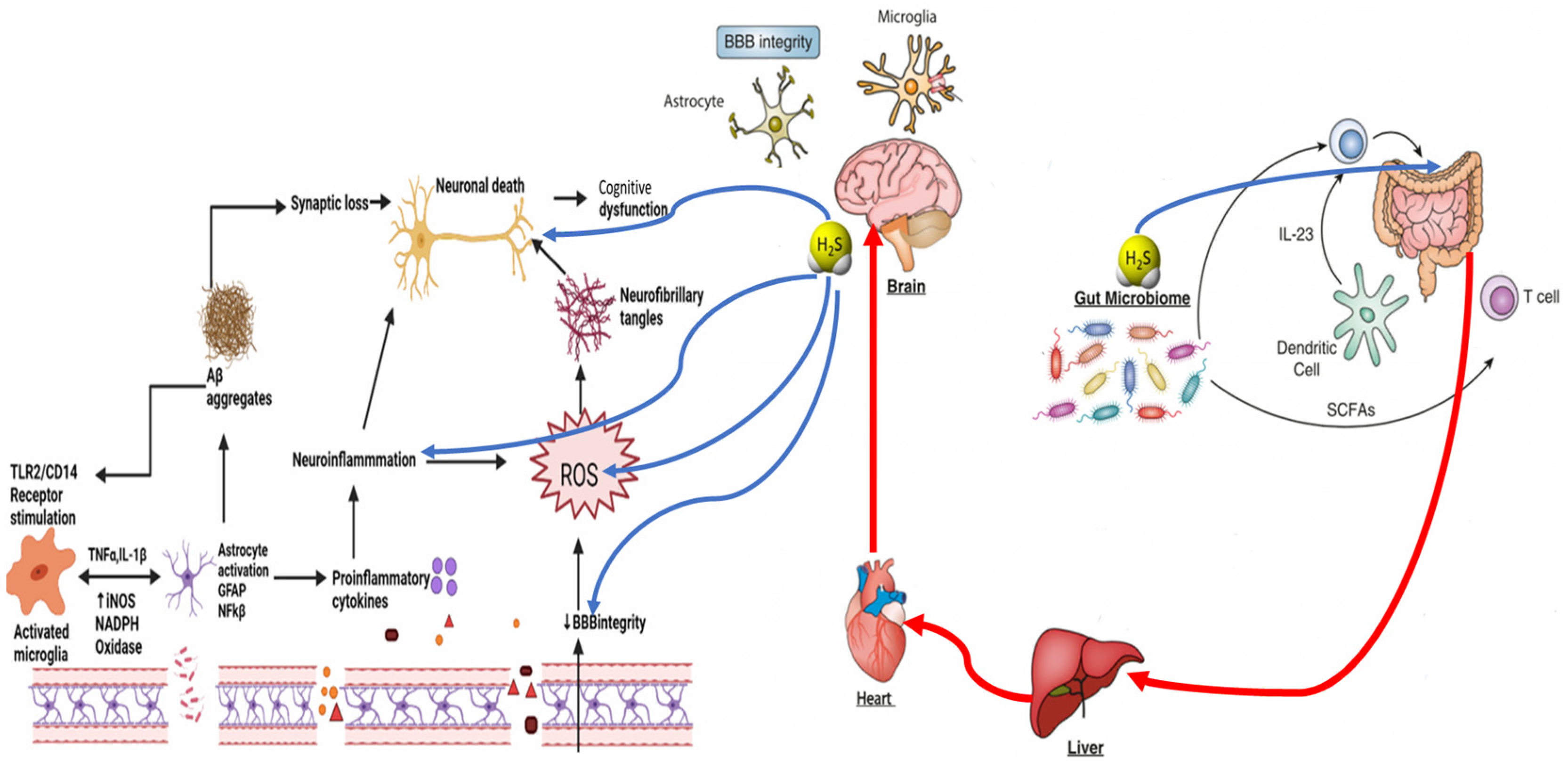
Figure 1. The dysbiosis-associated overproduction of H2S is proposed as a contributing factor in the progression of neurodegenerative diseases, where neuroinflammation plays a pivotal role. At the same time, H2S can have positive effects in a concentration-dependent manner.
Biomedicines 12 02670 g001
Figure 2. Illustrative interplay between diet, gut microbiota, and their metabolites in the context of neurodegenerative diseases, particularly Alzheimer’s and Parkinson’s. Diet shapes the gut microbial composition, including diverse groups such as Clostridium, Desulfovibrio, and Prevotella, responsible for metabolic activities with systemic impacts. Microbes metabolize dietary components into various products, including hydrogen sulfide (H2S) via sulfate reduction and short-chain fatty acids (SCFAs) like acetate, propionate, and butyrate from fiber fermentation. These metabolites influence gut permeability and barrier integrity, with butyrate supporting the gut lining. At the same time, excess H2S may compromise it, leading to a “leaky gut” and the release of inflammatory lipopolysaccharides (LPSs) into circulation. SCFAs and other microbial metabolites reach the brain through the bloodstream and vagus nerve, potentially promoting neuroinflammation—a known factor in neurodegeneration. The figure highlights how disruptions in gut microbiota balance can affect brain health, linking gut dysbiosis and altered barrier function to neurodegenerative disease risk and suggesting that therapeutic interventions targeting the gut may help mitigate these diseases.
Biomedicines 12 02670 g002
Figure 3. Bidirectional gut–brain axis, highlighting interactions among the hypothalamic–pituitary–adrenal (HPA) axis, gut microbiota, circulatory system, and neural pathways. The HPA axis produces cortisol, affecting immune cells, gut epithelium, and enteric muscles. Gut microbes modulate brain function via metabolites like short-chain fatty acids (SCFAs), neurotransmitters, and tryptophan metabolism. H2S was found to regulate mucosal integrity, support the anti-inflammatory environment of the gut, and maintain the protective mucus layer.
Biomedicines 12 02670 g003
Disclaimer/Publisher’s Note: The statements, opinions and data contained in all publications are solely those of the individual author(s) and contributor(s) and not of MDPI and/or the editor(s). MDPI and/or the editor(s) disclaim responsibility for any injury to people or property resulting from any ideas, methods, instructions or products referred to in the content.