Article

Recovery from Acute Kidney Injury and Long-Term Prognosis following Acute Myocardial Infarction

1
Department of Cardiology, Rabin Medical Center, Petach Tikva 4941492, Israel
2
Faculty of Medicine, Tel Aviv University, Tel Aviv 6997801, Israel
3
Division of Cardiovascular Medicine, Department of Medicine, Brigham and Women’s Hospital, Harvard Medical School, Boston, MA 02115, USA
4
Faculty of Health Sciences, Ben-Gurion University of the Negev, Beer Sheva 8410501, Israel
5
Department of Emergency Medicine, Soroka University Medical Center, Beer Sheva 8457108, Israel
*
Author to whom correspondence should be addressed.
Biomedicines 2024, 12(7), 1490; https://doi.org/10.3390/biomedicines12071490
Submission received: 4 June 2024 / Revised: 30 June 2024 / Accepted: 3 July 2024 / Published: 5 July 2024

Abstract

:
We investigated the recovery pattern from acute kidney injury (AKI) following acute myocardial infarction (AMI) and its association with long-term mortality. The retrospective study included AMI patients (2002–2027), who developed AKI during hospitalization. Creatinine (Cr) measurements were collected and categorized into 24 h timeframes up to 7 days from AKI diagnosis. The following groups of recovery patterns were defined: rapid (24–48 h)/no rapid and early (72–144 h)/no early recovery. Specific cut-off points for recovery at each AKI stage and timeframe were determined through receiver operating characteristic (ROC) curves. The probability of long-term (up to 10 years) mortality as a post-AKI recovery was investigated using a survival approach. Out of 17,610 AMI patients, 1069 developed AKI. For stage 1 AKI, patients with a Cr ratio <1.5 at 24 h and/or <1.45 at 48 h were defined as ‘rapid recovery’; for stages 2–3 AKI, a Cr ratio <2.5 at 96 h was defined as ‘early recovery’. Mortality risk in stage 1 AKI was higher among the non-rapidly recovered: AdjHR = 1.407; 95% CI: 1.086–1.824; p = 0.010. Among stages 2–3 AKI patients, the risk for long-term mortality was higher among patients who did not recover in the early period: AdjHR = 1.742; 95% CI: 1.085–2.797; p = 0.022. The absence of rapid recovery in stage 1 AKI and lack of early recovery in stages 2–3 AKI are associated with higher long-term mortality.

1. Introduction

Acute kidney injury (AKI) following acute myocardial infarction (AMI), also known as cardiorenal syndrome type 1 [1], is common, occurring in approximately 14.8% to 36.6% of cases [2,3]. The underlying mechanisms contributing to AKI post-AMI are multifactorial and often include a combination of hypotension, inflammation, oxidative stress, and direct tubular injury. Previous studies have shown post-AMI AKI to be independently associated with increased morbidity and mortality, such that patients experiencing complications are at increased risk of developing chronic kidney disease (CKD), end-stage kidney disease (ESKD), and adverse cardiovascular events [3,4,5,6,7,8,9].
Recovery from post-AMI AKI is variable and influenced by the severity of renal injury, the promptness of treatment, and the patient’s background. While in many cases, there is an initial improvement in renal function within the first week, and a full recovery by several weeks to months, a subset of patients may not regain their baseline renal function, leading to CKD and its associated long-term sequelae [10,11].
The aim of the current study is to characterize AKI following AMI based on the time to recovery and to assess its association with long-term prognosis.

2. Materials and Methods

2.1. Study Population

Our research relies on data from the Soroka Acute Myocardial Infarction (SAMI) registry, encompassing consecutive hospitalizations for AMI occurring at Soroka University Medical Center (SUMC), Israel, from 1 January 2002 to 31 October 2017 [12,13]. We included adult (i.e., ≥18-year-old) Israeli citizens who were hospitalized due to AMI at SUMC, developed AKI during their hospitalization, and were discharged alive. Additional inclusion criteria were estimated glomerular filtration rate (eGFR, ml/min/1.73 m2) >60 at admission and available data regarding at least two serum creatinine (Cr) tests during hospitalization: upon admission and at days 3 to 7. Patients with a diagnosis of hemodialysis/peritoneal dialysis at baseline and patients with a hospitalization period of less than three days were excluded from the analysis.
This project conformed to the Declaration of Helsinki and was approved by SUMC’s Institutional Review Board, which waived the need for informed consent in view of the investigation’s retrospective nature.

2.2. Follow-Up and Outcome

The study period spanned the timeframe between hospital discharge and either death or 30 December 2021. The study outcome was all-cause mortality up to 10 years after hospital discharge. Death events and dates were obtained from the Israeli Ministry of the Interior Population Registry.

2.3. Data Collection and Definitions

Patients’ clinical data were retrieved using electronic medical records. Baseline comorbidities were identified by International Classification of Diseases, Ninth Revision, Clinical Modification (ICD-9-CM) codes, as documented in real time by the treating medical team. Laboratory results and echocardiographic and angiographic findings were documented.
Patients hospitalized with AMI were allocated according to ICD-9-CM code 410. AMI diagnosis was based on ischemic signs and/or symptoms coupled with abrupt rise and fall in cardiac biomarkers levels consistent with acute myocardial injury, as dictated by the Universal Definition of Myocardial Infarction at the time (if applicable). Obstructive coronary artery disease (CAD) required the presence of a ≥70% vessel stenosis as assessed by angiography.

2.4. Study Strata and Groups

The first Cr measurement reported within the initial 24 h of admission was considered as the baseline Cr level for analysis. AKI was defined as an absolute increase in Cr ≥ 0.3 mg/dL (≥26.5 μmol/L) within 48 h or a relative increase in Cr to ≥1.5 times within 7 days, compared with the baseline level. The study strata, in accordance with AKI stage, was defined as 1 to 3 according to the following criteria: stage 1—an absolute increase in Cr ≥ 0.3 mg/dL or a relative increase of 1.5–1.9 from baseline; stage 2—a relative increase of 2–2.9 times baseline; and stage 3—an absolute increase to ≥4.0 mg/dL (≥353.6 μmol/L), a relative increase to 3.0 times baseline or initiation of renal replacement therapy [14,15,16].
All Cr measurements taken during hospitalization were retrieved, and categorized into 24 h timeframes, from the diagnosis of AKI and up to 7 days. Every time point encompassed all Cr tests conducted within a 12 h window both preceding and following that specific point. For instance, the 24 h interval comprised tests administered between 12 and 36 h. The recovery measure was calculated for each patient by dividing the Cr at a given time point by the baseline Cr level (Cr ratio). The recovery timeframes were defined as ‘rapid’, characterized as occurring within 24–48 h, and ‘early’, designated for the 72–144 h timeframe. Those patterns were based on previous publications referring to AKI recovery within 7 days as ‘early’ [17].

2.5. Statistical Analysis

All statistical analyses were performed separately for each study’s strata. The variables were reported as frequencies and percentages, medians, and interquartile ranges (IQRs), or means and standard deviations/standard errors (SD/SE), as appropriate. A comparison of the baseline characteristics between the patients who died and survivors was performed using Pearson’s Chi-Squared test for categorical variables and Student’s t test for count data. Particularly, using Student’s t tests, we compared the mean Cr rates for each timeframe.
The discriminative power of the Cr ratio was calculated using logistic regressions and receiver operating characteristic (ROC) curves. Based on these analyses, a cut-off point was calculated for each timeframe in which the Cr ratio was found to be significantly different among those who survived compared to those who died. In accordance with the cut-off point values and the timeframes, the following study groups were defined: rapid recovery/no rapid recovery and early recovery/no early recovery.
The probability of mortality as a post-AKI recovery was graphically displayed according to the survival approach (Kaplan–Meier method), and compared using the Log-rank test. Additionally, univariable and multivariable Cox regression analyses were utilized. The results of the models were expressed as Hazard Ratios/Adjusted Hazard Ratios (HR/AdjHR) and 95% Confidence Intervals (CI). The multivariable regressions incorporated the parameters of recovery and the baseline variables demonstrating a p-value of <0.1 on a preliminary univariable model. Statistical significance required a two-sided p-value of <0.05. All analyses were performed using SPSS, version 29 (IBM Corporation, Armonk, NY, USA).

3. Results

3.1. Study Population and Strata

During the years 2002 and 2017, 17,610 patients were admitted to SUMC with AMI. Of them, 10,555 were eligible for analysis according to the inclusion and exclusion criteria, and 1140 (10.8%) developed AKI during their hospitalization. Seventy-one (71) of them had no documented follow-up Cr tests, and were excluded from the analysis. Thus, 1069 patients were included in the final cohort (Supplementary Figure S1). Among them, 835 (78.1%) had stage 1 AKI, and 185 (17.3%) and 49 (4.6%) had stages 2 and 3 AKI, respectively. Due to the relatively low number of patients experiencing stage 2 and 3 AKI, the two groups were combined for analysis (n = 234).
The total mortality rate was (402/835 = 51.9%) among the patients with stage 1 AKI, whereas in stages 2–3 AKI, the incidence of mortality was higher (147/234 = 62.8%), p < 0.001.

3.2. Baseline Characteristics in Accordance with Status of Death

Baseline characteristics according to the AKI stage and the survival status are summarized in Table 1. Older patients with AKI at any stage were more likely to die. Additionally, heart diseases including cardiomegaly, congestive heart failure, and pulmonary heart disease, and conventional cardiovascular risk factors, including diabetes mellites, dyslipidemia, hypertension, smoking and peripheral vascular disease were found to be associated with increased mortality.

3.3. Baseline Characteristics in Accordance with Status of Death

Rapid recovery from stage 1 AKI was associated with a higher rate of long-term survival. The mean ± SD Cr ratio at 24 h among those who died was 1.49 ± 0.49, compared to 1.40 ± 0.31 among patients who survived (p < 0.001); and at 48 h, 1.44 ± 0.44 compared to 1.36 ± 0.37 (p = 0.006). No significant differences in the Cr ratio values between the patients who died and the survivors were found after 48 h (Figure 1a).
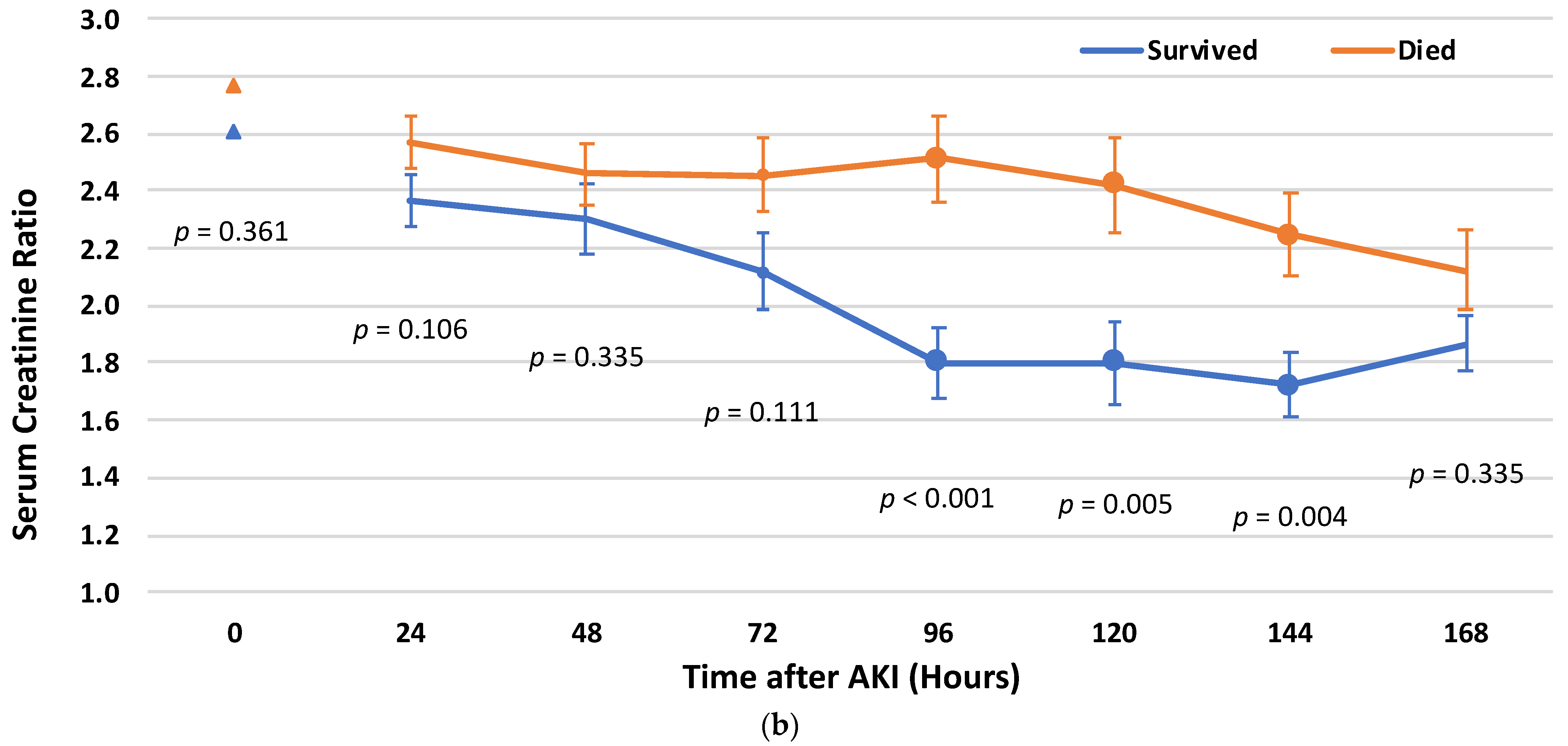
At stages 2–3 AKI, the differences in Cr ratio values became statistically significant in the early period after the AKI event. The mean ± SD Cr ratios among the patients who died, as compared with the survivors at the time periods of 96, 120, and 144 h, were 2.51 ± 1.62 vs. 1.80 ± 1.0 (p < 0.001), 2.42 ± 1.71 vs. 1.80 ± 0.95 (p = 0.005), and 2.25 ± 1.43 vs. 1.72 ± 0.69 (p = 0.004), respectively (Figure 1b).

3.4. Cr Rate Cut-Off Points for Post-AKI Recovery

The discrimination of the Cr ratio between the non-survivals and the survivals was based on ROC curves. A separate ROC curve was built for each timeframe in which there was a statistically significant difference in the Cr ratio between the patients who died and the survivors. For stage 1 AKI, a higher risk for death was associated with Cr ratio cut-off points of 1.50 for 24 h recovery (Odds Ratio [OR] = 1.384; 95% CI: 0.997–1.922; p = 0.052; sensitivity 42.9% and specificity 64.8%) and 1.45 for 48 h recovery (OR = 1.574; 95% CI: 1.090–2.273; p = 0.016, sensitivity 43.6% and specificity 67.0%).
For the patients with AKI stages 2–3, a Cr ratio cut-off point of 2.5 for 96 h recovery was associated with a higher risk of death (OR = 6.034; 95% CI: 1.998–18.219; p = 0.001, sensitivity 38.8% and specificity 90.5%).

3.5. Survival Analysis

Median follow-up was 2470 (IQR 1278–6352) days for stage 1 AKI and 1925 (514–3435) for stages 2–3 AKI. Cumulative mortalities were 0.523 and 0.676 for AKI stages 1 and 2–3, respectively.
Survival analysis compared the probability of dying during the follow-up period in the different groups of patients. Based on the previously defined cut-off points and the timeframes for AKI recovery, three groups of patients for each study strata were defined. Particularly, for stage 1 AKI—patients with a Cr ratio <1.5 at 24 h and/or <1.45 at 48 h were defined as ‘rapid recovery’ (619 patients). Those with a Cr ratio ≥1.5 within 24 h and a Cr ratio ≥1.45 within 48 h were defined as ‘not rapidly recovered’ (141 patients). The third group included patients with missing values of Cr within 24–48 h (75 patients). The baseline characteristics of the patients by the study groups are presented in Supplementary Table S1a. Females, along with individuals having diabetes mellitus and obesity, had a lower probability of achieving rapid recovery. The treatment approach varied between the two groups, with a higher incidence of percutaneous coronary intervention (PCI) among those who recovered rapidly, while the rate of surgical treatment (coronary artery bypass graft—CABG) was lower in this group.
For stages 2–3 AKI patients, a Cr ratio <2.5 at 96 h was defined as ‘early recovery’ (98 patients), and a Cr ratio ≥2.5 at this time was defined as ‘not early recovered’ (32 patients). The third group included those with missing values at this time (102 patients) (Supplementary Table S1b). Patients with diabetes mellitus and anemia demonstrated a reduced likelihood of experiencing early recovery, while the treatment strategy was similar in both groups.
Among the patients with stage 1 AKI, cumulative mortality was significantly higher in the group with no rapid recovery compared to the group with rapid recovery (0.630 vs. 0.505; p = 0.024). Cumulative mortality for the patients with missing values of Cr within this timeframe was 0.475, with no statistically significant differences from the group of rapid recovery (p = 0.580) (Figure 2a).
At stages 2–3 of AKI, cumulative mortality was higher among patients with no early recovery compared to those with early recovery (0.853 vs. 0.619; p = 0.024). Cumulative mortality for the patients with missing values of Cr within this timeframe was 0.655, with no statistically significant difference from the group of early recovery (p = 0.580) (Figure 2b).

3.6. Post-AKI Recovery and the Risk for Mortality

Among stage 1 AKI patients, the risk for long-term mortality in the no rapid recovery group was higher than the risk of patients with rapid recovery: HR = 1.331; 95% CI: 1.037–1.707; p = 0.025. The risk of patients with missing values of Cr within this timeframe was not different from the rapid recovery group: HR = 0.902; 95% CI: 0.632–1.287; p = 0.669.
Among stages 2–3 AKI patients, the risk for long-term mortality was higher among patients with no early recovery, compared to those with early recovery: HR = 1.755; 95% CI: 1.117–2.757; p = 0.015. The risk of patients with missing values of Cr within this timeframe was not different from the early recovery group: HR = 1.007; 95% CI: 0.701–1.447; p = 0.970.
After adjustment for the investigated confounders, the risk for long-term all-cause mortality has remained statistically significantly higher for those who did not recover rapidly or early compared with those who did. Among stage 1 AKI patients, no rapid recovery vs. rapid recovery, AdjHR was 1.407; 95% CI: (1.086–1.824); p = 0.010 (Table 2a). Accordantly, among stages 2–3 AKI, no early recovery vs. early recovery, the AdjHR was 1.742; 95% CI: 1.085–2.797; p = 0.022 (Table 2b).

4. Discussion

The present study investigated the phenomenon of AKI following AMI by the time to recovery and its association with all-cause long-term mortality. We have addressed the different AKI stages and recovery, according to a unique calculation, based on the baseline Cr of each patient. We found that patients with stages 2–3 AKI had higher mortality rates compared to patients with AKI stage 1. Next, we have characterized kidney recovery in association with long-term mortality after AMI associated with AKI. We found that patients with stage 1 AKI who experienced rapid recovery from the event (as compared with no rapid recovery), and patients with stages 2–3 AKI who experienced early recovery (as compared to no early recovery), had higher survival. We have found Cr ratio cut-off points for each AKI stage and timeframe: for stage 1 AKI—1.5 at 24 h and 1.45 at 48 h; for stages 2–3 AKI—2.5 at 96 h. Increased values of the Cr ratio above the cut-off points were found to be associated with a higher risk of death. There were two differences between stage 1 AKI and stages 2 and 3 AKI: first, the prognostic significance of kidney recovery in AKI stages 2–3 was only found after 96 h from the injury (early recovery), and second, the Cr ratios above these cut-off points were found to be associated with increased mortality. As outlined in the methods, each time point included all Cr tests performed within a 12 h period before and after that particular point.
The mechanism of the cardiorenal syndrome type 1 is complex. The syndrome is initiated through the combination of renal congestion, diminished renal perfusion and activation of the renin–angiotensin–aldosterone system, followed by inflammatory cell infiltration and direct tubular damage [1,18]. Several risk factors have been identified in the literature that increase the likelihood of AKI development, including advanced age, pre-existing kidney disease, diabetes mellitus, anemia, heart failure, and the use of nephrotoxic medications and high doses of contrast agents [19].
The mechanism of the renal repair process is based on hemodynamic stabilization and improved heart function accompanied by the renal regenerative process and repopulation of the surviving tubular cells, along with paracrine growth factor secretion [17,20]. In specific situations, renal restoration can become counterproductive, resulting in inflammation, fibrosis, and vascular rarefaction that all contribute to ongoing cellular and tissue dysfunction, ultimately culminating in CKD.
A rapid recovery from stage 1 AKI associated with a better prognosis is expected, as the kidney injury is transient and does not cause damage for more than a short period of time. The prognostic significance of the early recovery from stages 2–3 AKI can be explained in a different way—the initial injury is significant and somewhat extreme and the healing process lasts more than a couple of days. The success of this healing is revealed after 96 h, and this is the timeframe in which the Cr rate becomes significant. The higher Cr ratio cut-off point in stages 2–3 AKI is logical, as it represents the initial harm to the kidney function, which is more prominent than in stage 1 AKI. In both cases, kidney recovery implies the presence of premorbid renal reserve, which enables the adaptive repair process.
The terms renal recovery and time pattern are not clearly defined in the literature. Some studies define kidney recovery as a return to normal kidney function, while others address it as Cr decline and improvement in the kidney disease stage [21]. As for the time pattern—it varies, as early recovery can be defined as one that occurs within 72 h [17], 3–7 days [22], up to 10 days [23], or when there is ‘in-hospital recovery’ [11].
The KDIGO guideline characterizes AKI as an abrupt decline in kidney function within a span of 7 days or less, whereas CKD is defined by the enduring presence of kidney disease for a period exceeding 90 days, and renal injury present between days 7 to 90 is referred as acute kidney disease [15].
Our definition of the recovery as rapid (within 2 days) and early (3–6 days) is a combination of the above definitions. As for the recovery itself, we have used the serial Cr measurements and calculation of the Cr ratio, according to the Acute Disease Quality Initiative (ADQI) consensus report on AKI and renal recovery [21]. The calculation was based on the equation, in which the serum Cr following the AKI event is divided by the baseline Cr. The use of this method has the advantage of addressing each patient’s baseline Cr and their personal improvement. It can be simply calculated and used in the clinical evaluation of patients with AKI following AMI.
Early recognition and management of AKI are crucial for improving outcomes post-AMI. Timely intervention involves adequate hemodynamic support, discontinuation of nephrotoxic medications and optimizing volume status to enhance renal perfusion.
Moreover, the development of new therapeutic agents that provide both cardiovascular and renal benefits can be considered, as those can potentially act as disease modifiers, meaning they not only manage the symptoms, but also address the underlying causes of the illness. The medications that have been explored in this context include renin–angiotensin–aldosterone System (RAAS) inhibitors, sodium–glucose cotransporter 2 (SGLT2) inhibitors, mineralocorticoid receptor antagonists and GLP-1 receptor antagonists. Our study results enable identifying populations with a greater potential for kidney recovery and may guide physicians in their clinical decisions.

Limitations

The study exhibits several limitations. The primary constraint lies in its retrospective design and the derivation of the cohort from a single center. However, this cohort stands out as one of the largest reported to date, and its findings, particularly regarding AKI, align with prior publications, thereby bolstering the study’s validity. Secondly, the groups of patients with AKI stages 2 and 3 were relatively small, necessitating their combination for analysis. Additionally, a significant number of patients in these groups had missing values, potentially impacting the interpretation of results. Thirdly, our cohort lacks information on medical therapies administered during hospitalization and post-discharge, the number of radiocontrast agents given to each patient, and specific details about causes of death and other cardiovascular and renal outcomes. However, adjustments for confounding factors have been made, and the risk for the primary outcome, long-term all-cause mortality, remains statistically significant.

5. Conclusions

The findings from our study indicate an association between the AKI stage following AMI and the recovery patern from AKI with the risk of long-term all-cause mortality. Long-term follow-up reveal that patients experiencing stage 1 AKI without recovery within the initial 48 h and those with stages 2–3 AKI without recovery within 96 h are at a higher risk of mortality. We established specific cut-off points to address recovery at each AKI stage and timeframe. Pending further research, early intervention for AKI during the initial phase among AMI patients may offer potential benefits.

Supplementary Materials

The following supporting information can be downloaded at: https://www.mdpi.com/article/10.3390/biomedicines12071490/s1, Figure S1: The study flow chart; Table S1: Baseline characteristics of patients by Acute kidney injury (AKI) recovery group.

Author Contributions

Conceptualization, K.S.; methodology, Y.P. and A.S. (Arthur Shiyovich); validation, H.G. and A.S. (Alon Shechter); data curation, H.G. and Y.P.; writing—original draft preparation, K.S.; writing—review and editing, A.S. (Alon Shechter), H.G., A.S. (Arthur Shiyovich) and Y.P.; visualization, A.S. (Arthur Shiyovich). All authors have read and agreed to the published version of the manuscript.

Funding

This research received no external funding.

Institutional Review Board Statement

The study was conducted in accordance with the Declaration of Helsinki, and approved by Soroka’s Institutional Review Board (approval number SOR-0319-16).

Informed Consent Statement

Patient consent was waived due to the investigation’s retrospective nature.

Data Availability Statement

The data presented in this study are available on request from the corresponding author. The data are not publicly available due to privacy or ethical restrictions.

Conflicts of Interest

The authors declare no conflicts of interest.

References

  1. Ronco, C.; Bellasi, A.; Di Lullo, L. Cardiorenal Syndrome: An Overview. Adv. Chronic Kidney Dis. 2018, 25, 382–390. [Google Scholar] [CrossRef]
  2. Rodrigues, F.B.; Bruetto, R.G.; Torres, U.S.; Otaviano, A.P.; Zanetta, D.M.T.; Burdmann, E.A. Incidence and Mortality of Acute Kidney Injury after Myocardial Infarction: A Comparison between KDIGO and RIFLE Criteria. PLoS ONE 2013, 8, e69998. [Google Scholar] [CrossRef] [PubMed]
  3. Kaltsas, E.; Chalikias, G.; Tziakas, D. The Incidence and the Prognostic Impact of Acute Kidney Injury in Acute Myocardial Infarction Patients: Current Preventive Strategies. Cardiovasc. Drugs Ther. 2018, 32, 3281–3298. [Google Scholar] [CrossRef]
  4. Skalsky, K.; Shiyovich, A.; Steinmetz, T.; Kornowski, R. Chronic Renal Failure and Cardiovascular Disease: A Comprehensive Appraisal. J. Clin. Med. 2022, 11, 1335. [Google Scholar] [CrossRef] [PubMed]
  5. Fox, C.S.; Muntner, P.; Chen, A.Y.; Alexander, K.P.; Roe, M.T.; Wiviott, S.D. Short-Term Outcomes of Acute Myocardial Infarction in Patients With Acute Kidney Injury: A Report From the National Cardiovascular Data Registry. Circulation 2012, 125, 497–504. [Google Scholar] [CrossRef] [PubMed]
  6. Sun, Y.B.; Liu, B.C.; Zou, Y.; Pan, J.R.; Tao, Y.; Yang, M. Risk factors of acute kidney injury after acute myocardial infarction. Ren. Fail. 2016, 38, 1353–1358. [Google Scholar] [CrossRef]
  7. Chalikias, G.; Serif, L.; Kikas, P.; Thomaidis, A.; Stakos, D.; Makrygiannis, D.; Chatzikyriakou, S.; Papoulidis, N.; Voudris, V.; Lantzouraki, A.; et al. Long-term impact of acute kidney injury on prognosis in patients with acute myocardial infarction. Int. J. Cardiol. 2019, 283, 48–54. [Google Scholar] [CrossRef]
  8. Pickering, J.W.; Blunt, I.R.H.; Than, M.P. Acute Kidney Injury and mortality prognosis in Acute Coronary Syndrome patients: A meta-analysis. Nephrology 2018, 23, 237–246. [Google Scholar] [CrossRef]
  9. Parikh, C.R.; Coca, S.G.; Wang, Y.; Masoudi, F.A.; Krumholz, H.M. Long-term Prognosis of Acute Kidney Injury After Acute Myocardial Infarction. Arch. Intern. Med. 2008, 168, 987–995. [Google Scholar] [CrossRef]
  10. Itach, T.; Banai, A.; Paran, Y.; Zahler, D.; Merdler, I.; Eliashiv, D.; Banai, S.; Shacham, Y. Acute Kidney Injury Recovery Patterns in ST-Segment Elevation Myocardial Infarction Patients. J. Clin. Med. 2022, 11, 2169. [Google Scholar] [CrossRef]
  11. Goldberg, A.; Kogan, E.; Hammerman, H.; Markiewicz, W.; Aronson, D. The impact of transient and persistent acute kidney injury on long-term outcomes after acute myocardial infarction. Kidney Int. 2009, 76, 900–906. [Google Scholar] [CrossRef] [PubMed]
  12. Shvartsur, R.; Shiyovich, A.; Gilutz, H.; Azab, A.N.; Plakht, Y. Short and long-term prognosis following acute myocardial infarction according to the country of origin. Soroka acute myocardial infarction II (SAMI II) project. Int. J. Cardiol. 2018, 259, 227–233. [Google Scholar] [CrossRef]
  13. Plakht, Y.; Abu Eid, A.; Gilutz, H.; Shiyovich, A. Trends of Cardiovascular Risk Factors in Patients With Acute Myocardial Infarction: Soroka Acute Myocardial Infarction II (SAMI II) Project. Angiology 2019, 70, 530–538. [Google Scholar] [CrossRef] [PubMed]
  14. Skalsky, K.; Levi, A.; Bental, T.; Vaknin-Assa, H.; Assali, A.; Steinmetz, T.; Kornowski, R.; Perl, L. The Definition of “Acute Kidney Injury” Following Percutaneous Coronary Intervention and Cardiovascular Outcomes. Am. J. Cardiol. 2021, 56, 39–43. [Google Scholar] [CrossRef]
  15. Khwaja, A. KDIGO Clinical Practice Guidelines for Acute Kidney Injury. Nephron Clin. Pract. 2012, 120, c179–c184. [Google Scholar] [CrossRef]
  16. Mehta, R.L.; Kellum, J.A.; Shah, S.V.; Molitoris, B.A.; Ronco, C.; Warnock, D.G.; Levin, A.; Acute Kidney Injury Network. Acute Kidney Injury Network: Report of an initiative to improve outcomes in acute kidney injury. Crit. Care 2007, 11, R31. [Google Scholar] [CrossRef]
  17. Forni, L.G.; Darmon, M.; Ostermann, M.; Oudemans-van Straaten, H.M.; Pettilä, V.; Prowle, J.R.; Schetz, M.; Joannidis, M. Renal recovery after acute kidney injury. Intensive Care Med. 2017, 43, 855–866. [Google Scholar] [CrossRef]
  18. Ismail, Y.; Kasmikha, Z.; Green, H.L.; McCullough, P.A. Cardio-Renal Syndrome Type 1: Epidemiology, Pathophysiology, and Treatment. Semin. Nephrol. 2012, 32, 18–25. [Google Scholar] [CrossRef] [PubMed]
  19. Tsai, T.T.; Patel, U.D.; Chang, T.I.; Kennedy, K.F.; Masoudi, F.A.; Matheny, M.E.; Kosiborod, M.; Amin, A.P.; Messenger, J.C.; Rumsfeld, J.S.; et al. Contemporary Incidence, Predictors, and Outcomes of Acute Kidney Injury in Patients Undergoing Percutaneous Coronary Interventions. JACC Cardiovasc. Interv. 2014, 7, 1–9. [Google Scholar] [CrossRef]
  20. Venkatachalam, M.A.; Weinberg, J.M.; Kriz, W.; Bidani, A.K. Failed Tubule Recovery, AKI-CKD Transition, and Kidney Disease Progression. J. Am. Soc. Nephrol. 2015, 26, 1765–1776. [Google Scholar] [CrossRef]
  21. Chawla, L.S.; Bellomo, R.; Bihorac, A.; Goldstein, S.L.; Siew, E.D.; Bagshaw, S.M.; Bittleman, D.; Cruz, D.; Endre, Z.; Fitzgerald, R.L.; et al. Acute kidney disease and renal recovery: Consensus report of the Acute Disease Quality Initiative (ADQI) 16 Workgroup. Nat. Rev. Nephrol. 2017, 13, 241–257. [Google Scholar] [CrossRef] [PubMed]
  22. Kellum, J.A.; Sileanu, F.E.; Bihorac, A.; Hoste, E.A.J.; Chawla, L.S. Recovery after Acute Kidney Injury. Am. J. Respir. Crit. Care Med. 2017, 195, 784–791. [Google Scholar] [CrossRef] [PubMed]
  23. Heung, M.; Steffick, D.E.; Zivin, K.; Gillespie, B.W.; Banerjee, T.; Hsu, C.y.; Powe, N.R.; Pavkov, M.E.; Williams, D.E.; Saran, R.; et al. Acute Kidney Injury Recovery Pattern and Subsequent Risk of CKD: An Analysis of Veterans Health Administration Data. Am. J. Kidney Dis. 2016, 67, 742–752. [Google Scholar] [CrossRef] [PubMed]
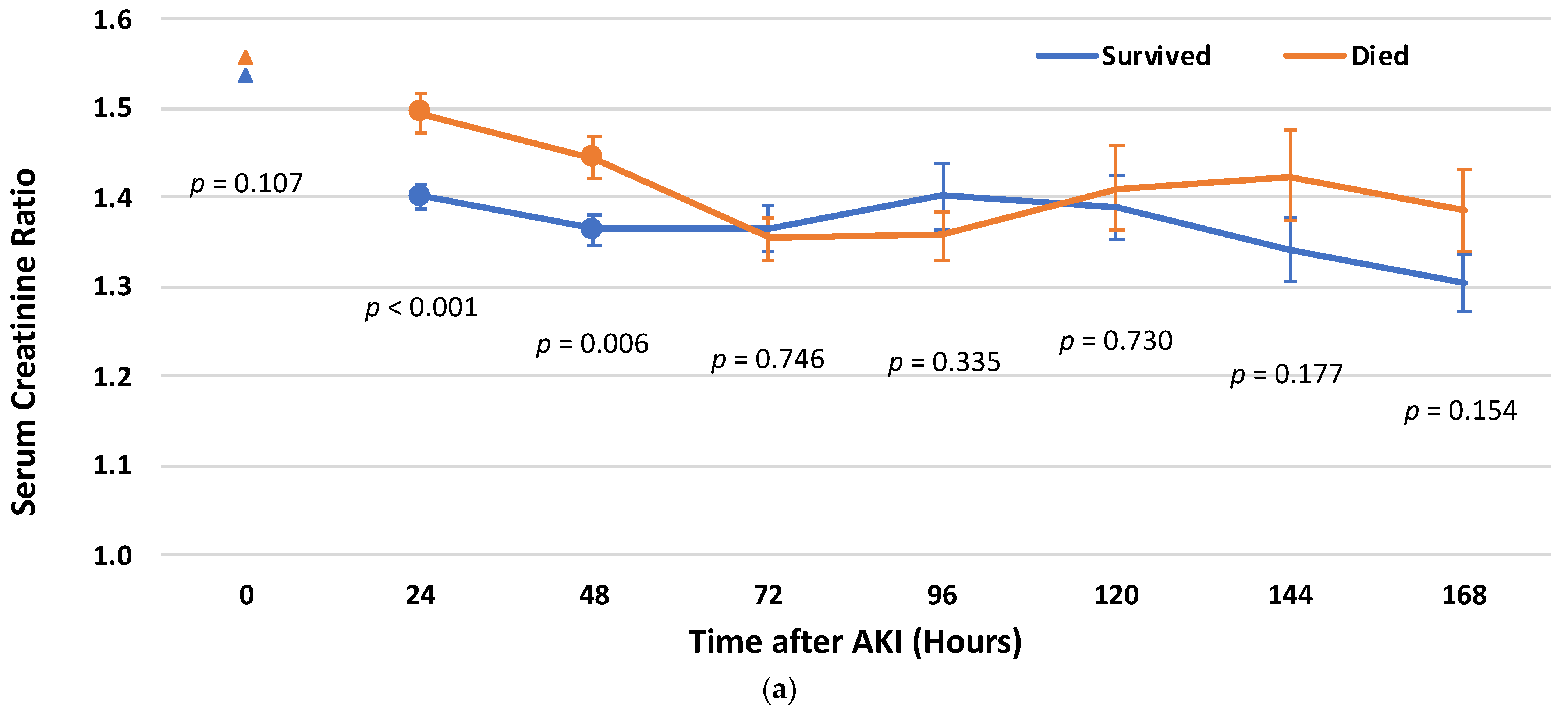
Figure 1. Serum Creatinine ratio (mean and standard error) by timeframe (hours after Acute kidney injury [AKI]) among the patients who died and among survivors: (a) among the patients with AKI stage 1; (b) among the patients with AKI stages 2 and 3.
Biomedicines 12 01490 g001aBiomedicines 12 01490 g001b
Figure 2. Cumulative long-term survival of the patients after Acute kidney injury (AKI) by the group of recovery: (a) among the patients with AKI stage 1; (b) among the patients with AKI stages 2 and 3.
Biomedicines 12 01490 g002
Table 1. Baseline characteristics and survival status: (a) among the patients with acute kidney injury (AKI) stage 1; (b) among the patients with AKI stages 2 and 3.
(a)
GroupSurvivedDiedTotalp
n433402835
Demographics
Age, Years, Mean (SD)62.05 (10.51)71.95 (10.63)66.82 (11.66)<0.001
<65273 (63)111 (27.6)384 (46.0)<0.001
65–75108 (24.9)129 (32.1)237 (28.4)
≥7552 (12.0)162 (40.3)214 (25.6)
Sex, Male367 (84.8)293 (72.9)660 (79.0)<0.001
Ethnicity, Arab/Other74 (17.1)72 (17.9)146 (17.5)0.755
Cardiac diseases
Cardiomegaly46 (10.6)65 (16.2)111 (13.3)0.018
Supraventricular arrhythmias44 (10.2)113 (28.1)157 (18.8)<0.001
Congestive heart failure77 (17.8)149 (37.1)226 (27.1)<0.001
Pulmonary heart disease21 (4.8)53 (13.2)74 (8.9)<0.001
Chronic ischemic heart disease410 (94.7)345 (85.8)755 (90.4)<0.001
Previous myocardial infarction65 (15.0)120 (29.9)185 (22.2)<0.001
Previous percutaneous coronary intervention85 (19.6)101 (25.1)186 (22.3)0.057
Previous coronary artery bypass graft22 (5.1)55 (13.7)77 (9.2)<0.001
Atrioventricular block19 (4.4)17 (4.2)36 (4.3)0.910
Cardiovascular risk factors
Diabetes mellitus178 (41.1)230 (57.2)408 (48.9)<0.001
Dyslipidemia376 (86.8)326 (81.1)702 (84.1)0.023
Hypertension267 (61.7)297 (73.9)564 (67.5)<0.001
Obesity112 (25.9)103 (25.6)215 (25.7)0.936
Smoking207 (47.8)151 (37.6)358 (42.9)0.003
Peripheral vascular disease39 (9.0)101 (25.1)140 (16.8)<0.001
Family history of ischemic heart disease64 (14.8)13 (3.2)77 (9.2)<0.001
Other disorders
Chronic obstructive pulmonary disease17 (3.9)70 (17.4)87 (10.4)<0.001
Neurological disorders52 (12.0)93 (23.1)145 (17.4)<0.001
Malignancy9 (2.1)33 (8.2)42 (5.0)<0.001
Anemia265 (61.2)269 (66.9)534 (64.0)0.086
Gastrointestinal bleeding14 (3.2)24 (6.0)38 (4.6)0.058
Schizophrenia/Psychosis8 (1.8)15 (3.7)23 (2.8)0.097
Alcohol/Drug addiction7 (1.6)16 (4.0)23 (2.8)0.037
History of malignancy18 (4.2)25 (6.2)43 (5.1)0.178
Administrative characteristics of the hospitalization
Admitted/transposed to ICCU345 (79.7)244 (60.7)589 (70.5)<0.001
Length of hospital stay, days, Mean (SD)16.46 (10.04)18.55 (14.87)17.47 (12.64)0.017
≥7375 (86.6)335 (83.3)710 (85.0)0.185
Clinical characteristics of the hospitalization
Type of AMI, STEMI234 (54.0)154 (38.3)388 (46.5)<0.001
Results of echocardiography
Echocardiography performance365 (84.3)309 (76.9)674 (80.7)0.007
Severe left ventricular dysfunction53 (14.5)76 (24.6)129 (19.1)0.001
Left ventricular hypertrophy13 (3.6)22 (7.1)35 (5.2)0.038
Mitral regurgitation17 (4.7)37 (12.0)54 (8.0)<0.001
Tricuspid regurgitation7 (1.9)22 (7.1)29 (4.3)0.001
Pulmonary hypertension17 (4.7)52 (16.8)69 (10.2)<0.001
Results of angiography
Angiography performance357 (82.4)251 (62.4)608 (72.8)<0.001
Measure of coronary artery disease
No/non-significant4 (1.1)5 (2.0)9 (1.5)0.175
One vessel48 (13.4)24 (9.6)72 (11.8)
Two vessels73 (20.4)43 (17.1)116 (19.1)
Three vessels/Left main artery232 (65.0)179 (71.3)411 (67.6)
Type of treatment
Noninvasive11 (2.5)110 (27.4)121 (14.5)<0.001
Percutaneous coronary intervention142 (32.8)140 (34.8)282 (33.8)
Coronary artery bypass graft280 (64.7)152 (37.8)432 (51.7)
Kidney function
eGFR (first), Mean (SD)84.30 (20.00)80.80 (18.09)82.62 (19.17)0.008
Creatinine at AKI, Mean (SD)1.20 (0.25)1.27 (0.25)1.23 (0.25)<0.001
(b)
GroupSurvivedDiedTotalp
n87147234
Demographics
Age, Years, Mean (SD)62.33 (11.70)71.39 (11.39)68.02 (12.29)<0.001
<6548 (55.2)38 (25.9)86 (36.8)<0.001
65–7528 (32.2)49 (33.3)77 (32.9)
≥7511 (12.6)60 (40.8)71 (30.3)
Sex, Male59 (67.8)77 (52.4)136 (58.1)0.021
Ethnicity, Arab/Other12 (13.8)22 (15.0)34 (14.5)0.806
Cardiac diseases
Cardiomegaly8 (9.2)29 (19.7)37 (15.8)0.033
Supraventricular arrhythmias16 (18.4)47 (32.0)63 (26.9)0.024
Congestive heart failure17 (19.5)60 (40.8)77 (32.9)0.001
Pulmonary heart disease6 (6.9)28 (19.0)34 (14.5)0.011
Chronic ischemic heart disease81 (93.1)111 (75.5)192 (82.1)0.001
Previous myocardial infarction15 (17.2)44 (29.9)59 (25.2)0.031
Previous percutaneous coronary intervention16 (18.4)27 (18.4)43 (18.4)0.996
Previous coronary artery bypass graft3 (3.4)20 (13.6)23 (9.8)0.012
Atrioventricular block4 (4.6)8 (5.4)12 (5.1)0.777
Cardiovascular risk factors
Diabetes mellitus37 (42.5)90 (61.2)127 (54.3)0.006
Dyslipidemia71 (81.6)118 (80.3)189 (80.8)0.802
Hypertension51 (58.6)108 (73.5)159 (67.9)0.019
Obesity19 (21.8)39 (26.5)58 (24.8)0.422
Smoking42 (48.3)48 (32.7)90 (38.5)0.018
Peripheral vascular disease12 (13.8)39 (26.5)51 (21.8)0.023
Family history of ischemic heart disease8 (9.2)6 (4.1)14 (6.0)0.111
Other disorders
Chronic obstructive pulmonary disease6 (6.9)31 (21.1)37 (15.8)0.004
Neurological disorders14 (16.1)49 (33.3)63 (26.9)0.004
Malignancy3 (3.4)8 (5.4)11 (4.7)0.486
Anemia47 (54.0)113 (76.9)160 (68.4)<0.001
Gastrointestinal bleeding4 (4.6)9 (6.1)13 (5.6)0.772
Schizophrenia/Psychosis1 (1.1)4 (2.7)5 (2.1)0.422
Alcohol/Drug addiction1 (1.1)1 (0.7)2 (0.9)0.706
History of malignancy3 (3.4)6 (4.1)9 (3.8)0.808
Administrative characteristics of the hospitalization
Admitted/transposed to ICCU76 (87.4)91 (61.9)167 (71.4)<0.001
Length of hospital stay, days, Mean (SD)19.37 (32.82)32.82 (28.18)27.82 (24.46)<0.001
≥781 (93.1)139 (94.6)220 (94.0)0.650
Clinical characteristics of the hospitalization
Type of AMI, STEMI42 (48.3)52 (35.4)94 (40.2)0.052
Results of echocardiography
Echocardiography performance73 (83.9)112 (76.2)185 (79.1)0.161
Severe left ventricular dysfunction13 (17.8)35 (31.3)48 (25.9)0.041
Left ventricular hypertrophy4 (5.5)8 (7.1)12 (6.5)0.653
Mitral regurgitation3 (4.1)8 (7.1)11 (5.9)0.394
Tricuspid regurgitation2 (2.7)9 (8.0)11 (5.9)0.205
Pulmonary hypertension4 (5.5)15 (13.4)19 (10.3)0.083
Results of angiography
Angiography performance74 (85.1)88 (59.9)162 (69.2)<0.001
Measure of coronary artery disease
No/non-significant2 (2.7)4 (4.5)6 (3.7)0.214
One vessel16 (21.6)10 (11.4)26 (16.0)
Two vessels16 (21.6)17 (19.3)33 (20.4)
Three vessels/Left main artery40 (54.1)57 (64.8)97 (59.9)
Type of treatment
Noninvasive3 (3.4)49 (33.3)52 (22.2)<0.001
Percutaneous coronary intervention30 (34.5)49 (33.3)79 (33.8)
Coronary artery bypass graft54 (62.1)49 (33.3)103 (44.0)
Kidney function
eGFR (first), Mean (SD)113.34 (109.81)91.98 (44.88)99.92 (76.29)0.086
Creatinine at AKI, Mean (SD)1.71 (1.00)1.82 (1.15)1.78 (1.10)0.434
Data are presented as numbers (percentage), unless specified otherwise. AKI—Acute kidney injury, AMI—Acute myocardial infarction, eGFR—Estimated glomerular filtration rate (mL/min/1.73 m2), ICCU—Intensive cardiac care unit, SD—standard deviation, STEMI—ST-Elevation myocardial infarction.
Table 2. Relationship between kidney recovery and the risk for long-term all-cause mortality—multivariable analysis: (a) among the patients with acute kidney injury (AKI) stage 1; (b) among the patients with AKI stages 2 and 3.
(a)
ParameterB (SE)AdjHR(95% CI)p
Recovery group *:
Rapid recovery 1 (ref.)
No rapid recovery0.341 (0.132)1.407(1.086–1.824)0.010
Missing values of serum Creatinine−0.023 (0.184)0.978(0.681–1.403)0.903
Age (years): ≥75 vs. <750.771 (0.113)2.162(1.731–2.700)<0.001
Supraventricular arrhythmias0.379 (0.116)1.461(1.163–1.836)0.001
Congestive heart failure0.387 (0.111)1.473(1.186–1.831)<0.001
Previous myocardial infarction0.383 (0.116)1.467(1.169–1.840)<0.001
Previous coronary artery bypass graft0.376 (0.153)1.457(1.079–1.968)0.014
Diabetes mellitus0.470 (0.108)1.600(1.296–1.976)<0.001
Chronic obstructive pulmonary disease0.938 (0.140)2.554(1.940–3.362)<0.001
Malignancy0.657 (0.190)1.929(1.330–2.797)<0.001
Schizophrenia/Psychosis0.746 (0.271)2.109(1.241–3.584)0.006
Alcohol/Drug addiction1.084 (0.262)2.955(1.770–4.934)<0.001
Type of treatment:
Type of AMI: NSTEMI vs. STEMI0.200 (0.109)1.221(0.986–1.513)0.067
Noninvasive 1 (ref.)
Percutaneous coronary intervention−0.947 (0.138)0.338(0.296–0.509)<0.001
Coronary artery bypass graft−1.326 (0.139)0.265(0.202–0.349)<0.001
(b)
ParameterB (SE)AdjHR(95% CI)p
Recovery group *:
Early recovery 1 (ref.)
No early recovery0.555 (0.242)1.742(1.085–2.797)0.022
Missing values of serum Creatinine−0.022 (0.189)0.978(0.676–1.416)0.908
Age (years): ≥75 vs. <750.969 (0.179)2.636(1.856–3.743)<0.001
Cardiomegaly1.137 (0.225)3.117(2.007–4.841)<0.001
Diabetes mellitus0.817 (0.190)2.263(1.560–3.282)<0.001
Neurological disorders0.472 (0.182)1.603(1.123–2.290)0.009
Malignancy0.830 (0.375)2.294(1.099–4.788)0.027
Type of treatment:
Noninvasive 1 (ref.)
Percutaneous coronary intervention−1.039 (0.214)0.354(0.233–0.538)<0.001
Coronary artery bypass graft−1.777 (0.226)0.169(0.109–0.263)<0.001
* Recovery groups were defined as: ‘Rapid recovery’—patients with creatinine ratio <1.5 at 12–36 h and/or <1.45 at 36–60 h after AKI diagnosis; ‘No rapid recovery’—patients with creatinine ratio ≥1.5 at 12–36 h and ≥1.45 at 36–60 h after AKI diagnosis; ‘Early recovery’—patients with creatinine ratio <2.50 at 84–108 h after AKI diagnosis; ‘No early recovery’—patients with creatinine ratio ≥2.50 at 84–108 h after AKI diagnosis. AdjHR—Adjusted hazard ratio, AKI—Acute kidney injury, B—Regression coefficient, CI—Confidence interval, NSTEMI—Non-ST-elevation myocardial infarction, STEMI- ST-elevation myocardial infarction, Ref.—Reference group, SE—Standard error.
Disclaimer/Publisher’s Note: The statements, opinions and data contained in all publications are solely those of the individual author(s) and contributor(s) and not of MDPI and/or the editor(s). MDPI and/or the editor(s) disclaim responsibility for any injury to people or property resulting from any ideas, methods, instructions or products referred to in the content.