Next Article in Journal
Cytocidal Effects of Interstitial Photodynamic Therapy Using Talaporfin Sodium and a Semiconductor Laser in a Rat Intracerebral Glioma Model
Previous Article in Journal
Breaking the Cycle of Pain: The Role of Graded Motor Imagery and Mirror Therapy in Complex Regional Pain Syndrome
 
 
biomedicines-logo
Article Menu

Article Menu

Article

COVID-19 Inflammatory Syndrome: Lessons from TNFRI and CRP about the Risk of Death in Severe Disease

by
Thaís Soares Farnesi-de-Assunção
1,
Ana Carolina de Morais Oliveira-Scussel
1,
Wellington Francisco Rodrigues
1,
Beatriz Sodré Matos
1,
Djalma Alexandre Alves da Silva
1,
Leonardo Eurípedes de Andrade e Silva
2,
Fabiano Vilela Mundim
1,
Fernanda Rodrigues Helmo
1,
Anna Victória Bernardes e Borges
1,
Chamberttan Souza Desidério
1,
Rafael Obata Trevisan
1,
Malu Mateus Santos Obata
1,
Laís Milagres Barbosa
1,
Marcela Rezende Lemes
1,
Juliana Cristina Costa-Madeira
1,
Rafaela Miranda Barbosa
1,
Andrezza Cristina Cancian Hortolani Cunha
3,
Loren Queli Pereira
3,
Sarah Cristina Sato Vaz Tanaka
3,
Fernanda Bernadelli de Vito
3,
Ivan Borges Monteiro
4,5,
Yulsef Moura Ferreira
5,
Guilherme Henrique Machado
6,
Hélio Moraes-Souza
3,
Denise Bertulucci Rocha Rodrigues
7,8,
Carlo José Freire de Oliveira
1,
Marcos Vinicius da Silva
1 and
Virmondes Rodrigues Júnior
1,*
add Show full author list remove Hide full author list
1
Department of Microbiology, Immunology and Parasitology, Federal University of Triângulo Mineiro, Uberaba 38025-180, MG, Brazil
2
Clinical Analysis and Pathological Anatomy Laboratory, Empresa Brasileira de Serviços Hospitalares (EBSERH), Federal University of Triângulo Mineiro, Uberaba 38025-180, MG, Brazil
3
Hematological Research Laboratory, Federal University of Triângulo Mineiro, Uberaba 38025-180, MG, Brazil
4
UNIMED São Domingos Hospital, Uberaba 38025-110, MG, Brazil
5
Alencar Gomes da Silva Regional Hospital, Uberaba 38060-200, MG, Brazil
6
Mário Palmério University Hospital, Uberaba 38050-501, MG, Brazil
7
Centro de Formação Especial em Saúde (CEFORES), Federal University of Triângulo Mineiro, Uberaba 38025-180, MG, Brazil
8
Department of Immunology, Medical School, University of Uberaba, Uberaba 38010-200, MG, Brazil
*
Author to whom correspondence should be addressed.
Biomedicines 2024, 12(9), 2138; https://doi.org/10.3390/biomedicines12092138
Submission received: 2 August 2024 / Revised: 10 September 2024 / Accepted: 12 September 2024 / Published: 20 September 2024
(This article belongs to the Section Immunology and Immunotherapy)

Abstract

:
Background/Objectives: Cytokine storm in severe COVID-19 is responsible for irreversible tissue damage and death. Soluble mediators from the TNF superfamily, their correlation with clinical outcome, and the use of TNF receptors as a potent predictor for clinical outcome were evaluated. Methods: Severe COVID-19 patients had the levels of soluble mediators from the TNF superfamily quantified and categorized according to the clinical outcome (death versus survival). Statistical modeling was performed to predict clinical outcomes. Results: COVID-19 patients have elevated serum levels from the TNF superfamily. Regardless of sex and age, the sTNFRI levels were observed to be significantly higher in deceased patients from the first weeks following the onset of symptoms. We analyzed hematological parameters and inflammatory markers, and there was a difference between the groups for the following factors: erythrocytes, hemoglobin, hematocrit, leukocytes, neutrophils, band cells, lymphocytes, monocytes, CRP, IL-8, IFN-γ, IL-10, IL-6, IL-4, IL-2, leptin MIF sCD40L, and sTNFRI (p < 0.05). A post hoc analysis showed an inferential capacity over 70% for some hematological markers, CRP, and inflammatory mediators in deceased patients. sTNFRI was strongly associated with death, and the sTNFRI/sTNFRII ratio differed between outcomes (p < 0.001; power above 90%), highlighting the impact of these proteins on clinical results. The final logistic model, including sTNFRI/sTNFRII and CRP, indicated high sensitivity, specificity, accuracy, and an eight-fold higher odds ratio for an unfavorable outcome. Conclusions: The joint use of the sTNFRI/sTNFRII ratio with CRP proves to be a promising tool to assist in the clinical management of patients hospitalized for COVID-19.

1. Introduction

SARS-CoV-2 infection, which has been responsible for millions of deaths, continues to be the focus of recent research aimed at filling gaps in our understanding. Numerous variables influence the severity and progression of COVID-19 [1]. Among these, the host’s immune response plays a crucial role in the disease’s pathogenesis, since an excessive and uncontrolled production of cytokines can lead to irreversible damage, multiple organ failure, and, ultimately, death [2,3]. The presence of a cytokine storm has been associated with the severity of COVID-19, as severe cases often exhibit elevated concentrations of various cytokines [3,4].
The cytokine storm mediated by viruses increases various pro-inflammatory molecules. Individuals affected by H5N1 exhibit abnormal levels of IL-8, IP-10, MCP-1, MIP-1, MIG, and CXCL9, while those with severe MERS (Middle East Respiratoty Syndrome) show significantly elevated serum levels of IL-6, IFN-γ, TNF-α, IL-15, IL-17, IL-8, CXCL10, and CCL15 [3]. The dysregulated production of inflammatory mediators is associated with organ dysfunction in COVID-19 [4].
Among the cytokines, TNF superfamily members stand out, such as the key proteins involved in the cytokine storm, and are inherently pro-inflammatory molecules with significant roles in viral infection processes [5]. TNF-α, a molecule closely associated with inflammation, can trigger PANoptosis while working in synergy with IFN-γ [6]. Furthermore, it is associated with pulmonary edema in COVID-19, as it mediates the breakdown of epithelial barrier integrity [7]. The effects of TNF-α are initiated by its interaction with its receptors, TNFRI and TNFRII, which display a differential expression in various tissues and cells. These receptors possess extracellular domains, which are similar to each other and rich in cysteines, as well as characteristic intracellular domains, all of which contribute to their distinct and specific biological functions [8,9].
Despite a decline in the number of COVID-19 cases and in the mortality rate, severe infections continue to pose a risk to the lives of affected individuals. Therefore, identifying effective biological markers that can predict disease severity is imperative for more precise therapeutic interventions as well as the development of biological tests and predictive scores that can significantly improve clinical management. Some inflammatory mediators, when adjusted for clinical, demographic, and laboratory factors, have demonstrated good prognostic capabilities [10]. However, until now, there is limited research that has provided insights into how often correct predictions occur in the studied population. Therefore, in this paper, we analyzed soluble inflammatory mediators from the TNF superfamily to assess if they can directly influence clinical decision making when used as predictors for the clinical outcomes of COVID-19 patients in combination with routine laboratory tests and statistical modeling.

2. Materials and Methods

2.1. Design Study and Participants

Individuals who required hospital care due to COVID-19 were invited to participate in this study. These individuals were admitted to reference hospitals for the treatment of COVID-19 in Uberaba, Minas Gerais State, Brazil, in the year 2020. The diagnosis of the infection was based on a positive result for SARS-CoV-2 by RT-PCR, clinical signs and symptoms, chest computed tomography findings, and clinical expert consensus. Initially, these patients were categorized into two groups: patients with active disease (Active Patients group: AP/COVID-19pos). Further subdivisions were made within the Active Patients group, including Survivor Patients (Survivor Patients group: SP/COVID-19pos) and Deceased Patients (Deceased COVID-19 Patients group: DP/COVID-19pos). Additionally, healthy individuals without a prior history of SARS-CoV-2 infection or immunological diseases were also recruited for this study (Healthy Donor group: HD/COVID-19neg).
This study was conducted in compliance with the Declaration of Helsinki and was approved by the National Research Ethics Commission (CAAE No. 30474020.2.0000.0008). All participants (or their legal representatives) provided their informed consent by signing a free and informed consent form. Individuals who did not sign the consent form were excluded from the study.

2.2. Procedures and Cytokine Measurement Methods

Serum samples were collected from hospitalized patients during their hospitalization. The blood was drawn into tubes without anticoagulant, followed by centrifugation, and the resulting serum was stored at −80 °C until it was ready for quantifying soluble factors. Clinical data about the hospitalized patients were obtained from their medical records, and additional information was gathered through personal communication with healthy donors.
The concentrations of soluble factors were measured with the help of enzyme-linked immunosorbent assay (ELISA) and cytometric bead array (CBA). The data were collected using an absorbance reader, EnSpire® (PerkinElmer, Cambridge, MA, USA), or a flow cytometer, FACS Calibur, before being analyzed on FCAP Array 2.0 software (BD Biosciences, San Jose, CA, USA). In both cases, the concentrations of cytokines and soluble factors were determined based on a standard curve. For more specific details regarding the measurement methods, please refer to the Supplementary Materials (Table S1).

2.3. Statistical Analysis

The tests used are described in the legend for each table and figure presented in this study. Analysis was performed using GraphPad Prism 8.0 Software (GraphPad Software, San Diego, CA, USA). G*Power version 3.1.7 (Uiversität Kiel, Kiel, Germany) was also used for sampling estimates and power of inferences (post hoc). The data were tabulated in a Microsoft® Excel (Microsoft Office Professional Plus 2016, Redmond, WA, USA) program and analyzed using IBM SPSS Statistics 27 (IBM Corp., Armonk, NY, USA). The evaluation of the distribution was performed using the Shapiro–Wilk test, whereas the homoscedasticity was assessed through Levene’s test. Welch correction was used for cases of unequal variances.
To generate association indicators, multivariate evaluations were initially conducted. MANOVA was applied to verify possible significant associations between the study parameters and disease outcome. The multivariate tests Pillai’s trace, Wilks’ lambda, Hotelling’s trace, and Roy’s largest root were used. Additionally, principal component analysis and classification tree (growing method: CHAID) were used to evaluate potential clusters and relationships to the outcome.
Fisher’s exact test was employed to analyze differences in a 2 × 2 contingency table, such as the sex of study groups and subgroups. The distribution was evaluated using the Shapiro–Wilk test. Comparisons between groups (female versus male and survivor versus deceased) were conducted with an unpaired t-test (for parametric data) or Mann–Whitney test (for non-parametric data). The ANOVA or Kruskal–Wallis test with Dunn’s post-test was conducted for a comparison between groups greater than three (e.g., sex of survivor and deceased), for parametric and non-parametric data, respectively. The multiple t-test was used to analyze differences in soluble factor kinetics between weeks of infection evolution. Mann–Whitney U test or the unpaired t-test was used to compare outcomes (discharge or death). Furthermore, the rank biserial correlation or Cohen’s d was used to determine the sizes of the effect. Multivariate logistic regression analyses were conducted to initially verify statistically significant associations between the variables and the outcome. Subsequently, a binomial logistic regression model was used to predict and estimate the effects (odds ratios [ORs] and confidence intervals [CIs]) of different potential explanatory variables for the death outcome. The parameters selected for modeling fulfilled the following three criteria: significant differences between potential explanatory variables, power of statistical inferences of at least 70% (refers to their ability to detect genuine effects or differences, based on the sampling design and statistical test applied), and tolerance of 80%. The Akaike information criterion (AIC), Schwarz Bayesian criterion (BIC), and R2 of McKelvey were also utilized to assess the complexity and adherence of the models. The accuracy, specificity, sensitivity, and AUC were determined for the predictive model presented [11,12,13,14,15]. A significance level of 5% was considered in all analyses. Lastly, the experimental design, database, methods, and data analysis were confirmed by a statistician.

3. Results

3.1. Study Population

Hospitalized patients, in 2020, with active SARS-CoV-2 infection (n = 214) were included in this study (mean age: 58.42 ± 16.07 years, Table S2). This group includes individuals who suffered from moderate and severe COVID-19 and thus required hospital care, whether for ventilatory support and/or highly complex medical care. After analyzing the data comparing healthy donors (HD/COVID-19neg) and those with active disease (AP/COVID-19pos), differences in terms of sex are evident (p = 0.0387, Table S2). Among them, 124 patients survived (SP/COVID-19pos group) and 90 were deceased (DP/COVID-19pos group), with a mean age of 53.44 ± 14.21 years and 65.14 ± 16 years, respectively (p < 0.0001, Table S2). The analysis of demographic data revealed no difference between the sexes; however, age proved to be an exception, which, regardless of sex, was found to be higher in individuals who passed on (p = 0.0414 for females and 0.0006 for males, Table S2). The patients in this study had provided at least one venous blood sample. The time, in days, between the onset of symptoms and the first blood collection was similar in both cohorts, with an average of 8.0 and 9.0 days for the DP/COVID-19pos and SP/COVID-19pos groups, respectively (Table S2). Furthermore, the average time, in days, between the first symptoms and the clinical outcome revealed no statistical difference between the groups. The DP/COVID-19pos group showed an average of 19.0 days between the onset of symptoms until the outcome, while the SP/COVID-19pos group depicted an average of 15.0 days (Table S2).
We evaluated the blood count data of the 214 patients included in this study, corresponding to the date on which the first blood sample was collected. For the red blood cell series, the DP/COVID-19pos group showed decreased red blood cells (p = 0.0048), hemoglobin percentage (p = 0.0053), and hematocrit percentage (p = 0.0183) when compared to the SP/COVID-19pos group (Table S3). In the white blood cell series, the DP/COVID-19pos group presented lymphopenia (p < 0.0001) and showed an increase in polymorphonuclear (p = 0.0093) compared to the SP/COVID-19pos group (Table S3). Furthermore, lymphopenia was more pronounced in men impacted by COVID-19 (p = 0.0243, AP/COVID-19pos group), with a significant reduction in the DP/COVID-19pos group (p = 0.0056) compared to females (Table S3). The DP/COVID-19pos group also depicted a reduction in monocytes compared to the SP/COVID-19pos group (p = 0.0071, Table S3). No changes were observed in total leukocytes and platelet series concerning outcome and sex (Table S3).
Initial multivariate analyses revealed a significant association for the selected biomarkers in the study (Pillai’s trace = 0.82, p = 0.002; Wilks’ lambda = 1.69, p < 0.001; Hotelling’s trace = 1.35, p < 0.001; and Roy’s largest root = 0.95, p < 0.001). The model used for the classification tree linked to inflammatory biomarkers indicated three clusters with significant power for separating the outcome of death or hospital discharge related to TNFRI.

3.2. TNF Family Soluble Factors

The study included multiple venous blood collections from COVID-19 patients during their hospitalization period. To minimize the potential interference of time and hospital stay length on the immune system activation, the analysis primarily focused on the data obtained from the first venous blood collection of each patient.
The initial analysis revealed that SARS-CoV-2 infection significantly increased serum levels of TNF family soluble factors, specifically sTNFα, sTNFRI, sTNFRII, and sCD40L when compared to healthy individuals’ serum levels (HD/COVID-19neg group) (p-values: sTNFα = 0.004, sTNFRI < 0.0001, sTNFRII < 0.0001, sCD40L < 0.0001, Figure 1A, Figure 1D, Figure 1G and Figure 1J, respectively).
When the COVID-19 patients were further categorized based on clinical outcomes (survivor or deceased), it was evident that both survivor and deceased groups depicted a significant serum increase in these mediators compared to the healthy group. However, the sTNFRI soluble form was significantly elevated in the group with an unfavorable clinical outcome (deceased patients) when compared to the survivor group (p < 0.0001, Figure 1E).
The study also explored the influence of age and sex on the immunological mediators’ serum levels (Tables S4–S7). The findings indicate that sTNFRI levels were significantly higher in both males and females who died due to SARS-CoV-2 infection (p < 0.001, Table S5). Additionally, elderly individuals over the age of 60 were found to have higher levels of sTNFRI (Table S6). Notably, sTNFRI levels were consistently elevated in deceased individuals, regardless of their age (below 59 years or above 60 years, Table S7).
Under the analyzed conditions, the levels of sTNF, sTNFRII, and sCD40L did not show significant differences between the groups (Tables S4–S7), except for decreased serum levels of sCD40L in deceased individuals over 60 years of age (p = 0.0229, Table S7).
This information provides valuable insights into the relationship between the immune response and clinical outcomes in COVID-19 patients, with a focus on age and sex as potential contributing factors.
The analysis of changes in serum levels over the weeks of hospitalization included all samples from patients in the study, amounting to 322 specimens. Of these, 168 samples were from the DP/COVID-19pos group (deceased patients), and 154 samples were from the SP/COVID-19pos group (survivor patients). The samples were grouped based on the number of weeks following the onset of symptoms.
The cytokine kinetics revealed that individuals who ultimately died had elevated sTNFRI levels from the first week after the onset of symptoms, and this elevation was maintained over subsequent weeks (Figure 1F). The statistical analysis demonstrated significant differences in sTNFRI levels at various time points after the onset of symptoms: first week: p = 0.004678; second week: p = 0.000027; third week: p = 0.000001; and fourth week: p = 0.0037797 (Figure 1F). These data indicate a sustained increase in sTNFRI levels in individuals who eventually did not survive the COVID-19 infection, starting as early as the first week after the onset of symptoms.

3.3. Effects of Biomarkers among COVID-19 Patients

To assess the impact of biomarkers on patients who either survived or succumbed to severe COVID-19, data from 322 samples were analyzed, including blood counts, C-reactive protein (CRP) (Table 1), and various inflammatory molecules (Table 2 and Table 3). In the group of patients who did not survive, significant differences were observed in several blood count parameters, including red blood cells (p < 0.001), hemoglobin % (p < 0.001), hematocrit percentage (p < 0.001), leukocytes (p < 0.001), neutrophils (p < 0.001), band cells (p = 0.031), lymphocytes (p < 0.001), and CRP (p < 0.001), compared to survivors. These variables demonstrate an inference power above 70%, emphasizing their substantial impact (Table 1).
Furthermore, various soluble inflammatory mediators were notably elevated in patients who did not survive, including IL-8 (p = 0.032), IL-10 (p = 0.002), IL-6 (p < 0.001), and MIF (p < 0.001, Table 2). Additionally, sCD40L was lower in patients who succumbed to the disease (Table S7 and Table 2). Other soluble mediators such as IFN-γ (p = 0.007), IL-4 (p = 0.002), IL-2 (p = 0.048), and leptin (p = 0.011) were also present in higher levels in this group (Table 2). Despite these findings, most variables showed an inferential power of less than 62% (Table 2).
Soluble TNFRI was significantly associated with death in COVID-19 patients (p < 0.001), and the ratios TNFRI/TNFα and TNFRI/TNFRII also depicted significant differences between the two groups (p < 0.001, Table 3). The effect size of these associations was verified, with an inference power greater than 90%, indicating a robust impact of these parameters on the outcome (Table 3).

3.4. Soluble TNFRs Predict Prognosis of Patients with Severe COVID-19

In the search for prognostic indicators in COVID-19 patients, logistic regression models were initially applied using the study variables. Although some variables showed differences between subgroups in inferential analyses, most did not demonstrate sufficient discriminative power between outcomes when grouped. Therefore, only variables with predictive potential were considered and are presented here. Variables such as hemoglobin, neutrophils, CRP, and sTNFRI were selected for modeling due to their predictive relevance (Table S8). However, after collinearity analysis, it was observed that both TNFRI and the TNFRI/TNFRII ratio held similar strengths in the model, leading to the exclusion of TNFRI. Therefore, a new model was constructed using the TNFRI/TNFRII ratio (Table 4).
The resulting model demonstrated that for every 10 mg/dL increase beyond the CRP reference value, the risk of death in COVID-19 patients increased by 7.3% (OR = 1.0073; CI = 1.002 to 1.01; p = 0.011). Simultaneously, the TNFRI/TNFRII ratio demonstrated a significant association with a 7.2332 times higher chance of death (OR = 7.2332; CI = 1.857 to 28.17; p = 0.004). This model achieved an 83% correct probability estimate (AUC = 0.83), with 74% accuracy, 87% specificity, and 52% sensitivity (Table 4).
In a refined model excluding a few variables (hemoglobin and neutrophils), the AUC remained high at 0.81, indicating an 81% correct probability estimate. This model showed improved accuracy (77%), specificity (90%), and sensitivity (59%) compared to the previous model. The CRP analysis in this model revealed that for every 10 mg/dL increase beyond the reference value, the risk of death increased by 8.4% (OR: 1.0084; CI = 1.0032 to 1.014; p = 0.002). Additionally, the TNFRI/TNFRII ratio indicated an approximately 8.3 times higher risk of death in COVID-19 patients (OR = 8.3405; CI = 2.4201 to 28.745; p < 0.001) when both variables were considered together (Table 4).
A separate model constructed solely using the TNFRI/TNFRII ratio revealed an odds ratio of 3.974 for an unfavorable outcome in a COVID-19 patient (OR = 3.974; CI = 2.070 to 7.628, p < 0.001). Despite slightly lower values for accuracy (0.68) and sensitivity (0.59), this model showcased higher specificity (0.92, Table 4). The dis(similarity) between the explanatory variables can be observed in the dendrogram (Figure 2).

4. Discussion

The study initially focused on evaluating various serum soluble factors from healthy individuals and the TNF superfamily in COVID-19 patients requiring hospital care. The analysis included inflammatory kinetics concerning clinical outcomes during weeks of hospitalization, effect size assessments of these molecules, and statistical modeling to predict mortality.
Demographic findings reveal a higher prevalence of males among COVID-19 patients, and individuals over 60 years old were more likely to succumb to the disease. Age, especially over 65, correlated with a threefold increase in the risk of death [16,17]. Patients who died exhibited changes in hematological indicators, such as reductions in the red series, lymphopenia, monocytopenia, and neutrophilia. Thrombocytopenia and thrombotic events were not identified in the platelet series; however, they are prevalent in severe COVID-19 [18,19,20].
The study delved into biomolecules involved in host defense, specifically the TNF superfamily cytokines. Regardless of the clinical outcome, hospitalized COVID-19 patients depicted elevated serum concentrations of sTNF-α, sTNFRI, sTNFRII, and sCD40L. Previous studies have associated elevated sTNF-α and sTNFR levels with COVID-19 [21,22,23]. Others associated sTNF-α levels with pulmonary COVID-19 [7]. A study also highlighted the role of ADAM17, a protease involved in the release of these soluble factors into circulation, and its increased activity in severe COVID-19 cases [23].
These findings contribute to the understanding of the pathophysiology of COVID-19, emphasizing the importance of inflammatory mediators and shedding light on potential prognostic indicators for severe outcomes.
Patients with severe COVID-19 require intensive care and are more likely to use invasive ventilatory support and succumb to the illness. Therefore, the cohort was analyzed in relation to clinical outcome (survivors vs. deceased). Previously, elevated levels of TNFR1, TNFR2, and CD40L have been associated with patients admitted to intensive care units and the use of invasive mechanical ventilation [21,22,24], in addition to being correlated with the severity of the disease, when scaled into mild, moderate, and severe cases [21]. From this perspective, the study demonstrates that regardless of sex and age, the deceased patients had even higher sTNFRI levels compared to those with a favorable outcome, in addition to being more evident in the first weeks after the onset of symptoms.
TNFRI is constitutively expressed in nucleated cells, whereas TNFRII is restricted to hematopoietic and endothelial cells [5,25]. Both receptors are activated by TNF, with TNFRI predominantly activated by the soluble form [5,26]. TNFRI, known as a death receptor [8], triggers pro-inflammatory cytokine production upon binding with sTNFα and subsequently leads to cell apoptosis or necroptosis [27,28]. This process contributes to lymphopenia observed in severe COVID-19 and activates the inflammasome [29,30], leading to increased IL-1β levels and neutrophilia.
The study highlights the intricate balance of TNF and its receptors in COVID-19 pathogenesis. Although controlled and local TNF/TNFRI activation aids infection resolution and tissue repair, excess TNF can be harmful [26]. The soluble forms of TNF and its receptors, particularly sTNFRI, act as a protective mechanism against TNF excess [5]. However, the complex formed by sTNF-α/sTNFR may act as a reservoir, influencing the long-term bioavailability of TNF and affecting the immune response [31].
The discussion emphasizes the need for a better understanding of the role of the TNF/TNFRI axis, especially in the post-infection context, as elevated sTNF-α levels in post-COVID individuals have been previously correlated with long COVID syndrome [32].
Under physiological conditions, the TNF-α/TNFRI complex does not harm endothelial cells, but excess TNF can activate apoptotic pathways and alter endothelial barrier permeability, contributing to a “pro-inflammatory” state [33]. Endothelial cell stress increases TNFRI expression and shedding, thereby contributing to soluble TNFRI levels [34]. In COVID-19, the coupling of sTNF-α/sTNFRI may lead to a prolonged release of sTNF-α, contributing to continuous and exacerbated inflammation, endothelial damage, and tissue injury.
This study emphasizes that sTNFRI acts as a sensitive indicator of immune response activation in COVID-19 patients. No other variable analyzed in the study demonstrated inferential power similar to sTNFRI. The sTNFRI/sTNFRII ratio, reflecting the biological effects of both receptors, also displayed significant inferential power. Binomial logistic regression modeling, combining the sTNFRI/sTNFRII ratio with CRP values, showed promising results in predicting clinical outcomes.
Given the multifactorial nature of the disease and the lack of consistent clinical management protocols during the survey period of the present study, there was difficulty in accessing metadata directly associated with patient deaths. This was one of the main limitations of the study.
While previous studies have identified cytokines like IL-6, IL-10, and TNF-α as early severity predictors or associated with mortality in severe COVID-19 [35,36], this study emphasizes the unique contribution of sTNFRI and sTNFRII levels. The joint use of these inflammatory mediators is proposed for assisting in the clinical management of hospitalized COVID-19 patients, potentially aiding in predicting the need for intensive treatment. The study suggests the development of low-cost tests for measuring sTNFRI and sTNFRII and mathematical models incorporating these parameters with routine CRP values.

Supplementary Materials

The following supporting information can be downloaded at https://www.mdpi.com/article/10.3390/biomedicines12092138/s1, Table S1: Soluble Protein Measurement Details; Table S2: Participants’ General Characteristics; Table S3: Laboratory Findings—Red, White, and Platelet Series; Table S4: TNF Family Soluble Factors versus Sex; Table S5: TNF Family Soluble Factors versus Sex/Outcomes; Table S6: TNF Family Soluble Factors versus Age ≤ 59 and ≥60 years; Table S7: TNF Family Soluble Factors versus Sex/Outcomes; Table S8: Binomial Logistic Regression Model for Predicting the Prognosis of Patients with COVID-19, Using sTNFRI.

Author Contributions

T.S.F.-d.-A.: Conceptualization, Data curation, Formal analysis, Investigation, Methodology, Validation, Visualization, Writing—original draft, and Writing—review and editing; A.C.d.M.O.-S.: Data curation, Investigation, and Methodology; W.F.R.: Formal analysis; B.S.M.: Data curation, Investigation, and Methodology; D.A.A.d.S.: Data curation; L.E.d.A.e.S.: Data curation; F.V.M.: Data curation; F.R.H.: Data curation; A.V.B.e.B.: Data curation; C.S.D.: Data curation; R.O.T.: Data curation; M.M.S.O.: Data curation; L.M.B.: Data curation; M.R.L.: Data curation; J.C.C.-M.: Data curation; R.M.B.: Data curation; A.C.C.H.C.: Data curation; L.Q.P.: Data curation; S.C.S.V.T.: Data curation; F.B.d.V.: Data curation; I.B.M.: Data curation; Y.M.F.: Data curation; G.H.M.: Data curation; H.M.-S.: Conceptualization and Supervision; D.B.R.R.: Writing—review and editing; C.J.F.d.O.: Conceptualization, Supervision, and Writing—review and editing; M.V.d.S.: Conceptualization, Formal analysis, Investigation, Resources, Supervision, Validation, and Writing—review and editing; V.R.J.: Conceptualization, Formal analysis, Funding acquisition, Project Administration; Resources, Supervision, Validation, Visualization, and Writing—review and editing. All authors have read and agreed to the published version of the manuscript.

Funding

This research was supported by the CAPES-Epidemic (Fundação Coordenação de Aperfeiçoamento de Pessoal de Nível Superior) under grant #668/2020 and FAPEMIG-Rede (Fundação de Amparo à Pesquisa do Estado de Minas Gerais) under grant #313-16. The funding source did not influence study design, conduct, or reporting.

Institutional Review Board Statement

The study was conducted in accordance with the Declaration of Helsinki and approved by the Institutional Review Board (or Ethics Committee) National Research Ethics Commission (CAAE No. 30474020.2.0000.0008, 7 April 2020).

Informed Consent Statement

All participants (or their legal representatives) provided their informed consent by signing a free and informed consent form. Individuals who did not sign the consent form were excluded from the study.

Data Availability Statement

The data presented in this study are available on request from the corresponding author due to ethical reasons.

Acknowledgments

We thank the medical and nursing teams for their support in collecting biological materials.

Conflicts of Interest

The authors declare no conflicts of interest. The funders had no role in the design of the study; in the collection, analyses, or interpretation of data; in the writing of the manuscript; or in the decision to publish the results.

References

  1. Clementi, N.; Ghosh, S.; De Santis, M.; Castelli, M.; Criscuolo, E.; Zanoni, I.; Clementi, M.; Mancini, N. Viral Respiratory Pathogens and Lung Injury. Clin. Microbiol. Rev. 2021, 34, 10–1128. [Google Scholar] [CrossRef] [PubMed]
  2. Akbari, H.; Tabrizi, R.; Lankarani, K.B.; Aria, H.; Vakili, S.; Asadian, F.; Noroozi, S.; Keshavarz, P.; Faramarz, S. The Role of Cytokine Profile and Lymphocyte Subsets in the Severity of Coronavirus Disease 2019 (COVID-19): A Systematic Review and Meta-Analysis. Life Sci. 2020, 258, 118167. [Google Scholar] [CrossRef] [PubMed]
  3. Tang, L.; Yin, Z.; Hu, Y.; Mei, H. Controlling Cytokine Storm Is Vital in COVID-19. Front. Immunol. 2020, 11, 3158. [Google Scholar] [CrossRef] [PubMed]
  4. Leisman, D.E.; Ronner, L.; Pinotti, R.; Taylor, M.D.; Sinha, P.; Calfee, C.S.; Hirayama, A.V.; Mastroiani, F.; Turtle, C.J.; Harhay, M.O.; et al. Cytokine Elevation in Severe and Critical COVID-19: A Rapid Systematic Review, Meta-Analysis, and Comparison with Other Inflammatory Syndromes. Lancet Respir. Med. 2020, 8, 1233–1244. [Google Scholar] [CrossRef]
  5. Herbein, G.; O’Brien, W.A. Tumor Necrosis Factor (TNF)-Alpha and TNF Receptors in Viral Pathogenesis. Proc. Soc. Exp. Biol. Med. 2000, 223, 241–257. [Google Scholar] [CrossRef]
  6. Karki, R.; Sharma, B.R.; Tuladhar, S.; Williams, E.P.; Zalduondo, L.; Samir, P.; Zheng, M.; Sundaram, B.; Banoth, B.; Malireddi, R.K.S.; et al. Synergism of TNF-α and IFN-γ Triggers Inflammatory Cell Death, Tissue Damage, and Mortality in SARS-CoV-2 Infection and Cytokine Shock Syndromes. Cell 2021, 184, 149–168.e17. [Google Scholar] [CrossRef]
  7. Eisenhut, M.; Shin, J. Il Pathways in the Pathophysiology of Coronavirus 19 Lung Disease Accessible to Prevention and Treatment. Front. Physiol. 2020, 11, 872. [Google Scholar] [CrossRef]
  8. Vanamee, É.S.; Faustman, D.L. Structural Principles of Tumor Necrosis Factor Superfamily Signaling. Sci. Signal. 2018, 11, eaao4910. [Google Scholar] [CrossRef]
  9. Cabal-Hierro, L.; Lazo, P.S. Signal Transduction by Tumor Necrosis Factor Receptors. Cell Signal. 2012, 24, 1297–1305. [Google Scholar] [CrossRef]
  10. Del Valle, D.M.; Kim-Schulze, S.; Huang, H.-H.; Beckmann, N.D.; Nirenberg, S.; Wang, B.; Lavin, Y.; Swartz, T.H.; Madduri, D.; Stock, A.; et al. An Inflammatory Cytokine Signature Predicts COVID-19 Severity and Survival. Nat. Med. 2020, 26, 1636–1643. [Google Scholar] [CrossRef]
  11. Ellis, P.D. The Essential Guide to Effect Sizes; Cambridge University Press: Cambridge, UK, 2010; ISBN 9780521194235. [Google Scholar]
  12. Fritz, C.O.; Morris, P.E.; Richler, J.J. Effect Size Estimates: Current Use, Calculations, and Interpretation. J. Exp. Psychol. Gen. 2012, 141, 2–18. [Google Scholar] [CrossRef] [PubMed]
  13. Rodrigues, W.F.; Miguel, C.B.; Marques, L.C.; da Costa, T.A.; de Abreu, M.C.M.; Oliveira, C.J.F.; Lazo-Chica, J.E. Predicting Blood Parasite Load and Influence of Expression of INOS on the Effect Size of Clinical Laboratory Parameters in Acute Trypanosoma Cruzi Infection with Different Inoculum Concentrations in C57BL/6 Mice. Front. Immunol. 2022, 13, 850037. [Google Scholar] [CrossRef] [PubMed]
  14. Arango, H.G. Bioestatística: Teórica E Computacional; Guanabara Koogan SA: Rio de Janeiro, Brazil, 2001. [Google Scholar]
  15. Casella, G.; Berger, R. Statistical Inference; Chapman and Hall/CRC: Boca Raton, FL, USA, 2024; ISBN 9781003456285. [Google Scholar]
  16. Ferretti, V.V.; Klersy, C.; Bruno, R.; Cutti, S.; Nappi, R.E. More on Age and Gender in COVID-19. Maturitas 2022, 163, 89. [Google Scholar] [CrossRef] [PubMed]
  17. Ramírez-Soto, M.C.; Ortega-Cáceres, G.; Arroyo-Hernández, H. Sex Differences in COVID-19 Fatality Rate and Risk of Death: An Analysis in 73 Countries, 2020–2021. Infez. Med. 2021, 29, 402. [Google Scholar] [CrossRef]
  18. Teodoro, A.G.F.; Rodrigues, W.F.; Farnesi-de-Assunção, T.S.; Borges, A.V.B.E.; Obata, M.M.S.; Neto, J.R.D.C.; da Silva, D.A.A.; Andrade-Silva, L.E.; Desidério, C.S.; Costa-Madeira, J.C.; et al. Inflammatory Response and Activation of Coagulation after COVID-19 Infection. Viruses 2023, 15, 938. [Google Scholar] [CrossRef]
  19. da Silva, D.A.A.; de Andrade-Silva, L.E.; Desidério, C.S.; Assunção, T.S.F.d.; Oliveira, A.C.d.M.; Trevisan, R.O.; Santos, M.M.; Helmo, F.R.; Barbosa, L.M.; Costa-Madeira, J.C.; et al. Relation between Hematological and Biochemical Parameters per Days of Symptoms in Hospitalized Patients with Flu-like Syndrome and COVID-19. Res. Soc. Dev. 2022, 11, e54411427439. [Google Scholar] [CrossRef]
  20. Jiang, S.Q.; Huang, Q.F.; Xie, W.M.; Lv, C.; Quan, X.Q. The Association between Severe COVID-19 and Low Platelet Count: Evidence from 31 Observational Studies Involving 7613 Participants. Br. J. Haematol. 2020, 190, e29–e33. [Google Scholar] [CrossRef]
  21. Gohda Id, T.; Murakoshi, M.; Suzuki, Y.; Hiki, M.; Naito, T.; Takahashi, K.; Tabe, Y.; Chen, R.J. Circulating Tumor Necrosis Factor Receptors Are Associated with Mortality and Disease Severity in COVID-19 Patients. PLoS ONE 2022, 17, e0275745. [Google Scholar] [CrossRef]
  22. Mortaz, E.; Tabarsi, P.; Jamaati, H.; Dalil Roofchayee, N.; Dezfuli, N.K.; Hashemian, S.M.R.; Moniri, A.; Marjani, M.; Malekmohammad, M.; Mansouri, D.; et al. Increased Serum Levels of Soluble TNF-α Receptor Is Associated with ICU Mortality in COVID-19 Patients. Front. Immunol. 2021, 12, 592727. [Google Scholar] [CrossRef]
  23. Palacios, Y.; Ruiz, A.; Ramón-Luing, L.A.; Ocaña-Guzman, R.; Barreto-Rodriguez, O.; Sánchez-Monciváis, A.; Tecuatzi-Cadena, B.; Regalado-García, A.G.; Pineda-Gudiño, R.D.; García-Martínez, A.; et al. Severe COVID-19 Patients Show an Increase in Soluble TNFR1 and ADAM17, with a Relationship to Mortality. Int. J. Mol. Sci. 2021, 22, 8423. [Google Scholar] [CrossRef]
  24. Fricke-Galindo, I.; Buendía-Roldán, I.; Ruiz, A.; Palacios, Y.; Pérez-Rubio, G.; de Jesus Hernández-Zenteno, R.; Reyes-Melendres, F.; Zazueta-Márquez, A.; Alarcón-Dionet, A.; Guzmán-Vargas, J.; et al. TNFRSF1B and TNF Variants Are Associated with Differences in Levels of Soluble Tumor Necrosis Factor Receptors in Patients With Severe COVID-19. J. Infect. Dis. 2022, 226, 778. [Google Scholar] [CrossRef] [PubMed]
  25. Marcos-Ramiro, B.; García-Weber, D.; Millán, J. TNF-Induced Endothelial Barrier Disruption: Beyond Actin and Rho. Thromb. Haemost. 2014, 112, 1088–1102. [Google Scholar] [CrossRef] [PubMed]
  26. Van Hauwermeiren, F.; Vandenbroucke, R.E.; Libert, C. Treatment of TNF Mediated Diseases by Selective Inhibition of Soluble TNF or TNFR1. Cytokine Growth Factor Rev. 2011, 22, 311–319. [Google Scholar] [CrossRef] [PubMed]
  27. Waters, J.P.; Pober, J.S.; Bradley, J.R. Tumour Necrosis Factor in Infectious Disease. J. Pathol. 2013, 230, 132–147. [Google Scholar] [CrossRef] [PubMed]
  28. Chen, Z.; Hua, S. Transcription Factor-Mediated Signaling Pathways’ Contribution to the Pathology of Acute Lung Injury and Acute Respiratory Distress Syndrome. Am. J. Transl. Res. 2020, 12, 5608–5618. [Google Scholar]
  29. Popescu, I.; Snyder, M.E.; Iasella, C.J.; Hannan, S.J.; Koshy, R.; Burke, R.; Das, A.; Brown, M.J.; Lyons, E.J.; Lieber, S.C.; et al. CD41 T-Cell Dysfunction in Severe COVID-19 Disease Is Tumor Necrosis Factor-a/Tumor Necrosis Factor Receptor 1–Dependent. Am. J. Respir. Crit. Care Med. 2022, 205, 1403–1418. [Google Scholar] [CrossRef]
  30. Sharma, D.; Malik, A.; Guy, C.; Vogel, P.; Kanneganti, T.D. TNF/TNFR Axis Promotes Pyrin Inflammasome Activation and Distinctly Modulates Pyrin Inflammasomopathy. J. Clin. Investig. 2019, 129, 150–162. [Google Scholar] [CrossRef]
  31. Su, Z.; Wu, Y. A Systematic Test of Receptor Binding Kinetics for Ligands in Tumor Necrosis Factor Superfamily by Computational Simulations. Int. J. Mol. Sci. 2020, 21, 1778. [Google Scholar] [CrossRef]
  32. Schultheiß, C.; Willscher, E.; Paschold, L.; Gottschick, C.; Klee, B.; Henkes, S.S.; Bosurgi, L.; Dutzmann, J.; Sedding, D.; Frese, T.; et al. The IL-1β, IL-6, and TNF Cytokine Triad Is Associated with Post-Acute Sequelae of COVID-19. Cell Rep. Med. 2022, 3, 100663. [Google Scholar] [CrossRef]
  33. Pober, J.S. Activation and Injury of Endothelial Cells by Cytokines. Pathol. Biol. 1998, 46, 159–163. [Google Scholar]
  34. Okuyama, M.; Yamaguchi, S.; Yamaoka, M.; Nitobe, J.; Fujii, S.; Yoshimura, T.; Tomoike, H. Nitric Oxide Enhances Expression and Shedding of Tumor Necrosis Factor Receptor I (P55) in Endothelial Cells. Arterioscler. Thromb. Vasc. Biol. 2000, 20, 1506–1511. [Google Scholar] [CrossRef] [PubMed]
  35. Kleymenov, D.A.; Bykonia, E.N.; Popova, L.I.; Mazunina, E.P.; Gushchin, V.A.; Kolobukhina, L.V.; Burgasova, O.A.; Kruzhkova, I.S.; Kuznetsova, N.A.; Shidlovskaya, E.V.; et al. A Deep Look Into COVID-19 Severity Through Dynamic Changes in Blood Cytokine Levels. Front. Immunol. 2021, 12, 771609. [Google Scholar] [CrossRef] [PubMed]
  36. Onuk, S.; Sipahioğlu, H.; Karahan, S.; Yeşiltepe, A.; Kuzugüden, S.; Karabulut, A.; Beştepe Dursun, Z.; Akın, A. Cytokine Levels and Severity of Illness Scoring Systems to Predict Mortality in COVID-19 Infection. Healthcare 2023, 11, 387. [Google Scholar] [CrossRef] [PubMed]
Figure 1. TNF family soluble factors, their relationship to COVID-19 active patient and their kinetic in weeks, after symptoms begin, according to clinical outcomes. TNF family soluble factor levels in the serum of healthy individuals and patients with active COVID-19 by the ELISA method. The results are expressed in pg/mL. In (A,D,G,J), soluble TNF-α, TNFRI, TNFRII, and CD40L levels can be seen, respectively, according to the clinical diagnosis (healthy and active COVID-19). In (B,E,H,K), soluble TNF-α, TNFRI, TNFRII, and CD40L levels are observed, respectively, according to the clinical outcomes (death and discharge). Patients with active disease AP/COVID-19pos group, n = 214), patients with active disease and evolution to death (DP/COVID-19pos group, n = 90), patients with active disease and evolution to hospital discharge (SP/COVID-19pos group, n = 124), and healthy donors (HD/COVID-19neg group, n = 14). The horizontal line represents the median; the bar shows the 25% to 75% quartiles, whereas the vertical line depicts the 10% and 90% percentiles. The statistical differences between groups are expressed in the graphic. In the graphs on the right, the p-values above the boxplot refer to the statistical differences in relation to the control group (healthy donors). Mann–Whitney test, Kruskal–Wallis test, Dunn’s post-test, and ANOVA were used for this study. In (C,F,I,L), soluble TNF-α, TNFRI, TNFRII, and CD40L levels are shown, respectively, following the symptom onset in weeks, according to the clinical course (Biomedicines 12 02138 i001 survivor and Biomedicines 12 02138 i002 death). Patients with active disease and evolution to death (DP/COVID-19pos group, n = 168) and patients with active disease and who survived (SP/COVID-19pos group, n = 154) are also presented. The points represent standard error of the mean (SEM). The statistical differences between groups during the weeks are expressed in the graphic. Multiple t-tests were used, and the results were considered statistically significant at p < 0.05.
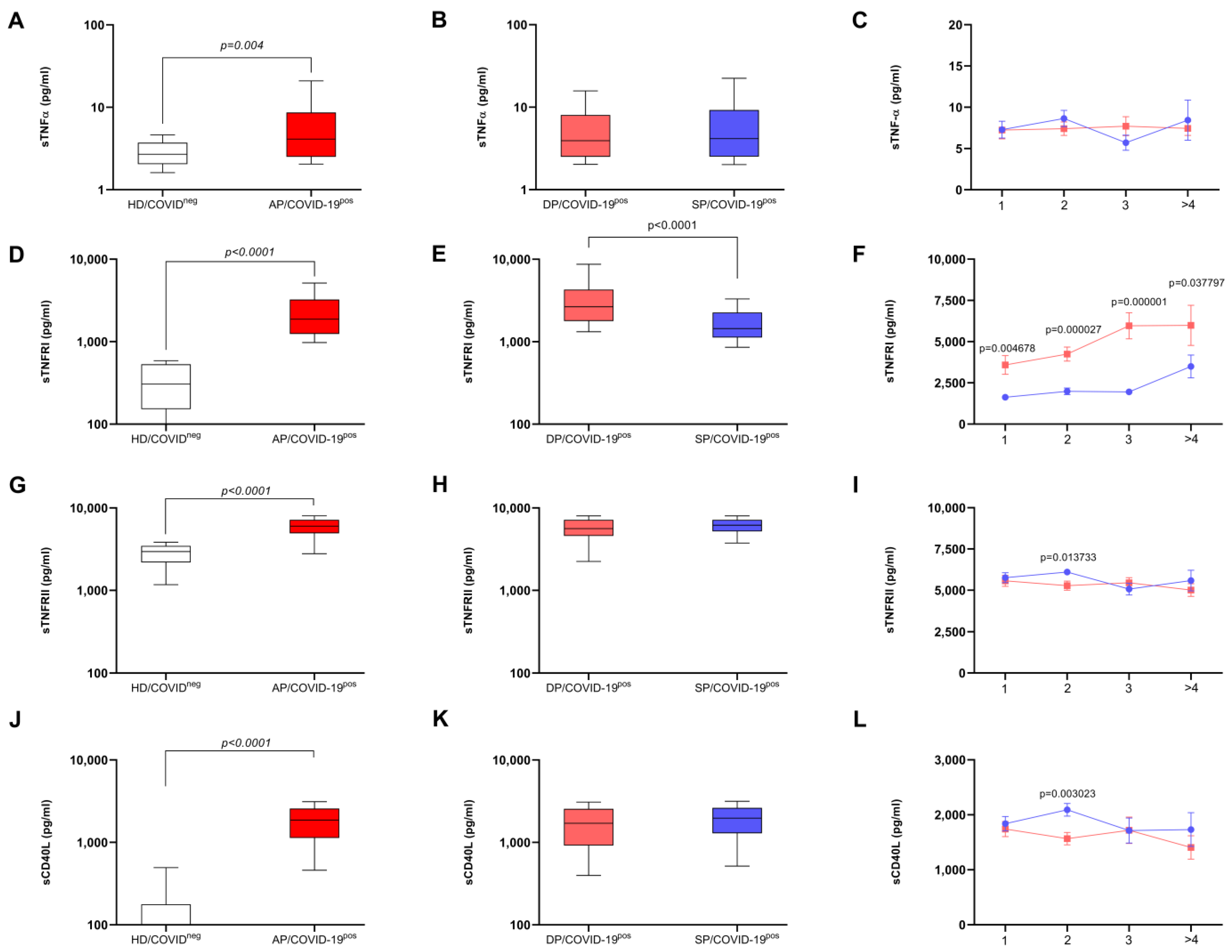
Biomedicines 12 02138 g001
Figure 2. Graphical representation for the dis(similarity) matrix between the explanatory variables for the association with the prognosis of symptomatic patients for COVID-19. A dis(similarity) matrix was generated for the explanatory variables using a multivariate grouping method, the Euclidean distance. In (A), the distribution of groups for patients whose outcome was discharge. In (B), matrix data for the groupings of patients whose outcome was death. TNFRI: tumor necrosis factor receptor I; TNFRII: tumor necrosis factor receptor II; CRP: C-reactive protein.
Biomedicines 12 02138 g002
Table 1. Effect of blood cell variables among patients who were discharged from hospital or died related to severe COVID-19.
BLOOD CELLS
VariablesOutcomeNẊ//MdσRBC—%Power—%p-Value
Erythrocytes—106/mm3Survivor1394.34//4.410.6835.8590.00<0.001
Deceased1423.88//3.810.85
Hemoglobin—g%Survivor13912.95//13.101.9134.2587.56<0.001
Deceased14211.63//11.802.41
Hematocrit—%Survivor13938.98//39.505.6729.2477.15<0.001
Deceased14254.45//35.55223.78
Platelets—103/mm3Survivor139226.00//205.0089.317.1540.360.821
Deceased142212.00//205.0083.6
Leukocytes—mm3Survivor13910,498.13//8800.005852.8225.1966.01<0.001
Deceased14213,000.22//11,735.006877.21
Neutrophils—mm3Survivor1398179.47//6966.004985.9028.6675.69<0.001
Deceased14110,748.47//9877.006038.81
Band cell—mm3Survivor139489.47//261.00890.0714.8133.180.031
Deceased141730.42//392.001036.80
Lymphocytes—mm3Survivor1391221.66//1014.00679.1732.1683.70<0.001
Deceased142881.98//815.50468.95
Monocytes—mm3Survivor139561.31//515.00320.5211.3223.570.101
Deceased142545.79//442.00433.50
Eosinophil—mm3Survivor13825.21//0.0055.430.545.470.915
Deceased14135.23//0.0085.32
CRP—mg/dLSurvivor8783.10//58.7069.0042.4383.04<0.001
Deceased73159.00//130.00113.00
N: sample number; Ẋ: mean; Md: median; σ: standard deviation; RBC: rank biserial correlation; %: percentage; CRP: C-reactive protein. The Mann–Whitney U test was used. Results were considered statistically significant at p < 0.05.
Table 2. Effect of immunological biomarkers among patients who were discharged from hospital or died related to severe COVID-19.
INFLAMMATORY MOLECULES
VariableOutcomeNẊ//MdσRBC—%Power—%p-Value
IL-12 p70—pg/mLSurvivor1534.58//4.052.512.587.780.690
Deceased1684.62//4.102.42
IL-1 β—pg/mLSurvivor1537.14//6.732.463.388.850.601
Deceased1687.40//6.963.14
IL-8—pg/mLSurvivor153178.58//21.95988.8613.933.280.032
Deceased168239.11//36.111342.21
IL-17—pg/mLSurvivor15354.67//26.50142.137.7216.570.232
Deceased16865.91//18.87134.45
IFN-γ—pg/mLSurvivor15311.52//6.8712.1417.3644.820.007
Deceased1688.85//5.739.64
IL-10—pg/mLSurvivor15314.88//9.1824.9720.2554.850.002
Deceased16830.62//15.1565.37
IL-6—pg/mLSurvivor15399.12//25.48471.9830.7284.98<0.001
Deceased168377.98//73.591420.58
IL-4—pg/mLSurvivor15314.62//10.7610.9619.8353.400.002
Deceased16811.32//8.867.58
IL-2—pg/mLSurvivor1535.95//6.283.1512.7629.750.048
Deceased1685.37//5.113.24
Adiponectin—µg/mLSurvivor1475.38 × 106//3.85 × 1063.74 × 1066.2813.440.339
Deceased1646.16 × 106//4.95 × 1064.89 × 106
Leptin—ng/mLSurvivor14730,052.30//21,348.8029,388.9016.6941.600.011
Deceased16426,778.00//12,097.0034,571.00
MIF—pg/mLSurvivor1535299.00//3655.555342.1022.1261.22<0.001
Deceased1687549.00//4732.007484.00
sCD40L—pg/mLSurvivor1511928.80//1952.70979.2019.5352.030.003
Deceased1671623.00//1539.001082.00
N: sample number; Ẋ: mean; Md: median; σ: standard deviation; RBC: rank biserial correlation; %: percentage; IL: interleukin; CD: cluster differentiation; INF: interferon; MIF: macrophage migration inhibitory factor; s: soluble. The Mann–Whitney U test was used. Results were considered statistically significant at p < 0.05.
Table 3. Effect of TNF family of biomarkers among patients who were discharged from hospital or died related to severe COVID-19.
RECEPTOR FOR TNF AND THE RATIO
VariableOutcomeNẊ//MdσRBC or Cohen’s d—%Power—%p-Value
sTNF-α—pg/mLSurvivor1547.75//4.487.452.697.930.677
Deceased1687.44//5.666.53
sTNFRI—pg/mLSurvivor1542034.00//1508.001629.0054.5899.91<0.001
Deceased1685224.00//3260.005840.00
sTNFRII—pg/mLSurvivor1545760.00//5997.001855.0011.1225.040.085
Deceased1685356.00//5453.002100.00
sTNFRI/sTNF-αSurvivor154496.00//388.00528.0035.8094.03<0.001
Deceased1681141.00//590.001392.00
sTNFRII/sTNF-αSurvivor1541555.00//1246.001245.009.3120.840.149
Deceased1681341.00//1014.001222.00
sTNFRI/sTNFRIISurvivor1540.419//0.2450.4951.9099.86<0.001
Deceased1681.33//0.5901.70
N: sample number; Ẋ: mean; Md: median; σ: standard deviation; RBC: rank biserial correlation; %: percentage; TNF: tumor necrosis factor; TNFRI: tumor necrosis factor receptor I; TNFRII: tumor necrosis factor receptor II; s: soluble. The Mann–Whitney U test was used. Results were considered statistically significant at p < 0.05.
Table 4. Binomial logistic regression model for predicting the prognosis of critically ill patients with COVID-19, using sTNFRI/sTNFRII ratio.
Predictor (Parameter) 1Odds Ratio95% Confidence Intervalp-Value
LowerUpper
Hemoglobin (g%)0.79480.5991.06000.112
Neutrophils (%)1.04520.9881.11000.122
CRP (mg/L)1.00731.0021.01000.011
sTNFRI/sTNFRII ratio7.23321.85728.17000.004
Performance Metrics
Accuracy0.740Specificity0.870
AUC0.830Sensitivity0.520
Predictor (Parameter) 1Odds ratio95% Confidence Intervalp-Value
LowerUpper
CRP (mg/L)1.00841.00321.01400.002
sTNFRI/sTNFRII ratio8.34052.420128.7450<0.001
Performance Metrics
Accuracy0.770Specificity0.900
AUC0.810Sensitivity0.590
Predictor (Parameter) 1Odds ratio95% Confidence Intervalp-Value
LowerUpper
sTNFRI/sTNFRII ratio3.97402.07007.6280<0.001
Performance Metrics
Accuracy0.680Specificity0.920
AUC0.740Sensitivity0.370
Legend: 1 Analyses refer to death/discharge; CRP: C-reactive protein; AUC: area under curve; %: percentage; TNFRI: tumor necrosis factor receptor I; TNFRII: tumor necrosis factor receptor II; s: soluble. Results were considered statistically significant at p < 0.05.
Disclaimer/Publisher’s Note: The statements, opinions and data contained in all publications are solely those of the individual author(s) and contributor(s) and not of MDPI and/or the editor(s). MDPI and/or the editor(s) disclaim responsibility for any injury to people or property resulting from any ideas, methods, instructions or products referred to in the content.

